Review

Impetigo Animal Models: A Review of Their Feasibility and Clinical Utility for Therapeutic Appraisal of Investigational Drug Candidates

1
Faculty of Health, University of Canberra, Bruce, Australian Capital Territory 2617, Canberra, Australia
2
Department of Pharmaceutics, School of Pharmacy, College of Health Sciences, Mekelle University, Mekelle 7000, Ethiopia
3
Daimantina Institute, University of Queensland, Wolloongabba, Brisbane 4102, Queensland, Australia
*
Author to whom correspondence should be addressed.
Submission received: 14 September 2020 / Revised: 6 October 2020 / Accepted: 7 October 2020 / Published: 14 October 2020

Abstract

:
Impetigo (school sores), a superficial skin infection commonly seen in children, is caused by the gram-positive bacteria Staphylococcus aureus and/or Streptococcus pyogenes. Antibiotic treatments, often topical, are used as the first-line therapy for impetigo. The efficacy of potential new antimicrobial compounds is first tested in in vitro studies and, if effective, followed by in vivo studies using animal models and/or humans. Animal models are critical means for investigating potential therapeutics and characterizing their safety profile prior to human trials. Although several reviews of animal models for skin infections have been published, there is a lack of a comprehensive review of animal models simulating impetigo for the selection of therapeutic drug candidates. This review critically examines the existing animal models for impetigo and their feasibility for testing the in vivo efficacy of topical treatments for impetigo and other superficial bacterial skin infections.

1. Introduction

Impetigo (school sores) is one of the eight dermatologic conditions listed in the 50 most common causes of infection globally [1], and is the only one of those eight with significant potential for life-threatening complications such as post-streptococcal glomerulonephritis and rheumatic heart diseases [2]. Over 162 million children suffer from impetigo globally at any given time, with a disproportionately high prevalence reported in Indigenous Australian children [3]. Impetigo is epidermal infection caused by the gram-positive bacteria Staphylococcus aureus and/or Streptococcus pyogenes (group A beta-hemolytic streptococcus; GAS) [3,4]. It can occur either as a primary infection or secondary to other underlying skin conditions which disrupt the skin barrier and allow the entry of pathogenic bacteria such as atopic dermatitis, eczema, or scabies [5,6]. Impetigo presents in two forms—bullous and non-bullous [4]. Bullous impetigo is generally caused by S. aureus and clinically presents with large fragile, flaccid, fluid-filled bullae (up to 2 cm in diameter) that less readily rupture into thin, brown crusts [4,7]. Non-bullous or crusted impetigo is the most common form of impetigo caused by both S. aureus and GAS [7]. It begins with a vesicle located on an erythematous base, which is easily ruptured, resulting in superficial ulceration covered with purulent discharge that then dries as an adhering yellow crust. Clinically, non-bullous impetigo caused by GAS is usually indistinguishable from non-bullous impetigo caused by S. aureus [8]. The clinical appearances of impetigo forms may be dependent on the type of disease-causing bacteria or etiologic agent; however, the histopathology of both forms is suggested as similar and is mainly denoted by the formation of intraepidermal pustules [9].
Antibiotics, often topical, are used as the first-line treatment for impetigo for faster symptom resolution [4,6,10,11]. However, as for other infections caused by S. aureus and S. pyogenes, antimicrobial resistance (AMR) poses a serious challenge in impetigo treatment and the rapid emergence and spread of multidrug-resistant bacterial strains (methicillin resistant S. aureus (MRSA) and macrolide-resistant S. pyogenes) is a global concern, limiting the effectiveness of existing treatment options for infections caused by these strains [12,13,14,15,16]. Given the seriousness of AMR, the World Health Organization (WHO) and the research community have recently highlighted the critical need for research of resistance patterns of pathogens to the existing antibiotics and the development of new and alternative antimicrobial agents with a number of new and existing candidates under investigation [10,17,18,19,20]. The antimicrobial activity of potential new compounds is first tested in vitro and, if effective, followed by study in animal models and clinical studies [21,22].
Appropriate animal models provide a highly controlled and cost-effective technique to predict efficacy in humans and refine treatment regimens and delivery vehicles bridging the gap between the in vitro drug discovery and clinical studies [22,23,24,25,26]. Animal models allow investigators to study the efficacy and safety of antimicrobial agents against specific pathogens in the presence of various host factors, metabolic processes, and anti-infective host defense mechanisms in the skin [22,23,27]. The use of animal models for the evaluation of new antimicrobial agents or new skin therapies is a long-established practice and has been acknowledged as a prerequisite and integral part of preclinical studies in drug development [21,28,29,30]. In addition to efficacy data, animal models provide other preliminary information, including safety, pharmacokinetic, and pharmacodynamic profiles of the potential antimicrobial agents necessary for the design and conduct of subsequent clinical studies [21,28,29,31,32].
To obtain the greatest scientific value from models, the choice of model must be optimized for the particular disease of interest and should react to the disease or its respective treatment in a way that resembles human physiology [26,33]. When selecting an optimum animal model, consideration should be given to the clinical relevance, reliability and reproducibility of the data generated, the technical complexity of conducting and interpreting the study, and the ethical acceptability of the animal use in the study [33,34].
For animal models aimed at screening and evaluating potential antibacterial agents for human use, the models should be able to establish the infections of the target human pathogens [34,35]. The models should allow for the observation of measurable study outcomes which are reproducible and readily controlled [34,35]. The study parameters should also be sensitive enough to allow detection and quantification of both bacteriostatic and bactericidal effects of the agents; enable clinically active drugs to demonstrate their efficacy; and provide results that show a high degree of clinical predictivity [34,35].
Although the results generated from animal models can and do provide a degree of clinical predictivity and are the best preliminary screening methods to select new antimicrobial agents or therapies for clinical trials, they cannot be directly translated to humans and the final judgment of an antimicrobial agent’s efficacy is ultimately dependent on the results of controlled clinical trials [21,22,26,28,29]. Over the years, several models of skin infection have been developed with considerable success in reproducing the pathogenesis of S. aureus and GAS diseases and are currently being used to study skin and soft tissue infections (SSTI) caused by these bacteria [30,34,36,37,38]. The experimental details of these models have been extensively summarized in a number of reviews [30,36,37,39]. However, the same cannot be said for impetigo at present, and there is a lack of a comprehensive review on the animal models investigated for impetigo. Here, we critically review the animal models investigated to mimic clinical impetigo and discuss their methodology, strengths, limitations, and the rationale for their application in evaluating impetigo drug candidates.
A summary of the various animal models is presented in Table 1 and Figure 1, and the strengths and limitations of the models are also summarized in Table 2.

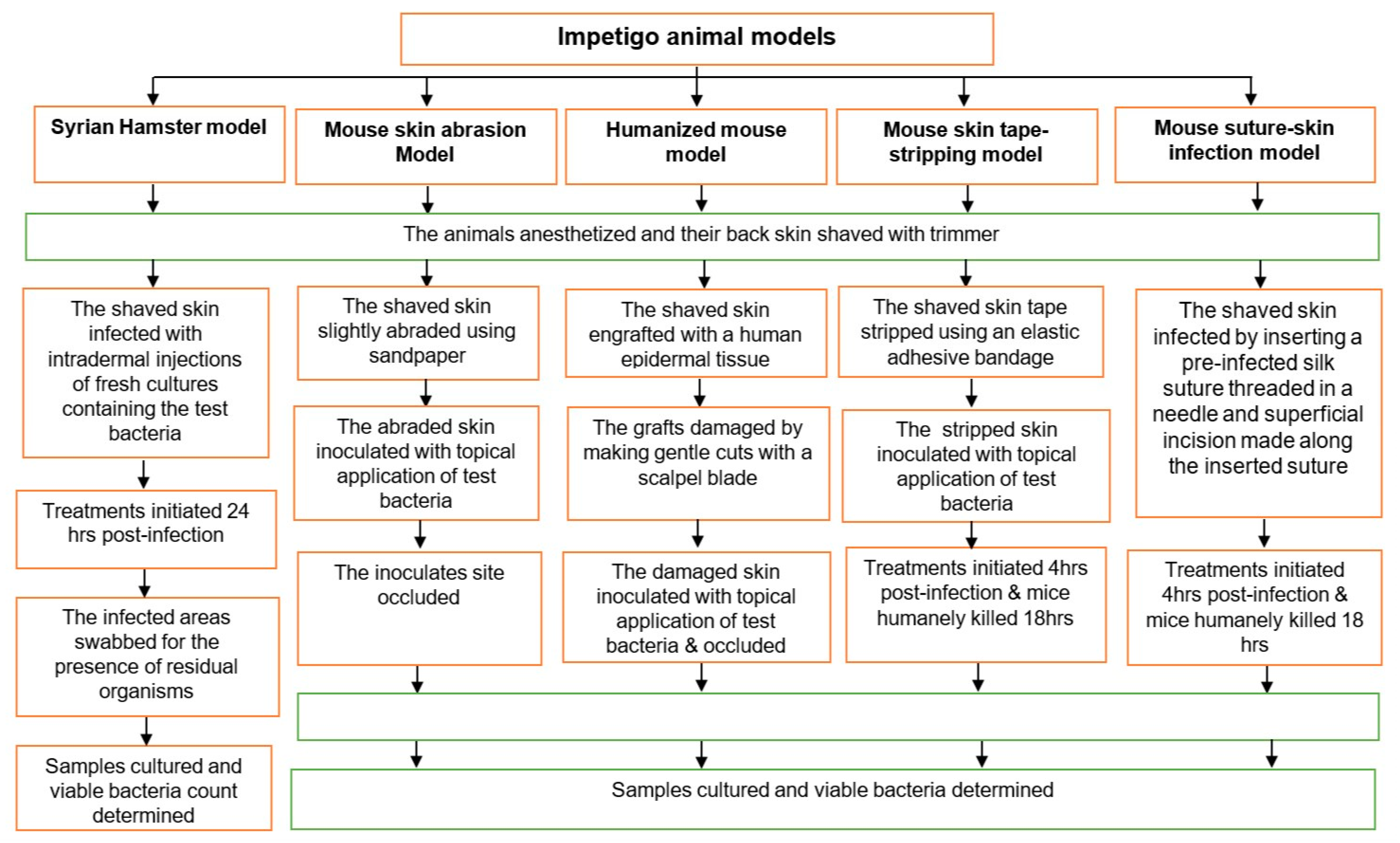
1.1. Syrian Hamster Impetigo Model

This model was established in 1970 by Dajani and his co-workers using experimental 6–8-week-old golden Syrian hamsters [9,40]. Subsequent studies have used this model to investigate the effect of various therapeutic regimens [41], the interaction between S. aureus and GAS strains [42], and the cellular responses following the infections [43]. In all studies, the authors have followed a similar procedure to establish the infection. The back of the hamster was carefully shaved and intradermal injections of 0.1 mL of fresh cultures containing 1.2 × 10⁸ Colony Forming Units (CFU)/mL of test organisms (S. aureus or GAS) were given at four to six sites at least 3 cm apart, with a 27-gauge needle fitted to a tuberculin syringe. After 24 h of the inoculation, various treatment regimens such as scrubbing with hexachlorophene soap (once daily, no report on dose and duration of treatment), gentamicin ointment (twice daily, no report on dose and duration of treatment), bacitracin ointment (twice daily, no report on dose and duration of treatment), benzathine penicillin G injection (30,000 U, one-time IM injection), or procaine penicillin injection (3000 U daily, for 7 days) were initiated for only GAS-infected hamsters and compared with a control (no treatment) group. Then, the infected hamsters were carefully observed daily for about 15 days for the gross clinical appearance of the lesions and the effect of therapy on the persistence of organisms in the lesions. The appearances of the lesions were examined photographically, and the effect of the treatments was investigated by swabbing the lesions for the presence of residual organisms.
In their results, the authors observed a formation of papular, erythematous areas, 4–6 h after injection, which progressed into vesicles within 24 h. The vesicular lesions then ruptured and subsequently became crusted within 2–3 days and remained in the crusted stage for 4–6 days. Histopathology demonstrated inflammatory cells invading muscle, adipose tissue, and the upper part of the dermis as well as the epidermis. Based on these observations, the authors concluded that the lesions developed in this model simulate the human disease in gross appearance, disease progression, and the histopathology of the infection process. Comparing features of the lesions produced by the test organisms, the authors noted that Staphylococcal and Streptococcal lesions were similar except that no vesicular stage was observed, and cellulitis was more pronounced with Staphylococcal infection.
The authors also found that the healing process for systemic treatments (mean duration: 4–5 days) was relatively faster than for topical treatments (mean duration: 7–8 days) compared with control groups (6 days), with complete eradication of the organism observed for only systemic treatments after 15 days of treatment, validating the applicability of this model for the therapeutic evaluation of drugs.
Even though the model succeeded in producing a human impetigo-like infection, the mode of bacterial inoculation (deep intradermal injection) is unlikely to represent the natural progression of impetigo development in humans. Human impetigo initially develops as a small vesicle after minor injuries or cuts on the skin, such as abrasions and skin scratches resulting from eczematous lesions or insect bites, rapidly progressing into pustular lesion, and subsequently evolving into an erosion covered by a thick crust [8,60,61,62]. Other authors [45,46,49,50,63] also suggest that the application of topical inoculation of organisms to the surface of slightly damaged or traumatized skin is likely to more closely resemble the natural route of transmission for GAS and S. aureus. Minor skin traumas perforate the stratum corneum, causing a temporary breach in the integrity of the skin barrier system that enables the inoculated bacteria to permeate or penetrate the epidermis with little hindrance [62,64]. In addition, the inoculum size of the test organisms (1.2 × 10⁷ CFU given at four sites) used to establish infection in the hamster model studies was too large in comparison to natural conditions. In human subjects, inoculum sizes of 106 cells (S. aureus) and 104 cells (GAS) are reportedly sufficient to produce severe Staphylococcus aureus skin infections [64].
The experimental impetigo produced in this model was also associated with a subepidermal abscess, a sign of the formation of dermal (e.g., cellulitis) and deep tissue infections, which is usually treated with systemic antibiotics in clinical settings [62,65]. This seems to negate the claim of establishing an impetigo-like infection (which is usually confined to the epidermis) [45,65]. The large inoculum delivered subcutaneously resulting in abscess formation creates the risk that results from the model will be biased toward systemically administered antimicrobials and lead to rejection of potentially clinically useful topical treatments [49].
Although the description of the sampling procedure in the studies lacked detail, swabbing and culturing of the samples were performed. This approach may confirm the presence of the test organisms, but it is unlikely to give a reliable estimate of the total count of organisms present in the lesion sites. Recent similar studies [52,53,54] suggest the use of quantification of viable lesion bacteria count through removing skin samples from the lesion sites with subsequent homogenization and culturing of the supernatant. This helps to quantify organisms present at both the surface and superficial tissue and provides a measure of the total count of bacteria contributing to the infection [52,53,54].
The hamster model was designed almost four decades ago, and currently little data on the response of this model to currently marketed topical antibiotics is available [44]. In addition, the use of hamsters for experimental purposes in the investigation of immune responses to skin infections has now been diminished due to a substantial difference between the human and hamster immune systems [66].
In sum, this model appears to have limited value in evaluating in vivo efficacy of potential topical antibacterial agents targeting impetigo and other SSTIs.

1.2. Mouse Skin Abrasion Impetigo Model

There has been a preference for mice (Mus musculus) as a model for SSTIs in recent years and mice are currently the most widely employed animal to model skin and other infectious diseases [24,36,67], not only because of their relative genomic similarity with humans, but also because of their availability, ease of handling, high reproductive rates, and relatively low cost [25,36,68]. Due to their ability to mimic the clinical presentations of S. aureus and GAS infections observed in humans, mouse models are most extensively used for studying the pathogenesis of infections caused by S. aureus and S. pyogenes (GAS) [24,30,36,59]. They are also capable of discriminating the clinical presentation of infections caused by different strains of disease-causing bacteria [69].
Recognizing the limitations of the hamster model, Abe et al. (1992) explored a mouse model for human skin infections [45]. In this study, female five-week-old mice (~20 g, ddY—Deutschland, Denken, and Yoken type) were treated with cyclophosphamide (Cy; 2 mg/mouse, intraperitoneal injection for 5 days), a potent immunosuppressive agent, to elicit leukocytopenia. The intent of the immunosuppression was to facilitate clear observation of the interaction of S. aureus and the epidermal cells during infection, as white blood cells (particularly neutrophil) infiltration during infection can obscure this interaction. Non-Cy treated mice were also used as a comparative control. After Cy-treatment, the back of the mice was carefully shaved using razor blades and the exposed skin was then slightly abraded three times using sandpaper. Fifty µL of S. aureus suspension (1.4 × 10⁷ CFU/mL or 7 × 10⁴ CFU) was then applied topically on each of the abraded areas, and the inoculated sites were occluded with sterile plastic plasters and sealed with vinyl adhesive tape. Subsequently, skin samples (1.5 × 1.0 cm2, n = 3) were excised from each mouse at different times (0.25–48 h) after the inoculation. The skin samples were washed with saline solution, cut into small pieces, and homogenized in saline solution (2 mL) in sterile mortars and aliquots (0.1 mL) cultured on appropriate Staphylococcus culture media. After 24 h of incubation, CFUs were counted and the resulting counts were converted to CFU/g skin sample. Additionally, skin samples were examined histopathologically and electron microscopically to assess the disease progression across the skin layers.
The study revealed the presence of bacteria in the horny skin layer (stratum corneum) of the Cy-treated mice at 0.5 h after inoculation, and the formation of subcorneal (immediately below stratum corneum) bullae at 1 h, with subsequent bacterial cluster formation inside the bullae at 3 h. After 6–12 h following inoculation, intraepidermal bullae containing many bacteria were developed, became enlarged, and spread to the upper dermis accompanied by epidermal necrosis within a day (at 24 h of the inoculation). Two days (at 48 h) after inoculation, bacteria further invaded into some surrounding hair follicles with a slight leukocyte infiltration. The non-Cy-treated mice showed the same outcome except for the abundant leukocytes and bacteria at 6 and 48 h; however, the authors suggested that further experimental models without the use of Cy should be explored.
In summary, the authors stated that the model has the potential to produce impetigo lesions that closely resemble the human blister lesions in terms of intraepidermal pustules formation and pathology findings. They also emphasized the importance of occlusion by an impermeable plastic film for the development of human-like S. aureus-induced blisters. Similar studies aimed at investigating the pathogenesis of S. aureus and GAS using in vitro (human skin explant culture) [70], in vivo (mouse model) [46], and human subjects [62,63,64,71] also reported that occlusion is necessary for the development of experimental skin infections. In two studies involving human subjects [64,71], bacterial inoculation was made on the intact skin, and occlusion was used to enhance the skin hydration, because moisture is suggested as the main factor promoting the growth of bacteria on the skin surface. The resulting moist and warm environment provides ideal conditions for the rapid growth of bacteria and subsequent development of the infection model [71].
The mouse skin abrasion model is designed specifically to simulate impetigo triggered by S. aureus sp. and further studies are required to confirm if the model is flexible enough to accommodate additional pathogens (e.g., S. pyrogens). Producing skin abrasion using sandpaper has also been viewed as a difficult procedure to monitor and control [46]. Moreover, we could not identify additional studies in the literature that used this model for the evaluation of topical antibiotic agents for SSTIs. Thus, it is difficult to validate the reproducibility and reliability of this model.

1.3. Humanized Mouse Model

Scaramuzzino et al. (2000) developed a humanized mouse (hu-mouse) model for impetigo by grafting human epidermal tissue from neonatal foreskin onto the back of 4–6-week-old female severe combined immunodeficiency disease (SCID) mice (mice that lack functional B and T lymphocytes), followed by infecting the graft with GAS four weeks after surgery [46]. To test the effect of compromised/damaged skin, the grafts were superficially damaged by one of three methods: a series of gentle cuts with a scalpel blade, gentle rubbing with sandpaper, or tape stripping (stripping the graft ten times in sequence) using fresh duct tape. An inoculum of GAS (50 µL of 103 CFU/mL) on a gauze pad was applied to the damaged area of skin under a circular bandage and occlusive dressing. A week after the inoculation, the mice were sacrificed, and their human skin grafts (1 × 1.5 cm) were removed for histopathology examination and determination of the bacterial count.
The study confirmed that the formation of an impetigo-like lesion characterized by erosion of the stratum corneum, and a typical murine innate immune system response (infiltration polymorphonuclear leukocytes), were observed. The typical honey-colored fibrin crust typical of non-bullous impetigo in humans was not observed, however. Only small inoculums of as low as 50 CFU of GAS were required to produce the impetigo-like lesions. In addition, the study confirmed that damaged skin and subsequent occlusion of the inoculated skin area were requirements for the induction of severe infection by virulent GAS. Other studies [45,62] have also recommended the requirement of breaching the integrity of the skin through minor superficial damage to elicit the Streptococcal infection.
The authors noted that tape stripping could not reliably remove the stratum corneum and it occasionally led to the separation of large stretches of the epidermal layer from the underlying dermis; whereas the use of sandpaper treatment was found difficult to monitor and control. These authors suggested that a gentle crosswise scalpel cut method was the most reproducible method tested to achieve superficial skin damage.
Given the use of grafted human skin followed by the topical application of low bacterial inoculum to superficially damaged grafted skin, this model potentially provides the closest simulation of the clinical pathology of impetigo [30,59]. This model has been employed in studies [47,48] seeking a better understanding of the pathogenic mechanisms associated with a GAS skin infection and has been suggested as an important model that could pave the way for the development of a vaccine for streptococcal-caused impetigo [59].
The hu-mouse model is associated with a number of important limitations. It is technically complex, requiring a source of human tissue and surgical and post-surgical animal nursing skills to successfully achieve the grafts [30,72]. The absence of an adaptive immune response in this mouse line prevented the rejection of the tissue grafts, but also removed a potentially relevant immune mechanism in the pathogenesis of impetigo [46]. The use of an immunocompromised SCID mouse is unlikely to closely simulate the physiological conditions encountered by the host under natural conditions, as the infection proceeds in the absence of adaptive immunity in this type of mouse [30]. The model is also likely limited by the requirement of a large number of mice and a huge effort to perfect the infection model [30]. Moreover, this model has been applied only to GAS, and its applicability for S. aureus-driven impetigo is unexplored. No relevant studies using this model for therapeutic evaluation of topical antibiotics for impetigo were located in the literature.
A reliable and reproducible mouse skin infection model with established pathogenesis of S. aureus and S. pyogenes is a prerequisite for further studies evaluating the therapeutic response of agents [63]. Our review identified a limited number of existing models developed specifically for impetigo and only the humanized mouse model seems to produce a human impetigo-like infection. In addition, the identified models have a number of limitations that restrict their feasibility in evaluating the clinical efficacy of topical antibacterial agents. A number of other models have been developed to investigate superficial skin infections more broadly [34,49] and for assessing the clinical efficacy of both marketed and investigational topical antibiotics (such as fusidic acid, mupirocin, retapamulin, and ozenoxacin) for impetigo treatment, and these are discussed below.

1.4. Mouse Skin Tape-Stripping Model

Kugelberg et al. (2005) established this model using 6–8-week-old female BALB/c mice, a readily available albino inbred mouse strain [49]. A reproducible degree of dermal damage was produced using tape stripping 7–10 times with an elastic adhesive bandage, standardized by measuring the trans epidermal water loss (TEWL) using a proprietary TEWL probe to yield a TEWL of approximately 70 g/(m2∙h). Following this procedure, the skin became visibly damaged, characterized by reddening and glistening but no regular bleeding. Then, an inoculum of 107 cells of S. aureus or S. pyogenes in a 5-µL droplet was placed on the stripped skin to initiate the infection. Four hours post infection, the mice were treated with 2% fusidic acid ointment twice daily (AM and PM, with an 8- hour interval) for a period of 4 days. Eighteen hours after the last topical treatment, the mice were killed, and the wounds (about 2 cm2) were excised, processed, and cultured to determine the number of viable bacteria per unit area of skin. In order to investigate the reproducibility of the infection with S. aureus and S. pyogenes, three independent experiments that included untreated and placebo-treated groups were performed.
This study concluded that the model is simple, reproducible, and useful to simulate localized skin infections caused by S. aureus or S. pyogenes. Topical treatment with fusidic acid significantly reduced the number of viable counts of S. aureus and S. pyogenes after the four-day treatment (p < 0.001), showing that this model can also be used for therapeutic evaluation of topical treatments. However, the authors stated that this model did not simulate the histology of impetigo. In human impetigo involving S. aureus, the first step for the bacterial skin invasion involves adhering to the outer layer of the epidermis (stratum corneum), followed by disruption of the epithelial barriers of the inner layers of the epidermis comprising cell-adhesion structures such as desmosomes and adherence junctions [73]. This process separates the epidermis just below the stratum granulosum and large intraepidermal pustules, which may contain bacteria, and these are described as typical histologic characteristics of impetigo [45,50,74]. Besides, in the case of impetigo, the upper dermis also contains an epidermal inflammatory infiltrate of neutrophils and lymphocytes which are reported to be crucial for the staphylococcal percutaneous invasion [50].
In this model [49], there was no intraepidermal or sub-corneal pustules nor the migration of neutrophils, which is inconsistent with the picture for human impetigo. Similar studies [50,51] that used this model and followed the same procedure have reported contrasting results. One study [50] claimed the formation of intraepidermal pustules resembling human impetigo through epicutaneous inoculation of S. aureus on the inner pinna of the mice. In contrast, other studies [51,75] demonstrated the formation of more of a cutaneous infection rather than human impetigo-like infection following a similar inoculation procedure and concluded that, in this mouse model, the test organisms are capable of rapidly penetrating epidermal layers and disseminate into dermal and cutaneous tissues. Similar results were also reported by other investigators [76,77] employing this model to evaluate a new topical antibiotic (HT61) for methicillin-sensitive S. aureus (MSSA) and MRSA infections. These studies showed that tape-stripping the mouse skin using autoclave tape ten times in succession damaged the skin by removing the top dermal layers, which became red and shiny but without observable bleeding—meaning the removal of most of the epidermal layer of the mouse skin. From these inconsistent findings, it is understood that the mouse skin tape-stripping is unlikely to be a reliable and reproducible model for impetigo.

1.5. Mouse Suture-Superficial Skin Infection Model

This model was originally investigated in two successive studies with the aims of characterizing the infection, evaluating its response to therapy through testing various topical antimicrobial formulations, and investigating the correlation of the in vitro antimicrobial activity of the tested formulations with their in vivo efficacy [34,35]. Accordingly, evaluation of in vitro antibacterial activity (minimum inhibitory concentration and minimum bactericidal concentration) of the topical agents was performed following the broth twofold dilution method. For the in vivo study, an infection model was developed using female CF-1, 18 to 20 g mice (n = 10 in each group). The study further explored the correlation of in vitro with in vivo study data.
For the in vivo study, mice were anesthetized; the dorsal area closely shaved; and on the day of infection, superficial surgical wounds were produced by making a longitudinal midline incision of 2.3 cm in length. The wounds were then infected either by direct seeding or by the insertion of a 5 cm length suture, which had been previously infected with a controlled inoculum (103–105 cells) of S. aureus or P. aeruginosa, using a surgical needle. To maintain its position, the ends of the suture were then secured with rubber cement.
In this model, two treatment regimens (immediate and delayed) were employed to assess the efficacy of the test formulations (gentamicin cream, neomycin-gramicidin-nystatin-triamcinolone acetonide cream, nitrofurazone cream, polymyxin B-bacitracin-neomycin ointment, and triclobisonium chloride ointment). In the immediate-treatment regimen, each wound was treated topically with 0.4 g of formulation 15 min and 6 h after the insertion of the suture; whereas the delayed treatment regimen was administered 24 and 30 h after suture insertion. For both regimens, the wounds were excised and cultured 18 h after the final treatment.
Quantitation of the viable bacteria in the wounds was performed before and after the treatments. The total pre-measured amount of viable bacteria counts loaded in the suture was used as a baseline, or pre-treatment viable count; whereas for post-treatment viable count determination, both surface rinse and biopsy (skin sample or tissue) homogenization techniques were performed in the study. The surface rinse technique was performed by tightly placing a sterile test tube containing distilled water in an inverted position on the back of a euthanized mouse in such a way that more than 90% of the wound was enclosed by the mouth of the tube. Then, the mouse and the test tube were held together, with the tube upright, and shaken vigorously for 25 times to remove the infecting organisms from the surface of the wound. The samples were then processed for determination of CFU. In the case of the biopsy homogenizing method, biopsy samples from infected wounds were taken, homogenized, diluted, and plated on the appropriate selective agar media for determination of CFU.
The results of these successive studies [34,35] indicated that clinical signs of infection like oedema, erythema, and suppuration were observed around the margins of the lesions, representing the secondary skin infections like impetigo which occur following skin damages. The developed infection persisted for 1 to 2 weeks and was found to be quite susceptible to both immediate and delayed regimes of topical treatments with efficacy consistent with in vitro activity against the tested organisms, demonstrating a reasonable correlation of the in vitro activity of the topical agents with their in vivo efficacy in the animal model.
Unlike the suture method, the directed seeding infection method resulted in greater variation in viable wound bacteria counts—this was chiefly attributed to inoculum runoff from the application site. However, only an inoculum size of 103–105 cells of the test organisms was required to elicit the desired experimental infection using the suture method. This inoculum size is even lower than the inoculum dose (106 cells) used to initiate S. aureus infection in human subjects [64,78]. The simplicity of the surgical procedure used in this model and a low inoculum dose needed to elicit the infection, are important advantages of this model. The suture method for eliciting bacterial skin infections (S. aureus or S. pyogenes) has also been used by other studies using mice [44,52,53,55,57,58,79], guinea pigs [56], and humans [80], reflecting some acceptance of the use of sutures to introduce a bacterial inoculum as a means to simulate superficial bacterial skin infections in humans [78].
For quantification of the viable bacteria count in skin, although the surface rinse method resulted in a lower count per unit area, it nonetheless correlated well with the dynamics of the total viable wound count obtained from the biopsy method. The skin surface rinse approach was faster, simpler and less labor intensive, and the authors therefore recommended this approach for screening type studies. For studies aiming to investigate the therapeutic efficacy of topical formulations, the biopsy homogenizing, as extensively used by similar studies [44,55,79], seems more appropriate, as it quantifies both the surface and superficial tissue-associated bacteria. Although expensive, technically complex, and relying heavily on a large number of approximations compared with conventional methods [81], using an optical in vivo bioluminescence imaging system could potentially ease the determination of in vivo bacterial burdens without excising the skin samples [82,83]. This technique uses bioluminescent bacteria strains, which are placed in the full thickness incision in the dermis of the animal skin, and capture the bioluminescent signals released by the bacteria with a camera to determine the bacterial burden [82].
Even though the mouse suture-superficial skin infection model was original established as a skin wound infection, it has been extensively advanced and optimized by researchers over the past few years to study the superficial skin infections like impetigo. In fact, this model is arguably the most widely used mouse model for the clinical evaluation of currently marketed topical antibiotics for the treatment of impetigo and SSTIs, such as retapamulin [54], fusidic acid [44], mupirocin [44,56], amoxicillin or amoxicillin-clavulanic acid [55], Gemifloxacin [57], as well as other new impetigo and SSTIs agents in the pipeline [52,53,58]. This could be because of the ability of the model to simulate topical skin infections like impetigo that are caused by either S. aureus alone, GAS alone, or both in combination [55], and this allows the researchers to study the effect of a broad spectrum of topical antibiotics against both bacteria. Moreover, the efficacy results obtained from this model have been shown to positively correlate with the efficacy results obtained from clinical trials in humans [44,52,54]. For instance, ozenoxacin 1% cream [84,85], retapamulin 1% ointment [86], mupirocin 2% ointment [87], and mupirocin 2% cream [88], studied using this model, have subsequently shown good efficacy in clinical trials, indicating the potential of this model in predicting the efficacy of topical antibiotics in human [54]. Its capability to respond to various antibiotics from a wide range of dosage forms, including cream, ointment, gel, solutions, and powder, makes this model uniquely suitable for the evaluation and comparison of the efficacy of topical and oral antibiotics [44,52,54,55,56]. Although most of the studies using this model followed the original procedure, which is the incision of the skin with a scalpel and placing the infected suture in the wound, it is worth mentioning the slight modification of this model reported in other studies [44,52,55], which performed insertion of the infected suture under the skin using a surgical needle followed by making a superficial incision using a scalpel along the length of the suture without reaching the panniculus carnosus. Piercing of the skin using a surgical needle followed by superficial skin incision could represent the natural progression of impetigo in humans, i.e., scratching an insect bite, minor injuries, or cuts on the skin, which subsequently lead to the entry of impetigo-causing bacteria. Two studies [44,56] also used an adhesive temporary skin closure to ensure the wound remained closed until treatment initiation.
In summary, the mouse suture-superficial skin infection model appears to be the most appropriate and practically viable in vivo method for evaluating the antimicrobial efficacy of topical skin formulations where the primary focus is impetigo.

2. Concluding Remarks

Relevant, predictive skin infection models are required for the preclinical, in vivo investigation of the safety and efficacy of new topical antimicrobial agents. In this regard, there is a limited range of in vivo models that can reliably simulate impetigo and the efficacy of prospective treatment. Given impetigo usually occurs as a mixed bacterial infection caused by S. aureus and GAS species, none of the models investigated for impetigo seem to simulate impetigo caused by co-infection with both bacteria. Almost all the models used topical inoculation of the bacteria to the surface of slightly damaged skin and this delivery method seems to correspond well with the route of transmission encountered during natural infection resulting in impetigo. In addition, the majority of the models recommend occlusion of the topically inoculated skin surface to maintain the moisture required for the growth of bacteria, particularly for the GAS species. It is also worth noting that all mouse models preferred female mice as opposed to their male counterparts. This seems to relate to the lesser likelihood of female mice involved in cage fights for dominance, resulting in fight wounds and related infections, which could considerably interfere with the experimental observations.
Given the strengths and limitations of models reviewed, when planning for the in vivo efficacy evaluation of potential topical antibacterial therapies, key considerations beyond their practical feasibility in a given laboratory should include: (1) the establishment of reproducible, controllable infections with measurable endpoints of efficacy, (2) reliable and simple quantification of pre- and post-treatment viable bacteria counts, and (3) the correlation of in vivo results with clinical effectiveness in humans.
While the humanized mouse model appears to be a promising infection model, as it can reproduce many features of human impetigo, it is limited by the requirements for human tissue and the need for experienced and surgically skilled personnel. So far it has only been studied for the pathogenesis of GAS and it lacks evidence to support its utility for the evaluation of topical antibiotics.
For practical simplicity and reproducibility, the mouse suture superficial skin infection model has many advantages and has been widely used to study the preclinical efficacy of marketed topical impetigo treatments. This method has been shown to be suitable to study the pathogenesis of impetigo-causing bacteria and appears to be the model of choice for researchers and pharmaceutical companies for both screening and therapeutic evaluation of investigational drugs for the topical treatment of impetigo.

Author Contributions

Project conceptualization and design: J.T. and S.A.; Literature search: S.A.; Data extraction: S.A.; First draft preparation and review: J.T. and S.A.; Critical review and editing: A.B., W.T., and J.T. All authors have read and agreed to the published version of the manuscript.

Funding

There is no funding relating to this article.

Conflicts of Interest

The authors declare no conflict of interest.

References

  1. Hay, R.J.; Johns, N.E.; Williams, H.C.; Bolliger, I.W.; Dellavalle, R.P.; Margolis, D.J.; Marks, R.; Naldi, L.; Weinstock, M.A.; Wulf, S.K.; et al. The Global Burden of Skin Disease in 2010: An Analysis of the Prevalence and Impact of Skin Conditions. J. Investig. Dermatol. 2014, 134, 1527–1534. [Google Scholar] [CrossRef] [PubMed] [Green Version]
  2. Bowen, A.C.; Carapetis, J.R.; Currie, B.J.; Fowler, V.; Chambers, H.F.; Tong, S.Y.C. Sulfamethoxazole-Trimethoprim (Cotrimoxazole) for Skin and Soft Tissue Infections Including Impetigo, Cellulitis, and Abscess. Open Forum Infect. Dis. 2017, 4. [Google Scholar] [CrossRef] [PubMed] [Green Version]
  3. Bowen, A.C.; Mahe, A.; Hay, R.J.; Andrews, R.M.; Steer, A.C.; Tong, S.Y.C.; Carapetis, J.R. The Global Epidemiology of Impetigo: A Systematic Review of the Population Prevalence of Impetigo and Pyoderma. PLoS ONE 2015, 10, e0136789. [Google Scholar] [CrossRef] [PubMed] [Green Version]
  4. Pereira, L.B. Impetigo—Review. An. Bras. Dermatol. 2014, 89, 293–299. [Google Scholar] [CrossRef]
  5. May, P.J.; Tong, S.Y.C.; Steer, A.C.; Currie, B.J.; Andrews, R.M.; Carapetis, J.R.; Bowen, A.C. Treatment, prevention and public health management of impetigo, scabies, crusted scabies and fungal skin infections in endemic populations: A systematic review. Trop. Med. Int. Heal. 2019, 24, 280–293. [Google Scholar] [CrossRef]
  6. Koning, S.; Van Der Sande, R.; Verhagen, A.P.; A Van Suijlekom-Smit, L.W.; Morris, A.D.; Butler, C.C.; Berger, M.; Van Der Wouden, J.C. Interventions for impetigo. Cochrane Database Syst. Rev. 2012, 1, CD003261. [Google Scholar] [CrossRef] [Green Version]
  7. D’Cunha, N.M.; Peterson, G.M.; Baby, K.E.; Thomas, J. Impetigo: A need for new therapies in a world of increasing antimicrobial resistance. J. Clin. Pharm. Ther. 2017, 43, 150–153. [Google Scholar] [CrossRef] [Green Version]
  8. Steer, A.C.; Danchin, M.H.; Carapetis, J.R. Group A streptococcal infections in children. J. Paediatr. Child Health 2007, 43, 203–213. [Google Scholar] [CrossRef]
  9. Dajani, A.S.; Wannamaker, L.W. Experimental Infection of the Skin in the Hamster Simulating Human Impetigo. I. Natural History of the Infection. J. Infect. Dis. 1970, 122, 196–204. [Google Scholar] [CrossRef]
  10. Vila, J.; Hebert, A.A.; Torrelo, A.; López, Y.; Tato, M.; García-Castillo, M.; Cantón, R. Ozenoxacin: A review of preclinical and clinical efficacy. Expert Rev. Anti Infect. Ther. 2019, 17, 159–168. [Google Scholar] [CrossRef]
  11. Cole, C.; Gazewood, J. Diagnosis and treatment of impetigo. Am. Fam. Physician 2007, 75, 859–864. [Google Scholar] [PubMed]
  12. WHO. Antimicrobial Resistance: Global Report on Surveillance Geneva; WHO Press, World Health Organization: Geneva, Switzerland, 2014. Available online: https://www.who.int/drugresistance/documents/surveillancereport/en/ (accessed on 5 July 2019).
  13. Bowen, A.C.; Tong, S.Y.C.; Chatfield, M.; Carapetis, J.R. The microbiology of impetigo in indigenous children: Associations between Streptococcus pyogenes, Staphylococcus aureus, scabies, and nasal carriage. BMC Infect. Dis. 2014, 14, 727. [Google Scholar] [CrossRef] [PubMed] [Green Version]
  14. Yeoh, D.K.; Anderson, A.; Cleland, G.; Bowen, A.C. Are scabies and impetigo “normalised”? A cross-sectional comparative study of hospitalised children in northern Australia assessing clinical recognition and treatment of skin infections. PLoS Negl. Trop. Dis. 2017, 11, e0005726. [Google Scholar] [CrossRef] [PubMed] [Green Version]
  15. Pangilinan, R.; Tice, A.; Tillotson, G. Topical antibiotic treatment for uncomplicated skin and skin structure infections: Review of the literature. Expert Rev. Anti Infect. Ther. 2009, 7, 957–965. [Google Scholar] [CrossRef]
  16. Bohaty, B.R.; Choi, S.; Cai, C.; Hebert, A.A. Clinical and bacteriological efficacy of twice daily topical retapamulin ointment 1% in the management of impetigo and other uncomplicated superficial skin infections. Int. J. Women’s Dermatol. 2015, 1, 13–20. [Google Scholar] [CrossRef] [PubMed] [Green Version]
  17. Bonev, B.B.; Hooper, J.; Parisot, J. Principles of assessing bacterial susceptibility to antibiotics using the agar diffusion method. J. Antimicrob. Chemother. 2008, 61, 1295–1301. [Google Scholar] [CrossRef] [Green Version]
  18. WHO. WHO Publishes List of Bacteria for Which New Antibiotics are Urgently Needed Geneva. 2017. Available online: https://www.who.int/en/news-room/detail/27-02-2017-who-publishes-list-of-bacteria-for-which-new-antibiotics-are-urgently-needed (accessed on 1 October 2019).
  19. Balouiri, M.; Sadiki, M.; Ibnsouda, S.K. Methods for in vitro evaluating antimicrobial activity: A review. J. Pharm. Anal. 2016, 6, 71–79. [Google Scholar] [CrossRef] [Green Version]
  20. The Pew Charitable Trusts. Antibiotics Currently in Clinical Development 2017 (cited 2019 October 17). Available online: http://www.pewtrusts.org/~/media/assets/2017/05/antibiotics-currently-in-clinical-development-03-2017.pdf?la=en (accessed on 17 October 2019).
  21. FDA. Guideline for Clinical Evaluation of Anti-Nfective Drugs for Systemic Administration USA: U.S Department of Health and Human Service, Food and Drug Adminstration 1977. Available online: https://catalog.hathitrust.org/Record/000090753 (accessed on 10 July 2020).
  22. Zak, O.; O’Reilly, T. Animal models in the evaluation of antimicrobial agents. Antimicrob. Agents Chemother. 1991, 35, 1527–1531. [Google Scholar] [CrossRef] [PubMed] [Green Version]
  23. Petersen, T.K. In vivo Pharmacological Disease Models for Psoriasis and Atopic Dermatitis in Drug Discovery. Basic Clin. Pharmacol. Toxicol. 2006, 99, 104–115. [Google Scholar] [CrossRef] [PubMed]
  24. Avci, P.; Sadasivam, M.; Gupta, A.; De Melo, W.C.; Huang, Y.-Y.; Yin, R.; Chandran, R.; Kumar, R.; Otufowora, A.; Nyame, T.; et al. Animal models of skin disease for drug discovery. Expert Opin. Drug Discov. 2013, 8, 331–355. [Google Scholar] [CrossRef] [PubMed] [Green Version]
  25. Simmons, D. The use of animal models in studying genetic disease: Transgenesis and induced mutation. Nat. Educ. 2008, 1, 70. [Google Scholar]
  26. Barré-Sinoussi, F.; Montagutelli, X. Animal models are essential to biological research: Issues and perspectives. Futur. Sci. OA 2015, 1, 1–3. [Google Scholar] [CrossRef] [Green Version]
  27. Craig, W. Relevance of animal models for clinical treatment. Eur. J. Clin. Microbiol. Infect. Dis. 1993, 12 (Suppl. 1), S55–S57. [Google Scholar] [CrossRef]
  28. Beam, J.T.R.; Gilbert, D.N.; Kunin, C.M. General Guidelines for the Clinical Evaluation of Anti-Infective Drug Products. Clin. Infect. Dis. 1992, 15 (Suppl. 1), S5–S32. [Google Scholar] [CrossRef] [PubMed]
  29. Soriano, F.; Aguilar, L.; Ponte, C. In vitro antibiotic sensitivity testing breakpoints and therapeutic activity in induced infections in animal models. J. Chemother. 1997, 9 (Suppl. 1), 36–46. [Google Scholar]
  30. Watson, M.E.J.; Neely, M.N.; Caparon, M.G. Animal models of Streptococcus pyogenes infection. In Streptococcus pyogenes: Basic Biology to Clinical Manifestations; Ferretti, J.J., Stevens, D.L., Fischetti, V.A., Eds.; University of Oklahoma Health Sciences Center: Oklahoma City, OK, USA, 2016. [Google Scholar]
  31. Zhao, M.; Lepak, A.J.; Andes, D. Animal models in the pharmacokinetic/pharmacodynamic evaluation of antimicrobial agents. Bioorg. Med. Chem. 2016, 24, 6390–6400. [Google Scholar] [CrossRef] [PubMed]
  32. Ghiselli, R.; Cirioni, O.; Giacometti, A.; Scalise, A.; Simonetti, O.; Mocchegiani, F.; Orlando, F.; Goteri, G.; Della Vittoria, A.; Filosa, A.; et al. Comparative Efficacy of Topical Versus Systemic Teicoplanin in Experimental Model of Wound Infections. J. Surg. Res. 2008, 144, 74–81. [Google Scholar] [CrossRef] [PubMed]
  33. Van Der Staay, F.J.; Arndt, S.S.; Nordquist, R.E. Evaluation of animal models of neurobehavioral disorders. Behav. Brain Funct. 2009, 5, 11. [Google Scholar] [CrossRef] [PubMed] [Green Version]
  34. McRipley, R.J.; Whitney, R.R. Characterization and Quantitation of Experimental Surgical-Wound Infections Used to Evaluate Topical Antibacterial Agents. Antimicrob. Agents Chemother. 1976, 10, 38–44. [Google Scholar] [CrossRef] [PubMed] [Green Version]
  35. McRipley, R.J.; Whitney, R.R. Responsiveness of Experimental Surgical-Wound Infections to Topical Chemotherapy. Antimicrob. Agents Chemother. 1976, 10, 45–51. [Google Scholar] [CrossRef] [Green Version]
  36. Kim, H.K.; Missiakas, D.; Schneewind, O. Mouse models for infectious diseases caused by Staphylococcus aureus. J. Immunol. Methods 2014, 410, 88–99. [Google Scholar] [CrossRef] [PubMed]
  37. Malachowa, N.; Kobayashi, S.D.; Braughton, K.R.; DeLeo, F.R. Mouse Model of Staphylococcus aureus Skin Infection. Recent Results Cancer Res. 2013, 1031, 109–116. [Google Scholar] [CrossRef]
  38. Yamakawa, T.; Mitsuyama, J.; Hayashi, K. In vitro and in vivo antibacterial activity of T-3912, a novel non-fluorinated topical quinolone. J. Antimicrob. Chemother. 2002, 49, 455–465. [Google Scholar] [CrossRef] [PubMed]
  39. Dai, T.; Kharkwal, G.B.; Tanaka, M.; Huang, Y.-Y.; De Arce, V.J.B.; Hamblin, M.R. Animal models of external traumatic wound infections. Virulence 2011, 2, 296–315. [Google Scholar] [CrossRef] [Green Version]
  40. Cushing, A.H.; Mortimer, E.A. A Hamster Model for Streptococcal Impetigo. J. Infect. Dis. 1970, 122, 224–226. [Google Scholar] [CrossRef]
  41. Dajani, A.S.; Hill, P.L.; Wannamaker, L.W. Experimental infection of the skin in the hamster simulating human impetigo. II. Assessment of various therapeutic regimens. Pediatrics 1971, 48, 83–90. [Google Scholar]
  42. Dajani, A.S.; Wannamaker, L.W. Experimental infection of the skin in the hamster simulating human impetigo. III. Interaction between Staphylococci and group A Streptococci. J. Exp. Med. 1971, 134, 588–599. [Google Scholar] [CrossRef] [Green Version]
  43. Dajani, A.S.; Wannamaker, L.W. Experimental infection of the skin in the hamster simulating human impetigo. IV. Cellular responses after Streptococcal and Staphylococcal infections. Infect. Immun. 1972, 5, 942–946. [Google Scholar] [CrossRef] [Green Version]
  44. Gisby, J.; Bryant, J. Efficacy of a New Cream Formulation of Mupirocin: Comparison with Oral and Topical Agents in Experimental Skin Infections. Antimicrob. Agents Chemother. 2000, 44, 255–260. [Google Scholar] [CrossRef] [Green Version]
  45. Abe, Y.; Akiyama, H.; Arata, J. Production of experimental Staphylococcal impetigo in mice. J. Dermatol. Sci. 1992, 4, 42–48. [Google Scholar] [CrossRef] [Green Version]
  46. Scaramuzzino, D.A.; McNiff, J.M.; Bessen, D.E. Humanized In Vivo Model for Streptococcal Impetigo. Infect. Immun. 2000, 68, 2880–2887. [Google Scholar] [CrossRef] [PubMed] [Green Version]
  47. Svensson, M.D.; Scaramuzzino, D.A.; Sjobring, U.; Olsen, A.; Frank, C.; Bessen, D.E. Role for a secreted cysteine proteinase in the establishment of host tissue tropism by group A streptococci. Mol. Microbiol. 2000, 38, 242–253. [Google Scholar] [CrossRef] [PubMed]
  48. Svensson, M.D.; Sjöbring, U.; Luo, F.; Bessen, D.E. Roles of the plasminogen activator streptokinase and the plasminogen-associated M protein in an experimental model for streptococcal impetigo. Microbiology 2002, 148, 3933–3945. [Google Scholar] [CrossRef] [PubMed] [Green Version]
  49. Kugelberg, E.; Norström, T.; Petersen, T.K.; Duvold, T.; Andersson, D.I.; Hughes, D. Establishment of a Superficial Skin Infection Model in Mice by Using Staphylococcus aureus and Streptococcus pyogenes. Antimicrob. Agents Chemother. 2005, 49, 3435–3441. [Google Scholar] [CrossRef] [Green Version]
  50. Imanishi, I.; Hattori, S.; Hisatsune, J.; Ide, K.; Sugai, M.; Nishifuji, K. Staphylococcus aureus penetrate the interkeratinocyte spaces created by skin-infiltrating neutrophils in a mouse model of impetigo. Vet. Dermatol. 2016, 28, 126-e27. [Google Scholar] [CrossRef] [Green Version]
  51. Hahn, B.L.; Onunkwo, C.C.; Watts, C.J.; Sohnle, P.G. Systemic dissemination and cutaneous damage in a mouse model of Staphylococcal skin infections. Microb. Pathog. 2009, 47, 16–23. [Google Scholar] [CrossRef] [Green Version]
  52. Tarragó, C.; Esquirol, L.P.; Arañó, A.; Lachamp, L.; D’Aniello, F.; Zsolt, I. Therapeutic efficacy of ozenoxacin in animal models of dermal infection with Staphylococcus aureus. Futur. Microbiol. 2018, 13, 21–30. [Google Scholar] [CrossRef] [Green Version]
  53. Håkansson, J.; Björn, C.; Lindgren, K.; Sjöström, E.; Sjöstrand, V.; Mahlapuu, M. Efficacy of the Novel Topical Antimicrobial Agent PXL150 in a Mouse Model of Surgical Site Infections. Antimicrob. Agents Chemother. 2014, 58, 2982–2984. [Google Scholar] [CrossRef] [Green Version]
  54. Rittenhouse, S.; Singley, C.; Hoover, J.; Page, R.; Payne, D. Use of the Surgical Wound Infection Model to Determine the Efficacious Dosing Regimen of Retapamulin, a Novel Topical Antibiotic. Antimicrob. Agents Chemother. 2006, 50, 3886–3888. [Google Scholar] [CrossRef] [Green Version]
  55. Boon, R.J.; Beale, A.S. Response of Streptococcus pyogenes to therapy with amoxicillin or amoxicillin-clavulanic acid in a mouse model of mixed infection caused by Staphylococcus aureus and Streptococcus pyogenes. Antimicrob. Agents Chemother. 1987, 31, 1204–1209. [Google Scholar] [CrossRef] [Green Version]
  56. Boon, R.J.; Beale, A.S.; Sutherland, R. Efficacy of topical mupirocin against an experimental Staphylococcus aureus surgical wound infection. J. Antimicrob. Chemother. 1985, 16, 519–526. [Google Scholar] [CrossRef] [PubMed]
  57. Berry, V.; Page, R.; Satterfield, J.; Singley, C.; Straub, R.; Woodnutt, G. Comparative efficacy of gemifloxacin in experimental models of pyelonephritis and wound infection. J. Antimicrob. Chemother. 2000, 45, 87. [Google Scholar] [CrossRef] [Green Version]
  58. Håkansson, J.; Ringstad, L.; Umerska, A.; Johansson, J.; Andersson, T.; Boge, L.; Rozenbaum, R.T.; Sharma, P.K.; Tollbäck, P.; Björn, C.; et al. Characterization of the in vitro, ex vivo, and in vivo Efficacy of the Antimicrobial Peptide DPK-060 Used for Topical Treatment. Front. Cell. Infect. Microbiol. 2019, 9, 174. [Google Scholar] [CrossRef] [PubMed] [Green Version]
  59. Medina, E.; Lengeling, A. Genetic regulation of host responses to Group A Streptococcus in mice. Briefings Funct. Genom. Proteom. 2005, 4, 248–257. [Google Scholar] [CrossRef] [PubMed] [Green Version]
  60. Sladden, M.J.; Johnston, A.G. Common skin infections in children. BMJ 2004, 329, 95–99. [Google Scholar] [CrossRef] [PubMed] [Green Version]
  61. Bangert, S.; Levy, M.; Hebert, A.A. Bacterial Resistance and Impetigo Treatment Trends: A Review. Pediatr. Dermatol. 2012, 29, 243–248. [Google Scholar] [CrossRef]
  62. Leyden, J.J.; Stewart, R.; Kligman, A.M. Experimental Infections with Group A Streptococci in Humans. J. Investig. Dermatol. 1980, 75, 196–201. [Google Scholar] [CrossRef] [Green Version]
  63. Duncan, W.C.; McBride, M.E.; Knox, J.M. Experimental Production of Infections in Humans. J. Investig. Dermatol. 1970, 54, 319–323. [Google Scholar] [CrossRef] [Green Version]
  64. Singh, G.; Marples, R.R.; Kligman, A.M. Experimental Staphylococcus aureus Infections in Humans. J. Investig. Dermatol. 1971, 57, 149–162. [Google Scholar] [CrossRef] [Green Version]
  65. Ikeda, M.; Arata, J.; Kashiwa, N. Antibiotic effects on bacterial counts in skin lesions of experimental Staphylococcal skin infections in the hamster. J. Dermatol. 1984, 11, 67–72. [Google Scholar] [CrossRef]
  66. Baxter, V.K.; Griffin, D.E. Chapter 10—Animal models: No model is perfect, but many are useful. In Viral Pathogenesis, 3rd ed.; Katze, M.G., Korth, M.J., Law, G.L., Nathanson, N., Eds.; Academic Press: Boston, MA, USA, 2016; pp. 125–138. [Google Scholar]
  67. Justice, M.J.; Dhillon, P. Using the mouse to model human disease: Increasing validity and reproducibility. Dis. Model. Mech. 2016, 9, 101–103. [Google Scholar] [CrossRef] [PubMed] [Green Version]
  68. Ericsson, A.C.; Crim, M.J.; Franklin, C.L. A Brief History of Animal Modeling. Mo. Med. 2013, 110, 201–205. [Google Scholar] [PubMed]
  69. Bunce, C.; Wheeler, L.; Reed, G.; Musser, J.; Barg, N. Murine model of cutaneous infection with gram-positive cocci. Infect. Immun. 1992, 60, 2636–2640. [Google Scholar] [CrossRef] [PubMed] [Green Version]
  70. Abe, Y.; Akiyama, H.; Arata, J. Production of Staphylococcal impetigo-like lesion on human skin explants in culture. J. Dermatol. Sci. 1993, 5, 150–164. [Google Scholar] [CrossRef]
  71. Marples, R.R.; Kligman, A.M. Methods for Evaluating Topical Antibacterial Agents on Human Skin. Antimicrob. Agents Chemother. 1974, 5, 323–329. [Google Scholar] [CrossRef] [Green Version]
  72. Sumitomo, T.; Mori, Y.; Nakamura, Y.; Honda-Ogawa, M.; Nakagawa, S.; Yamaguchi, M.; Matsue, H.; Terao, Y.; Nakata, M.; Kawabata, S. Streptococcal Cysteine Protease-Mediated Cleavage of Desmogleins Is Involved in the Pathogenesis of Cutaneous Infection. Front. Cell. Infect. Microbiol. 2018, 8, 10. [Google Scholar] [CrossRef]
  73. Iwatsuki, K.; Yamasaki, O.; Morizane, S.; Oono, T. Staphylococcal cutaneous infections: Invasion, evasion and aggression. J. Dermatol. Sci. 2006, 42, 203–214. [Google Scholar] [CrossRef]
  74. Mempel, M.; Schnopp, C.; Hojka, M.; Fesq, H.; Weidinger, S.; Schaller, M.; Korting, H.; Ring, J.; Abeck, D. Invasion of human keratinocytes by Staphylococcus aureus and intracellular bacterial persistence represent haemolysin-independent virulence mechanisms that are followed by features of necrotic and apoptotic keratinocyte cell death. Br. J. Dermatol. 2002, 146, 943–951. [Google Scholar] [CrossRef]
  75. Onunkwo, C.C.; Hahn, B.L.; Sohnle, P.G. Clearance of experimental cutaneous Staphylococcus aureus infections in mice. Arch. Dermatol. Res. 2010, 302, 375–382. [Google Scholar] [CrossRef] [Green Version]
  76. Hu, Y.; Shamaei-Tousi, A.; Liu, Y.; Coates, A.R.M. A New Approach for the Discovery of Antibiotics by Targeting Non-Multiplying Bacteria: A Novel Topical Antibiotic for Staphylococcal Infections. PLoS ONE 2010, 5, e11818. [Google Scholar] [CrossRef]
  77. Hu, Y.; Coates, A.R.M. Enhancement by novel anti-methicillin-resistant Staphylococcus aureus compound HT61 of the activity of neomycin, gentamicin, mupirocin and chlorhexidine: In vitro and in vivo studies. J. Antimicrob. Chemother. 2012, 68, 374–384. [Google Scholar] [CrossRef] [PubMed] [Green Version]
  78. Russell, E.H.; Gutekunst, D.P.; Chamberlain, E.R. Evaluation of furazolium chloride in topical treatment of model infections in laboratory animals. Antimicrob. Agents Chemother. 1967, 7, 497–501. [Google Scholar]
  79. Alexander, J.W.; Kaplan, J.Z.; Altemeier, W.A. Role of Suture Materials in the Development of Wound Infection. Ann. Surg. 1967, 165, 192–199. [Google Scholar] [CrossRef] [PubMed]
  80. Elek, S.D. Experimental Staphylococcal infections in the skin of man. Ann. N. Y. Acad. Sci. 1956, 65, 85–90. [Google Scholar] [CrossRef] [PubMed]
  81. Hillman, E.M.; Amoozegar, C.B.; Wang, T.; McCaslin, A.F.H.; Bouchard, M.B.; Mansfield, J.; Levenson, R.M. In vivo optical imaging and dynamic contrast methods for biomedical research. Philos. Trans. R. Soc. A Math. Phys. Eng. Sci. 2011, 369, 4620–4643. [Google Scholar] [CrossRef] [Green Version]
  82. Cho, J.S.; Zussman, J.; Donegan, N.P.; Ramos, R.I.; Garcia, N.C.; Uslan, D.Z.; Iwakura, Y.; Simon, S.I.; Cheung, A.L.; Modlin, R.L.; et al. Noninvasive In Vivo Imaging to Evaluate Immune Responses and Antimicrobial Therapy against Staphylococcus aureus and USA300 MRSA Skin Infections. J. Investig. Dermatol. 2011, 131, 907–915. [Google Scholar] [CrossRef] [Green Version]
  83. Guo, Y.; Ramos, R.I.; Cho, J.S.; Donegan, N.P.; Cheung, A.L.; Miller, L.S. In Vivo Bioluminescence Imaging to Evaluate Systemic and Topical Antibiotics against Community-Acquired Methicillin-Resistant Staphylococcus aureus—Infected Skin Wounds in Mice. Antimicrob. Agents Chemother. 2012, 57, 855–863. [Google Scholar] [CrossRef] [Green Version]
  84. Albareda, N.; Zeichner, J.; Rosenberg, N. A Randomized Vehicle-Controlled Trial to Assess the Efficacy, Safety, and Tolerability of Ozenoxacin 1% Cream in 412 Patients 2 Months and Older with Impetigo. Ski. J. Cutan. Med. 2017, 1, s103. [Google Scholar] [CrossRef]
  85. Gropper, S.; Albareda, N.; Chelius, K.; Kruger, D.; Mitha, I.; Vahed, Y.; Gani, M.; García-Alonso, F. Ozenoxacin 1% cream in the treatment of impetigo: A multicenter, randomized, placebo- and retapamulin-controlled clinical trial. Futur. Microbiol. 2014, 9, 1013–1023. [Google Scholar] [CrossRef]
  86. Koning, S.; Van Der Wouden, J.; Chosidow, O.; Twynholm, M.; Singh, K.; Scangarella, N.; Oranje, A.; Van Der Wouden, J.C. Efficacy and safety of retapamulin ointment as treatment of impetigo: Randomized double-blind multicentre placebo-controlled trial. Br. J. Dermatol. 2008, 158, 1077–1082. [Google Scholar] [CrossRef]
  87. Goldfarb, J.; Crenshaw, D.; O’Horo, J.; Lemon, E.; Blumer, J.L. Randomized clinical trial of topical mupirocin versus oral erythromycin for impetigo. Antimicrob. Agents Chemother. 1988, 32, 1780–1783. [Google Scholar] [CrossRef] [PubMed] [Green Version]
  88. Rist, T.; Parish, L.C.; Capin, L.R.; Sulica, V.; Bushnell, W.D.; Cupo, M.A. A comparison of the efficacy and safety of mupirocin cream and cephalexin in the treatment of secondarily infected eczema. Clin. Exp. Dermatol. 2002, 27, 14–20. [Google Scholar] [CrossRef] [PubMed]
Publisher’s Note: MDPI stays neutral with regard to jurisdictional claims in published maps and institutional affiliations.
Figure 1. Summary flow diagram of the rodent models investigated for impetigo.
Antibiotics 09 00694 g001
Table 1. A summary of rodent models investigated for impetigo.
Model Bacteria Tested Host Animal Anaesthetic Agent Inoculation Route Inoculum Dose Time for Infection OccurrenceAntimicrobial Agents TestedSampling Method
Hamster impetigo model [9,40,41,42,43,44]S. aureus or S. pyogenesHamster (Golden Syrian type, 6–8 weeks, 80–120 g, n = 4–75)Isoflurane (3%, inhalation)Intradermal injections1.2 × 107 CFU24 h post-inoculationGentamicin ointment, Bacitracin ointment, Benzathine penicillin G injection,
Procaine penicillin injection
Swabbing the lesion surface
Mouse skin abrasion impetigo model [45]S. aureusMouse (ddY type, female, 5 weeks old, ~20 g, n = 5)Not reportedTopical/epicutaneous inoculation to slightly damaged skin by sandpaper 7 × 10⁴ CFU24 h post-inoculationNo antimicrobial agent usedBiopsy of infected skin area
Humanized mouse impetigo Model [46,47,48]S. pyogenesMouse (SCID type, female, 4–6-week-old, n = no report)Ketamine-Xylazine
(Intraperitoneal injection)
Topical/epicutaneous inoculation to slightly damaged skin by sandpaper, scalpel, or tape stripping50 CFU1 week post-inoculationNo antimicrobial agent usedBiopsy of infected skin area
Mouse skin tape-stripping model [49,50,51]S. aureus or S. pyogenesMouse (BALB/c type, female, 6–8-weeks-old, n = no report)1:1:2 v/v mixture of hypnorm (fentanyl, fluanisone), dormicum (midazolam) and distilled water, Intraperitoneal injection)Topical/epicutaneous inoculation to slightly damaged skin by tape stripping107 cells4 h post-inoculationFusidic acid ointmentBiopsy of infected skin area
Mouse suture-superficial skin infection model [34,35,44,52,53,54,55,56,57,58]S. aureus and/or S. pyogenesMouse (CF-1, CD1, and MF1 type, female and male, 18–20 g, n = 10–50)Sodium pentobarbital (30 mg/kg, Intraperitoneal injection)
Or
Diazepam plus fentanyl Fluanisone (1.25 mg/kg plus 0.5 mL/kg Intramuscular injection)
Topical/epicutaneous inoculation by insertion of an infected suture 103–105 cells6 h post-inoculationGentamicin cream, Polymyxin B-bacitracin-neomycin ointment, Retapamulin ointment, Fusidic acid cream, Muprocin ointment and cream, Ozenoxacin cream, amoxicillin or amoxicillin-clavulanic acid oral, Gemifloxacin oralSwabbing the lesion surface
Or
Biopsy of infected skin area
NB: The data included in the table are collated from multiple studies employing closely related methods/models where available.
Table 2. A summary of the strengths and limitations of the rodent models investigated for impetigo.
ModelsGeneral Strengths General Limitations
Hamster impetigo model [9,40,41,42,43,44]
  • Widely studied and standardized method [9,40,41,42,43,44]
  • Applicable to impetigo like infection from S. aureus or GAS species
  • Relatively rapid development of impetigo-like infection, 2–6 days
  • Simulates the human diseases in gross appearance, progression of the lesion (popular-vesicular-crusted-healing without scar), and histology
  • Sample collection can be made through swabbing and culturing
  • Rapid data generation—5 to 7 days
  • The area of infection is usually on the dorsal side so that likely interference with the grooming behavior of the animal is limited
  • The mode of infection (intradermal inoculation) is different from that of human impetigo where infection initially develops following a trauma in the form of cuts, abrasions, and insect bites
  • High bacteria inoculum is required to initiate the infection
  • The experimental impetigo is associated with deep tissue infection (subdermal abscess) which potentially limits the clinical relevance of this model for drugs for topical or superficial skin infections
  • Treatment starts 24 h post-infection as compared to other models [34] in which treatment is initiated 4–6 h after inoculation
  • Hamsters have a different immune response as compared to humans
Mouse skin abrasion impetigo model [45]
  • Technically quick and simple model to perform
  • Infection is initiated through topical inoculation, which is similar to human impetigo
  • Experimental impetigo similar to bullous human impetigo
  • Experimental duration 5–7 days
  • The area of infection is usually on the dorsal side, hence less interference with the grooming behavior of the test animal
  • Designed for only S. aureus related impetigo
  • Needs cyclophosphamide treatment to establish impetigo
  • Could allow bacterial penetration through the skin surfaces if Stratum corneum is completely removed as a result of abrasion
  • Sandpaper abrasion is difficult to standardize
  • Utility for evaluation of treatments is not yet studied
Humanized mouse impetigo Model [46,47,48]
  • Produces an impetigo-like lesion [30,59]
  • Topical inoculation, resembles natural process of human impetigo
  • Can faithfully reproduce many features of human impetigo for S. pyogenes infection
  • Sensitive to a relatively low inoculum to elicit the infection
  • Modeled only for GAS-caused impetigo
  • No reports on the use of this model for treatment evaluation
  • Complex model requiring human tissue and advanced surgical skills
  • Needs humanized mice lacking functional B and T lymphocytes
  • Time consuming, engrafted mice need 4 weeks of recovery before inoculation
Mouse skin tape-stripping model [49,50,51]
  • Technically quick and simple model to perform
  • Developed to simulate skin infections caused by S. aureus or S. pyogenes
  • Mode of infection mimics natural development of impetigo
  • Can maintain the infection for 4 days after inoculation
  • The tape-stripping is relatively painless and nonintrusive for the animals
  • Cannot accurately simulate impetigo as tape stripping could potentially remove the Stratum corneum and lead to separation of large stretches of the epidermal layer from the underlying dermis
  • Could allow bacterial penetration into deeper skin structure
  • No optimization of tape striping is done so it is difficult to regulate and monitor the tape stripping process
Mouse suture-superficial skin infection model [34,35,44,52,53,54,55,56,57,58]
  • Designed for both S. aureus and/or S. pyogenes
  • Low dose is required to elicit the infection
  • Well-established and widely used model to study efficacy of topical antibiotics marketed for impetigo (fusidic acid [44], mupirocin [44,56], and retapamulin [54])
  • Established for use as a screening tool for identifying and evaluating substances that may be employed as topical antibacterial agents either for systemic or local effects
  • Widely used to select the dose and dosing regimen of a newer topical antibiotics for treating impetigo
  • Confirmed responsiveness for various topical antimicrobial agents
  • Can be used to conduct comparative study of oral, injectable, and/or topical antibiotics
  • The infection could persist in the wounds for up to 7 days
  • Results from this model are shown to correlate closely with efficacy in clinical trials with human subjects [44,52,54]
  • The area of infection is usually dorsally located to hinder grooming or cleaning behavior of the animals
  • The throughput time per mouse (the total time taken to prepare a wound and inoculate it with bacteria) is about 20 min for this model as compared to 2 min for the tape-stripping model
  • The time required to process each animal by this model could be labor intensive and time consuming when this model is used for antimicrobial screening purpose requiring many animals

