Article

Effective Photodynamic Inactivation of 26 Escherichia coli Strains with Different Antibiotic Susceptibility Profiles: A Planktonic and Biofilm Study

1
Institut Químic de Sarrià, Universitat Ramon Llull, 08017 Barcelona, Spain
2
Laboratori d’anàlisis M. Bayó, 08221 Terrassa, Spain
*
Authors to whom correspondence should be addressed.
Submission received: 4 December 2019 / Revised: 18 February 2020 / Accepted: 21 February 2020 / Published: 25 February 2020
(This article belongs to the Special Issue Photodynamic Therapy in the Inactivation of Microorganisms)

Abstract

:
The emergence of multidrug-resistant bacteria is a growing problem and alternative therapies are being sought to effectively address this issue. The aim of this study is to assess a range of Escherichia coli strains’ susceptibility to Methylene Blue-mediated antimicrobial photodynamic therapy and determine if this is affected by their antibiotic-resistance profile. Two reference and twenty-four uropathogenic clinical E. coli strains were used in this study. All were tested in vitro for antimicrobial susceptibility against sixteen antibiotics. Strains underwent photodynamic treatments using the photosensitizer Methylene Blue with red light and tested in both planktonic and biofilm state. It was found that reference strain ATCC 25922 was susceptible to all tested antibiotics whereas reference strain ATCC 35218 showed resistance only to Ampicillin. With the exception of strains number 16 and 22, all of the isolated strains were multidrug-resistant according to the criteria established by the European Centre for Disease Prevention and Control and the Centre for Disease Control and Prevention, where acquired non-susceptibility to at least one agent in three or more antimicrobial categories is outlined. Photodynamic therapy induced more than 3 log10 colony-forming units’ reduction to all strains in planktonic state. Whereas when tested in biofilm state, two and a half times the original dose of methylene blue was necessary to cause a 3 log10 antimicrobial effect. There were statistically significant differences in susceptibility among the strains tested in both the planktonic and biofilm experiments. Nevertheless, antimicrobial photodynamic therapy could inactivate all multidrug-resistant strains in the planktonic and biofilm state.

1. Introduction

Since the discovery of penicillin by Fleming in 1928, many different families of antibiotics were approved and are currently being used to treat a great variety of infections [1]. However, their therapeutic potential has been threatened by the emergence of increasingly resistant bacterial strains, first observed by Abraham and Chain in 1940. Antimicrobial resistance (AMR) is an increasingly serious threat that endangers human and animal health [2]. It has been estimated that more than 670,000 cases of infections with multidrug-resistant bacteria occurred in the EU in 2015, which caused about 33,000 deaths [3]. Four pathogens have been identified of major concern: cephalosporin-resistant Escherichia coli, methicillin-resistant Staphylococcus aureus (MRSA), carbapenem-resistant Pseudomonas aeruginosa and cephalosporin-resistant Klebsiella pneumoniae [3]. There is a gap between the burden of infections due to multidrug-resistant bacteria and the development of new antibiotics to tackle this problem. Therefore, an important research effort is being made to find alternative antimicrobial therapies to which these microorganisms cannot easily develop resistance, such as the use of bacteriophages, antibodies, probiotics, lysins, antimicrobial peptides, genetically modified phages, antibacterial oligonucleotides, and CRISPR-Cas9 [4]. Each of them has advantages and disadvantages relative to current antibiotics and their implementation is currently at different stages. Their main drawbacks are the associated costs, potential toxicity and, in some cases, development of resistance [4].
One additional and actively-explored alternative is antimicrobial photodynamic therapy (aPDT), which is devoid of most of the problems of the above candidates [5]. aPDT uses the combination of light-absorbing chemicals (photosensitizers, PSs), light and oxygen, each devoid of any harmful effects, which result in the production of reactive oxygen species that react with cellular components, consequently leading to microbial cell inactivation. In a clinical setting, aPDT has the benefit of being a safe and effective treatment with the added ability to kill microbial cells rapidly [6], whereas antibiotics can take several days to take effect. Another advantage of aPDT is that the likelihood of developing resistance is considerably low, presumably because of the non-specific, multi-target nature of the damage that leads to cell death [7,8,9,10,11]. Furthermore, some studies have demonstrated that repeated PDT treatments do not induce resistance in the bacteria against aPDT treatment [12,13], although some bacteria may develop tolerance to aPDT upon sub-lethal treatments [14]. The effectiveness of aPDT is limited in situations such as hypoxia or when deeply-seated infections require treatment, the latter is due to the poor penetration of light in tissue. Nevertheless, several strategies are currently being developed to overcome these drawbacks, which do not detract from the intrinsic potential of the photodynamic effect. Moreover, PDT can be used in combination with antibiotics, which aids in enhancing their effect, reducing their dose and hence collateral damages in the surrounding healthy tissues, and even turning susceptive an initially resistant microorganism [15].
A great variety of dyes can efficiently kill Gram-positive bacteria in aPDT, while only cationic PSs, or non-cationic PSs in combination with agents that permeabilize the outer membrane, are able to kill Gram-negative species such as E. coli [16]. In this work, we have selected Methylene Blue (MB) as the PS. MB is a clinically-approved cationic dye demonstrated to inactivate several kinds of microorganisms and viruses [17,18].
The susceptibility of antibiotic-resistant strains to aPDT is of interest in the context of AMR because some of the antibiotic resistance mechanisms might affect the effectiveness of aPDT [19,20,21]. Several studies have concluded that aPDT can kill drug-resistant microbes as efficiently as the drug-susceptible ones [22,23,24,25]. However, other studies suggest that the bactericidal effect of aPDT is strain-dependent [26,27]. Thus, the aim of the study is to assess whether the susceptibility of a large variety of antibiotic-resistant strains of E. coli to MB aPDT is affected by their antibiotic-resistance profile. The studies are conducted both in planktonic and in biofilm state.

2. Results

2.1. Antibiotic Susceptibility Profiles

The reference E. coli ATCC 25922 was susceptible to all the antibiotics tested, whereas E. coli ATCC 35218 showed resistance only to Ampicillin. Table 1 shows the results obtained after performing the antimicrobial susceptibility tests on all of the clinical isolates which details the strains that demonstrated resistance to the tested antibiotics. If a strain is not mentioned in the list, it means that it is susceptible to the drug. The antimicrobial category and the bacterial target of the different tested antibiotics are also specified.

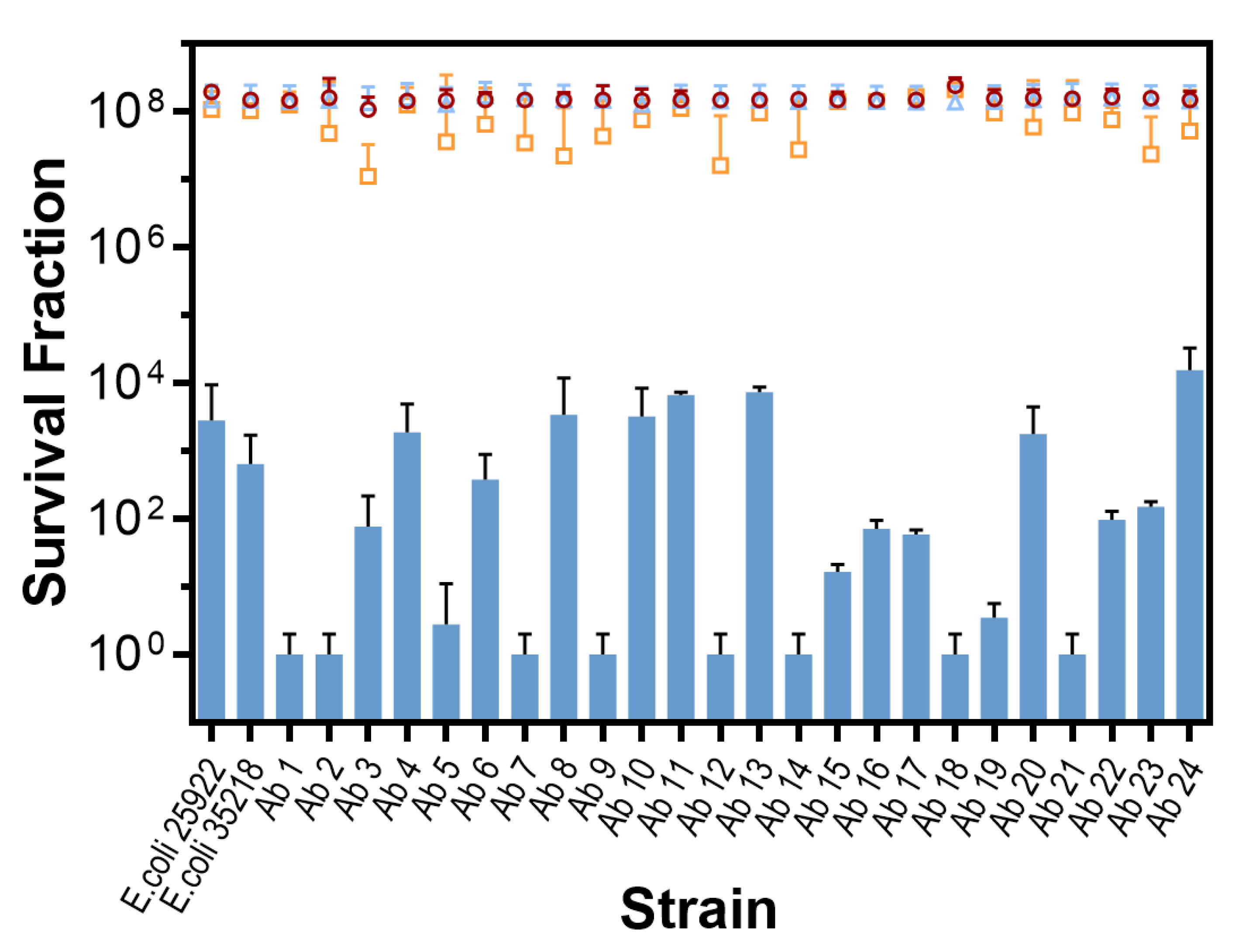
2.2. In Vitro Photodynamic Inactivation of E. coli Growing in Planktonic State

Using 31 µM of MB and 18 J/cm2 of red light, at least a 3 log10 CFU/mL (99.9%) reduction was achieved for all strains and more than 6 log10 for half of them. Data are shown in Figure 1. Control experiments showed that the effect of light alone is negligible for all strains (p > 0.9) while MB alone exerts some toxicity (<1 log10; p < 0.0001). On the other hand, there were significant differences among the control and the PDT-treated groups (p < 0.0001) and there were also significant differences in PDT susceptibility among the strains (p < 0.0001).

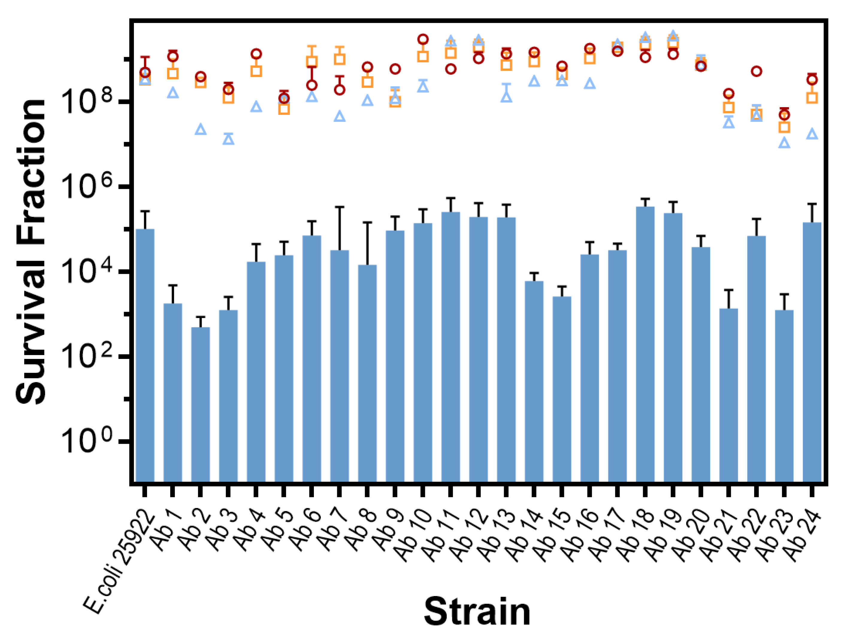
In vitro Photodynamic Inactivation of E. coli Growing in Biofilm

All the strains studied in this work grew in biofilm. The photodynamic inactivation treatments using the same conditions employed for the cells in planktonic state were ineffective, therefore the MB concentration was increased. An antimicrobial effect (99.9% or 3 log10 CFU/mL reduction) could be achieved for all the strains at a concentration of 78.2 µM. Data are shown in Figure 2.
Control experiments showed that the effect of light or MB alone is negligible for all strains (p > 0.9). On the other hand, there were significant differences among the control and the PDT-treated groups (p < 0.0001) and there were also significant differences in PDT susceptibility among the strains (p = 0.0007).

3. Discussion

The susceptibility of 24 clinical isolates of E. coli to a panel of antibiotics commonly used in the clinics to treat urinary infections has been studied. The antibiotics were selected from different families with different modes of action. Table 1 shows that the antibiotics nitrofurantoin and tetracycline have the highest number of strains which are resistant (>80%). Moreover, nine out of the sixteen antibiotics tested were ineffective against at least 50% of the clinical strains isolated in this work. In fact, only three antibiotics (cefotaxime, cefoxitin, and ciprofloxacin) were effective in vitro against all the isolates. Additional insights can be gained if the data in Table 1 are analyzed in terms of the individual strains (Table 2). All the isolated strains are resistant to at least four antibiotics and one of them (strain 4) is remarkably resistant to eleven out of the sixteen antibiotics. On average, strains were resistant to seven antibiotics. With the exception of strains number 16 and 22, all the isolated strains are multidrug-resistant according to the criteria established by the European Centre for Disease Prevention and Control and the Centre for Disease Control and Prevention, namely acquired non-susceptibility to at least one agent in three or more antimicrobial categories [28]. This does not detract from the fact that strains 16 and 22 show resistance to four of the antibiotics.
In contrast, the results (Figure 1; Figure 2) demonstrate that aPDT exerts an antimicrobial effect (>3 log10 CFU/mL reduction) in 100% of the clinical E. coli strains tested, irrespective of their antibiotic susceptibility profile, both in planktonic and biofilm state. Detailed inspection of the planktonic state results shows that aPDT achieves a disinfecting effect (>5 log CFU/mL reduction [29]) for 17 isolates, including 15 of the multidrug-resistant strains. It should be noted that these results were obtained with relatively mild conditions, namely 31 µM MB and 18 J/cm2 red light, chosen to reveal the different susceptibility of the clinical isolates based on our previous experience with the reference strains and preliminary experiments. Higher MB concentrations or light doses would have resulted in even higher extent of photoinactivation, thereby masking any potential differences among the isolates. Consequently, the study demonstrates that all tested strains of E. coli, independent of their antibiotic-resistance profile, can be successfully photo-inactivated by MB, corresponding to previous data for reference strains [30].
Although the bactericidal effect of the treatment is consistent across all strains, there are statistically significant differences in susceptibility amongst them (Figure 1). These results are consistent with those found on S. aureus [20]. It has been proposed that some of the resistance factors present in the strains have weight in the susceptibility of S. aureus to PDT [31], but no clear correlation has been found between the strains’ response to aPDT and antibiotic resistance.
The situation is less clear in E. coli. While some studies had found that the reference strain ATCC 25922 was slightly more susceptible to photoinactivation than multidrug resistant clinical isolates [21,31], our results show that this is the case only for a few isolates (strains 11, 13, and 24), while the majority are significantly more susceptible (Figure 1). Since MB is internalized by E. coli [32,33] and is known to be a substrate for efflux pumps in E. coli as well as in other bacteria [19,34,35], one could reasonably expect some tolerance to aPDT in the resistant strains. We can speculate that resistance mechanisms such as reduced permeability to antimicrobial agents, active efflux of the antimicrobial from the cell, enzymatic alterations, or degradation of the antimicrobial agent [36] may contribute to modulate the photodynamic activity of MB, but they are not determinants for aPDT efficiency.
Biofilms play a major role in bacterial infections and strongly affect the susceptibility of the bacteria to antibiotics and also to aPDT [37]. E. coli biofilms evolve from early stage to mature within 16 h [38], concomitantly increasing the thickness of the extracellular matrix. In our study, biofilms were allowed to grow for at least 16 h to let them reach the mature state. Figure 2 shows the aPDT results obtained for the 24 clinical isolates growing in biofilms. The main finding is that aPDT was able to produce an antimicrobial effect (99.9% kill) on all strains, irrespective of their antibiotic resistance profile. Comparing with the results in planktonic state (Figure 1), it is apparent that photoinactivation is not as efficient and no disinfecting effect (>99.999% kill) could be achieved for any isolate despite increasing the concentration of MB by 2.5-fold. It is well known that photoinactivation of cells living in biofilms require higher concentration of MB compared to their planktonic counterparts [39,40], owing mainly to the mechanical barrier to MB diffusion posed by the extracellular matrix and to alterations in gene expression. As in planktonic cells, statistically significant differences were detected among the strains: Firstly, none of the multidrug-resistant strains appear to be more tolerant to aPDT than the reference ATCC 25922. Thus, as in the planktonic state, the antibiotic-resistance mechanisms do not confer tolerance to aPDT in the biofilm state. In fact, some of the clinical isolates were found more susceptible to aPDT than the reference ATCC 25922 also in biofilm state. Secondly, the differences among strains are less marked than in planktonic state, which indicates that the major determinant of photoinactivation efficacy must be a factor common to all strains, likely the existence of an extracellular matrix. Thus, not surprisingly, no clear correlation is observed between the planktonic and biofilm results.

4. Materials and Methods

4.1. Bacterial Strains

Twenty-four strains of uropathogenic E. coli and two strains obtained from the American Type Culture Collection (E. coli ATCC 35218 and E. coli ATCC 25922) were tested in this study. The clinical specimens were isolated from twenty-four different anonymized urine samples with pyuria obtained from twenty-four patients on different days after receiving their consent for the use of microorganisms obtained from their samples in investigational work. The patients had received prior instruction in the proper methods for the collection of midstream urine specimens. Specimens were cultured quantitatively for bacteria on brolacin agar (C.L.E.D. agar, Merck, Darmstadt, Germany) and tryptic soy agar (TSA, Merck, Darmstadt, Germany) within one hour of collection unless they were refrigerated. Species identifications were determined after obtaining a pure culture. All the isolated strains were maintained at −80 °C at our premises.

4.2. Antibiotic Susceptibility Testing

Antibiotic susceptibility tests of the strains were carried out by means of the disc diffusion method using Rosco Neo-Sensitabs™ tablets commercialized by ROSCO Diagnostica (Taastrup, Denmark). Details of the tested antibiotics and their references are depicted in Table 3. The method was adapted from that described by Kirby-Bauer in 1966 [41]. A few colonies of the strain to be tested were picked from an 18–24 h non-selective agar plate and introduced into a tube containing 4 mL 0.9% NaCl solution obtaining a turbidity equivalent to 0.5 McFarland. One milliliter of this bacterial suspension was then introduced onto a Petri dish containing Mueller-Hinton agar as culture media (Sigma, Saint Louis, United States) and rotated over its entire surface. The surplus suspension was removed using a micropipette. After the inoculum had dried (3–15 min) the antibiotic disks were placed on the agar and the plates were incubated side up in a 35 °C incubator. After 16–18 h incubation, the diameters of the zones of complete inhibition were measured to the nearest whole millimeter to classify the strain as susceptible, intermediate or resistant to each specific antibiotic [42]. Classification of the strains as multidrug-resistant was done according to the criteria established by the European Centre for Disease Prevention and Control (ECDC) and the Centre for Disease Control and Prevention (CDC) [28].

4.3. Photosensitizer and Light Source

MB was supplied by Panreac (Montcada i Reixac, Spain). It was dissolved in Milli-Q sterile water to give stock solutions with a dye concentration of 78.2 µM. Stock solution was stored in the dark for no more than 1 week and was diluted in PBS immediately before experiments. Red light at 625 ± 25 nm was delivered using an 18 LED light source (Showtec, Par64 Short, Sussex, UK).

4.4. Photodynamic Inactivation of E. coli Growing in Planktonic State:

For each strain, three independent tests were carried out as follows: Bacteria were grown aerobically overnight at 37 °C in Tryptic Soy Broth (TSB, Panreac 413820.1210, Castellar del Vallès, Spain) in an orbital shaking incubator. A volume of 10 µL of this pre-inoculum was added to 10 mL of sterile TSB and incubated, until the culture reached an optical density of 0.5 at 600 nm (OD600, measured in a Jenway 6305 spectrophotometer), corresponding to ca. 1.5 × 10⁸ CFU/mL.
The suspensions were then centrifuged (5 min, 3000 rpm) and the pellet was resuspended in 5 mL of phosphate-buffered saline at pH 4 (PBS, Fisher BP399, New Jersey, USA). Two 0.5 mL aliquots of this bacterial suspension were then diluted with PBS to 1 mL (BS1 and BS2) and two additional 0.5 mL aliquots were diluted with a 62 µM MB solution in PBS also to 1 mL (BS3 and BS4; final MB concentration 31 µM). The four resulting suspensions were then incubated in the dark in an orbital shaking incubator (Rotabit-JP Selecta) at 37 °C for 15 min and 60 rpm.
Then, three 300 µL aliquots of BS1 and BS3 were placed in different wells of a 96-well plate and kept in the dark until the end of the experiments (dark controls BS1 -L -MB and BS3 -L +MB, respectively). In turn, three 300 µL aliquots of BS2 and BS4 were placed in different wells of a second 96-well plate and irradiated with red light (625 ± 25 nm, 8 mW/cm2, 18 J/cm2) 25 cm from the top of the plates (light control BS2 +L -MB and test BS4 +L +MB, respectively).
At the end of the experiments, all suspensions were ten-fold serially diluted in PBS in triplicate, and 10 µL of each dilution was streaked on Tryptic Soy Agar plates (TSA, TSB added with 1.5% agar-agar, Fisher BP9744, Geel, Belgium) and incubated aerobically in the dark for 18 to 20 h at 37 °C. After the incubation period, the number of CFUs/mL was determined, yielding 9 results for each of the four BSs. These conditions were selected based on our previous experience with the reference E. coli strains.

4.5. Photodynamic Inactivation of E. coli Growing in Biofilm

This assay was performed against the reference E. coli ATCC 25922, a strain known to produce biofilms [43], along with the clinical strains of uropathogenic E. coli. For all strains, it was checked that they were capable to live in biofilm as described by Crémet et al. [44].
For each strain, three independent tests were carried out as follows: Bacteria were grown aerobically overnight (minimum 16 h) at 37 °C in TSB in an orbital shaking incubator. Twelve 2-µL aliquots of this pre-inoculum were placed in the wells of two different 96-well polystyrene plates (six aliquots in each plate) containing each 130 µL of TSB and were incubated overnight at 37 °C without stirring to allow the formation of biofilms (BFs).
The supernatant liquid was then removed from the wells, and the benthonic cells were gently washed three times with sterile PBS solution. After the third wash, three wells in each plate were refilled with 130 µL of PBS (BF1 and BF2, respectively), while the other three were refilled with a 78 µM MB solution (BF3 and BF4, respectively).
The BFs were then incubated at 37 °C for 15 min in darkness. After the incubation period, the plate containing the three replicates of BF1 and BF3 was kept in the dark until the end of the experiments (dark controls BF1 -L -MB and BF3 -L +MB, respectively), while the plate containing the three replicates of BF2 and BF4 was irradiated with red light (625 ± 25 nm, 8 mW/cm2, 18 J/cm2) 25 cm from the top of the plates (light control BF2 +L -MB and test BF4 +L +MB, respectively).
At the end of the experiments, all BFs were washed three times with PBS and the remaining surface-attached cells of the wells were resuspended with 180 µL of PBS by pipetting up and down. All suspensions were then ten-fold serially diluted in PBS in triplicate, and 10 µL of each dilution was streaked on Tryptic Soy Agar plates and incubated aerobically in the dark for 18 to 20 h at 37 °C. After the incubation period, the number of CFUs/mL was determined, yielding 9 results for each of the four BFs.

4.6. Statistics

Survival fractions are presented as mean + standard deviation (SD). Differences between the means were compared for significance by a one-way ANOVA using the GraphPad Prism 7.04 software. Values of p < 0.05 were considered significant.

5. Conclusions

The usefulness of photodynamic therapy against 26 clinical isolates of E. coli with different antibiotic susceptibility profiles, most of them multidrug resistant, has been demonstrated. aPDT was able to exert an antimicrobial effect in 100% of the strains, both in planktonic and biofilm state, with significant differences in susceptibility among strains. Antibiotic resistance did not confer tolerance to aPDT in any of the clinical isolates tested.

Author Contributions

Conceptualization, M.A., S.N.; Investigation, Ò.G., G.M., M.B.; Writing—original draft preparation, Ò.G.; Writing—review and editing, Ò.G., G.M., M.B., M.A., S.N.; Supervision, M.A., S.N.; Funding Acquisition, S.N. All authors have read and agreed to the published version of the manuscript.

Funding

This research was funded by Spanish Ministerio de Economía y Competitividad (Grant Numbers CTQ2013-48767-C3-1-R and CTQ2016-78454-C2-1-R).

Conflicts of Interest

The authors declare no conflict of interest.

References

  1. Buchholz, K.; Collins, J. The roots—A short history of industrial microbiology and biotechnology. Appl. Microbiol. Biotechnol. 2013, 97, 3747–3762. [Google Scholar] [CrossRef]
  2. O’Neill. Wellcome Trust. Available online: https://amr-review.org/sites/default/files/160518_Final%20paper_with%20cover.pdf/ (accessed on 20 January 2020).
  3. Cassini, A.; Högberg, D.; Plachouras, D.; Quattrocchi, A.; Hoxha, A.; Simonsen, G.S.; Colomb-Cotinat, M.; Kretzschmar, M.E.; Devleesschauwer, B.; Cecchini, M.; et al. Attributable deaths and disability-adjusted life-years caused by infections with antibiotic-resistant bacteria in the EU and the European Economic Area in 2015: A population-level modelling analysis. Lancet Infect. Dis. 2019, 19, 56–66. [Google Scholar] [CrossRef] [Green Version]
  4. Ghosh, C.; Sarkar, P.; Issa, R.; Haldar, J. Alternatives to conventional antibiotics in the era of antimicrobial resistance. Trends Microbiol. 2019, 27, 323–338. [Google Scholar] [CrossRef]
  5. Wainwright, M.; Maisch, T.; Nonell, S.; Plaetzer, K.; Almeida, A.; Tegos, G.P.; Hamblin, M.R. Photoantimicrobials—Are we afraid of the light? Lancet Infect. Dis. 2017, 17, e49–e55. [Google Scholar] [CrossRef]
  6. Maisch, T.; Spannberger, F.; Regensburger, J.; Felgenträger, A.; Bäumler, W. Fast and effective: Intense pulse light photodynamic inactivation of bacteria. J. Ind. Microbiol. Biotechnol. 2012, 39, 1013–1021. [Google Scholar] [CrossRef]
  7. Jori, G.; Fabris, C.; Soncin, M.; Ferro, S.; Coppellotti, O.; Dei, D.; Fantetti, L.; Chiti, G.; Roncucci, G. Photodynamic therapy in the treatment of microbial infections: Basic principles and perspective applications. Lasers Surg. Med. 2006, 38, 468–481. [Google Scholar] [CrossRef] [PubMed]
  8. Tavares, A.; Carvalho, C.M.; Faustino, M.A.; Neves, M.G.; Tomé, J.P.; Tomé, A.C.; Cavaleiro, J.A.; Cunha, A.; Gomes, N.C.; Alves, E.; et al. Antimicrobial photodynamic therapy: Study of bacterial recovery viability and potential development of resistance after treatment. Mar. Drugs 2010, 8, 91–105. [Google Scholar] [CrossRef] [PubMed] [Green Version]
  9. Cieplik, F.; Deng, D.; Crielaard, W.; Buchalla, W.; Hellwig, E.; Al-Ahmad, A.; Maisch, T. Antimicrobial photodynamic therapy—What we know and what we don’t. Crit. Rev. Microbiol. 2018, 571–589. [Google Scholar] [CrossRef] [PubMed] [Green Version]
  10. Romanova, N.A.; Brovko, L.Y.; Moore, L.; Pometun, E.; Savitsky, A.P.; Ugarova, N.N.; Griffiths, M.W. Assessment of photodynamic destruction of Escherichia coli O157:H7 and Listeria monocytogenes by using ATP bioluminescence. Appl. Environ. Microbiol. 2003, 69, 6393–6398. [Google Scholar] [CrossRef] [PubMed] [Green Version]
  11. Malik, Z.; Faraggi, A.; Savion, N. Ultrastructural damage in photosensitized endothelial cells: Dependence on hematoporphyrin delivery pathways. J. Photochem. Photobiol. B 1992, 14, 359–368. [Google Scholar] [CrossRef]
  12. Wainwright, M.; Crossley, K.B. Photosensitising agents—Circumventing resistance and breaking down biofilms: A review. Int. Biodeter. Biodeg. 2004, 53, 119–126. [Google Scholar] [CrossRef]
  13. Giuliani, F.; Martinelli, M.; Cocchi, A.; Arbia, D.; Fantetti, L.; Roncucci, G. In vitro resistance selection studies of RLP068/Cl, a new Zn(II) phthalocyanine suitable for antimicrobial photodynamic therapy. Antimicrob. Agents Chemother. 2010, 54, 637–642. [Google Scholar] [CrossRef] [PubMed] [Green Version]
  14. Rapacka-Zdonczyk, A.; Wozniak, A.; Pieranski, M.; Woziwodzka, A.; Bielawski, P.K.; Grinholc, M. Development of Staphylococcus aureus tolerance to antimicrobial photodynamic inactivation and antimicrobial blue light upon sublethal treatment. Sci. Rep. 2019, 9, 9423. [Google Scholar] [CrossRef] [PubMed] [Green Version]
  15. Laguna, V.; Gilaberte, Y.; Millán-Lou, M.I.; Agut, M.; Nonell, S.; Rezusta, A.; Hamblin, M.R. Combination of photodynamic therapy and antimicrobial compounds to treat skin and mucosal infections: A systematic review. Photochem. Photobiol. Sci. 2019, 18, 1020–1029. [Google Scholar] [CrossRef] [PubMed]
  16. Hamblin, M.R.; O’Donnell, D.A.; Murthy, N.; Rajagopalan, K.; Michaud, N.; Sherwood, M.E. Polycationic photosensitizer conjugates: Effects of chain length and Gram classification on the photodynamic inactivation of bacteria. J. Antimicrob. Chemother. 2002, 49, 941–951. [Google Scholar] [CrossRef] [PubMed] [Green Version]
  17. Wainwright, M.; Crossley, K.B. Methylene Blue—A therapeutic dye for all seasons? J. Chemother. 2002, 14, 431–443. [Google Scholar] [CrossRef]
  18. Floyd, R.A.; Schneider, J.E., Jr.; Dittmer, D.P. Methylene blue photoinactivation of RNA viruses. Antiviral Res. 2004, 61, 141–151. [Google Scholar] [CrossRef]
  19. Tegos, G.P.; Masago, K.; Aziz, F.; Higginbotham, A.; Stermitz, F.R.; Hamblin, M.R. Inhibitors of bacterial multidrug efflux pumps potentiate antimicrobial photoinactivation. Antimicrob. Agents Chemother. 2008, 52, 3202–3209. [Google Scholar] [CrossRef] [Green Version]
  20. Grinholc, M.; Rapacka-Zdonczyk, A.; Rybak, B.; Szabados, F.; Bielawski, K.P. Multiresistant strains are as susceptible to photodynamic inactivation as their naïve counterparts: Protoporphyrin IX-mediated photoinactivation reveals differences between methicillin-resistant and methicillin-sensitive Staphylococcus aureus strains. Photomed. Laser Surg. 2014, 32, 121–129. [Google Scholar] [CrossRef] [Green Version]
  21. Caires, C.S.A.; Leal, C.R.B.; Rodrigues, A.C.S.; Lima, A.R.; Silva, C.M.; Ramos, C.A.N.; Chang, M.R.; Arruda, E.J.; Oliveira, S.L.; Nascimento, V.A.; et al. Photoinactivation of mcr-1 positive Escherichia coli. Laser Phys. Lett. 2018, 15. [Google Scholar] [CrossRef]
  22. Wilson, M.; Yianni, C. Killing of methicillin-resistant Staphylococcus aureus by low-power laser light. J. Med. Microbiol. 1995, 42, 62–66. [Google Scholar] [CrossRef] [PubMed]
  23. Soncin, M.; Fabris, C.; Busetti, A.; Dei, D.; Nistri, D.; Roncucci, G.; Jori, G. Approaches to selectivity in the Zn(II)-phthalocyanine-photosensitized inactivation of wild-type and antibiotic-resistant Staphylococcus aureus. Photochem. Photobiol. 2002, 10, 815–819. [Google Scholar] [CrossRef]
  24. Tang, H.M.; Hamblin, M.R.; Yow, C.M. A comparative in vitro photoinactivation study of clinical isolates of multidrug-resistant pathogens. J. Infect. Chemother. 2007, 13, 87–91. [Google Scholar] [CrossRef] [PubMed] [Green Version]
  25. Maisch, T. A new strategy to destroy antibiotic resistant microorganisms: Antimicrobial photodynamic treatment. Mini Rev. Med. Chem. 2009, 9, 974–983. [Google Scholar] [CrossRef] [PubMed]
  26. Grinholc, M.; Szramka, B.; Kurlenda, J.; Graczyk, A.; Bielawski, K.P. Bactericidal effect of photodynamic inactivation against methicillin-resistant and methicillin-susceptible Staphylococcus aureus is strain-dependent. J. Photochem. Photobiol. B 2008, 90, 57–63. [Google Scholar] [CrossRef]
  27. Parente, T.M.A.L.; Rebouças, E.L.; dos Santos, V.C.V.; Barbosa, F.C.B.; Zaninc, I.C.J. Serratia marcescens resistance profile and its susceptibility to photodynamic antimicrobial chemotherapy. Photodiagnosis Photodyn. Ther. 2016, 14, 185–190. [Google Scholar] [CrossRef]
  28. Magiorakos, A.P.; Srinivasan, A.; Carey, R.B.; Carmeli, Y.; Falagas, M.E.; Giske, C.G.; Harbarth, S.; Hindler, J.F.; Kahlmeter, G.; Olsson-Liljequist, B.; et al. Multidrug-resistant, extensively drug-resistant and pandrug-resistant bacteria: An international expert proposal for interim standard definitions for acquired resistance. Clin. Microbiol. Infect. 2012, 18, 268–281. [Google Scholar] [CrossRef] [Green Version]
  29. Boyce, J.M.; Pittet, D. Guideline for hand hygiene in health-care settings: Recommendations of the Healthcare Infection Control Practices Advisory Committee and the HICPAC/SHEA/APIC/IDSA hand hygiene task force. Infect. Control Hosp. Epidemiol. 2002, 23, S3–S40. [Google Scholar] [CrossRef]
  30. Planas, O.; Bresolí-Obach, R.; Nos, J.; Gallavardin, T.; Ruiz-González, R.; Agut, M.; Nonell, S. Synthesis, photophysical characterization, and photoinduced antibacterial activity of Methylene Blue-loaded amino- and mannose-targeted mesoporous silica nanoparticles. Molecules 2015, 20, 6284–6298. [Google Scholar] [CrossRef] [Green Version]
  31. Kashef, N.; Ravaei, S.A.G.; Djavid, G.E. Phototoxicity of phenothiazinium dyes against methicillin-resistant Staphylococcus aureus and multi-drug resistant Escherichia coli. Photodiagnosis Photodyn. Ther. 2012, 9, 11–15. [Google Scholar] [CrossRef]
  32. Garcez, A.S.; Núñez, S.C.; Baptista, M.S.; Daghastanli, N.A.; Itri, R.; Hamblin, M.R.; Ribeiro, M.S. Antimicrobial mechanisms behind photodynamic effect in the presence of hydrogen peroxide. Photochem. Photobiol. Sci. 2011, 10, 483–490. [Google Scholar] [CrossRef] [Green Version]
  33. George, S.; Hamblin, M.R.; Kishen, A. Uptake pathways of anionic and cationic photosensitizers into bacteria. Photochem. Photobiol. Sci. 2009, 8, 788–795. [Google Scholar] [CrossRef] [PubMed] [Green Version]
  34. Tegos, G.P.; Hamblin, M.R. Phenothiazinium antimicrobial photosensitizers are substrates of bacterial multidrug resistance pumps. Antimicrob. Agents Chemother. 2006, 50, 196–203. [Google Scholar] [CrossRef] [PubMed] [Green Version]
  35. Rineh, A.; Dolla, N.K.; Ball, A.R.; Magana, M.; Bremner, J.B.; Hamblin, M.R.; Tegos, G.P.; Kelso, M.J. Attaching the NorA efflux pump inhibitor INF55 to Methylene Blue enhances antimicrobial photodynamic inactivation of methicillin-resistant Staphylococcus aureus in vitro and in vivo. ACS Infect Dis. 2017, 3, 756–766. [Google Scholar] [CrossRef] [PubMed]
  36. McDermott, P.F.; Walker, R.D.; White, D.G. Antimicrobials: Modes of action and mechanisms of resistance. Int. J. Toxicol. 2003, 22, 135–143. [Google Scholar] [CrossRef]
  37. Cieplik, F.; Tabenski, L.; Buchalla, W.; Maisch, T. Antimicrobial photodynamic therapy for inactivation of biofilms formed by oral key pathogens. Front. Microbiol. 2014, 12. [Google Scholar] [CrossRef] [PubMed] [Green Version]
  38. Gu, H.; Lee, S.W.; Carnicelli, J.; Jiang, Z.; Ren, D. Antibiotic susceptibility of Escherichia coli cells during early-stage biofilm formation. J. Bacteriol. 2019, 201, e00034-19. [Google Scholar] [CrossRef] [Green Version]
  39. Ronqui, M.R.; de Aguiar Coletti, T.M.; de Freitas, L.M.; Miranda, E.T.; Fontana, C.R. Synergistic antimicrobial effect of photodynamic therapy and ciprofloxacin. J. Photochem. Photobiol. B 2016, 158, 122–129. [Google Scholar] [CrossRef] [Green Version]
  40. Sousa, A.S.; Prates, R.A.; de Santi, M.E.; Lopes, R.G.; Bussadori, S.K.; Ferreira, L.R.; Deana, A.M. Photodynamic inactivation of Candida albicans biofilm: Influence of the radiant energy and photosensitizer charge. Photodiagnosis Photodyn. Ther. 2016, 14, 111–114. [Google Scholar] [CrossRef]
  41. Bauer, A.W.; Kirby, W.M.; Sherris, J.C.; Turck, M. Antibiotic susceptibility testing by a standardized single disk method. Am. J. Clin. Pathol. 1966, 45, 493–496. [Google Scholar] [CrossRef]
  42. CLSI. Performance Standards for Antimicrobial Disk Susceptibility Tests, Approved Standard—12th ed.; CLSI document M02-A12; Clinical and Laboratory Standards Institute: Wayne, PA, USA, 2015. [Google Scholar]
  43. Naves, P.; del Prado, G.; Huelves, L.; Gracia, M.; Ruiz, V.; Blanco, J.; Dahbi, G.; Blanco, M.; del Carmen Pontea, M.; Soriano, F. Correlation between virulence factors and in vitro biofilm formation by Escherichia coli strains. Microb. Pathog. 2008, 45, 86–91. [Google Scholar] [CrossRef] [PubMed]
  44. Crémet, L.; Corvec, S.; Batard, E.; Auger, M.; Lopez, I.; Pagniez, F.; Dauvergne, S.; Caroff, N. Comparison of three methods to study biofilm formation by clinical strains of Escherichia coli. Diagn. Microbiol. Infect. Dis. 2013, 75, 252–255. [Google Scholar] [CrossRef] [PubMed]
Figure 1. In vitro photodynamic inactivation of E. coli growing in planktonic state (18 J/cm2 at 625 ± 25 nm, 31 µM MB concentration). Survival fractions are presented as mean + standard deviation (SD). Circles: cell control (BS1 -L -MB); triangles: light control (BS2 +L -MB); squares: MB control (BS3 -L +MB); bars: test (BS4 +L +MB).
Antibiotics 09 00098 g001
Figure 2. In vitro inactivation of E. coli growing in biofilm state (18 J/cm2 at 625 ± 25 nm, 78 µM MB concentration). Survival fractions are presented as mean + standard deviation (SD). Circles: cell control (BF1 −L −MB); triangles: light control (BF2 +L −MB); squares: MB control (BF3 −L +MB); bars: test (BF4 +L +MB).
Antibiotics 09 00098 g002
Table 1. Resistance profile of the tested clinical strains of uropathogenic Escherichia coli to various antibiotics.
Antimicrobial CategoryAntibioticBacterial TargetIdentification Number of the Resistant Strains % of Resistant Strains
PenicillinsAmpicillinCell wall synthesis1–7, 9–14, 17, 19, and 2067
Penicillins + β-lactamase inhibitorsAmoxicillin + Clavulanic acid21 and 238
First-generation CephalosporinsCephalotin1, 2, 4, 6–10, 14–2479
Second-generation CephalosporinsCefuroxime1, 6–8, 14, 17, 19, and 2033
Third-generation CephalosporinsCefotaximeNone of them0
CephamycinsAztreonam1, 4–9, 12, 14–16, 18, 22, and 2458
CefoxitinNone of them0
QuinolonesCiprofloxacinDNA gyraseNone of them0
Nalidixic acid3, 4–9, 11, 13, 15–20, 22, and 24.71
Norfloxacin44
MacrolidesAzithromycin50S subunit of the ribosome 4, 9, 17, 19, and 2021
TetracyclinesTetracycline30S subunit of the ribosome1–15, 18, 20, 21, 23, and 24.83
AminoglycosidesGentamycin1–4, 6, 7, 9, 10, 13, 14, 17, and 1950
PhosphoenolpyruvatesFosfomycinUDP-N-acetylglucosamine enolpyruvyl transferase1, 2, 4, 5, 8–12, 14, 15, 17–20, and 24 67
FurantoinsNitrofurantoinVarious bacterial enzymes and DNA2–13, 15–19, 21–2488
Diaminopyrimidine + SulfamideTrimethoprim + SulfamethoxazoleSynthesis of folic acid1–5, 8, 11–15, 18, 20, 21, 23, and 24.67
Table 2. Resistance profile of the tested clinical strains of uropathogenic Escherichia coli to various antibiotics.
AntibioticStrains
123456789101112131415161718192021222324
AmpicillinRRRRRRR RRRRRR R RR
Amoxicillin + Clavulanic acid R R
CephalotinRR R RRRRR RRRRRRRRRRR
CefuroximeR RRR R R RR
Cefotaxime
AztreonamR RRRRRR R RRR R R R
Cefoxitin
Ciprofloxacin
Nalidixic acid RRRRRRR R R RRRRRR R R
Norfloxacin R
Azithromycin R R R RR
TetracyclineRRRRRRRRRRRRRRR R RR RR
GentamycinRRRR RR RR RR R R
FosfomycinRR RR RRRRR RR RRRR R
Nitrofurantoin RRRRRRRRRRRR RRRRR RRRR
Trimethoprim + SulfamethoxazoleRRRRR R RRRRR R RR RR
Number of resistances8761178889666687487885457
Multidrug resistant?YYYYYYYYYYYYYYYNYYYYYNYY
Table 3. Antibiotics tested against the E. coli strains.
AntibioticDose/µgReference of the Rosco Neo-Sensitabs™ tablets
Ampicillin10567NR 60212
Amoxicillin + Clavulanic acid20 + 10567NR 60112
Cephalothin30567NR 60612
Cefuroxime30567NR 60512
Cefotaxime30567NR 63912
Aztreonam30567NR 63612
Cefoxitin10567NR 62912
Ciprofloxacin5567NR 60812
Nalidixic acid30567NR 61412
Norfloxacin10567NR 76212N
Azithromycin15567NR 60312
Tetracycline30567NR 62012
Gentamycin10567NR 61112
Fosfomycin200567NR 62312
Nitrofurantoin300567NR 62612
Trimethoprim + Sulfamethoxazole1.25 + 23.7567NR 62212

