Article

Antibacterial Activities of Selected Pure Compounds Isolated from Gut Bacteria of Animals Living in Polluted Environments

1
Department of Biological Sciences, School of Science and Technology, Sunway University, Bandar Sunway 47500, Malaysia
2
Department of Biology, Chemistry and Environmental Sciences, College of Arts and Sciences, American University of Sharjah, Sharjah 26666, UAE
3
National Institute for Biotechnology and Genetic Engineering, Faisalabad 44000, Pakistan
*
Author to whom correspondence should be addressed.
Submission received: 8 March 2020 / Revised: 4 April 2020 / Accepted: 7 April 2020 / Published: 17 April 2020

Abstract

:
Antibiotic resistance is a global threat to public health, further accelerated by the misuse of antibiotics in humans and animals. Our recent studies have shown that gut bacteria of animals living in polluted environments are a potential source of antibacterials. Gut bacteria of cockroaches, water monitor lizards and the turtle exhibited molecules such as curcumenol, docosanedioic acid, N-acyl-homoserine lactone, L-homotyrosine and Di-rhamnolipids. Using purified compounds, assays were performed to determine their antibacterial properties using serial dilution method, cytotoxic effects using lactate dehydrogenase release, and cell viability using MTT assay. The results revealed that the purified compounds exhibited significant antibacterial activities (p < 0.05) against selected Gram-negative (Pseudomonas aeruginosa) and Gram-positive bacteria (Streptococcus pyogenes) with effective MIC50 and MIC90 at µg concentrations, and with minimal effects on human cells as observed from LDH and MTT assays. These findings are significant and provide a basis for the rational development of therapeutic antibacterials. Future studies are needed to determine in vivo effects of the identified molecules together with their mode of action, which could lead to the development of novel antibacterial(s).

1. Introduction

The emergence of antibiotic resistance is a public health crisis [1,2]. Several lines of evidence suggest that bacterial pathogens have evolved dramatic multi-drug resistance in recent years resulting in a major public health threat globally [1,2,3]. For example, Pseudomonas aeruginosa have emerged as a major cause of nosocomial infections, particularly in immuno-compromised patients [4]. In 2015, European Centers for Disease Prevention and Control (ECDC) reported that five strains out of every 100 invasive P. aeruginosa isolates were found to be resistant against all five antibacterial groups (piperacillin-tazobactam, fluoroquinolones, ceftazidime, aminoglycosides and carbapenems) under surveillance (EARS-Net) whereas, 13.7% were resistant to three antimicrobial groups available [5]. In the United States, multi-drug resistant P. aeruginosa causes 13% of severe health care-associated infections [5]. Similarly, in 2017 reports from World Health Organization (WHO) have shown that carbapenem-resistant P. aeruginosa was listed in the “critical” group [6]. Of the Gram-positive pathogens, Streptococcus pyogenes is responsible for more than 600 million infections annually [7]. S. pyogenes causes a wide range of conditions and diseases from minor skin and soft tissue infections to severe clinical manifestations [8]. Collectively, these findings suggest that novel antibacterials are needed to counter increasing bacterial resistance [9,10].
Microbial derived natural products possess a wide range of biological properties including antibacterial activities. Most antibacterial classes are derived from readily-available soil bacteria [10,11], but this resource has been mined for several decades [12]. In addition to soil, bacteria isolated from unusual environments can be a potential source of antibacterials. Recent studies have shown that gut bacteria of animals living in polluted environments are a potential source of antibacterials [12,13,14,15,16,17,18,19,20]. The microbiota associated with animal’s gastrointestinal tract (GIT) is a novel and fascinating area of research [21]. The gut microflora signifies the ecological community of microbes inhabiting the GI tract that ultimately influence development, immunity and physiology of all animals [22].
In this study, we used compounds isolated from the gut bacteria of cockroach and turtle. These compounds include (curcumenol and L-Homotyrosine) produced by Bacillus subtilis, (docosanedioic acid) produced by E. coli, and (N-tetradecanoyl homoserine lactone and Di-rhamnolipids) isolated from P. aeruginosa. These compounds were tested against selected Gram-positive and Gram-negative bacteria and human cell lines. Overall, the isolated compounds showed promising antibacterial activities against both Gram-positive and Gram-negative bacteria.

2. Results

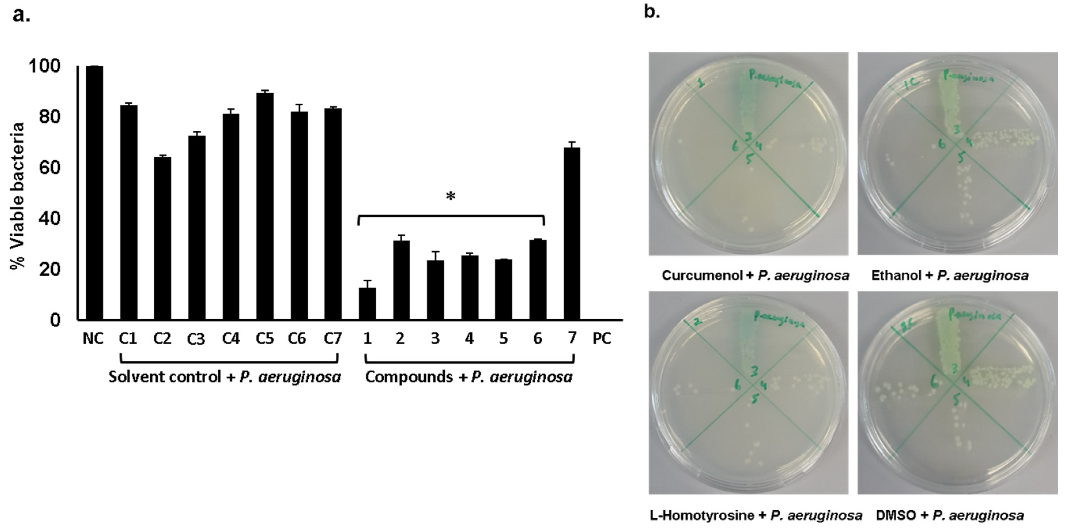
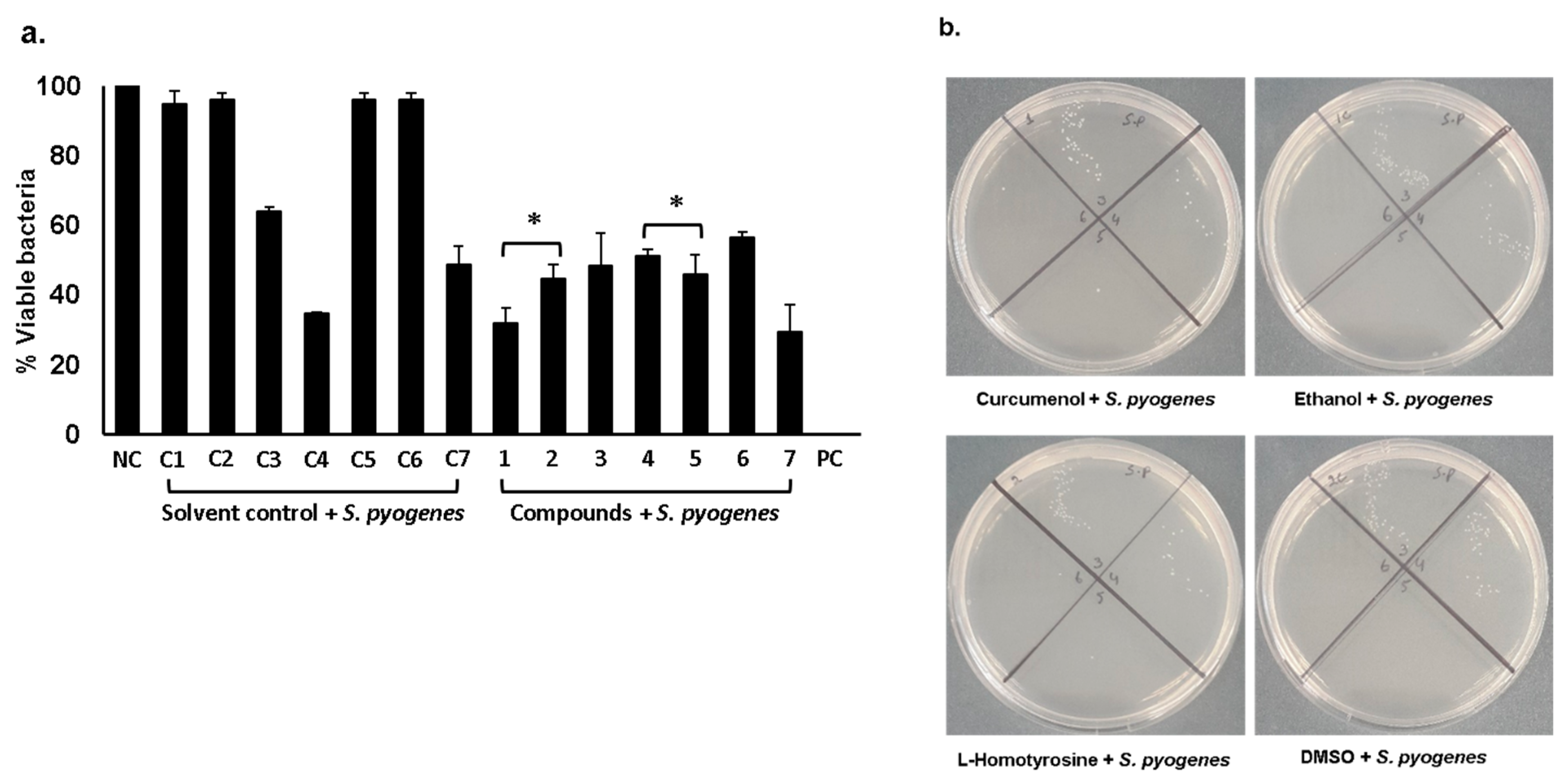
2.1. Curcumenol, L-Homotyrosine and Docosanedioic Acid Showed Significant Antibacterial Activities Against Gram-Negative and Gram-Positive Pathogenic Bacteria Tested

In our previous studies, we have isolated gut bacteria from animals/pests and tested their conditioned media (extracts) against multi-drug resistant Gram-negative and Gram-positive bacteria [9,15]. Bioactive molecules were identified (see Supplementary Figures S1–S5) [9,23] and out of which a few selected compounds were obtained from Sigma Aldrich and tested for their antibacterial properties. The results revealed that all three compounds exhibited significant bactericidal activities (Figure 1a,b) (P < 0.05, using student’s t-test, two-tailed distribution). Among all tested compounds, curcumenol showed exceptional bactericidal effects (Figure 1a) killing 87% bacteria. The minimum inhibitory concentrations (MIC50 and MIC90) for curcumenol, L-homotyrosine and docosanedioic acid against P. aeruginosa are shown in Table 1.
When these compounds were tested against S. pyogenes, the results showed that among all the three tested compounds, curcumenol and L-homotyrosine exhibited significant bactericidal activities (Figure 2a,b) (P < 0.05). Curcumenol alone as well as in combination with L-homotyrosine and docosanedioic acid showed important antibacterial properties (Figure 2a) (P < 0.05). Docosanedioic alone and in combination with L-homotyrosine did not show antibacterial effects. The MIC values of these compounds against S. pyogenes are shown in Table 1.

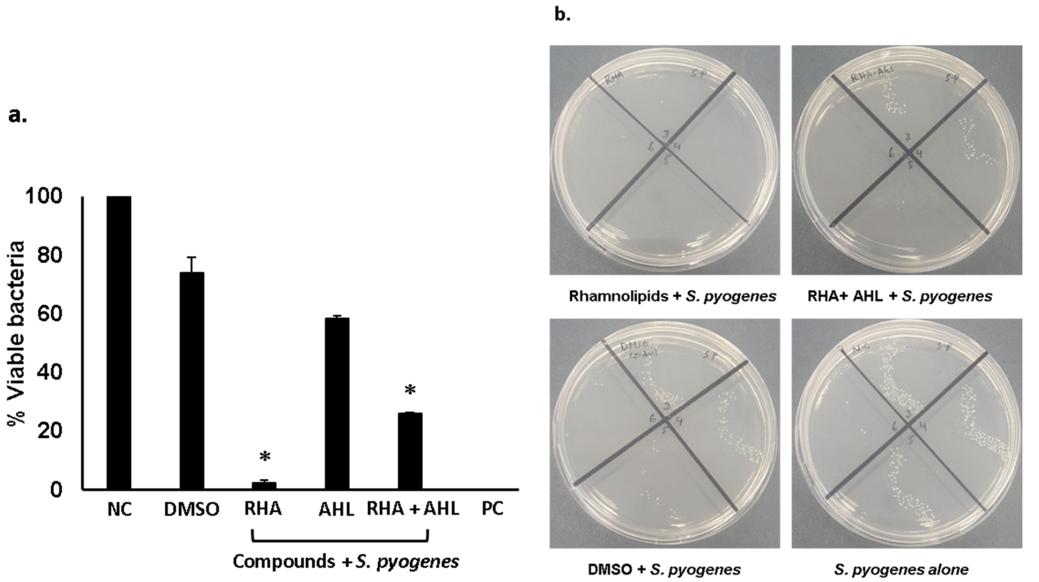
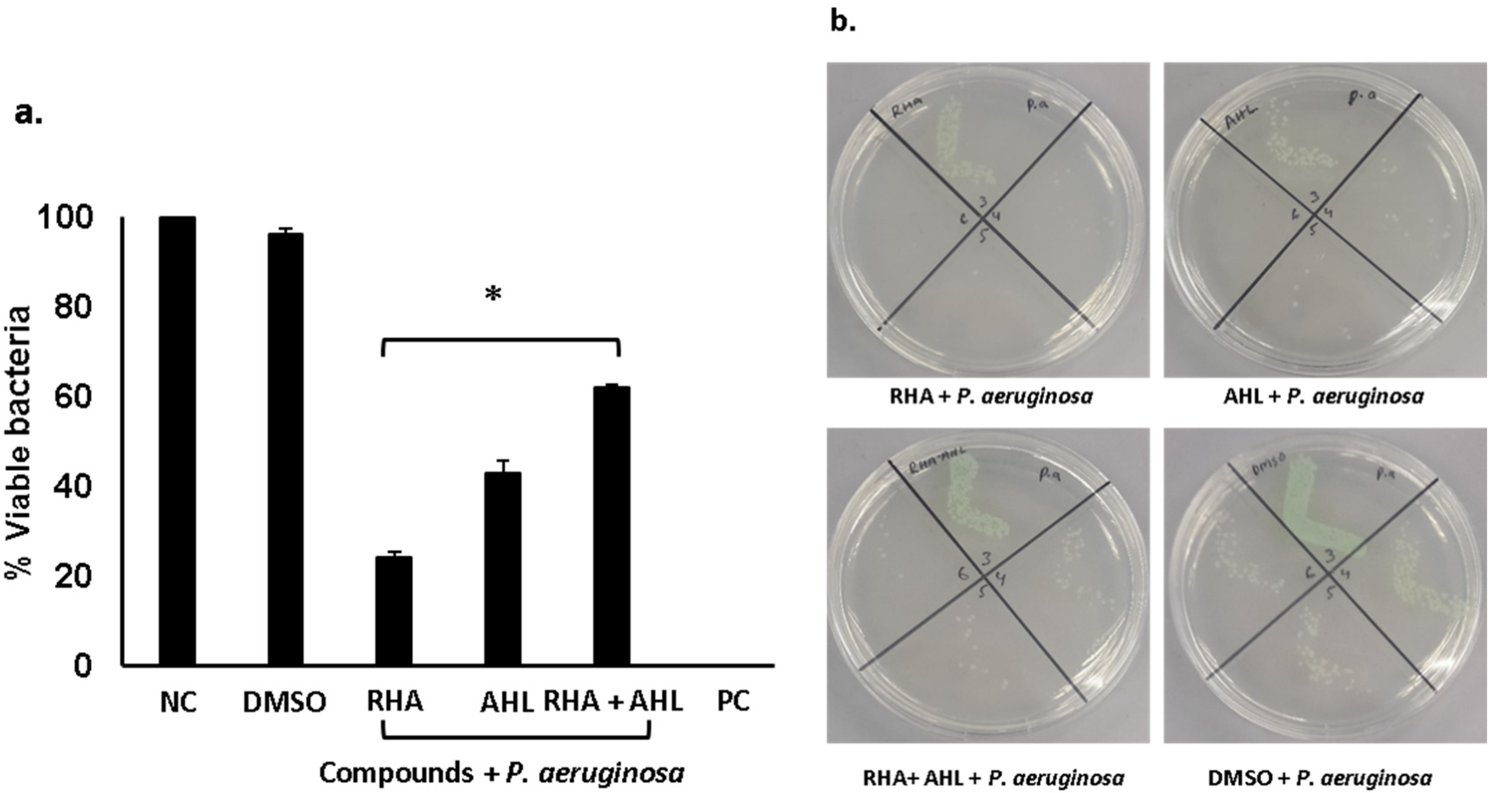
2.2. Di-Rhamnolipids and N-Tetradecanoyl Homoserine Lactones Showed Promising Antibacterial Activities Against Gram-Positive and Gram-Negative Bacteria

Di-rhamnolipids and N-tetradecanoyl homoserine (AHL) lactone (see supplementary Figure S1) from P. aeruginosa isolated from turtle’s gut bacteria and were tested for their antibacterial activities [9]. The results revealed that rhamnolipids showed exceptional bactericidal activity (>90%) against S. pyogenes (Figure 3) (P < 0.05). When tested in combination with AHL, synergistic effects were observed and significant antibacterial activity against S. pyogenes was revealed (Figure 3) (P < 0.05). When Di-rhamnolipids were tested against P. aeruginosa, rhamnolipids alone as well as in combination showed important bactericidal activities (Figure 4) (P < 0.05). Similarly, AHL were tested against both S. pyogenes and P. aeruginosa. AHL alone failed to show antibacterial activity against S. pyogenes whereas combined with RHA showed notable bactericidal activity (Figure 3) (P < 0.05). Unlike against P. aeruginosa, AHL showed promising antibacterial effects alone as well as in combination with rhamnolipids (Figure 4) (P < 0.05). The MIC50 and MIC90 for Di-rhamnolipids and AHLs against P. aeruginosa and S. pyogenes are shown in Table 1.

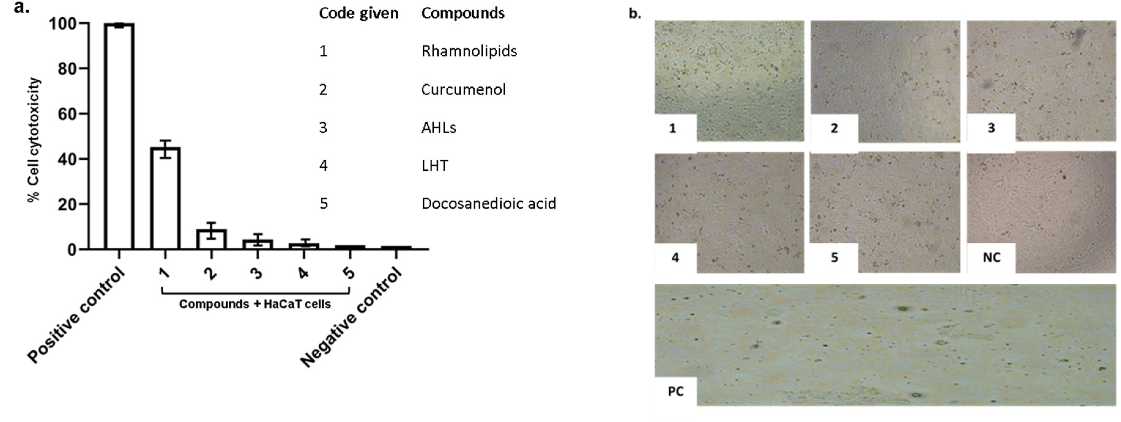
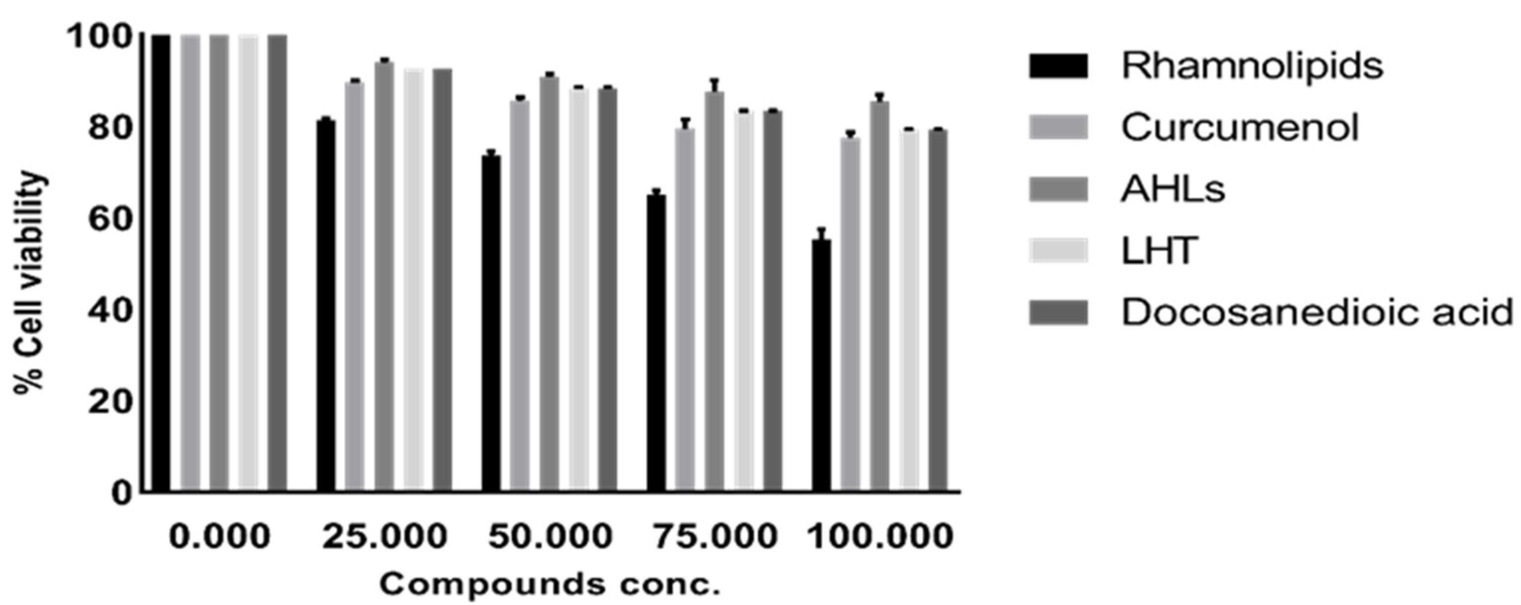
2.3. Pure Compounds Showed Minimal Cytotoxic Effects against Human Cell Lines

Lactate dehydrogenase assays were performed to determine the cytotoxic effects of pure compounds, against HaCaT cell lines. The results showed that all pure compounds tested showed minimal/ or no cytotoxic effects against human cells lines except Di-rhamnolipids that showed 45% cytotoxicity (Figure 5). Moreover, the compounds were tested at various concentrations to determine their effects on cell viability. The results revealed that among all compounds tested, Di-rhamnolipids showed cytotoxicity at 50 µg/mL and 100 µg/mL against HaCaT cell lines (Figure 6). All other compounds exhibited minimal cytotoxicity which is considered as non-toxic according to ISO 10993-5 [9].

3. Discussion

Antibiotic resistance is a major threat to public health [15,24,25,26]. Most of the antibacterial agents available commercially are derived from plants, and microbes, especially bacteria [27]. The producer’s species are utilizing these molecules as competitive weapons against other bacteria, parasites and pathogenic fungi [28]. Natural products especially bacterial secondary metabolites have been exploited successfully to target pathogenic microbes [15,29]. For example, Rahayu et al., [30] isolated curcumenol from the Curcuma aeruginosa plant rhizomes with significant antibacterial activity against Gram-negative (Salmonella typhi and E. coli) bacteria. Similarly, Diastuti et al., [31] isolated terpenoids and sesquiterpenes from Curcuma heyneana rhizomes that showed antibacterial activities. Kacem et al., [32] tested hydrodistilled oil contained curcumenol as component obtained from plant i.e., Genista quadriflora against S. aureurs. Crude extracts showed significant antibacteria activities against S. aureurs. In these studies, curcumenol was isolated either from plant rhizomes or oil but here in this study we identified curcumenol from Bacillus subtilis isolated from the gut of cockroaches (Blaptica dubia) that showed antibacterial activities against both Gram-negative (P. aeruginosa) and Gram-positive (S. pyogenes) bacteria. The bactericidal activity of curcumenol was higher against P. aeruginosa as compared to S. pyogenes.
Bacillus subtilis also produced L-homotyrosine. This compound showed broad-spectrum antibacterial activities against both S. pyogenes and P. aeruginosa. L-homotyrosine exhibited antibacterial activities against P. aeruginosa and its possible mode of action is inhibition of 4-hydroxyphenylpyruvate dioxygenase enzymes of P. aeruginosa [33]. This compounds also showed bactericidal activities against Staphylococcus aureus [34]. L-homotyrosine has been shown to possess antifungal activity against Candida albicans and C. glabrata by inhibiting β-1,3-glucan synthesis [35]. Docosanedioic acid was identified from E. coli isolated from cockroach (Gromphadorhina portentosa) gut. Docosanedioic acid showed antibacterial activities against P. aeruginosa but failed to show bactericidal activity against S. pyogenes. This compound possesses several biological activities reported previously such as anti-HIV, anti-inflammatory, anti-cancer and antifungal activities against A. fumigatus, C. neoformans and C. albicans [36]. We tested Docosanedioic acid for their antibacterial activities and revealed notable bactericidal effects against Gram-negative P. aeruginosa.
Similarly, P. aeruginosa isolated from turtle gut produced strong quorum sensing molecules i.e., N-acyl homoserine lactones (AHL) (N-Tetradecanoyl homoserine) and biological biosurfactant i.e., Di-rhamnolipids [9]. The antibacterial assays of these molecules showed that AHL showed promising results against P. aeruginosa while no antibacterial activity was observed against S. pyogenes. Rhamnolipids showed exceptional bactericidal activity against S. pyogenes and P. aeruginosa. Similar molecules have been isolated from P. aeruginosa isolated from different sources. For example, Patel et al., [37] screened several Gram-negative bacteria including P. aeruginosa that produced AHLs involved in cell-to-cell communication. In another study, Kušar et al., [38] quantified AHLs from clinical samples of P. aeruginosa isolated from dog with otitis externa. Rhamnolipids isolated from P. aeruginosa OBP1 significantly alters the cell membrane of K. pneumoniae and S. aureus and showed significant antibacterial activities [39]. Dusane et al., [40] tested rhamnolipids against Bacillus pumilus and results showed dislodging of biofilm formed by B. pumilus at very low concentrations, signifying their role in eradicating pre-formed biofilms [40].
In summary, these findings suggest that gut bacteria of animals living in polluted environments are a potential source of antibacterials. Gut bacteria of cockroaches, water monitor lizards and turtle exhibited molecules such as curcumenol, docosanedioic acid, N-acyl-homoserine lactone, L-homotyrosine and Di-rhamnolipids with significant antibacterial activities and minimal effects on human cells. These findings are significant and provide a basis for the rational development of therapeutic antibacterials. Future studies are needed to determine in vivo effects of the identified molecules together with their mode of action, which could lead to the development of novel antibacterial(s).

4. Materials and Methods

4.1. Bacterial Cultures

Bacterial species used in this study include multi-drug resistant Pseudomonas aeruginosa (ATCC 10145) and Streptococcus pyogenes (ATCC 49399) and grown overnight at 37 °C and maintained aerobically prior to experimentations as previously described [23].

4.2. Preparation of Compounds Stock Solution

The purified compounds identified from animal’s gut bacteria were dissolved in their respective solvents. Di-rhamnolipids, N-tetradecanoyl homoserine lactones and L-Homotyrosine were dissolved in Di-methyl sulfoxide (DMSO), Curcumenol was dissolved in ethanol and docosanedioic acid was dissolved in n-hexane. All compounds were isolated and identified using high performance liquid chromatography and mass spectrometry (See supplementary Figure S1) [9,15]. All the required stock solutions were prepared and stored at their optimum temperature before testing for their antibacterial and cytotoxic activities.

4.3. Evaluation of Pure Compounds for Antibacterial Properties

Antibacterial assays were performed as described previously [9] with some modifications. Pure compounds at final concentration of 50 µg/mL (Table 2) were incubated with 1 × 10⁶ bacterial inoculums for 2 h at 37 °C. Next, cultures were serially diluted and plated on agar plates. Plates were incubated overnight at 37 °C and viable bacterial colonies were counted. In some of the experiments, the compounds were evaluated at various concentrations (100, 50, 25, 12.5 and 6.25 µg/mL) to determine minimum inhibitory concentrations i.e., MIC50 and MIC90 against S. pyogenes and P. aeruginosa. For MIC, 1 × 10⁵ bacteria per well for controls and pure compounds were tested. Bacteria grown in Muller Hinton broth (MHB) alone and different solvents used for compounds preparation were used as negative control, while gentamicin was used as a positive control.

4.4. In Vitro Cytotoxicity Assays

Cell cytotoxicity assays were performed using MTT (3-(4,5-dimethylthiazol-2-Yl)-2,5-diphenyltetrazolium bromide) assays [9] and lactate dehydrogenase (LDH) assays [41] to determine the effects of pure compounds against human cell lines. Briefly, for LDH assays, confluent HaCaT cell monolayers in a 96-well plate was challenged with 50 µg/mL of pure compounds and plates were incubated for 24 h at 37 °C in the presence of 5% CO2 and humidified conditions. Next, the supernatant was mixed with an equal volume of LDH kit reagents and data analysis was performed spectrophotometrically at 490 nm. For MTT assays, pure compounds were incubated at various concentrations with overnight grown confluent monolayers of human cells and plates were incubated at 37 °C for overnight at 95% humidity and 5% CO2. After this incubation, 10 µL of MTT solution was added to each well and plates was incubated for 3–4 h at 37 °C. The MTT solution was withdrawn carefully and 100 µL of DMSO was added to dissolve the crystals formed by viable cells. The plates were incubated for 15 min at 37 °C and the absorbance determined at 540 nm immediately.

4.5. Statistical Analysis

Student T-test was used to analyze data for antibacterial assays using GraphPad Prism 8.0.2 software. LCMS data analysis (Supplementary data was analyzed using Agilent MassHunter Qualitative Analysis B.05.00 and Thermo Scientific Xcalibur software). The data are expressed as the mean ± standard error of three independent experiments carried out in duplicate. P-value < 0.05 was defined as data is significant.

4.6. Ethical Approval and Consent to Participate

This article does not contain any studies using humans and animals. We also confirm that all experiments were performed in accordance with relevant guidelines and regulations.

5. Conclusions

In summary, here we tested selected pure compounds isolated from animal gut bacteria for their bactericidal activities against Gram-positive and Gram-negative bacteria. Most of the compounds showed significant antibacterial activities with minimum and/or no cytotoxicity with the exception of Di-rhamnolipids that exhibited minimal cytotoxic activities against human cells. These outcomes are important and should lead to the rational development of novel curative antibacterial drugs. In future, such bioactive molecules possessing antibacterial activities could be tested in vivo using mouse animal model.

Supplementary Materials

The following are available online at https://0-www-mdpi-com.brum.beds.ac.uk/2079-6382/9/4/190/s1, Figure S1: Curcumenol, Figure S2: L-Homotyrosine; Figure S3: Docosanedioic acid; Figure S4: N-Tetradecanoyl-homoserine lactone; Figure S5 Di-Rhamnolipids.

Author Contributions

N.A.K. and R.S. conceived the idea; N.A. carried out all experiments under the supervision of R.S. and N.A.K.; N.A. carried out LC/MS analyses under the supervision of M.I.; N.A. prepared the first draft of the manuscript; N.A.K. and R.S. corrected the manuscript. All authors approved the manuscript. All authors have read and agreed to the published version of the manuscript.

Funding

This research was funded by FRGS, Malaysia (FRGS/1/2018/SKK08/SYUC/01/2), and, in part, by the Open Access Program from the American University of Sharjah.

Acknowledgments

This paper represents the opinions of the author(s) and does not mean to represent the position or opinions of the American University of Sharjah.

Conflicts of Interest

No conflict of interest exists.

References

  1. Sprenger, M.; Fukuda, K. New mechanisms, new worries. Science 2016, 351, 1263–1264. [Google Scholar] [CrossRef] [PubMed]
  2. Brown, E.D.; Wright, G.D. Antibacterial drug discovery in the resistance era. Nature 2016, 529, 336–343. [Google Scholar] [CrossRef] [PubMed]
  3. Potron, A.; Poirel, L.; Nordmann, P. Emerging broad-spectrum resistance in Pseudomonas aeruginosa and Acinetobacter baumannii: Mechanisms and Epidemiology. Int. J. Antimicrob. Agents 2015, 45, 568–585. [Google Scholar] [CrossRef] [PubMed] [Green Version]
  4. Bassetti, M.; Vena, A.; Croxatto, A.; Righi, E.; Guery, B. How to manage Pseudomonas aeruginosa infections. Drugs. Context 2018, 7, 212527. [Google Scholar] [CrossRef]
  5. Horcajada, J.P.; Montero, M.; Oliver, A.; Sorlí, L.; Luque, S.; Gómez-Zorrilla, S.; Benito, N.; Grau, S. Epidemiology and Treatment of Multidrug-Resistant and Extensively Drug-Resistant Pseudomonas aeruginosa Infections. Clin. Microbiol. Rev. 2019, 32, e00031-19. [Google Scholar] [CrossRef]
  6. Tacconelli, E.; Carrara, E.; Savoldi, A.; Harbarth, S.; Mendelson, M.; Monnet, D.L.; Pulcini, C.; Kahlmeter, G.; Kluytmans, J.; Carmeli, Y.; et al. Discovery, research, and development of new antibiotics: The WHO priority list of antibiotic-resistant bacteria and tuberculosis. Lancet. Infect. Dis. 2018, 18, 318–327. [Google Scholar] [CrossRef]
  7. Lynskey, N.N.; Lawrenson, R.A.; Sriskandan, S. New understandings in Streptococcus pyogenes. Curr. Opin. Infect. Dis. 2011, 24, 196–202. [Google Scholar] [CrossRef]
  8. Lu, B.; Fang, Y.; Fan, Y.; Chen, X.; Wang, J.; Zeng, J.; Li, Y.; Zhang, Z.; Huang, L.; Li, H.; et al. High prevalence of Macrolide-resistance and molecular characterization of Streptococcus pyogenes isolates circulating in China from 2009 to 2016. Front. Microbiol. 2017, 8, 1–10. [Google Scholar] [CrossRef]
  9. Akbar, N.; Khan, N.A.; Sagathevan, K.; Iqbal, M.; Tawab, A.; Siddiqui, R. Gut bacteria of Cuora amboinensis (turtle) produce broad-spectrum antibacterial molecules. Sci. Rep. 2019, 9, 1–19. [Google Scholar] [CrossRef] [Green Version]
  10. Chevrette, M.G.; Carlson, C.M.; Ortega, H.E.; Thomas, C.; Ananiev, G.E.; Barns, K.J.; Book, A.J.; Cagnazzo, J.; Carlos, C.; Flanigan, W.; et al. The antimicrobial potential of Streptomyces from insect microbiomes. Nat. Commun. 2019, 10, 516. [Google Scholar] [CrossRef] [Green Version]
  11. Newman, D.J.; Cragg, G.M. Natural products as sources of new drugs from 1981 to 2014. J. Nat. Prod 2016, 79, 629–661. [Google Scholar] [CrossRef] [PubMed] [Green Version]
  12. Ling, L.L.; Schneider, T.; Peoples, A.J.; Spoering, A.L.; Engels, I.; Conlon, B.P.; Mueller, A.; Schäberle, T.F.; Hughes, D.E.; Epstein, S.; et al. A new antibiotic kills pathogens without detectable resistance. Nature 2015, 517, 455. [Google Scholar] [CrossRef] [PubMed]
  13. Qin, Z.; Munnoch, J.T.; Devine, R.; Holmes, N.A.; Seipke, R.F.; Wilkinson, K.A.; Wilkinson, B.; Hutchings, M.I. Formicamycins, antibacterial polyketides produced by Streptomyces formicae isolated from African Tetraponera plant-ants. Chem. Sci. 2017, 8, 3218–3227. [Google Scholar] [CrossRef] [PubMed] [Green Version]
  14. Akbar, N.; Siddiqui, R.; Sagathevan, K.A.; Khan, N.A. Gut bacteria of animals/pests living in polluted environments are a potential source of antibacterials. Appl. Microbiol. Biotechnol. 2019, 103, 3955–3964. [Google Scholar] [CrossRef]
  15. Akbar, N.; Siddiqui, R.; Iqbal, M.; Sagathevan, K.; Khan, N.A. Gut bacteria of cockroaches are a potential source of antibacterial compound (s). Lett. Appl. Microbiol. 2018, 66, 416–426. [Google Scholar] [CrossRef]
  16. Ichikawa, S.; Yamaguchi, M.; Matsuda, A. Antibacterial nucleoside natural products inhibiting phospho-MurNAc-pentapeptide translocase; Chemistry and structure-activity relationship. Curr. Med. Chem. 2015, 22, 3951–3979. [Google Scholar] [CrossRef]
  17. Hartmann, M.; Berditsch, M.; Hawecker, J.; Ardakani, M.F.; Gerthsen, D.; Ulrich, A.S. Damage of the bacterial cell envelope by antimicrobial peptides gramicidin S and PGLa as revealed by transmission and scanning electron microscopy. Antimicrob. Agents Chemother. 2010, 54, 3132–3142. [Google Scholar] [CrossRef] [Green Version]
  18. Kohanski, M.A.; Dwyer, D.J.; Collins, J.J. How antibiotics kill bacteria: From targets to networks. Nat. Rev. Microbiol. 2010, 8, 423. [Google Scholar] [CrossRef] [Green Version]
  19. Miller, I.; Chevrette, M.; Kwan, J. Interpreting microbial biosynthesis in the genomic age: Biological and practical considerations. Mar. Drugs 2017, 15, 165. [Google Scholar] [CrossRef]
  20. Kostylev, M.; Kim, D.Y.; Smalley, N.E.; Salukhe, I.; Greenberg, E.P.; Dandekar, A.A. Evolution of the Pseudomonas aeruginosa quorum-sensing hierarchy. Proc. Natl. Acad. Sci. USA 2019, 116, 7027–7032. [Google Scholar] [CrossRef] [Green Version]
  21. Abdelrhman, K.F.A.; BaccI, G.; Mancusi, C.; Mengoni, A.; Serena, F.; Ugolini, A. A First Insight into the Gut Microbiota of the Sea Turtle Caretta caretta. Front. Microbiol 2016, 7, 1–5. [Google Scholar] [CrossRef] [PubMed]
  22. Arizza, V.; VecchionI, L.; Caracappa, S.; Sciurba, G.; Berlinghieri, F.; Gentile, A.; Persichetti, M.F.; Arculeo, M.; Alduina, R. New insights into the gut microbiome in loggerhead sea turtles Caretta caretta stranded on the Mediterranean coast. PLoS ONE 2019, 14, e0220329. [Google Scholar] [CrossRef] [PubMed] [Green Version]
  23. Akbar, N.; Siddiqui, R.; Sagathevan, K.; Iqbal, M.; Khan, N.A. Gut Bacteria of Water Monitor Lizard (Varanus salvator) Are a Potential Source of Antibacterial Compound (s). Antibiotics 2019, 8, 164. [Google Scholar] [CrossRef] [PubMed] [Green Version]
  24. Wenzel, R.P.; Edmond, M.B. Managing antibiotic resistance. N. Engl. J. Med. 2000, 343, 1961–1963. [Google Scholar] [CrossRef]
  25. Rachakonda, S.; Cartee, L. Challenges in antimicrobial drug discovery and the potential of nucleoside antibiotics. Curr. Med. Chem. 2004, 11, 775–793. [Google Scholar] [CrossRef]
  26. Von Nussbaum, F.; Brands, M.; Hinzen, B.; Weigand, S.; Häbich, D. Antibacterial natural products in medicinal chemistry-exodus or revival? Angew. Chem. Int. Ed. 2006, 45, 5072–5129. [Google Scholar] [CrossRef]
  27. Ruiz, B.; Chávez, A.; Forero, A.; García-Huante, Y.; Romero, A.; Sánchez, M.; Rocha, D.; Sánchez, B.; Rodríguez-Sanoja, R.; Sánchez, S.; et al. Production of microbial secondary metabolites: Regulation by the carbon source. Crit. Rev. Microbiol. 2010, 36, 146–167. [Google Scholar] [CrossRef]
  28. Demain, A.L.; Fang, A. The natural functions of secondary metabolites. Adv. Biochem. Eng. Biotechnol. 2000, 69, 1–39. [Google Scholar]
  29. Schneider, T.; Sahl, H.G. An oldie but a goodie–cell wall biosynthesis as antibiotic target pathway. Int. J. Med. Microbiol. 2010, 300, 161–169. [Google Scholar] [CrossRef]
  30. Rahayu, D.U.C.; Sugita, P. Antibacterial activity of curcumenol from rhizomes of Indonesian Curcuma aeruginosa (Zingiberaceae). Rasayan J. Chem. 2018, 11, 762–765. [Google Scholar] [CrossRef]
  31. Diastuti, H.; Syah, Y.M.; Juliawaty, L.D.; Singgih, M. Antibacterial Activity of Germacrane Type Sesquiterpenes from Curcuma heyneana Rhizomes. Indones. J. Chem. 2014, 14, 32–36. [Google Scholar] [CrossRef] [Green Version]
  32. Kacem, N.; Roumy, V.; Duhal, N.; Merouane, F.; Neut, C.; Christen, P.; Hostettmann, K.; Rhouati, S. Chemical composition of the essential oil from Algerian Genista quadriflora Munby and determination of its antibacterial and antifungal activities. Ind Crops Prod 2016, 90, 87–93. [Google Scholar] [CrossRef]
  33. Pascal, R.A., Jr.; Oliver, M.A.; Chen, Y.J. Alternate substrates and inhibitors of bacterial 4-hydroxyphenylpyruvate dioxygenase. Biochemistry 1985, 24, 3158–3165. [Google Scholar] [CrossRef] [PubMed]
  34. Or, Y.S. Preparation of Tricyclic Erythromycins as Bactericides. International Patent WO1997017356, 15 March 1997. [Google Scholar]
  35. Klein, L.L.; Li, L.; Chen, H.J.; Curty, C.B.; DeGoey, D.A.; Grampovnik, D.J.; Leone, C.L.; Thomas, S.A.; Yeung, C.M.; Funk, K.W.; et al. Total synthesis and antifungal evaluation of cyclic aminohexapeptides. Bioorg. Med. Chem. 2000, 8, 1677–1696. [Google Scholar] [CrossRef]
  36. Ali, S.M.; Khan, N.A.; Sagathevan, K.; Anwar, A.; Siddiqui, R. Biologically active metabolite(s) from haemolymph of red-headed centipede Scolopendra subspinipes possess broad spectrum antibacterial activity. AMB Express 2019, 9, 95. [Google Scholar] [CrossRef]
  37. Patel, N.M.; Moore, J.D.; Blackwell, H.E.; Amador-Noguez, D. Identification of unanticipated and novel N-acyl L-homoserine lactones (AHLs) using a sensitive non- targeted LC-MS/MS method. PLoS ONE 2016, 11, e0163469. [Google Scholar] [CrossRef]
  38. Kušar, D.; Šrimpf, K.; Isaković, P.; Kalšek, L.; Hosseini, J.; Zdovc, I.; Kotnik, T.; Vengušt, M.; Tavčar-Kalcher, G. Determination of N-acylhomoserine lactones of Pseudomonas aeruginosa in clinical samples from dogs with otitis externa. BMC Vet. Res. 2016, 12, 233. [Google Scholar] [CrossRef] [Green Version]
  39. Bharali, P.; Saikia, J.P.; Ray, A.; Konwar, B.K. Rhamnolipid (RL) from Pseudomonas aeruginosa OBP1: A novel chemotaxis and antibacterial agent. Colloids Surf B Biointerfaces 2013, 103, 502–509. [Google Scholar] [CrossRef]
  40. Dusane, D.H.; Nancharaiah, Y.V.; Zinjarde, S.S.; Venugopalan, V.P. Rhamnolipid mediated disruption of marine Bacillus pumilus biofilms. Colloids Surf B Biointerfaces 2010, 81, 242–248. [Google Scholar] [CrossRef]
  41. Ali, S.M.; Siddiqui, R.; Ong, S.K.; Shah, M.R.; Anwar, A.; Heard, P.J.; Khan, N.A. Identification and characterization of antibacterial compound (s) of cockroaches (Periplaneta americana). Appl. Microbiol. Biotechnol 2017, 101, 253–286. [Google Scholar] [CrossRef] [PubMed] [Green Version]
Figure 1. Antibacterial assays of pure compounds against P. aeruginosa. Briefly, 50 µg/mL of each compound was incubated with 1 × 10⁶ bacteria at 37 °C for 2 h. Next, cultures were serially diluted and plated onto agar plates. Following this incubation, viable bacterial colonies were counted and c.f.u. was recorded. (a) Bactericidal activities of curcamenol, L-homotyrosine and docosanedioic acid against P. aeruginosa. (b) Representative effects of compounds on P. aeruginosa. The data are expressed as the mean ±SE of several independent experiments performed in duplicate. P-values were determined by student’s T-test where (*) represents P < 0.05 using GraphPad Prism 8.0.2.
Antibiotics 09 00190 g001
Figure 2. Antibacterial assays of pure compounds against S. pyogenes. Briefly, 1 × 10⁶ bacteria were exposed to 50 µg/mL of each compound at 37 °C for 2 h. After this, cultures were serially diluted, plated onto agar plates and plates were incubated overnight at 37 °C. Next day, bacterial c.f.u was determined by counting viable colonies. (a) Antibacterial activities of curcamenol, L-homotyrosine and docosanedioic acid against S. pyogenes. (b) Representative effects of compounds on S. pyogenes. P-values were determined by student’s T-test where (*) represents P < 0.05 using GraphPad Prism 8.0.2. The data are expressed as the mean ±SE of several independent experiments performed in duplicate.
Antibiotics 09 00190 g002
Figure 3. Antibacterial assays of pure compounds against S. pyogenes. Briefly, 1 × 10⁶ bacteria were incubated with pure compounds at 50 µg/mL for 2 h at 37 °C. Next, cultures were diluted, plated onto agar plates and plates were incubated overnight at 37 °C. Following this incubation, bacterial c.f.u was determined by counting viable colonies. (a) bactericidal and (b) bacteriostatic activities of rhamnolipids and N-acyl homoserine lactones against S. pyogenes. P-values were determined by student’s T-test where (*) represents P < 0.05 using GraphPad Prism 8.0.2. The data are expressed as the mean ± SE of several independent experiments performed in duplicate.
Antibiotics 09 00190 g003
Figure 4. Antibacterial assays of pure compounds against P. aeruginosa. Briefly, pure compounds at 50 µg/mL were incubated with 1 × 10⁶ bacteria for 2 h at 37 °C. Next, cultures were diluted and plated onto agar plates. The plates were incubated at 37 °C for overnight. After this incubation, viable bacterial colonies were counted to determine, (a) bactericidal and (b) bacteriostatic activities of rhamnolipids and N-acyl homoserine lactones against P. aeruginosa. The data are expressed as the mean ± SE of several independent experiments performed in duplicate. P-values were determined by student’s T-test where (*) represents P < 0.05 using GraphPad Prism software 8.0.2.
Antibiotics 09 00190 g004
Figure 5. In vitro cytotoxic effects of compounds against HaCaT cell lines. Briefly, pure compounds were incubated with HaCaT cells monolayer in a 96 well plate at 37 °C for 24 h in the presence of 5% CO2 and humidified conditions. Following day, LDH released by cells was measured at 490 nm and results were recorded. (a) Pure compounds tested were non-toxic against HaCaT cells. (b) Representative images and compounds effects incubated with cell monolayer.
Antibiotics 09 00190 g005
Figure 6. Cytotoxic effects of compounds isolated from gut bacteria in a concentration dependent manner on HaCaT cell viability using MTT assays. Briefly, overnight grown confluent HaCaT cells monolayer was exposed to different concentration of the compounds in a 96-well plate and plate was incubated at 37 °C for 24 h in the presence of 5% CO2 and humidified conditions. HaCaT cells alone was taken as negative control. All data are expressed as the mean ± standard error of three independent experiments performed in duplicates. Data was analyzed using GraphPad prism software version 8.0.2.
Antibiotics 09 00190 g006
Table 1. MIC50 and MIC90 values of pure compounds against Gram-positive and Gram-negative bacteria.
S. NoCompoundsS. PyogenesP. Aeruginosa
MIC50MIC90MIC50MIC90
1Rhamnolipids34.6 µg/mL98.36 µg/mL44. 06 µg/mL86.87 µg/mL
2AHL74.1 µg/mL140.06 µg/mL44.53 µg/mL87.73 µg/mL
3Curcumenol82.26 µg/mL156 µg/mL82.96 µg/mL165.69 µg/mL
4L-Homotyrosine73.27 µg/mL141.45 µg/mL51.39 µg/mL94.25 µg/mL
5Docosanedioic acid111.1 µg/mL204.76 µg/mL220.8 µg/mL405.48 µg/mL
AHL: N-acyl homoserine lactone.
Table 2. Pure compounds identified from animal’s gut bacteria used in this study.
Codes GivenCompounds/Combinations and Their Concentrations
1Curcumenol (50 µg/mL)
2L-Homotyrosine (50 µg/mL)
3Docosanedioic acid (50 µg/mL)
4Curcumenol (25 µg/mL) + L-Homotyrosine (25 µg/mL)
5Curcumenol (25 µg/mL) + Docosanedioic acid (25 µg/mL)
6L-Homotyrosine (25 µg/mL) + Docosanedioic acid (25 µg/mL)
7Curcumenol (16.67 µg/mL) + L-Homotyrosine (16.67 µg/mL) + Docosanedioic acid (16.67 µg/mL)
C1Ethanol
C2DMSO
C3n- hexane
C4Ethanol + DMSO + n- hexane
C5Ethanol + DMSO
C6Ethanol + n-hexane
C7DMSO + n- hexane
NCBacteria alone
PCBacteria + Gentamicin
RHARhamnolipids (50 µg/mL)
AHLN-Tetradecanonyl homoserine lactone (50 µg/mL)
RHA + AHLRhamnolipids (25 µg/mL+ N-Tetradecanonyl homoserine lactone (25 µg/mL)

