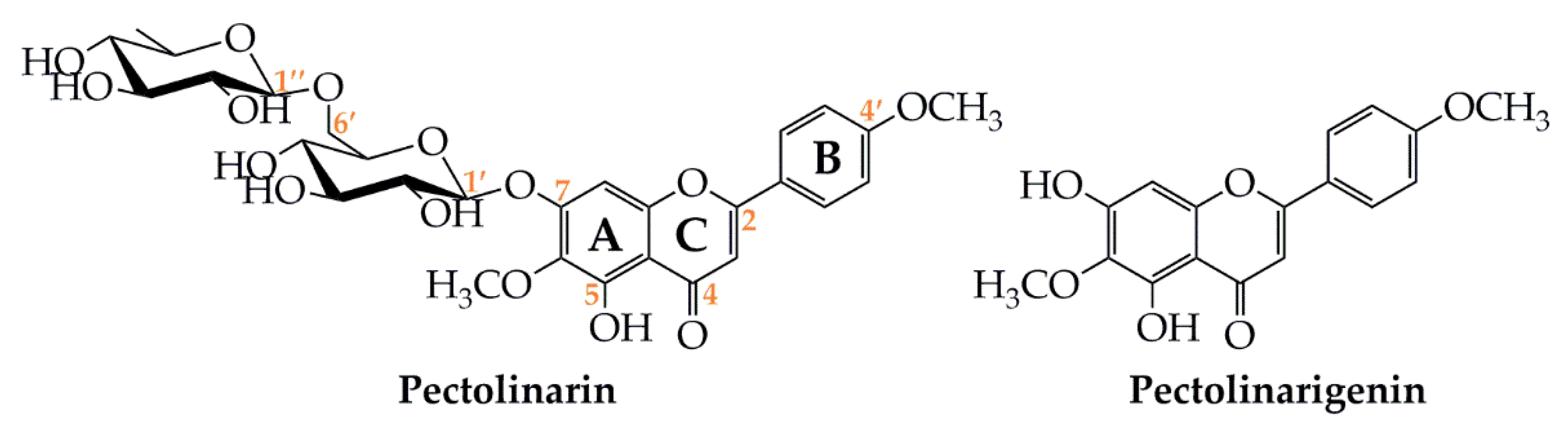
Isolation and Biological Properties of the Natural Flavonoids Pectolinarin and Pectolinarigenin—A Review


Abstract
:1. Introduction
2. Isolation of Pectolinarin and Pectolinarigenin
3. Biological Activities of Pectolinarin and Pectolinarigenin
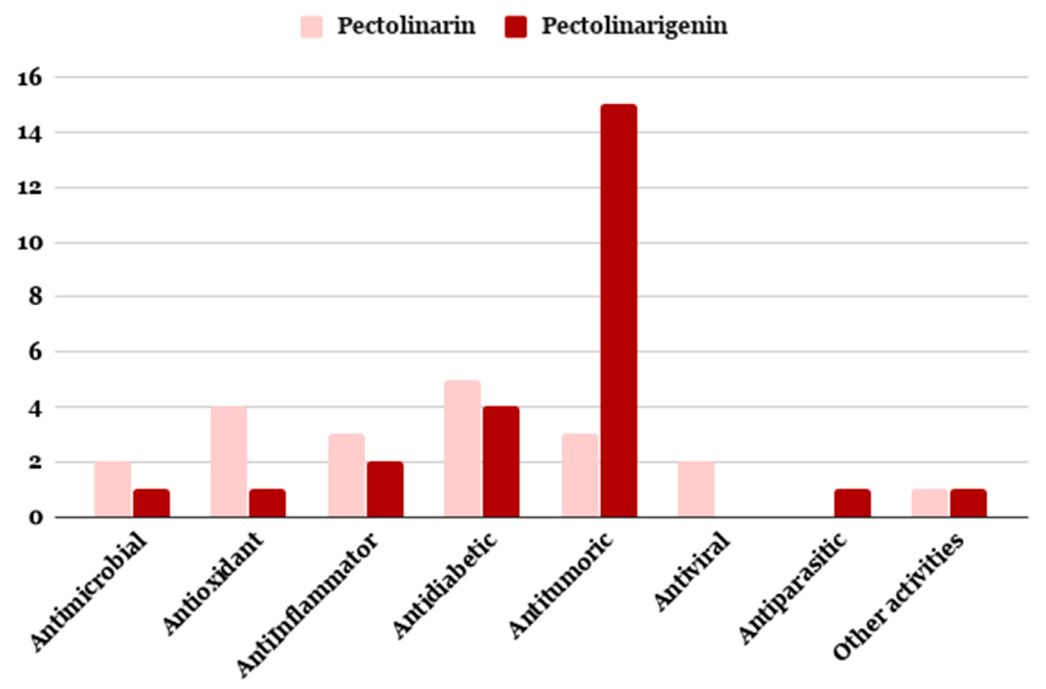
3.1. Antimicrobial Activity
3.2. Antioxidant, Anti-Inflammtory and Antidiabetic Activities
3.3. Cytotoxic and Antitumor Activities
3.4. Other Biological Activities
3.4.1. Antiviral
3.4.2. Antiparasitic
4. Conclusions
Author Contributions
Funding
Conflicts of Interest
References
- Tungmunnithum, D.; Thongboonyou, A.; Pholboon, A.; Yangsabai, A. Flavonoids and Other Phenolic Compounds from Medicinal Plants for Pharmaceutical and Medical Aspects: An Overview. Medicines (Basel) 2018, 5, 93. [Google Scholar] [CrossRef] [PubMed]
- Klobb, T. Two new glucosides: Linarine and pectolinarine. Compt. Rend. 1907, 145, 331–334. [Google Scholar]
- Gruenwald, J. PDR for Herbal Medicines, 1st ed.; Medical Economics Company: Montvale, NJ, USA, 1998. [Google Scholar]
- Hua, H.; Cheng, M.; Li, X.; Pei, Y. A new pyrroloquinazoline alkaloid from Linaria vulgaris. Chem. Pharm. Bull. 2002, 50, 1393–1394. [Google Scholar] [CrossRef] [PubMed] [Green Version]
- Lee, S.J. Korean Folk Medicine; Seoul National University Press: Seoul, Korea, 1966; pp. 145–146. [Google Scholar]
- Jeong, B.; Shin, M. Dictionary of Folk Medicine; Yeungrim Pub.: Seoul, Korea, 1989; p. 1041. [Google Scholar]
- Liu, S.; Zhang, J.; Li, D.; Liu, W.; Luo, X.; Zhang, R.; Li, L.; Zhao, J. Anticancer activity and quantitative analysis of flavone of Cirsium japonicum DC. Nat. Prod. Res. 2007, 21, 915–922. [Google Scholar] [CrossRef] [PubMed]
- Pandya, P.N.; Aghera, H.B.; Ashok, B.K. Diuretic activity of Linaria ramosissima (Wall.) Janch. leaves in albino rats. Ayu 2012, 33, 576–578. [Google Scholar] [CrossRef] [PubMed] [Green Version]
- Jain, A.; Katewa, S.S.; Galave, P.; Nag, A. Some therapeutic uses of biodiversity among the tribals of Rajasthan. Ind. J. Tradit. Med. 2008, 7, 256–262. [Google Scholar]
- Bole, P.V.; Pathak, J.M. Flora of Saurashtra; The director botanical survey of India: New Delhi, India, 1988. [Google Scholar]
- Ghisalberti, E.L. Lantana camara L. (Verbenaceae). Fitoterapia 2000, 71, 467–486. [Google Scholar] [CrossRef]
- Dioscorides; Robert, T.G. The Greek Herbal of Dioscorides; Gunther, R.T., Ed.; Hafner Publishing Company: New York, NY, USA, 1968; p. 247. [Google Scholar]
- Muhaisen, H.M.H.; Ilyas, M.; Mushfiq, M.; Parveen, M.; Basudan, O.A. Flavonoid from Viburnum cotinifolium. J. Chem. Res. 2002, 10, 480–481. [Google Scholar] [CrossRef]
- Singh, H.; Lily, M.K.; Dangwal, K. Viburnum mullaha D.DON fruit (Indian Cranberry): A potential source of polyphenol with rich antioxidant, anti-elastase, anti-collagenase, and anti-tyrosinase activities. Int. J. Food Prop. 2017, 20, 1729–1739. [Google Scholar] [CrossRef] [Green Version]
- Martínez-Vázquez, M.T.O.; Apan, R.; Lastra, A.L.; Bye, R. A comparative study of the analgesic and anti-inflammatory activities of pectolinarin isolated from Cirsium subcoriaceum and linarin isolated from Buddleia cordata. Planta Med. 1998, 64, 134–137. [Google Scholar] [CrossRef]
- Morita, N.; Shimizu, M.; Arisawa, M. Two new flavone glycosides from Cirsium lineare. Phytochemistry 1973, 12, 421–423. [Google Scholar] [CrossRef]
- Park, J.C.; Lee, J.H.; Choi, J.W. Isolation and biological activity of flavone glycosides from the aerial part of Cirsium japonicum var. ussuriense in Korea. Han’guk Yongyang Siklyong Hakhoechi 1995, 24, 906–910. [Google Scholar]
- Ganzera, M.; Pöcher, A.; Stuppner, H. Differentiation of Cirsium japonicum and C. setosum by TLC and HPLC-MS. Phytochem. Anal 2005, 16, 205–209. [Google Scholar] [CrossRef] [PubMed]
- Jeong, D.M.; Jung, H.A.; Choi, J.S. Comparative antioxidant activity and HPLC profiles of some selected Korean thistles. Arch. Pharm. Res. 2008, 31, 28–33. [Google Scholar] [CrossRef]
- Ma, Q.; Jiang, J.G.; Zhang, X.M.; Zhu, W. Identification of luteolin 7-O-β-D-glucuronide from Cirsium japonicum and its anti-inflammatory mechanism. J. Funct. Foods 2018, 46, 521–528. [Google Scholar] [CrossRef]
- Nazaruk, J.; Jakoniuk, P. Flavonoid composition and antimicrobial activity of Cirsium rivulare (Jacq.) All. flowers. J. Ethnopharmacol. 2005, 102, 208–212. [Google Scholar] [CrossRef]
- Do, J.C.; Jung, K.Y.; Son, K.H. Isolation of pectolinarin from the aerial parts of Cirsium nipponicum. Saengyak Hakhoe Chi. 1994, 25, 73–75, CA 121:104074. [Google Scholar]
- Yoo, Y.M.; Nam, J.H.; Kim, M.Y.; Choi, J.; Park, H.J. Pectolinarin and pectolinarigenin of Cirsium setidens prevent the hepatic injury in rats caused by D-galactosamine via an antioxidant mechanism. Biol. Pharm. Bull. 2008, 31, 760–764. [Google Scholar] [CrossRef] [Green Version]
- Nugroho, A.; Lim, S.C.; Karki, S.; Choi, J.S.; Park, H.J. Quantitative determination of five phenolic peroxynitrite-scavengers in nine korean native compositae herbs. Nat. Prod. Sci. 2015, 21, 155–161. [Google Scholar]
- Lee, J.H.; Jung, H.K.; Han, Y.S.; Yoon, Y.M.; Yun, C.W.; Sun, H.Y.; Cho, H.W.; Lee, S.H. Antioxidant effects of Cirsium setidens extract on oxidative stress in human mesenchymal stem cells. Mol. Med. Rep. 2016, 14, 3777–3784. [Google Scholar] [CrossRef] [Green Version]
- Kim, M.S.; Nam, M.; Hwang, G.S. Metabolic alterations in two Cirsium Species identified at distinct phenological stages using UPLC-QTOF/MS. Phytochem. Anal. 2018, 29, 77–86. [Google Scholar] [CrossRef] [PubMed]
- Uehara, A.; Nakata, M.; Kitajima, J.; Iwashina, T. Internal and external flavonoids from the leaves of Japanese Chrysanthemum species (Asteraceae). Biochem. Syst. Ecol. 2012, 41, 142–149. [Google Scholar] [CrossRef]
- Lee, H.B.; Kwak, J.H.; Zee, O.P.; Yoo, S.J. Flavonoids from Cirsium rhinoceros. Arch. Pharmacal. Res. 1994, 17, 273–277. [Google Scholar] [CrossRef]
- Yim, S.H.; Kim, H.J.; Lee, I.S. A polyacetylene and flavonoids from Cirsium rhinoceros. Arch. Pharm. Res. 2003, 26, 128–131. [Google Scholar]
- Gardner, R.C. Acacetin-7-O-rutinoside and pectolinarin from Cirsium coloradense. Phytochemistry 1973, 12, 223. [Google Scholar] [CrossRef]
- Shelyuto, V.L.; Glyzin, V.I.; Ban’kovskii, A.I.; Bubon, N.T. Flavonoid glycosides of Cirsium oleraceum. Chem. Nat. Compd. 1971, 7, 372–373. [Google Scholar] [CrossRef]
- Shelyuto, V.L.; Glyzin, V.I.; Yurchenko, G.N.; Smirnova, L.P. Flavonoids from Cirsium oleraceum flowers. Chem. Nat. Compd. 1978, 14, 336. [Google Scholar] [CrossRef]
- Gardner, R.C. Systematics of Cirsium (Compositae) in Wyoming. Madrono 1974, 22, 239–265. [Google Scholar]
- Morita, N.; Fukuta, M.; Shimizu, M. Studies on the medicinal resources XXIII, Flavonoids of Cirsium plants (Compositae) in Japan. Components of the leaves of Cirsium microspicatum Nakai var. kiotense Kitam., C. dipsicolepis Matsum., C. brevicaule A. Gray, C. matsumurae Nakai, C. yakusimense Masamune, C. amplexifolium Kitam., C. spinosum Kitam., C. tanakae Matsum. subsp. aomorense Kitam. and C. arvense scop. var. setosum Ledeb. Yakugaku Zasshi 1964, 18, 9–11. [Google Scholar]
- Nakaoki, T.; Morita, N. Studies on the medicinal resources XIII, Flavonoids of Cirsium plants (Compositae) in Japan. Components of the leaves of Cirsium microspicatum Nakai, C. otayae Kitamura, C. yoshizawae Koidz., C. japonicum DC.; C. purpuratum Matusum. Yakugaku Zasshi 1959, 79, 1338–1340. [Google Scholar] [CrossRef] [Green Version]
- Glyzinm, V.I.; Shelyuto, V.L.; Patudin, A.V.; Bubon, N.T. Flavonoids of Cirsium Mill species. Mater S’ezde. Farm. B SSR 1977, 153–156. [Google Scholar]
- Iwashina, T.; Kadota, Y.; Ueno, T.; Ootani, S. Foliar flavonoid composition in Japanese Cirsium species (Compositae), and their chemotaxonomic significance. J. Jpn. Bot. 1995, 70, 280–290. [Google Scholar]
- Christian, A.; Ivan, S.; Elisabetta, C.; Maria, D.M.; Matthias, H.; Olivier, P. Comprehensive analysis of Cirsium spinosissimum Scop., a wild alpine food plant. Food Chem. 2014, 160, 165–170. [Google Scholar]
- Morita, N.; Lin, C.N. Studies on the components of Formosan Cirsium species. Part IV. Components of Cirsium arisanense Kitamura and Cirsium ferum Kitamura. Tiawan Yao xue Zazhi 1976, 28, 40–42. [Google Scholar]
- Nakaoki, T.; Morita, N. Studies on the medicinal resources XIV, Flavonoids of Cirsium plants (Compositae) in Japan. Components of the leaves of Cirsium kagamontanum Nakai, C. inundatum Makino, and C. matsumurae Nakai var. pubescens Kitamura. Yakugaku Zasshi 1960, 80, 1296–1297. [Google Scholar] [CrossRef]
- Lin, C.N. Components of formosan Cirsium species. III. Flavonoids of Cirsium kawakamii and Cirsium wallichii. J. Chin. Chem. Soc. (Taipei-Taiwan) 1975, 22, 275–277. [Google Scholar] [CrossRef]
- Wallace, J.W.; Bohm, B.A. Cirsimaritin-4′-O-rutinoside, a new flavone glycoside from Cirsium brevistylum. Phytochemistry 1971, 10, 452–454. [Google Scholar] [CrossRef]
- Cho, S.; Lee, J.; Lee, Y.K.; Chung, M.J.; Kwon, K.H.; Lee, S. Determination of pectolinarin in Cirsium spp. using HPLC/UV analysis. J. Appl. Biol. Chem. 2016, 59, 107–112. [Google Scholar] [CrossRef] [Green Version]
- Makboul, A.M.; Abdel-Baki, A.M. Flavonoids from the leaves of Duranta plumieri. Fitoterapia 1981, 52, 219–220. [Google Scholar]
- Nugroho, A.; Lim, S.C.; Byeon, J.S.; Choi, J.S.; Park, H.J. Simultaneous quantification and validation of caffeoylquinic acids and flavonoids in Hemistepta lyrata and peroxynitrite-scavenging activity. J. Pharm. Biomed. Anal. 2013, 76, 139–144. [Google Scholar] [CrossRef]
- Laskaris, G.G.; Gourneus, D.C.; Kokkalou, E. Phenolics of Picnomon acarna. J. Nat. Prod. 1995, 58, 1248–1250. [Google Scholar] [CrossRef]
- Simões, L.R.; Maciel, G.M.; Brandão, G.C.; Filho, J.D.; Oliveira, A.B.; Castilho, R.O. Chemical constituents of Distictella elongata (Vahl) Urb. (Bignoniaceae). An. Acad. Bras. Cienc. 2013, 85, 873–879. [Google Scholar] [CrossRef] [PubMed] [Green Version]
- Brindha, P.; Ragamanvitha, A.; Narendran, R.; Sriram, S.; Vadivel, V. Antioxidant activity and phytochemical composition of aqueous extract of Markhamia lutea (Benth) K. Schum. leaves. Trop. J. Nat. Prod. Res. 2017, 1, 63–68. [Google Scholar] [CrossRef] [Green Version]
- Chen, X.; Wang, L.; Wei, T.; Liang, M.; Huang, X. A Method for Extraction of Pectolinarin in Buddleja officinalis Flower. CN 105954405 A 20160921, 21 September 2016. [Google Scholar]
- Kamil, M.S.; Ilyas, M. Flavonoidic constituents of Rhododendron arboreum leaves. Fitoterapia 1995, 66, 371. [Google Scholar]
- Yao, Y.; Wu, C.Y.; Hao, Q.; Li, H.Z.; Li, R.T. Study on chemical constituents of Corallodiscus flabellatus. J. Kunming Univ. Sci. Tech. (Nat. Sci. Ed.) 2012, 37, 64–68. [Google Scholar]
- Liu, H.; Liao, H.; Yuan, K. Chemical constituents contained in Aeschynanthus moningeriae. Zhongguo Zhongyao Zazhi 2012, 37, 1963–1967. [Google Scholar]
- Feng, L.; Zhang, Y.; Liu, Y.C.; Liu, Y.; Luo, S.H.; Huang, C.S.; Li, S.H. Leucoflavonine, a new bioactive racemic flavoalkaloid from the leaves of Leucosceptrum canum. Bioorg. Med. Chem. 2019, 27, 27,442–446. [Google Scholar] [CrossRef]
- Oganesyan, G.B.; Mnatsakanyan, V.A.; Gacs-Baitz, E.; Radics, L. Flavonoid glycosides of Teucrium hyrcanicum L. Armyanskii Khimicheskii Zhurnal 1989, 42, 646–653. [Google Scholar]
- Manivannan, R.; Aeganathan, R.; Prabakaran, K. Anti-microbial and anti-inflammatory flavonoid constituents from the leaves of Lawsonia inermis. J. Phytopharmacol. 2015, 4, 212–216. [Google Scholar]
- Khare, C.P. Indian Medicinal Plants, An Illustrated Dictionary; Springer: Berlin/Heidelberg, Germany, 2007; p. 423. [Google Scholar]
- Monteiro, J.; Schuquel, I.T.A.; de Almeida, T.L.; de Oliveira Santin, S.M.; da Silva, C.C.; Chiavelli, L.U.R.; Ruiz, A.L.T.G.; de Carvalho, J.E.; Vendramini-Costa, D.B.; Nakamura, C.V.; et al. Oncibauerins A and B, new flavanones from Oncidium baueri (Orchidaceae). Phytochem. Lett. 2014, 9, 141–148. [Google Scholar] [CrossRef]
- Ferreira, N.P.; Chiavelli, L.U.R.; Savaris, C.R.; Silvana, M.O.; Lucca, D.L.; Milaneze-Gutierre, M.A.; Faria, R.T.; Pomini, A.M. Chemical study of the flowers of the orchid Oncidium baueri Lindley and their visiting bees Trigona spinipes Fabricius. Biochem. Syst. Ecol. 2019, 86, 103918. [Google Scholar] [CrossRef]
- Roh, J.H.; Zee, O.P.; Moon, H.I. Phytochemical Constituents from Melampyrum roseum var. hirsutum Beauv. Korean J. Pharmacol. 2006, 31, 157–162. [Google Scholar]
- Liu, Q.; Yang, Q.M.; Hu, H.J.; Yang, L.; Yang, Y.B.; Chou, G.X.; Wang, Z.T. Bioactive diterpenoids and flavonoids from the aerial parts of Scoparia dulcis. J. Nat. Prod. 2014, 77, 1594–1600. [Google Scholar] [CrossRef]
- Moon, K.I.; Min, B.S.; Lee, H.K.; Zee, O.P. Antioxidant compounds of Oryza sativa L. Saengyak Hakhoechi 2002, 33, 173–176. [Google Scholar]
- Liao, M.; Cheng, X.; Zhang, X.; Diao, X.; Liang, C.; Zhang, L. Qualitative and quantitative analyses of active constituents in Trollius ledebourii. J. Chromatogr. Sci. 2018, 56, 619–635. [Google Scholar] [CrossRef] [PubMed] [Green Version]
- Zhou, J.; Xie, G.; Yan, X. Encyclopedia of Traditional Chinese Medicines—Molecular Structures, Pharmacological Activities, Natural Sources and Applications; Springer: Berlin/Heidelberg, Germany, 2010; Volume 4, p. 173. [Google Scholar]
- Karar, M.G.E.; Kuhnert, N. UPLC-ESI-Q-TOF-MS/MS Characterization of phenolics from Crataegus monogyna and Crataegus laevigata (Hawthorn) leaves, fruits and their herbal derived drops (Crataegutt Tropfen). J. Chem. Biol. Ther. 2015, 1, 102–125. [Google Scholar]
- Zou, Y.; Hong, M.; Yang, X.f. Isolation of chemical components from Thesium chinense. Zhongguo Shiyan Fangjixue Zazhi 2016, 22, 74–77. [Google Scholar]
- Kuptsova, L.P.; Ban’kovskii, A.I. New flavonoid from some species of toadflax. Chem. Nat. Compd. 1970, 6, 128–129. [Google Scholar] [CrossRef]
- Ilieva, E.; Handjieva, N.; Bankova, V.; Popov, S.; Evstatieva, L. Iridoid and flavonoid glycosides from Linaria species. Bulg. Chem. Commun. 1992, 25, 400–406. [Google Scholar]
- Hua, H.; Sun, J.; Li, X. Flavonoids from yellow toadflax (Linaria vulgaris). Chin. Tradit. Herb. Drugs. 1999, 30, 332–334. [Google Scholar]
- Mun, G.S.; Song, H.Y.; Kim, I.G. Analysis of flavonoid-components of Linaria Japonica Miq. Punsok Hwahak 1979, 3, 28–32. [Google Scholar]
- Otsuka, H. Isolation of isolinariins A and B, new flavonoid glycosides from Linaria japonica. J. Nat. Prod. 1992, 55, 1252–1255. [Google Scholar] [CrossRef]
- Widyowati, R.; Sugimoto, S.; Yamano, Y.; Sukardiman, Y.; Otsuka, H.; Matsunami, K. New Isolinariins C, D and E, flavonoid glycosides from Linaria japonica. Chem. Pharm. Bull. 2016, 64, 517–521. [Google Scholar] [CrossRef] [Green Version]
- Tundis, R.; Deguin, B.; Loizzo, M.R.; Bonesi, M.; Statti, G.A.; Tillequin, F.; Menichini, F. Potential antitumor agents: Flavones and their derivatives from Linaria reflexa Desf. Bioorg. Med. Chem. Lett. 2005, 15, 4757–4760. [Google Scholar] [CrossRef]
- Cheriet, T.; Aouabdia, S.; Mancini, I.; Defant, A.; Seghiri, R.; Boumaza, O.; Mekkiou, R.; Sarri, D.; León, F.; Brouard, I.; et al. Chemical constituents of Linaria reflexa Desf. (Scrophulariaceae). Der Pharm. Lett. 2014, 6, 54–57. [Google Scholar]
- Cheriet, T.; Hanfer, M.; Boudjelal, A.; Baali, N.; Mancini, I.; Seghiri, R.; Ameddah, S.; Menad, A.; Benayache, F.; Benayache, S. Glycosyl flavonoid profile, in vivo antidiabetic and in vitro antioxidant properties of Linaria reflexa Desf. Nat. Prod. Res. 2017, 31, 2042–2048. [Google Scholar] [CrossRef]
- Cheriet, T.; Hanfer, M.; Mancini, I.; Benelhadj, S.; Laouas, N.E.; Ameddah, S.; Menad, A.; Seghiri, R. Anti-inflammatory and hemostatic effects of Linaria reflexa Desf. Nat. Prod. Res. 2019, 1–6. [Google Scholar] [CrossRef]
- Smirnova, L.P.; Boryaev, K.I.; Ban’kovskii, A.I. Acacetin and its glycosides in plants of the genus Linaria. Chem. Nat. Compd. 1974, 10, 96–97. [Google Scholar] [CrossRef] [Green Version]
- Lahloub, M.F. Flavonoid, phenylpropanoid and iridoid glycosides of Linaria haelava (Forssk.) Dil. Mansoura J. Pharm. Sci. 1992, 8, 78–95. [Google Scholar]
- Ahmed-Chaouch, M.; Cheriet, T.; Beretta, G.; Sarri, D.; Bensouici, C.; Ouelbani, R.; Mancini, I.; Sekhara, I.; Seghiri, R. Chemical composition, in vitro antioxidant, anticholinesterase and antibacterial activities of Linaria scariosa Desf. Nat. Prod. Res. 2019, 1–5. [Google Scholar] [CrossRef]
- Yuldashev, M.P.; Batirov, E.K.H.; Malikov, V.M. Flavonoids of the epigeal part of Kickxia elatine. Chem. Nat. Compd. 1996, 32, 30–32. [Google Scholar] [CrossRef]
- Amer, M.M.A. Glycosides of Kickxia heterophylla (Schousb.) Dandy in Andrews. Alex. J. Pharm. Sci. 1993, 7, 58–61. [Google Scholar]
- Khan, I.Z.; Aqil, M. Isolation and identification of pectolinarin and mannitol from Kickxia ramosissima (Wall). Chem. Environ. Res. 1993, 2, 287–289. [Google Scholar]
- Ahmad, V.A.; Kousar, F.; Zubair, M.; Khan, A.; Ali, M.S.; Choudhary, M.I.; Sener, B. A new iridoid glycoside from Linaria genestifolia. Fitoterapia 2006, 77, 12–14. [Google Scholar] [CrossRef]
- Al-Rehaily, A.J.; Abdel-Kader, M.S.; Ahmad, M.S.; Mossa, J.S. Iridoid glucosides from Kickxia abhaica D.A. Sutton from Scrophulariaceae. Phytochemistry 2006, 67, 429–432. [Google Scholar] [CrossRef]
- Venditti, A.; Frezza, C.; Serafini, I.; Ciccòla, A.; Sciubba, F.; Serafini, M.; Bianco, A. Iridoids of chemotaxonomy relevance, a new antirrhinoside ester and other constituents from Kickxia spuria subsp. integrifolia (Brot.) R. Fern. Chem. Biodiv. 2018, 15, e1700473. [Google Scholar] [CrossRef]
- Kassem, F.F. Flavonoids of Kickxia aegyptiaca (Dum.) Nabelek. Alex. J. Pharm. Sci. 1992, 6, 62–65. [Google Scholar]
- Jeon, Y.S.; Kim, M.W. The antioxidative effects and isolation and characterization of the extracts from Morus alba L. Korean J. Food Nutr. 2011, 24, 94–100. [Google Scholar] [CrossRef]
- Pan, W.D.; Mai, L.T.; Li, Y.J.; Xu, X.L.; Yu, D.Q. Studies on the chemical constituents of the leaves of Lantana camara. Acta Pharma. Sin. 1993, 28, 35–39. [Google Scholar]
- Mahato, S.B.; Sahu, N.P.; Roy, S.K.; Sharma, O.P. Potential antitumor agents from Lantana camara: Structures of flavonoid, and phenylpropanoid glycosides. Tetrahedron 1994, 50, 9439–9446. [Google Scholar] [CrossRef]
- Begum, S.; Wahab, A.; Siddiqui, B.S.; Qamar, F. Nematicidal constituents of the aerial parts of Lantana camara. J. Nat. Prod. 2000, 63, 765–767. [Google Scholar] [CrossRef] [PubMed]
- Juang, F.C.; Chen, Y.F.; Lin, F.M.; Huang, K.F. Constituents from the leaves of Lantana camara (IV). J. Chin. Med. 2005, 16, 149–155. [Google Scholar]
- Sousa, E.O.; Rocha, J.B.T.; Barros, L.M.; Barros, A.R.C.; Costa, J.G.M. Phytochemical characterization and in vitro antioxidant properties of Lantana camara L. and Lantana montevidensis Briq. Ind. Crops Prod. 2013, 43, 517–522. [Google Scholar] [CrossRef]
- Abdjul, D.B.; Yamazaki, H.; Maarisit, W.; Rotinsulu, H.; Wewengkang, D.S.; Sumilat, D.A.; Kapojos, M.M.; Losung, F.; Ukai, K.; Namikoshi, M. Oleanane triterpenes with protein tyrosine phosphatase 1B inhibitory activity from aerial parts of Lantana camara collected in Indonesia and Japan. Phytochemistry 2017, 144, 106–112. [Google Scholar] [CrossRef] [PubMed]
- Martins, G.R.; da Fonseca, T.S.; Martínez-Fructuoso, L.; Simas, R.C.; Silva, F.T.; Salimena, F.R.G.; Alviano, D.S.; Alviano, C.S.; Leitão, G.G.; Pereda-Miranda, R.; et al. Antifungal phenylpropanoid glycosides from Lippia rubella. J. Nat. Prod. 2019, 82, 566–572. [Google Scholar] [CrossRef]
- Winnett, V.; Boyer, H.; Sirdaarta, J.; Cock, I.E. The potential of Tasmannia lanceolata as a natural preservative and medicinal agent: Antimicrobial activity and toxicity. Pharmacogn. Commnun. 2014, 4, 42–52. [Google Scholar] [CrossRef] [Green Version]
- Noh, H.; Lee, H.; Kim, E.; Mu, L.; Rhee, Y.K.; Cho, C.W.; Chung, J. Inhibitory effect of a Cirsium setidens extract on hepatic fat accumulation in mice fed a high-fat diet via the induction of fatty acid β-oxidation. Biosci. Biotech. Bioch. 2013, 77, 1424–1429. [Google Scholar] [CrossRef] [Green Version]
- Shen, Y.M.; Mu, Q.Z. New Furans from Cirsium chlorolepis. Planta Med. 1990, 56, 472–474. [Google Scholar] [CrossRef]
- Stuart, R.G.A. Chinese Materia Medica; Southern Materials Centre: Taipei, Taiwan, 1911. [Google Scholar]
- Cho, J.W.; Choi, S.Y.; Hong, H.D.; Lee, Y.G.; Jung, H.C. Method for Separating Flavonoids from Cirsium setidens with High Yield and Purity by Extracting with Aqueous Ethanol and Crystallizing with Methanol. Repub. Korean Kongkae Taeho Kongbo KR 2015017148 A 20150216, 7 March 2016. [Google Scholar]
- Hussain, F.; Jahan, N.; Rahman, K.; Sultana, B.; Jamil, S. Identification of hypotensive biofunctional compounds of Coriandrum sativum and evaluation of their angiotensin-converting enzyme (ACE) inhibition potential. Oxid. Med. Cell Longev. 2018, 2018, 4643736. [Google Scholar] [CrossRef] [Green Version]
- Umikalsom, Y.; Harborne, J.B. Flavonoid distribution in Asplenioid ferns. Pertanika 1991, 14, 297–300. [Google Scholar]
- Umikalsom, Y. Flavone O-glycosides and other flavonoids of Malaysian Asplenium L. Pertanika 1991, 14, 149–152. [Google Scholar]
- Valant-Vetschera, K.M.; Wollenweber, E. Flavonoid patterns of Achillea. Part 7. Leaf flavonoids of the Achillea millefolium group. Part II: Distribution patterns of free aglycons in leaf exudates. Biochem. Syst. Ecol. 1988, 16, 605–614. [Google Scholar] [CrossRef]
- Liang, J.Y.; Xu, J.; Shao, Y.Z.; Yang, Y.Y.; Lu, P.Y.; Wang, J.L.; Du, S.S. Chemical constituents from the aerial sections of Ajania potaninii. Biochem. Syst. Ecol. 2019, 84, 64–66. [Google Scholar] [CrossRef]
- Wollenweber, E.; Hradetzky, D.; Mann, K.; Roitman, J.N.; Yatskievych, G.; Proksch, M.; Proksch, P. Exudate flavonoids from aerial parts of five Ambrosia species. J. Plant Physiol. 1987, 131, 37–43. [Google Scholar] [CrossRef]
- Schmidt, T.J.; Willuhn, G. Sesquiterpene lactone and flavonoid variability of the Arnica angustifolia aggregate (Asteraceae). Biochem. Syst. Ecol. 2000, 28, 133–142. [Google Scholar] [CrossRef]
- Merfort, I. Methylated flavonoids from Arnica montana and Arnica chamissonis. Planta Med. 1984, 50, 107–108. [Google Scholar] [CrossRef] [PubMed]
- Todorova, M.; Staneva, J.; Evstatieva, L. Phytochemical study of Arnica chamissonis less. subsp. foliosa (Nutt.) Maguire. C. R. Acad. Bulg. Sci. 2008, 61, 451–454. [Google Scholar]
- Poplawski, J.; Holub, M.; Samek, Z.; Herout, V. Terpenes. CCIX. Arnicolides - sesquiterpenic lactones from the leaves of Arnica montana. Coll. Czech Chem. Commun. 1971, 36, 2189–2199. [Google Scholar] [CrossRef]
- Hu, J.F.; Zhu, Q.X.; Bai, S.P.; Jia, Z.J. New eudesmane sesquiterpene and other constituents from Artemisia mongolica. Planta Med. 1996, 62, 477–478. [Google Scholar] [CrossRef]
- Saleh, N.A.M.; El-Negoumy, S.I.; Abou-Zaid, M.M. Flavonoids of Artemisia judaica, A. monosperma and A. herba-alba. Phytochemistry 1987, 26, 3059–3064. [Google Scholar] [CrossRef]
- Belenovskaya, L.M.; Markova, L.P.; Kapranova, G.I. Phenolic compounds of Artemisia xerophytica. Chem. Nat. Compd. 1982, 18, 115. [Google Scholar] [CrossRef]
- Kul’magambetova, E.A.; Pribytkova, L.N.; Adekenov, S.M. Flavonoids of Artemisia glabella. Chem. Nat. Compd. 2000, 36, 95–96. [Google Scholar] [CrossRef]
- Yin, Y.; Gong, F.Y.; Wu, X.X.; Sun, Y.; Li, Y.H.; Chen, T.; Xu, Q. Anti-inflammatory and immunosuppressive effect of flavones isolated from Artemisia vestita. J. Ethnopharmacol. 2008, 120, 1–6. [Google Scholar] [CrossRef] [PubMed]
- Sharp, H.; Bartholomew, B.; Bright, C.; Latif, Z.; Sarker, S.D.; Nash, R.J. 6-Oxygenated flavones from Baccharis trinervis (Asteraceae). Biochem. Syst. Ecol. 2000, 29, 105–107. [Google Scholar] [CrossRef]
- Rojas, J.; Morales, A. Study of the chemical components of Baccharis decussata (K) hieron. Ciencia (Maracaibo) 2000, 8, 251–256. [Google Scholar]
- Zamorano, R.; Aguirre, M.E.; Munoz de la Pena, A.; Cordano, G.; Medina, J.; Timmermann, B. Flavonoids from Baccharis concava Pers. Bol. Soc. Chil. Quim. 1987, 32, 101–103. [Google Scholar]
- Passero, L.F.; Bonfim-Melo, A.; Corbett, C.E.; Laurenti, M.D.; Toyama, M.H.; de Toyama, D.O.; Romoff, P.; Fávero, O.A.; dos Grecco, S.S.; Zalewsky, C.A.; et al. Anti-leishmanial effects of purified compounds from aerial parts of Baccharis uncinella C. DC. (Asteraceae). Parasitol. Res. 2011, 108, 529–536. [Google Scholar] [CrossRef]
- Weimann, C.; Goransson, U.; Pongprayoon-Claeson, U.; Claeson, P.; Bohlin, L.; Rimpler, H.; Heinrich, M. Spasmolytic effects of Baccharis conferta and some of its constituents. J. Pharm. Pharmacol. 2002, 54, 99–104. [Google Scholar] [CrossRef]
- Mosharrafa, S.A.M.; Mansour, R.M.A.; Abou-Zaid, M.; Saleh, N.A.M. Some biologically active flavonoids from Egyptian members of the Compositae. Bull. Chem. Soc. Ethiopia 1994, 8, 9–13. [Google Scholar]
- Cardona, M.L.; Fernandez, I.; Pedro, J.R.; Perez, B. Sesquiterpene lactones and flavonoids from Centaurea aspera. Phytochemistry 1991, 30, 2331–2333. [Google Scholar] [CrossRef]
- Halfon, B.; Oksuz, S.; Cirpici, A. Flavonoids from Centaurea cariensis Boiss. Doga: Turk. Saglik. Bilimleri. Dergisi. 1989, 13, 138–140. [Google Scholar]
- Fernandez, I.; Garcia, B.; Grancha, F.J.; Pedro, J.R. Sesquiterpene lactones, flavonoids and coumarins from Centaurea collina. Phytochemistry 1989, 28, 2405–2407. [Google Scholar] [CrossRef]
- Csupor, D.; Widowitz, U.; Blazso, G.; Laczko-Zold, E.; Tatsimo, J.S.N.; Balogh, A.; Boros, K.; Danko, B.; Bauer, R.; Hohmann, J. Anti-inflammatory activities of eleven Centaurea species occurring in the Carpathian Basin. Phytother. Res. 2013, 27, 540–544. [Google Scholar] [CrossRef]
- Trendafilova, A.; Todorova, M.; Bancheva, S. Secondary metabolites from Centaurea moesiaca. Biochem. Syst. Ecol. 2007, 35, 544–548. [Google Scholar] [CrossRef]
- Mosaddegh, M.; Tavakoli, M.; Behzad, S. Constituents of the aerial parts of Centaurea behen. Chem. Nat. Comp. 2018, 54, 1015–1017. [Google Scholar] [CrossRef]
- Kumkarnjana, S.; Nimmannit, U.; Koobkokkruad, T.; Pattamadilok, C.; Suttisri, R.; Vardhanabhuti, N. Anti-adipogenic effect of flavonoids from Chromolaena odorata leaves in 3T3-L1 adipocytes. J. Integr. Med. 2018, 16, 427–434. [Google Scholar] [CrossRef] [PubMed]
- Perez Gutierrez, R.M.; Ramirez, E.; Vargas, R. Effect of Cirsium pascuarense on blood glucose levels of normoglycaemic and alloxan-diabetic mice. Phytother. Res. 2001, 15, 552–554. [Google Scholar] [CrossRef] [PubMed]
- Lim, H.; Son, K.H.; Chang, H.W.; Bae, K.H.; Kang, S.S.; Kim, H.P. Anti-inflammatory activity of pectolinarigenin and pectolinarin isolated from Cirsium chanroenicum. Biol. Pharm. Bull. 2008, 31, 2063–2067. [Google Scholar] [CrossRef] [PubMed] [Green Version]
- Lu, M.; Xu, X.; Lu, H.; Lu, Z.; Xu, B.; Tan, C.; Shi, K.; Guo, R.; Kong, Q. Evaluation of anti-tumor and chemoresistance-lowering effects of pectolinarigenin from Cirsium japonicum Fisch ex DC in breast cancer. Trop. J. Pharm. Res. 2016, 15, 547–553. [Google Scholar] [CrossRef] [Green Version]
- Khan, Z.U.H.; Ali, F.; Khan, S.U.; Ali, I. Phytochemical study on the constituents from Cirsium arvense. Mediter. J. Chem. 2011, 1, 64–69. [Google Scholar] [CrossRef]
- Lee, J.H.; Lee, K.R. Phytochemical constituents of Cirsium nipponicum (MAX.) Makino. Saengyak Hakhoechi 2005, 36, 145–150. [Google Scholar]
- Chung, A.K.; Kwon, H.C.; Choi, S.Z.; Min, Y.D.; Lee, S.O.; Lee, W.B.; Yang, M.C.; Lee, K.H.; Nam, J.H.; Kwak, J.H. Norisoprenoids from Cirsium rhinoceros. Saengyak Hakhoechi 2002, 33, 81–84. [Google Scholar]
- Zhu, S.H.; Zhang, Q.j.; Chen, Q.; Zhou, T.; Yao, R.J. Study on chemical constituents of Dichrocephala integrifolia. Zhongguo Shiyan Fangjixue Zazhi 2010, 16, 34–36. [Google Scholar]
- Bierner, M.W. Pectolinarigenin from Dugaldia pinetorum (Standl.) Bierner. Biochem. Syst. Ecol. 1994, 22, 109–110. [Google Scholar] [CrossRef]
- Zdero, C.; Bohlmann, F.; Mueller, M. Sesquiterpene lactones and other constituents from Eriocephalus species. Phytochemistry 1987, 26, 2763–2775. [Google Scholar] [CrossRef]
- Stevens, J.F.; Elema, E.T.; Wollenweber, E. Exudate flavonoids of Eupatorium cannabinum. Biochem. Syst. Ecol. 1995, 23, 451–452. [Google Scholar] [CrossRef]
- Yuan, J.; Yang, J.; Miao, J. Chemical constituents of Eupatorium odoratum. Zhongcaoyao 2005, 36, 1771–1773. [Google Scholar]
- Wu, B.; Liang, J. Pectolinarigenin promotes functional recovery and inhibits apoptosis in rats following spinal cord injuries. Exp. Ther. Med. 2019, 17, 3877–3882. [Google Scholar] [CrossRef] [Green Version]
- Herz, W.; Govindan, S.V.; Kumar, N. Sesquiterpene lactones and other constituents of Eupatorium lancifolium and E. semiserratum. Phytochemistry 1981, 20, 1343–1347. [Google Scholar] [CrossRef]
- Xiao, Y.; Li, K.; Wang, Z.; Fu, F.; Shao, S.; Song, F.; Zhao, J.; Chen, W.; Liu, Q.; Xu, J. Pectolinarigenin prevents bone loss in ovariectomized mice and inhibits RANKL-induced osteoclastogenesis via blocking activation of MAPK and NFATc1 signaling. J. Cell Physiol. 2019, 234, 13959–13968. [Google Scholar] [CrossRef]
- Timmermann, B.; Wollenweber, E.; Doerr, M.; Valant-Vetschera, K.M.; Fuentes, E.R. External flavonoids in two Grindelia species. Z. Naturforsch. 1994, 49, 395–397. [Google Scholar] [CrossRef]
- Alarcon, R.; Ocampos, S.; Pacciaroni, A.; Colloca, C.; Sosa, V. Constituents of Gutierrezia mandonii (Asteraceae). Biochem. Syst. Ecol. 2009, 37, 683–685. [Google Scholar] [CrossRef]
- Ozawa, A.T.; Rivera, P.A.; Romo de Vivar, A. Active principles of the toxic plant Helenium integrifolium. Rev. Latinoam. Quím. 1983, 14, 40–43. [Google Scholar]
- Rojo, A.L.; Palacios, P.S.; Acevedo, C.; Spegazzini, E.D.; Debenedetti, S.L. 6-Methoxyflavonoids from Heterotheca latifolia (Asteraceae). Biochem. Syst. Ecol. 2004, 32, 351–353. [Google Scholar] [CrossRef]
- Ahmed, A.A.; Mohamed, A.Y.; Spring, O.; Bierner, M.W.; Mabry, T.J. Sesquiterpene lactones and flavonoids from Hymenoxys jamesii (Asteraceae) and their systematic significance. Biochem. Syst. Ecol. 2002, 30, 487–491. [Google Scholar] [CrossRef]
- Farkas, L.; Nogradi, M.; Sudarsanam, V.; Herz, W. Constituents of Iva species. V. Isolation, structure, and synthesis of nevadensin, a new flavone from Iva nevadensis and Iva acerosa. J. Org. Chem. 1966, 31, 3228–3232. [Google Scholar] [CrossRef]
- Herz, W.; Bhat, S.V.; Sudarsanam, V. Constituents of Iva species. XII. Sesquiterpene lactones and flavones of Iva frutescens. Phytochemistry 1972, 11, 1829–1831. [Google Scholar] [CrossRef]
- Ybarra, M.I.; Catalan, C.A.N.; Diaz, J.G.; Herz, W. A cyperane and trixanes from Jungia polita. Phytochemistry 1992, 31, 3627–3629. [Google Scholar] [CrossRef]
- Chivers, H.; Corbett, R.E.; Mitchell, R.E.M. Extractives from the leaves of Olearia paniculata. J. Chem. Soc. C: Org. 1966, 20, 1814–1816. [Google Scholar] [CrossRef]
- Cardona, M.L.; Garcia, B.; Pedro, J.R.; Sinisterra, J.F. Flavonoids, flavonolignans and a phenylpropanoid from Onopordon corymbosum. Phytochemistry 1990, 29, 629–631. [Google Scholar] [CrossRef]
- Gonzalez Collado, I, Macias, FA, Massanet, GM, Oliva, J, Maria Rodriguez Luis, F, Vergara, C Chemical components of Onopordum nervosum Boiss. Anales de Quimica, Serie C Quimica Organica y Bioquimica 1984, 80, 100–101.
- Becchi, M.; Carrier, M. 6-Methoxyflavones of Santolina chamaecyparissus. Planta Med. 1980, 38, 267–268. [Google Scholar] [CrossRef]
- Flamini, G.; Ghelli, G.C.; Pistelli, L.; Morelli, I. Phenolic compounds from Santolina pinnata. Planta Med. 1994, 60, 97. [Google Scholar] [CrossRef] [PubMed]
- Sham’yanov, I.D.; Batirov, E.K.; Yuldashev, M.P.; Mallabaev, A. Components of Saussurea elegans. Chem. Nat. Compd. 1983, 19, 763–764. [Google Scholar] [CrossRef]
- Pacciaroni, A.D.V.; Sosa, V.E.; Espinar, L.A.; Oberti, J.C. Sesquiterpene lactones from Schkuhria pinnata. Phytochemistry 1995, 39, 127–131. [Google Scholar] [CrossRef]
- Deng, Y.R.; Song, A.X.; Wang, H.Q. Chemical components of Seriphidium santolium Poljak. J. Chin. Chem. Soc. 2004, 51, 629–636. [Google Scholar] [CrossRef]
- Ortega, A.; Mondragon, P.; Maldonado, E. Guaianolides of Stevia laxiflora. Rev. Soc. Quim. Mex. 1999, 43, 100–102. [Google Scholar]
- Alara, O.R.; Abdurahman, N.H.; Ukaegbu, C.I.; Azhari, N.H.; Kabbashi, N.A. Metabolic profiling of flavonoids, saponins, alkaloids, and terpenoids in the extract from Vernonia cinerea. J. Liq. Chromatogr. Relat. Technol. 2018, 41, 722–731. [Google Scholar] [CrossRef]
- Wollenweber, E.; Bouillant, M.L.; Lebreton, P.; Egger, K. Rare flavonoid-aglycons in the lipophile excretion of Alnus glutinosa. Z. Naturforsch. B 1971, 26, 1188–1190. [Google Scholar] [CrossRef]
- Wollenweber, E. Flavonoids from Alnus crispa, A. japonica, A. koehnei and A. sinuate. Phytochemistry 1974, 13, 2318–2319. [Google Scholar] [CrossRef]
- Popravko, S.A.; Kononenko, G.P.; Tikhomirova, V.I.; Vul’fson, N.S. Secondary metabolites of the birch. IV. Identification of the group of flavonoid aglycons in birch buds (Betula verrucosa). Bioorg. Khim. 1979, 5, 1662–1667. [Google Scholar]
- Isidorov, V.; Szoka, L.; Nazaruk, J. Cytotoxicity of white birch bud extracts: Perspectives for therapy of tumours. PLoS ONE 2018, 13, e0201949. [Google Scholar] [CrossRef] [PubMed]
- Hase, T.; Ohtani, K.; Kasai, R.; Yamasaki, K.; Picheansoonthon, C. Revised structure for hortensin, a flavonoid from Millingtonia hortensis. Phytochemistry 1995, 40, 287–290. [Google Scholar] [CrossRef]
- Wang, K.; Li, M.M.; Chen, X.Q.; Peng, L.Y.; Cheng, X.; Li, Y.; Zhao, Q.S. Phenolic constituents from Brainea insignis. Chem. Pharm. Bull. 2010, 58, 868–871. [Google Scholar] [CrossRef] [PubMed] [Green Version]
- Bacon, J.D.; Hannan, G.L.; Fang, N.; Mabry, T.J. Chemosystematics of the Hydrophyllaceae: Flavonoids of three species of Eriodictyon. Biochem. Syst. Ecol. 1986, 14, 591–595. [Google Scholar] [CrossRef]
- Agnese, A.M.; Juliani, H.R.; Cabrera, J.L. Phytochemical study of species of genus Adesmia (Fabaceae). Anales de la Asociacion Quimica Argentina 1989, 77, 287–291. [Google Scholar]
- Wollenweber, E.; Doerr, M.; Rivera, D.; Roitman, J.N. Externally accumulated flavonoids in three Mediterranean Ononis species. Z. Nat. C J. Biosci. 2003, 58, 771–775. [Google Scholar] [CrossRef]
- Wollenweber, E. On the distribution of exudate flavonoids among angiosperms. Rev. Lat. Quim. 1990, 21, 115–121. [Google Scholar]
- He, X.G.; Lin, L.Z.; Lian, L.Z. Analysis of flavonoids from red clover by liquid chromatography-electrospray mass spectrometry. J. Chromatogr. A 1996, 755, 127–132. [Google Scholar] [CrossRef]
- Pu, X.; Zhou, J. Studies on the chemical components from Leucosceptrum canum. Yunnan Zhiwu Yanjiu 1989, 11, 263–266. [Google Scholar]
- Zaidi, F.; Voirin, B.; Jay, M.; Viricelt, M.R. Free flavonoid aglycons from leaves of Mentha pulegium and Mentha suaveolens (Labiatae). Phytochemistry 1998, 48, 991–994. [Google Scholar] [CrossRef]
- Vieira, R.F.; Grayer, R.J.; Paton, A.J. Chemical profiling of Ocimum americanum using external flavonoids. Phytochemistry 2003, 63, 555–567. [Google Scholar] [CrossRef]
- Abdelshafeek, K.A.; Elgendy, H.A.; El Missiry, M.M.; Seif El Nasr, M.M. Structure elucidation of phenolic acids, flavonoids and hypocholesterolemic activity of Nepeta septemcrenata and Otostegia fruticosa. Der Pharm. Chem. 2016, 8, 357–362. [Google Scholar]
- Ulubelen, A.; Öztürk, S.; Iśildatici, S. A new flavone from Salvia triloba L.f (Labiatae). J. Pharm. Sci. 1968, 57, 1037–1038. [Google Scholar] [CrossRef]
- Wollenweber, E.; Dörr, M.; Rustaiyan, A.; Roitman, J.N.; Graven, E.H. Exudate flavonoids of some Salvia and a Trichostema species. Z. Naturforsch. 1992, 47, 782–784. [Google Scholar] [CrossRef] [Green Version]
- Ulubelen, A.; Tuzlaci, E. Flavonoids and triterpenoids from Salvia euphratica and S. longipedicellata. Fitoterapia 1990, 61, 185. [Google Scholar]
- Topcu, G.; Ulubelen, A.; Tam, T.C.M.; Tao-Che, C. Ses-terterpenes and other constituents of Salvia yosgadensis. Phytochemistry 1996, 42, 1089–1092. [Google Scholar] [CrossRef]
- Han, G.H.; Li, Z.L.; Sun, L.; Hua, H.M. Chemical constituents of the whole herbs of Salvia plebeia R. Br. Shenyang Yaoke Daxue Xuebao 2009, 26, 896–899. [Google Scholar]
- Kolak, U. New diterpenoids from the aerial parts of Salvia pilifera. Turk. J. Chem. 2007, 31, 363–369. [Google Scholar]
- Nikolova, M.T.; Grayer, R.J.; Genova, E.; Porter, E.A. Exudate flavonoids from Bulgarian species of Salvia. Biochem. Syst. Ecol. 2006, 34, 360–364. [Google Scholar] [CrossRef]
- Davydov, V.S.; Nikitina, G.K.; Bandyukova, V.A. Flavonoids in aerial parts of Scutellaria polyodon Juz. Rastitel’nye Resursy 1991, 27, 50–54. [Google Scholar]
- Denikeeva, M.F.; Litvinenko, V.I.; Borodin, L.I. Flavonoid compounds of Scutellaria przewalskii. Khimiya Prirodnykh Soedinenii 1970, 6, 534–539. [Google Scholar] [CrossRef]
- Gonzalez, A.G.; Fraga, B.M.; Hernandez, M.G.; Larruga, F.; Luis, J.G.; Ravelo, A.G. Flavones from some canary species of Sideritis. Lloydia 1978, 41, 279–280. [Google Scholar]
- Ulubelen, A.; Topcu, G.; Kaya, U. Steroidal compounds from Teucrium chamaedrys subsp. chamaedrys. Phytochemistry 1994, 36, 171–173. [Google Scholar] [CrossRef]
- Marin, P.D.; Grayer, R.J.; Kite, G.C.; Matevski, V. External leaf flavonoids of Thymus species from Macedonia. Biochem. Syst. Ecol. 2003, 31, 1291–1307. [Google Scholar] [CrossRef]
- Thoison, O.; Sévenet, T.; Niemeyer, H.M.; Russell, G.B. Insect antifeedant compounds from Nothofagus dombeyi and N. pumilio. Phytochemistry 2004, 65, 2173–2176. [Google Scholar] [CrossRef]
- Aqil, M. Flavonoids from Striga passargei. Ultra Sci. Phys. Sci. 1995, 7, 105–107. [Google Scholar]
- Aqil, M.; Khan, I.Z. Cirsimiaritin 5-galactoside from Striga aspera. Sci. Phys. Sci. 1993, 5, 95–97. [Google Scholar]
- Olalere, O.A.; Abdurahman, H.N.; Gan, C.Y. Microwave-enhanced extraction and mass spectrometry fingerprints of polyphenolic constituents in Sesamum indicum leaves. Ind. Corps Prod. 2019, 131, 151–159. [Google Scholar] [CrossRef]
- Imre, S.; Islimyeli, S.; Oztunc, A.; Buyuktimkin, N. Flavonoid aglycons in some Digitalis species. Planta Med. 1984, 50, 360. [Google Scholar] [CrossRef]
- Hiermann, A.; Kartnig, T. Flavonoids in the leaves of Digitalis lanata (Ehrhart). Part 2. Planta Med. 1978, 34, 225–226. [Google Scholar] [CrossRef]
- Perry, N.B.; Foster, L.M. Antiviral and antifungal flavonoids, plus a triterpene, from Hebe cupressoides. Planta Med. 1994, 60, 491–492. [Google Scholar] [CrossRef] [PubMed]
- Nikolova, M.; Gevrenova, R. A HPLC analysis on interpopulational variations in the flavonoid composition of Veronica chamaedrys. Int. J. Bot. 2007, 3, 7–10. [Google Scholar]
- Yin, L.; Han, H.; Zheng, X.; Wang, G.; Li, Y.; Wang, W. Flavonoids analysis and antioxidant, antimicrobial, and anti-inflammatory activities of crude and purified extracts from Veronicastrum latifolium. Ind. Crops Prod. 2019, 137, 652–661. [Google Scholar] [CrossRef]
- Liu, X.; Yang, Q.; Lu, Y.; Li, Y.; Li, T.; Zhou, B.; Qiao, L. Effect of purslane (Portulaca oleracea L.) extract on anti-browning of freshcut potato slices during storage. Food Chem. 2019, 283, 445–453. [Google Scholar] [CrossRef]
- Wei, J.; Li, D.; Hua, H.; Li, Z. Isolation and identification of chemical constituents from flowers of Trollius chinensis (II). J. Shenyang Pharm. Univ. 2012, 29, 12–15. [Google Scholar]
- Papanov, G.; Malakov, P.; Tomova, K. Aromatic compounds, flavones, and glycosides from extracted flowers of Rosa damascena. Nauchni Trudove-Plovdivski Universitet Paisii Khilendarski 1984, 22, 221–226. [Google Scholar]
- Wollenweber, E.; Doerr, M. Flavonoid aglycones from the lipophilic exudates of some species of Rosaceae. Biochem. Syst. Ecol. 2008, 36, 481–483. [Google Scholar] [CrossRef]
- Singhal, A.K.; Sharma, R.P.; Thyagarajan, G.; Herz, W.; Govindan, S.V. New prenylated isoflavones and a prenylated dihydroflavonol from Millettia pachycarpa. Phytochemistry 1980, 19, 929–934. [Google Scholar] [CrossRef]
- Singh, M.; Prakash, L. A new flavone glycoside and other chemical constituents from Kickxia ramosissima Wall. (Scrophulariaceae). Pharmazie 1987, 42, 490–491. [Google Scholar]
- Khan, I.Z.; Aqil, M.; Kolo, B.G. A new flavone glycoside Kickxia ramosissima (Wall). Ultra Sci. Phys. Sci. 2001, 13, 112–115. [Google Scholar]
- Amin, A.; Tuenter, E.; Foubert, K.; Iqbal, J.; Cos, P.; Maes, L.; Exarchou, V.; Apers, S.; Pieters, L. In vitro and in silico antidiabetic and antimicrobial evaluation of constituents from Kickxia ramosissima (Nanorrhinum ramosissimum). Front. Pharmacol. 2017, 8, 232. [Google Scholar] [CrossRef] [PubMed] [Green Version]
- Bui, M.L.; Grayer, R.J.; Veitch, N.C.; Kite, G.C.; Tran, H.; Nguyen, Q.C.K. Uncommon 8-oxygenated flavonoids from Limnophila aromatica (Scrophulariaceae). Biochem. Syst. Ecol. 2004, 32, 943–947. [Google Scholar] [CrossRef]
- Stetskov, V.V.; Krivut, B.A. Chromatospectrophotometric method for the quantitative determination of pectolinarigenin in Linaria vulgaris and L. kurdica. Chem. Nat. Compd. 1982, 5, 553–555. [Google Scholar] [CrossRef]
- Pal, S.; Chowdhury, A.; Adityachaudhury, N. Isolation of rice weevil feeding inhibitors uncinatone and pectolinarigenin from Clerodendron siphonenthus. J. Agric. Food Chem. 1989, 37, 234–236. [Google Scholar] [CrossRef]
- Barua, A.S.; Pal, S.; Chowdhury, A.; Adityachaudhury, N. Occurrence of pectolinarigenin and cirsimaritin in Clerodendron siphonanthus. Ind. J. Chem. Sect. B Org. Chem. Includ. Med. Chem. 1989, 28, 198. [Google Scholar]
- Subramanian, S.S.; Nair, A.G.R. Scutellarein and pectolinarigenin from Clerodendron phlomides and Duranta rupens. Phytochemistry 1972, 11, 3095–3096. [Google Scholar] [CrossRef]
- Seth, K.K.; Pandey, V.B.; Dasgupta, B. Flavonoids of Clerodendron phlomidis flowers. Pharmazie 1982, 37, 74–75. [Google Scholar]
- Roy, R.; Pandey, V.B. Flavonoids of Clerodendron phlomidis. Indian J. Nat. Prod. 1995, 11, 13–14. [Google Scholar]
- Muthu, C.; Baskar, K.; Duraipandiyan, V.; Ignacimuthu, S.; Al-Dhabi, N.A. Bioefficacy of pectolinaringenin from Clerodendrum phlomidis Linn. F. against Anopheles stephensi and bhendi fruit borer, Earias vittella fab. Braz. Arch. Biol. Technol. 2015, 58, 358–366. [Google Scholar] [CrossRef] [Green Version]
- Singh, M.K.; Khare, G.; Iyer, S.K.; Sharwan, G.; Tripathi, D.K. Clerodendrum serratum: A clinical approach. J. Appl. Pharm. Sci. 2012, 2, 11–15. [Google Scholar]
- Vendantham, T.N.; Subramanian, S.S.; Harborne, J.B. 4′-methyl-scutellarein and pectolinarigenin from Clerodendron inerme. Phytochemistry 1977, 16, 294–295. [Google Scholar] [CrossRef]
- Chethana, G.S.; Savitha, H.; Jyothi, N.; Hari Venkatesh, K.R.; Gopinath, S.M. Pharmacognostic investigations on different parts of Clerodendrum inerme. Glob. J. Res. Med. Plants Indigen. Med. 2013, 2, 485–491. [Google Scholar]
- Ganapaty, S.; Rao, D.V. Constituents of Clerodendrum neriifolium. Fitoterapia 1989, 60, 381. [Google Scholar]
- Rahman, A.A.; Azam, A.T.M.Z.; Gafur, M.A. Brine shrimp lethality bioassay with extracts and two flavonoids from Clerodendrum indicum Linn. Pak. J. Pharmacol. 2000, 17, 1–6. [Google Scholar]
- Somwong, P.; Suttisri, R. Cytotoxic activity of the chemical constituents of Clerodendrum indicum and Clerodendrum villosum roots. J. Integr. Med. 2018, 16, 57–61. [Google Scholar] [CrossRef] [PubMed]
- Ganapaty, S.; Babu, G.J.; Naidu, K.C. Phytochemical studies of roots of Duranta repens. Ind. J. Nat. Prod. 1997, 13, 11–14. [Google Scholar]
- Babu, G.J.; Naidu, K.C.; Ganapaty, S. Phytoconstituents from the stem of Duranta plumieri Jacq. Indian Drugs 1998, 35, 514–516. [Google Scholar]
- Skaltsa, H.; Shammas, G. Flavonoids from Lippia citriodora. Planta Med. 1988, 54, 465. [Google Scholar] [CrossRef]
- Takagi, M.; Funahashi, S.; Ohta, K.; Nakabayashi, T. Flavonoids in the sea-grass, Phyllospadix japonica. Agric. Biol. Chem. 1979, 43, 2417–2418. [Google Scholar]
- Angiosperm Phylogeny Group. An update of the angiosperm phylogeny group classification for the orders and families of flowering plants: APG III. Bot. J. Linn. Soc. 2009, 161, 105–121. [Google Scholar] [CrossRef] [Green Version]
- Jeong, H.C.; Shim, Y.S.; Rhee, Y.K.; Choi, S.Y.; Hong, H.D.; Chung, J.; Han, M.J.; Cho, C.W. Quantification of Marker Compounds in Cirsium setidens Nakai by HPLC-DAD. Food Sci. Biotechnol. 2013, 22, 1481–1486. [Google Scholar] [CrossRef]
- Nijveldt, R.J.; van Nood, E.; van Hoorn, D.E.; Boelens, P.G.; van Norren, K.; van Leeuwen, P.A. Flavonoids: A review of probable mechanisms of action and potential applications. Am. J. Clin. Nutr. 2001, 74, 418–425. [Google Scholar] [CrossRef]
- Cushnie, T.P.T.; Lamb, A.J. Recent advances in understanding the antibacterial properties of flavonoids. Int. J. Antimicrob. Agents 2011, 38, 99–107. [Google Scholar] [CrossRef]
- Cushnie, T.P.T.; Lamb, A.J. Antimicrobial activity of flavonoids. Int. J. Antimicrob. Agents 2005, 26, 343–356. [Google Scholar] [CrossRef]
- Gutierrez, R.M.; Mitchell, S.; Solis, R.V. Psidium guajava: A review of its traditional uses, phytochemistry and pharmacology. J. Ethnopharmacol. 2008, 117, 1–27. [Google Scholar] [CrossRef]
- Shan, B.; Cai, Y.Z.; Brooks, J.D.; Corke, H. Antibacterial properties and major bioactive components of cinnamon stick (Cinnamomum burmannii): Activity against foodborne pathogenic bacteria. J. Agric. Food Chem. 2007, 55, 5484–5490. [Google Scholar] [CrossRef]
- Ngueyem, T.A.; Brusotti, G.; Caccialanza, G.; Finzi, P.V. The genus Bridelia: A phytochemical and ethnopharmacological review. J. Ethnopharmacol. 2009, 124, 339–349. [Google Scholar] [CrossRef]
- Gould, I.M. The epidemiology of antibiotic resistance. Int. J. Antimicrob. Agents 2008, 32, S2–S9. [Google Scholar] [CrossRef]
- Roccaro, A.S.; Blanco, A.R.; Giuliano, F.; Rusciano, D.; Enea, V. Epigallocatechin-gallate enhances the activity of tetracycline in staphylococci by inhibiting its efflux from bacterial cells. Antimicrob. Agents Chemother. 2004, 48, 1968–1973. [Google Scholar] [CrossRef] [Green Version]
- Eumkeb, G.; Siriwong, S.; Phitaktim, S.; Rojtinnakorn, N.; Sakdarat., S. Synergistic activity and mode of action of flavonoids isolated from smaller galangal and amoxicillin combinations against amoxicillin-resistant Escherichia coli. J. Appl. Microbiol. 2012, 112, 55–64. [Google Scholar] [CrossRef]
- Daglia, M. Polyphenols as antimicrobial agents. Curr. Opin. Biotechnol. 2012, 23, 174–181. [Google Scholar] [CrossRef]
- Fast, W.; Sutton, L.D. Metallo-beta-lactamase: Inhibitors and reporter substrates. Biochim. Biophys. Acta-Proteins Proteom. 2013, 1834, 1648–1659. [Google Scholar] [CrossRef]
- Zheng, Y.; Jiang, X.; Gao, F.; Song, J.; Sun, J.; Wang, L.; Sun, X.; Lu, Z.; Zhang, H. Identification of plant-derived natural products as potential inhibitors of the Mycobacterium tuberculosis proteasome. BMC Complement. Altern. Med. 2014, 14, 400–406. [Google Scholar] [CrossRef] [Green Version]
- Karioti, A.; Sokovic, M.; Ciric, A.; Koukoulitsa, C.; Bilia, A.R.; Skaltsa, H. Antimicrobial properties of Quercus ilex L. proanthocyanidin dimers and simple phenolics: Evaluation of their synergistic activity with conventional antimicrobials and prediction of their pharmacokinetic profile. J. Agric. Food Chem. 2011, 59, 6412–6422. [Google Scholar] [CrossRef]
- Zhong, L.Y.; Zhou, L.G.; Zhou, Y.M.; Chen, Y.Q.; Sui, P.; Wang, J.G.; Wang, M.G. Antimicrobial flavonoids from the twigs of Populus nigra x Populus deltoides. Nat. Prod. Res. 2012, 26, 307–313. [Google Scholar] [CrossRef]
- Wu, T.; Zang, X.X.; He, M.Y.; Pan, S.Y.; Xu, X.Y. Structure activity relationship of flavonoids on their anti-escherichia coli activity and inhibition of DNA gyrase. J. Agric. Food Chem. 2013, 61, 8185–8190. [Google Scholar] [CrossRef]
- Banjarnahor, S.D.; Artanti, N. Antioxidant properties of flavonoids. Med. J. Indones. 2015, 23, 239–244. [Google Scholar] [CrossRef] [Green Version]
- Procházková, D.; Bousová, I.; Wilhelmová, N. Antioxidant and prooxidant properties of flavonoids. Fitoterapia 2011, 82, 513–523. [Google Scholar] [CrossRef]
- Lee, S.H.; Heo, S.I.; Li, L.; Lee, M.J. Antioxidant and hepatoprotective activities of Cirsium setidens Nakai against CCl4-Induced Liver Damage. Am. J. Chin. Med. 2008, 36, 107. [Google Scholar] [CrossRef]
- Ferrándiz, M.L.; Bustos, G.; Payá, M.; Gunasegaran, R.; Alcaraz, M.J. Hispidulin protection against hepatotoxicity induced by bromobenzene in mice. Life Sci. 1994, 55, PL145–PL150. [Google Scholar] [CrossRef]
- Yuan, L.P.; Chen, F.H.; Ling, L.; Dou, P.F.; Bo, H.; Zhong, M.M.; Xia, L.J. Protective effects of total flavonoids of Bidens pilosa L. (TFB) on animal liver injury and liver fibrosis. J. Ethnopharmacol. 2008, 116, 539–546. [Google Scholar] [CrossRef] [PubMed]
- Jin, X.F.; Qian, J.; Lu, Y.H. The role of hepatoprotective effect of a flavonoid-rich extract of Salvia plebeia R.Br. on carbon tetrachloride induced acute hepatic injury in mice. J. Med. Plant Res. 2011, 9, 1558–1563. [Google Scholar]
- Lee, Y.J.; Lee, J.H.; Kim, Y.H.; Kim, J.H.; Yu, S.Y.; Kim, D.B.; Lee, J.S.; Cho, M.L.; Cho, J.H.; Kim, B.K.; et al. Assessment of the pectolinarin content and the radical scavenging-linked antiobesity activity of Cirsium setidens Nakai extracts. Food Sci. Biotech. 2015, 24, 2235–2243. [Google Scholar] [CrossRef]
- Bhakta, D.; Ganjewala, D. Effect of leaf positions on total phenolics, flavonoids and proantho-cyanidins content and antioxidant activities in Lantana camara (L). J. Sci. Res. 2009, 1, 363–369. [Google Scholar] [CrossRef]
- Chung, M.J.; Lee, S.; Park, Y.I.; Lee, J.; Kwon, K.H. Neuroprotective effects of phytosterols and flavonoids from Cirsium setidens and Aster scaber in human brain neuroblastoma SK-N-SH cells. Life Sci. 2016, 148, 173–182. [Google Scholar] [CrossRef]
- Kim, S.M.; Chung, M.J.; Ha, T.J.; Choi, H.N.; Jang, S.J.; Kim, S.O.; Chun, M.H.; Do, S.I.; Choo, Y.K.; Park, Y.I. Neuroprotective effects of black soybean anthocyanins via inactivation of ASK1-JNK/p38 pathways and mobilization of cellular sialic acids. Life Sci. 2012, 90, 874–882. [Google Scholar] [CrossRef]
- Choi, H.N.; Chung, M.J.; Park, J.K.; Park, Y.I. Neuroprotective effects of nacetylglucosamine against hydrogen peroxide-induced apoptosis in human neuronal SK-N-SH cells by inhibiting the activation of caspase-3, PARP, and p38. Food Sci. Biotechnol. 2013, 22, 853–858. [Google Scholar] [CrossRef]
- Nam, Y.; Choi, M.; Hwang, H.; Lee, M.G.; Kwon, B.M.; Lee, M.G.; Kwon, B.M.; Lee, W.H.; Suk, K. Natural flavone jaceosidin is a neuroinflammation inhibitor. Phytother. Res. 2013, 27, 404–411. [Google Scholar] [CrossRef]
- Cai, M.; Phan, P.T.; Hong, J.G.; Kim, D.H.; Kim, J.M.; Park, S.J.; Liu, X.; Han, J.E.; Park, H.; Choi, J.W.; et al. The neuroprotective effect of eupatilin against ischemia/reperfusioninduced delayed neuronal damage in mice. Eur. J. Pharmacol. 2012, 689, 104–110. [Google Scholar] [CrossRef]
- Rahman, T.; Hosen, I.; Towhidul Islam, M.M.; Shekhar, H.U. Oxidative stress and human health. Adv. Biosci. Biotechnol. 2012, 3, 997–1019. [Google Scholar] [CrossRef] [Green Version]
- Mittal, M.; Siddiqui, M.R.; Tran, K.; Reddy, S.P.; Malik, A.B. Reactive oxygen species in inflammation and tissue injury. Antioxid. Redox Signal. 2014, 20, 1126–1167. [Google Scholar] [CrossRef] [PubMed] [Green Version]
- Verri, W.A.; Vicentini, F.T.M.C.; Baracat, M.M.; Georgetti, S.R.; Cardoso, R.D.R.; Cunha, T.M.; Ferreira, S.H.; Cunha, F.Q.; Fonseca, M.J.V.; Casagrande, R. Flavonoids as anti-inflammatory and analgesic drugs: Mechanisms of action and perspectives in the development of pharmaceutical forms. Stud. Nat. Prod. Chem. 2012, 36, 297–330. [Google Scholar]
- Ali, B.H.; Bashir, A.K.; Tanira, M.O.M. Anti-inflammatory antipyretic and analgesic effects of Lawsonia inermis l. (henna) in rats. Pharmacology 1995, 51, 356–363. [Google Scholar] [CrossRef] [PubMed]
- Celik, H.; Kosar, M. Inhibitory effects of dietary flavonoids on purified hepatic NADH cytochrome b5 reductase: Structureactivity relationships. Chem. Biol. Interact. 2012, 197, 103–109. [Google Scholar] [CrossRef] [PubMed]
- During, A.; Larondelle, Y. The O-methylation of chrysin markedly improves its intestinal anti-inflammatory properties: Structure-activity relationships of flavones. Biochem. Pharmacol. 2013, 86, 1739–1746. [Google Scholar] [CrossRef] [PubMed]
- Isoda, H.; Motojima, H.; Onaga, S.; Samet, I.; Villareal, M.O.; Han, J. Analysis of the erythroid differentiation effect of flavonoid apigenin on K562 human chronic leukemia cells. Chem. Biol. Interact. 2014, 220, 269–277. [Google Scholar] [CrossRef] [Green Version]
- Ogurtsova, K.; da Rocha Fernandes, J.D.; Huang, Y.; Linnenkamp, U.; Guariguata, L.; Cho, N.H.; Cavan, D.; Shaw, J.E.; Makaroff, L.E. IDF Diabetes Atlas: Global estimates for the prevalence of diabetes for 2015 and 2040. Diabetes Res. Clin. Pract. 2017, 128, 40–50. [Google Scholar] [CrossRef] [Green Version]
- Sarian, M.N.; Ahmed, Q.U.; Mat So’ad, S.Z.; Alhassan, A.M.; Murugesu, S.; Perumal, V.; Syed Mohamad, S.N.A.; Khatib, A.; Latip, J. Antioxidant and antidiabetic effects of flavonoids: A structure-activity relationship based study. BioMed Res. Int. 2017, 2017, 8386065. [Google Scholar] [CrossRef]
- Testa, R.; Bonfigli, A.R.; Genovese, S.; De Nigris, V.; Ceriello, A. The possible role of flavonoids in the prevention of diabetic complications. Nutrients 2016, 8, 310. [Google Scholar] [CrossRef]
- Kim, J.; Kwon, C.; Son, K. Inhibition of alpha-glucosidase and amylase by luteolin, a flavonoid. Biosci. Biotechnol. Biochem. 2000, 64, 2458–2461. [Google Scholar] [CrossRef]
- Yin, J.; Heo, S.I.; Wang, M.H. Antioxidant and antidiabetic activities of extracts from Cirsium japonicum roots. Nut. Res. Pract. 2008, 2, 247–251. [Google Scholar] [CrossRef] [PubMed]
- Liao, Z.; Chen, X.; Wu, M. Antidiabetic effect of flavones from Cirsium japonicum DC in diabetic rats. Arch. Pharmacal. Res. 2010, 33, 353–362. [Google Scholar] [CrossRef]
- Liao, Z.; Wu, Z.; Wu, M. Cirsium japonicum Flavones Enhance Adipocyte Differentiation and Glucose Uptake in 3T3-L1 Cells. Biol. Pharm. Bull. 2012, 35, 855–860. [Google Scholar] [CrossRef] [PubMed] [Green Version]
- Juárez-Reyes, K.; Brindis, F.; Medina-Campos, O.N.; Pedraza-Chaverri, J.; Bye, R.; Linares, E.; Mata, R. Hypoglycemic, antihyperglycemic, and antioxidant effects of the edible plant Anoda cristata. J. Ethnopharmacol. 2015, 161, 36–45. [Google Scholar] [CrossRef]
- Matin, A.; Gavande, N.; Kim, M.S.; Yang, N.X.; Salam, N.K.; Hanrahan, J.R.; Roubin, R.H.; Hibbs, D.E. 7-Hydroxy-benzopyran-4-one derivatives: A novel pharmacophore of peroxisome proliferator-activated receptor α and -γ (PPARα and γ) dual agonists. J. Med. Chem. 2009, 52, 6835–6850. [Google Scholar] [CrossRef]
- Matin, A.; Doddareddy, M.R.; Gavande, N.; Nammi, S.; Groundwater, P.W.; Roubin, R.H.; Hibbs, D.E. The discovery of novel isoflavone pan peroxisome proliferatoractivated receptor agonists. Bioorg. Med. Chem. 2013, 21, 766–778. [Google Scholar] [CrossRef] [PubMed]
- Madunić, J.; Madunić, I.V.; Gajskic, G.; Popić, J.; Vrhovac, V.G. Apigenin: A dietary flavonoid with diverse anticancer properties. Cancer Lett. 2018, 413, 11–22. [Google Scholar] [CrossRef] [PubMed]
- Bonesi, M.; Tundis, R.; Deguin, B.; Loizzo, M.R.; Menichini, F.; Tillequin, F.; Menichini, F. In vitro biological evaluation of novel 7-O-dialkylaminoalkyl cytotoxic pectolinarigenin derivatives against a panel of human cancer cell lines. Bioorg. Med. Chem. Lett. 2008, 18, 5431–5434. [Google Scholar] [CrossRef]
- Siegel, R.L.; Miller, K.D.; Jemal, A. Cancer statistics. CA Cancer J. Clin. 2018, 68, 7–30. [Google Scholar] [CrossRef]
- Lu, M.; Kong, Q.; Xu, X.; Lu, H.; Lu, Z.; Yu, W.; Zuo, B.; Su, J.; Guo, R. Pectolinarigenin - A flavonoid compound from Cirsium Japonicum with potential anti-proliferation activity in MCF-7 breast cancer cell. Trop. J. Pharm. Res. 2014, 13, 225–228. [Google Scholar] [CrossRef] [Green Version]
- Sudhakaran, M.; Sardesai, S.; Doseff, A.I. Flavonoids: New Frontier for Immuno-Regulation and Breast Cancer Control. Antioxidants (Basel) 2019, 8, 103. [Google Scholar] [CrossRef] [PubMed] [Green Version]
- Wang, C.; Cheng, Y.; Liu, H.; Xu, Y.; Peng, H.; Lang, J.; Liao, J.; Liu, H.; Liu, H.; Fan, J. Pectolinarigenin suppresses the tumor growth in nasopharyngeal carcinoma. Cell Physiol. Biochem. 2016, 39, 1795–1803. [Google Scholar] [CrossRef] [PubMed]
- Zhang, T.; Li, S.; Li, J.; Yin, F.; Hua, Y.; Wang, Z.; Lin, B.; Wang, H.; Zou, D.; Zhou, Z.; et al. Natural product pectolinarigenin inhibits osteosarcoma growth and metastasis via SHP-1-mediated STAT3 signaling inhibition. Cell Death Dis. 2016, 7, e2421–e2434. [Google Scholar] [CrossRef] [PubMed] [Green Version]
- Liu, S.; Luo, X.; Li, D.; Zhang, J.; Qiu, D.L.; Liu, W.; She, L.; Yang, Z. Tumor inhibition and improved immunity in mice treated with flavone from Cirsium japonicum DC. Int. Immunopharmacol. 2006, 6, 1387–1393. [Google Scholar] [CrossRef] [PubMed]
- Zhou, B.; Hong, Z.; Zheng, H.; Chen, M.; Shi, L.; Zhao, C.; Qian, H. Pectolinarigenin Suppresses Pancreatic Cancer Cell Growth by Inhibiting STAT3 Signaling. Nat. Prod. Commun. 2017, 12, 1861–1864. [Google Scholar] [CrossRef] [Green Version]
- Wu, T.; Dong, X.; Yu, D.; Shen, Z.; Yu, J.; Yan, S. Natural product pectolinarigenin inhibits proliferation, induces apoptosis, and causes G2/M phase arrest of HCC via PI3K/AKT/mTOR/ERK signaling pathway. Onco Targets Ther. 2018, 11, 8633–8642. [Google Scholar] [CrossRef] [Green Version]
- Lee, H.J.; Venkatarame Gowda Saralamma, V.; Kim, S.M.; Ha, S.E.; Raha, S.; Lee, W.S.; Kim, E.H.; Lee, S.J.; Heo, J.D.; Kim, G.S. Pectolinarigenin induced cell cycle arrest, autophagy, and apoptosis in gastric cancer cell via PI3K/AKT/mTOR signaling pathway. Nutrients 2018, 10, 1043. [Google Scholar] [CrossRef] [Green Version]
- Lee, H.J.; Venkatarame Gowda Saralamma, V.; Kim, S.M.; Ha, S.E.; Vetrivel, P.; Kim, E.H.; Lee, S.J.; Heo, J.D.; Rampogu, S.; Lee, K.W.; et al. Comparative proteomic profiling of tumor-associated proteins in human gastric cancer cells treated with pectolinarigenin. Nutrients 2018, 10, 1596. [Google Scholar] [CrossRef] [Green Version]
- Xu, F.; Gao, X.; Pan, H. Pectolinarigenin inhibits non-small cell lung cancer progression by regulating the PTEN/PI3K/AKT signaling pathway. Oncol. Rep. 2018, 40, 3458–3468. [Google Scholar] [CrossRef]
- Wang, L.; Wang, N.; Zhao, Q.; Zhang, B.; Ding, Y. Pectolinarin inhibits proliferation, induces apoptosis, and suppresses inflammation in rheumatoid arthritis fibroblast-like synoviocytes by inactivating the phosphatidylinositol 3 kinase/protein kinase B pathway. J. Cell Biochem. 2019, 120, 15202–15210. [Google Scholar] [CrossRef]
- Gan, G.; Li, Y.; Yu, Y.; Yu, X.; Liu, H.; Zhang, Q.; Yin, W.; Yu, L.; Ye, T. Natural product pectolinarigenin exhibits potent anti-metastatic activity in colorectal carcinoma cells in vitro and in vivo. Bioorg. Med. Chem. 2019, 27, 115089. [Google Scholar] [CrossRef]
- Huang, Z.; Fang, F.; Wang, J.; Wong, C.W. Structural activity relationship of flavonoids with estrogen-related receptor gamma. FEBS Lett. 2010, 584, 22–26. [Google Scholar] [CrossRef] [PubMed] [Green Version]
- Kothandan, G.; Gadhe, C.G.; Madhavan, T.; Choi, C.H.; Cho, S.J. Docking and 3DQSAR (quantitative structure activity relationship) studies of flavones, the potent inhibitors of p-glycoprotein targeting the nucleotide binding domain. Eur. J. Med. Chem. 2011, 46, 4078–4088. [Google Scholar] [CrossRef] [PubMed]
- Chidambara Murthy, K.N.; Kim, J.; Vikram, A.; Patil, B.S. Differential inhibition of human colon cancer cells by structurally similar flavonoids of Citrus. Food Chem. 2012, 132, 27–34. [Google Scholar] [CrossRef] [PubMed]
- Zhang, J.; Wu, Y.; Zhao, X.; Luo, F.; Li, X.; Zhu, H.; Sun, C.; Chen, K. Chemopreventive effect of flavonoids from Ougan (Citrus reticulata cv. Suavissima) fruit against cancer cell proliferation and migration. J. Funct. Foods 2014, 10, 511–519. [Google Scholar] [CrossRef]
- De Clercq, E.; Field, H.J. Antiviral prodrugs – the development of successful prodrug strategies for antiviral chemotherapy. Br. J. Pharmacol. 2006, 147, 1–11. [Google Scholar] [CrossRef] [Green Version]
- Kaul, T.N.; Middleton, E., Jr.; Ogra, P.L. Antiviral effect of flavonoids on human viruses. J. Med. Virol. 1985, 15, 71–79. [Google Scholar] [CrossRef]
- Mukoyama, A.; Ushijima, H.; Nishimura, S.; Koike, H.; Toda, M.; Hara, Y.; Shimamura, T. Inhibition of rotavirus and enterovirus infections by tea extracts. Jpn. J. Med. Sci. Biol. 1991, 44, 181–186. [Google Scholar] [CrossRef] [Green Version]
- Li, B.Q.; Fu, T.; Dongyan, Y.; Mikovits, J.A.; Ruscetti, F.W.; Wang, J.M. Flavonoid baicalin inhibits HIV-1 infection at the level of viral entry. Biochem. Biophys. Res. Commun. 2000, 276, 534–538. [Google Scholar] [CrossRef]
- Zandi, K.; Teoh, B.T.; Sam, S.S.; Wong, P.F.; Mustafa, M.R.; Abubakar, S. Antiviral activity of four types of bioflavonoid against dengue virus type-2. Virol. J. 2011, 8, 560. [Google Scholar] [CrossRef] [Green Version]
- Johari, J.; Kianmehr, A.; Mustafa, M.R.; Abubakar, S.; Zandi, K. Antiviral activity of baicalein and quercetin against the Japanese encephalitis virus. Int. J. Mol. Sci. 2012, 13, 16785–16795. [Google Scholar] [CrossRef]
- Sithisarn, P.; Michaelis, M.; Schubert-Zsilavecz, M.; Cinatl, J., Jr. Differential antiviral and anti-inflammatory mechanisms of the flavonoids biochanin A and baicalein in H5N1 influenza A virus-infected cells. Antivir. Res. 2013, 97, 41–48. [Google Scholar] [CrossRef]
- Sarwar, M.W.; Riaz, A.; Dilshad, S.M.R.; Al-Qahtani, A.; Nawaz-Ul-Rehman, M.S.; Mubin, M. Structure activity relationship (SAR) and quantitative structure activity relationship (QSAR) studies showed plant flavonoids as potential inhibitors of dengue NS2B-NS3 protease. BMC Struct. Biol. 2018, 18, 6–16. [Google Scholar] [CrossRef] [PubMed] [Green Version]
- Simões, L.R.; Maciel, G.M.; Brandão, G.C.; Kroon, E.G.; Castilho, R.O.; Oliveira, A.B. Antiviral activity of Distictella elongata (Vahl) Urb. (Bignoniaceae), a potentially useful source of anti-dengue drugs from the state of Minas Gerais. Braz. Lett. Appl. Microbiol. 2011, 53, 602–607. [Google Scholar] [CrossRef]
- Jo, S.; Kim, S.; Shin, D.H.; Kim, M.S. Inhibition of SARS-CoV 3CL protease by flavonoids. J. Enzym. Inhib. Med. Chem. 2020, 35, 145–151. [Google Scholar] [CrossRef] [PubMed] [Green Version]
- Nguyen, T.T.; Moon, Y.H.; Ryu, Y.B.; Kim, Y.M.; Nam, S.H.; Kim, M.S.; Kimura, A.; Kim, D. The influence of flavonoid compounds on the in vitro inhibition study of a human fibroblast collagenase catalytic domain expressed in E. coli. Enzym. Microb. Technol. 2013, 52, 26–31. [Google Scholar] [CrossRef]
- Wang, T.Y.; Li, Q.; Bi, K.S. Bioactive flavonoids in medicinal plants: Structure, activity and biological fate. Asian J. Pharm. Sci. 2018, 13, 12–23. [Google Scholar] [CrossRef] [PubMed]
- Wink, M. Medicinal plants: A source of anti-parasitic secondary metabolites. Molecules 2012, 17, 12771–12791. [Google Scholar] [CrossRef] [Green Version]
- Jordaõ, C.O.; Vichnewski, W.; de Souza, G.E.P.; Albuquerque, S.; Lopes, J.L.C. Trypanocidal activity of chemical constituents of Lychnophora salicifolia Mart. Phytother. Res. 2004, 18, 332–334. [Google Scholar] [CrossRef] [PubMed]
- Grael, C.F.; Albuquerque, S.; Lopes, J.L. Chemical constituents of Lychnophora pohlii and trypanocidal activity of crude plant extracts and of isolated compounds. Fitoterapia 2005, 76, 73–82. [Google Scholar] [CrossRef] [PubMed]
- Mead, J.R.; McNair, N. Antiparasitic activity of flavonoids and isoflavones against Cryptosporidium parvum and Encephalitozoon intestinalis. FEMS Microbiol. Lett. 2006, 259, 153–157. [Google Scholar] [CrossRef] [PubMed] [Green Version]
- Bai, J.; Zhao, S.; Fan, X.; Chen, Y.; Zou, X.; Hu, M.; Wang, B.; Jin, J.; Wang, X.; Hu, J.; et al. Inhibitory effects of flavonoids on P-glycoprotein in vitro and in vivo: Food/herb-drug interactions and structure–activity relationships. Toxicol. Appl. Pharm. 2019, 369, 49–59. [Google Scholar] [CrossRef] [PubMed]
- Kuang, W.; Zhang, X.; Lan, Z. Flavonoids extracted from Linaria vulgaris protect against hyperlipidemia and hepatic steatosis induced by western-type diet in mice. Arch. Pharm. Res. 2018, 41, 1190–1198. [Google Scholar] [CrossRef] [PubMed]
- Berim, A.; Gang, D.R. Methoxylated flavones: Occurrence, importance, biosynthesis. Phytochem. Rev. 2016, 15, 363–390. [Google Scholar] [CrossRef]


| Genus | Species | Collection Place | Reference |
|---|---|---|---|
| Family: Adoxaceae | |||
| Viburnum | V. cotinifolium | Kashmir/India | [13] |
| V. mullaha | Indian Himalayan region | [14] | |
| Family: Asteraceae | |||
| Cirsium | C. subcoriaceum | Pahuatlan/Mexico | [15] |
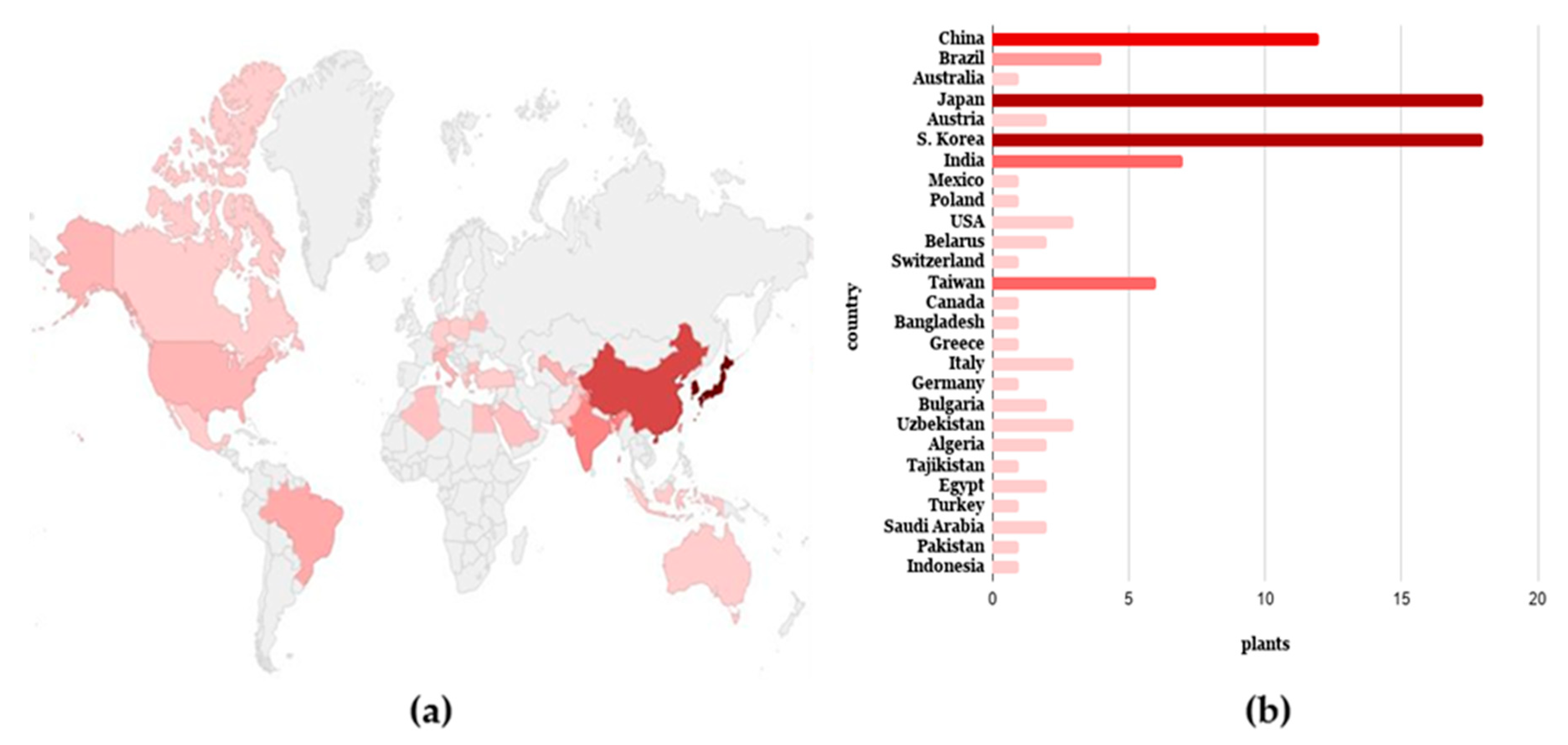
| C. japonicum | Daejeon/S. Korea, Oberndorf/Austria, Chengdu/China, Henan/China | [7,16,17,18,19,20] | |
| Oberndorf/Austria | |||
| Bialystok/Poland | |||
| Daejeon/S. Korea | |||
| C. setosum | Daejeon/S. Korea | [18] | |
| C. rivulare | Daejeon, Wonju, Pyongchang-gun, Gangwondo, Jeongseon-gun, Yanggu/S. Korea | [21] | |
| C. lineare | Daejeon/S. Korea | [19] | |
| C. nipponicum | Daejeon, Sancheong/S. Korea | [19,22] | |
| C. setidens | Jeongseon-gun, Jeju Island/S. Korea | [19,23,24,25,26] | |
| Laramie/USA | |||
| Laramie/USA, Vitebsk/Belarus | |||
| C. pendulum | Nemuro, Hatimandake, Memanbetsu, Onsen Kyushu, Hokkaido/Japan | [19] | |
| C. chanroenicum | [19,26,27] | ||
| C. rhinoceros | [28,29] | ||
| C. coloradense | Wyoming/USA, Japan | [30] | |
| C. arisanense | Mount Akaishi, Mount Senmai, Shizuoka, Takanomori, Nekura Valley/Japan, Vitebsk/Belarus, | ||
| C. tioganum | Mount Shirouma/Japan | ||
| C. oleraceum | la Dotze/Switzerland | [30,31,32] | |
| Hsien/Taiwan | |||
| C. microspicatum | Nemuro, Mount Shirouma /Japan, Mount Ali, Chiayi Hsien/Taiwan | [16] | |
| C. babanum | Ku Kuan, Taichung Haien/Taiwan | ||
| C. kagamontanum | Mount Shirouma, Mount Hakusan ad pedem/Japan | ||
| C. inundatum | Vancouver/Canada | ||
| C. dipsacolepis | Seongnam/Korea | ||
| C. brevicaule | |||
| C. yezoense | |||
| C. kamtschaticum | |||
| C. pectinellum | |||
| C. bitchuense | [33,34] | ||
| C. senjonse | [17,31,35,36,37] | ||
| C. spicatum | |||
| C. yezonese | |||
| C. vallis-demonii | |||
| C. gratiosum | |||
| C. indundatum | |||
| C. otayae | [35] | ||
| C. purpuratum | |||
| C. spinosissimum | [38] | ||
| C. spinosum | [34] | ||
| C. ferum | [39] | ||
| C. kawakamii | [16,40,41] | ||
| C. wallichii | [41] | ||
| C. yoshizawae | [35] | ||
| C. matsumurae | [34,40] | ||
| C. brevistylum | [42] | ||
| C. chlorolepis | [43] | ||
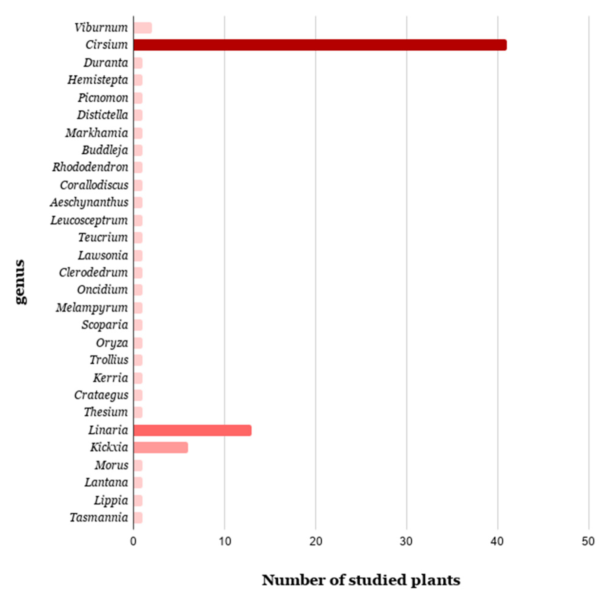
| Duranta | D. plumieri | Rajshahi/Bangladesh | [44] |
| Hemistepta | H. lyrata | Kangwon/S. Korea | [45] |
| Picnomon | P. acarna | Mount Hortiatis/Greece | [46] |
| Family: Bignoniaceae | |||
| Distictella | D. elongata | Minas Gerais State/Brazil | [47] |
| Markhamia | M. lutea | Benguluru/India | [48] |
| Family: Buddlejaceae | |||
| Buddleja | B. officinalis | Anhui/China | [49] |
| Family: Ericaceae | |||
| Rhododendron | R. arboreum | Aligarh/India | [50] |
| Family: Gesneriaceae | |||
| Corallodiscus | C. flabellate | Kunming/China | [51] |
| Aeschynanthus | A. moningeriae | Jinhua/China | [52] |
| Family: Lamiaceae | |||
| Leucosceptrum | L. canum | Tibet/China | [53] |
| Teucrium | T. hyrcanicum | Sicily/Italy | [54] |
| Family: Lythraceae | |||
| Lawsonia | L. inermis | Thanjavur/India | [55] |
| Family: Moraceae | |||
| Clerodedrum | C. phlomides | Tamil Nadu/India | [56] |
| Family: Orchidaceae | |||
| Oncidium | O. baueri | Londrina/Brazil | [57,58] |
| Family: Orobanchaceae | |||
| Melampyrum | M. roseum | Suwon/S. Korea | [59] |
| Family: Plantaginaceae | |||
| Scoparia | S. dulcis | Nanning/China | [60] |
| Family: Poaceae | |||
| Oryza | O. sativa | a) | [61] |
| Family: Ranunculaceae | |||
| Trollius | T. ledebourii | Hebei/China | [62] |
| Family: Rosaceae | |||
| Kerria | K. japonica var. | Chongqing/China | [63] |
| Crataegus | C. laevigata | Bremen/Germany | [64] |
| Family: Santalaceae | |||
| Thesium | T. chinense | Anhui/China | [65] |
| Family: Scrophulariaceae | |||
| Linaria | L. vulgaris | Sofia/Bulgaria, Tachkent/Uzbekistan | [2,66,67,68] |
| L. japonica | Heilongjiang/China | [69,70,71] | |
| L. reflexa | Tottori Prefecture/Japan | [72,73,74,75] | |
| L. vulgariformis | Constantine/Algeria, Calabria/Italy | [66] | |
| L. popovii | Tachkent/Uzbekistan | ||
| L. kurdica | |||
| L. sessili | [76] | ||
| L. kokanica | Pamir/Tajikistan | ||
| L. haelava | [77] | ||
| L. simplex | Mansoura/Egypt | [67] | |
| L. genistifolia | Sofia/Bulgaria | ||
| L. dalmatica | |||
| L. scariosa | Msila/Algeria | [78] | |
| Kickxia | K. elatine | Dustlik/Uzbekistan | [79] |
| K. heterophylla | Mansoura/Egypt | [80] | |
| K. ramosissima | Ankara/Turkey | [81,82] | |
| K. abhaica | Baljurashi/Saudi Arabia | [83] | |
| Appennines hills/Italy | |||
| K. spuria | Saudi Arabia | [84] | |
| K. aegyptiaca | [85] | ||
| Family: Verbenaceae | |||
| Morus | M. alba L. | Hongseong/Korea | [86] |
| Lantana L. camara | Taichung/Taiwan, Palampur/India, Karachi/Pakistan, Ceará state/Brazil, Manado/Indonesia, Okinawa/Japan | [87,88,89,90,91,92] | |
| Lippia L. rubella | Minas Gerais/Brazil | [93] | |
| Family: Winteraceae | |||
| Tasmannia | T. lanceolata | Go Wild Harvest/Australia | [94] |
| Genus | Species | Collection Place | Reference |
|---|---|---|---|
| Family: Apiaceae | |||
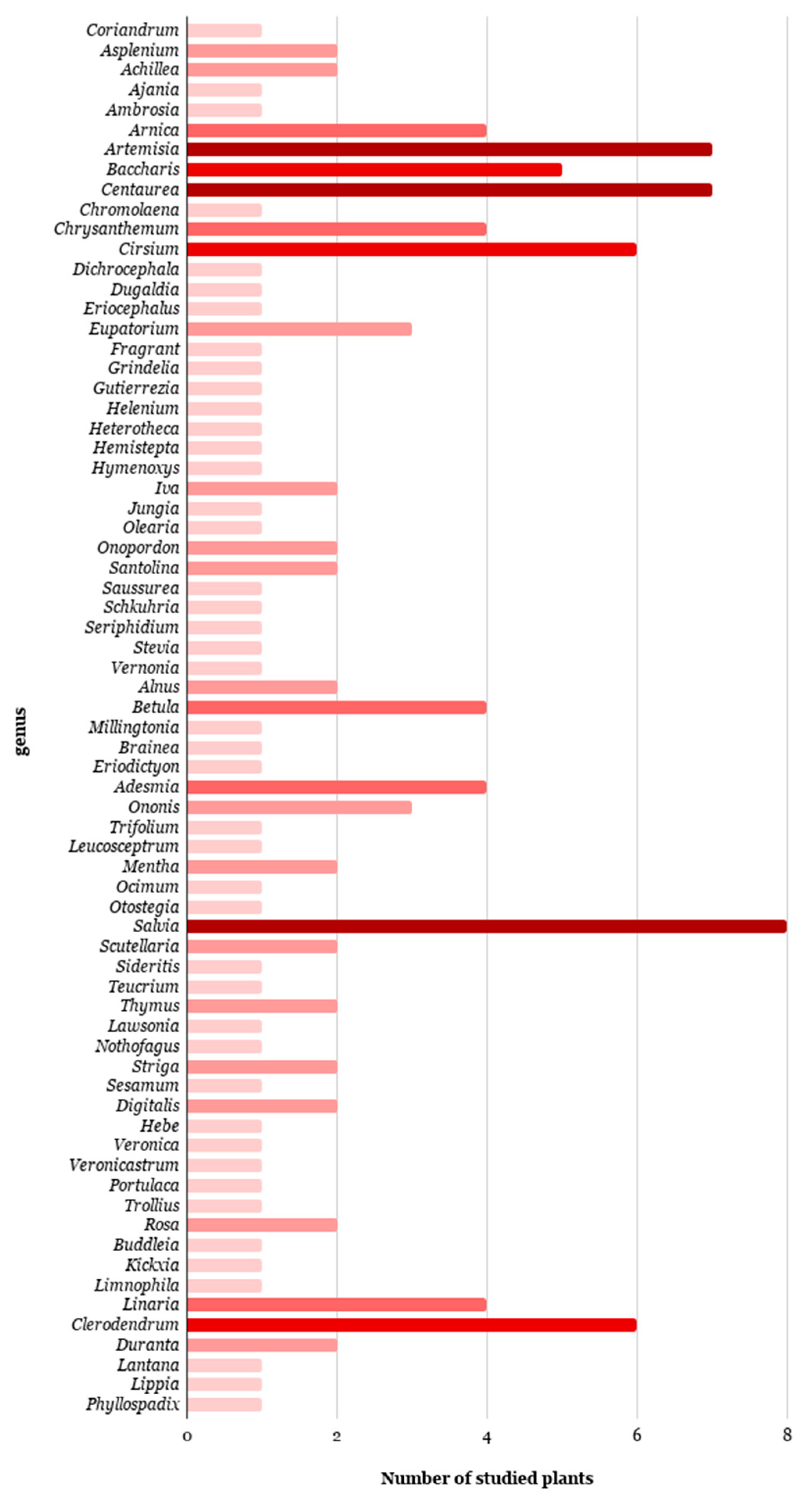
| Coriandrum | C. sativum | Faisalabad/Pakistan | [99] |
| Family: Aspleniaceae | |||
| Asplenium | A. glaucophyllum | West Malysia | [100] |
| A. normale | West Malaysia | [101] | |
| Family: Asteraceae | |||
| Achillea | A. collina | wet lowlandmeadows/UK | [102] |
| A. asplenifolia | |||
| Ajania | A. potaninii | Gansu/China | [103] |
| Ambrosia | A. camphorate | Baja California/Mexico | [104] |
| Arnica | A. angustifolia | northwestCanada and Alaska | [105] |
| A. Montana | California/USA | [106] | |
| A. chamissonis | Graines Voltz/France | [107] | |
| A. montana | Šumava Mounts/Czech | [108] | |
| Artemisia | A. mongolica | Gansu/China | [109] |
| A. judaica | St. Catherine, Sinai/Egypt | [110] | |
| A. monosperma | Cairo/Egypt | ||
| A. herba-alba | Mount Moses/Egypt | ||
| A. xerophytica | South Gobi Aimak/Mongolia | [111] | |
| A. glabella | Karaganda/Kazakhstan | [112] | |
| A. vestita | Lhasa/Tibet | [113] | |
| Baccharis | B. trinervis | Costa Rica | [114] |
| B. decussata | Venezuela | [115] | |
| B. concave | [116] | ||
| B. uncinella | Campos do Jordão/Brazil | [117] | |
| B. conferta | Veracruz/Mexico | [118] | |
| Centaurea | C. alexandrina | Alexandria/Egypt | [119] |
| C. aspera | Ribera Baixa/Spain | [120] | |
| C. cariensis | [121] | ||
| C. collina | Valencia/Spain | [122] | |
| C. sadleriana | Jakabszállás/Hungary | [123] | |
| C. moesiaca | Malashevska planina/Bulgaria | [124] | |
| C. behen | Iran | [125] | |
| Chromolaena | C. odorata | Chonburi/Thailand | [126] |
| Chrysanthemum | C. pacificum | Tsukuba/Japan | [27] |
| C. shiwogiku | Muroto-misaki/Japan | ||
| C. kinokuniense | Tsukuba/Japan | ||
| C. rupestre | Mount Mikuni/Japan | ||
| Cirsium | C. setidens | Jeongseon-gun, Halla of jejudo, Daejeon, Kangwon, Yanggu/S. Korea; Guerrero/Mexico; | [19,24,25,26,27,28,127] |
| C. chanroenicum | Daejeon, Ulsan, Sancheong/S. Korea | [26,128] | |
| C. japonicum | Jiang Xi/China | [129] | |
| C. arvense | Musa Khel Bannu/Pakistan | [130] | |
| C. nipponicum | Suwon/S. Korea | [131] | |
| C. rhinoceros | [132] | ||
| Dichrocephala | D. integrifolia | Shanghai/China | [133] |
| Dugaldia | D. pinetorum | Nuevo Lebn/Mexico | [134] |
| Eriocephalus | E. giessii | Aus-Koppies/Namibia | [135] |
| Eupatorium | E. cannabinum | Gronigen/Netherlands | [136] |
| E. odoratum | Kuala Pilah/Malaysia | [137,138] | |
| E. semiserratum | Arkansas/USA | [139] | |
| Fragrant | F. Eupatorium | Guangxi/China | [140] |
| Grindelia | G. glutinosa | Poconchile, Valle deLiuta, Tarapaca/Chile | [141] |
| Gutierrezia | G. mandonii | Salta/Argentina | [142] |
| Helenium | H. integrifolium | [143] | |
| Heterotheca | H. latifolia | San Luis/Argentina | [144] |
| Hemistepta | H. lyrata | Kangwon/S. Korea | [45] |
| Hymenoxys | H. jamesii | Coconino/USA | [145] |
| Iva | I. nevadensis | Tonopah/USA | [146] |
| I. frutescens | Franklin/USA | [147] | |
| Jungia | J. polita | San Martin/Argentina | [148] |
| Olearia | O. paniculata | Dunedin/New Zealand | [149] |
| Onopordon | O. corymbosum | Barracas, Castellon/Spain | [150] |
| O. nervosum | a) | [151] | |
| Santolina | S. chamaecyparissus | Lyon/France | [152] |
| S. pinnata | Pisa/Italy | [153] | |
| Saussurea | S. elegans | Murghab/Tajikistan | [154] |
| Schkuhria | S. pinnata | Cordoba/Argentina | [155] |
| Seriphidium | S. santolium | Xinjiang Uigour/China | [156] |
| Stevia | S. laxiflora | Cuernavaca, Morelos/Mexico | [157] |
| Vernonia | V. cinerea | Pahang/Malaysia | [158] |
| Family: Betulaceae | |||
| Alnus | A. glutinosa | Darmstadt/Germany | [159] |
| A. japonica | [160] | ||
| Betula | B. ermanii | [159] | |
| B. verrucosa | a) | [161] | |
| B. pubescens | Biebrza/Poland | [162] | |
| B. pendula | |||
| Family: Bignoniaceae | |||
| Millingtonia | M. hortensis | Khon Kaen/Thailand | [163] |
| Family: Blechnaceae | |||
| Brainea | B. insignis | Yunnan/China | [164] |
| Family: Boraginaceae | |||
| Eriodictyon | E. tomentosum | Placer Co./USA | [165] |
| Family: Fabaceae | |||
| Adesmia | A. grandiflora | a) | [166] |
| A. trijuga | |||
| A. horrida | |||
| A. retrofracta | |||
| Ononis | O. fruticosa | Los Castanõs/Spain | [167] |
| O. natrix | |||
| O. rotundifolia | a) | [168] | |
| Trifolium | T. pratense | Trout Lake/USA | [169] |
| Family: Lamiaceae | |||
| Leucosceptrum | L. canum | a) | [170] |
| Mentha | M. pulegium | Petite Kabylie/Algeria | [171] |
| M. suaveolens | |||
| Ocimum | O. americanum | RoyalBotanic Gardens, Kew/England | [172] |
| Otostegia | O. fruticosa | St. Catherine/Egypt | [173] |
| Salvia | S. trilobu | Marmara island/Turkey | [174] |
| S. hypoleuca | Elbruz moun/Russia | [175] | |
| S. pedicellata | a) | [176] | |
| S. yosgadensis | Sultanhani/Turkey | [177] | |
| S. plebeia | a) | [178] | |
| S. pilifera | Berit Mount/Turkey | [179] | |
| S. tomentosa | Sofia/Bulgaria | [180] | |
| S. argentea | |||
| Scutellaria | S. polyodon | a) | [181] |
| S. przewalskii | Susamyr/Kyrgyzstan | [182] | |
| Sideritis | S. gomerae | Canary islands/Spain | [183] |
| Teucrium | T. chamaedrys | Eskisehir/Turkey | [184] |
| Thymus | T. longicaulis | Sar planina/Macedonia | [185] |
| T. glabrescens | Skopje/Macedonia | ||
| Family: Lythraceae | |||
| Lawsonia | L. inermis | Thanjavur/India | [153] |
| Family: Nothofagaceae | |||
| Nothofagus | N. dombeyi | Altos de Lircay/Chile | [186] |
| Family: Orobanchaceae | |||
| Striga | S. passargei | a) | [187] |
| S. aspera | a) | [188] | |
| Family: Padaliacea | |||
| Sesamum | S. indicum | Gambang/Malaysia | [189] |
| Family: Plantaginaceae | |||
| Digitalis | D. trojana | Kizilcahamam, DemirkOy/Turkey | [190] |
| D. orientalis | |||
| D. lanata | a) | [191] | |
| Hebe | H. cupressoides | Dunedin/New Zealand | [192] |
| Veronica | V. chamaedrys | Rila Mount/Bulgaria | [193] |
| Veronicastrum | V. latifolium | Yongkang/China | [194] |
| Family: Portulacaceae | |||
| Portulaca | P. oleracea | Tianjin/China | [195] |
| Family: Ranunculaceae | |||
| Trollius | T. chinensis | Hebei/China | [196] |
| Family: Rosaceae | |||
| Rosa | R. damascena | Plovdiv/Bulgaria | [197] |
| R. rugosa | Botanischer Garten der TU Darmstadt/Germany | [198] | |
| Family: Scrophulariaceae | |||
| Buddleia | B. macrostachya | Sibsagar/India | [199] |
| Kickxia | K. ramosissima | Takht-e-Nusrati/Pakistan | [200,201,202] |
| Limnophila | L. aromatica | Ho Chi Minh/Vietnam | [203] |
| Linaria | L. vulgaris | Ukrania; China | [68,204] |
| L. reflexa | Constantine/Algeria | [73] | |
| L. kurdica | Ukrania | [204] | |
| L. scariosa | Msila/Algeria | [78] | |
| Family: Verbenaceae | |||
| Clerodendrum | C. siphonenthus | Calcutta, Kalyani/India | [205,206] |
| C. phlomidis | Pondicherry, Alanthurai/India | [205,206,207,208,209,210] | |
| C. serratum | Bhilai/India | [211] | |
| C. inerme | Pondicherry/India | [212,213] | |
| C. neriifolium | a) | [214] | |
| C. indicum | a)Khao Kho/Thailand | [215,216] | |
| Duranta | D. repens | a) | [217] |
| D. plumieri | a) | [44,218] | |
| Lantana | L. camara | Taichung/Taiwan, Palampur/India, Karachi/Pakistan, Ceará state/Brazil, Manado/Indonesia, Okinawa/Japan | [87,88,89,90,91,92] |
| Lippia | L. citriodora | Athens/Greece | [219] |
| Family: Zosteraceae | |||
| Phyllospadix | P. japonica | Omaezaki/Japan | [220] |
© 2020 by the authors. Licensee MDPI, Basel, Switzerland. This article is an open access article distributed under the terms and conditions of the Creative Commons Attribution (CC BY) license (http://creativecommons.org/licenses/by/4.0/).
Share and Cite
Cheriet, T.; Ben-Bachir, B.; Thamri, O.; Seghiri, R.; Mancini, I. Isolation and Biological Properties of the Natural Flavonoids Pectolinarin and Pectolinarigenin—A Review. Antibiotics 2020, 9, 417. https://0-doi-org.brum.beds.ac.uk/10.3390/antibiotics9070417
Cheriet T, Ben-Bachir B, Thamri O, Seghiri R, Mancini I. Isolation and Biological Properties of the Natural Flavonoids Pectolinarin and Pectolinarigenin—A Review. Antibiotics. 2020; 9(7):417. https://0-doi-org.brum.beds.ac.uk/10.3390/antibiotics9070417
Chicago/Turabian StyleCheriet, Thamere, Balkeis Ben-Bachir, Oumelkhir Thamri, Ramdane Seghiri, and Ines Mancini. 2020. "Isolation and Biological Properties of the Natural Flavonoids Pectolinarin and Pectolinarigenin—A Review" Antibiotics 9, no. 7: 417. https://0-doi-org.brum.beds.ac.uk/10.3390/antibiotics9070417