Article

Selective Eradication of Staphylococcus aureus by the Designer Genetically Programmed Yeast Biocontrol Agent

by
Sofiya O. Pipiya
1,
Yuliana A. Mokrushina
1,2,
Alexander G. Gabibov
1,2,3,*,
Ivan V. Smirnov
1,2,* and
Stanislav S. Terekhov
1,2,*
1
Shemyakin-Ovchinnikov Institute of Bioorganic Chemistry, Russian Academy of Sciences, Moscow 117997, Russia
2
Department of Chemistry, Lomonosov Moscow State University, Moscow 119991, Russia
3
Department of Life Sciences, Higher School of Economics, Moscow 101000, Russia
*
Authors to whom correspondence should be addressed.
Submission received: 17 July 2020 / Revised: 7 August 2020 / Accepted: 18 August 2020 / Published: 19 August 2020
(This article belongs to the Special Issue Antimicrobial Action of Biomaterials)

Abstract

:
Staphylococcus aureus is a common human pathogen that is particularly often associated with antibiotic resistance. The eradication of this ubiquitous infectious agent from its ecological niches and contaminated surfaces is especially complicated by excessive biofilm formation and persisting cells, which evade the antibacterial activity of conventional antibiotics. Here, we present an alternative view of the problem of specific S. aureus eradication. The constitutive heterologous production of highly specific bacteriolytic protease lysostaphin in yeast Pichia pastoris provides an efficient biocontrol agent, specifically killing S. aureus in coculture. A yeast-based anti-S. aureus probiotic was efficient in a high range of temperatures and target-to-effector ratios, indicating its robustness and versatility in eliminating S. aureus cells. The efficient eradication of S. aureus by live lysostaphin-producing P. pastoris was achieved at high scales, providing a simple, biocompatible and cost-effective strategy for S. aureus lysis in bioproduction and surface decontamination. Future biomedical applications based on designer yeast biocontrol agents require evaluation in in vivo models. However, we believe that this strategy is very promising since it provides highly safe, efficient and selective genetically programmed probiotics and targeted biocontrol agents.

Graphical Abstract

1. Introduction

Antibiotic resistance (AR) is a persistent threat to global healthcare, resulting in up to 11 million deaths annually [1,2]. Staphylococcus aureus is one of the most common pathogens, accounting for nearly 120,000 severe cases of bloodstream infections and 20,000 deaths per year in the United States alone [3]. S. aureus is a highly specialized human pathogen, notorious for an extensive number of methicillin-resistant (MRSA) strains resistant to the entire β-lactam antibiotic class [4]. Moreover, S. aureus strains have adopted a variety of virulence factors to efficiently colonize and counteract host immunity [5]. The exceptionally flexible S. aureus adaptation to antibiotics arises not only from the horizontal transfer of mobile genetic elements. It is also enhanced by increased mutation rates of S. aureus strains. The estimated mutation rate for the S. aureus isolate collection is 3.3 × 10⁻⁶ per site per year [6]. This rate is about 1000 times faster than the canonical substitution rate estimate for E. coli [7]. A high mutation rate of S. aureus isolates relates to their low doubling time in the wild [8]. This mutability provides the increased evolutionary space for S. aureus that it implements for its rapid adaptation. Hence, the eradication of S. aureus is an incredibly difficult task because of its plasticity, ubiquity, propensity to AR and simplicity of transmission [5]. Extensive biofilm formation and persisters in turn worsen the situation dramatically, making S. aureus non-sensitive to antibiotic drugs [9].
Bacteriolytic enzymes, disrupting cell integrity, represent a promising alternative to conventional antibiotics. The most promising example of the former is lysostaphin, which not only kills planktonic S. aureus but also disrupts S. aureus biofilms [10]. Lysostaphin is a specific zinc-containing metalloprotease secreted by strains of S. simulans biovar staphylolyticus [11]. Lysostaphin cleaves the pentaglycine crossbridges present in peptidoglycan (PG), the essential component of the bacterial cell wall, leading to rapid cell lysis. The recognition of the pentaglycine crossbridge and the peptide stem is shared by two independent SH3b domains, allowing protein clustering on the PG [12]. This unusual mechanism underpins the potent activity of lysostaphin, and its capacity to punch holes in the cell walls so as to cause rapid cell lysis.
Lysostaphin displays bacteriolytic activity against numerous Staphylococcus species [13,14]. It is highly active against virtually any natural S. aureus strain, including clinically relevant MRSA and vancomycin intermediately susceptible S. aureus (VISA) [15,16]. This makes lysostaphin a promising agent in the experimental therapy of S. aureus infections overperforming conventional vancomycin therapy in different animal models [17,18,19], thus providing an essential therapeutic agent against VISA and vancomycin-resistant S. aureus (VRSA) [20].
A wide palette of applications illustrates the versatility and efficiency of lysostaphin in the eradication of S. aureus from different environments. Hydrogel-based materials eliminating implant S. aureus infections represent an example of an advanced biomaterial facilitating healing [21]. The heterologous production of lysostaphin was applied to protect transgenic animals from mastitis, eradicating S. aureus from milk and mammary tissue [22]. Encapsulated mammalian cells with a synthetic gene circuit to sense and respond to MRSA infection could provide potential prophylactic, diagnostic or therapeutic options for treating medical implant-associated infections [23]. Deimmunized lysostaphin variants are valuable in this case as they expand the boundaries of lysostaphin systemic administration and therapy [24].
Here we describe an alternative paradigm of specific S. aureus eradication. We consider applying the high viability, robustness, and safety of yeasts to create a genetically programmed biocontrol agent, killing S. aureus cells and destroying its biofilms. The heterologous inducible production of recombinant lysostaphin in yeast Pichia pastoris was previously achieved via the codon optimization of its natural sequence [25]. Alternatively, we focused on the constitutive production of lysostaphin in yeast cells to obtain live cell cultures, eradicating S. aureus. P. pastoris produced up to 30 mg/L/day of recombinant lysostaphin in shaking flask conditions. This culture medium was highly active against S. aureus, leading to growth inhibition and bactericidal effects at 1.6 × 10⁴- and 4 × 10³-fold dilutions, respectively. Moreover, 107 CFU/mL of P. pastoris eradicated up to 1010 CFU/mL S. aureus after 12 h in coculture. Recombinant lysostaphin-producing (rLys) P. pastoris was active against S. vitulinus and S. xylosus, which have similar PG crossbridge sequences, correlating with the lysostaphin activity spectrum. The anti-S. aureus activity of live rLys P. pastoris culture was prominent at the 20–33 °C temperature range, with some decrease at 37 °C that we associate with the elevated growth temperature, non-optimal for P. pastoris. The efficient eradication of S. aureus via engineered P. pastoris in coculture was achieved in a broad range of target (S. aureus):effector (rLys P. pastoris) ratios, both at high and low S. aureus cell densities. These results illustrate the considerable potential of live lysostaphin-producing cultures in S. aureus elimination, creating a background for their further applications in vivo. Yeast-based bacteriolytic live cultures represent a particularly promising approach to the creation of highly safe, efficient and selective genetically programmed probiotics and targeted designer biocontrol agents.

2. Results

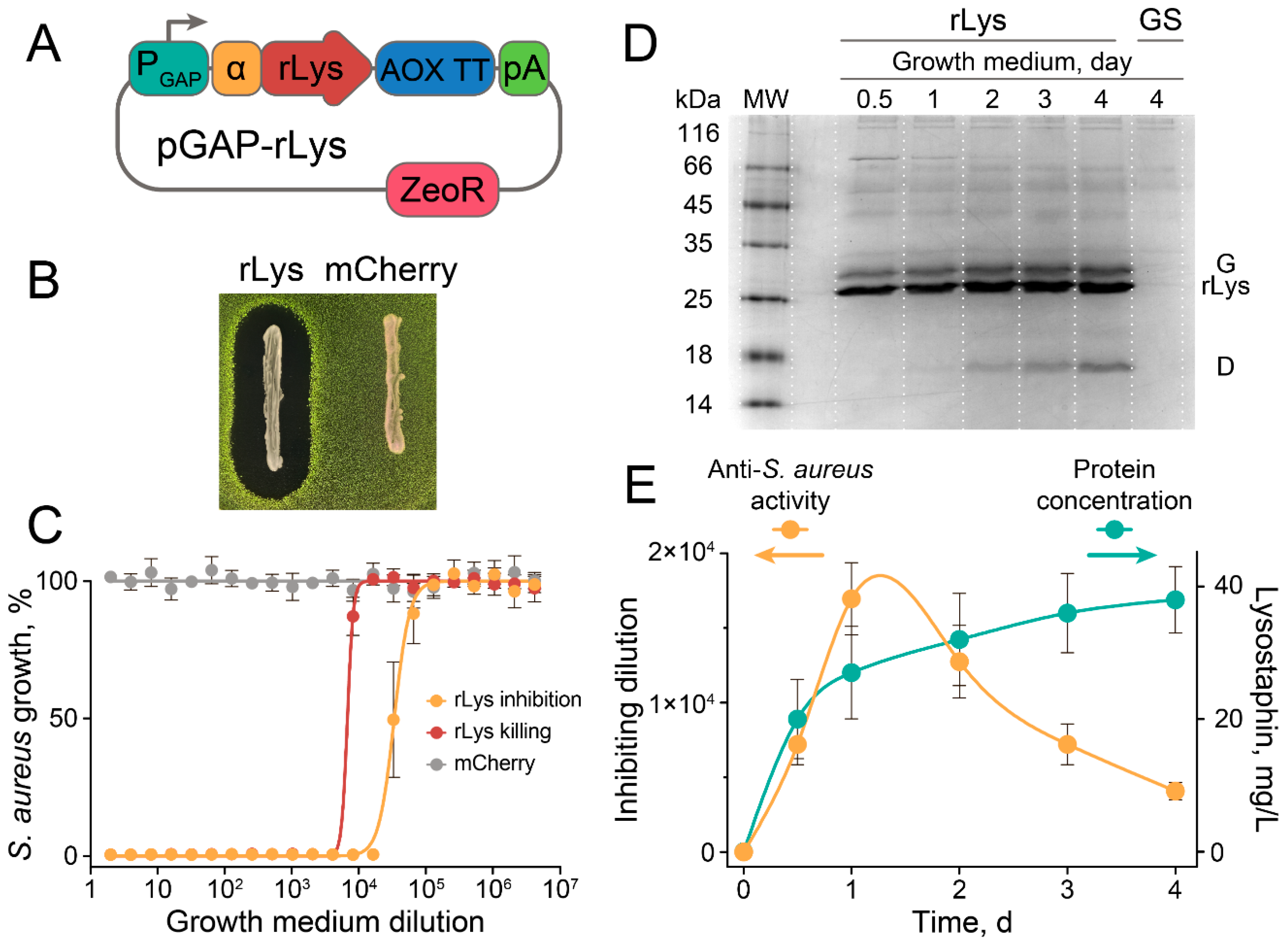
2.1. Constitutive Production of Recombinant Lysostaphin in P. pastoris

The codon-optimized sequence of lysostaphin [25] was cloned into pGAPα under control of the strong constitutive glyceraldehyde-3-phosphate dehydrogenase (GAP) promoter, so as to maintain constitutive production of the recombinant lysostaphin in P. pastoris (Figure 1A). The P. pastoris transformed with pGAPα-Lys vector produced recombinant lysostaphin, and secreted it into the culture medium, giving clear zones of S. aureus inhibition in an agar overlay assay (Figure 1B).
The culture medium of rLys P. pastoris was highly active against S. aureus, leading to growth inhibition and bactericidal effects at 1.6 × 10⁴- and 4 × 10³-fold dilutions, respectively (Figure 1C). Control P. pastoris strain producing mCherry fluorescent protein did not influence S. aureus growth and viability (Figure 1B,C). P. pastoris produced up to 30 mg/L/day of recombinant lysostaphin in shaking flask conditions (Figure 1D,E), and we estimate that this productivity could be increased by several folds using bioreactor cultivation conditions. However, proteolysis of the recombinant lysostaphin was observed (Figure 1D), resulting in the maximal anti-S. aureus activity detected at the 24 h cultivation time point (Figure 1E). Since the specific activity of the rLys P. pastoris culture medium gradually decreased after 24 h of cultivation, we speculate that the production of recombinant lysostaphin by live rLys P. pastoris culture in situ is preferred for enhanced S. aureus eradication.

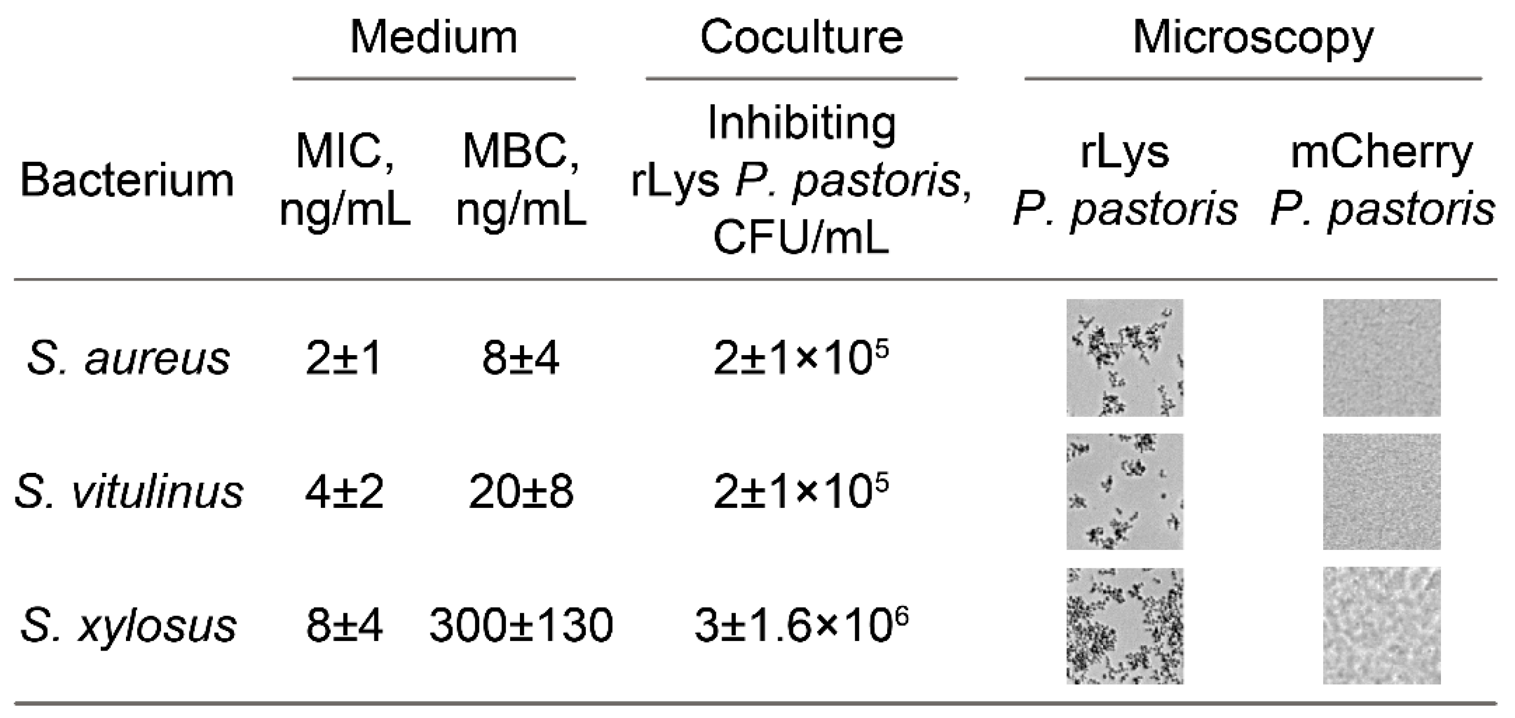
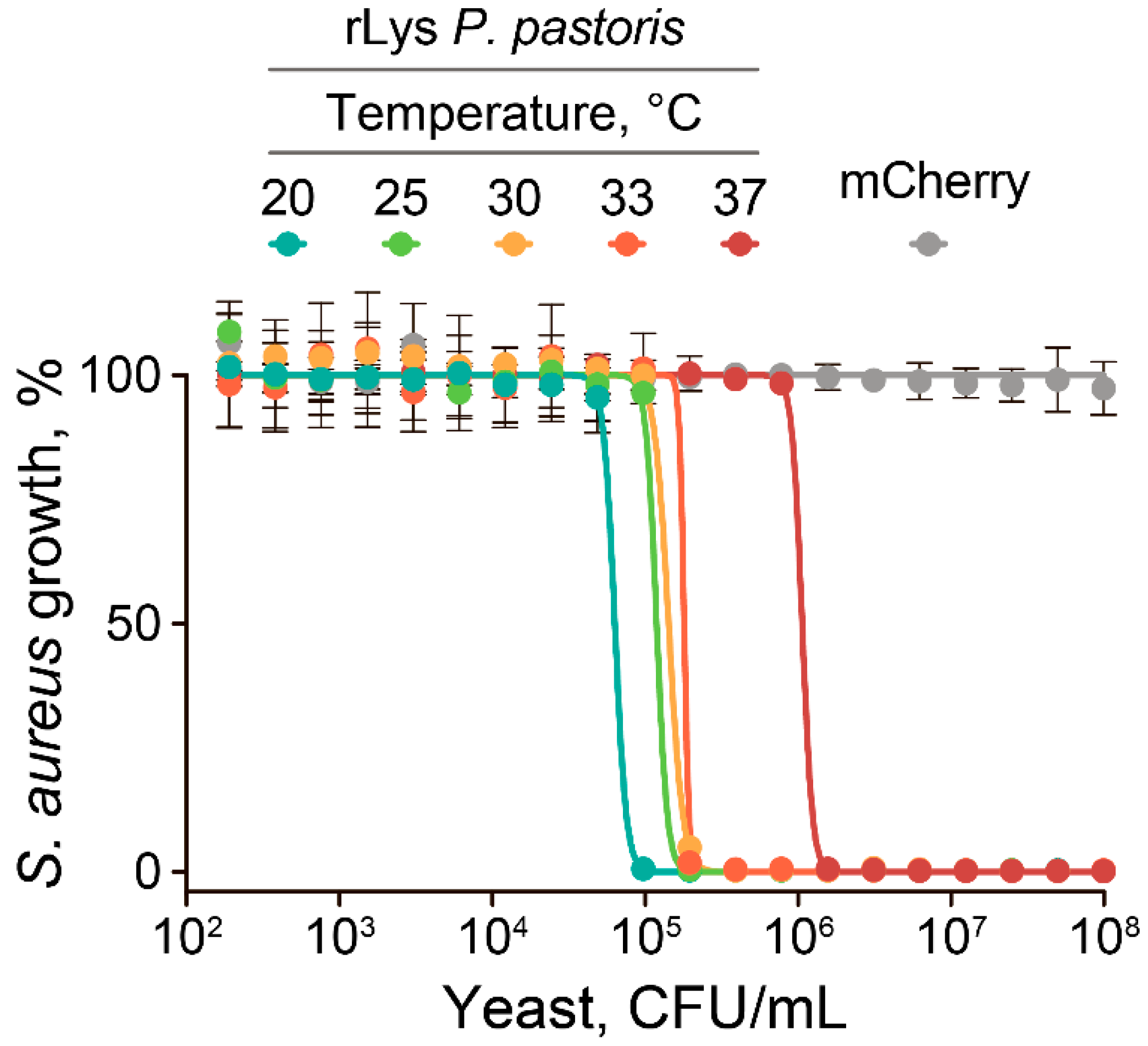
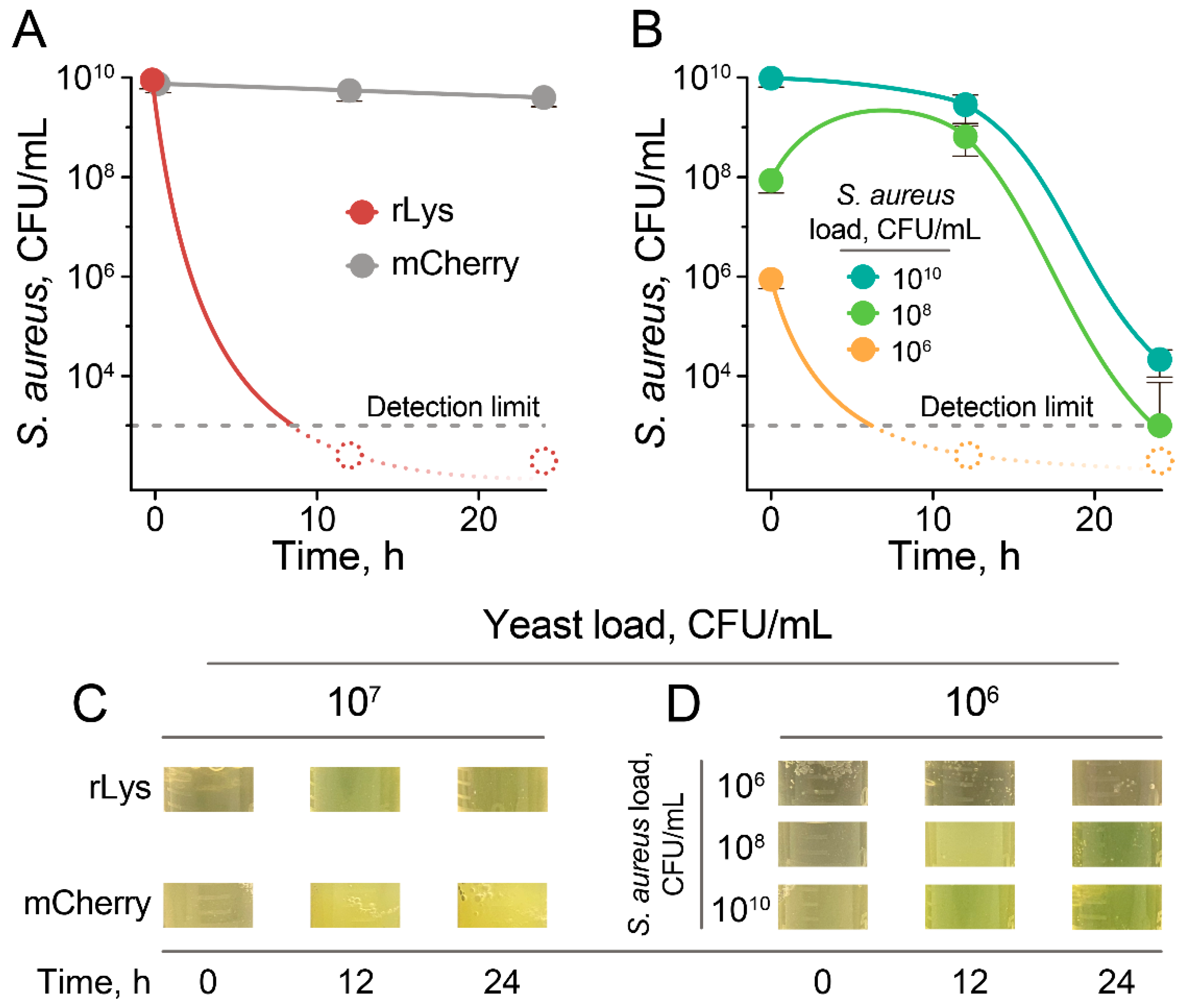
2.2. The Anti-Stapylococcal Activity of Engineered rLys P. pastoris

Unlike a parent P. pastoris GS115 strain, engineered rLys P. pastoris displays antagonistic properties, mediated by constitutive lysostaphin production. To trace the correlation between lysostaphin activity spectrum and the antagonistic properties of rLys P. pastoris against different bacteria, we used a panel of Staphylococci and several other bacteria naturally resistant toward lysostaphin (Figure 2). The antagonistic properties of rLys P. Pastoris were similar to the lysostaphin activity spectrum. The most efficient growth inhibition was observed for S. aureus and S. vitulinus. Mediocre inhibition was observed for S. xylosus. Several Staphylococci (Staphylococcus pasteuri, Staphylococcus warneri, Staphylococcus haemolyticus and Staphylococcus epidermidis) and other bacteria (Macrococcus caseolyticus, Bacillus subtilis, Enterococcus faecium, Enterococcus faecalis, and Escherichia coli) were not inhibited by rLys P. pastoris. This is in line with the lysostaphin activity spectrum, and it is associated with the alternative PG structures among these bacteria [26].
Hence, engineered rLys P. pastoris displays quite a narrow activity spectrum. rLys P. pastoris acts efficiently on S. aureus and several Staphylococci, these having similar PG structures. This selectivity is favored for the practical application of rLys P. pastoris as it could be exploited as a targeted biocontrol agent for S. aureus eradication.

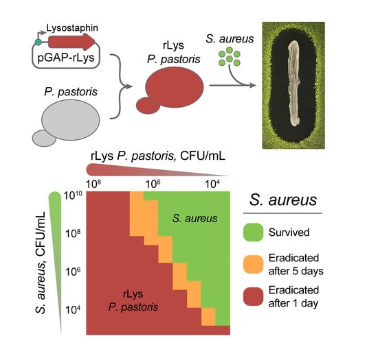
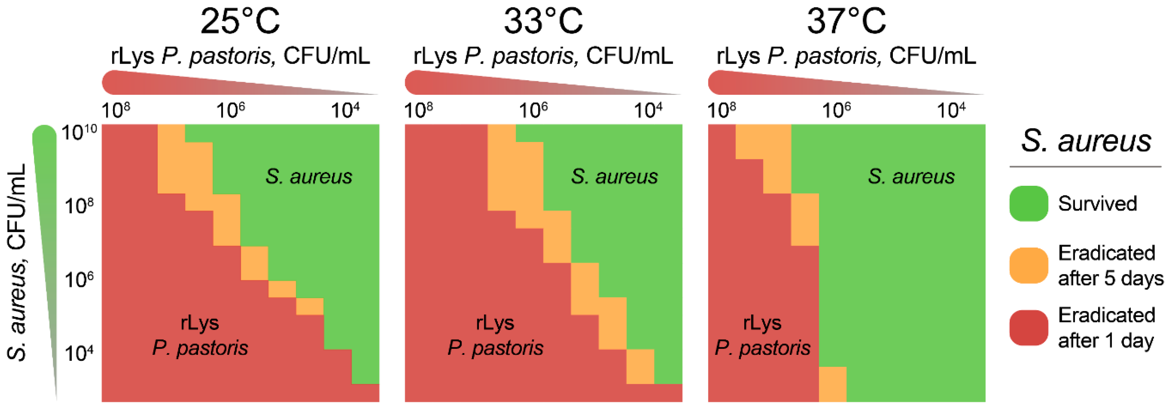
2.3. Eradication of S. aureus by Live rLys P. pastoris Biocontrol Agent

Focusing on the inhibitory properties of rLys P. pastoris cells alone, we screened different cocultivation modes of S. aureus and rLys P. pastoris. S. aureus and P. pastoris have different optimal growth temperatures, 37 °C and 30 °C, respectively. Hence, elevated temperatures could decrease the anti-S. aureus activity of rLys P. pastoris (Figure 3).
However, efficient S. aureus inhibition by rLys P. pastoris was observed up to 33 °C, corresponding to the temperature of the skin surface, which is an ecological niche of S. aureus. The further increase in cocultivation temperature resulted in an eight-fold decrease of anti-S. aureus activity at 37 °C (Figure 3). S. aureus inhibition was particularly efficient at temperatures below 25 °C, which is close to natural P. pastoris growth conditions.
To estimate the kinetics of S. aureus eradication, we varied the target (S. aureus):effector (rLys P. pastoris) ratios and measured cell viability after cocultivation at 33 °C (Figure 4).
At relatively high rLys P. pastoris loads (107 CFU/mL up to 1010 CFU/mL), S. aureus cells were killed within 12 h of cocultivation (Figure 4A). The control P. pastoris strain producing the mCherry fluorescent protein did not influence S. aureus viability, as was expected from the culture medium activity testing (Figure 4A). Decreasing the load of rLys P. pastoris to 106 CFU/mL resulted in different scenarios. At a low S. aureus load, 106 CFU/mL, rLys P. pastoris controlled S. aureus growth and eliminated viable cells within less than 12 h (Figure 4B). Increasing the S. aureus loads to 108 CFU/mL resulted in a situation wherein rLys P. pastoris could not control S. aureus growth from the beginning of cocultivation. This led to S. aureus proliferation at the 12 h time point, while after that rLys P. pastoris produced enough lysostaphin to kill the majority of S. aureus cells, decreasing its titer by 5.8 orders of magnitude after 24 h of cocultivation (Figure 4B). In dense S. aureus cultures with loads up to 1010 CFU/mL, rLys P. pastoris gradually decreased S. aureus titer by 0.5 and 5.1 orders of magnitude at the 12 h and 24 h time points, respectively (Figure 4B). The decrease in viable S. aureus cells was associated with the massive S. aureus lysis detected by culture clearance (Figure 4C,D).
Thus, the complete or near-complete lysis of S. aureus cells is achieved by considerably low rLys P. pastoris concentrations at 12 h. P. pastoris cultures reach cell densities exceeding 1010 CFU/mL when cultivated in bioreactors. Hence, more than 1000-fold dilutions of rLys P. pastoris live cell cultures are efficient in S. aureus eradication. This makes it a safe and cost-effective biocontrol agent for eliminating S. aureus cells.
To further estimate the eradication landscapes of the rLys P. pastoris biocontrol agent, we performed time-lapse cocultivation of S. aureus and rLys P. pastoris at different target:effector ratios (Figure 5).
rLys P. pastoris eradicated S. aureus at various target:effector ratios at temperatures from 25 °C to 37 °C (Figure 5). However, the eradication of S. aureus was not observed at low (106 CFU/mL) loads of rLys P. pastoris at 37 °C, indicating the necessity of effector proliferation for complete S. aureus removal (Figure 5). Both 25 °C and 33 °C temperatures were convenient for rLys P. pastoris growth resulting in S. aureus elimination even at yeast titers less than 104 CFU/mL. However, only up to 104 CFU/mL of S. aureus could be eradicated at such a low yeast level. The temperature of 25 °C resulted in a more efficient S. aureus eradication than 33 °C at low yeast loads, while this relation was the opposite at high effector concentrations (Figure 5). We associate this with the counteraction of the more efficient proliferation of rLys P. pastoris at low temperatures, and the increased bacteriolytic activity at high temperatures.

3. Discussion

S. aureus is a ubiquitous human bacterium with outstanding pathogenic potential. Infections that are caused by AR S. aureus, particularly MRSA strains, result in epidemic waves still presenting a global threat [4,5]. Conventional antibiotics are often inefficient in S. aureus infections nowadays. Potent probiotics antagonizing S. aureus could be an alternative, or could provide synergistic effects in S. aureus eradication. Efficient S. aureus-killing bacteria are presented in bacterial communities of human [27] and animal [28,29] origins. However, the native properties of these killing bacteria may considerably hinder their application as a probiotic and a safe biocontrol agent in vivo, or even make it dangerous [27]. Designer genetically programmed probiotics, on the other hand, are free of these side effects. Here we suggest engineering anti-S. aureus yeasts, since they are safe, simple to manipulate, and generally easily tolerate stress conditions. Similar to the majority of non-sporulating bacteria, S. aureus displays a rapid decrease in cell viability under starvation, resulting in 99–99.9% cell death after 2 days [30]. Unlike bacteria, yeasts have additional molecular pathways to adapt themselves to the various environmental conditions, e.g., introns promote yeast growth under starvation [31]. Hence, yeasts have an improved plasticity to survive under stress conditions and starvation, maintaining more than 50% survival for weeks and even months [32,33]. This is especially relevant to P. pastoris, which efficiently maintains viability by transcriptional reprogramming toward near-zero growth rates [34]. Thus, the genetically programmed S. aureus-killing yeast represents a proof-of-concept of the targeted designer yeast biocontrol agent.
rLys yeast efficiently eradicated S. aureus under a broad range of conditions and target:effector ratios. Even the 1000-fold numerical superiority of S. aureus over rLys P. pastoris in a dense culture resulted in S. aureus eradication. Moreover, complete S. aureus lysis could be achieved by cocultivation with only 107 CFU/mL in less than 12 h. Trace amounts of rLys P. pastoris eliminated similar concentrations of S. aureus in coculture at 25–33 °C. The optimal growth temperature for P. pastoris is 28–30 °C. However, efficient S. aureus eradication was observed up to 33 °C. Even at 37 °C, the antagonistic effect of rLys P. pastoris was substantial. Although the reduction of S. aureus’ virulence factors at colder temperatures may take place [35] and influence eradication efficacy, we attribute the decreased efficiency of rLys P. pastoris at 37 °C to the impaired protein production, endoplasmic reticulum stress, and yeast cell death extensively reported previously [36,37]. Hence, rLys P. pastoris seems to be more suitable for S. aureus eradication from the environment or skin surfaces at temperatures around 33 °C. For in vivo applications based on oral probiotic administration, this methodology could be easily transferred to other non-pathogenic yeast strains displaying elevated growth temperatures. Several strains of Saccharomyces and Candida are culturable at temperatures up to 42 °C, providing this option, while strict safety precautions must be taken into account.
An extensive number of cryptic AR enzymes yet to be discovered [38] and resistance toward lysostaphin was documented for laboratory strains [19,39]. The major mechanism of acquired lysostaphin resistance relates to mutations in the femA gene, which encodes the factor responsible for the addition of the second and third glycines to the pentaglycine bridge of the cell wall [40]. However, lysostaphin-resistant variants of S. aureus demonstrate reduced fitness in vitro and in vivo [41]. Moreover, resistance to lysostaphin is associated with a loss of resistance toward β-lactams, resulting from a change in the PG crossbridge from pentaglycine to a single glycine [42]. Hence, the application of rLys yeast biocontrol agents may provide an artificial selective pressure for the amplification of S. aureus strains sensitive to conventional β-lactam antibiotics, thus passively targeting β-lactam AR. The combination of rLys yeast with β-lactam antibiotics may be another straightforward option, since lysostaphin and β-lactams have synergistic effects on oxacillin-resistant Staphylococci [43].
The efficient eradication of S. aureus by live rLys P. pastoris was achieved at high scales, resulting in a simple and cost-effective strategy for S. aureus lysis in bioproduction and surface decontamination. The reported strategy is easily scalable, providing an almost unlimited source of the biocompatible S. aureus-killing agent. Live rLys P. pastoris cultures diluted more than 1000 folds efficiently eradicate S. aureus. The exceptional viability of yeast, in turn, prevents subsequent re-colonization, serving as a selective artificial biocontrol agent. Future biomedical applications based on designer yeast biocontrol agents require their evaluation in vivo. However, we believe that this strategy is very promising, since it provides highly safe, efficient and selective genetically programmed probiotics and targeted biocontrol agents.

4. Materials and Methods

4.1. Bacterial and Yeast Strains

A bacterial collection of clinical isolates including Enterococcus faecium, Enterococcus faecalis, Macrococcus caseolyticus, Staphylococcus vitulinus, Staphylococcus pasteuri, Staphylococcus warneri, Staphylococcus xylosus, Staphylococcus haemolyticus and Staphylococcus epidermidis was kindly provided by Lytech Co. Ltd. (Moscow, Russia). Escherichia coli BL21(DE3), Bacillus subtilis 168 ATCC 23857 and clinical isolates were used as indicator bacteria in activity tests. Staphylococcus aureus, constitutively producing GFP, was described previously [27]. Pichia pastoris GS115 was used as a heterologous host for secreted lysostaphin production. rLys P. pastoris and the control mCherry-producing strain were obtained from Pichia pastoris GS115, transformed with pGAP-rLys or pGAP-mCherry, respectively.

4.2. Cultivation Conditions

Pichia pastoris was cultured in a 2YT medium (10 g/L yeast extract, 16 g/L tryptone, 5 g/L NaCl) at 30 °C in an orbital shaker at 200 rpm. All bacteria were grown in 2YT. The bacteria were inoculated with overnight culture using 1:100 dilutions, and were grown for 4 h at 37 °C and 250 rpm shaking. Chloramphenicol was added to a final concentration of 10 µg/mL for the S. aureus overnight culture.

4.3. Plasmid Construction and Yeast Transformation

Plasmid pGAPZalpha_BsmBI was constructed based on the pGAPZalpha A vector for the constitutive extracellular production of target proteins. An additional BsmBI restriction site was cloned into the MCS using annealed oligonucleotides. It enabled the insertion of a gene immediately downstream of the leader peptide. A codon-optimized lysostaphin sequence (GenBank: KF724949.1) was chemically synthesized and sub-cloned into the plasmid pGAPZalpha_BsmBI flanked by BsmBI/SalI restriction sites. The resulting plasmid pGAP-rLys was linearized by the AvrII restriction site and the electroporated P. pastoris GS115 strain using lithium acetate and dithiothreitol [44]. The same procedure was done for a similar pGAP-mCherry construct. Transformed cells were selected on YPDS plates (2% peptone, 1% yeast extract, 2% glucose, 1M sorbitol, 2% agar) supplemented with 100 µg/mL of zeocin.

4.4. Recombinant Lysostaphin Production

The overnight culture of rLys P. pastoris was used to inoculate 100 mL 2YT to a final optical density at 600 nm (OD600) of 3, corresponding to 1.5 × 10⁸ CFU/mL. The yeast culture was grown for 96 h in 750 mL shake flasks. Aliquots were collected every 24 h, centrifuged at 5000 rpm for 10 min, and supernatants were stored at −80 °C. Protein concentration was estimated using comparative protein densitometry with a VersaDoc Molecular Imager MP 4000 system, followed by Quantity One software quantification and a Bradford Protein Assay of samples obtained by repetitive concentration with Amicon Ultra 10 kDa Centrifugal Filters.

4.5. Agar Overlay Assay

The agar overlay assay was conducted using soft-agar (5 g/L yeast extract, 8 g/L tryptone, 2.5 g/L NaCl, 0.7% agar, 10 µg/mL chloramphenicol). Tested P. pastoris strains were grown for 2 days on 2YT agar plates at 30 °C. Soft agar was melted, cooled to 42 °C, and inoculated with S. aureus using 1:2000 overnight culture dilution. A quantity of 5 mL of liquid soft agar was used to overlay P. pastoris streaks. Plates were incubated overnight at 30 °C after agar solidification.

4.6. Estimation of Lysostaphin Activity in Culture Media

The activity of culture media was tested in 96-well plates using serial dilutions in 2YT medium at 33 °C in a microplate shaker with 600 rpm shaking. The tested bacteria were diluted in 2YT medium to a final ~106 CFU/mL. MICs were determined as the lowest concentration of lysostaphin that inhibited the growth of tested bacteria after 16 h of incubation. MBCs were determined as the lowest concentration of lysostaphin that inhibited the growth of the tested bacteria after 7 days of incubation. The bacterial growth time course was monitored by OD600 using a Varioskan Flash multimode reader. The growth of S. aureus was monitored using GFP fluorescence and OD600.

4.7. Cocultivation

The cocultivation analysis included the following: (i) rLys P. pastoris activity spectrum, (ii) temperature dependence of rLys P. pastoris activity, (iii) S. aureus killing kinetics, and (iv) S. aureus eradication landscapes. Both (i) and (ii) indicator bacterial strains were diluted in 2YT to ~106 CFU/mL in 96-well plates. P. pastoris cells were washed with fresh 2YT and added to bacteria using various yeast concentrations in the 104–108 CFU/mL range. Plates were incubated in a microplate shaker at 33 °C and 600 rpm for 7 days. The bacterial growth was monitored using GFP fluorescence, OD600 and light microscopy. (ii) Plates were incubated at 20 °C, 25 °C, 30 °C, 33 °C and 37 °C for 7 days. (iii) S. aureus-killing kinetics were determined in 750 mL flasks at 33 °C with 200 rpm shaking. A quantity of 50 mL of 2YT was inoculated with microorganisms at different target:effector (S. aureus/rLys P. pastoris) titers: 1010/107 CFU/mL, 1010/106 CFU/mL, 108/106 CFU/mL and 106/106 CFU/mL. mCherry-producing P. pastoris was used in a 107 CFU/mL concentration as a negative control. Aliquots were collected after 0, 12 and 24 h of incubation. Ten-fold serial dilutions were plated on 2YT agar plates supplemented with 10 µg/mL of chloramphenicol and incubated overnight at 37 °C for S. aureus count. (iv) Eradication landscapes were obtained by cocultivation of S. aureus and rLys P. pastoris, using three-fold serial dilutions starting from 1010 CFU/mL and 108 CFU/mL, respectively. Plates were incubated at 25 °C, 33 °C and 37 °C for 5 days in a microplate shaker with 600 rpm shaking. The bacterial growth was monitored using GFP fluorescence, OD600 and light microscopy. The viability of S. aureus was assayed by plating, as was described above in (iii).

5. Conclusions

The constitutive heterologous production of highly specific bacteriolytic protease lysostaphin in yeast Pichia pastoris provides an efficient biocontrol agent, specifically killing S. aureus in coculture. Live rLys P. pastoris cultures diluted more than 1000 folds efficiently eradicate S. aureus. A yeast-based anti-S. aureus probiotic was efficient in a high range of temperatures and target-to-effector ratios, indicating its robustness and versatility in eliminating S. aureus cells. The efficient eradication of S. aureus by live rLys P. pastoris was achieved at high scales, resulting in a simple and cost-effective strategy for S. aureus lysis in bioproduction and surface decontamination. These results illustrate the considerable potential of live lysostaphin-producing cultures in S. aureus elimination, creating a background for their further applications in vivo.

Author Contributions

I.V.S., A.G.G. and S.S.T. designed research; S.O.P., Y.A.M. and S.S.T. performed research; S.O.P. and S.S.T. analyzed data; I.V.S., A.G.G. and S.S.T. wrote the paper. All authors have read and agreed to the published version of the manuscript.

Funding

This work was supported by Grant 18-29-08054 from the Russian Foundation for Basic Research and Grant 19-14-00331 from the Russian Science Foundation.

Conflicts of Interest

The authors declare no conflict of interest.

References

  1. Fleischmann, C.; Scherag, A.; Adhikari, N.K.J.; Hartog, C.S.; Tsaganos, T.; Schlattmann, P.; Angus, D.C.; Reinhart, K. Assessment of Global Incidence and Mortality of Hospital-treated Sepsis. Current Estimates and Limitations. Am. J. Respir. Crit. Care Med. 2016, 193, 259–272. [Google Scholar] [CrossRef] [PubMed]
  2. Rudd, K.E.; Johnson, S.C.; Agesa, K.M.; Shackelford, K.A.; Tsoi, D.; Kievlan, D.R.; Colombara, D.V.; Ikuta, K.S.; Kissoon, N.; Finfer, S.; et al. Global, regional, and national sepsis incidence and mortality, 1990–2017: Analysis for the Global Burden of Disease Study. Lancet 2020, 395, 200–211. [Google Scholar] [CrossRef] [Green Version]
  3. Kourtis, A.P.; Hatfield, K.; Baggs, J.; Mu, Y.; See, I.; Epson, E.; Nadle, J.; Kainer, M.A.; Dumyati, G.; Petit, S.; et al. Vital Signs: Epidemiology and Recent Trends in Methicillin-Resistant and in Methicillin-Susceptible Staphylococcus aureus Bloodstream Infections—United States. MMWR Morb. Mortal. Wkly. Rep. 2019, 68, 214–219. [Google Scholar] [CrossRef] [PubMed] [Green Version]
  4. Chambers, H.F.; DeLeo, F.R. Waves of resistance: Staphylococcus aureus in the antibiotic era. Nat. Rev. Microbiol. 2009, 7, 629–641. [Google Scholar] [CrossRef] [PubMed]
  5. Turner, N.A.; Sharma-Kuinkel, B.K.; Maskarinec, S.A.; Eichenberger, E.M.; Shah, P.P.; Carugati, M.; Holland, T.L.; Fowler, V.G. Methicillin-resistant Staphylococcus aureus: An overview of basic and clinical research. Nat. Rev. Microbiol. 2019, 17, 203–218. [Google Scholar] [CrossRef] [PubMed]
  6. Harris, S.R.; Feil, E.J.; Holden, M.T.G.; Quail, M.A.; Nickerson, E.K.; Chantratita, N.; Gardete, S.; Tavares, A.; Day, N.; Lindsay, J.A.; et al. Evolution of MRSA During Hospital Transmission and Intercontinental Spread. Science 2010, 327, 469–474. [Google Scholar] [CrossRef] [PubMed] [Green Version]
  7. Ochman, H.; Elwyn, S.; Moran, N.A. Calibrating bacterial evolution. Proc. Natl. Acad. Sci. USA 1999, 96, 12638–12643. [Google Scholar] [CrossRef] [PubMed] [Green Version]
  8. Gibson, B.; Wilson, D.J.; Feil, E.; Eyre-Walker, A. The distribution of bacterial doubling times in the wild. Proc. Biol. Sci. 2018, 285, 20180789. [Google Scholar] [CrossRef]
  9. Waters, E.M.; Rowe, S.E.; O’Gara, J.P.; Conlon, B.P. Convergence of Staphylococcus aureus Persister and Biofilm Research: Can Biofilms Be Defined as Communities of Adherent Persister Cells? PLoS Pathog. 2016, 12, e1006012. [Google Scholar] [CrossRef]
  10. Wu, J.A.; Kusuma, C.; Mond, J.J.; Kokai-Kun, J.F. Lysostaphin disrupts Staphylococcus aureus and Staphylococcus epidermidis biofilms on artificial surfaces. Antimicrob. Agents Chemother. 2003, 47, 3407–3414. [Google Scholar] [CrossRef] [Green Version]
  11. Schindler, C.A.; Schuhardt, V.T. Lysostaphin: A New Bacteriolytic Agent for the Staphylococcus. Proc. Natl. Acad. Sci. USA 1964, 51, 414–421. [Google Scholar] [CrossRef] [Green Version]
  12. Gonzalez-Delgado, L.S.; Walters-Morgan, H.; Salamaga, B.; Robertson, A.J.; Hounslow, A.M.; Jagielska, E.; Sabała, I.; Williamson, M.P.; Lovering, A.L.; Mesnage, S. Two-site recognition of Staphylococcus aureus peptidoglycan by lysostaphin SH3b. Nat. Chem. Biol. 2020, 16, 24–30. [Google Scholar] [CrossRef]
  13. Bastos, M.D.; Coutinho, B.G.; Coelho, M.L. Lysostaphin: A Staphylococcal Bacteriolysin with Potential Clinical Applications. Pharmaceuticals 2010, 3, 1139–1161. [Google Scholar] [CrossRef] [PubMed] [Green Version]
  14. Whitman, W.B. Staphylococcus. In Bergey’s Manual of Systematics of Archaea and Bacteria; John Wiley & Sons: Hoboken, NJ, USA, 2015; pp. 1–43. [Google Scholar] [CrossRef]
  15. Kusuma, C.M.; Kokai-Kun, J.F. Comparison of four methods for determining lysostaphin susceptibility of various strains of Staphylococcus aureus. Antimicrob. Agents Chemother. 2005, 49, 3256–3263. [Google Scholar] [CrossRef] [PubMed] [Green Version]
  16. Yang, X.Y.; Li, C.R.; Lou, R.H.; Wang, Y.M.; Zhang, W.X.; Chen, H.Z.; Huang, Q.S.; Han, Y.X.; Jiang, J.D.; You, X.F. In vitro activity of recombinant lysostaphin against Staphylococcus aureus isolates from hospitals in Beijing, China. J. Med. Microbiol. 2007, 56, 71–76. [Google Scholar] [CrossRef] [PubMed]
  17. Placencia, F.X.; Kong, L.; Weisman, L.E. Treatment of methicillin-resistant Staphylococcus aureus in neonatal mice: Lysostaphin versus vancomycin. Pediatr. Res. 2009, 65, 420–424. [Google Scholar] [CrossRef] [PubMed]
  18. Dajcs, J.J.; Hume, E.B.; Moreau, J.M.; Caballero, A.R.; Cannon, B.M.; O’Callaghan, R.J. Lysostaphin treatment of methicillin-resistant Staphylococcus aureus keratitis in the rabbit. Investig. Ophthalmol. Vis. Sci. 2000, 41, 1432–1437. [Google Scholar]
  19. Climo, M.W.; Patron, R.L.; Goldstein, B.P.; Archer, G.L. Lysostaphin treatment of experimental methicillin-resistant Staphylococcus aureus aortic valve endocarditis. Antimicrob. Agents Chemother. 1998, 42, 1355–1360. [Google Scholar] [CrossRef] [Green Version]
  20. Kokai-Kun, J.F.; Chanturiya, T.; Mond, J.J. Lysostaphin as a treatment for systemic Staphylococcus aureus infection in a mouse model. J. Antimicrob. Chemother. 2007, 60, 1051–1059. [Google Scholar] [CrossRef] [Green Version]
  21. Johnson, C.T.; Wroe, J.A.; Agarwal, R.; Martin, K.E.; Guldberg, R.E.; Donlan, R.M.; Westblade, L.F.; García, A.J. Hydrogel delivery of lysostaphin eliminates orthopedic implant infection by Staphylococcus aureus and supports fracture healing. Proc. Natl. Acad. Sci. USA 2018, 115, E4960–E4969. [Google Scholar] [CrossRef] [Green Version]
  22. Kerr, D.E.; Plaut, K.; Bramley, A.J.; Williamson, C.M.; Lax, A.J.; Moore, K.; Wells, K.D.; Wall, R.J. Lysostaphin expression in mammary glands confers protection against staphylococcal infection in transgenic mice. Nat. Biotechnol. 2001, 19, 66–70. [Google Scholar] [CrossRef] [PubMed]
  23. Liu, Y.; Bai, P.; Woischnig, A.K.; Charpin-El Hamri, G.; Ye, H.; Folcher, M.; Xie, M.; Khanna, N.; Fussenegger, M. Immunomimetic Designer Cells Protect Mice from MRSA Infection. Cell 2018, 174, 259–270. [Google Scholar] [CrossRef] [PubMed] [Green Version]
  24. Blazanovic, K.; Zhao, H.; Choi, Y.; Li, W.; Salvat, R.S.; Osipovitch, D.C.; Fields, J.; Moise, L.; Berwin, B.L.; Fiering, S.N.; et al. Structure-based redesign of lysostaphin yields potent antistaphylococcal enzymes that evade immune cell surveillance. Mol. Ther. Methods Clin. Dev. 2015, 2, 15021. [Google Scholar] [CrossRef] [PubMed]
  25. Zhao, H.; Blazanovic, K.; Choi, Y.; Bailey-Kellogg, C.; Griswold, K.E. Gene and protein sequence optimization for high-level production of fully active and aglycosylated lysostaphin in Pichia pastoris. Appl. Environ. Microbiol. 2014, 80, 2746–2753. [Google Scholar] [CrossRef] [PubMed] [Green Version]
  26. Vollmer, W.; Blanot, D.; De Pedro, M.A. Peptidoglycan structure and architecture. FEMS Microbiol. Rev. 2008, 32, 149–167. [Google Scholar] [CrossRef] [Green Version]
  27. Terekhov, S.S.; Smirnov, I.V.; Stepanova, A.V.; Bobik, T.V.; Mokrushina, Y.A.; Ponomarenko, N.A.; Belogurov, A.A.; Rubtsova, M.P.; Kartseva, O.V.; Gomzikova, M.O.; et al. Microfluidic droplet platform for ultrahigh-throughput single-cell screening of biodiversity. Proc. Natl. Acad. Sci. USA 2017, 114, 2550–2555. [Google Scholar] [CrossRef] [Green Version]
  28. Terekhov, S.S.; Smirnov, I.V.; Malakhova, M.V.; Samoilov, A.E.; Manolov, A.I.; Nazarov, A.S.; Danilov, D.V.; Dubiley, S.A.; Osterman, I.A.; Rubtsova, M.P.; et al. Ultrahigh-throughput functional profiling of microbiota communities. Proc. Natl. Acad. Sci. USA 2018, 115, 9551–9556. [Google Scholar] [CrossRef] [Green Version]
  29. Terekhov, S.S.; Nazarov, A.S.; Mokrushina, Y.A.; Baranova, M.N.; Potapova, N.A.; Malakhova, M.V.; Ilina, E.N.; Smirnov, I.V.; Gabibov, A.G. Deep Functional Profiling Facilitates the Evaluation of the Antibacterial Potential of the Antibiotic Amicoumacin. Antibiotics 2020, 9, 157. [Google Scholar] [CrossRef] [Green Version]
  30. Watson, S.P.; Clements, M.O.; Foster, S.J. Characterization of the starvation-survival response of Staphylococcus aureus. J. Bacteriol. 1998, 180, 1750–1758. [Google Scholar] [CrossRef] [Green Version]
  31. Parenteau, J.; Maignon, L.; Berthoumieux, M.; Catala, M.; Gagnon, V.; Abou Elela, S. Introns are mediators of cell response to starvation. Nature 2019, 565, 612–617. [Google Scholar] [CrossRef]
  32. Petti, A.A.; Crutchfield, C.A.; Rabinowitz, J.D.; Botstein, D. Survival of starving yeast is correlated with oxidative stress response and nonrespiratory mitochondrial function. Proc. Natl. Acad. Sci. USA 2011, 108, E1089–E1098. [Google Scholar] [CrossRef] [PubMed] [Green Version]
  33. Sideri, T.; Rallis, C.; Bitton, D.A.; Lages, B.M.; Suo, F.; Rodríguez-López, M.; Du, L.-L.; Bähler, J. Parallel profiling of fission yeast deletion mutants for proliferation and for lifespan during long-term quiescence. G3 2014, 5, 145–155. [Google Scholar] [CrossRef] [PubMed] [Green Version]
  34. Rebnegger, C.; Vos, T.; Graf, A.B.; Valli, M.; Pronk, J.T.; Daran-Lapujade, P.; Mattanovich, D. Pichia pastoris Exhibits High Viability and a Low Maintenance Energy Requirement at Near-Zero Specific Growth Rates. Appl. Environ. Microbiol. 2016, 82, 4570–4583. [Google Scholar] [CrossRef] [PubMed] [Green Version]
  35. Anderson, K.L.; Roberts, C.; Disz, T.; Vonstein, V.; Hwang, K.; Overbeek, R.; Olson, P.D.; Projan, S.J.; Dunman, P.M. Characterization of the Staphylococcus aureus Heat Shock, Cold Shock, Stringent, and SOS Responses and Their Effects on Log-Phase mRNA Turnover. J. Bacteriol. 2006, 188, 6739–6756. [Google Scholar] [CrossRef] [PubMed] [Green Version]
  36. Li, Z.; Xiong, F.; Lin, Q.; d’Anjou, M.; Daugulis, A.J.; Yang, D.S.C.; Hew, C.L. Low-Temperature Increases the Yield of Biologically Active Herring Antifreeze Protein in Pichia pastoris. Protein Expr. Purif. 2001, 21, 438–445. [Google Scholar] [CrossRef] [Green Version]
  37. Zhong, Y.; Yang, L.; Guo, Y.; Fang, F.; Wang, D.; Li, R.; Jiang, M.; Kang, W.; Ma, J.; Sun, J.; et al. High-temperature cultivation of recombinant Pichia pastoris increases endoplasmic reticulum stress and decreases production of human interleukin-10. Microbial. Cell Factories 2014, 13, 163. [Google Scholar] [CrossRef] [Green Version]
  38. Terekhov, S.S.; Mokrushina, Y.A.; Nazarov, A.S.; Zlobin, A.; Zalevsky, A.; Bourenkov, G.; Golovin, A.; Belogurov, A.; Osterman, I.A.; Kulikova, A.A.; et al. A kinase bioscavenger provides antibiotic resistance by extremely tight substrate binding. Sci. Adv. 2020, 6, eaaz9861. [Google Scholar] [CrossRef]
  39. Strandén, A.M.; Ehlert, K.; Labischinski, H.; Berger-Bächi, B. Cell wall monoglycine cross-bridges and methicillin hypersusceptibility in a femAB null mutant of methicillin-resistant Staphylococcus aureus. J. Bacteriol. 1997, 179, 9–16. [Google Scholar] [CrossRef] [Green Version]
  40. Ehlert, K.; Schröder, W.; Labischinski, H. Specificities of FemA and FemB for different glycine residues: FemB cannot substitute for FemA in staphylococcal peptidoglycan pentaglycine side chain formation. J. Bacteriol. 1997, 179, 7573–7576. [Google Scholar] [CrossRef] [Green Version]
  41. Kusuma, C.; Jadanova, A.; Chanturiya, T.; Kokai-Kun, J.F. Lysostaphin-resistant variants of Staphylococcus aureus demonstrate reduced fitness in vitro and in vivo. Antimicrob. Agents Chemother. 2007, 51, 475–482. [Google Scholar] [CrossRef] [Green Version]
  42. Climo, M.W.; Ehlert, K.; Archer, G.L. Mechanism and suppression of lysostaphin resistance in oxacillin-resistant Staphylococcus aureus. Antimicrob. Agents Chemother. 2001, 45, 1431–1437. [Google Scholar] [CrossRef] [PubMed] [Green Version]
  43. Kiri, N.; Archer, G.; Climo, M.W. Combinations of lysostaphin with beta-lactams are synergistic against oxacillin-resistant Staphylococcus epidermidis. Antimicrob. Agents Chemother. 2002, 46, 2017–2020. [Google Scholar] [CrossRef] [PubMed] [Green Version]
  44. Wu, S.; Letchworth, G.J. High efficiency transformation by electroporation of Pichia pastoris pretreated with lithium acetate and dithiothreitol. BioTechniques 2004, 36, 152–154. [Google Scholar] [CrossRef] [PubMed]
Figure 1. Production of recombinant lysostaphin (rLys) in P. pastoris and its anti-S. aureus activity. (A) Schematic representation of vector pGAPα-Lys engineered for heterologous constitutive production of rLys in P. pastoris. PGAP—glyceraldehyde-3-phosphate dehydrogenase (GAP) promoter, α—alpha-factor signal sequence, rLys—codon-optimized sequence of lysostaphin, AOX TT—AOX1 transcriptional terminator, pA—polyadenylation signal, ZeoR—zeocin resistance. (B) Agar overlay assay of P. Pastoris GS115 transformed with pGAPα-Lys (rLys) or control vector pGAPα-mCherry (mCherry). GFP-producing S. aureus was used as an indicator strain. (C) Inhibition of S. aureus growth by rLys P. pastoris culture medium. S. aureus growth was estimated by the fluorescence of GFP reporter. rLys inhibition (orange) and rLys killing (red) was measured after 1 day and 1 week, respectively. mCherry (gray) indicates the control medium obtained from mCherry-producing P. Pastoris. (D) rLys daily production into the growth medium, visualized by 15% PAGE. G—glycosylated rLys; D—a byproduct of rLys proteolytic degradation; GS—parent P. pastoris GS115 strain. rLys production was monitored in rLys P. Pastoris culture supernatant. (E) Anti-S. aureus activity in culture medium (orange) and protein production (aquamarine) time curves. Data represent the mean of three biological replicates ± SD.
Antibiotics 09 00527 g001
Figure 2. Antibacterial activity of rLys produced in a culture medium, and inhibitory activity of live rLys P. pastoris cell culture. The growth medium was collected after 1 d of cultivation. Live rLys P. pastoris cells were washed with a fresh medium before cocultivation with bacteria. ∼106 CFU/mL of indicator bacteria was used. Staphylococcus pasteuri, Staphylococcus warneri, Staphylococcus haemolyticus, Staphylococcus epidermidis, Macrococcus caseolyticus, Bacillus subtilis, Enterococcus faecium, Enterococcus faecalis and Escherichia coli were not inhibited by rLys P. pastoris and rLys up to 50 µg/mL concentration. MIC—minimum inhibitory concentration, MBC—minimum bactericidal concentration.
Antibiotics 09 00527 g002
Figure 3. Growth temperature influences the inhibitory activity of the live rLys P. pastoris cell culture. The cocultivation of rLys P. pastoris with a GFP-producing S. aureus indicator strain was performed at different temperatures in the 20–37 °C range. S. aureus growth was estimated by the fluorescence of the GFP reporter. mCherry (gray) indicates control mCherry-producing P. pastoris strain. Data represent the mean of three biological replicates ± SD.
Antibiotics 09 00527 g003
Figure 4. The cocultivation of S. aureus with rLys P. pastoris results in S. aureus killing. (A) Viability of S. aureus in coculture with 107 CFU/mL of rLys P. pastoris (red) or control mCherry-producing P. pastoris (gray). Dashed dots indicate that no viable S. aureus was detected at the respective time point. The detection limit of viable S. aureus cells was ∼103 CFU/mL. (B) Viability of S. aureus in coculture with 106 CFU/mL of rLys P. pastoris at various S. aureus loads. 1010 (aquamarine), 108 (green) and 106 (orange) CFU/mL of S. aureus were used as a starting point. Data represent the mean of three biological replicates ± SD. (C,D) illustrate representative images of S. aureus/P. pastoris cocultures at various time points, corresponding to panels (A,B), respectively.
Antibiotics 09 00527 g004
Figure 5. S. aureus eradication landscapes obtained by cocultivation with rLys P. pastoris. Various concentrations of S. aureus were cocultivated with rLys P. pastoris, resulting in S. aureus growth or eradication. Viable S. aureus cells were assayed after 1 day and 5 days. Areas indicating S. aureus/rLys P. pastoris ratios where live S. aureus was not detected after 1 day and 5 days are colored with red and orange, respectively. Areas with live S. aureus are colored with green. The detection limit of viable S. aureus cells is ∼103 CFU/mL.
Antibiotics 09 00527 g005

