Article

Comparison of Three Endodontic Irrigant Regimens against Dual-Species Interkingdom Biofilms: Considerations for Maintaining the Status Quo

Glasgow Endodontology Group, Glasgow Dental School, School of Medicine, Dentistry and Nursing, College of Medical, Veterinary and Life Sciences, Glasgow G12 8QF, UK
*
Author to whom correspondence should be addressed.
Submission received: 4 September 2020 / Revised: 19 September 2020 / Accepted: 19 September 2020 / Published: 22 September 2020
(This article belongs to the Special Issue Oral Microorganisms and Inactivation of Oral Biofilms)

Abstract

:
Endodontic infections are often interkingdom biofilms, though current clinical management rarely considers this phenomenon. This study aimed to evaluate new and standard endodontic antimicrobial regimens against simple and complex Candida albicans and Enterococcus faecalis mono- and dual-species biofilms. C. albicans and E. faecalis mono- and dual-species biofilms were grown upon Thermanox™ coverslips and treated for 5 min with 3% NaOCl, 3% NaOCl followed by 17% EDTA, or 9% HEDP dissolved in 3% NaOCl. The number of cells remaining immediately after treatment at 0 h and after 72 h of regrowth were assessed using real-time quantitative PCR. All three treatment arms showed a similar positive antimicrobial effect on C. albicans and E. faecalis in both mono- and dual-species biofilms following initial treatment, resulting in ≥98% reduction in colony forming equivalent (CFE). Regardless of species or biofilm type (mono- or dual- species), the antimicrobial effect of NaOCl:HEDP mixture was comparable to that of NaOCl alone, with both showing significant regrowth after 72 h, whereas sequential treatment with NaOCl and EDTA consistently prevented significant regrowth. Our data suggest that sequential irrigation with NaOCl and EDTA remains the antimicrobial strategy of choice as it significantly reduces biofilm persistence and regrowth in our experimental dual-species biofilm conditions.

1. Introduction

Apical periodontitis arises from micro-organisms mainly organized as microbial biofilm in the intricate anatomy of the root canal system [1,2]. It is now widely accepted that biofilms rarely exist as single species entities. In fact, they exist as complex, diverse and heterogeneous cellular communities of organisms spanning different phylogenetic kingdoms [3]. Interactions between the contributing organisms can have a negative impact on health through intensification of pathogenicity and increased tolerance to antimicrobial challenge [4]. Enterococcus faecalis has been classically associated with endodontic infections, and whilst occasionally detected in primary infections, it is more frequently associated with post-treatment disease [5]. Microbially less well recognised with post-treatment disease, but arguably as important is the yeast Candida albicans. Its prevalence in canals of failing root filled teeth varies from one study to another [6,7], though it has been reported to be as high as 36.7% [8]. Interestingly, it is frequently co-isolated from polymicrobial biofilm infections and can interact with a number of different bacteria in a variety of ways [9], particularly driving enhanced antimicrobial tolerance. Given its phenotypic plasticity to change from yeast cells to form long hyphal elements, it creates a physical structure to support and protect bacterial biofilms [10].
The main goal of endodontic treatment is to eliminate or reduce the microbial biofilm burden to a subcritical level that promotes healing of periapical lesions. It is recognised that mechanical debridement and chemical disinfection should be utilised to achieve this aim. A number of chemical disinfection agents have been explored, however, sodium hypochlorite (NaOCl) remains the most widely used irrigant for root canal disinfection. NaOCl is considered the gold standard disinfectant in endodontic applications due to its proven effectiveness against biofilms and its ability to dissolve organic material [11]. It is important to recognise that despite employing robust mechanical and chemical regimens, the complete eradication of microorganisms from the root canal system is difficult to achieve [12]. This is mainly due to the complexity of the root canal system, the ability of microorganisms to invade the dentinal tubules, and the overriding biofilm lifestyle [13]. The complex anatomy of root canal system has been well described by Vertucci [14]. The presence of lateral canals, multiple foramina, deltas, isthmus and c-shaped canals are some forms of the complex anatomy that may render root canal disinfection more challenging. Indeed, recent work from our group has highlighted notable persistence of microorganisms following the use of standard endodontic irrigants (sequential application of NaOCl and EDTA), which has potential clinical relevance when considering polymicrobial biofilms [15]. Recently 1-hydroxyethane 1,1-diphosphonic acid (HEDP) has been suggested as an alternative chelating agent [16]. HEDP is a mild chelating agent that, unlike others can be used simultaneously with NaOCl. This has led to the development of the concept of “continuous chelation” [17], which is in contrast to the sequential chelation that is required when other chelators such as EDTA are utilised. This single chemotherapeutic strategy has the potential to mitigate any tolerance and persistence observed in conventional approaches. Notably, there have been few studies actively addressing the effectiveness of HEDP, and these have been limited to single E. faecalis biofilm studies [16,18,19]. In light of the convenience of HEDP as a potential replacement for EDTA, the aim of the present study was to explore the effect of the use of a continuous or sequential chelator on persistence and regrowth of dual-species C. albicans and E. faecalis biofilms; biofilms representing a tougher and more meaningful challenge.

2. Results

2.1. Effect of Endodontic Irrigants on Planktonic Cells

Initially, the effect of treatment regimens on planktonic mono-species and dual-species microorganisms was established through planktonic minimum inhibitory concentration (PMIC) tests (Table 1). Data indicated that C. albicans was most sensitive to all agents tested compared to E. faecalis, except HEDP dissolved in NaOCl. Moreover, apart from this treatment, all dual-species inoculum were equivalent to the MICs of E. faecalis. Both organisms were most sensitive to NaOCl alone or along with HEDP. These data provided reassurances that the endodontic irrigants have antimicrobial activity for subsequent experiments, except for HEDP alone (PMIC = 4.5%).

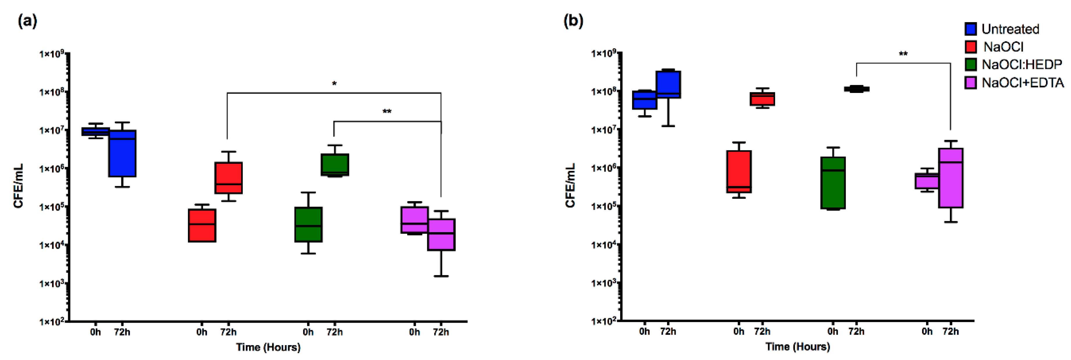
2.2. Effect of Endodontic Irrigants on Single Species Biofilms

Next, we tested each endodontic irrigant against C. albicans mono-species biofilms, where it was shown that all the three treatments have a comparably high initial killing effect at 0 h, resulting in >99% reduction in CFE compared to untreated controls (Figure 1a). The median number of CFE/mL in the control group was 8.7 × 10⁶, reducing to approximately ≤3.5 × 10⁴ CFE/mL for all treatment arms (Table 2). After 72 h, NaOCl+EDTA was significantly the most effective at inhibiting subsequent regrowth (p < 0.05). Only residual yeast cells can be observed when viewed by Calcofluor staining (Figure 2). Conversely, NaOCl alone and the NaOCl:HEDP mixture exhibited a significant regrowth (p < 0.05), showing a 17- and 23.5-fold increase in CFE/mL at 72 h compared to 0 h respectively. There was no significant difference between NaOCl alone and NaOCl:HEDP at both these time points (p > 0.05).
For E. faecalis, the immediate killing effect of the three treatments at 0 h was comparable to that of C. albicans, resulting in ≥98% reduction in CFE compared to untreated controls (Figure 1b). The median number of CFE/mL was reduced from 6.22 × 10⁷ in the control group to ≤8.5 × 10⁵ for all treatments. There was no statistically significant difference between the three treatments at 0 h (Table 2). However, at 72 h E. faecalis was more resilient and showed a greater ability to regrow with the NaOCl and NaOCl:HEDP treatments compared to the effect on C. albicans. There was approximately a 54- and 66-fold increased cell number at 72 h compared to 0 h with NaOCl alone and NaOCl:HEDP treatments, respectively. Again, NaOCl+EDTA was the most effective in inhibiting regrowth at 72 h, with only a three-fold increase in CFE/mL (p < 0.01) (Figure 1b). Small aggregates of cells can be observed retained on the surface of coverslips when viewed by fluorescence microscopy (Figure 2).

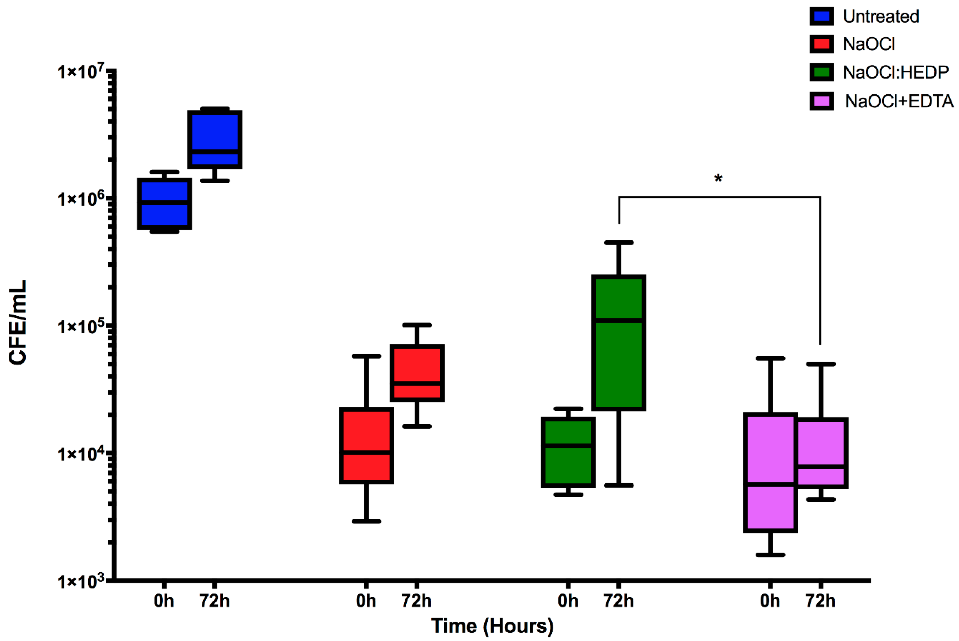
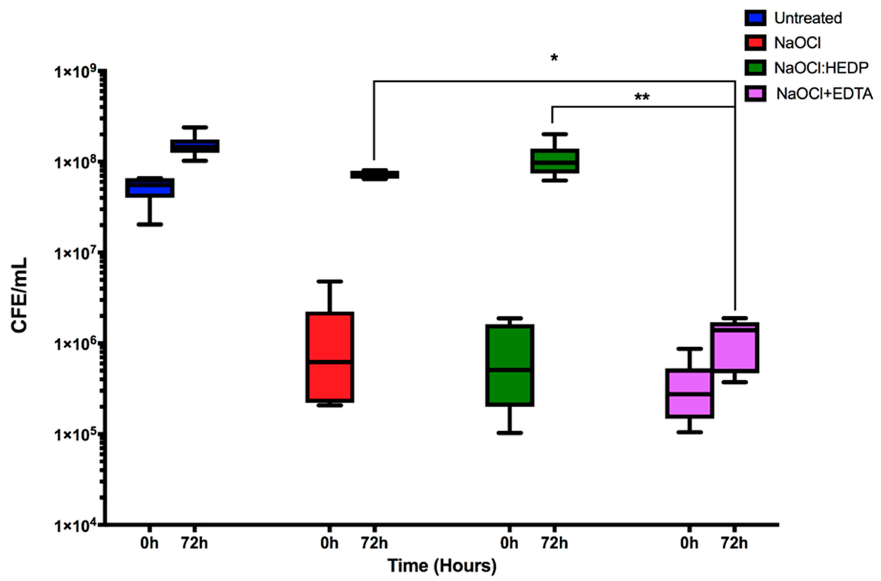
2.3. Effect of Endodontic Irrigants on Dual Species Biofilms

Next, dual-species biofilms were treated and assessed as described above for mono-species biofilms, and C. albicans quantified (Figure 3). The effect of the three treatments at 0 h (immediately after treatment) was similar to that of mono-species biofilms, with all treatments significantly reduced by ≥98.4% compared to controls (9.24 × 10⁵ CFE/mL, p < 0.05). NaOCl+EDTA was the most effective regimen overall, but also notably with no significant regrowth detected after 72 h (7.8 × 10³ CFE/mL) compared to the initial treatment (5.7 × 10³ CFE/mL). Conversely, despite their initial effectiveness both NaOCl alone and NaOCl:HEDP were associated with increased residual biofilm by three- and 12-fold between initial treatment at 0 h compared 72 h post-treatment, respectively. Notably, the residual C. albicans cells were significantly lower in NaOCl+EDTA at 72 h compared NaOCl:HEDP treatment (p < 0.05, Figure 3).
Finally, dual-species biofilms were treated and assessed as described above for mono-species biofilms, and E. faecalis quantified. In dual-species, E. faecalis was significantly reduced immediately after all three treatments by ≥97.4% (p < 0.01), though neither treatment was more effective than one another (Figure 4). However, both NaOCl and NaOCl:HEDP treatments showed significant levels of regrowth after 72 h (p < 0.01), returning to baseline control levels of up to and greater than 7 × 10⁷ CFE/mL (55- and 137-fold increase, respectively (Table 2)). Conversely, for the NaOCl+EDTA treatment after 72 h only a five-fold increase was observed, which was significantly reduced compared to NaOCl (p > 0.05) and NaOCl:HEDP (p < 0.01). This is visually evident when microscopic examination is performed (Figure 2). Here we show that compared to the untreated dual-species biofilm where coccus, yeasts and hyphae are observed, that when treated and observed at 72 h then very low levels of representative morphotypes are evident.

3. Discussion

In the present study we tested the anti-biofilm properties of HEDP, which in endodontics is used as a mild chelating agent to remove the smear layer. In contrast to other chelating agents such as EDTA, it can be mixed with NaOCl to simplify chemical cleaning. It can be also dissolved in normal saline and used separately to remove the smear layer. Its effectiveness in smear layer removal and its effect on radicular dentine has been previously evaluated and shown to be as effective as NaOCl followed by 17% EDTA, but resulted in less dentine erosion and is less toxic [20]. However, augmentative antimicrobial properties of chelating agents would also be highly desirable in root canal disinfection. Here, its antimicrobial properties were compared with standard endodontic irrigants such as NaOCl alone, and more importantly those of NaOCl followed by EDTA. This later sequential chelation system is considered the gold standard in irrigation protocols as it allows for not only biofilm disruption but also smear layer removal. Results of the present study show that HEDP has no significant impact on the antimicrobial activity of NaOCl, either positively or negatively, within a simple mono-species and more complex dual-species challenge. Indeed, our studies have indicated that treatment with NaOCl followed by EDTA is consistently more effective, though caveats exist with respect to resilient biofilm populations.
Our direct post-treatment data (0 h) for both mono- and dual-species E. faecalis biofilms did not show a significant difference between either of the treatment arms. In contrast, when considering regrowth after treatment over time, NaOCl+EDTA was significantly more effective in inhibiting regrowth. HEDP alone (dissolved in normal saline) has no antimicrobial effect under the experimental conditions of the present study. Contrary to this, comparison of the antimicrobial effect of combined NaOCl and dual rinse HEDP to a standard NaOCl+EDTA irrigating sequence against E. faecalis grown inside root canals from extracted human teeth showed that a NaOCl:HEDP mixture resulted in a significantly lower residual bacterial viability [21]. However, Zehnder, Schmidlin [16] found that HEDP did not affect E. faecalis viability and did not impact the antibacterial activity of NaOCl, findings echoed elsewhere [18,19]. These experimental conclusions are likely explained by differences in the in vitro substrates used for these studies, so some caution must be exercised in making bold clinical judgements based on these findings.
Irrespective of treatment regimen tested, there were still remaining populations of C. albicans and E. faecalis following initial treatment which were able to persist and regrow. The antifungal effect of the traditional chelator EDTA is well-documented in the literature [22,23], and as discussed in our previous work EDTA significantly reduced C. albicans regrowth [15]. Nevertheless, it has limited or no antibacterial activity on E. faecalis at any time or concentration [24,25]. Therefore, it may be somewhat unexpected to see that EDTA significantly inhibited E. faecalis regrowth after NaOCl treatment. It is possible that this observed effect of EDTA is related to its membrane permeabilising effect. Lipopolysaccharide molecules have a strong affinity for divalent cations which are required for the thermodynamic stability of the cell membrane [26]. EDTA disrupts the outer membrane of gram-negative bacteria by removing divalent cations [27]. This membrane destabilising effect may be potentiated by the prior treatment of the bacterial cells with NaOCl. HEDP in our experiments did not show an effect, this may be a result of its weaker chelating activity.
Perhaps the most well studied mechanism of Candida-bacteria interactions is physical attachment. A scaffold of hyphae within a biofilm provides a potential niche for the colonization of various gram-positive and gram-negative bacteria, a phenomenon that has been termed as a “mycofilm” [28]. Increasing evidence suggests that E. faecalis and C. albicans are usually found co-isolated in persistent endodontic infections [29]. Understanding the inter-kingdom interaction between bacteria and fungi in endodontic infections might help in developing more effective treatment strategies. We have previously demonstrated that C. albicans is resilient with high regrowth potential following NaOCl treatment [15]. These findings were confirmed in the present study for mono-species biofilms. However, when co-cultured with E. faecalis, C. albicans become more susceptible to treatment with decreased ability for regrowth in comparison to their mono-species biofilms. This can be explained due to the interaction between the two species. E. faecalis has been shown to inhibit hyphal morphogenesis and biofilm formation in C. albicans [30]. Cruz and colleagues also showed that C. albicans and E. faecalis attenuate each other’s virulence in a Caenorhabditis elegans model. This might also explain the low fungal prevalence in endodontic infections, although this effect may be also modified by the conditions of the environment in which this interaction occurred. C. albicans virulence attenuation was largely due to the inhibition of hyphal morphogenesis, a major virulence factor in C. albicans. However, we did not find that C. albicans affected E. faecalis response to irrigants in dual-species biofilms. Ishijima, Hayama [31] also showed that heat killed E. faecalis EF2001 [commercially available probiotic) administered orally has a protective effect against oral candidiasis of mice tongue. Although the effect of C. albicans on E. faecalis is partially revealed, it still unknown how C. albicans affects E. faecalis virulence and response to treatment. Future work should explore the molecular basis for the bidirectional modulation of E. faecalis and C. albicans behaviour and how that would affect these microorganism’s’ response to endodontic treatment.
As outlined previously, the yeast C. albicans and bacteria E. faecalis have been frequently isolated from secondary endodontic infection. Historically, both have been used as model endodontic pathogens to test various endodontic antimicrobials. It is clear from the findings of the present study that the use of mono-species models is no longer appropriate. Any model system must recognise the potential for inter-species/inter-kingdom interactions. The impact of E. faecalis on C. albicans ability to persist and regrow is both interesting and important, as these have a significant impact on how we may view the effectiveness of tested irrigants or medicaments. As such, further studies are required to explore and validate model systems of endodontic disease. This study also highlights the importance of persistence and regrowth in endodontic disease, albeit in a limited number of clinical strains. The present model represents an open system with no anatomical irregularities or complex topography for biofilm to persist, yet we still demonstrate persistence. This is significant problem and further work is required to optimise irrigation strategies and other adjuncts to achieve adequate disinfection.
These results indicate the need for additional work to elucidate the potential clinical implications should a continuous chelation strategy be adopted. C. albicans is significantly more susceptible to NaOCl and NaOCl:HEDP treatment when co-cultured with E. faecalis, suggesting some level of interkingdom antagonism. Future work should investigate the effect of different endodontic antimicrobials against multi-species biofilms on dentine substrate to allow for wider interspecies interactions to be assessed.

4. Materials and Methods

4.1. Microbial Growth Conditions and Standardisation

C. albicans, a clinical isolate from oral rinse of patients attending restorative clinic at Glasgow Dental Hospital and School for routine dental care was used [32]. This isolate has been previously characterised in terms of antifungal susceptibility, proteolytic activity and biofilm formation ability [15,33,34]. The strain was maintained on Sabouraud’s dextrose agar (SAB (Sigma–Aldrich, Dorset, UK)) plates at 30 °C for 48 h. E. faecalis root canal clinical isolate ER5/1 was used [35]. The isolate was cultured on 5% horse blood Columbia agar plate and incubated in 5% CO2 at 37 °C for 24 h. An overnight culture was then prepared in yeast peptone dextrose ((YPD) (Sigma-Aldrich, Dorset, UK)) for C. albicans and in Brain Heart Infusion ((BHI) Sigma–Aldrich, Dorset, UK) for E. faecalis. C. albicans culture was incubated at 30 °C at 120 rpm in an orbital shaker (IKA KS 4000 i control, Berlin, Germany) for 18 h while E. faecalis culture was incubated in 5% CO2 at 37 °C for 18 h. Cells in cultures were then pelleted by centrifugation for 5 min at 3000 rpm and washed twice with PBS. Finally, yeast cells were counted using a haemocytometer and bacterial cells were standardized to OD600 of 0.3 which equivalent of 2 × 10⁸ cells/mL using colorimeter.

4.2. Planktonic Minimum Inhibitory Concentration

First, to ensure the active agents were effective against planktonic (free-floating cells), we determined the PMIC for mono-species and dual-species microorganisms. 3% NaOCl (Parcan, Septodont, Saint-Maur-des-Fosses, France), 17% EDTA (Endo-Solution, CERKAMED, Stalowa Wola, Poland) and 9% HEDP [(Dual Rinse HEDP® Medcem GmbH, Weinfelden, Switzerland) dissolved in either 3% NaOCl or normal saline as per manufacturer’s instructions] were tested using broth microdilution method according to the M27-A3 standard for fungi [36] and the M07-A10 standard for E. faecalis [37]. Briefly, C. albicans was adjusted to a cellular density of (2 × 10⁴ cells/mL) into 1:1 v/v mixture of Roswell Park Memorial Institute (RPMI)-1640 (Sigma–Aldrich, Dorset, UK)/Todd Hewitt broth (THB) (Sigma–Aldrich, Dorset, UK) growth media. E. faecalis cells were adjusted to a density of (2 × 10⁵ cells/mL). Serial double-fold dilutions of each treatment were performed in a 96-well round-bottom microtiter plates (Corning Incorporated, Corning, NY, USA) using 1:1 v/v RPMI/THB media. Treatment concentrations were ranging from (1.5–0.0029%) for NaOCl, (8.5–0.066%) for EDTA and (4.5–0.035%) for HEDP dissolved in 3% NaOCl or normal saline. After serial dilution of treatments, 100 μL of standardized microorganisms were added to 100 μL of each concentration and plates were incubated in 5% CO2 at 37 °C for 24 h. All PMIC tests contained 4 technical repeats and performed at 3 different occasions. Finally, the PMIC concentration was determined as the lowest concentration of each treatment that prevents visible growth or colony formation at the bottom of the plate wells.

4.3. Biofilm Development, Treatment and Regrowth Assessment

Next, treatments were tested against C. albicans and E. faecalis mono-species biofilms (surface attached cells). A cellular density of (1 × 10⁶ cells/mL) for C. albicans, (1 × 10⁷ cells/mL) for E. faecalis was prepared in 1:1 v/v RPMI/THB growth media. 24 h biofilms of mono-species and dual-species were then formed onto sterile Thermanox™ Coverslips (Thermo Fisher Scientific, Paisley, UK) contained in a polystyrene, 24-well microtiter plates (Corning Incorporated, Corning, NY, USA) and incubated in 5% CO2 at 37 °C. Afterwards, unattached cells were removed by washing with PBS and biofilms treated for 5 min by adding 500 μL of either 3% NaOCl only, 3% NaOCl followed by 17% EDTA (5 min each) or 9% HEDP dissolved in 3% NaOCl. Untreated controls of each biofilm were also included. After the 5 min treatment, 500 μL of 5% sodium thiosulfate (Fisher Chemicals, London, UK) was used for 10 min at room temperature to inactivate NaOCl and NaOCl:HEDP mixture, while 500 μL of Dey Engley Neutralising broth (Sigma-Aldrich, London, UK) was used for 15 min at 37 °C to inactivate EDTA. Treatments and neutralizers were removed, and coverslips were washed with PBS. Neutralizers were also added to untreated controls to ensure that the observed effect is solely due to applied treatments and not neutralizers. To further assess the effect of treatments on biofilms’ ability of to persist and regrow, 500 μL of a fresh 1:1 v/v RPMI/THB growth media was added to the treated and washed biofilms and plates re-incubated for further 72 h in 5% CO2 at 37 °C. As an outcome measures, the number of cells remaining immediately after treatment (0 h) and those regrowing after 72 h of reincubation with growth media (72 h) was assessed using real-time quantitative polymerization chain reaction (qPCR). All treatment experiments were performed in duplicates and repeated at 3 different occasions.

4.4. Quantitative Analysis Using Real-Time Quantitative PCR

Thermanox™ coverslips were first washed with PBS and sonicated in 1 mL of PBS at 35 kHz in an ultrasonic water-bath (Fisher Scientific, Paisley, UK) for 10 min to dislodge cells. DNA was extracted from sonicated samples using the QIAamp DNA mini kit, as per manufacturer’s instructions (Qiagen, Crawley, UK). PCR was carried out as previously prescribed [38], using the Step-One plus real time PCR machine and StepOne software V2.3 (Life Technologies, Paisley, UK). Briefly, C. albicans/E. faecalis DNA was added to a PCR mastermix containing Fast SYBR® Green (Thermo Fisher Scientific, Paisley, UK) and specific forward and reverse primers (Table 3) within irradiated RNase-free water. Serial dilutions of bacterial/fungal DNA extracted from 1 × 10⁸ cells/mL were also included to create a standard curve for each species. The applied thermal cycles were as follow: 50 °C for 2 min, 95 °C for 2 min, 40 cycles of 95 °C for 3 s and 60 °C for 30 s. Finally, colony forming equivalents (CFE) was calculated in relation to each species standard curve.

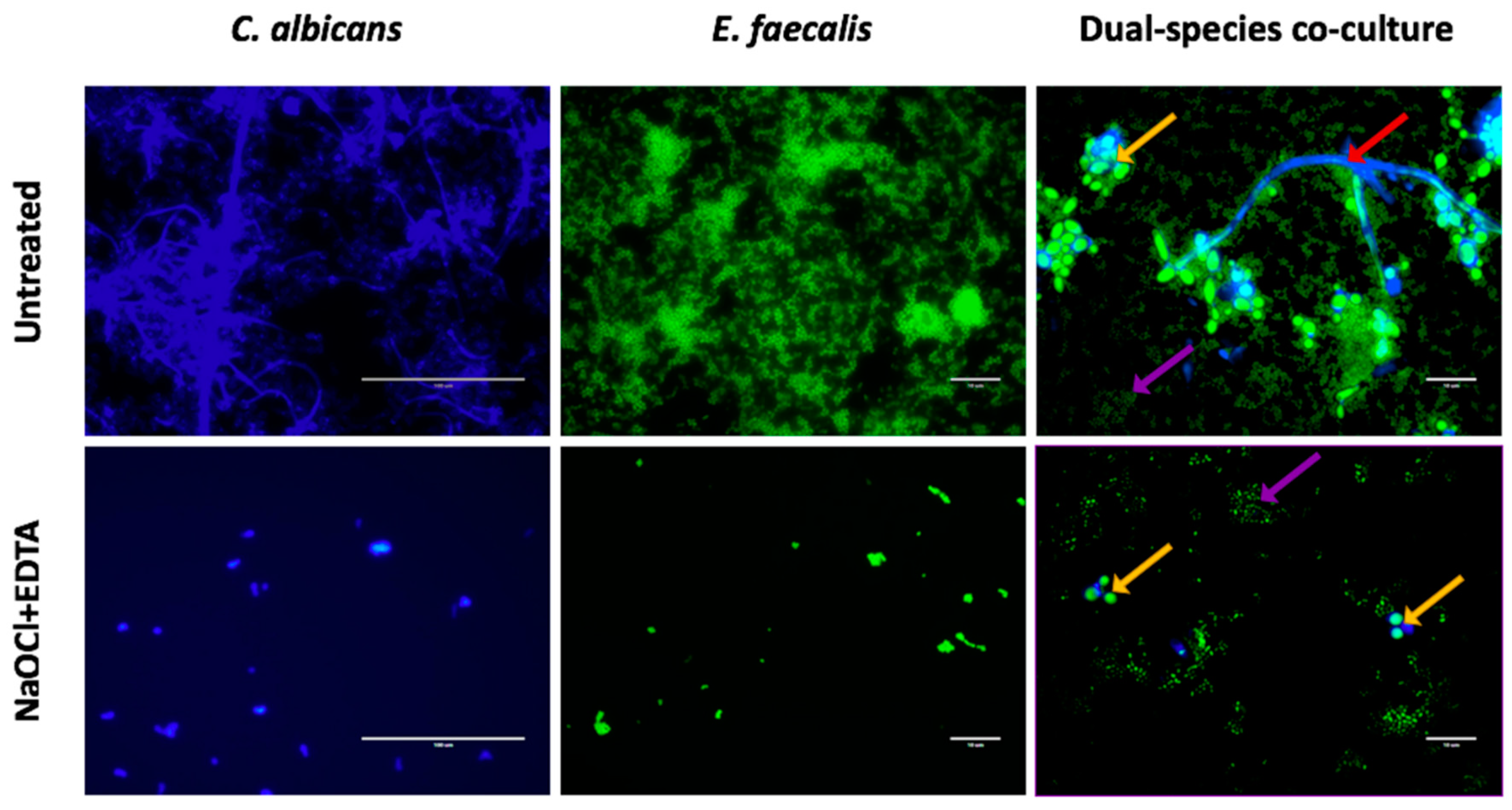
4.5. Fluorescence Microscopic Imaging

Thermanox™ coverslips from each treatment condition in the two time points (0 h and 72 h) were washed with PBS. 5 μM calcofluor white (Invitrogen, Paisley, UK) which specifically stains chitin and beta-glucans of fungal cell wall was used to stain C. albicans while 5 μM SYTO9 (Thermo Fisher Scientific, Paisley, UK) was used to stain both bacterial and fungal cells with high affinity to nucleic acids. After incubating the samples in the dark for 20 min, excess stain was washed with sterile water. Afterwards, stained samples were fixed with 2% paraformaldehyde for 1 h and imaged using EVOS FL Cell Imaging system (Thermo Fisher Scientific, Waltham, MA, USA).

4.6. Statistical Analysis

GraphPad Prism (version 7.0 d) was used for graphs production, data distribution and statistical analysis. D’Agostino-Pearson omnibus normality test was used to test the normal distribution of the data. For statistical analysis, non-parametric Kruskal–Wallis test with Dunn’s multiple comparison test was used to compare CFE values of the three treatment conditions at 0 h and 72 h. Mann–Whitney test was also used to compare mono- and dual-species biofilms of the same treatment.

5. Conclusions

Although continuous chelation is an admirable goal, simplifying protocols and potentially minimising the risk of deleterious interactions between irrigants, the current work raises questions of whether we should depart from sequential irrigation. The use of NaOCl followed by EDTA has an enhanced effect on biofilm disruption and significantly reduces C. albicans and E. faecalis persistence and regrowth. The current study also highlights the need for appropriate biofilm model choices. Both inhibitory and promotory effects can be observed between the participating microorganisms within the biofilm, evidenced here where C. albicans is significantly more susceptible to NaOCl and NaOCl:HEDP treatment when co-cultured with E. faecalis. It may be that with the addition of further complexity within the biofilm model that the interactions between microorganisms modifies to an even greater extent irrigation effectiveness.

Author Contributions

O.A.A., S.A., S.S. and K.A. undertook the laboratory studies, designed the studies, undertook data analysis and contributed to the writing of the manuscript. W.M. and G.R. conceived the study, designed the studies, oversaw analysis, participated in the writing of the manuscript. All authors have read and agreed to the published version of the manuscript.

Funding

This research was funded by the British Endodontic Society.

Conflicts of Interest

None of the authors have any conflict of interest.

References

  1. Kakehashi, S.; Stanley, H.R.; Fitzgerald, R.J. The Effects of Surgical Exposures of Dental Pulps in Germ-Free and Conventional Laboratory Rats. Oral Surg. Oral Med. Oral Pathol. 1965, 20, 340–349. [Google Scholar] [CrossRef]
  2. Moller, A.J.; Fabricius, L.; Dahlen, G.; Ohman, A.E.; Heyden, G. Influence on periapical tissues of indigenous oral bacteria and necrotic pulp tissue in monkeys. Scand. J. Dent. Res. 1981, 89, 475–484. [Google Scholar] [CrossRef] [PubMed]
  3. O’Donnell, L.E.; Millhouse, E.; Sherry, L.; Kean, R.; Malcolm, J.; Nile, C.J.; Ramage, G. Polymicrobial Candida biofilms: Friends and foe in the oral cavity. FEMS Yeast Res. 2015, 15, fov077. [Google Scholar] [CrossRef] [PubMed] [Green Version]
  4. Kong, E.F.; Tsui, C.; Kucharikova, S.; Andes, D.; Van Dijck, P.; Jabra-Rizk, M.A. Commensal Protection of Staphylococcus aureus against Antimicrobials by Candida albicans Biofilm Matrix. mBio 2016, 7. [Google Scholar] [CrossRef] [PubMed] [Green Version]
  5. Rocas, I.N.; Siqueira, J.F., Jr.; Santos, K.R. Association of Enterococcus faecalis with different forms of periradicular diseases. J. Endod. 2004, 30, 315–320. [Google Scholar] [CrossRef]
  6. Persoon, I.F.; Buijs, M.J.; Ozok, A.R.; Crielaard, W.; Krom, B.P.; Zaura, E.; Brandt, B.W. The mycobiome of root canal infections is correlated to the bacteriome. Clin. Oral Investig. 2017, 21, 1871–1881. [Google Scholar] [CrossRef] [Green Version]
  7. Persoon, I.F.; Crielaard, W.; Ozok, A.R. Prevalence and nature of fungi in root canal infections: A systematic review and meta-analysis. Int. Endod. J. 2017, 50, 1055–1066. [Google Scholar] [CrossRef] [Green Version]
  8. Ashraf, H.; Samiee, M.; Eslami, G.; Ghodse Hosseini, M.R. Presence of Candida Albicans in Root Canal System of Teeth Requiring Endodontic Retreatment with and without Periapical Lesions. Iran. Endod. J. 2007, 2, 24–28. [Google Scholar]
  9. Delaney, C.; Kean, R.; Short, B.; Tumelty, M.; McLean, W.; Nile, C.J.; Ramage, G. Fungi at the scene of the crime: Innocent bystanders or accomplices in oral infections? Curr. Clin. Microbiol. Rep. 2018, 5, 190–200. [Google Scholar] [CrossRef] [Green Version]
  10. Ovchinnikova, E.S.; Krom, B.P.; Busscher, H.J.; van der Mei, H.C. Evaluation of adhesion forces of Staphylococcus aureus along the length of Candida albicans hyphae. BMC Microbiol. 2012, 12, 281. [Google Scholar] [CrossRef] [Green Version]
  11. Zehnder, M. Root canal irrigants. J. Endod. 2006, 32, 389–398. [Google Scholar] [CrossRef] [PubMed]
  12. Byström, A.; Sunvqvist, G. The antibacterial action of sodium hypochlorite and EDTA in 60 cases of endodontic therapy. Int. Endod. J. 1985, 18, 35–40. [Google Scholar] [CrossRef] [PubMed]
  13. Brook, I. Microbiology and management of endodontic infections in children. J. Clin. Pediatr Dent. 2003, 28, 13–17. [Google Scholar] [CrossRef] [PubMed]
  14. Vertucci, F.J. Root canal morphology and its relationship to endodontic procedures. Endod. Top. 2005, 10, 3–29. [Google Scholar] [CrossRef]
  15. Alshanta, O.A.; Shaban, S.; Nile, C.J.; McLean, W.; Ramage, G. Candida albicans Biofilm Heterogeneity and Tolerance of Clinical Isolates: Implications for Secondary Endodontic Infections. Antibiotics 2019, 8, 204. [Google Scholar] [CrossRef] [Green Version]
  16. Zehnder, M.; Schmidlin, P.; Sener, B.; Waltimo, T. Chelation in root canal therapy reconsidered. J. Endod. 2005, 31, 817–820. [Google Scholar] [CrossRef]
  17. Neelakantan, P.; Varughese, A.A.; Sharma, S.; Subbarao, C.V.; Zehnder, M.; De-Deus, G. Continuous chelation irrigation improves the adhesion of epoxy resin-based root canal sealer to root dentine. Int. Endod. J. 2012, 45, 1097–1102. [Google Scholar] [CrossRef]
  18. Arias-Moliz, M.T.; Ordinola-Zapata, R.; Baca, P.; Ruiz-Linares, M.; Ferrer-Luque, C.M. Antimicrobial activity of a sodium hypochlorite/etidronic acid irrigant solution. J. Endod. 2014, 40, 1999–2002. [Google Scholar] [CrossRef]
  19. Morago, A.; Ordinola-Zapata, R.; Ferrer-Luque, C.M.; Baca, P.; Ruiz-Linares, M.; Arias-Moliz, M.T. Influence of smear layer on the antimicrobial activity of a sodium hypochlorite/etidronic acid irrigating solution in infected dentin. J. Endod. 2016, 42, 1647–1650. [Google Scholar] [CrossRef]
  20. Lottanti, S.; Gautschi, H.; Sener, B.; Zehnder, M. Effects of ethylenediaminetetraacetic, etidronic and peracetic acid irrigation on human root dentine and the smear layer. Int. Endod. J. 2009, 42, 335–343. [Google Scholar] [CrossRef]
  21. Giardino, L.; Del Fabbro, M.; Morra, M.; Pereira, T.; de Andrade, F.B.; Savadori, P.; Generali, L. Dual Rinse® HEDP increases the surface tension of NaOCl but may increase its dentin disinfection efficacy. Odontology 2019, 107, 521–529. [Google Scholar] [CrossRef] [PubMed]
  22. Ates, M.; Akdeniz, B.G.; Sen, B.H. The effect of calcium chelating or binding agents on Candida albicans. Oral Surg. Oral Med. Oral Pathol. Oral Radiol. Endod. 2005, 100, 626–630. [Google Scholar] [CrossRef] [PubMed]
  23. Sen, B.H.; Akdeniz, B.G.; Denizci, A.A. The effect of ethylenediamine-tetraacetic acid on Candida albicans. Oral Surg. Oral Med. Oral Pathol. Oral Radiol. Endod. 2000, 90, 651–655. [Google Scholar] [CrossRef] [PubMed]
  24. Arias-Moliz, M.T.; Ferrer-Luque, C.M.; Espigares-García, M.; Baca, P. Enterococcus faecalis biofilms eradication by root canal irrigants. J. Endod. 2009, 35, 711–714. [Google Scholar] [CrossRef]
  25. Arias-Moliz, M.T.; Ferrer-Luque, C.M.; Espigares-Rodríguez, E.; Liébana-Ureña, J.; Espigares-García, M. Bactericidal activity of phosphoric acid, citric acid, and EDTA solutions against Enterococcus faecalis. Oral Surg. Oral Med. Oral Pathol. Oral Radiol. Endod. 2008, 106, e84–e89. [Google Scholar] [CrossRef]
  26. Hancock, R.E. Alterations in outer membrane permeability. Annu. Rev. Microbiol. 1984, 38, 237–264. [Google Scholar] [CrossRef]
  27. Leive, L. Studies on the permeability change produced in coliform bacteria by ethylenediaminetetraacetate. J. Biol. Chem. 1968, 243, 2373–2380. [Google Scholar]
  28. Kean, R.; Rajendran, R.; Haggarty, J.; Townsend, E.M.; Short, B.; Burgess, K.E.; Lang, S.; Millington, O.; Mackay, W.G.; Williams, C. Candida albicans Mycofilms Support Staphylococcus aureus Colonization and Enhances Miconazole Resistance in Dual-Species Interactions. Front. Microbiol. 2017, 8, 258. [Google Scholar] [CrossRef] [Green Version]
  29. Pinheiro, E.T.; Gomes, B.P.; Ferraz, C.C.; Sousa, E.L.; Teixeira, F.B.; Souza-Filho, F.J. Microorganisms from canals of root-filled teeth with periapical lesions. Int. Endod. J. 2003, 36, 1–11. [Google Scholar] [CrossRef] [Green Version]
  30. Cruz, M.R.; Graham, C.E.; Gagliano, B.C.; Lorenz, M.C.; Garsin, D.A. Enterococcus faecalis inhibits hyphal morphogenesis and virulence of Candida albicans. Infect. Immun. 2013, 81, 189–200. [Google Scholar] [CrossRef] [Green Version]
  31. Ishijima, S.A.; Hayama, K.; Ninomiya, K.; Iwasa, M.; Yamazaki, M.; Abe, S. Protection of mice from oral Candidiasis by heat-killed enterococcus faecalis, possibly through its direct binding to Candida albicans. Med. Mycol. J. 2014, 55, E9–E19. [Google Scholar] [CrossRef] [PubMed] [Green Version]
  32. Coco, B.; Bagg, J.; Cross, L.; Jose, A.; Cross, J.; Ramage, G. Mixed Candida albicans and Candida glabrata populations associated with the pathogenesis of denture stomatitis. Oral Microbiol. Immunol. 2008, 23, 377–383. [Google Scholar] [CrossRef] [PubMed]
  33. Ramage, G.; Coco, B.; Sherry, L.; Bagg, J.; Lappin, D.F. In Vitro Candida albicans Biofilm Induced Proteinase Activity and SAP8 Expression Correlates with In Vivo Denture Stomatitis Severity. Mycopathologia 2012, 174, 11–19. [Google Scholar] [CrossRef]
  34. Ramage, G.; Jose, A.; Coco, B.; Rajendran, R.; Rautemaa, R.; Murray, C.; Lappin, D.F.; Bagg, J. Commercial mouthwashes are more effective than azole antifungals against Candida albicans biofilms in vitro. Oral Surg. Oral Med. Oral Pathol. Oral Radiol. Endod. 2011, 111, 456–460. [Google Scholar] [CrossRef] [PubMed]
  35. Johnson, E.M.; Flannagan, S.E.; Sedgley, C.M. Coaggregation interactions between oral and endodontic Enterococcus faecalis and bacterial species isolated from persistent apical periodontitis. J. Endod. 2006, 32, 946–950. [Google Scholar] [CrossRef]
  36. CLSI. Reference Method for Broth Dilution Antifungal Susceptibility Testing of Yeasts. Approved Standard-Third Edition M27-A3; Clinical and Laboratory Standard Institute: Wayne, PA, USA, 2008. [Google Scholar]
  37. CLSI. Methods for Dilution Antimicrobial Susceptibility Tests for Bacteria That Grow Aerobically. Approved Standard-Tenth Edition M07-A10; Clinical and Laboratory Standard Institute: Wayne, PA, USA, 2015. [Google Scholar]
  38. O’Donnell, L.E.; Smith, K.; Williams, C.; Nile, C.J.; Lappin, D.F.; Bradshaw, D.; Lamber, M.; Robertson, D.P.; Bagg, J.; Hannah, V. Dentures are a reservoir for respiratory pathogens. J. Prosthodont. 2016, 25, 99–104. [Google Scholar] [CrossRef] [Green Version]
  39. Fujita, S.-I.; Lasker, B.A.; Lott, T.J.; Reiss, E.; Morrison, C.J. Microtitration plate enzyme immunoassay to detect PCR-amplified DNA from Candida species in blood. J. Clin. Microbiol. 1995, 33, 962–967. [Google Scholar] [CrossRef] [Green Version]
  40. Rathnayake, I.; Hargreaves, M.; Huygens, F. Genotyping of Enterococcus faecalis and Enterococcus faecium isolates by use of a set of eight single nucleotide polymorphisms. J. Clin. Microbiol. 2011, 49, 367–372. [Google Scholar] [CrossRef] [Green Version]
Figure 1. The effect of the three endodontic irrigants on pre-formed 24 h mono-species biofilms. NaOCl alone, NaOCl:HEDP and NaOCl+EDTA were used to treat (a) C. albicans and (b) E. faecalis. CFE at 0 h and 72 h are presented as box and whisker plot of each treatment on log10 Y axis. Statistical significance between the three treatment conditions at 72 h was presented as * p < 0.05, ** p < 0.01. Results represent data from three independent experiments.
Antibiotics 09 00634 g001
Figure 2. Fluorescence microscopic examination of C. albicans mono-species, E. faecalis mono-species and dual species biofilms. Upper panel represents untreated biofilms at 72 h. Lower panel represents regrowth of biofilms treated with NaOCl+EDTA at 72 h. C. albicans in mono-species and dual-species biofilms was stained blue with calcofluor white while the green fluorescence SYTO 9 stained both C. albicans and E. faecalis cells. Yellow arrows show C. albicans yeast cells, red arrow shows C. albicans hyphae and purple arrows show E. faecalis. Scale bars is 100 μm in C. albicans mono-species and 10 μm in E. faecalis mono-species and dual-species co-culture.
Antibiotics 09 00634 g002
Figure 3. The effect of the three endodontic irrigants on C. albicans in pre-formed 24 h dual-species biofilm. NaOCl alone, NaOCl:HEDP and NaOCl+EDTA were used to treat mixed species biofilms and C. albicans quantified. Box and whisker plot of CFE at 0 h and 72 h for each treatment. Values were plotted as log10 on the Y axis. Statistical significance between the three treatment conditions at 72 h was presented as * p < 0.05. Results represent data from three independent experiments.
Antibiotics 09 00634 g003
Figure 4. The effect of the three endodontic irrigants on E. faecalis in pre-formed 24 h dual-species biofilm. NaOCl alone, NaOCl:HEDP and NaOCl+EDTA were used to treat mixed species biofilms and E. faecalis quantified. Box and whisker plot of CFE at 0 h and 72 h for each treatment. Values were plotted as log10 on the Y axis. Statistical significance between the three treatment conditions at 72 h was presented as * p < 0.05, ** p < 0.01. Results represent data from three independent experiments.
Antibiotics 09 00634 g004
Table 1. Susceptibility of planktonic Candida albicans, Enterococcus faecalis and their co-cultures to sodium hypochlorite, EDTA and HEDP. Numbers represent PMIC values.
Endodontic IrrigantC. albicansE. faecalisCo-Culture
NaOCl0.093%0.187%0.187%
EDTA0.13%0.265%0.265%
HEDP: NaOCl0.093%0.093%0.187%
HEDP: saline2.25%4.5%4.5%
Table 2. The median number and range of CFE/mL for untreated controls, NaOCl, NaOCl:HEDP and NaOCl+EDTA treatment at 0 h and 72 h.
Biofilm TypeTime PointTreatmentMedian CFE/mLRange (Minimum to Maximum)
C. albicans mono-species0 hUntreated control8.70 × 106(6.05 × 10⁶ to 1.46 × 10⁷)
NaOCl3.47 × 104(1.16 × 10⁴ to 1.13 × 10⁵)
NaOCl:HEDP3.10 × 104(5.97 × 10³ to 2.34 × 10⁵)
NaOCl+EDTA3.57 × 104(1.89 × 10⁴ to 1.32 × 10⁵)
72 hUntreated control5.83 × 106(3.26 × 10⁵ to 1.58 × 10⁷)
NaOCl3.82 × 105(1.40 × 10⁵ to 2.71 × 10⁶)
NaOCl:HEDP7.76 × 105(6.07 × 10⁵ to 3.99 × 10⁶)
NaOCl+EDTA2.01 × 104(1.53 × 10³ to 7.65 × 10⁴)
E. faecalis Mono-Species0 hUntreated control6.22 × 107(2.19 × 10⁷ to 1.04 × 10⁸)
NaOCl3.11 × 105(1.64 × 10⁵ to 4.53 × 10⁶)
NaOCl:HEDP8.51 × 105(8.01 × 10⁴ to 3.36 × 10⁶)
NaOCl+EDTA5.97 × 105(2.37 × 10⁵ to 9.57 × 10⁵)
72 hUntreated control8.61 × 107(1.22 × 10⁷ to 3.64 × 10⁸)
NaOCl7.41 × 107(3.62 × 10⁷ to 1.18 × 10⁸)
NaOCl:HEDP1.10 × 108(9.44 × 10⁷ to 1.36 × 10⁸)
NaOCl+EDTA1.38 × 106(3.81 × 10⁴ to 4.989 × 10⁶)
C. albicans Dual-Species0 hUntreated control9.24 × 105(5.472 × 10⁵ to 1.60 × 10⁶)
NaOCl1.01 × 104(2.92 × 10³ to 5.77 × 10⁴)
NaOCl:HEDP1.14 × 104(4.724 × 10³ to 2.23 × 10⁴)
NaOCl+EDTA5.70 × 103(1.60 × 10³ to 5.54 × 10⁴)
72 hUntreated control2.31 × 106(1.37 × 10⁶ to 5.01 × 10⁶)
NaOCl3.52 × 104(1.62 × 10⁴ to 1.01 × 10⁵)
NaOCl:HEDP1.10 × 105(5.59 × 10³ to 4.49 × 10⁵)
NaOCl+EDTA7.85 × 103(4.34 × 10³ to 4.50 × 10⁴)
E. faecalis Dual-Species0 hUntreated control5.54 × 107(2.935 × 10⁷ to 6.61 × 10⁷)
NaOCl6.22 × 105(2.081 × 10⁵ to 4.81 × 10⁶)
NaOCl:HEDP5.10 × 105(1.029 × 10⁵ to 1.89 × 10⁶)
NaOCl+EDTA2.76 × 105(1.05 × 10⁵ to 8.70 × 10⁵)
72 hUntreated control1.46 × 108(1.03 × 10⁸ to 2.39 × 10⁸)
NaOCl7.24 × 107(6.44 × 10⁷ to 8.06 × 10⁷)
NaOCl:HEDP9.76 × 107(6.21 × 10⁷ to 2.02 × 10⁸)
NaOCl+EDTA1.39 × 106(3.73 × 10⁵ to 1.89 × 10⁶)
Table 3. C. albicans and E. faecalis primers for qPCR.
PrimerSequence (5′—3′)References
C. albicansITS3—GCA TCG ATG AAG AAC GCA GC
ITS4—TCC TCC GCT TAT TGA TAT GC
[39]
E. faecalisF—CAAACTG TTGGCATTCCACAA
R—TGGATTTCCTTTCCAGTC ACTTC3
[40]

