1. Introduction
The need for compatible materials for the preservation of cultural heritagehas resulted in the revival of lime-based mortar technology and other applications. Quick lime is the traditional ingredient used in ancient mortars and plasters; early examples have been found in Palestine and Turkey dating back to 12,000 before present (BP) as well as in Asia, Egypt, Greece and throughout the Roman Empire, in North and South America [
1,
2]. Currently, lime is the binder of choice for the preparation of compatible mortars used to construct masonry and monuments and also in the conservation of architectural heritage. Despite the apparent simplicity of the technology involved in mortar preparation, the different chemical reactions occurring during the various steps are quite complex. In particular, the hardening of slaked lime (reaction with air and progressive drying) and the choice of the filler are two important steps that influence both durability and workability. Ancient recipes describe the use of animal glues, such as casein and other dairy products, fat, albumen, and blood, but also vegetable glues, such as linseed oil, beer, and resins [
3,
4] as an additive to improve quality mortars.
Modern diagnostic techniques are now able to identify the large variety of natural products used in historical mortars; for example, animal glue, lipid and proteinaceous materials were identified in medieval mortars in Southern Italy [
5]; egg white, juice of plants, drying oil, molasses were found in mortars from Spanish Colonial Period in the Philippines [
6]; blood, crop flour, sticky-rice have been used as binders in the mortars of Chinese wooden buildings [
7]. The Mesoamerican historical buildings tradition [
8], a pre-Hispanic practice, includes the addition of mucilage from different plant species. The use of cacti mucilage as an additive to the plasters and as a binder for the wall paintings was discovered in the mural paintings of Cacaxtla, showing a good state of conservation [
9]. The use of Opuntia mucilage has continued over Mexican history to modern times through traditional construction techniques, being transmitted from generation-to-generation. Apart fromempirical knowledge, very few data are available on mucilage-based mortar for restoration and their physical and mechanical properties. From the chemical point of view, Opuntia mucilage is a complex carbohydrate with a molecular weight between 1.5 and 4 × 10
6 g/mol, composed of a variable amount of arabinose (24.6–42%), galactose (21–40.1%), galacturonic acid (8–12.7%), rhamnose (7–13.1%), and xylose (22–22.2%) [
10]. In general, the composition and percentage of carbohydrates varyin the mucilage ofdifferent species. However, their presence is important because the carboxylic groups react with divalent cations to form an egg-box structure, which allows water retention in plants [
11,
12]. As an additional component, it contains mono and divalent cations, DNA, proteins in small amounts and phytomolecules, such as polyphenols. This chemical composition makes the mucilage an excellent additive to lime, which may prevent surface breaks of mortars guaranteeing the desirable moisture [
13]. Other functions of the vegetable mucilage used in the preparation of lime mortar areto promote the plasticity of the mortar allowing its use in thin layers [
14] and to act as a consolidator thanks to the high viscosity.
On these premises, the aim of our work was the study of the cohesion and integrity of lime mortars added with fresh mucilage from five plants and the evaluation of their bioreceptivity to substantiate the improvement in durability.
Therefore, in order to fulfill our aim, we: (i) extracted and characterized by FTIR the mucilage from fiveplant species; (ii) prepared samples of bio-mortars with the different mucilages; (iii) evaluated physical properties of the bio-mortars after threemonths of drying and after threeyears aging using non-destructive tests (ultrasound and X-rays measurements, colorimetry); (iv) assessed the bioreceptivity of the bio-mortars inoculated with fungi, bacteria and photosynthetic biofilm isolated from the walls of archeological sites in order to employ the mucilages in restoration and conservation intervention.
2. Materials and Methods
2.1. Plant Materials and Mucilage Extraction
Cladodes (1–3 years old) of Opuntia ficus-indica and Opuntia engelmannii, stems of Cylindropuntia californica and leaves of Aloe vera were collected from a wild bush at early hours in the morning in Maccarese (Rome) and immediately used for the experimental trials. The plant materials were washed with tap water in order to remove impurities and spines, peeled from epidermis on both sides and chopped into pieces of 1–2 cm. Commercial seeds from Chia (Salvia hyspanica) were bought from a local retailer (NaturaSi shop, Rome, Italy). Fresh chopped plant material of Opuntia ficus-indica, Opuntia engelmannii, Cylindropuntia californica, and Aloe vera (200 g each) was mixed with distilled water (200 mL each) in different flasks (biomass to water ratios: 1:1, w/v). The samples were left to soak for 24 h at 25 °C, in dark conditions without stirring. The mucilage was removed by percolation through a 0.5 mm mesh. Seeds from Salvia hyspanica were mixed with distilled water in biomass to water ratioof 1:5 (w/v). Seeds were removed from mucilage by centrifugation at 5000 rpm for 10 min.
2.2. Mucilages Characterization
The mucilages extracted from the different plants were immediately analyzed with a digital refractometer DBR35/45/SALT (Metricon Corporation, Pennington, NJ, USA) in order to measure the Brix degrees (°Bx), i.e., the percentage of sugar content by weight (grams per 100 mL of water). Furthermore, the pH of each mucilage was measured with a pH-meter (Mettler Toledo, Columbus, OH, USA). For further analyses, the extracts were kept at −20 °C.
For the infrared analysis, the mucilages were freeze-dried in order to obtain anhydrous powders. Infrared absorption spectra were measured with a Spectrum 100 Fourier-transform infrared (FTIR) spectrometer (PerkinElmer, Waltham, MA, USA) in the wavenumber range between 400 and 4000 cm
−1 with a spectral resolution of 4 cm
−1. Absorbance spectra of the dried powders were calculated as:
from the transmissivity measured with attenuated total reflectance (ATR) accessory equipped with a multipass KRS5 crystal. After the blank subtraction, absorbance spectra were normalized in order to facilitate the comparison.
Since with the ATR technique it is impossible to know the exact quantity of the sample which is actually probed, the resulting spectra cannot be used for the quantitative analysis of the chemical content, i.e., the absolute value of the intensity of the absorption peaks cannot be related to the number of the corresponding chemical bonds in the sample; nevertheless, the positions and the relative intensities of the bands give useful information about the composition of the specimen. Hence, after the blank subtraction, each absorbance spectrum was normalized to one by dividing by the maximum value of the intensity of a selected band (in this work, we selected the intense band centered at 1040 cm−1) in order to facilitate the comparison to the other spectra.
2.3. Bio-Mortar Samples Preparation
Samples were prepared with a ratio of aged lime putty and marble powder: 1:2 (
v/v) and added with mucilage from the different plant species. Optimal additive concentration for the improvement of mechanical resistance was assessed in previous work [
7]; for this reason, in this study, a 2.5% (
v/w) of mucilage was used. The bio-mortar samples were then put in a mold (size 5 cm × 5 cm × 5 cm) according to normal UNE-EN-1015-11 [
10] and dried at room temperature for 30 days.
Six replicas were prepared with each kind of mucilage to be used for colorimetric, radiographic, ultrasound and bioreceptivity tests. The tests were performed on the fresh samples and after 3 years of natural aging.
In order to compare the cohesive performance of the bio-mortars prepared with Opuntia ficus-indica, 10% v/w (OFI) with respect to mortars prepared with synthetic additive, a preliminary trial was carried out on replicated mortar samples added with 10% of Primal AC 33, an acrylic polymer widely used for restoration and conservation purposes.
2.4. Evaluation of Physical Properties of Bio-Mortars by Non-Destructive Tests
Colorimetric measurements were carried out to evaluate color changes induced by the presence of the plant mucilage with respect to the mortars prepared without vegetable additives. Color values, reported in the
CIEL*
a*
b*space, were measured with a Techkon SP 820 λ (TECHKON GmbH, Königstein, Germany), with a 3 mm spot, using a standard D65 illumination. In the color representation space,
L*
a*
b*,
L*is the lightness,
a* is the red/green position, and
b* is the blue/yellow position. In the
CIEL*
a*
b* system, the distance between two color points represents their color difference Δ
E* and it is calculated from the differences of its components Δ
L*, Δ
a*, Δ
b*, according to the Equation (2):
The measurements were performed on the same face of each cubic sample in three points (a mask was used to repeat the measurement). Reported data are the mean of 10 replicas. These measurements allowed calculating the total color difference ΔE, relative to the same area of the sample.
Ultrasonic velocity (US) measurements were carried out with the aim of verifying the internal cohesion in each sample. The US in a mortar sample is related to its compactness due to density, porosity and composition. A portable instrument, a Krautkramer USM 23 (GE Inspection Technologies GmbH, Wunstorf, Germany) equipped with a low-frequency probe (50 kHz), was used in the transmission method, and no sample preparation was necessary. Ultrasonic wave velocity was measured in all specimens in the three directions X, Y, and Z. For each direction, three values were recorded, and the average was calculated. A thin layer of water was used as an acoustic coupling medium between the stone and the transducer.
The compressive strength (
RCK) obtained from ultrasonic tests was estimated as for the Equation (3):
In which
Vel US is the measured US expressed in m/s, and
a is a suitable constant that must be calibrated on a reference sample [
15].
Digital radiography was carried out with Seifert ISOVOLT Titan 160 M2 0.4–1.5 (GE Inspection Technologies GmbH, Wunstorf, Germany) to study the density of the mortar. This method allows the internal vision of the object in a non-invasive way [
16]. X-rays, electromagnetic radiation more energetic than visible light, cross the objects highlighting the macro structural nonuniformity and the defects present inside the object’s volume. Therefore, if defects exist in the object under investigation such as cavities, cracks, large inclusions less absorbent than the matrix, or discontinuities of denser and more absorbent material, spots of different tones and proportional to the thickness of the defect will appear on the plate, and the defect will appear delimited by its perspective projection. The shades of gray represent the absorption of the rays proportional to the density of the sample passed through. In black, the denser objects and inclusions;in white, the pores. The elaboration of the digital radiography images isnondimensional values.
2.5. Bioreceptivity Tests
Bioreceptivity is the aptitude of a material to be colonized by living organisms, as conceived by Guillitte [
17]. For bio-receptivity tests, the bio-mortar samples were prepared following the procedure described in
Section 2.3, but adding 10% of mucilage (
v/w) instead of 2.5% to enhance the organic content in the mortar and increase the possibility of biodeteriogens growth. The mortar was distributed into sterile Petri dishes and set to dry for 30 days. To perform the contamination, three fungi and three bacteria strains were chosen among those commonly found in bio-deterioration studies, isolated from Domus Aurea (Rome, Italy) and an Etruscan tomb in Cerveteri (Cerveteri, Italy), as well as a photosynthetic biofilm collected in the Catacomb of San Callisto in Rome (courtesy of prof. Laura Bruno).
Penicillium commune Fcont,
Cladosporium sp. DAF6 and
Fusarium sp. CERV14F1 spores were inoculated onto the samples (10
6 cells/mL, in triplicate) and incubated at 28 °C and 96% RH.
Rhodococcus opacus CER14.3
, Bacillus amyloliquefacens CER14.4 and
Virgibacillus sp. NOT1 bacterial cells were inoculated onto the samples (10
6 cells/mL, in triplicate) and incubated at about 26 °C (room temperature). Phototrophic biofilm CSC3W (composed ofalgae, cyanobacteria, moss protonemata) was inoculated onto the samples (in triplicate) and incubated at room temperature in the daylight.
The growth of the biodeteriogens was daily monitored by stereomicroscope and digital microscope, and recorded by photograph. Positive and negative controls were included in the experimental trial: negative controls were mortars and bio-mortars without artificial contamination to check if any spontaneous biological growth could be detected; positive controls were solid agar media inoculated with the same microorganisms used in the artificial contamination to assess the viability of the contaminants in optimal conditions.
2.6. Statistical Analyses
Statistical analyses were used to compare results obtained in replicates (9 values for each variable), and data were expressed as mean ± standard deviation (SD). All analyses were performed by SPSS statistics software v 25 (IBM, Armonk, New York, USA). Using the analysis of variance (ANOVA), differences among means were determined for significance at p < 0.05. A comparison of means was made by Duncan and Tukey’s tests (n = 5 replicates).
3. Results and Discussion
The extraction method of mucilage from succulent plants was optimized and standardized in a previous study [
10,
18]. The water extraction for 24 h at room temperature yielded a mucilage that showed visible viscosity and was colorless, indicating the absence of chlorophyll in the samples. Usually, the °Bx is a measure of the sugar content of an aqueous solution with one Brix degree equivalent to 1 g of sucrose in 100 g of the solution; it represents the strength of the solution as a percentage by mass. Since mucilage in water is a complex solution containing different solids other than pure sucrose, the °Bx is an indicative measure thatonly approximates the dissolved solid content.
Values of pH and°Bx of the mucilage extract from
Opuntia ficus-indica,
Opuntia engelmannii,
Cylindropuntia californica,
Aloe vera and
Salvia hispanica were compared, and the results are shown in
Table 1. Mucilage extract of
Salvia hispanica showed alower value in soluble sugar content and the highest in pH. The cacti mucilages (Opuntias and
Cylindropuntia) showed similar values, 0.9 to 1.0 °Bx, and
Aloe vera exhibited the highest soluble sugar content. Among the succulent species, whose mucilage is characterized by a pH around 4, only
Cylindropuntia showed a sub-acidic value.
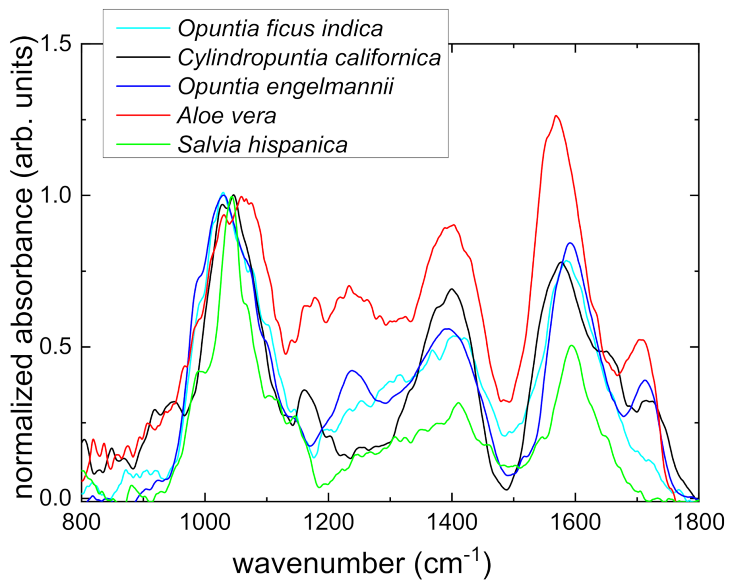
Infrared spectroscopy can help in the identification of the chemical composition of a sample. The normalized absorbance spectra are reported in
Figure 1 in the most characteristic spectral interval between 800 and 1800 cm
−1, the so-called sugar fingerprint region. Comparison of the experimental data with spectra reported in literature enables to attribute some diagnostic peaks to specific sugars, and hence identify the composition of the samples [
19,
20,
21,
22]. In particular, the peak around 1051 cm
−1 is due to the presence of arabinose and rhamnose, the one around 940 cm
−1 is due to the presence of galactose, while the peaks at 1104 cm
−1 and 1179 cm
−1 to the xylose. Furthermore, in the spectra reported in
Figure 1, the peaks at 1104 cm
−1 and 1118 cm
−1are due to the vibrations of C-O bonds in uronic acids, and the band centered at 1429 cm
−1 can be ascribed to the carboxylic group of the galacturonic acid. The infrared spectra of the different
Opuntia and
Cylindropuntia mucilages are quite similar, demonstrating similarity in the sugar content, as also measured with the refractometric technique. On the other hand, the peaks centered at 1400 cm
−1and 1573 cm
−1 in the spectrum of the mucilage extracted from
Aloe vera are much more intense than those in the spectra of
Opuntia and
Cylindropuntia species, indicating higher content of sugars, as also confirmed by the presence of two broad, intense peaks at around 1180 cm
−1and 1240 cm
−1 and by the measured Brix degrees. The spectrum of the
Salvia hispanica mucilage, on the contrary, shows poorly intense bands in the sugar fingerprints region, demonstrating a low content of carbohydrates with respect to the other samples reported in this work. All in all, the results of the FTIR analysis are in very good agreement with those obtained by the refractometric analysis.
Since it is recognized that the gelling capacity of mucilage and its functional value as an additive in mortars is principally due to the presence of uronic acids [
11,
19], the analysis of the carboxylic acids is of the utmost importance. In particular, in all the spectra of mucilages extracted from
Opuntia and
Cylindropuntia species, the diagnostic peaks at 1118 cm
−1 and 1429 cm
−1 have the same intensity, indicating that the use of the extracts as an additive in bio-mortars could have almost equivalent positive effects, as also demonstrated by non-destructive tests (see below). Furthermore, the very high-intensity of the same peaks in the spectrum of
Aloe vera mucilage suggests that its use could result in even higher mortar mechanical performances. On the other hand, the spectrum of the
Salvia hispanica extract, with less pronounced peaks indicates a poor gelling capacity.
Colorimetric analyses showed very small variations of chromatic coordinates values induced by the presence of the mucilages in the mortars, which did not significantly change the colors; indeed,Δ
E* remains always <4. Hence, since, according to [
23], only a Δ
E* > 5 is perceived by the human eye, no color alteration occurs. Furthermore, no color changes result after samples of aging.
The results of non-destructive tests on bio-mortars supplemented with 2.5% of mucilage from
Opuntia ficus-indica (OFI)
Opuntia engelmannii (OE),
Cylindropuntia californica (CC),
Aloe vera (Aloe) and
Salvia hispanica (SH) are reported in
Figure 2,
Figure 3 and
Figure 4. The analysis wasperformed after three months of drying and after three years of natural aging of the bio-mortars supplemented by the different mucilages.
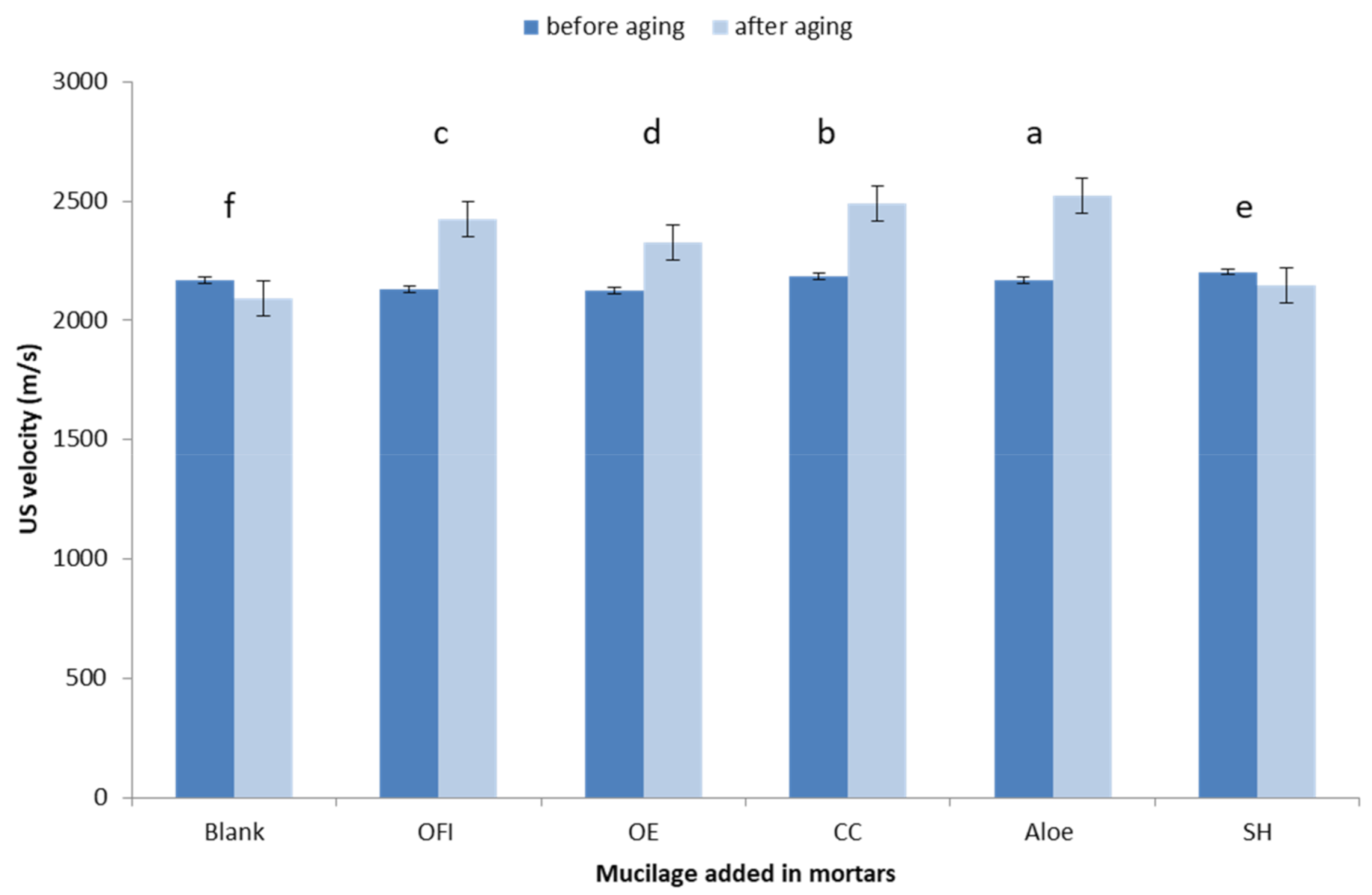
The ultrasonic pulse velocity is an indirect method for quantifying homogeneity in the samples [
24]. The results of the US measurements performed three months after hardening showed that the addition of mucilage to mortars promote a small variation in the propagation velocity of the ultrasonic waves into the samples (
Figure 2); after three years of natural aging, a slight increase was observed in those bio-mortars containing succulent plant extracts: Aloe, CC and OFI mucilages seem to induce the more significant change in the ultrasound propagation. Similar results were reported by [
25] showing the addition of
Opuntia ficus-indica mucilage at water replacement concentration between 4–8% as suitable for durability enhancing applications in cement-based mortar.
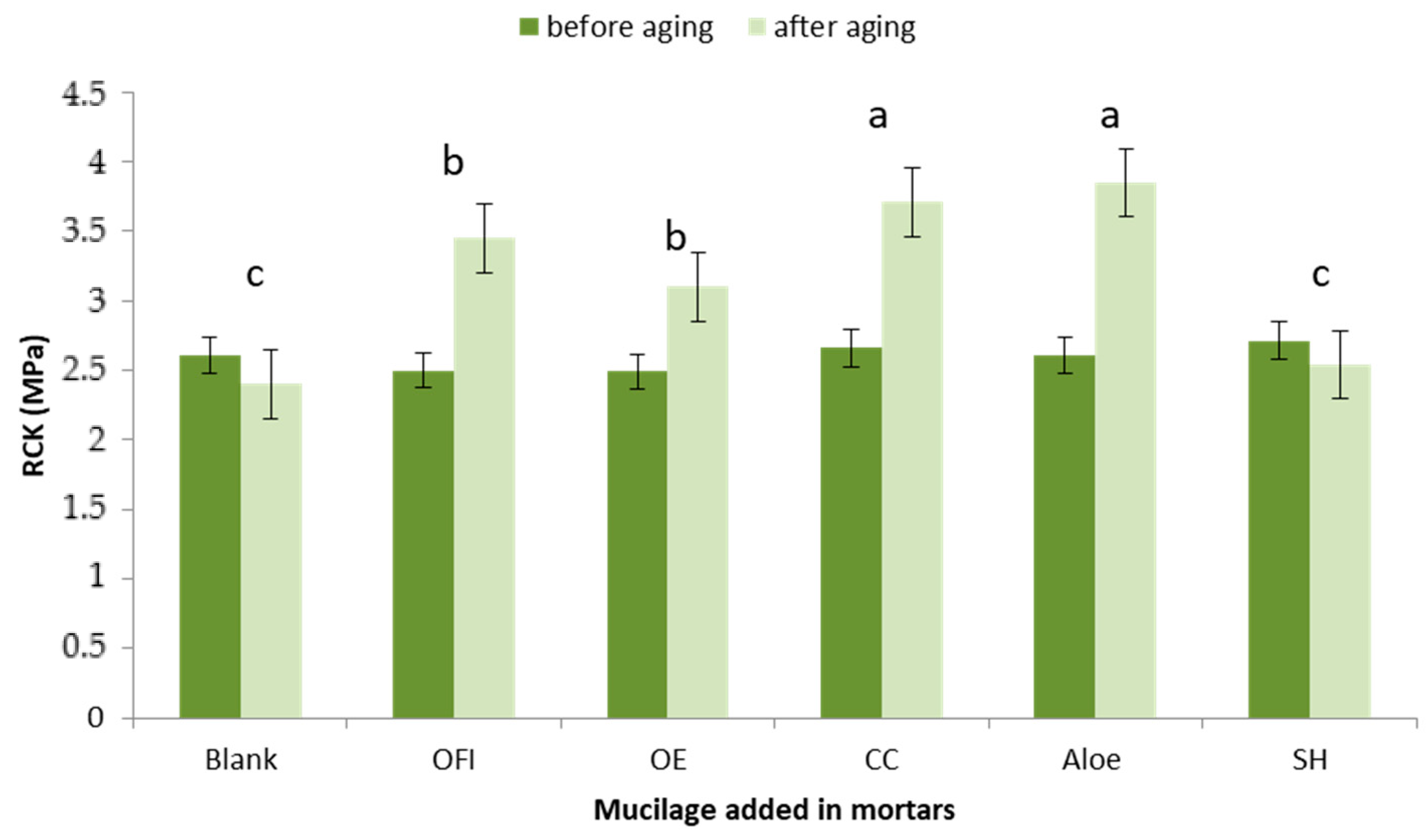
The measure of the elastic module in all the samples just after hardening showed a similar behavior (
Figure 3); a significant increase in the samples of bio-mortars added with Cacti mucilages (Aloe, CC, OFI, OE) was observed after aging. A possible explanation of this behavior can be due to the delay of the hardening (carbonating) of the bio-mortars, thus favoring the formation of a more regular crystalline structure.
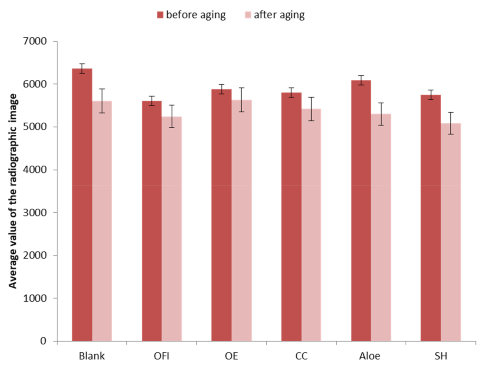
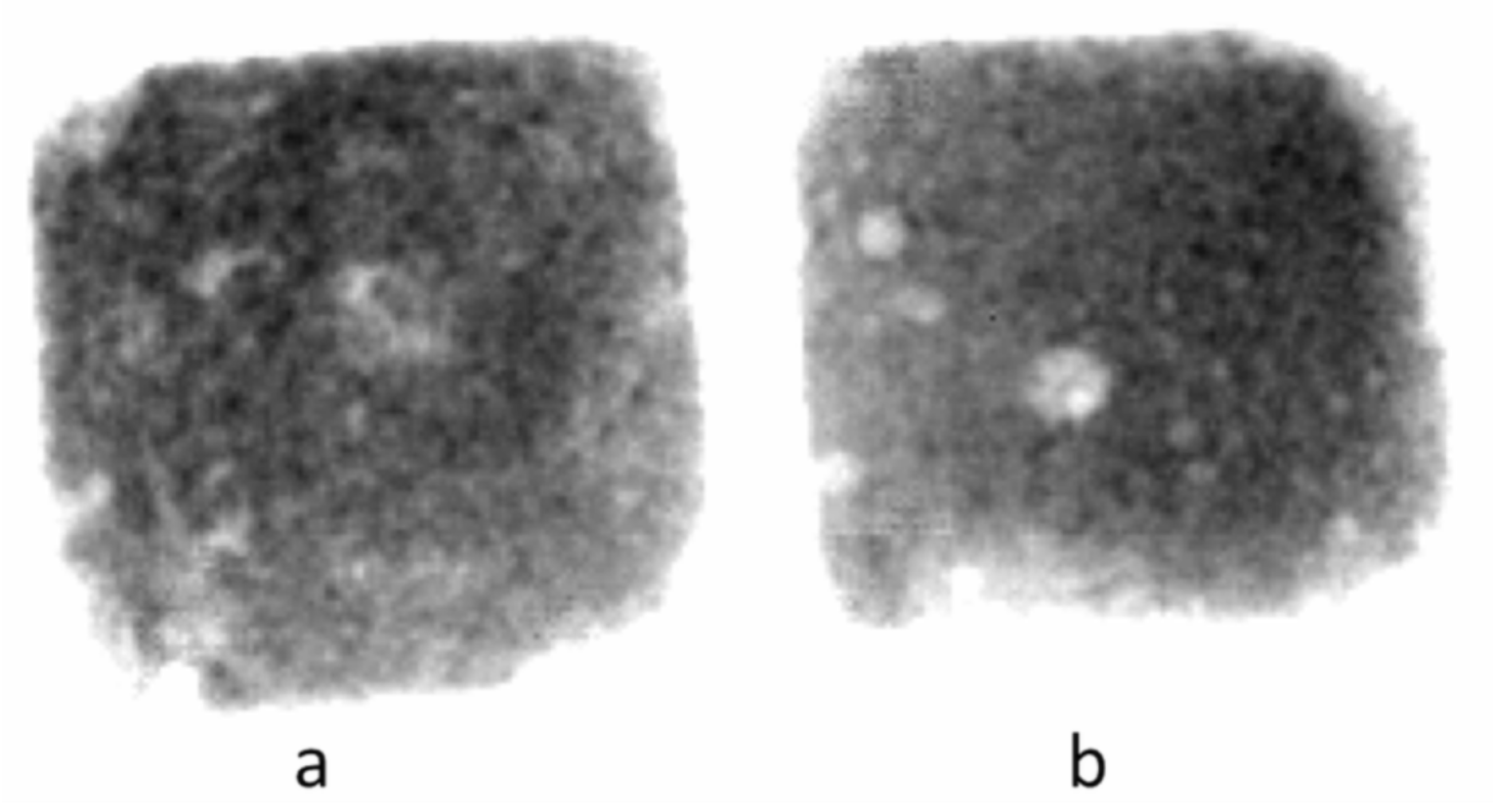
Radiographic density showed few differences in the bio-mortars in comparison with the blank three months after drying. Nevertheless, differences were observed in the samples supplemented by mucilage after long term aging. These materials seem to be more homogenous with high-density. These results are also in agreement with those of the ultrasound pulse velocity test, which provides an indirect evaluation of the porous material void content because sound travels faster in a homogeneous solid material than through air. Highly porous material shows a low velocity of the ultrasound wave, as shown in the blank (
Figure 2).
Furthermore, a preliminary radiographic test performed to compare the use of Primal AC 33 with OFI as an additive (
Figure 5) produced a promising result. The analysis of mortar added with primal acrylic resin showed comparable compactness to the mortar added with the same percentage of
Opuntia ficus-indica mucilage.
These encouraging results contribute to the idea of the potential utilization of Opuntia ficus indica cladodes as raw material for colloid production. In Italy, for instance, where Sicily is the first cactus pear supplier, the cladodes are not utilized byproducts. Pruning in the specialized orchards produces annually a range of 6–8 tons/hectare of cladodes and immature fruits, which are one of the main costs for farmers, not easy to dispose of. The possibility of recycling this biomass from waste into high added value byproducts implies non only a reduction of crop management costs, but it could generate income diversification for farmers.
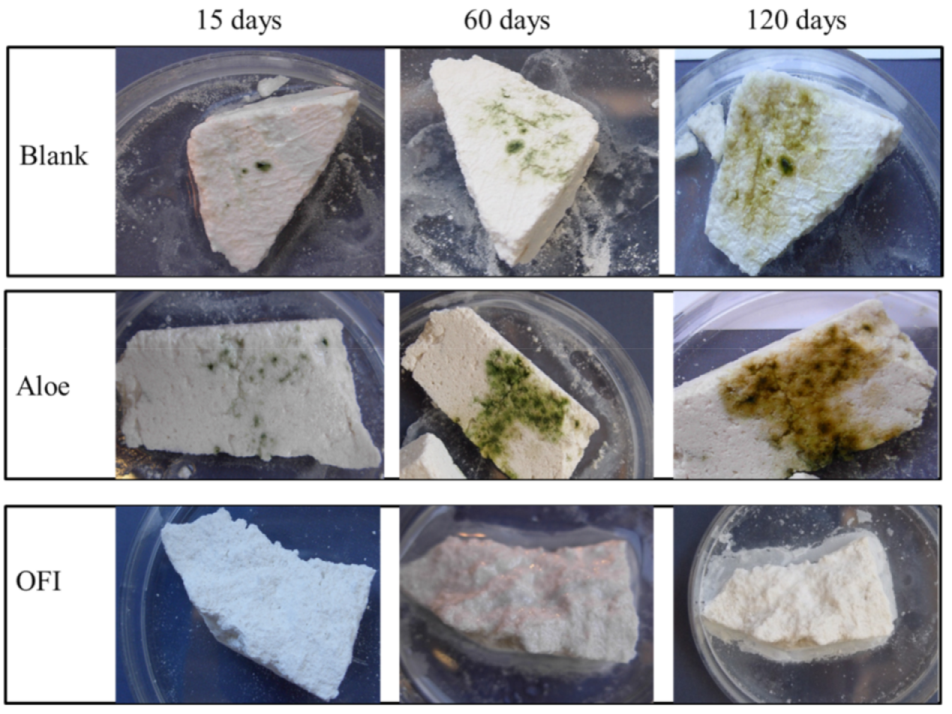
The bioreceptivity trials were performed with a variety of biodeteriogens (bacteria, fungi and mixed photosynthetic biofilm), but only a few of them were able to grow on the samples of bio-mortars, as shown in
Table 2. A little growth of fungi was detected on the Control (mortar without additives), but no evidence of biodeterioration was observed in the bio-mortar samples inoculated with bacteria and fungi.
The bio-mortars artificially contaminated with the photosynthetic biofilm showed a different bioreceptivity, as shown in
Table 2 and
Figure 6. A major biofilm development was observed in the control sample, as well as in the bio-mortars containing CC and Aloe. The result indicates the natural bioreceptivity of the mortar, which can be colonized by microorganisms if kept in suitable environmental conditions. The bio-mortars added with OFI, OE and SH were not susceptible to the microbiological attack nor of bacteria, or fungi or photosynthetic biofilm, during all the time of investigation (3 months), while the CC and Aloe mucilage addition increased the bioreceptivity of the mortar since the biofilm development started at 15 days of incubation and went on up to 120 days when started to die as indicated by the change of color for green to brown.

 ,
, 


