Article

Compositional and Morphological Comparison among Three Coeval Violins Made by Giuseppe Guarneri “del Gesù” in 1734

1
Arvedi Laboratory of Non-Invasive Diagnostics, CISRiC, University of Pavia, 26100 Cremona, Italy
2
Department of Chemistry, University of Torino, 10125 Torino, Italy
3
DEIB, Cremona Campus, Polytechnic of Milan, 20133 Milano, Italy
4
Department of Physics, Polytechnic of Milan, 20133 Milano, Italy
5
Department of Electrical, Computer and Biomedical Engineering, University of Pavia, 27100 Pavia, Italy
6
Department of Chemistry, University of Pavia, 27100 Pavia, Italy
7
Department of Musicology and Cultural Heritage, University of Pavia, 26100 Cremona, Italy
*
Author to whom correspondence should be addressed.
Submission received: 30 June 2021 / Revised: 16 July 2021 / Accepted: 19 July 2021 / Published: 23 July 2021

Abstract

:
In the present work, we had the opportunity to study the coating systems of three different coeval violins, namely “Spagnoletti”, “Stauffer”, and “Principe Doria”, made by Giuseppe Guarneri “del Gesù” in 1734. These three violins were non-invasively investigated by reflection Fourier transform infrared spectroscopy and X-ray fluorescence. These two techniques were combined for the first time with a 3D laser scanner. The analytical campaign enabled the characterization of the materials and their distribution within the stratigraphy, mainly composed of varnish and, when present, of a proteinaceous ground coat. Some restoration materials were also identified, suggesting the application of different maintenance treatments undertaken during their history. The preliminary information about morphological and geometrical differences between the three coeval violins were acquired through the 3D laser scanner in order to observe similarities and differences in the design features among the three violins.

1. Introduction

In the 17th and 18th centuries, during the so-called Golden Age period of historical violin making, hundreds of musical instruments were made in the workshops of the most important luthiers based in Cremona (Italy), namely Antonio Stradivari, Nicola Amati, Giuseppe Guarneri “del Gesù”, and other members of the great luthier families. Traditional manufacturing techniques, materials, and recipes employed in violin making were passed down from master artisans to apprentices, gradually changing through the decades. The decline of the art of violin making in Cremona, which occurred from the beginning of the 19th century, caused the loss of these traditions but left us numerous masterpieces of inimitable beauty. Nevertheless, starting from the 20th century, the high interest in the topic has led some violin makers and scientists to study the lost manufacturing techniques and the materials used by the old Cremonese Masters [1,2]. In recent years, some research teams have considered the investigation with a scientific approach [3,4,5] with the aims of characterizing the nature of materials involved in the Cremonese finishing treatments, i.e., the varnish [6,7], and understanding how they affect the acoustical properties of the musical instrument [8,9,10]. For this purpose, a large set of non-invasive and micro-invasive techniques—when possible—were widely used to investigate the physical and chemical features of the materials. The most common techniques were X-ray fluorescence (XRF) [11,12], Fourier transform infrared (FTIR) spectroscopy in different geometries [13,14,15,16,17], tomographic techniques, like computed axial tomography (CAT) or optical coherence tomography (OCT), imaging techniques, and chromatographic and different spectroscopic analyses [18,19,20].
Under this scenario, analytical investigations enabled researchers to obtain a clear picture of the expected materials and their multi-layered coating systems, consisting in multiple superimposed films of varnishes, generally applied on wood, which was previously treated with a sealer to prevent varnish penetration. The most common materials involved in the finishing processes are siccative oils, metal-based driers, natural resins, animal glues, inorganic fillers, organic colorants, inorganic pigments, and lake pigments [21,22].
In this research, we had the opportunity to study and compare three esteemed coeval violins, namely the “Spagnoletti”, the “Stauffer”, and the “Principe Doria” (Figure 1 and Figure S1) made by Giuseppe Guarneri “del Gesù” (Cremona, 1698—1744) in 1734. They were made in the period between 1730 and 1742, the most fruitful for the Master [23]. Instruments of this period are considered by contemporary violin makers unsurpassed specimens of established shape, with faultless workmanship [24].
A spectroscopic non-invasive approach through reflection FTIR and XRF was combined with a 3D model obtained with a 3D laser scanner. XRF and FTIR allowed us to identify the elemental and molecular composition of the materials, and the 3D scan provided decisive information regarding the constructive features such as arching, corners, and f-hole shapes. To our knowledge, an analytical strategy that combines a diagnostic campaign focused on material characterization with a structural assessment of three contemporary instruments with such a high historical significance has never been accomplished before.
The present study intends to (i) non-invasively characterize the chemical composition of the materials as well as understand their distribution within the coating systems, (ii) obtain preliminary valuable information about the design peculiarities adopted by Guarneri del Gesù in 1734 when he built the “Spagnoletti”, the “Stauffer”, and the “Principe Doria” violins, and, finally, (iii) observe similarities and differences in the material and design features among the three violins.

2. Experimental

Visible and ultraviolet-induced fluorescence (UVIFL) images were acquired following the protocol described in [25]. Visible illumination was provided by two soft-box LEDs (T = 5400K), while UV illumination was provided by two Wood’s lamp tubes TL-D 36 W BBL IPP low-pressure Hg tubes, 40 Watt, emission peak approximately 365nm (Philips, Amsterdam, Netherlands). Photos were taken with a Nikon D4 full-frame digital camera (Nikon Corporation, Tokyo, Japan) with a 50mm f/1.4 Nikkor objective (Nikon Corporation, Tokyo, Japan) (for visible: 1/10-1/15 exposure time, aperture f/8, ISO 100; for UV: 30s exposure time, aperture f/8, ISO 400). Violins were vertically placed (using the end-button hole) on a laboratory-built rotating platform, moved by a stepper motor (NEMA 17, Changzhou Jinsanshi Mechatronics Co. Ltd., Changzhou, China) controlled with an EasyDriver V44 driver board, in turn controlled by an Arduino Nano board. A simplified graphical interface was also developed to send instruction to the Arduino in a comfortable way [25].
For the preliminary analysis of the UVIFL images, we used UV Analyzer, an interactive tool developed in our lab that implements a series of image processing algorithms able to quickly highlight and label regions of interest on the surface of the instrument [25,26].
Reflection FTIR spectroscopy was performed using the Alpha portable spectrometer (Bruker Optics, Billerica, MA, USA) equipped with the R-Alpha external reflectance module that is composed of an optical layout of 23°/23°. The compact optical bench was composed of a SiC Globar source (Bruker Optics, Billerica, MA, USA), a RockSolid interferometer (with gold mirrors, Bruker Optics, Billerica, MA, USA), and an uncooled DLaTGS detector (Bruker Optics, Billerica, MA, USA). The reflection module allowed contactless and non-invasive analysis of measurement areas of about 5 mm in diameter at a working distance of 15 mm. Pseudo-absorbance spectra [log(1/R); R—reflectance] were collected between 7500 and 375 cm−1, at a resolution of 4 cm−1 and with an acquisition time of 1 min. The background was acquired using a gold flat mirror. For the study of derivative bands, reflection infrared spectra were transformed to absorbance spectra by applying the Kramers–Kronig transformation (KKT) included in the OPUS software package (7.2 version, 2013, Bruker, Billerica, MA, USA) and a portion of the mid-IR spectral range (3600–400 cm−1) is considered.
X-Ray fluorescence (XRF) spectroscopy was performed in single-point mode, using an ELIO portable X-ray fluorescence spectrometer by Bruker Optics Corporation (Billerica, MA, USA). It uses an X-ray source working with an Rh anode and an analytical spot diameter of 1.3 mm. Spectra were collected on 2048 channels at 40 kV, 80 μA, 480 s. For each element taken into consideration, the displayed value corresponds to the net area counts of the peak (Kα) normalized to time and to the average of net area counts of the coherent scattering Rh-Kα peak [27,28]. Data processing was carried out with ELIO software (1.6.0.57 version, 2020, Bruker, Billerica, MA, USA).
The 3D scan of the violins was performed using a RS3 Integrated Scanner (a linear laser scanner with a stated accuracy of 30 μm) mounted on a mobile arm with 7 degrees of freedom (Romer Absolute Arm 7-Axis "SI"), both produced by Hexagon Metrology (Cobham, United Kingdom). PolyWorks suite (12.1, 2012, InnovMetric, Quebec City, Canada) was used as the main scanning and editing software. Blender (2.79, 2017, Blender, Amsterdam, Netherlands) was then used for some final refinements of the meshes. Before the 3D scan, all the mobile components of the violin that could interfere with the acquisition (the strings, the tailpiece, the bridge, the chin rest, and the pegs) were removed. To maintain a stable position of the violins during the scanning process without damaging the varnishes, we used two Plexiglas supports provided with clamps covered with gum. During the acquisition, the laboratory was maintained in a stable climatic condition with a temperature of 20 °C and a humidity of 50% (the same as the showcases of the museum). All the steps of the acquisition and editing procedure were described in [29]. For the analysis of the scans, we used the 3D point cloud and post-processed them with Mathematica (11.3, 2019, Wolfram Research, Champaign, IL, USA). To smooth the data of the arching (discussed in Section 3.2), we subsampled the data using half the available points. As for the corners (discussed in Section 3.2), we computed the alpha shape with a resolution of 5 mm. The outline plots (discussed in Section 3.2) are not smoothed, as the variations are smaller than the line thickness at the scale we plotted. All the tools used are discussed in more detail in [30,31].

3. Results and Discussion

3.1. Non-Invasive Analytical Investigation by Reflection FTIR and XRF

The UVIFL images allowed highlighting the best-conserved (BC), worn-out (WO), and restored (RE) areas [11], according to their different UV fluorescence levels (Figure 1 and Figure S1). These images were also run through the UV Analyzer [25] (Figure 2 and Figure S2) to precisely select the analytical spots. Three areas were identified on the soundboard and five on the back plate of the “Spagnoletti”; three on the soundboard and five on the back plate of the “Stauffer”; five on the soundboard and four on the back plate of the “Principe Doria”. In order to obtain a complete stratigraphic picture of the different materials spreading from the external layers to the deeper ones, all the BC, WO, and RE areas were analyzed (“Spagnoletti”: 35 FTIR spots, 20 XRF spots; “Stauffer”: 35 FTIR spots, 16 XRF spots; “Principe Doria”: 31 FTIR spots, 15 XRF spots).
Reflection FTIR spectra collected on WO areas of the “Principe Doria” and the “Stauffer” show characteristic signals of a proteinaceous material, possibly casein or animal glue [18], which had been spread on the entire body of the violins before varnishing. As regards the “Spagnoletti”, instead, proteins were only revealed in circumscribed areas of the upper and lower corners, suggesting their correlation with subsequent restoration work. The KKT diagnostic bands of this compound fall at 3310 cm−1asNH), 1660 cm−1 (νC=O or amide I), and 1550–45 cm−1 (combination of νCN and δNH or amide II) [32,33,34,35] (Figure 3a).
Some inorganic compounds were also investigated in the pseudo-absorbance spectra of the wide FTIR data set. In particular, silicate-based materials were detected in WO areas of the “Spagnoletti”: bands at 3625 cm−1 (derivative, νOH inner hydroxyl groups), 1030 and 1005 cm−1 (inverted, νasSiO), 910 cm−1 (inverted, δOH inner hydroxyl groups), and 535 and 465 cm−1 (inverted, δAl–O–Si) are attributed to kaolin [14], whereas signals at 1080 cm−1 (inverted, νasSiO), 800 and 780 cm−1sSiO), and ca. 450 cm−1 (inverted, δSiO) derive from quartz [14] (Figure 3b). Concerning the “Principe Doria”, features of quartz (not shown here) were identified together with those of sulfate compounds, likely gypsum, with bands being positioned at 3410 cm−1 (derivative, νOH), 1140 and 1115 cm−1 (inverted, νasSO4), and 670 and 600 cm−1 (inverted δasSO4) [14] (Figure 3b). As already highlighted in [14,18], however, we cannot assign the sulfates to a precise layer of the “Principe Doria” stratigraphy due to their heterogeneous results on the plates. Conversely, FTIR spectra collected on the “Stauffer” did not enable a reliable identification of inorganic compounds, and only a tentative attribution to silicate-based minerals was possible (Figure 3b). XRF counts of low-weight elements Si and S, even if underestimated (atomic number Z < K), increased in the WO areas. These results can further confirm the presence of silicate- and sulfate-based phases dispersed in the ground coat or filling the wood pores.
Within each violin, the BC areas of the soundboard and back plate exhibit similar FTIR spectral profiles. In detail, the spectra collected on the “Principe Doria” display the KKT characteristic bands of an oil–resinous varnish at 2955 cm−1 (shoulder, νasCH3), 2925 cm−1asCH2), 2855 cm−1sCH2), 1730–10 cm−1 (νC=O), 1460 cm−1 (δCH2), 1380 cm−1 (δCH3), 1250–40 and 1170–65 cm−1, and 1100 cm−1 (νC–O) [14] (Figure 3c), possibly laid on the ground. If these signals can be definitely attributed to a mixture of oil(s) and vegetal resin(s) in the “Principe Doria”, the identification is more uncertain in the “Spagnoletti” and the “Stauffer” due to the strong spectral similarities between oil-based varnish and shellac resin [32]. Interestingly, XRF analysis performed on the same BC areas highlighted an increase in counts of Fe and Mn and a possibly a correlation between the two elements in the “Spagnoletti” and the “Stauffer”, suggesting the presence of iron-based pigments like umber earth [11] in the varnish. Conversely, these signals underwent only slight and non-significant variations between WO and BC areas of the “Principe Doria”, probably due to invasive restoration processes, which removed the upper part of the external pigmented varnish [18]. In addition, higher concentrations of Pb were detected in some spots on the soundboard and back plate of the three violins, suggesting the use of a lead-based siccative dispersed in the oil binder [12].
As regards the restored areas, reflection FTIR highlighted KKT signals of shellac (not shown here), mainly in the RE areas of the “Spagnoletti” and the “Stauffer”, on both sides of the body. The characteristic bands of this animal resin fall at 2925 (νasCH2), 2855 cm−1sCH2), 1730 cm−1 (νC=O esters), 1710 cm−1 (νC=O acids), 1460 cm−1 (δCH2), 1375 cm−1 (δCH3), 1255 cm−1 (νC–O esters), 1155 cm−1 (νC–O acids), 940 and 925 cm−1 (δH2C=C), and 720 cm−1asCH2) [15,32,36]. Additionally, a metal soap identifiable as calcium stearate was found in some RE and WO areas on both the soundboard and back plate of the “Spagnoletti” violin, and residues of benzoin resin—commonly used to polish a varnished surface—were revealed in the same areas of the “Stauffer”. The detected marker bands of the calcium soap, acting as a lubricant, are located in the pseudo-absorbance spectra at 2955 cm−1 (derivative, νasCH3), 2920 cm−1 (derivative, νasCH2), 2850 cm−1 (derivative, νsCH2), 1575 cm−1 (inverted, νasCOO–), 1540 cm−1 (inverted, νsCOO–), and 720 cm−1 (derivative, δCH2) [37,38] (Figure 3d). As reported in [37,38], the strong doublet at 1575 and 1540 cm−1 corresponds to the asymmetric carboxylate stretching vibrations of the calcium salts. The KKT diagnostic bands of benzoin resin were identified at 2930 and ca. 2865 cm−1 (νCH), 1710 cm−1 (νC=O conjugate ketone), 1610, 1600, and 1515 cm−1 (aromatic skeletal), 1460 and 1450 cm−1 (aromatic skeletal + δCH), ca. 1380 cm−1 (δCH), 1265, 1165, 1120, and 1030 cm−1 (νC–O), and 835, 770, and 710 cm−1 (undefined) [18] (Figure 3d). As regards the “Principe Doria”, few FTIR analytical areas highlighted markers of benzoin resins and shellac. Interestingly, an aliphatic hydrocarbon compound (e.g., paraffin) was further singled out (Figure 3d). In the absence of some markers of the varnish (mainly νC=O and νC–O), or when their intensity is low, the very sharp and intense CH2 peaks can be attributed to long aliphatic chains [39,40]. Furthermore, evidence of sulfate-based phases, such as gypsum, was identified in heterogeneously diffused areas of the instrument. The presence of the latter inorganic compound was difficult to locate in a precise layer since it occurs in some areas at different varnish thicknesses. XRF signals of Br detected on the three violins can be attributed to bromomethane, commonly used in the recent past as a treatment against woodworms [41]. All these different materials highlighted on the surfaces of the three violins, commonly used for maintenance practice and as surface polish in restoration, are significant signs of different maintenance treatments undertaken during their long history.
Elemental composition of purflings was studied by XRF and the results obtained are reported in Figure 4. Measurements highlight the presence in all the instruments of significant amounts of Fe, Cu, and Zn, typically associated with iron-based dyes/stain [42]. Other elements, namely Si, S, K, Ca, Ti, Mn, Pb, and Ba, were also identified in the same analytical spots but they are not considered in the discussion since they are not related to the dyeing process but probably to the wood and/or other compounds, such as fillers, used in the finishing treatments. Depending on the composition of the raw materials and on the procedures used to mix them, the ratios between the elemental abundances can vary. In any event, purfling batches made in the same period, and thus stained with the same methods and substances, should generally show a comparable amount of marker elements (Fe, Cu, and Zn).
The results obtained on the soundboard (SPA_Pur_SB in Figure 4) and back plate (SPA_Pur_BP1, BP2 in Figure 4) of the “Spagnoletti” and the “Stauffer” (STA_Pur_SB1, SB2, BP in Figure 4) purflings highlight, respectively, a similar amount of the selected marker elements (Figure 4). This could be attributable to a similar wood-dyeing procedure and, thus, probably to the same batch. On the other hand, if we compare the results of the two violins, slight variations of net area counts can be observed in the Fe results, as well as in the results obtained on the “Principe Doria”. In fact, purflings of the “Principe Doria” revealed higher counts of Fe and lower counts of Cu, possibly due to a different iron-based dyeing process preparation procedure.

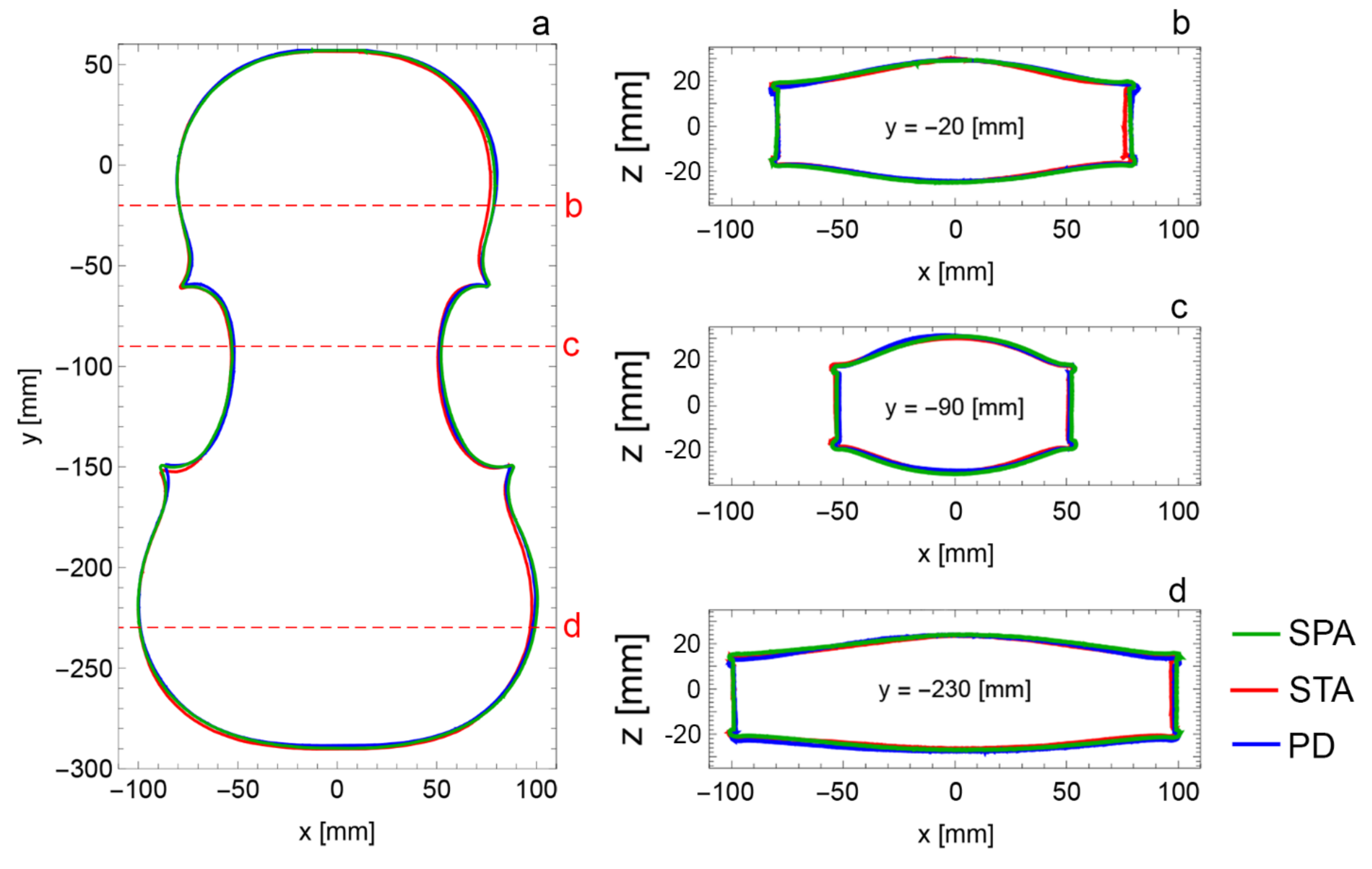
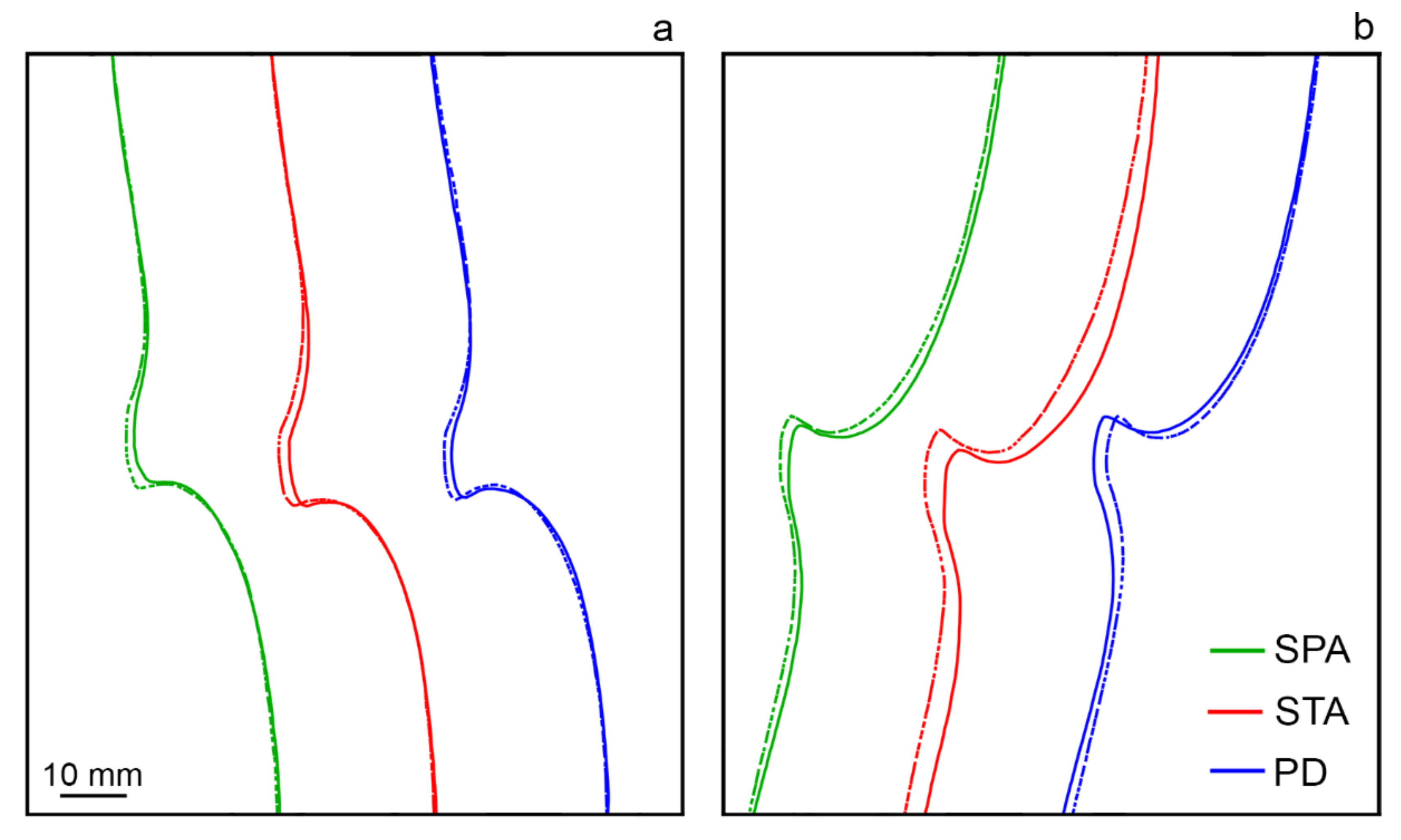
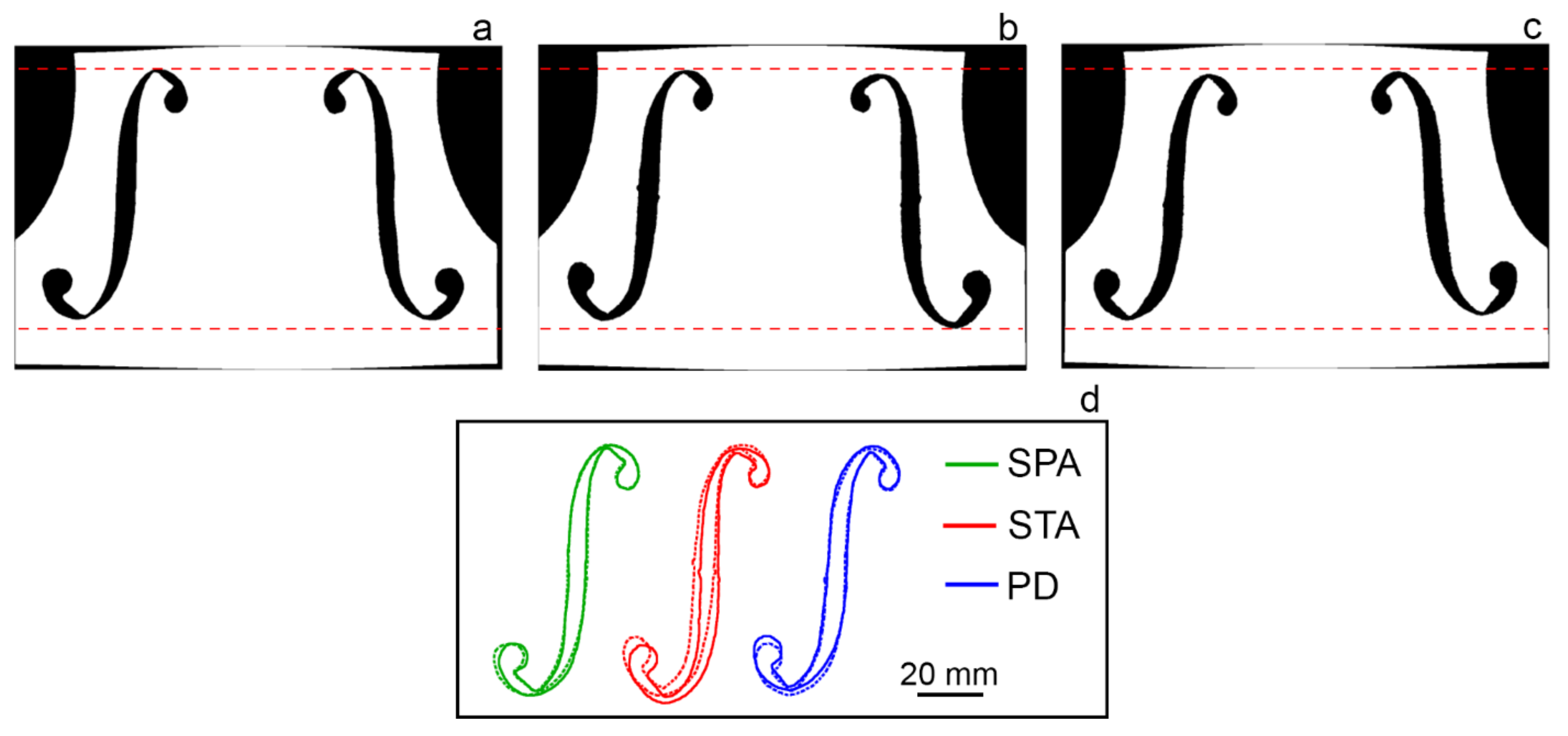
3.2. Morphological Investigation through 3D Laser Scanner

The outlines of the three violins (Figure 5a) were computed using the scan of the rib sections, as they provide a more accurate representation since they see less wear compared to the top or the back. To center and align the violins, we computed the moment of inertia of the point cloud, centered the violin with respect to the center of mass of the 3D scan, and rotated them along their principal axis. The computation was done with the whole violin, so the origin of the y-axis was in the upper part of the violin body rather than in the center of the violin. The measures of the three violins are shown in Table S1 in the Supplementary Materials.
The outlines of the violins studied here are quite similar, yet the lower left corner presents the largest variation between them. The measures collected corresponding to the cross sections shown in Figure 5b–d revealed some slight variations between the violins in the upper and lower right parts (Figure 5b,d; Figure S3f,h). As reported in Table S1, the “Principe Doria” revealed the maximum width in both measurements, while the “Stauffer” and the “Spagnoletti” are smaller. In particular, the “Stauffer” shows a significant variation both in the upper and lower parts of the soundboard, while the “Spagnoletti” only in the upper part. The width at the center (Figure S3g; Table S1g) is similar for all three. A different picture appears when looking at the back plate (Figure 5; Figure S3f,g,h). Here, the “Spagnoletti” shows the maximum values in the three measures while the “Principe Doria” and the “Stauffer” revealed slightly smaller values. As for the total lengths of the back plates of the “Spagnoletti”, the “Stauffer”, and the “Principe Doria”, they are, respectively, 349.5 mm, 349.37 mm, and 349.95 mm, and those of the soundboard are 348.82 mm, 349.38 mm, and 348.81 mm. Despite these slight differences, the creation of the 3D models reveals a good match between the shape of the bodies, suggesting the use of the same mold and templates for all the three violins.
Figure 5b–d show cross sections of the violins at three different y locations: −20 mm, −90 mm, and −230 mm. The violins have roughly the same arching profile, except for the central part of the “Principe Doria”, which exhibits a higher curvature on the left side. This is where the bass bar is located, and it could be due to a poorly fitted bass bar at some point in the history of the violin or to a conscious choice by the maker. The ribs present some places where they are not perfectly vertical, which is most visible at y = −20 mm (Figure 5b, green and blue lines) on both sides of the “Spagnoletti” and the “Principe Doria” and at y = −230 mm on the left side of the “Principe Doria” (Figure 5d, blue line). As regards the rib height, the three violins exhibit a consistent value of around 30 mm (Table S1i,j,j1,k), with that of the “Spagnoletti” seeming to be slightly higher than others. The small variations in the relative height are likely due to twisting of the wood and to the invasive restoration interventions that the instruments have undergone over the decades.
The corners of the top plates show a distinct wear pattern on the right side of the instrument, both in the upper and lower ones (Figure 6, solid lines). This wear pattern is most visible in the right side of the “Spagnoletti” (green solid line), where the bottom part of the upper corner has been worn till completely losing its shape. The lower corners look more dissimilar, in particular in the case of the “Stauffer”, where the variation in the outline is also more noticeable. This aspect could be related to the high concert activity that the violins have undergone, whose effects of wear mainly fall on that side of the violin.
The f-holes are the most different features among the three violins (Figure 7) in terms of position and dimensions. As reported in Table S1, the f-holes of the “Spagnoletti” and the “Stauffer” show similar a length of around 77 mm, whereas the ones of the “Principe Doria” are noticeably smaller in the total length (right hole: 74.59 mm, left hole: 75.41 mm). In terms of their width at the center (o, p, q in Table S1), they present similar values, except for the lower lobe distance in the “Principe Doria”, where a larger variation between left and right holes is present.
In Figure 7d, the f-holes are mirrored and compared for each violin. The f-holes of the “Stauffer” and the “Principe Doria” are less symmetric, with either the left or the right one slightly higher than the other and, in the case of the “Stauffer”, slightly out of alignment (left hole higher, right hole lower).

4. Conclusions

The combination of non-invasive spectroscopic approaches, like reflection FTIR and XRF, with the 3D laser scanner, allowed us to investigate both chemical and morphological features of three historical violins. In fact, the three masterpieces made by Giuseppe Guarneri del Gesù in 1734, namely the “Spagnoletti”, the “Stauffer”, and the “Principe Doria”, were deeply characterized in terms of the finishing treatments, such as varnishes, ground coats, inorganic fillers, dispersed pigments, and restoration materials, as well as in the morphological and geometrical features, such as the shape of the body, the arching, the corners, and the f-holes. These are decisive peculiarities that help to better understand the artisanship of Guarneri “del Gesù”.
Integrating and comparing the results, it is possible to observe how the three coeval violins underwent numerous restorations aimed to maintain the aesthetics of the finishing treatment. These repeated interventions led to the almost total removal or coverage of the original materials on most of the investigated areas. On the contrary, the geometry of the body remained almost unchanged, except for the central part of the “Principe Doria”, which exhibits a higher curvature where the bass bar is located.
Lastly, the innovative combination of non-invasive spectroscopic techniques and the 3D laser scanner has proved to be a powerful tool to deeply characterize both the compositional and morphological features, providing detailed information about the finishing layers and past restoration actions, besides disclosing specific aspect of the shapes, morphology, and geometries of the violins. Moreover, the unique chance to directly compare the results of three instruments by the same maker and from the same year of production allowed us to make a step forward in the understanding of the Guarneri manufacturing, which seems to share common features in different instruments. Indeed, the outcome regarding the Master’s manufacturing also plays a key role for modern violin makers, who often attempt to reproduce the excellent features of the Cremonese historical lutherie.

Supplementary Materials

The following are available online at https://0-www-mdpi-com.brum.beds.ac.uk/article/10.3390/coatings11080884/s1, Figure S1: Visible (left) and UV (right) images of the backplate of the Spagnoletti (a), Stauffer (b), and Principe Doria (c) made by Giuseppe Guarneri del Gesù in 1734. Figure S2: Images of the soundboard of the Spagnoletti (a), Stauffer (b), and Principe Doria (c) violins obtained through UV Analyzer. Different colors correspond to best-conserved (orange), worn-out (light blue), and restoration (red, blue, and green) areas, according to their different UV fluorescence levels. Figure S3: Legend of the measurement positions acquired on the violins. Table S1: Measurements obtained on the 3D models of the three violins. Measurement letters in the first column are related to the measurement positions shown in Figure S3.

Author Contributions

Conceptualization, G.F. and M.M.; methodology, G.F. and M.M.; software, G.F., C.I., T.R., M.A., P.D., and S.G.; validation, G.F., C.I., T.R., M.A., P.D., S.G., F.A., M.L., and M.M.; formal analysis, G.F., C.I., T.R., M.A., and P.D.; investigation, G.F., C.I., and T.R.; resources, M.M., F.A., and M.L.; data curation, G.F., C.I., and T.R.; writing—original draft preparation, G.F., C.I., and S.G.; writing—review and editing, G.F., C.I., T.R., M.A., P.D., S.G., F.A., M.L., and M.M.; visualization, G.F. and C.I.; supervision, M.M. and F.A. All authors have read and agreed to the published version of the manuscript.

Funding

This research received no external funding

Institutional Review Board Statement

Not applicable.

Informed Consent Statement

Not applicable.

Data Availability Statement

Data are contained within the article or Supplementary Materials.

Acknowledgments

We would like to thank the Fondazione Museo del Violino Antonio Stradivari, Cremona, for its collaboration and availability, which contributed to improving this research study. We are grateful to the “Friends of Stradivari” partners and to the owners of the violins for their important collaboration in this study.

Conflicts of Interest

The authors declare no conflict of interest.

References

  1. Sacconi, S.F. I segreti di Stradivari. Con il catalogo dei cimeli stradivariani del Museo civico “Ala Ponzone” di Cremona; Libreria del Convegno: Cremona, Italy, 1979. [Google Scholar]
  2. Michelman, J. Violin Varnish: A Plausible Re-Creation of the Varnish Used by the Italian Violin Makers Between the Years 1550 and 1750, A.D., 1st ed.; Joseph Michelman: Cincinnati, OH, USA, 1946; pp. 58–154. [Google Scholar]
  3. Tai, B.H. Stradivari’s Varnish: A review of scientific findings—Part I. J. Violin Soc. Am. 2007, 21, 119–144. [Google Scholar]
  4. Tai, B.H. Stradivari’s Varnish: A review of scientific findings—Part II. J. Violin Soc. Am. 2009, 22, 1–31. [Google Scholar]
  5. Von Bohlen, A.; Meyer, F. Microanalysis of old violin varnishes by total-reflection X-ray fluorescence. Spectrochim. Acta Part B At. Spectrosc. 1997, 52, 1053–1056. [Google Scholar] [CrossRef]
  6. Echard, J.-P.; Bertrand, L.; Von Bohlen, A.; Le Hô, A.-S.; Paris, C.; Bellot-Gurlet, L.; Soulier, B.; Lattuati-Derieux, A.; Thao, S.; Robinet, L.; et al. The Nature of the Extraordinary Finish of Stradivari’s Instruments. Angew. Chem. Int. Ed. 2009, 49, 197–201. [Google Scholar] [CrossRef]
  7. Nagyvary, J.; Ehrman, J.M. The composite nature of the antique Italian varnish. Naturwissenschaften 1988, 75, 513–515. [Google Scholar] [CrossRef]
  8. Lämmlein, S.; Künniger, T.; Rüggeberg, M.; Schwarze, F.W.; Mannes, D.; Burgert, I. Frequency dependent mechanical properties of violin varnishes and their impact on vibro-mechanical tonewood properties. Results Mater. 2021, 9, 100137. [Google Scholar] [CrossRef]
  9. Lämmlein, S.L.; Mannes, D.; Van Damme, B.; Burgert, I.; Schwarze, F.W. Influence of varnishing on the vi-bro-mechanical properties of wood used for violins. J. Mater. Sci. 2019, 54, 8063–8095. [Google Scholar] [CrossRef]
  10. Setragno, F.; Zanoni, M.; Antonacci, F.; Sarti, A.; Malagodi, M.; Rovetta, T.; Invernizzi, C. Feature-Based Analysis of the Impact of Ground Coat and Varnish on Violin Tone Qualities. Acta Acust. United Acust. 2017, 103, 80–93. [Google Scholar] [CrossRef]
  11. Rovetta, T.; Invernizzi, C.; Licchelli, M.; Cacciatori, F.; Malagodi, M. The elemental composition of Stradivari’s musical instruments: New results through non-invasive EDXRF analysis. X-Ray Spectrom. 2018, 47, 159–170. [Google Scholar] [CrossRef]
  12. Echard, J.-P. In situ multi-element analyses by energy-dispersive X-ray fluorescence on varnishes of historical violins. Spectrochim. Acta Part B At. Spectrosc. 2004, 59, 1663–1667. [Google Scholar] [CrossRef]
  13. Fiocco, G.; Invernizzi, C.; Grassi, S.; Davit, P.; Albano, M.; Rovetta, T.; Stani, C.; Vaccari, L.; Malagodi, M.; Licchelli, M.; et al. Reflection FTIR spectroscopy for the study of historical bowed string instruments: Invasive and non-invasive approaches. Spectrochim. Acta Part A Mol. Biomol. Spectrosc. 2021, 245, 118926. [Google Scholar] [CrossRef]
  14. Invernizzi, C.; Fichera, G.V.; Licchelli, M.; Malagodi, M. A non-invasive stratigraphic study by reflection FT-IR spectroscopy and UV-induced fluorescence technique: The case of historical violins. Microchem. J. 2018, 138, 273–281. [Google Scholar] [CrossRef]
  15. Invernizzi, C.; Daveri, A.; Vagnini, M.; Malagodi, M. Non-invasive identification of organic materials in historical stringed musical instruments by reflection infrared spectroscopy: A methodological approach. Anal. Bioanal. Chem. 2017, 409, 3281–3288. [Google Scholar] [CrossRef]
  16. Caruso, F.; Martino, D.F.C.; Saverwyns, S.; Van Bos, M.; Burgio, L.; Di Stefano, C.; Peschke, G.; Caponetti, E. Micro-analytical identification of the components of varnishes from South Italian historical musical instruments by PLM, ESEM–EDX, microFTIR, GC–MS, and Py–GC–MS. Microchem. J. 2014, 116, 31–40. [Google Scholar] [CrossRef]
  17. Daher, C.; Drieu, L.; Bellot-Gurlet, L.; Percot, A.; Paris, C.; Le Hô, A.-S. Combined approach of FT-Raman, SERS and IR micro-ATR spectroscopies to enlighten ancient technologies of painted and varnished works of art. J. Raman Spectrosc. 2014, 45, 1207–1214. [Google Scholar] [CrossRef] [Green Version]
  18. Invernizzi, C.; Fiocco, G.; Iwanicka, M.; Kowalska, M.; Targowski, P.; Blümich, B.; Rehorn, C.; Gabrielli, V.; Bersani, D.; Licchelli, M.; et al. Non-invasive mobile technology to study the stratigraphy of ancient Cremonese violins: OCT, NMR-MOUSE, XRF and reflection FT-IR spectroscopy. Microchem. J. 2020, 155, 104754. [Google Scholar] [CrossRef]
  19. Blümich, B.; Baias, M.; Rehorn, C.; Gabrielli, V.; Jaschtschuk, D.; Harrison, C.; Invernizzi, C.; Malagodi, M. Comparison of historical violins by non-destructive MRI depth profiling. Microchem. J. 2020, 158, 105219. [Google Scholar] [CrossRef]
  20. Fiocco, G.; Rovetta, T.; Malagodi, M.; Licchelli, M.; Gulmini, M.; Lanzafame, G.; Zanini, F.; Giudice, A.L.; Re, A. Synchrotron radiation micro-computed tomography for the investigation of finishing treatments in historical bowed string instruments: Issues and perspectives. Eur. Phys. J. Plus 2018, 133, 525. [Google Scholar] [CrossRef]
  21. Fiocco, G.; Rovetta, T.; Gulmini, M.; Piccirillo, A.; Licchelli, M.; Malagodi, M. Spectroscopic Analysis to Characterize Finishing Treatments of Ancient Bowed String Instruments. Appl. Spectrosc. 2017, 71, 2477–2487. [Google Scholar] [CrossRef]
  22. Bucur, V. Handbook of Materials for String Musical Instruments, 1st ed.; Springer International Publishing: Basel, Switzerland, 2016; Chapter 9: The Varnish; pp. 373–453. [Google Scholar]
  23. Chiesa, C.; Hargrave, R.G.; Pollens, S. Giuseppe Guarneri del Gesù; Peter Biddulph: London, UK, 1998. [Google Scholar]
  24. Jalovec, K. Beautiful Italian Violins; P. Hamlyn: London, UK, 1963. [Google Scholar]
  25. Dondi, P.; Lombardi, L.; Rocca, I.; Malagodi, M.; Licchelli, M. Multimodal workflow for the creation of interactive presentations of 360 spin images of historical violins. Multimedia Tools Appl. 2018, 77, 28309–28332. [Google Scholar] [CrossRef]
  26. Dondi, P.; Lombardi, L.; Invernizzi, C.; Rovetta, T.; Malagodi, M.; Licchelli, M. Automatic Analysis of UV-Induced Fluorescence Imagery of Historical Violins. J. Comput. Cult. Heritage 2017, 10, 1–13. [Google Scholar] [CrossRef]
  27. Invernizzi, C.; Fiocco, G.; Iwanicka, M.; Targowski, P.; Piccirillo, A.; Vagnini, M.; Licchelli, M.; Malagodi, M.; Bersani, D. Surface and Interface Treatments on Wooden Artefacts: Potentialities and Limits of a Non-Invasive Multi-Technique Study. Coatings 2020, 11, 29. [Google Scholar] [CrossRef]
  28. Poggialini, F.; Fiocco, G.; Campanella, B.; Legnaioli, S.; Palleschi, V.; Iwanicka, M.; Targowski, P.; Sylwestrzak, M.; Invernizzi, C.; Rovetta, T.; et al. Stratigraphic analysis of historical wooden samples from ancient bowed string instruments by laser induced breakdown spectroscopy. J. Cult. Heritage 2020, 44, 275–284. [Google Scholar] [CrossRef]
  29. Dondi, P.; Lombardi, L.; Malagodi, M.; Licchelli, M. 3D modelling and measurements of historical violins. Acta IMEKO 2017, 6, 29–34. [Google Scholar] [CrossRef]
  30. Gonzalez, S.; Salvi, D.; Baeza, D.; Antonacci, F.; Sarti, A. A data-driven approach to violin making. Sci. Rep. 2021, 11, 1–9. [Google Scholar] [CrossRef] [PubMed]
  31. Gonzalez, S.; Salvi, D.; Antonacci, F.; Sarti, A. Eigenfrequency optimisation of free violin plates. J. Acoust. Soc. Am. 2021, 149, 1400–1410. [Google Scholar] [CrossRef]
  32. Fiocco, G.; Rovetta, T.; Invernizzi, C.; Albano, M.; Malagodi, M.; Licchelli, R.A.; Giudice, A.; Gabriele, N.; Lanzafame, G.; Zanini, F.; et al. A Micro-tomographic insight into the coating systems of historical bowed string instruments. Coatings 2019, 9, 81. [Google Scholar] [CrossRef] [Green Version]
  33. Invernizzi, C.; Rovetta, T.; Licchelli, M.; Malagodi, M. Mid and Near-Infrared Reflection Spectral Database of Natural Organic Materials in the Cultural Heritage Field. Int. J. Anal. Chem. 2018, 2018, 1–16. [Google Scholar] [CrossRef]
  34. Pellegrini, D.; Duce, C.; Bonaduce, I.; Biagi, S.; Ghezzi, L.; Colombini, M.P.; Tinè, M.R.; Bramanti, E. Fourier transform infrared spectroscopic study of rabbit glue/inorganic pigments mixtures in fresh and aged reference paint reconstruc-tions. Microchem. J. 2016, 124, 31–35. [Google Scholar] [CrossRef]
  35. Bertrand, L.; Robinet, L.; Cohen, S.X.; Sandt, C.; Le Hô, A.-S.; Soulier, B.; Lattuati-Derieux, A.; Echard, J.-P. Identification of the finishing technique of an early eighteenth century musical instrument using FTIR spectromicroscopy. Anal. Bioanal. Chem. 2011, 399, 3025–3032. [Google Scholar] [CrossRef] [PubMed]
  36. Stuart, B. Infrared Spectroscopy: Fundamentals and Applications; John Wiley & Sons: West Sussex, UK, 2004; ISBN 0470854278. [Google Scholar]
  37. Invernizzi, C.; Daveri, A.; Rovetta, T.; Vagnini, M.; Licchelli, M.; Cacciatori, F.; Malagodi, M. A multi-analytical non-invasive approach to violin materials: The case of Antonio Stradivari “Hellier” (1679). Microchem. J. 2016, 124, 743–750. [Google Scholar] [CrossRef]
  38. Rosi, F.; Cartechini, L.; Monico, L.; Gabrieli, F.; Vagnini, M.; Buti, D.; Doherty, B.; Anselmi, A.; Brunetti, B.G.; Miliani, C. Tracking metal oxalates and carboxylates on painting surfaces by non-invasive reflection mid-FTIR spectroscopy. In Metal Soaps in Art; Casadio, F., Keune, K., Noble, P., van Loon, A., Hendriks, E., Eds.; Springer: Cham, Switzerland, 2019; pp. 173–193. [Google Scholar]
  39. Helwig, K.; Forest, É.; Turcotte, A.; Baker, W.; Binnie, N.E.; Moffatt, E.; Poulin, J. The formation of calcium fatty acid salts in oil paint: Two case studies. In Metal Soaps in Art; Casadio, F., Keune, K., Noble, P., van Loon, A., Hendriks, E., Eds.; Springer: Cham, Switzerland, 2019; pp. 173–193. [Google Scholar]
  40. Rosi, F.; Grazia, C.; Fontana, R.; Gabrieli, F.; Buemi, L.P.; Pampaloni, E.; Romani, A.; Stringari, C.; Miliani, C. Disclosing Jackson Pollock’s palette in Alchemy (1947) by non-invasive spectroscopies. Heritage Sci. 2016, 4, 18. [Google Scholar] [CrossRef] [Green Version]
  41. Saiz-Jimenez, C. Molecular Biology and Cultural Heritage; Routledge: London, UK, 2003. [Google Scholar]
  42. Canevari, C.; Delorenzi, M.; Invernizzi, C.; Licchelli, M.; Malagodi, M.; Rovetta, T.; Weththimuni, M. Chemical characterization of wood samples colored with iron inks: Insights into the ancient techniques of wood coloring. Wood Sci. Technol. 2016, 50, 1057–1070. [Google Scholar] [CrossRef]
Figure 1. Visible (left) and UV (right) images of the soundboard of (a) the “Spagnoletti”, (b) the “Stauffer”, and (c) the “Principe Doria” made by Giuseppe Guarneri “del Gesù” in 1734. Visible and UV images of the back plates are shown in Figure S1 in the Supplementary Materials.
Coatings 11 00884 g001
Figure 2. Images of the soundboard of (a) the “Spagnoletti”, (b) the “Stauffer”, and (c) the “Principe Doria” violins obtained through UV Analyzer. Different colors correspond to best-conserved (orange), worn-out (light blue), and restored (red, blue, and green) areas, according to their different UV-induced fluorescence colors. Areas identified on the back plates are shown in Figure S2 in the Supplementary Materials.
Coatings 11 00884 g002
Figure 3. Reflection FTIR spectra in pseudo-absorbance (gray line) and after KKT (black line) collected in areas of the three violins (SPA—“Spagnoletti”, STA—“Stauffer”, PD—“Principe Doria”) containing: (a) proteins, (b) inorganic compounds, (c) varnishes, and (d) restoration substances. Marker bands of proteins (∗), silicates (▲), sulfates (⬤), varnishes (▼), metal soap (◼), benzoin resin (▽), and aliphatic hydrocarbon () are highlighted.
Coatings 11 00884 g003
Figure 4. Estimates of Fe, Cu, and Zn detected by XRF analysis, corresponding to the purflings of the soundboard (SB) and back plate (BP) of the “Spagnoletti” (SPA), the “Stauffer” (STA), and the “Principe Doria” (PD). The y-axis values correspond to the net area counts of the element peaks (Kα) normalized to time and net area counts of the Rh peak (Kα).
Coatings 11 00884 g004
Figure 5. Outlines (a) and cross sections at different y locations (bd) of the “Spagnoletti” (SPA, green line), the “Stauffer” (STA, red line), and the “Principe Doria” (PD, blue line).
Coatings 11 00884 g005
Figure 6. Outlines of the upper (a) and lower (b) corners of the “Spagnoletti” (SPA), the “Stauffer” (STA), and the “Principe Doria” (PD). In both images, the dotted lines are related to the left corners while the solid lines to the right ones.
Coatings 11 00884 g006
Figure 7. F-hole shapes of the “Spagnoletti” (a), the “Stauffer” (b), and the “Principe Doria” (c) and their position on the soundboard between the waists. Red dotted lines mark the maximum upper and lower position of the f-holes. Outlines of the f-holes (d) of the “Spagnoletti” (SPA), the “Stauffer” (STA), and the “Principe Doria” (PD).
Coatings 11 00884 g007
Publisher’s Note: MDPI stays neutral with regard to jurisdictional claims in published maps and institutional affiliations.

