
Fluid Films as Models for Understanding the Impact of Inhaled Particles in Lung Surfactant Layers


1
Departamento de Química Física, Facultad de Ciencias Químicas, Universidad Complutense de Madrid, Ciudad Universitaria s/n, 28040 Madrid, Spain
2
Instituto Pluridisciplinar, Universidad Complutense de Madrid, Paseo Juan XXIII 1, 28040 Madrid, Spain
Coatings 2022, 12(2), 277; https://0-doi-org.brum.beds.ac.uk/10.3390/coatings12020277
Submission received: 27 December 2021
/
Revised: 23 January 2022
/
Accepted: 16 February 2022
/
Published: 19 February 2022
(This article belongs to the Special Issue Current Perspective on the Study of Liquid-Fluid Interfaces: From Fundamentals to Innovative Applications)
Abstract
:Pollution is currently a public health problem associated with different cardiovascular and respiratory diseases. These are commonly originated as a result of the pollutant transport to the alveolar cavity after their inhalation. Once pollutants enter the alveolar cavity, they are deposited on the lung surfactant (LS) film, altering their mechanical performance which increases the respiratory work and can induce a premature alveolar collapse. Furthermore, the interactions of pollutants with LS can induce the formation of an LS corona decorating the pollutant surface, favoring their penetration into the bloodstream and distribution along different organs. Therefore, it is necessary to understand the most fundamental aspects of the interaction of particulate pollutants with LS to mitigate their effects, and design therapeutic strategies. However, the use of animal models is often invasive, and requires a careful examination of different bioethics aspects. This makes it necessary to design in vitro models mimicking some physico-chemical aspects with relevance for LS performance, which can be done by exploiting the tools provided by the science and technology of interfaces to shed light on the most fundamental physico-chemical bases governing the interaction between LS and particulate matter. This review provides an updated perspective of the use of fluid films of LS models for shedding light on the potential impact of particulate matter in the performance of LS film. It should be noted that even though the used model systems cannot account for some physiological aspects, it is expected that the information contained in this review can contribute on the understanding of the potential toxicological effects of air pollution.
Keywords:
dynamics; fluid interfaces; inhalation; lung surfactant; nanoparticles; pollutants; rheology1. Introduction
In recent years, the injection of pollutants into the atmosphere as a result of the combustion processes of fossil fuels in industries, power plants, heating systems and vehicles, and the residues of the production processes of nanotechnological industries has undergone a spectacular growth. This opens many questions related to the potential harmful effects of such compounds for human health [1] as is evidenced from the recent statistics of the World Health Organization (WHO) [2], which include long-term exposure to air pollutants as one of the most important sources of cardiovascular diseases, and mortality (around one third of the deaths by strokes, lung cancer, or cardiac diseases are associated with air pollution) [3,4]. Therefore, air pollution should currently be included as a very important public health issue in modern s·ociety [5,6], making it necessary to evaluate the potential impact of pollutants in normal physiological functions, paying special attention to the role of inhaled pollutants [7,8,9].
Pollutant inhalation is associated with the emergence of pathogenic states and respiratory diseases, e.g., acute respiratory distress syndrome (ARDS), asthma, chronic obstructive pulmonary diseases, fibrosis, or lung cancer [10,11]. Therefore, it is critical to perform a careful examination of the effect of particles in respiratory function [12].
Among pollutants, fine particulate matter (PM2.5, particles with diameter ≤2.5 μm) is counted as one of the most important sources of respiratory diseases [13,14,15]. This type of particulate matter can be inhaled and transported along the respiratory tract to the alveoli, where they interact with the lung surfactant (LS) film [16,17], modifying the LS chemical composition and lateral organization upon incorporation, which in turn alter LS functionality. Furthermore, the interaction between LS and particles can induce different pathways which favor particle translocation towards other tissues and organs [18,19]. This may be the origin of several adverse health effects [20,21].
LS is a mixture containing mainly lipids and proteins which overlays the inner wall of alveoli in mammals (i.e., the alveolar lining) [7,22], contributing to two very important physiological aspects: (i) regulation of the mechanical properties of the alveoli during breathing, preventing alveolar collapse during exhalation [23,24,25], and (ii) protection against inhaled matter, mainly pathogens (e.g., SARS-CoV-2) [7,26,27,28,29,30,31]. Therefore, any dysfunction on the LS mechanical properties or modification of its composition may be harmful for health [32,33,34,35,36,37,38]. However, to date there is an important lack of knowledge about the true impact of inhaled particles in the respiratory cycle, even though it is clear that the deposition of particles on LS alters the breathing mechanics, the use of particles, even inhaled ones, in therapy and diagnosis is undergoing continuous growth [39,40,41,42,43]. Therefore, it is necessary to deepen the current understanding of the interactions of different types of particulate pollutants with LS for predicting their potential toxicity [44,45,46]. However, it is not always easy to perform a direct evaluation of the interaction of pollutants with LS under physiological relevant conditions, and most in vivo tests are invasive, including exposure to particles followed by bronchoalveolar lavage or even the sacrifice of small animals [15,47,48]. This makes it necessary to seek non-invasive in vitro approaches providing a first evaluation of the impact of inhaled particles in the physico-chemical properties of LS films [49,50,51,52].
The use of models based on fluid films of different lipids or mixtures of lipids and proteins at water/vapor interfaces, or their deposits onto solid surfaces as Langmuir–Blodgett or Langmuir–Schaefer films, is widespread for the evaluation of the impact of specific chemicals on the activity of different biological systems under physiological relevant conditions [49,53,54,55,56,57,58,59,60,61,62,63,64]. Therefore, the use of interfacial science may help in the evaluation of the effect of inhaled pollutants or particles in LS films [58,59,65,66,67,68] by exploiting the importance of different physical and chemical aspects of the liquid/vapor interface formed between the alveolar lining and the gas contained in the alveolar cavity in LS physiology [69,70]. It should be stressed that studies using model systems are in most of the cases an oversimplification, allowing mimicking of only some specific physico-chemical aspects related to the complex biophysical function of LS film. Furthermore, the most common practice relies on the use of models including a limited number of components, i.e., minimal systems, which many times makes it difficult to extrapolate the results obtained by using model systems to the real in vivo situation. On the other side, model systems do not include, in most of the cases, some specific hydrodynamics and aerodynamics aspects guiding the interaction between LS and inhaled particles under true in vivo conditions.
It should be noted that the limitations associated with the use of model systems based on fluid interfaces for evaluating the potential toxicity of inhaled particles upon their interaction with LS cannot hide the many possibilities offered by these types of model systems for preliminary assessment of the harmful effects of inhaled pollutants in the normal respiratory function [59,65,67]. This review tries to provide an updated physico-chemical perspective about the current understanding on the potential toxicity of inhaled materials upon interaction with LS films, which requires the introduction of some general points related to the LS performance, including its chemical composition and the role of such composition in LS interfacial properties. Furthermore, considering that the aim of this work is focused on the incorporation of inhaled particles into LS films, some general aspects of such process will be also discussed. On the other side, considering that the review tries to provide insights into the utility of fluid films as models for studying the impact of particles on LS performance, it is necessary to introduce the most common experimental tools used for exploring the interaction of LS with particulate matter, and the chemical composition of such models together with the influence of the physico-chemical characteristics of the particles in the model properties. The last part of the review will highlight some relevant information about the interaction of particles with LS extracted from the exploitation of the tools offered for the interfacial science.
2. Lung Surfactant from Chemistry to a Functional Layer
LS is a very complex mixture containing lipids (around 90% of the total weight) and different proteins (comprising about the 10% of the total weight) [24,71], with the specific proteolipidic composition of LS playing a key role and being essential for the control of its structure, properties, and function. This means that the specific composition of LS determines the formation of a surfactant film at the surface of the liquid film covering the alveolar epithelium, contributing to the surface tension reduction and facilitating the respiratory mechanics [32,72]. Furthermore, the formation of the LS film acts as the main biological barrier protecting against the entrance of inhaled particles into the mammal bloodstream. Figure 1 presents a scheme summarizing the average composition of mammal LS.
The lipid fraction includes three major components, phospholipids, cholesterol, and neutral lipids, corresponding to around 79%, 8%, and 5% of the total weight of LS, respectively. Among phospholipids, zwitterionic phosphatidylcholines (PC) emerge as the most important contribution to the average composition of LS (in the range of 60–70% of the total weight of LS), with anionic species, such phosphatidylglycerols (PG) and phosphatidylinositols (PI) together with other minor components, e.g., phosphatidylethanolamines (PE) and sphingomyelin, appearing in smaller concentrations [73,74]. A more detailed analysis of the average phospholipid composition of mammal LS shows that 1,2-dipalmitoyl-sn-glycero-3-phosphocholine (DPPC) is the main component of the lipid fraction, and in turn, of the LS (in the range 40–50% of the total LS weight) [75]. It should be noted that the exact content of DPPC in LS is inversely proportional to the rate of surface area change, i.e., the respiratory rate, occurring during breathing [71]. Other phosphatidylcholines that appear commonly in mammal LS are 1-palmitoyl-2-oleyl-sn-glycerol-3-phosphatidylcholine, 1-palmitoyl-2-palmitoleyl-sn-glycero-3-phosphatidylcholine, and 1-palmitoyl-2-myristoil-sn-glycero-3-phosphatidylcholine. It should be noted that the specific proportion between DPPC and other phosphatidylcholines is correlated to the specific respiratory rate and the alveolar development [73,76,77,78,79]. Beyond phosphatidylcholines, anionic phospholipids, e.g., phosphatidylglycerols and some phosphatidylinositols, account for 10% of the total weight of the LS, with an absence of myristoyl components and a low concentration of dipalmitoyl among these types of compounds [73,80]. The presence of anionic PG and PI presents a very important role in the interactions between the lipid fraction and the cationic surfactant proteins (SP-B and SP-C) [22].
The presence of cholesterol is also a very important feature in LS, allowing the modulation of lipid packing in LS structures, and hence any change of the cholesterol concentration may dramatically impact the lateral organization and functionality of LS [81]. Furthermore, the presence of neutral lipids in LS, e.g., cholesterol esters, triglycerides, diglycerides, and free fatty acids, is very important for surfactant properties, and its content has been modulated during evolution for optimizing the activity of the surfactant [82].
The protein fraction is commonly formed by four proteins, the so-called surfactant proteins (SP), SP-A, SP-B, SP-C, and SP-D, which are named following the order of their first detection. Furthermore, some additional proteins, e.g., SP-G and SP-H, can appear in specific mammal species [83]. Among the four most common proteins, there are two oligomeric fibrous ones with hydrophilic characters (SP-A and SP-D), presenting a very important role in the innate defense of the lung. Furthermore, they perform a certain role in maintaining the alveolar surfactant homeostasis [84]. On the other side, SP-B and SP-C are hydrophobic proteins, playing a very important role in the control of the interfacial properties of LS, and hence in its functionality [75,85].
SP-B is a positively charged polypeptide with a molecular weight of about 8.7 kDa, adopting α-helical structure [86]. This protein performs a very important role on the LS adsorption at the liquid/gas interface, and in the dynamics exchange between the interfacial LS layer and the subphase enriched in compounds with low surface activity [86,87]. Furthermore, SP-B plays a very important role in the storage of LS modulating the assembly of lipids into the lamellar bodies, and their deficiencies are lethal [88]. On the other side, SP-C is a small hydrophobic protein (molecular weight around 3.7 kDa) with helical structure, and their deficiency is not lethal but can induce chronic respiratory disorders [72,89]. SP-C is associated with the promotion of vesicle fragmentation helping in the membrane curvature [90,91].
Lung Surfactant Interfacial Activity: A Key Aspect for the Respiratory Function
The interfacial activity of LS is closely correlated to the phospholipid ability for self-organizing at the interface but also to the LS composition, temperature, and lateral pressure [72,92]. Thus, below the melting temperature (temperature for the order-disorder transition), the lateral mobility of phospholipids is hindered due to the strong interfacial packing, whereas once the melting temperature is passed, the fluidity of phospholipid membranes is enhanced. The emergence of the transition between ordered and disordered phases is correlated to the specific molecular characteristics of the considered phospholipid. In particular, for the main component of the LS, i.e., the 1,2-Dipalmitoyl-sn-glycero-3-phosphatidylcholine (DPPC), the transition occurs around 41 °C, whereas the transition temperature can drop down to values below 0 °C for membranes of phospholipid having unsaturated acyl chains. Furthermore, the presence of cholesterol is essential for modulating the lipid packing. On the other side, when interfacial films, i.e., monolayers, are considered, the modulation of the phase behavior is commonly associated with surface pressure changes Π = γ0 − γ, with γ0 and γ being the surface tension for a pristine water/vapor (around 72 mN/m at 37 °C), and the surface tension of the monolayer, respectively. Therefore, if the interfacial density of phospholipids at the interface is low, it can be assumed that the monolayer behaves as a gas-like system, with the increase of the interfacial density pushing the monolayer through phases with higher lateral order, where the lipid mobility starts to be hindered, e.g., liquid expanded phase (LE) or liquid condensed phase (LC), to reach a 2D solid-like conformation for high values of Π [93].
Moreover, lipid membranes can undergo 3D assembly processes in aqueous medium to form a broad range of structure with lamellar and non-lamellar ordering. It should be noted that the specific molecular characteristics of lipids, and in particular their hydrophilic-lipophilic balance (HLB), are essential in the modulation of the LS adsorption at the liquid/gas interface and reorganization during breathing [72]. Figure 2 show a sketch of different lipid structures that can emerge in LS under physiological conditions.
The lipid polymorphism in LS surfactant plays a very important role for ensuring the correct respiratory function, which requires the fulfillment of three conditions: (i) efficient interfacial adsorption in a small time-scale; (ii) surface tension reduction down to very low values under compression without any weakening of the mechanical stability, and (iii) efficient respreading during expansion, ensuring the stabilization of the surface of the alveoli under recurrent compression-expansion cycles [72].
De novo secreted LS undergoes a continuous adsorption to the gas/liquid interface for guarantying the normal respiratory function. This entails that lipids are incorporated into the interface following a process governed by LS composition, and lipid concentration and structure, with the transition bilayer-to-monolayer being essential for the formation of functional interfacial films (see Figure 2). This requires the formation of non-lamellar structures at specific locations with the contribution of anionic lipids and SP-B and SP-C proteins [92,94,95,96,97]. In particular, SP-B protein presents a very important role in the first contact of LS with the interface, and also promotes membrane aggregation, fusion, and permeabilization, which may be related to the rapid lipid exchange between the interfacial film and the fluid phase underneath, commonly enriched in the less surface-active components, forming the so-called reservoirs [86,87,98,99,100,101,102].
The adsorption and spreading of the LS at the gas/liquid interface leads to the formation of an interfacial film which leads to a strong decrease of the surface tension during compression (at exhalation), minimizing the respiratory work [22]. Experimental evidences shown that the absorption of LS at water/vapor interface leads to an immediate drop of the surface tension from 70 mN/m to a value around 25 mN/m, and this latter value can be furtherly reduced to an almost negligible one upon a change of the interfacial area in the range 10–15%, which is equivalent to the final steps of the exhalation. This is possible because the composition of the interfacial film can be remodeled (squeezing-out process) to reduce the surface tension during exhalation [103], which requires excluding those molecules from the interface which cannot attain high values of surface pressure (unsaturated phospholipids, cholesterol, and surfactant proteins), a film enriched in DPPC remaining at the interface. This can lead to the formation of rather solid structures under high lateral pressures as those occurring in the alveolar surface during exhalation. This increases the lung compliance, and stabilizes the alveolar volume, which in turn contributes towards prevention of the premature alveolar collapse during exhalation [23,24,25]. Simultaneously to the compositional remodeling, the interfacial film is bound through protein-mediated interactions to reservoirs containing excluded lipids, proteins, and freshly secreted LS [92]. The association of the reservoirs with the interface plays a central role in the new remodeling of the interfacial composition occurring during inspiration by supplementing new molecules to the interface [102,104,105,106,107,108,109]. Furthermore, these types of structures contribute to the mechanical stability as a result of the high cohesion induced by protein-protein and protein-lipid interactions [87,104]. Therefore, disaturated lipids drive the surface tension decrease during exhalation, with surface-active proteins (SP-B and SP-C) acting as helper systems for ensuring such minimization of the surface tension.
The lateral organization of the LS interfacial film is reminiscent of a coexistence between two liquid phases, where domains enriched in DPPC appear surrounded by a disorder lipid phase. These domains grow during exhalation leading to a solid structure at maximum compression [92], which is reverted during inspiration as result of the quick replenishment of the gas/liquid interface by the adsorption of lipids from the reservoirs. This leads to the increase of the surface tension leading to the formation disordered phase. Therefore, the continuous remodeling of the LS films modulated by the surface proteins SP-B and SP-C connecting the interfacial film with the reservoirs ensures effortless breathing [32,72,110]. Figure 3 present a sketch showing the different phenomena occurring during the compression-expansion cycles of alveolar cavity, indicating the remodeling processes and their effect on the surface tension.
A last important process associated with the interfacial activity of the LS is related to the recycling of lipids and proteins excluded from the interfacial layer as result of the activity of alveolar macrophages and type II pneumocytes [111]. In particular, the SP-C protein presents a very central role in the recycling process contribution to the formation of small vesicles that can be easily taken up [90]. On the other side, SP-B stimulated the secretion of lamellar bodies in type II pneumocyte [112], whereas SP-A contributes to the mechanism regulating the inhibition of LS secretion when their concentration is enough in the alveoli [113].
3. Impact of Inhaled Particle Deposition in Lung Surfactant Film
The transport of inhaled particles along the respiratory tract involves several processes: Brownian diffusion, gravitational sedimentation, inertial impaction, and interception [114]. However, the specific role of each individual process is far from clear due to the high complexity of the breathing dynamics. On the other side, inhaled aerosols commonly present a high polydispersity, which limits their penetration into deep lung regions, and only a small fraction of particles with sizes below 4 μm can go through the respiratory airways to reach the alveolar cavity. This can be understood considering the tortuous way, including 23 tubular bifurcations, encountered for the particle from their inhalation through the nose to their final destination inside the alveoli [7].
It is commonly accepted that the biophysical effects of inhaled substances start when they penetrate into the alveoli, leading to a partial inhibition of the LS function, and in turn in the lung function [32]. It should be stressed that the high surface area of the alveoli (up to 100 m2) leads to relatively small concentrations of deposited particles on the alveolar fluid. However, the high surface-to-volume (or surface-to-mass) ratio of deposited particles leads to a situation in which inhaled particles induce stronger effects than those that are expected considering only the deposited mass, i.e., the acquired dose [42,55,57,65,115,116]. Therefore, the evaluation of the safety of inhaled particles must consider the role of several parameters, including the dimensions of the particulate material and its concentration, the particle molecular structure, and other physico-chemical properties (hydrophobicity and charge) [69,114,117,118,119,120,121,122,123,124,125].
It should be stressed that the deposition of particles on the LS layer is commonly associated with several events that are triggered after the interaction of the particles with the LS. Such interaction may occur directly with specific components of the LS film at the interface, or indirectly through the competition of the inhaled particles with the LS components for the space available at the interface [63,126,127,128,129]. Thus, the interaction of particles with the LS film may induce an inhibition of the LS functionality. In particular, particles can critically impact normal respiratory function [66], with the alteration of the Marangoni flows occurring in the LS upon particle deposition playing a critical role [130,131]. This is particularly important because Marangoni flows are essential in the control of the compositional remodeling of the LS film during breathing. Therefore, if the Young–Laplace law is assumed as an oversimplified representation of the alveolar mechanics during breathing [132], it may be expected that an alveolar compression under high surface tension conditions may induce an alveolar collapse. However, the ability of LS to decrease the surface tension during exhalation, and the consequent reduction of the respiratory work, takes the system far from collapse conditions. This is, in part, a result of the high content of LS in DPPC, and other disaturated phospholipids, which lead to the formation, at room temperature, of highly condensed phase with thigh packing at very low surface tensions. However, the incorporation of particles into the LS film may influence the mechanical aspects associated with the normal physiological respiratory function in different ways [32,65,66].
The deposition of particles on the LS film may inhibit the formation of condensed phases during alveolar compression, which in turn limits its ability to reduce the surface tension, and drives a premature alveolar collapse, e.g., hydrophobic particles may remain trapped within the hydrophobic regions of the LS film, modifying the intricate balance of the interactions emerging between the components and hindering the formation of tightly packed phases [133,134]. This can lead to a situation in which the surface tension is not low enough during compression, and it is impossible to overcome the forces pulling the alveolar walls together [135]. It should be noted that collapsed alveoli can be reopened during inspiration. However, alveolar collapse leads to a reduction of the tidal volume [32]. On the other side, particle incorporation modifies the remodeling dynamics of LS film, modifying the area of the hysteresis loop corresponding to the compression-expansion cycles. This is the result of the modification of the boundary conditions controlling the mass transport between the interfacial film and the adjacent fluid due to two possible effects: (i) incomplete clearance of inhaled particles from LS film or (ii) modification of the fluid phase composition as a result of the expulsion of particles from the interface during the compression [130,131,136,137,138]. The latter effect adds an additional mechanism in which particles can influence the functionality of LS as a result of the adsorption of LS compounds onto the particle surface, contributing to the formation of an LS corona onto particle surface which influence their fate. This modifies the particle wettability and the LS composition, which alters the ability of LS to reduce the surface tension and the remodeling process [139]. It is commonly accepted that SP-A binds to hydrophilic particles, facilitating the subsequent adsorption of the rest of the LS components, which allows particle clearance from the lung [140]. The situation changes when hydrophobic particles are considered, with the binding of hydrophobic components starting the formation of the corona [141]. According to the above discussion, it may be expected that particles penetrating into the alveolar cavity can be embedded within the LS membrane and undergo different processes in a highly dynamic environment. Thus, particles will be subjected to the same compression-expansion processes than the LS film, which can induce their release from the LS towards the alveolar spaces, making it possible for particles to interact and be internalized by the cellular barrier [142,143]. Therefore, it may be expected that particle inhalation can present short-term and long-term harmful effects in the normal physiology of the respiratory process.
4. Surface Science Approaches for Evaluating the Interaction of Particles with the Lung Surfactant Film: Tools and Models
4.1. Experimental Tools
The use of colloidal and interfacial approaches allows overcoming some of the main limitations associated with the in vivo evaluation of the interaction between inhaled particles and LS, providing important insights on the most fundamental bases governing the interaction of particles with LS, avoiding the use of invasive methodologies [58,59,66].
The use of Langmuir film balances for studying spread films of lipids or lipid-protein mixtures at liquid/vapor interfaces are probably the most exploited approaches for deepening on the knowledge of LS mechanics, and its modification as results of the incorporation of particles or different types of chemicals, providing very insightful information on the quasi-static mechanical properties of LS films [58,144]. It is true that the studies based on the use of Langmuir film balances do not provide true biophysical information. However, they help in the understanding of several aspects with relevance for the incorporation of particles into LS films in terms of the modification of the surface pressure of pristine LS layers as a result of the incorporation of particles, i.e., the changes in the surface pressure vs. molecular area plot for the interfacial film, the so-called interfacial isotherm [108,117,118,128,129,145].
The use of Langmuir film balances also allows the study of the rheological response of interfacial layers under compression-expansion cycles (oscillatory barrier experiments) which are somehow reminiscent of the deformations of the alveoli during breathing. During these experiments, the surface pressure is monitored by using a surface balance fitted with a contact probe, normally a Wilhelmy plate, whereas the area available for the film is modified by the coupled motion of two barriers arranged parallel in opposite extremes of the trough [146,147]. These experiments require surface pressure changes occurring in comparable time-scales to the area changes. Despite the many possibilities offered for the use of Langmuir film balance, it should be stressed that the probed frequencies and deformation amplitudes are far from that what occur during normal breathing, and in turn they only give semi-quantitative information that can be exploited for evaluating the incorporation of particles into LS films.
Langmuir film balances can also be exploited for transferring the interfacial film from the surface of the liquid to a solid substrate by the Langmuir–Blodgett or Langmuir–Schaefer methods, which makes an ex situ study possible of the interaction of the LS film with particles using microscopy techniques, commonly atomic force microscopy (AFM). These types of studies provide insights on how particles modify the interfacial morphology of LS films [148,149,150]. Figure 4 shows sketches of the Langmuir film balance and the two alternatives for depositing films on solid surfaces.
The highly dynamic character of the alveoli during breathing requires deepening of the mechanical characterization of LS layers under dilational deformations mimicking the expiration-inspiration cycles [116,151]. This information is difficult to obtain by using Langmuir film balances as was stated above (further details can be found in the recent review by Ravera et al. [68]). However, the use of techniques based on oscillating drops or bubbles, e.g., pulsating bubble surfactometer or captive bubble tensiometer [152,153,154,155,156], help in the evaluation of the potential impact of the particle incorporation in the mechanics of LS films under realistic simulations of the physiological situation. Thus, it is possible to evaluate the remodeling process and stability of LS films upon their exposure to inhaled particles.
The use of the captive bubble tensiometer relies on the creation of an air bubble suspended in a liquid phase commonly containing the LS which adsorbs to the liquid/vapor interface. Once the LS film is ready at the interface, particles are deposited on the interface by injection in its vicinity through the liquid phase. In some cases, the particles can be premixed with the LS before the formation of the bubble, which provides a less realistic representation of the inhalation process. The use of a captive bubble tensiometer makes it possible to evaluate the fast adsorption of the LS film to the water/vapor interface, and the remodeling processes, as well as the surface activity of LS under changes of the interfacial and its stability [157,158]. Another alternative for evaluating the impact of particles in the mechanics of LS films is the use of a pulsating bubble surfactometer. This technique is based on the creation of an air bubble suspended on a capillary tube inside a chamber filled with LS which undergoes compression-expansion cycles by the action of a piston pulsator [152,157].
Conventional oscillating bubbles have also been used for exploring the effect of particles in LS [159,160]. This approach relies on the oscillation of the bubble surface at a frequency close to that corresponding to the breathing rate (around 15 min−1), allowing the evaluation of different relevant properties for the breathing cycle, including the minimum surface tension reached under compression and the area of the hysteresis loop associated with the compression-expansion of the area available, and how these parameters are influenced as a result of the incorporation of particles [68]. Oscillating drops in pendant drop tensiometers also allow obtaining information about the mechanical performance of LS layers under sinusoidal changes of the interfacial area [159,161,162]. Thus, the analysis of the dependence of the dilational viscoelasticity of the layer on the deformation frequency helps in understanding how particles modify the kinetics associated with the exchange of material between the interfacial film and the adjacent fluid layer. This becomes very important because any modification of the remodeling kinetics may be either a signature of inhibition, degradation of the LS activity, or in combination [137,163,164]. Therefore, the combination of experimental results and suitable theoretical models may help in the evaluation of the particle impact in the interfacial relaxation [165]. This is in agreement with the work by Kondej and Sosnowski [166], where the changes in surface viscosity and elasticity obtained against dilational deformations of the interface are proposed as a signature of the inhibition of the LS activity due to the particle incorporation. As an alternative, the analysis of the evolution of the linearity of the rheological response in terms of the total harmonics distortion (THD) is also a very promising tool for evaluating the inhibition of the LS film mechanics [165].
The use of constrained drop surfactometer (see Figure 5) has recently emerged as a very promising tool for studying LS films under conditions which are very close to the true physiological one [70].
Constrained drop surfactometer consists of a single sessile droplet constrained on a knife-sharp edge pedestal where the LS is accommodated. This configuration avoids surfactant leakage even at the lowest values of surface tension [167], allowing real time evaluation of the volume, surface area, and surface tension of the droplets using a modified axisymmetric drop analysis (closed-loop axisymmetric drop analysis). These act as feedback control systems enabling the direct measurement and modification of the above-mentioned parameters. The main novelty of the use of constrained drop surfactometer in relation to other traditional configurations relies on the possibility to perform precise harmonic oscillatory deformations of the interface with predefined amplitudes and frequencies (up to 0.5 Hz), which is useful for determining the surface dilational modulus before and during the exposure of the LS film to particulate matter [167].
The above discussion shows clearly that the evaluation of the effect of particles on the interfacial properties of LS films is a very interesting tool for deepening knowledge on the effect of pollutants in the dynamic response of the air/liquid interface in the alveoli during breathing [168]. This is possible by evaluating aspects, such as the minimum and maximum surface tension values reached upon compression and expansion, respectively, which provides important insights on the surfactant functionality. However, in recent years, many researches have turned their interests towards the understanding of the relationship existing between the changes in the interfacial packing of LS layers and the worsening of their mechanical properties [22,169]. This is important because as was stated above, the LS present a very complex organization at the interface and in the adjacent liquid phase [170], and the use of techniques such as Brewster angle microscopy (BAM), AFM, surface force apparatus (SFA), ellipsometry, infrared reflection absorption spectroscopy (IRRAS), or epifluorescence microscopy, and their combinations with tensiometric techniques (mainly Langmuir film balances) are very powerful tools for deepening knowledge on the impact of particles on the organization of LS films in the micrometric and submicrometric scale [171,172,173,174]. Furthermore, there are more sophisticated tools, including neutron reflectivity or synchrotron grazing angle X-ray diffractions which may also be useful in the evaluation of the impact of particulate matter on LS interfacial organization [108,134,175,176,177,178,179,180,181,182].
4.2. Chemistry of Lung Surfactant Models
The methodologies used for evaluating the interaction between pollutants and LS components rely on the use of different models, ranging from simple lipid monolayers to complex surfactant extracts obtained from bronchoalveolar lavage fluid. Therefore, it is necessary to know the main differences of the physico-chemical properties of the chosen model with respect to the true LS to draw conclusions with biophysical relevance. Therefore, any LS model should mimic some of the physical properties related to the physiological function of LS: (i) ability to decrease the surface tension of the water/vapor interface down to a quasi-null value (<2 mN/m) upon reduction of the interfacial area, (ii) effective compositional remodeling during compression at very low surface tension values, and (iii) fast re-adsorption and respreading during expansion of the area of the interface [7,58,66,67].
DPPC fulfills the first requirement, which has pushed the use of DPPC monolayers as minimal models for understanding some physico-chemical aspects related to the impact of particles in LS films. Furthermore, the interfacial behavior of this lipid has been extensively studied by many researchers [59,66,175,183,184,185,186,187]. However, the use of DPPC as a model for understanding the performance of LS is rather limited because its inefficiency in the reservoir formation and its slow respreading at the interface during expansion. This has driven the research on the use of more complex models, including DPPC in combination with other lipids or fatty acids, e.g., palmitic acid, cholesterol or DOPC (1,2-dioleoyl-sn-glycero-3-phosphocholine), among others [117,136,165,173,188,189,190,191], which provide a simplified picture of the complex behavior of LS under physiological conditions. The use of these models can provide a deeper understanding of the behavior of LS films because some studies have evidenced the important role of specific lipids, e.g., 1-palmitoyl-2-oleoyl-sn-glycero-3-phosphocholine (POPC) or 1-palmitoyl-2-oleoyl-sn-glycero-3-(phospho-rac-(1-glycerol)) (POPG), on the modulation of the phase behavior of LS film [173,189,190,192]. On the other side, it has been reported that the inclusion of DOPC in LS models provides a suitable environment for gaining information of the remodeling process, contributing to the squeezing-out of lipids and reservoir formation upon compression, and to the respreading of the LS upon expansion [193].
It should be noted that the biophysical interpretation of the results obtained from studies using LS models composed by single lipids or their mixtures is very difficult, especially because the absence of the surface-active proteins makes it difficult to control the exchange of molecules between the liquid/vapor interface and the adjacent aqueous subphase during the compression-expansion cycles of the interfacial layers, and hence the control over the formation of reservoirs and the re-adsorption/respreading of material at the fluid interface becomes very poor, which are very critical processes for the normal physiological function of the LS related to the surface properties and mechanical stability of the interfacial layer [22,104,106,194]. Therefore, the use of most realistic models, based on natural extracts or laboratory mixtures, having a composition similar to that what is found in mammal LS is required for a biophysical quantification of the potential effects of particles in LS films. This has been solved by using different commercial LS formulations (clinical LS) as models [195]. These types of surfactants are commonly used in surfactant replacement therapy (SRT), and for treating neonatal respiratory distress syndrome (NRDS) [196,197], having gained interest as palliative treatment for reducing the impact of SARS-CoV-2 in respiratory physiology [30,198]. Table 1 summarized some of the currently used clinical lung surfactants and their origins.
The importance of the use of realistic LS models is easily understood considering the differences on the effect of particles in the behavior of interfacial films of DPPC and commercial LS formulation [199]. In particular, DPPC films containing particles do not easily undergo any remodeling process, emerging as a fast destabilization of the interfacial film when the surface tension reaches a low value. This pushes the monolayer to a premature collapse [133,200], which drives the formation of bilayer folds and other 3D structures. These structures are not easily re-adsorbed into the interfacial film upon expansion. However, the presence of other lipids and the surface-active proteins SP-B and SP-C results in a minimization of the destabilization of the interfacial film. Thus, whereas from the studies of the interaction of particles with DPPC films a clear inhibition of the LS activity may be expected, this can be, at least in part, ruled out from similar studies in which commercial LS formulations are used as model [199,201,202,203]. Similar conclusions can be extracted from the analysis of the minimum surface tension that can be reached by LS model in presence of particles. Thus, the incorporation of particles into interfacial films of commercial LS formulations leads to a smaller increase of the minimum surface tension reached upon compression than when particles are incorporated in DPPC layers. This can be understood considering that the presence of surface-active proteins favors the remodeling process of the interfacial layer, and in particular the reservoir formation and the re-adsorption/respreading kinetics of the squeezed out material during the expansion of the interfacial area [115,199].
The Tanaka mixture containing only lipids (DPPC, POPG, and palmitic acid in weight ratio 68:22:9) emerges as a very simple and robust model for study involving the interfacial properties of LS [204]. This is possible because the addition of POPG to DPPC monolayers plays a crucial role in the modulation of the interfacial compressibility of the interfacial film by reducing the rigidity of pristine DPPC films, which is essential for the LS packing and its ability to reduce the interfacial tension. Furthermore, the presence of POPG has a very important contribution in the formation and stabilization of reservoirs. On the other side, palmitic acid is essential for the fluidization of films at high values of surface tension, making the molecular rearrangement in the LS films easy. Furthermore, palmitic acid contributes to the interfacial packing, improving the film rigidity at low surface tension values, avoiding a premature collapse.
Despite that many studies dealing with LS models consider the use of single monolayer, the LS complexity can be modelled by using other types of colloidal structures, including bilayers or multilayers [49]. This presents interest for gaining additional insights into the LS behavior and how this is modified by particle incorporation, especially considering the important role of bulk structures, e.g., reservoirs, in the exchange of material between the interface and the adjacent fluid phase during the compression-expansion cycles [205].
From the above discussion, it is clear that there are many possibilities for exploring LS films, especially from a mechanical point of view. However, the used of models with biophysical relevance should consider two main aspects: (i) the temperature used should be close to the physiological one (37 °C) and (ii) the specific hydrodynamic conditions of the experiments which govern the Marangoni flows within the interfacial film, and between the interface and the adjacent fluid phase [65,66,107,109,130,131].
5. Evaluating the Interaction of Lung Surfactant and Particles: Methodological Approaches
The investigation of the interaction of particles with LS films requires, in most cases, to put into contact a film preformed at the water/vapor interface and the particles, which can be commonly done by aerosolization or deposition. In particular cases, the studies are performed by premixing the lung surfactant and the particles before the preparation of the interfacial films [32]. The use of these different approaches can decisively impact the biophysical interpretation of the experimental results [206]. Furthermore, the existence of different approaches for incorporating particles into LS layers makes it difficult to compare the different impacts found for similar particles in different studies. Thus, the incorporation of hydrophilic particles into LS layers is commonly explored by spreading the LS model film onto a subphase containing a dispersion of particles [119,188], or by the injection of the particles into the subphase once the LS layer is preformed at the water/vapor interface [116,207]. These methodologies are far from the real situation that is found during particle inhalation. However, they emerge as very useful alternatives for evaluating the direct interaction of particles with LS layers [67].
The situation becomes even more complex when the interaction of hydrophobic particles with LS layers is considered. In this case, there are multiple possibilities for incorporating particles into LS films. The first approach used for the incorporation of hydrophobic into LS films relies on the co-spreading at the pristine water/vapor interface of a mixed dispersion containing the LS model and the particles [42,208]. Thus, it is possible to tune, at will, the exposure dose in relation to the LS concentration, ensuring that the exposure of the surfactant to the particles is high enough. However, the obtained films are probably very far from what are expected once inhaled particles interact with LS layer, the formation of particles decorated with a surfactant corona emerging even before the formation of the LS layers at the water/vapor interface. A more realistic approach considers the deposition of particles at the interface from the airside once the surface layer is formed at the water/vapor interface [118,133], which leads to a more realistic picture of what happens in the LS film upon pollutant inhalation. However, organic solvent is used for the spreading of either the LS layer, the particles, or both [206,209], which may modify the LS-particle interactions, or the lateral packing and homogeneity of the film, and the difficulties associated with the mimicking of the specific mass transport conditions occurring during in vivo breathing are two main drawbacks of the studies using the approach discussed above [67,70,130,131].
Aerosolization of the particles onto a preformed LS film by using a dry power insufflator can provide a more realistic representation of the inhalation process, even though the use of organic solvents continues being necessary for spreading the LS layer [199]. Figure 6 presents sketches of three commonly used approaches for the study of particles with LS models, as well as examples of the differences in the surface pressure-area per molecule isotherms obtained using different preparation procedures.
The above discussion considers the study of the interaction of particles with spread monolayers of LS models obtained by mixing different raw materials in an organic solvent. However, a very different situation emerges when commercial LS formulations are used as models. These are commonly supplied as saline aqueous dispersions of LS components as vesicles and micelles of lipids associated with LS proteins. Therefore, the use of these types of formulations require the preparation of interfacial films by the direct adsorption of the LS components from the bulk dispersion to the aqueous solution/vapor interface. This requires depositing the particles on the interfacial film by injection into the aqueous subphase after the formation of the interfacial layer (hydrophilic particles) or by spreading through the airside onto the formed layer (hydrophobic particles). On the other side, hydrophilic particles can also be premixed with the LS dispersion before forming the interfacial film. The latter leads to the co-adsorption of LS molecules and particles at the liquid/vapor interface [210]. It should be stressed that commercial LS formulation can also be extracted in an organic solvent, and then applied by spreading at a pristine/water vapor interface [211].
It is true that most of the studies using LS models for exploring the impact of particles in the respiratory mechanics present a situation that appears very far from what happens when the pollutants are inhaled. However, these studies are very useful for gaining an important understanding of the most fundamental physico-chemical bases of the interaction of inhaled pollutants and LS films, and their potential harmful effects [59,65,66].
6. How Do the Particle Physico-Chemical Properties Affect Their Interaction with LS Layers?
Once particles are deposited on the LS layer, their specific physico-chemical properties define the particle-LS interactions, and their consequences. Furthermore, it is very important to control the particle dose that interacts with the LS film because in most cases the harmful effects associated with inhaled particles present a strong dose dependence [70,193,212,213,214]. It should be noted that in general, particles’ affects to the respiratory function are at two different levels: (i) LS biological function and (ii) LS metabolism [7]. Therefore, a deep understanding of the impact of particles on the physico-chemical properties of LS layers requires examination of the specific characteristics of the probed particles [65,128]. Figure 7 summarized the potential impact of particles with different physico-chemical properties in the interfacial properties of layers formed by LS models.
6.1. Interaction of Particles and Lung Surfactant Films: A Matter of Size
Particle size is a very important parameter controlling the interaction of particles with cells, affecting particle uptake. Furthermore, particle size affects their cytotoxicity and the induction of inflammatory response [215,216]. Therefore, it may be expected that size can also influence the interaction between inhaled pollutants and the LS film.
Dwidedi et al. [57] studied the interaction of hydrophobic poly(organosiloxane) particles with two different sizes (12 and 136 nm) with two LS models, and found that the smallest particles do not induce any significant modification of the phase behavior of DPPC layers, even though the morphology of the Lc domains undergoes a slight change. However, when the incorporation of the biggest particles was considered, the lifting-off of the surface-pressure isotherm was shifted to lower values of the compression degree, and the DPPC film undergoes a strong modification of its phase behavior. The situation changes when 1,2-dipalmitoyl-sn-glycero-3-phosphoglycerol (DPPG) and the SP-C were used together with DPPC as model LS. In this case, it was again found that the smaller the particle size the smaller the modification of the phase behavior. However, the incorporation of big particles (diameter 136 nm) induces a strong modification of the interfacial phase behavior of the LS model. These modifications were found to be stronger at the highest surface pressures (above 35–40 mN/m), which is critical for the normal physiological function of the LS film, and in particular for the remodeling of the composition of the interfacial, inhibiting the incorporation of vesicles from the adjacent fluid phase to the interface. Furthermore, particles with a diameter of 136 nm lead to a fluidization of the interfacial film, i.e., reduce the elasticity of the LS film, independently of the model used. This may be explained considering that particle incorporation leads to a disruption of the lateral packing of the LS film, modifying the cohesion between the molecules in the monolayer. This results from the complexity of the interaction balance governing the interfacial packing, which includes different contributions, e.g., steric hindrance, excluded area effects, and other type of interactions [200]. This leads to a strong influence of the particle size and their concentration in the performance of LS film [118,119,133,179].
The effect of the particle size in the biophysical properties of LS films is strongly dependent on the specific chemistry of the probed particles. This is clear considering the existence of some studies in which the effect of particles is rather independent of their size, and emerges only dependent on the particle nature [173,178,217], whereas in other cases, size emerges as the main aspect governing the impact of particles with LS films.
Orsi et al. [178] reported that the modification of the phase behavior, lateral packing of the molecules at the interface and interfacial dynamics of DPPC films upon the incorporation of particles with size in the range 9–60 nm emerged rather independent of the specific particle size. Furthermore, the incorporation of particles into the DPPC films hindered the formation of condensed phase, leading to an interfacial organization that was reminiscent of what is expected for Pickering emulsions (2D Pickering emulsion-like structure) in which particles decorated with DPPC molecules are distributed around ordered domains of DPPC. This organization results in a reduction of the line tension of the domains in relation to that which is found for pure DPPC, and consequently their growth is hindered, i.e., the formation of domains with smaller size is found [179] in agreement with the molecular dynamic simulations by Curtis et al. [217]. Figure 8 presents a schematic representation of the structure emerging from the incorporation of hydrophilic particles into DPPC films.
Contrary to what was reported for the incorporation of hydrophilic silicon dioxide particles, Ku et al. [218] found that the incorporation of gelatin particles into DPPC monolayers leads to a modification of the interfacial behavior of the lipid in such a way that the resultant emerges strongly dependent on the specific dimensions of the incorporated particles. Furthermore, it was found that the largest particles (in this case with an average diameter around 236 nm) present the strongest interaction with the LS model layer altering both the surface pressure-area per molecule and the surface potential-area per molecule isotherms, and the reduction of particle dimensions weakens such interaction. Particle incorporation into DPPC pushes the phase behavior to more expanded states, making the rearrangement of lipid molecules at the fluid interface difficult.
The above discussion evidences clearly that the influence of the size on the impact of particles in the physico-chemical properties of LS model is far from clear, and the current framework is controversial. An additional contribution to this controversy emerges from the study by Kodama et al. [219] in which the interaction of particles with different dimensions with a commercial LS formulation (Survanta) was explored. They found that only very small particles (average diameter about 20 nm) drive a significant modification of the interfacial phase behavior of the LS model. The multiple scenarios found for the interaction of particles and LS models can be understood considering that the effect of particles is dependent on a complex interplay of different factors, including the types of particles and LS model as well as the specific interfacial behavior of the model used.
It should be noted that together with particle size, there are physical parameters related to the particles, e.g., total surface area and specific surface area, chemical nature, and surface charge, that influence their impact on the behavior of LS layers. This is better understood considering that the alteration of LS behavior as a result of the particle incorporation emerges from two different directions: (i) particle aggregation in the LS layers and (ii) specific particle-LS interactions [219]. This perspective agrees with the results found when the interaction of particles in vivo is analyzed. The association of particles with LS films is strongly correlated to the specific nature of the interactions occurring within the system, and the ability of particles to be coated for an LS corona [220]. On the other side, the aggregation plays a very important role in the deposition of particles along the respiratory tract, as well as on the interaction with LS and clearance mechanism. It should be noted that together with the size, there are many other physico-chemical parameters of the particles modulating their interactions with LS [7,66].
6.2. Role of Particle Surface Charge and Wettability on Their Interactions with Lung Surfactant Films
The ability of particles to interact with biological structures is strongly dependent on the particle surface charge and their hydrophilic-lipophilic balance, i.e., wettability. This is understood considering that both parameters present a very critical role in how particles are incorporated within biological interfaces and interact with biomolecules, affecting metabolic pathways of the LS and their biophysical function [65,143,221]. In particular, it has been found that charged polystyrene particles lead to a stronger inflammatory response in lung than neutral ones [222], and hence it may be expected that electrostatic interactions present a prominent role in the modification of the respiratory physiology [200,212,223]. This can be ascribed to the higher strength of electrostatic interactions in relation to other forces [224].
The incorporation of negatively charged hydrophilic silicon dioxide particles into DPPC monolayers can occur through their electrostatic interaction with the ammonium terminal group of the lipid, allowing a modification of the dipolar moment of the DPPC molecules at the interface which in turn modifies its ability for reorienting at the water/vapor interface and the molecular packing. This hinders the formation of ordered phases, and reduces the rigidity of the film. On the other side, hydrophilic silicon particles undergo an effective clearance from the interface at the highest values of the surface pressure, suggesting the formation of a lipid corona on the particle surface [136]. Similar conclusions were found by Farnoud and Fiegel [55] when analyzing the interaction of negatively charged polystyrene carboxylate with DPPC films. Furthermore, they also reported that the incorporation of charged particles into lipid monolayers affects the size of the compression-expansion hysteresis loop, which plays a critical role in the exchange of material between the interface and the adjacent subphase, affecting to the compositional remodeling of LS layers, and in turn, to its normal physiological function.
The inhibitory character of the LS function associated with its interaction with charge particles was also evidenced by molecular dynamic simulations [225]. It is worth mentioning that the specific nature of the particle charge does not present a significant effect for the interaction of monolayers formed by zwitterionic lipids such as DPPC. However, it may be expected that the effect of the cationic and anionic particles on true LS becomes more complex due to the presence of lipids and proteins with different positively and negatively charged moieties.
Negatively charged polylactide particles induce a strong inhibitory effect of the behavior of bovine LS extract (Curosurf) as a result of the association between the particles and the SP-B protein. Furthermore, the interaction of particles with SP-C protein also has a very important influence in the inhibitory effect of the negatively charged polylactide particles. However, the interaction of positively charged particles with the LS model results in a reduced inhibitory effect which confirms the importance of the charge nature on their inhibitory character [19,103,226]. This agrees with the different scenario found upon the interaction of positively and negatively charged aluminum oxide, silicon dioxide, and latex nanoparticles, with LS layers. Thus, negatively charged particles present a negligible impact on the LS function, whereas positive charged ones lead to the formation of aggregates with LS vesicles. This interaction, which occurs with an LS having an average negative charge, is enhanced as the charge density of the particles is increased [208]. The different impact of charged particles on the interfacial properties of lipid mixtures and LS formulations containing proteins was independently demonstrated by Behyan et al. [189] in their study about the interaction of positively and negatively charged silicon dioxide particles with different LS models, a DPPC/POPG mixture, and an extract from calf lung (Infasurf). They found that whereas the behavior of Infasurf was only modified by the cationic particles, that of the natural extract was influenced by both, positively and negatively charged particles.
The particle wettability also emerges as a very important parameter influencing the particle interaction with LS films [200]. Thus, the incorporation of hydrophobic particles into DPPC layers modifies lateral packing of molecules at the interface and the cohesion interactions between the lipids at the interface, which results in the inhibition of the ability of DPPC for reducing the surface tension of the interface, and the reduction of the film rigidity. Furthermore, hydrophobic particles do not undergo an effective clearance upon the compression of the interface [118,133]. On the contrary, hydrophilic particles undergo an effective clearance from the interface upon compression [200]. The above picture is in agreement with the work by Zhang et al. [214] on the interactions of hydrophobic gold particles and DPPC monolayers, in which the particle hydrophobicity was found to be critical for ensuring the retention of the particles within the alveolar lining film for long periods of time. Furthermore, the interactions of particles with the lipids result in a decrease of the layer rigidity in accordance with the finding by Guzmán et al. [118,133]. Similar results in relation to the retention of hydrophobic and hydrophilic particles were found when the interaction of particles with different hydrophilicity and natural LS models was analyzed. The enhanced retention of the hydrophobic particles is associated with the emergence of strong van der Waals interactions between particles and the hydrophobic tails of the lipid molecules [57,227], which makes the modification of the monolayer organization easy as result of the particle trapping [228].
In general, it was found that the incorporation of hydrophobic particles, e.g., hydrophobic montmorillonite, silicon dioxide, carbon black, or graphene oxide, into DPPC films shifts the surface pressure-area per molecule isotherm to more expanded states as a result of an excluded area effect induced by particles [118,129,133,229]. On the other side, hydrophilic particles, including halloysite or bentonice, induce the opposite effect, pushing DPPC monolayers towards more compressed states and reducing the average distance between lipid molecules [229]. The above situation is very different to what emerges when commercial LS formulations are used as models. In these cases, the isotherm appears shifted to more compressed states with independence of the wetting properties of the particles. This leads to a strong inhibition of the LS performance, which is characterized by a worsening of the ability of LS to reduce the surface tension, and compositional remodeling [19,227]. However, the influence of hydrophilic and hydrophobic particles follows very different pathways. Thus, hydrophilic particles undergo a direct penetration into LS layers, whereas hydrophobic particles require being wrapped by LS components to be incorporated in LS films [228,230] in agreement with the finding by Hu et al. [19]. They combined experiments and simulations, and found that hydrophilic particles undergo a fast translocation through the LS films during compression, whereas hydrophobic particles lead to the formation of structural protrusion in the LS film, and hence the inhibition induced by hydrophilic particles emerge faster than those induced for the hydrophobic one.
6.3. Impact of Particle Shape on the Interactions with LS Layers
Particle shape, and in particular shape anisotropy, emerges as a very important parameter controlling the physico-chemical properties of colloidal particles, and their ability for self-organizing at the fluid interface [231], and hence it may be expected that it plays a very important role in the modification of the LS performance as a result of the particle incorporation [232]. This is clear by comparing the modifications of the phase behavior of DPPC layers upon the incorporation of three different types of particles, two surface inactive anisotropic clays (plate-like bentonite and halloysite nanotubes) and spherical silicon dioxide particles. The results found that the increase of the particle anisotropy facilitates the particle clearance upon compression [229]. However, there are no systematic experimental studies evaluating the impact of the particle anisotropy on LS layers. However, some simulations (molecular dynamics) evidenced that the length-to-diameter aspect ratio of the particles plays a very important role on the control of their penetration and disturbance of LS function [233].
The influence of the particle anisotropy on the modification of LS layers was further explored by Kondej and Sosnowski [161]. They studied how carbon particles with different geometry (nanotubes and nanohorns) modify the performance of LS layers and found that the increase of the surface area of the particles induces a stronger frustration in the LS behavior. It should be stressed that most of the effect on LS behavior associated with particle anisotropy can be ascribed to the influence of the capillary forces [234].
6.4. Does the Particle Chemistry Matter in Their Interactions with Lung Surfactant Films?
The impact of the particle chemical nature on LS performance is difficult to systematize [59,65], and only some general aspects will be included about the impact of particle chemistry in LS layers. Silicon dioxide particles alter the LS performance in such a way that it is strongly dependent on the number of SiOH groups on the particle surface. Thus, the increase of the surface density of silanol groups leads to the emergence of silicosis and other lung diseases associated with the inhalation of silicon dioxide particles [235]. The importance of the chemistry of the particles in the modification of LS properties is also found by comparing the effect of carbon black and fumed silicon dioxide in the interfacial properties of DPPC layers [128]. Despite the physical characteristic of the particles possibly being very similar, the specific chemical nature of the particles governs the balance of interactions occurring within the interface, and that consequently modifies the degree of interfacial disruption and the aggregation of the particles at the interface.
Figure 9 presents a summary of the impact of different physico-chemical properties of the particles in the LS function.
7. Some Experimental Results of the Interaction of Particles with Interfacial Lung Surfactant Models
The understanding of the potential harmful effects associated with the incorporation of particles in LS layers requires a deeper analysis of the impact of particles in the tensiometric properties and dynamic response of the interfacial films, as well as their effect on the lateral organization and structure. Furthermore, it is also very important to evaluate the distribution of particles between phases with different orders because it can impact on the translocation of particles across the pulmonary fluid and the clearance processes. This section will review the effect of different types of particles on the behavior of LS models, paying attention mainly to realistic systems including lipids and the surface-active proteins.
The incorporation of particles, mainly those of hydrophobic nature, into LS films is commonly associated with a partial inhibition of the LS function. This was evidenced in the study by Valle et al. [227] where the interaction of polymer particles with LS films was evaluated. These particles lead to a contraction of the area available for LS molecules, i.e., shift the isotherm to more compressed states, which significantly alter the ability for surface tension reduction (see Figure 10a). The modification of the tensiometric properties as a result of the incorporation of particles is also reflected in changes in the lateral organization of the molecules at the interface as evidenced by AFM micrographs of Langmuir-Blodgett films at different surface pressures (see Figure 10b). These images evidence that particle incorporation makes the lateral packing of the molecules difficult, hindering the nucleation and growth of domains containing ordered phases, which disturbs the monolayer-to-multilayer transition. On the other side, the analysis of the AFM images also evidences a higher tendency of particles to aggregate into LS layers as their hydrophobicity increases, which may be a signature of the important role of hydrophobicity in the control of the particle retention and translocation across the pulmonary fluid. Last but not least, the incorporation of particles increases the area of the hysteresis loop of the compression-expansion cycles of the LS layers, which is expected to present a critical impact on the normal function of LS (see Figure 10c). Recently, Beck-Broichsitter et al. [236] evidenced that the inhibition of the LS activity upon particle deposition is strongly correlated to their ability to sequester the surface-active proteins. Therefore, the shielding of the particles to avoid the formation of an LS corona reduces their harmful effects and allows their exploitation as carriers for inhalable drugs.
The deposition of hydrophilic hydroxyapatite particles leads to similar effects than those discussed above for hydrophobic polymer particles [69]. In addition, there is a strong time dependent behavior in the inhibitory character of the LS function as a result of the inhalation of hydroxyapatite particles, reaching the maximum inhibition after 7 h of exposure. It should be noted that even though hydroxyapatite particles are effectively cleared from LS films, they modify the lateral packing of the LS molecules at the fluid interface by hindering the formation of condensed phase, altering the remodeling process of the film and the reservoir formation. LS films at physiologically relevant conditions present a morphology characterized by the presence of a homogeneously distributed fluid multilayer in which insertions of DPPC domains appear. This structure changes upon the deposition of particles on the LS films, with the appearance of crystalline folds along the direction of lateral compression, which leads to the inhibition of the LS performance.
It should be noted that the interaction of particles with LS models containing proteins emerges very different in most of the cases to what happens for model mixtures in absence of surface-active proteins. The role of these proteins in the formation of the LS corona on the surface of the particles is essential for the inhibitory role of particles [236]. The important role of the proteins is clear considering that in most cases the interaction of hydrophilic particles, e.g., silicon dioxide, Fe3O4, or titanium dioxide, with models based only on lipids, takes the surface pressure-area per molecule to more expanded states. This shifting is enhanced with the increase of the particle concentration, which may be explained considering that the interaction occurs through electrostatic particles, with the particles being retained as granular domains within the monolayer [117,119,188,223].
The importance of the model composition is also supported by the results obtained by Tatur and Badia [115] using a multi-technique approach (surface tension measurements, ellipsometry, Brewster angle microscopy, and AFM of Langmuir-Schaeffer films). They compared the effect of alkylated gold particles on the interfacial behavior of DPPC and discovered that the particles do not alter the tensiometric properties of Survanta films, but they lead to a strong modification to those of the DPPC layers, changing both the phase behavior and the lateral packing of the molecules. Thus, gold nanoparticles hinder the nucleation and growth of condensed phase domains, leading to the change of the shape of the domains from a multilobe geometry for pristine DPPC monolayers to a circular one in the presence of particles. This is similar to what was reported for the incorporation of silico dioxide particles into DPPC films [179]. The different effect of gold particles on DPPC and Survanta films can be understood by considering that their accumulation occurs within the disorder region, and this leads to the incorporation of particles within the phase containing the surface-active proteins. This minimizes their effect on the condensation of ordered phases when Survanta films are considered. The above results suggest that alkylated gold particles present a rather limited inhibition of the LS function under the experimental conditions. However, a true biophysical interpretation of the results should consider its effect under conditions mimicking the physiological one. This becomes very important because Hossain et al. [237] reported the inhibition of LS function as a result of the interaction with gold nanoparticles. These make the reduction of the surface tension of the water/vapor interface difficult, reducing the lateral packing of LS molecules at the interface and dragging LS molecules into the adjacent subphase
Tobacco smoke results in similar inhibition of the LS performance as particles, as was evidenced for Survanta and Curosurf films under physiological relevant conditions [238]. Thus, the response of LS layers after exposure to tobacco smoke to consecutive compression-expansion cycle evidences a clear reduction of the efficiency of the compositional remodeling process, which agrees with the reduced formation of reservoir evidenced from the analysis of the Langmuir–Blodgett deposits. Furthermore, the tobacco smoke reduces the ability of the LS to reduce the surface tension under compression conditions, increasing the respiratory work. The comparison of the effect of tobacco smoke with that associated with the vapor ejected by electronic cigarettes evidenced unexpected results [172]. On one side, both alter the lateral packing of LS film, whereas on the other side only tobacco smoke modifies the tensiometric properties of LS films, resulting in a premature collapse of the layer. These differences can be understood only by considering the different chemical nature of the vapor obtained from each type of cigarette. The influence of tobacco smoke can be correlated to the effect of other types of hydrophobic carbonaceous particles (nanotubes or nanohorns), which alter the viscoelastic response of LS layers (Survanta) under dynamic conditions, enhancing the monolayer rigidity. However, the effect of hydrophilic carbonaceous particles is rather limited which may be explained considering their effective clearance [161].
The inhibition of the LS function is not only associated with particle inhalation, with the exposure to other chemicals (trimethoxyoctylsilane, methyl 3-oxo-2-pentylcyclopentaneacetate, and diisopentyl ether) also being associated with the emergence of acute inhalation toxicity as was evidenced by combining interfacial science techniques and biophysical methodologies. It should be noted that the understanding of the potential inhalation toxicity associated with these types of chemicals is very important because LS is the main target of impregnation spray products [239]. The interaction of LS layers with the above products leads to a partial inactivation of the LS function, resulting in a fluidization of the condensed phases due to the insertion of the chemical between LS molecules which weakens the cohesion in the layer and reduces the stability of the LS function. There are many other chemicals, including benzalkonium chloride and cetylpyridinium chloride, that present a strong inhibitory characteristic of the LS function. These molecules alter the lateral organization of the molecules at the water/vapor interface, affecting the ability of LS to reduce the surface tension [240].
8. Beyond Experiments: Exploiting Computational Tools for Elucidating the Interaction of Lung Surfactant with Inhaled Particles
The use of computational tools for the evaluation of the interaction of particles with LS is rare due to the complexity of these types of systems, which require a strong computational effort. However, some studies have evidenced that molecular dynamic simulations can help the understanding of the dynamics of the composition remodeling process, and how this is modified by the incorporation of particles [86].
Hu et al. [19] demonstrated, by using coarse-grained molecular dynamics, the differences in the translocation pathway of hydrophilic and hydrophobic particles, which agrees with experimental results. Thus, hydrophilic particles undergo an easy translocation through the LS films upon compression, whereas hydrophobic particles were retained within the LS layer. This different behavior was found to be the result of the different abilities of particles for the binding of the surface-active material, with the binding of SP-B being essential for the clearance of hydrophilic particles.
The interaction of hydrophobic carbonaceous particles with LS films has also been explored by using molecular dynamics, which has shown the direct interaction of such particles with the surfactant proteins, altering the interfacial organization of the LS film. Furthermore, the simulations evidence that asymmetric particles are placed at the interface under compression in a direction perpendicular to the monolayer [241,242], which agrees with the experimental picture found for the interaction of graphene nanosheets and LS model films [129,243]. On the other side, molecular dynamic simulations have provided additional evidences on the dose-dependent inhibition of the LS performance upon the deposition of carbonaceous particles [244], which agrees with the experimental findings [63,118,129].
Molecular dynamic simulations were also used for confirming that particles alter the exchange of material between the interface and the adjacent fluid subphase, which plays a critical role in the LS functioning [164]. This agrees with the molecular dynamic simulations by Li et al. [245] which evidenced that the transport of particles to the LS film can disrupt the ultrastructure and fluidity of the interface, resulting in a premature alveolar collapse.
9. What Can We Learn Using Model Lung Surfactant Films?
The effect of particle incorporation into LS films is the emergence of specific dependency on the specific nature of the considered particles, and the use LS model. However, most of the studies present some general features that are relevant for extracting biophysical relevant information. In particular, the incorporation of particles into LS films worsens the mechanical properties of the surfactant-laden interface by hindering the reorientation and lateral packing of the surfactant molecules at the water/vapor interface. This alters the compositional remodeling of the interfacial layer. Furthermore, the use of model systems evidences that the interaction of LS with particles results many times in the formation of a surfactant corona on the particles surface, which leads to a compositional modification of the LS, and can facilitate the translocation of the particles beyond the alveoli, resulting in their transport along the bloodstream to different organs and tissues. Thus, the interaction of particles with LS can alter and inhibit the LS functionality through different mechanisms.
Moreover, the formation of the surfactant corona can also modify the dynamic of material exchange between the interfacial layer and the adjacent fluid subphase, which alters the particle clearance during breathing. However, it is necessary to obtain a further understanding on how specific components of LS may influence the biological alterations induced by particles, and in particular the influence of the adsorption of surfactant proteins on the particle fate, and the physiological function of LS.
The use of LS models allows inferring that the inhalation, and subsequent deposition of particles on LS leads to an important alteration of the normal physiological respiratory function. However, it is necessary to deepen knowledge of the specific effects induced by particles with different properties (charge, wettability, chemical nature, morphology, or size) for obtaining a suitable evaluation of the potential harmful effects associated with inhaled pollutants. This is a very important for unraveling the possible elimination pathways of the particles after their deposition on the LS film. Furthermore, the understanding of the specific effects associated with particle nature is very important in the modulation of the in vivo interaction of LS and pollutants.
In summary, the use of fluid films as LS models presents an important role in the safety evaluation of particulate pollutants. However, there are many open questions that require refining of some specific physico-chemical and mechanical aspects of the currently available models.
10. Conclusions
This review analyzes some of the most relevant colloidal and interfacial approaches exploited for the in vitro evaluation of the potential toxicity of inhaled pollutants. It is true that the results obtained by using these types of tools are often times difficult to extrapolate to a true biophysical situation, they are useful for a fundamental understanding of some of the physico-chemical aspects associated with the harmful effects of particulate matter in the LS function. In particular, the use of model based on monolayers at fluid interfaces can help to understand how the deposition of particle leads to either inhibition, inactivation, or both, of the LS function due to the alteration of the packing/organization of the molecules at the interface. This hinders the formation of ordered phases, and inhibits the compositional remodeling of LS layers at low values of the surface tensions, which reduces the LS ability for reaching surface tension values close to zero, increasing the mechanical work associated with breathing. However, the use of colloidal and interfacial models makes it difficult to mimic the entire respiratory cycle and its modification by particles, thus they provide very limited access to aerodynamic and hydrodynamic aspects of the problems which many times are very important aspects for understanding the true effect of inhaled materials in respiratory physiology. Despite the clear limitations associated with the use of model systems, it is clear that these types of systems provide a general perspective of the potential risks and hazards associated with particle inhalation. However, a complete picture of these aspects can only be obtained by combining physico-chemical models with complementary biophysical and medical studies.
Funding
This work was funded by MICINN (Spain) under grant PID2019-106557GB-C21 and by the E.U. in the framework of the European Innovative Training Network-Marie Sklodowska-Curie Action NanoPaInt (grant agreement 955612).
Institutional Review Board Statement
Not applicable.
Informed Consent Statement
Not applicable.
Data Availability Statement
Not applicable.
Conflicts of Interest
The author declares no conflict of interest. The funders had no role in the design of the study; in the collection, analyses, or interpretation of data; in the writing of the manuscript, or in the decision to publish the results.
References
- Kishor, R.; Purchase, D.; Saratale, G.D.; Saratale, R.G.; Ferreira, L.F.R.; Bilal, M.; Chandra, R.; Bharagava, R.N. Ecotoxicological and health concerns of persistent coloring pollutants of textile industry wastewater and treatment approaches for environmental safety. J. Environ. Chem. Eng. 2021, 9, 105012. [Google Scholar] [CrossRef]
- WHO. How Air Pollution Is Destroying Our Health. Available online: https://www.who.int/airpollution/news-and-events/how-air-pollution-is-destroying-our-health (accessed on 16 August 2021).
- Miller, K.A.; Siscovick, D.S.; Sheppard, L.; Shepherd, K.; Sullivan, J.H.; Anderson, G.L.; Kaufman, J.D. Long-Term Exposure to Air Pollution and Incidence of Cardiovascular Events in Women. N. Engl. J. Med. 2007, 356, 447–458. [Google Scholar] [CrossRef] [PubMed]
- Brook, R.D.; Franklin, B.; Cascio, W.; Hong, Y.; Howard, G.; Lipsett, M.; Luepker, R.; Mittleman, M.; Samet, J.; Smith, S.C.; et al. Air Pollution and Cardiovascular Disease. Circulation 2004, 109, 2655–2671. [Google Scholar] [CrossRef]
- Pavese, G.; Alados-Arboledas, L.; Cao, J.; Satheesh, S.K. Carbonaceous Particles in the Atmosphere: Experimental and Modelling Issues. Adv. Meteorol. 2014, 2014, 529850. [Google Scholar] [CrossRef]
- Aili, A.; Xu, H.; Kasim, T.; Abulikemu, A. Origin and Transport Pathway of Dust Storm and Its Contribution to Particulate Air Pollution in Northeast Edge of Taklimakan Desert, China. Atmosphere 2021, 12, 113. [Google Scholar] [CrossRef]
- Garcia-Mouton, C.; Hidalgo, A.; Cruz, A.; Pérez-Gil, J. The Lord of the Lungs: The essential role of pulmonary surfactant upon inhalation of nanoparticles. Eur. J. Pharm. Biopharm. 2019, 144, 230–243. [Google Scholar] [CrossRef]
- Oberdörster, G.; Maynard, A.; Donaldson, K.; Castranova, V.; Fitzpatrick, J.; Ausman, K.; Carter, J.; Karn, B.; Kreyling, W.; Lai, D.; et al. Principles for characterizing the potential human health effects from exposure to nanomaterials: Elements of a screening strategy. Part. Fibre Toxicol. 2005, 2, 8. [Google Scholar] [CrossRef]
- Chio, C.-P.; Liao, C.-M. Assessment of atmospheric ultrafine carbon particle-induced human health risk based on surface area dosimetry. Atmos. Environ. 2008, 42, 8575–8584. [Google Scholar] [CrossRef]
- Fan, L.; Liu, S. Respirable nano-particulate generations and their pathogenesis in mining workplaces: A review. Int. J. Coal Sci. Technol. 2021, 8, 179–198. [Google Scholar] [CrossRef]
- Serra, D.S.; Araujo, R.S.; Oliveira, M.L.M.; Cavalcante, F.S.A.; Leal-Cardoso, J.H. Lung injury caused by occupational exposure to particles from the industrial combustion of cashew nut shells: A mice model. Arch. Environ. Occup. Health 2021, 76, 1–11. [Google Scholar] [CrossRef]
- Ghorani-Azam, A.; Riahi-Zanjani, B.; Balali-Mood, M. Effects of air pollution on human health and practical measures for prevention in Iran. J. Res. Med. Sci. 2016, 21, 65. [Google Scholar] [CrossRef] [PubMed]
- Peters, A.; Dockery, D.W.; Muller, J.E.; Mittleman, M.A. Increased Particulate Air Pollution and the Triggering of Myocardial Infarction. Circulation 2001, 103, 2810–2815. [Google Scholar] [CrossRef] [Green Version]
- Liu, S.-T.; Liao, C.-Y.; Kuo, C.-Y.; Kuo, H.-W. The Effects of PM2.5 from Asian Dust Storms on Emergency Room Visits for Cardiovascular and Respiratory Diseases. Int. J. Environ. Res. Public Health 2017, 14, 428. [Google Scholar] [CrossRef] [PubMed] [Green Version]
- Kendall, M.; Guntern, J.; Lockyer, N.P.; Jones, F.H.; Hutton, B.M.; Lippmann, M.; Tetley, T.D. Urban PM2.5 surface chemistry and interactions with bronchoalveolar lavage fluid. Inhal. Toxicol. 2004, 16 (Suppl. S1), 115–129. [Google Scholar] [CrossRef] [PubMed]
- Manojkumar, N.; Srimuruganandam, B. Investigation of on-road fine particulate matter exposure concentration and its inhalation dosage levels in an urban area. Build. Environ. 2021, 198, 107914. [Google Scholar] [CrossRef]
- Borghi, F.; Spinazzè, A.; Mandaglio, S.; Fanti, G.; Campagnolo, D.; Rovelli, S.; Keller, M.; Cattaneo, A.; Cavallo, D.M. Estimation of the Inhaled Dose of Pollutants in Different Micro-Environments: A Systematic Review of the Literature. Toxics 2021, 9, 140. [Google Scholar] [CrossRef]
- Cao, Y.; Zhao, Q.; Geng, Y.; Li, Y.; Huang, J.; Tian, S.; Ning, P. Interfacial interaction between benzo[a]pyrene and pulmonary surfactant: Adverse effects on lung health. Environ. Pollut. 2021, 287, 117669. [Google Scholar] [CrossRef]
- Hu, G.; Jiao, B.; Shi, X.; Valle, R.P.; Fan, Q.; Zuo, Y.Y. Physicochemical Properties of Nanoparticles Regulate Translocation across Pulmonary Surfactant Monolayer and Formation of Lipoprotein Corona. ACS Nano 2013, 7, 10525–10533. [Google Scholar] [CrossRef]
- Muthusamy, S.; Peng, C.; Ng, J.C. Effects of binary mixtures of benzo[a]pyrene, arsenic, cadmium, and lead on oxidative stress and toxicity in HepG2 cells. Chemosphere 2016, 165, 41–51. [Google Scholar] [CrossRef]
- Widziewicz, K.; Rogula-Kozłowska, W.; Loska, K.; Kociszewska, K.; Majewski, G. Health Risk Impacts of Exposure to Airborne Metals and Benzo(a)Pyrene during Episodes of High PM10 Concentrations in Poland. Biomed. Environ. Sci. 2018, 31, 23–36. [Google Scholar] [CrossRef]
- Lopez-Rodriguez, E.; Pérez-Gil, J. Structure-function relationships in pulmonary surfactant membranes: From biophysics to therapy. Biochim. Biophys. Acta 2014, 1838, 1568–1585. [Google Scholar] [CrossRef] [PubMed] [Green Version]
- Sosnowski, T.; Podgórski, A. Assessment of the Pulmonary Toxicity of Inhaled Gases and Particles with Physicochemical Methods. Int. J. Occup. Saf. Ergon. 1999, 5, 431–447. [Google Scholar] [CrossRef] [PubMed] [Green Version]
- Parra, E.; Pérez-Gil, J. Composition, structure and mechanical properties define performance of pulmonary surfactant membranes and films. Chem. Phys. Lipids 2015, 185, 153–175. [Google Scholar] [CrossRef] [PubMed]
- De Souza Carvalho, C.; Daum, N.; Lehr, C.M. Carrier interactions with the biological barriers of the lung: Advanced in vitro models and challenges for pulmonary drug delivery. Adv. Drug Deliv. Rev. 2014, 75, 129–140. [Google Scholar] [CrossRef] [PubMed]
- Kuroki, Y.; Takahashi, M.; Nishitani, C. Pulmonary collectins in innate immunity of the lung. Cell Microbiol. 2007, 9, 1871–1879. [Google Scholar] [CrossRef] [PubMed]
- Griese, M. Pulmonary surfactant in health and human lung diseases: State of the art. Eur. Respir. J. 1999, 13, 1455–1476. [Google Scholar] [CrossRef] [PubMed]
- Sosnowski, T.R. Inhaled aerosols: Their role in COVID-19 transmission, including biophysical interactions in the lungs. Curr. Opin. Colloid Interface Sci. 2021, 54, 101451. [Google Scholar] [CrossRef]
- Rezaei, M.; Netz, R.R. Airborne virus transmission via respiratory droplets: Effects of droplet evaporation and sedimentation. Curr. Opin. Colloid Interface Sci. 2021, 55, 101471. [Google Scholar] [CrossRef]
- Veldhuizen, R.A.W.; Zuo, Y.Y.; Petersen, N.O.; Lewis, J.F.; Possmayer, F. The COVID-19 pandemic: A target for surfactant therapy? Exp. Rev. Respir. Med. 2021, 15, 597–608. [Google Scholar] [CrossRef]
- Zuo, Y.Y.; Uspal, W.E.; Wei, T. Airborne Transmission of COVID-19: Aerosol Dispersion, Lung Deposition, and Virus-Receptor Interactions. ACS Nano 2020, 14, 16502–16524. [Google Scholar] [CrossRef]
- Da Silva, E.; Vogel, U.; Hougaard, K.S.; Pérez-Gil, J.; Zuo, Y.Y.; Sørli, J.B. An adverse outcome pathway for lung surfactant function inhibition leading to decreased lung function. Curr. Res. Toxicol. 2021, 2, 225–236. [Google Scholar] [CrossRef] [PubMed]
- Lucassen, J. Dynamic dilational properties of composite surfaces. Colloids Surf. 1992, 65, 139–149. [Google Scholar] [CrossRef]
- Ling, X.; Mayer, A.; Yang, X.; Bournival, G.; Ata, S. Motion of Particles in a Monolayer Induced by Coalescing of a Bubble with a Planar Air-Water Interface. Langmuir 2021, 37, 3648–3661. [Google Scholar] [CrossRef] [PubMed]
- Noskov, B.A.; Bykov, A.G. Dilational rheology of monolayers of nano- and micropaticles at the liquid-fluid interfaces. Curr. Opin. Colloid Interface Sci. 2018, 37, 1–12. [Google Scholar] [CrossRef]
- Maestro, A. Tailoring the interfacial assembly of colloidal particles by engineering the mechanical properties of the interface. Curr. Opin. Colloid Interface Sci. 2019, 39, 232–250. [Google Scholar] [CrossRef]
- Guzmán, E.; Abelenda-Núñez, I.; Maestro, A.; Ortega, F.; Santamaria, A.; Rubio, R.G. Particle-laden fluid/fluid interfaces: Physico-chemical foundations. J. Phys. Cond. Matter 2021, 33, 333001. [Google Scholar] [CrossRef]
- Maestro, A.; Santini, E.; Guzmán, E. Physico-chemical foundations of particle-laden fluid interfaces. Eur. Phys. J. E 2018, 41, 97. [Google Scholar] [CrossRef]
- Sohail, M.; Guo, W.; Li, Z.; Xu, H.; Zhao, F.; Chen, D.; Fu, F. Nanocarrier-based Drug Delivery System for Cancer Therapeutics: A Review of the Last Decade. Curr. Med. Chem. 2021, 28, 3753–3772. [Google Scholar] [CrossRef]
- Radivojev, S.; Luschin-Ebengreuth, G.; Pinto, J.T.; Laggner, P.; Cavecchi, A.; Cesari, N.; Cella, M.; Melli, F.; Paudel, A.; Fröhlich, E. Impact of simulated lung fluid components on the solubility of inhaled drugs and predicted in vivo performance. Int. J. Pharm. 2021, 606, 120893. [Google Scholar] [CrossRef]
- Kirkpatrick, C.J.; Bonfield, W. NanoBioInterface: A multidisciplinary challenge. J. R. Soc. Interface 2010, 7, S1–S4. [Google Scholar] [CrossRef] [Green Version]
- Harishchandra, R.K.; Saleem, M.; Galla, H.-J. Nanoparticle interaction with model lung surfactant monolayers. J. R. Soc. Interface 2010, 7, S15–S26. [Google Scholar] [CrossRef] [PubMed] [Green Version]
- Chaudhury, A.; Debnath, K.; Bu, W.; Jana, N.R.; Basu, J.K. Penetration and preferential binding of charged nanoparticles to mixed lipid monolayers: Interplay of lipid packing and charge density. Soft Matter 2021, 17, 1963–1974. [Google Scholar] [CrossRef] [PubMed]
- Iannelli, R.; Bianchi, V.; Macci, C.; Peruzzi, E.; Chiellini, C.; Petroni, G.; Masciandaro, G. Assessment of pollution impact on biological activity and structure of seabed bacterial communities in the Port of Livorno (Italy). Sci. Total Environ. 2012, 426, 56–64. [Google Scholar] [CrossRef]
- Wan, F.; Nylander, T.; Foged, C.; Yang, M.; Baldursdottir, S.G.; Nielsen, H.M. Qualitative and quantitative analysis of the biophysical interaction of inhaled nanoparticles with pulmonary surfactant by using quartz crystal microbalance with dissipation monitoring. J. Colloid Interface Sci. 2019, 545, 162–171. [Google Scholar] [CrossRef] [PubMed]
- Halappanavar, S.; Rahman, L.; Nikota, J.; Poulsen, S.S.; Ding, Y.; Jackson, P.; Wallin, H.; Schmid, O.; Vogel, U.; Williams, A. Ranking of nanomaterial potency to induce pathway perturbations associated with lung responses. NanoImpact 2019, 14, 100158. [Google Scholar] [CrossRef]
- Kadoya, C.; Ogami, A.; Morimoto, Y.; Myojo, T.; Oyabu, T.; Nishi, K.; Yamamoto, M.; Todoroki, M.; Tanaka, I. Analysis of Bronchoalveolar Lavage Fluid Adhering to Lung Surfactant-Experiment on Intratracheal Instillation of Nickel Oxide with Different Diameters. Ind. Health 2012, 50, 31–36. [Google Scholar] [CrossRef] [Green Version]
- Kendall, M. Fine airborne urban particles (PM2.5) sequester lung surfactant and amino acids from human lung lavage. Am. J. Physiol.-Lung Cell. Mol. Physiol. 2007, 293, L1053–L1058. [Google Scholar] [CrossRef] [Green Version]
- Clifton, L.A.; Campbell, R.A.; Sebastiani, F.; Campos-Terán, J.; Gonzalez-Martinez, J.F.; Björklund, S.; Sotres, J.; Cárdenas, M. Design and use of model membranes to study biomolecular interactions using complementary surface-sensitive techniques. Adv. Colloid Interface Sci. 2020, 277, 102118. [Google Scholar] [CrossRef]
- Cedervall, T.; Lynch, I.; Lindman, S.; Berggård, T.; Thulin, E.; Nilsson, H.; Dawson, K.A.; Linse, S. Understanding the nanoparticle-protein corona using methods to quantify exchange rates and affinities of proteins for nanoparticles. Proc. Natl. Acad. Sci. USA 2007, 104, 2050–2055. [Google Scholar] [CrossRef] [Green Version]
- Lundqvist, M.; Stigler, J.; Cedervall, T.; Berggård, T.; Flanagan, M.B.; Lynch, I.; Elia, G.; Dawson, K. The Evolution of the Protein Corona around Nanoparticles: A Test Study. ACS Nano 2011, 5, 7503–7509. [Google Scholar] [CrossRef]
- Casals, E.; Pfaller, T.; Duschl, A.; Oostingh, G.J.; Puntes, V. Time Evolution of the Nanoparticle Protein Corona. ACS Nano 2010, 4, 3623–3632. [Google Scholar] [CrossRef] [PubMed]
- Stefaniu, C.; Brezesinski, G.; Möhwald, H. Langmuir monolayers as models to study processes at membrane surfaces. Adv. Colloid Interface Sci. 2014, 208, 197–213. [Google Scholar] [CrossRef] [PubMed]
- Podgórski, A.; Sosnowski, T.R.; Gradoń, L. Deactivation of the Pulmonary Surfactant Dynamics by Toxic Aerosols and Gases. J. Aerosol Med. 2001, 14, 455–466. [Google Scholar] [CrossRef] [PubMed]
- Farnoud, A.M.; Fiegel, J. Low concentrations of negatively charged sub-micron particles alter the microstructure of DPPC at the air–water interface. Colloids Surf. A 2012, 415, 320–327. [Google Scholar] [CrossRef]
- Sosnowski, T.R.; Koliński, M.; Gradoń, L. Alteration of Surface Properties of Dipalmitoyl Phosphatidylcholine by Benzo[a]pyrene: A Model of Pulmonary Effects of Diesel Exhaust Inhalation. J. Biomed. Nanotechnol. 2012, 8, 818–825. [Google Scholar] [CrossRef] [PubMed]
- Dwivedi, M.V.; Harishchandra, R.K.; Koshkina, O.; Maskos, M.; Galla, H.-J. Size Influences the Effect of Hydrophobic Nanoparticles on Lung Surfactant Model Systems. Biophys. J. 2014, 106, 289–298. [Google Scholar] [CrossRef] [PubMed] [Green Version]
- Sosnowski, T.R.; Kubski, P.; Wojciechowski, K. New experimental model of pulmonary surfactant for biophysical studies. Colloids Surf. A 2017, 519, 27–33. [Google Scholar] [CrossRef]
- Guzmán, E.; Santini, E. Lung surfactant-particles at fluid interfaces for toxicity assessments. Curr. Opin. Colloid Interface Sci. 2019, 39, 24–39. [Google Scholar] [CrossRef]
- Ariga, K. Don’t Forget Langmuir-Blodgett Films 2020: Interfacial Nanoarchitectonics with Molecules, Materials, and Living Objects. Langmuir 2020, 36, 7158–7180. [Google Scholar] [CrossRef]
- Bertsch, P.; Bergfreund, J.; Windhab, E.J.; Fischer, P. Physiological fluid interfaces: Functional microenvironments, drug delivery targets, and first line of defense. Acta Biomater. 2021, 130, 32–53. [Google Scholar] [CrossRef]
- Rubio, R.G.; Guzmán, E.; Ortega, F.; Liggieri, L. Monolayers of Cholesterol and Cholesteryl Stearate at the Water/Vapor Interface: A Physico-Chemical Study of Components of the Meibum Layer. Colloids Interfaces 2021, 5, 30. [Google Scholar] [CrossRef]
- Guzmán, E.; Santini, E.; Ferrari, M.; Liggieri, L.; Ravera, F. Evaluation of the impact of carbonaceous particles in the mechanical performance of lipid Langmuir monolayers. Colloids Surf. A 2022, 634, 127974. [Google Scholar] [CrossRef]
- Guzmán, E.; Santini, E.; Ferrari, M.; Liggieri, L.; Ravera, F. Evaluating the impact of hydrophobic silicon dioxide in the interfacial properties of lung surfactant films. Environ. Sci. Technol. 2022, 56. [Google Scholar] [CrossRef] [PubMed]
- Arick, D.Q.; Choi, Y.H.; Kim, H.C.; Won, Y.-Y. Effects of nanoparticles on the mechanical functioning of the lung. Adv. Colloid Interface Sci. 2015, 225, 218–228. [Google Scholar] [CrossRef]
- Sosnowski, T.R. Particles on the lung surface—Physicochemical and hydrodynamic effects. Curr. Opin. Colloid Interface Sci. 2018, 36, 1–9. [Google Scholar] [CrossRef]
- Wang, F.; Liu, J.; Zeng, H. Interactions of particulate matter and pulmonary surfactant: Implications for human health. Adv. Colloid Interface Sci. 2020, 284, 102244. [Google Scholar] [CrossRef]
- Ravera, F.; Miller, R.; Zuo, Y.Y.; Noskov, B.A.; Bykov, A.G.; Kovalchuk, V.I.; Loglio, G.; Javadi, A.; Liggieri, L. Methods and models to investigate the physicochemical functionality of pulmonary surfactant. Curr. Opin. Colloid Interface Sci. 2021, 55, 101467. [Google Scholar] [CrossRef]
- Fan, Q.; Wang, Y.E.; Zhao, X.; Loo, J.S.C.; Zuo, Y.Y. Adverse Biophysical Effects of Hydroxyapatite Nanoparticles on Natural Pulmonary Surfactant. ACS Nano 2011, 5, 6410–6416. [Google Scholar] [CrossRef] [Green Version]
- Valle, R.P.; Wu, T.; Zuo, Y.Y. Biophysical Influence of Airborne Carbon Nanomaterials on Natural Pulmonary Surfactant. ACS Nano 2015, 9, 5413–5421. [Google Scholar] [CrossRef] [Green Version]
- Bernhard, W. Lung surfactant: Function and composition in the context of development and respiratory physiology. Ann. Anat. 2016, 208, 146–150. [Google Scholar] [CrossRef]
- Castillo-Sánchez, J.C.; Cruz, A.; Pérez-Gil, J. Structural hallmarks of lung surfactant: Lipid-protein interactions, membrane structure and future challenges. Arch. Biochem. Biophys. 2021, 703, 108850. [Google Scholar] [CrossRef] [PubMed]
- Lang, C.J.; Postle, A.D.; Orgeig, S.; Possmayer, F.; Bernhard, W.; Panda, A.K.; Jürgens, K.D.; Milsom, W.K.; Nag, K.; Daniels, C.B. Dipalmitoylphosphatidylcholine is not the major surfactant phospholipid species in all mammals. Am. J. Physiol. Regul. Integr. Comp. Physiol. 2005, 289, R1426–R1439. [Google Scholar] [CrossRef] [PubMed] [Green Version]
- Goerke, J. Pulmonary surfactant: Functions and molecular composition. Biochim. Biophys. Acta 1998, 1408, 79–89. [Google Scholar] [CrossRef] [Green Version]
- Bernhard, W.; Mottaghian, J.; Gebert, A.; Rau, G.A.; von der Hardt, H.; Poets, C.F. Commercial versus Native Surfactants. Am. J. Respir. Crit. Care Med. 2000, 162, 1524–1533. [Google Scholar] [CrossRef] [PubMed] [Green Version]
- Pynn, C.J.; Henderson, N.G.; Clark, H.; Koster, G.; Bernhard, W.; Postle, A.D. Specificity and rate of human and mouse liver and plasma phosphatidylcholine synthesis analyzed in vivo. J. Lipid Res. 2011, 52, 399–407. [Google Scholar] [CrossRef] [Green Version]
- Bernhard, W.; Gebert, A.; Vieten, G.; Rau, G.A.; Hohlfeld, J.M.; Postle, A.D.; Freihorst, J. Pulmonary surfactant in birds: Coping with surface tension in a tubular lung. Am. J. Physiol. Regul. Integr. Comp. Physiol. 2001, 281, R327–R337. [Google Scholar] [CrossRef]
- Bernhard, W.; Hoffmann, S.; Dombrowsky, H.; Rau, G.A.; Kamlage, A.; Kappler, M.; Haitsma, J.J.; Freihorst, J.; von der Hardt, H.; Poets, C.F. Phosphatidylcholine Molecular Species in Lung Surfactant. Am. J. Respir. Cell Mol. Biol. 2001, 25, 725–731. [Google Scholar] [CrossRef] [Green Version]
- Bernhard, W.; Postle, A.D.; Rau, G.A.; Freihorst, J. Pulmonary and gastric surfactants. A comparison of the effect of surface requirements on function and phospholipid composition. Comp. Biochem. Physiol. A Mol. Integr. Physiol. 2001, 129, 173–182. [Google Scholar] [CrossRef]
- Rau, G.A.; Vieten, G.; Haitsma, J.J.; Freihorst, J.; Poets, C.; Ure, B.M.; Bernhard, W. Surfactant in newborn compared with adolescent pigs: Adaptation to neonatal respiration. Am. J. Respir. Cell Mol. Biol. 2004, 30, 694–701. [Google Scholar] [CrossRef]
- Bernardino de la Serna, J.; Perez-Gil, J.; Simonsen, A.C.; Bagatolli, L.A. Cholesterol rules: Direct observation of the coexistence of two fluid phases in native pulmonary surfactant membranes at physiological temperatures. J. Biol. Chem. 2004, 279, 40715–40722. [Google Scholar] [CrossRef] [Green Version]
- Orgeig, S.; Daniels, C.B.; Johnston, S.D.; Sullivan, L.C. The pattern of surfactant cholesterol during vertebrate evolution and development: Does ontogeny recapitulate phylogeny? Reprod. Fertil. Dev. 2003, 15, 55–73. [Google Scholar] [CrossRef] [PubMed]
- Schröder, H.; Sollfrank, L.; Paulsen, F.; Bräuer, L.; Schicht, M. Recombinant expression of surfactant protein H (SFTA3) in Escherichia coli. Ann. Anat. 2016, 208, 129–134. [Google Scholar] [CrossRef] [PubMed]
- Haagsman, H.P.; Diemel, R.V. Surfactant-associated proteins: Functions and structural variation. Comp. Biochem. Physiol. A Mol. Integr. Physiol. 2001, 129, 91–108. [Google Scholar] [CrossRef]
- Zasadzinski, J.A.; Ding, J.; Warriner, H.E.; Bringezu, F.; Waring, A.J. The physics and physiology of lung surfactants. Curr. Opin. Colloid Interface Sci. 2001, 6, 506–513. [Google Scholar] [CrossRef]
- Liekkinen, J.; Enkavi, G.; Javanainen, M.; Olmeda, B.; Pérez-Gil, J.; Vattulainen, I. Pulmonary Surfactant Lipid Reorganization Induced by the Adsorption of the Oligomeric Surfactant Protein B Complex. J. Mol. Biol. 2020, 432, 3251–3268. [Google Scholar] [CrossRef] [PubMed]
- Martinez-Calle, M.; Prieto, M.; Olmeda, B.; Fedorov, A.; Loura, L.M.S.; Pérez-Gil, J. Pulmonary surfactant protein SP-B nanorings induce the multilamellar organization of surfactant complexes. Biochim. Biophys. Acta 2020, 1862, 183216. [Google Scholar] [CrossRef] [PubMed]
- Edwards, V.; Cutz, E.; Viero, S.; Moore, A.M.; Nogee, L. Ultrastructure of Lamellar Bodies in Congenital Surfactant Deficiency. Ultrastruct. Pathol. 2005, 29, 503–509. [Google Scholar] [CrossRef]
- Lukovic, D.; Cruz, A.; Gonzalez-Horta, A.; Almlen, A.; Curstedt, T.; Mingarro, I.; Pérez-Gil, J. Interfacial Behavior of Recombinant Forms of Human Pulmonary Surfactant Protein SP-C. Langmuir 2012, 28, 7811–7825. [Google Scholar] [CrossRef]
- Roldan, N.; Nyholm, T.K.M.; Slotte, J.P.; Pérez-Gil, J.; García-Álvarez, B. Effect of Lung Surfactant Protein SP-C and SP-C-Promoted Membrane Fragmentation on Cholesterol Dynamics. Biophys. J. 2016, 111, 1703–1713. [Google Scholar] [CrossRef] [Green Version]
- Roldan, N.; Pérez-Gil, J.; Morrow, M.R.; García-Álvarez, B. Divide & Conquer: Surfactant Protein SP-C and Cholesterol Modulate Phase Segregation in Lung Surfactant. Biophys. J. 2017, 113, 847–859. [Google Scholar] [CrossRef]
- Pérez-Gil, J. Structure of pulmonary surfactant membranes and films: The role of proteins and lipid–protein interactions. Biochim. Biophys. Acta 2008, 1778, 1676–1695. [Google Scholar] [CrossRef] [PubMed] [Green Version]
- Kaganer, V.M.; Möhwald, H.; Dutta, P. Structure and phase transitions in Langmuir monolayers. Rev. Mod. Phys. 1999, 71, 779–819. [Google Scholar] [CrossRef] [Green Version]
- Perkins, W.R.; Dause, R.B.; Parente, R.A.; Minchey, S.R.; Neuman, K.C.; Gruner, S.M.; Taraschi, T.F.; Janoff, A.S. Role of Lipid Polymorphism in Pulmonary Surfactant. Science 1996, 273, 330–332. [Google Scholar] [CrossRef]
- Chavarha, M.; Khoojinian, H.; Schulwitz, L.E.; Biswas, S.C.; Rananavare, S.B.; Hall, S.B. Hydrophobic Surfactant Proteins Induce a Phosphatidylethanolamine to Form Cubic Phases. Biophys. J. 2010, 98, 1549–1557. [Google Scholar] [CrossRef] [Green Version]
- Chavarha, M.; Loney, R.W.; Kumar, K.; Rananavare, S.B.; Hall, S.B. Differential Effects of the Hydrophobic Surfactant Proteins on the Formation of Inverse Bicontinuous Cubic Phases. Langmuir 2012, 28, 16596–16604. [Google Scholar] [CrossRef] [Green Version]
- Chavarha, M.; Loney, R.W.; Rananavare, S.B.; Hall, S.B. An Anionic Phospholipid Enables the Hydrophobic Surfactant Proteins to Alter Spontaneous Curvature. Biophys. J. 2013, 104, 594–603. [Google Scholar] [CrossRef] [Green Version]
- Hobi, N.; Giolai, M.; Olmeda, B.; Miklavc, P.; Felder, E.; Walther, P.; Dietl, P.; Frick, M.; Pérez-Gil, J.; Haller, T. A small key unlocks a heavy door: The essential function of the small hydrophobic proteins SP-B and SP-C to trigger adsorption of pulmonary surfactant lamellar bodies. Biochim. Biophys. Acta 2016, 1863, 2124–2134. [Google Scholar] [CrossRef]
- Olmeda, B.; García-Álvarez, B.; Gómez, M.J.; Martínez-Calle, M.; Cruz, A.; Pérez-Gil, J. A model for the structure and mechanism of action of pulmonary surfactant protein B. FASEB J. 2015, 29, 4236–4247. [Google Scholar] [CrossRef]
- Parra, E.; Moleiro, L.H.; López-Montero, I.; Cruz, A.; Monroy, F.; Pérez-Gil, J. A combined action of pulmonary surfactant proteins SP-B and SP-C modulates permeability and dynamics of phospholipid membranes. Biochem. J. 2011, 438, 555–564. [Google Scholar] [CrossRef] [Green Version]
- Possmayer, F.; Keating, N.; Zuo, Y.; Petersen, N.; Veldhuizen, R. Pulmonary Surfactant Reduces Surface Tension to Low Values near Zero through a Modified Squeeze-Out Mechanism. Biophys. J. 2012, 102, 414a. [Google Scholar] [CrossRef] [Green Version]
- Keating, E.; Zuo, Y.Y.; Tadayyon, S.M.; Petersen, N.O.; Possmayer, F.; Veldhuizen, R.A. A modified squeeze-out mechanism for generating high surface pressures with pulmonary surfactant. Biochim. Biophys. Acta 2012, 1818, 1225–1234. [Google Scholar] [CrossRef] [PubMed] [Green Version]
- Pérez-Gil, J.; Keough, K.M. Interfacial properties of surfactant proteins. Biochim. Biophys. Acta 1998, 1408, 203–217. [Google Scholar] [CrossRef] [Green Version]
- Bernardino de la Serna, J.; Vargas, R.; Picardi, V.; Cruz, A.; Arranz, R.; Valpuesta, J.M.; Mateu, L.; Pérez-Gil, J. Segregated ordered lipid phases and protein-promoted membrane cohesivity are required for pulmonary surfactant films to stabilize and protect the respiratory surface. Faraday Discuss. 2013, 161, 535–548. [Google Scholar] [CrossRef] [PubMed]
- Akanno, A.; Guzmán, E.; Fernández-Peña, L.; Llamas, S.; Ortega, F.; Rubio, R.G. Equilibration of a Polycation–Anionic Surfactant Mixture at the Water/Vapor Interface. Langmuir 2018, 34, 7455–7464. [Google Scholar] [CrossRef] [PubMed]
- Schürch, D.; Ospina, O.L.; Cruz, A.; Pérez-Gil, J. Combined and independent action of proteins SP-B and SP-C in the surface behavior and mechanical stability of pulmonary surfactant films. Biophys. J. 2010, 99, 3290–3299. [Google Scholar] [CrossRef] [Green Version]
- Campbell, R.A.; Tummino, A.; Noskov, B.A.; Varga, I. Polyelectrolyte/surfactant films spread from neutral aggregates. Soft Matter 2016, 12, 5304–5312. [Google Scholar] [CrossRef] [Green Version]
- Sheridan, A.J.; Slater, J.M.; Arnold, T.; Campbell, R.A.; Thompson, K.C. Changes to DPPC Domain Structure in the Presence of Carbon Nanoparticles. Langmuir 2017, 33, 10374–10384. [Google Scholar] [CrossRef] [Green Version]
- Tummino, A.; Toscano, J.; Sebastiani, F.; Noskov, B.A.; Varga, I.; Campbell, R.A. Effects of Aggregate Charge and Subphase Ionic Strength on the Properties of Spread Polyelectrolyte/Surfactant Films at the Air/Water Interface under Static and Dynamic Conditions. Langmuir 2018, 34, 2312–2323. [Google Scholar] [CrossRef]
- Cañadas, O.; Olmeda, B.; Alonso, A.; Pérez-Gil, J. Lipid–Protein and Protein–Protein Interactions in the Pulmonary Surfactant System and Their Role in Lung Homeostasis. Int. J. Mol. Sci. 2020, 21, 3708. [Google Scholar] [CrossRef]
- Olmeda, B.; Martínez-Calle, M.; Pérez-Gil, J. Pulmonary surfactant metabolism in the alveolar airspace: Biogenesis, extracellular conversions, recycling. Ann. Anat. 2017, 209, 78–92. [Google Scholar] [CrossRef]
- Martínez-Calle, M.; Olmeda, B.; Dietl, P.; Frick, M.; Pérez-Gil, J. Pulmonary surfactant protein SP-B promotes exocytosis of lamellar bodies in alveolar type II cells. FASEB J. 2018, 32, 4600–4611. [Google Scholar] [CrossRef] [PubMed] [Green Version]
- White, M.K.; Strayer, D.S. Surfactant Protein a Regulates Pulmonary Surfactant Secretion via Activation of Phosphatidylinositol 3-Kinase in Type II Alveolar Cells. Exp. Cell Res. 2000, 255, 67–76. [Google Scholar] [CrossRef] [PubMed]
- Mortensen, N.P.; Durham, P.; Hickey, A.J. The role of particle physico-chemical properties in pulmonary drug delivery for tuberculosis therapy. J. Microencapsul. 2014, 31, 785–795. [Google Scholar] [CrossRef] [PubMed]
- Tatur, S.; Badia, A. Influence of Hydrophobic Alkylated Gold Nanoparticles on the Phase Behavior of Monolayers of DPPC and Clinical Lung Surfactant. Langmuir 2012, 28, 628–639. [Google Scholar] [CrossRef] [PubMed]
- Farnoud, A.M.; Fiegel, J. Interaction of Dipalmitoyl Phosphatidylcholine Monolayers with a Particle-Laden Subphase. J. Phys. Chem. B 2013, 117, 12124–12134. [Google Scholar] [CrossRef]
- Guzmán, E.; Ferrari, M.; Santini, E.; Liggieri, L.; Ravera, F. Effect of silica nanoparticles on the interfacial properties of a canonical lipid mixture. Colloids Surf. B 2015, 136, 971–980. [Google Scholar] [CrossRef]
- Guzmán, E.; Santini, E.; Zabiegaj, D.; Ferrari, M.; Liggieri, L.; Ravera, F. Interaction of Carbon Black Particles and Dipalmitoylphosphatidylcholine at Water/Air Interface: Thermodynamics and Rheology. J. Phys. Chem. C 2015, 119, 26937–26947. [Google Scholar] [CrossRef]
- Guzmán, E.; Santini, E.; Ferrari, M.; Liggieri, L.; Ravera, F. Effect of the Incorporation of Nanosized Titanium Dioxide on the Interfacial Properties of 1,2-Dipalmitoyl-sn-glycerol-3-phosphocholine Langmuir Monolayers. Langmuir 2017, 33, 10715–10725. [Google Scholar] [CrossRef]
- Beck-Broichsitter, M.; Ruppert, C.; Schmehl, T.; Guenther, A.; Betz, T.; Bakowsky, U.; Seeger, W.; Kissel, T.; Gessler, T. Biophysical investigation of pulmonary surfactant surface properties upon contact with polymeric nanoparticles in vitro. Nanomedicine 2011, 7, 341–350. [Google Scholar] [CrossRef]
- Santamaria, A.; Batchu, K.C.; Matsarskaia, O.; Prévost, S.F.; Russo, D.; Natali, F.; Seydel, T.; Hoffmann, I.; Laux, V.; Haertlein, M.; et al. Strikingly Different Roles of SARS-CoV-2 Fusion Peptides Uncovered by Neutron Scattering. J. Am. Chem. Soc. 2022, 144. [Google Scholar] [CrossRef]
- Wang, R.; Guo, Y.; Liu, H.; Chen, Y.; Shang, Y.; Liu, H. The effect of chitin nanoparticles on surface behavior of DPPC/DPPG Langmuir monolayers. J. Colloid Interface Sci. 2018, 519, 186–193. [Google Scholar] [CrossRef] [PubMed]
- Park, S.S.; Wexler, A.S. Size-dependent deposition of particles in the human lung at steady-state breathing. J. Aerosol Sci. 2008, 39, 266–276. [Google Scholar] [CrossRef]
- Hofmann, W. Modelling inhaled particle deposition in the human lung—A review. Aerosol Sci. 2011, 42, 693–724. [Google Scholar] [CrossRef]
- Lynch, I.; Elder, A. Disposition of nanoparticles as a function of their interactions with biomolecules. In Nanomaterials: Risks and Benefits; NATO Science for Peace and Security Series C: Environmental Security; Linkov, I., Steevens, J., Eds.; Springer: Dordrecht, Germany, 2009; pp. 31–41. [Google Scholar] [CrossRef]
- Stachowicz-Kuśnierz, A.; Cwiklik, L.; Korchowiec, J.; Rogalska, E.; Korchowiec, B. The impact of lipid oxidation on the functioning of a lung surfactant model. Phys. Chem. Chem. Phys. 2018, 20, 24968–24978. [Google Scholar] [CrossRef] [PubMed]
- Long, D.L.; Hite, R.D.; Grier, B.L.; Suckling, B.N.; Safta, A.M.; Morris, P.E.; Waite, B.M.; Seeds, M.C. Secretory Phospholipase A2-Mediated Depletion of Phosphatidylglycerol in Early Acute Respiratory Distress Syndrome. Am. J. Med. Sci. 2012, 343, 446–451. [Google Scholar] [CrossRef] [PubMed] [Green Version]
- Guzmán, E.; Santini, E.; Ferrari, M.; Liggieri, L.; Ravera, F. Interaction of Particles with Langmuir Monolayers of 1,2-Dipalmitoyl-Sn-Glycero-3-Phosphocholine: A Matter of Chemistry? Coatings 2020, 10, 469. [Google Scholar] [CrossRef]
- Muñoz-López, R.; Guzmán, E.; Velázquez, M.M.; Fernández-Peña, L.; Merchán, M.D.; Maestro, A.; Ortega, F.; Rubio, G.R. Influence of Carbon Nanosheets on the Behavior of 1,2-Dipalmitoyl-sn-glycerol-3-phosphocholine Langmuir Monolayers. Processes 2020, 8, 94. [Google Scholar] [CrossRef] [Green Version]
- Gradon, L.; Podgorski, A. Hydrodynamical model of pulmonary clearance. Chem. Eng. Sci. 1989, 44, 741–749. [Google Scholar] [CrossRef]
- Gradon, L.; Podgórski, A.; Sosnowski, T.R. Experimental and theoretical investigations of transport properties of DPPC monolayer. J. Aerosol Med. 1996, 9, 357–367. [Google Scholar] [CrossRef]
- Wrobel, S.; Clements, J.A. Bubbles, babies and biology: The story of surfactant—Second breath: A medical mystery solved. FASEB J. 2004, 18, 1624e. [Google Scholar] [CrossRef]
- Guzmán, E.; Santini, E.; Ferrari, M.; Liggieri, L.; Ravera, F. Interfacial Properties of Mixed DPPC–Hydrophobic Fumed Silica Nanoparticle Layers. J. Phys. Chem. C 2015, 119, 21024–21034. [Google Scholar] [CrossRef]
- Orsi, D.; Rimoldi, T.; Guzmán, E.; Liggieri, L.; Ravera, F.; Ruta, B.; Cristofolini, L. Hydrophobic Silica Nanoparticles Induce Gel Phases in Phospholipid Monolayers. Langmuir 2016, 32, 4868–4876. [Google Scholar] [CrossRef] [PubMed]
- Notter, R.H.; Wang, Z. Pulmonary surfactant: Physical chemistry, physiology, and replacement. Rev. Chem. Eng. 1997, 13, 1–118. [Google Scholar] [CrossRef]
- Guzmán, E.; Liggieri, L.; Santini, E.; Ferrari, M.; Ravera, F. Influence of silica nanoparticles on phase behavior and structural properties of DPPC—Palmitic acid Langmuir monolayers. Colloids Surf. A 2012, 413, 280–287. [Google Scholar] [CrossRef]
- Kondej, D.; Sosnowski, T.R. Changes in the Activity of the Pulmonary Surfactant after Contact with Bentonite Nanoclay Particles. Chem. Eng. Trans. 2012, 26, 531–536. [Google Scholar] [CrossRef]
- Kramek-Romanowska, K.; Odziomek, M.; Sosnowski, T.R. Dynamic tensiometry studies on interactions of novel therapeutic inhalable powders with model pulmonary surfactant at the air-water interface. Colloids Surf. A 2015, 480, 149–158. [Google Scholar] [CrossRef]
- Maestro, A.; Guzmán, E.; Ortega, F.; Rubio, R.G. Contact angle of micro- and nanoparticles at fluid interfaces. Curr. Opin. Colloid Interface Sci. 2014, 19, 355–367. [Google Scholar] [CrossRef]
- Ariki, S.; Nishitani, C.; Kuroki, Y. Diverse functions of pulmonary collectins in host defense of the lung. J. Biomed. Biotechnol. 2012, 2012, 532071. [Google Scholar] [CrossRef] [Green Version]
- Raesch, S.S.; Tenzer, S.; Storck, W.; Rurainski, A.; Selzer, D.; Ruge, C.A.; Perez-Gil, J.; Schaefer, U.F.; Lehr, C.-M. Proteomic and Lipidomic Analysis of Nanoparticle Corona upon Contact with Lung Surfactant Reveals Differences in Protein, but Not Lipid Composition. ACS Nano 2015, 9, 11872–11885. [Google Scholar] [CrossRef]
- Hidalgo, A.; Cruz, A.; Pérez-Gil, J. Barrier or carrier? Pulmonary surfactant and drug delivery. Eur. J. Pharm. Biopharm. 2015, 95, 117–127. [Google Scholar] [CrossRef]
- Hidalgo, A.; Cruz, A.; Pérez-Gil, J. Pulmonary surfactant and nanocarriers: Toxicity versus combined nanomedical applications. Biochim. Biophys. Acta 2017, 1859, 1740–1748. [Google Scholar] [CrossRef] [PubMed]
- Schüer, J.J.; Arndt, A.; Wölk, C.; Pinnapireddy, S.R.; Bakowsky, U. Establishment of a Synthetic In Vitro Lung Surfactant Model for Particle Interaction Studies on a Langmuir Film Balance. Langmuir 2020, 36, 4808–4819. [Google Scholar] [CrossRef] [PubMed]
- Barrow, R.E.; Hills, B.A. A critical assessment of the Wilhelmy method in studying lung surfactants. J. Physiol. 1979, 295, 217–227. [Google Scholar] [CrossRef] [PubMed]
- Mendoza, A.J.; Guzmán, E.; Martínez-Pedrero, F.; Ritacco, H.; Rubio, R.G.; Ortega, F.; Starov, V.M.; Miller, R. Particle laden fluid interfaces: Dynamics and interfacial rheology. Adv. Colloid Interface Sci. 2014, 206, 303–319. [Google Scholar] [CrossRef]
- Bykov, A.G.; Loglio, G.; Ravera, F.; Liggieri, L.; Miller, R.; Noskov, B.A. Dilational surface elasticity of spread monolayers of pulmonary lipids in a broad range of surface pressure. Colloids Surf. A 2018, 541, 137–144. [Google Scholar] [CrossRef]
- Ulman, A. An Introduction to Ultrathin Organic Films: From Langmuir-Blodgett to Self-Assembly; Academic Press: Cambridge, MA, USA, 1991. [Google Scholar]
- Rubinger, C.P.L.; Moreira, R.L.; Cury, L.A.; Fontes, G.N.; Neves, B.R.A.; Meneguzzi, A.; Ferreira, C.A. Langmuir–Blodgett and Langmuir–Schaefer films of poly(5-amino-1-naphthol) conjugated polymer. Appl. Surf. Sci. 2006, 253, 543–548. [Google Scholar] [CrossRef]
- Hui, S.W.; Viswanathan, R.; Zasadzinski, J.A.; Israelachvili, J.N. The structure and stability of phospholipid bilayers by atomic force microscopy. Biophys. J. 1995, 68, 171–178. [Google Scholar] [CrossRef] [Green Version]
- Alonso, C.; Alig, T.; Yoon, J.; Bringezu, F.; Warriner, H.; Zasadzinski, J.A. More Than a Monolayer: Relating Lung Surfactant Structure and Mechanics to Composition. Biophys. J. 2004, 87, 4188–4202. [Google Scholar] [CrossRef] [Green Version]
- Enhorning, G. Pulmonary surfactant function studied with the pulsating bubble surfactometer (PBS) and the capillary surfactometer (CS). Comp. Biochem. Physiol. Part A Mol. Integr. Physiol. 2001, 129, 221–226. [Google Scholar] [CrossRef]
- Zuo, Y.Y.; Li, D.; Acosta, E.; Cox, P.N.; Neumann, A.W. Effect of Surfactant on Interfacial Gas Transfer Studied by Axisymmetric Drop Shape Analysis−Captive Bubble (ADSA-CB). Langmuir 2005, 21, 5446–5452. [Google Scholar] [CrossRef]
- Chen, Z.; Zhong, M.; Luo, Y.; Deng, L.; Hu, Z.; Song, Y. Determination of rheology and surface tension of airway surface liquid: A review of clinical relevance and measurement techniques. Respir. Res. 2019, 20, 274. [Google Scholar] [CrossRef] [PubMed]
- Echaide, M.; Autilio, C.; López-Rodríguez, E.; Cruz, A.; Pérez-Gil, J. In Vitro Functional and Structural Characterization of a Synthetic Clinical Pulmonary Surfactant with Enhanced Resistance to Inhibition. Sci. Rep. 2020, 10, 1385. [Google Scholar] [CrossRef] [PubMed]
- Zuo, Y.Y.; Ding, M.; Li, D.; Neumann, A.W. Further development of Axisymmetric Drop Shape Analysis-captive bubble for pulmonary surfactant related studies. Biochim. Biophys. Acta 2004, 1675, 12–20. [Google Scholar] [CrossRef] [PubMed]
- Autilio, C.; Pérez-Gil, J. Understanding the principle biophysics concepts of pulmonary surfactant in health and disease. Arch. Dis. Child. Fetal Neonatal Ed. 2019, 104, F443–F451. [Google Scholar] [CrossRef] [PubMed]
- Bakshi, M.S.; Zhao, L.; Smith, R.; Possmayer, F.; Petersen, N.O. Metal Nanoparticle Pollutants Interfere with Pulmonary Surfactant Function In Vitro. Biophys. J. 2008, 94, 855–868. [Google Scholar] [CrossRef] [PubMed] [Green Version]
- Kondej, D.; Sosnowski, T. Physicochemical mechanisms of mineral nanoparticles effects on pulmonary gas/liquid interface studied in model systems. Physicochem. Probl. Miner. Process. 2014, 50, 57–69. [Google Scholar] [CrossRef]
- Dobrowolska, K.; JabBczzDska, K.; Kondej, D.; Sosnowski, T.R. Interactions of insoluble micro- and nanoparticles with the air-liquid interface of the model pulmonary fluids. Physicochem. Probl. Miner. Process. 2017, 54, 151–162. [Google Scholar] [CrossRef]
- Kondej, D.; Sosnowski, T.R. Interactions of Carbon Nanotubes and Carbon Nanohorns with a Model Membrane Layer and Lung Surfactant In Vitro. J. Nanomater. 2019, 2019, 9457683. [Google Scholar] [CrossRef] [Green Version]
- Llamas, S.; Fernández-Peña, L.; Akanno, A.; Guzmán, E.; Ortega, V.; Ortega, F.; Csaky, A.G.; Campbell, R.A.; Rubio, R.G. Towards understanding the behavior of polyelectrolyte–surfactant mixtures at the water/vapor interface closer to technologically-relevant conditions. Phys. Chem. Chem. Phys. 2018, 20, 1395–1407. [Google Scholar] [CrossRef]
- Notter, R.H.; Taubold, R.; Mavis, R.D. Hysteresis in Saturated Phospholipid Films and its Potential Relevance for Lung Surfactant Function In Vivo. Exp. Lung Res. 1982, 3, 109–127. [Google Scholar] [CrossRef]
- Sosnowski, T.R.; Koliński, M.; Gradón, L. Interactions of benzo[a]pyrene and diesel exhaust particulate matter with the lung surfactant system. Ann. Occup. Hyg. 2011, 55, 329–338. [Google Scholar] [CrossRef] [PubMed]
- Guzmán, E.; Liggieri, L.; Santini, E.; Ferrari, M.; Ravera, F. Influence of silica nanoparticles on dilational rheology of DPPC–palmitic acid Langmuir monolayers. Soft Matter 2012, 8, 3938–3948. [Google Scholar] [CrossRef]
- Kondej, D.; Sosnowski, T.R. Interfacial rheology for the assessment of potential health effects of inhaled carbon nanomaterials at variable breathing conditions. Sci. Rep. 2020, 10, 14044. [Google Scholar] [CrossRef]
- Yang, J.; Yu, K.; Tsuji, T.; Jha, R.; Zuo, Y.Y. Determining the surface dilational rheology of surfactant and protein films with a droplet waveform generator. J. Colloid Interface Sci. 2019, 537, 547–553. [Google Scholar] [CrossRef]
- Sørli, J.B.; Låg, M.; Ekeren, L.; Perez-Gil, J.; Haug, L.S.; Da Silva, E.; Matrod, M.N.; Gützkow, K.B.; Lindeman, B. Per- and polyfluoroalkyl substances (PFASs) modify lung surfactant function and pro-inflammatory responses in human bronchial epithelial cells. Toxicol. In Vitro 2020, 62, 104656. [Google Scholar] [CrossRef]
- Liekkinen, J.; de Santos Moreno, B.; Paananen, R.O.; Vattulainen, I.; Monticelli, L.; Bernardino de la Serna, J.; Javanainen, M. Understanding the Functional Properties of Lipid Heterogeneity in Pulmonary Surfactant Monolayers at the Atomistic Level. Front. Cell Dev. Biol. 2020, 8, 581016. [Google Scholar] [CrossRef]
- Arroyo, R.; Martín-González, A.; Echaide, M.; Jain, A.; Brondyk, W.H.; Rosenbaum, J.; Moreno-Herrero, F.; Pérez-Gil, J. Supramolecular Assembly of Human Pulmonary Surfactant Protein SP-D. J. Mol. Biol. 2018, 430, 1495–1509. [Google Scholar] [CrossRef]
- Finot, E.; Markey, L.; Hane, F.; Amrein, M.; Leonenko, Z. Combined atomic force microscopy and spectroscopic ellipsometry applied to the analysis of lipid-protein thin films. Colloids Surf. B 2013, 104, 289–293. [Google Scholar] [CrossRef]
- Przybyla, R.J.; Wright, J.; Parthiban, R.; Nazemidashtarjandi, S.; Kaya, S.; Farnoud, A.M. Electronic cigarette vapor alters the lateral structure but not tensiometric properties of calf lung surfactant. Respir. Res. 2017, 18, 193. [Google Scholar] [CrossRef]
- Guzmán, E.; Liggieri, L.; Santini, E.; Ferrari, M.; Ravera, F. DPPC–DOPC Langmuir monolayers modified by hydrophilic silica nanoparticles: Phase behaviour, structure and rheology. Colloids Surf. A 2012, 413, 174–183. [Google Scholar] [CrossRef]
- Arora, S.; Kappl, M.; Haghi, M.; Young, P.M.; Traini, D.; Jain, S. An investigation of surface properties, local elastic modulus and interaction with simulated pulmonary surfactant of surface modified inhalable voriconazole dry powders using atomic force microscopy. RSC Adv. 2016, 6, 25789–25798. [Google Scholar] [CrossRef] [Green Version]
- Carrascosa-Tejedor, J.; Santamaria, A.; Pereira, D.; Maestro, A. Structure of DPPC Monolayers at the Air/Buffer Interface: A Neutron Reflectometry and Ellipsometry Study. Coatings 2020, 10, 507. [Google Scholar] [CrossRef]
- Lee, K.Y.C.; Majewski, J.; Kuhl, T.L.; Howes, P.B.; Kjaer, K.; Lipp, M.M.; Waring, A.J.; Zasadzinski, J.A.; Smith, G.S. Synchrotron X-Ray Study of Lung Surfactant-Specific Protein SP-B in Lipid Monolayers. Biophys. J. 2001, 81, 572–585. [Google Scholar] [CrossRef]
- Liu, X.; Counil, C.; Shi, D.; Mendoza-Ortega, E.E.; Vela-Gonzalez, A.V.; Maestro, A.; Campbell, R.A.; Krafft, M.P. First quantitative assessment of the adsorption of a fluorocarbon gas on phospholipid monolayers at the air/water interface. J. Colloid Interface Sci. 2021, 593, 1–10. [Google Scholar] [CrossRef]
- Orsi, D.; Guzmán, E.; Liggieri, L.; Ravera, F.; Ruta, B.; Chushkin, Y.; Rimoldi, T.; Cristofolini, L. 2D dynamical arrest transition in a mixed nanoparticle-phospholipid layer studied in real and momentum spaces. Sci. Rep. 2015, 5, 17930. [Google Scholar] [CrossRef] [Green Version]
- Guzmán, E.; Orsi, D.; Cristofolini, L.; Liggieri, L.; Ravera, F. Two-Dimensional DPPC Based Emulsion-like Structures Stabilized by Silica Nanoparticles. Langmuir 2014, 30, 11504–11512. [Google Scholar] [CrossRef]
- Mandal, P.; Bhatta, F.; Kooijman, E.E.; Allender, D.W.; Mann, E.K. Combined Brewster Angle and Fluorescence Microscopy of DMPC/D-Cholesterol Mixed Langmuir Monolayers. Biophys. J. 2012, 102, 96a–97a. [Google Scholar] [CrossRef] [Green Version]
- Mandal, P.; Noutsi, P.; Chaieb, S. Cholesterol Depletion from a Ceramide/Cholesterol Mixed Monolayer: A Brewster Angle Microscope Study. Sci. Rep. 2016, 6, 26907. [Google Scholar] [CrossRef] [Green Version]
- Mendelsohn, R.; Mao, G.; Flach, C.R. Infrared reflection–absorption spectroscopy: Principles and applications to lipid–protein interaction in Langmuir films. Biochim. Biophys. Acta 2010, 1798, 788–800. [Google Scholar] [CrossRef] [Green Version]
- Can, S.Z.; Chang, C.F.; Walker, R.A. Spontaneous formation of DPPC monolayers at aqueous/vapor interfaces and the impact of charged surfactants. Biochim. Biophys. Acta 2008, 1778, 2368–2377. [Google Scholar] [CrossRef] [Green Version]
- Phillips, M.C.; Chapman, D. Monolayer characteristics of saturated 1,2-diacyl phosphatidylcholines (lecithins) and phosphatidylethanolamines at the air-water interface. Biochim. Biophys. Acta 1968, 163, 301–313. [Google Scholar] [CrossRef]
- Klopfer, K.J.; Vanderlick, T.K. Isotherms of Dipalmitoylphosphatidylcholine (DPPC) Monolayers: Features Revealed and Features Obscured. J. Colloid Interface Sci. 1996, 182, 220–229. [Google Scholar] [CrossRef] [Green Version]
- Arriaga, L.R.; López-Montero, I.; Ignés-Mullol, J.; Monroy, F. Domain-Growth Kinetic Origin of Nonhorizontal Phase Coexistence Plateaux in Langmuir Monolayers: Compression Rigidity of a Raft-Like Lipid Distribution. J. Phys. Chem. B 2010, 114, 4509–4520. [Google Scholar] [CrossRef] [PubMed]
- Hifeda, Y.F.; Rayfield, G.W. Evidence for first-order phase transitions in lipid and fatty acid monolayers. Langmuir 1992, 8, 197–200. [Google Scholar] [CrossRef]
- Guzmán, E.; Liggieri, L.; Santini, E.; Ferrari, M.; Ravera, F. Mixed DPPC–cholesterol Langmuir monolayers in presence of hydrophilic silica nanoparticles. Colloids Surf. B 2013, 105, 284–293. [Google Scholar] [CrossRef]
- Behyan, S.; Borozenko, O.; Khan, A.; Faral, M.; Badia, A.; DeWolf, C. Nanoparticle-induced structural changes in lung surfactant membranes: An X-ray scattering study. Environ. Sci. Nano 2018, 5, 1218–1230. [Google Scholar] [CrossRef]
- Stachowicz-Kuśnierz, A.; Trojan, S.; Cwiklik, L.; Korchowiec, B.; Korchowiec, J. Modeling Lung Surfactant Interactions with Benzo[a]pyrene. Chem. Eur. J. 2017, 23, 5307–5316. [Google Scholar] [CrossRef]
- Rose, D.; Rendell, J.; Lee, D.; Nag, K.; Booth, V. Molecular dynamics simulations of lung surfactant lipid monolayers. Biophys. Chem. 2008, 138, 67–77. [Google Scholar] [CrossRef]
- Klenz, U.; Saleem, M.; Meyer, M.C.; Galla, H.-J. Influence of Lipid Saturation Grade and Headgroup Charge: A Refined Lung Surfactant Adsorption Model. Biophys. J. 2008, 95, 609–799. [Google Scholar] [CrossRef] [Green Version]
- Zhao, Q.; Li, Y.; Chai, X.; Zhang, L.; Xu, L.; Huang, J.; Ning, P.; Tian, S. Interaction of nano carbon particles and anthracene with pulmonary surfactant: The potential hazards of inhaled nanoparticles. Chemosphere 2019, 215, 746–752. [Google Scholar] [CrossRef]
- Suri, L.N.M.; McCaig, L.; Picardi, M.V.; Ospina, O.L.; Veldhuizen, R.A.W.; Staples, J.F.; Possmayer, F.; Yao, L.-J.; Perez-Gil, J.; Orgeig, S. Adaptation to low body temperature influences pulmonary surfactant composition thereby increasing fluidity while maintaining appropriately ordered membrane structure and surface activity. Biochim. Biophys. Acta 2012, 1818, 1581–1589. [Google Scholar] [CrossRef] [PubMed] [Green Version]
- Walther, F.J.; Gordon, L.M.; Waring, A.J. Advances in synthetic lung surfactant protein technology. Exp. Rev. Med. Respir. 2019, 13, 499–501. [Google Scholar] [CrossRef] [PubMed] [Green Version]
- Robertson, B. Lung surfactant for replacement therapy. Clin. Physiol. 1983, 3, 97–110. [Google Scholar] [CrossRef] [PubMed]
- Egan, E.A.; Notter, R.H.; Kwong, M.S.; Shapiro, D.L. Natural and artificial lung surfactant replacement therapy in premature lambs. J. Appl. Physiol. Respir. Environ. Exerc. Physiol. 1983, 55, 875–883. [Google Scholar] [CrossRef]
- Mirastschijski, U.; Dembinski, R.; Maedler, K. Lung Surfactant for Pulmonary Barrier Restoration in Patients With COVID-19 Pneumonia. Front. Med. 2020, 7, 254. [Google Scholar] [CrossRef]
- Farnoud, A.M.; Fiegel, J. Calf Lung Surfactant Recovers Surface Functionality After Exposure to Aerosols Containing Polymeric Particles. J. Aerosol Med. Pulm. Drug Deliv. 2015, 29, 10–23. [Google Scholar] [CrossRef]
- Guzmán, E.; Liggieri, L.; Santini, E.; Ferrari, M.; Ravera, F. Effect of Hydrophilic and Hydrophobic Nanoparticles on the Surface Pressure Response of DPPC Monolayers. J. Phys. Chem. C 2011, 115, 21715–21722. [Google Scholar] [CrossRef]
- Baoukina, S.; Rozmanov, D.; Mendez-Villuendas, E.; Tieleman, D.P. The Mechanism of Collapse of Heterogeneous Lipid Monolayers. Biophys. J. 2014, 107, 1136–1145. [Google Scholar] [CrossRef] [Green Version]
- Phan, M.D.; Lee, J.; Shin, K. Collapsed States of Langmuir Monolayers. J. Oleo Sci. 2016, 65, 385–397. [Google Scholar] [CrossRef] [Green Version]
- Kuo, C.-C.; Kodama, A.T.; Boatwright, T.; Dennin, M. Particle Size Effects on Collapse in Monolayers. Langmuir 2012, 28, 13976–13983. [Google Scholar] [CrossRef]
- Tanaka, Y.; Takei, T.; Aiba, T.; Masuda, K.; Kiuchi, A.; Fujiwara, T. Development of synthetic lung surfactants. J. Lipid Res. 1986, 27, 475–485. [Google Scholar] [CrossRef]
- Casals, C.; Cañadas, O. Role of lipid ordered/disordered phase coexistence in pulmonary surfactant function. Biochim. Biophys. Acta 2012, 1818, 2550–2562. [Google Scholar] [CrossRef] [PubMed] [Green Version]
- Miguel Diez, M.; Buckley, A.; Tetley, T.D.; Smith, R. The method of depositing CeO2 nanoparticles onto a DPPC monolayer affects surface tension behaviour. NanoImpact 2019, 16, 100186. [Google Scholar] [CrossRef]
- Alhakamy, N.A.; Elandaloussi, I.; Ghazvini, S.; Berkland, C.J.; Dhar, P. Effect of Lipid Headgroup Charge and pH on the Stability and Membrane Insertion Potential of Calcium Condensed Gene Complexes. Langmuir 2015, 31, 4232–4245. [Google Scholar] [CrossRef] [Green Version]
- Mousseau, F.; Berret, J.F. The role of surface charge in the interaction of nanoparticles with model pulmonary surfactants. Soft Matter 2018, 14, 5764–5774. [Google Scholar] [CrossRef] [Green Version]
- Yang, Y.; Wu, Y.; Ren, Q.; Zhang, L.G.; Liu, S.; Zuo, Y.Y. Biophysical Assessment of Pulmonary Surfactant Predicts the Lung Toxicity of Nanomaterials. Small Methods 2018, 2, 1700367. [Google Scholar] [CrossRef]
- Sosnowski, T.R.; Jabłczyńska, K.; Odziomek, M.; Schlage, W.K.; Kuczaj, A.K. Physicochemical studies of direct interactions between lung surfactant and components of electronic cigarettes liquid mixtures. Inhal. Toxicol. 2018, 30, 159–168. [Google Scholar] [CrossRef] [Green Version]
- Taneva, S.G.; Stewart, J.; Taylor, L.; Keough, K.M.W. Method of purification affects some interfacial properties of pulmonary surfactant proteins B and C and their mixtures with dipalmitoylphosphatidylcholine. Biochim. Biophys. Acta 1998, 1370, 138–150. [Google Scholar] [CrossRef] [Green Version]
- Beck-Broichsitter, M.; Ruppert, C.; Schmehl, T.; Günther, A.; Seeger, W. Biophysical inhibition of synthetic vs. naturally-derived pulmonary surfactant preparations by polymeric nanoparticles. Biochim. Biophys. Acta 2014, 1838, 474–481. [Google Scholar] [CrossRef] [Green Version]
- Sachan, A.K.; Harishchandra, R.K.; Bantz, C.; Maskos, M.; Reichelt, R.; Galla, H.-J. High-Resolution Investigation of Nanoparticle Interaction with a Model Pulmonary Surfactant Monolayer. ACS Nano 2012, 6, 1677–1687. [Google Scholar] [CrossRef]
- Zhang, K.; Liu, L.; Bai, T.; Guo, Z. Interaction Between Hydrophobic Au Nanoparticles and Pulmonary Surfactant (DPPC) Monolayers. J. Biomed. Nanotechnol. 2018, 14, 526–535. [Google Scholar] [CrossRef] [PubMed]
- Anderson, D.S.; Patchin, E.S.; Silva, R.M.; Uyeminami, D.L.; Sharmah, A.; Guo, T.; Das, G.K.; Brown, J.M.; Shannahan, J.; Gordon, T.; et al. Influence of Particle Size on Persistence and Clearance of Aerosolized Silver Nanoparticles in the Rat Lung. Toxicol. Sci. 2015, 144, 366–381. [Google Scholar] [CrossRef] [PubMed] [Green Version]
- Chen, Y.; Yang, Y.; Xu, B.; Wang, S.; Li, B.; Ma, J.; Gao, J.; Zuo, Y.Y.; Liu, S. Mesoporous carbon nanomaterials induced pulmonary surfactant inhibition, cytotoxicity, inflammation and lung fibrosis. J. Environ. Sci. 2017, 62, 100–114. [Google Scholar] [CrossRef]
- Curtis, E.M.; Bahrami, A.H.; Weikl, T.R.; Hall, C.K. Modeling nanoparticle wrapping or translocation in bilayer membranes. Nanoscale 2015, 7, 14505–14514. [Google Scholar] [CrossRef] [Green Version]
- Ku, T.; Gill, S.; Löbenberg, R.; Azarmi, S.; Roa, W.; Prenner, E.J. Size Dependent Interactions of Nanoparticles with Lung Surfactant Model Systems and the Significant Impact on Surface Potential. J. Nanosc. Nanotechnol. 2008, 8, 2971–2978. [Google Scholar] [CrossRef]
- Kodama, A.T.; Kuo, C.-C.; Boatwright, T.; Dennin, M. Investigating the Effect of Particle Size on Pulmonary Surfactant Phase Behavior. Biophys. J. 2014, 107, 1573–1581. [Google Scholar] [CrossRef] [PubMed] [Green Version]
- Lundqvist, M.; Stigler, J.; Elia, G.; Lynch, I.; Cedervall, T.; Dawson, K.A. Nanoparticle size and surface properties determine the protein corona with possible implications for biological impacts. Proc. Natl. Acad. Sci. USA 2008, 105, 14265–14270. [Google Scholar] [CrossRef] [Green Version]
- Schleh, C.; Hohlfeld, J.M. Interaction of nanoparticles with the pulmonary surfactant system. Inhal. Toxicol. 2009, 21, 97–103. [Google Scholar] [CrossRef]
- Kim, J.; Chankeshwara, S.V.; Thielbeer, F.; Jeong, J.; Donaldson, K.; Bradley, M.; Cho, W.-S. Surface charge determines the lung inflammogenicity: A study with polystyrene nanoparticles. Nanotoxicology 2016, 10, 94–101. [Google Scholar] [CrossRef]
- Hao, C.; Li, J.; Mu, W.; Zhu, L.; Yang, J.; Liu, H.; Li, B.; Chen, S.; Sun, R. Adsorption behavior of magnetite nanoparticles into the DPPC model membranes. Appl. Surf. Sci. 2016, 362, 121–125. [Google Scholar] [CrossRef]
- Adair, J.H.; Suvaci, E.; Sindel, J. Surface and Colloid Chemistry. In Encyclopedia of Materials: Science and Technology; Buschow, K.H.J., Cahn, R.W., Flemings, M.C., Ilschner, B., Kramer, E.J., Mahajan, S., Veyssière, P., Eds.; Elsevier: Oxford, UK, 2001; pp. 1–10. [Google Scholar]
- Chen, P.; Zhang, Z.; Gu, N.; Ji, M. Effect of the surface charge density of nanoparticles on their translocation across pulmonary surfactant monolayer: A molecular dynamics simulation. Mol. Simul. 2018, 44, 85–93. [Google Scholar] [CrossRef]
- Beck-Broichsitter, M. Biophysical Activity of Impaired Lung Surfactant upon Exposure to Polymer Nanoparticles. Langmuir 2016, 32, 10422–10429. [Google Scholar] [CrossRef] [PubMed]
- Valle, R.P.; Huang, C.L.; Loo, J.S.C.; Zuo, Y.Y. Increasing Hydrophobicity of Nanoparticles Intensifies Lung Surfactant Film Inhibition and Particle Retention. ACS Sustain. Chem. Eng. 2014, 2, 1574–1580. [Google Scholar] [CrossRef]
- Konduru, N.V.; Damiani, F.; Stoilova-McPhie, S.; Tresback, J.S.; Pyrgiotakis, G.; Donaghey, T.C.; Demokritou, P.; Brain, J.D.; Molina, R.M. Nanoparticle Wettability Influences Nanoparticle–Phospholipid Interactions. Langmuir 2018, 34, 6454–6461. [Google Scholar] [CrossRef]
- Kondej, D.; Sosnowski, T.R. Effect of clay nanoparticles on model lung surfactant: A potential marker of hazard from nanoaerosol inhalation. Environ. Sci. Poll. Res. 2016, 23, 4660–4669. [Google Scholar] [CrossRef] [Green Version]
- Xu, Y.; Deng, L.; Ren, H.; Zhang, X.; Huang, F.; Yue, T. Transport of nanoparticles across pulmonary surfactant monolayer: A molecular dynamics study. Phys. Chem. Chem. Phys. 2017, 19, 17568–17576. [Google Scholar] [CrossRef]
- Sacanna, S.; Pine, D.J. Shape-anisotropic colloids: Building blocks for complex assemblies. Curr. Opin. Colloid Interface Sci. 2011, 16, 96–105. [Google Scholar] [CrossRef]
- Yang, K.; Ma, Y.-Q. Computer simulation of the translocation of nanoparticles with different shapes across a lipid bilayer. Nat. Nanotechnol. 2010, 5, 579–583. [Google Scholar] [CrossRef]
- Lin, X.; Zuo, Y.Y.; Gu, N. Shape affects the interactions of nanoparticles with pulmonary surfactant. Sci. China Mater. 2015, 58, 28–37. [Google Scholar] [CrossRef]
- Aramrak, S.; Flury, M.; Harsh, J.B.; Zollars, R.L.; Davis, H.P. Does Colloid Shape Affect Detachment of Colloids by a Moving Air-Water Interface? Langmuir 2013, 29, 5770–5780. [Google Scholar] [CrossRef]
- Tarn, D.; Ashley, C.E.; Xue, M.; Carnes, E.C.; Zink, J.I.; Brinker, C.J. Mesoporous Silica Nanoparticle Nanocarriers: Biofunctionality and Biocompatibility. Acc. Chem. Res. 2013, 46, 792–801. [Google Scholar] [CrossRef] [PubMed] [Green Version]
- Beck-Broichsitter, M.; Ruppert, C.; Schmehl, T.; Günther, A.; Seeger, W. Biophysical inhibition of pulmonary surfactant function by polymeric nanoparticles: Role of surfactant protein B and C. Acta Biomater. 2014, 10, 4678–4684. [Google Scholar] [CrossRef] [PubMed]
- Hossain, S.I.; Luo, Z.; Deplazes, E.; Saha, S.C. Shape matters—The interaction of gold nanoparticles with model lung surfactant monolayers. J. R. Soc. Interface 2021, 18, 20210402. [Google Scholar] [CrossRef] [PubMed]
- Stenger, P.C.; Alonso, C.; Zasadzinski, J.A.; Waring, A.J.; Jung, C.-L.; Pinkerton, K.E. Environmental tobacco smoke effects on lung surfactant film organization. Biochim. Biophys. Acta 2009, 1788, 358–370. [Google Scholar] [CrossRef] [Green Version]
- Da Silva, E.; Autilio, C.; Hougaard, K.S.; Baun, A.; Cruz, A.; Perez-Gil, J.; Sørli, J.B. Molecular and biophysical basis for the disruption of lung surfactant function by chemicals. Biochim. Biophys. Acta 2021, 1863, 183499. [Google Scholar] [CrossRef]
- Kanno, S.; Hirano, S.; Kato, H.; Fukuta, M.; Mukai, T.; Aoki, Y. Benzalkonium chloride and cetylpyridinium chloride induce apoptosis in human lung epithelial cells and alter surface activity of pulmonary surfactant monolayers. Chem. Biol. Interact. 2020, 317, 108962. [Google Scholar] [CrossRef]
- Nisoh, N.; Karttunen, M.; Monticelli, L.; Wong-Ekkabut, J. Lipid monolayer disruption caused by aggregated carbon nanoparticles. RSC Adv. 2015, 5, 11676–11685. [Google Scholar] [CrossRef]
- Choe, S.; Chang, R.; Jeon, J.; Violi, A. Molecular Dynamics Simulation Study of a Pulmonary Surfactant Film Interacting with a Carbonaceous Nanoparticle. Biophys. J. 2008, 95, 4102–4114. [Google Scholar] [CrossRef] [Green Version]
- Li, S.; Stein, A.J.; Kruger, A.; Leblanc, R.M. Head Groups of Lipids Govern the Interaction and Orientation between Graphene Oxide and Lipids. J. Phys. Chem. C 2013, 117, 16150–16158. [Google Scholar] [CrossRef]
- Kanishtha, T.; Banerjee, R.; Venkataraman, C. Effect of particle emissions from biofuel combustion on surface activity of model and therapeutic pulmonary surfactants. Environ. Toxicol. Pharmacol. 2006, 22, 325–333. [Google Scholar] [CrossRef]
- Li, L.; Xu, Y.; Li, S.; Zhang, X.; Feng, H.; Dai, Y.; Zhao, J.; Yue, T. Molecular modeling of nanoplastic transformations in alveolar fluid and impacts on the lung surfactant film. J. Hazard. Mater. 2022, 427, 127872. [Google Scholar] [CrossRef] [PubMed]
Figure 1.
Average composition, as percentage in relation to the total weight, of mammal LS. PL: phospholipids; PC: phosphatidylcholines; PG: phosphatidylglycerol; Chol: cholesterol; NL: neutral lipids; SP-A/B/C/D: surfactant protein A/B/C/D. Reprinted from Parra and Pérez-Gil [24], with permission from Elsevier, Copyright 2015.

Figure 2.
Different structures emerging in LS. Reprinted from Castillo-Sánchez et al. [72], with permission under Open access CC BY 4.0 license, https://creativecommons.org/licenses/by/4.0/ (accessed on 20 December 2021).
Figure 2.
Different structures emerging in LS. Reprinted from Castillo-Sánchez et al. [72], with permission under Open access CC BY 4.0 license, https://creativecommons.org/licenses/by/4.0/ (accessed on 20 December 2021).

Figure 3.
Sketch of the different remodeling processes occurring during compression-expansion cycles of the alveolar cavity. Reprinted from Da Silva et al. [32], with permission from Elsevier, Copyright 2021.
Figure 3.
Sketch of the different remodeling processes occurring during compression-expansion cycles of the alveolar cavity. Reprinted from Da Silva et al. [32], with permission from Elsevier, Copyright 2021.

Figure 4.
(A) Scheme of a Langmuir film balance fitted with a contact probe (Wilhelmy plate) and two symmetric barriers. The numbers indicate: 1. Fluid interface, 2. Symmetric barriers and 3. Force balance fitted with a Wilhelmy plate. (B) Scheme of the vertical transference of a film from a fluid interface to a solid substrate following the Langmuir–Blodgett method. (C) Scheme of the horizontal transference of a film from a fluid interface to a solid substrate following the Langmuir–Schaefer method. Reprinted from Wang et al. [67], with permission from Elsevier, Copyright 2020.

Figure 5.
Sketch of the typical configuration of a constrained drop surfactometer. Reprinted from Valle et al. [70], with permission from ACS, Copyright 2015.
Figure 5.
Sketch of the typical configuration of a constrained drop surfactometer. Reprinted from Valle et al. [70], with permission from ACS, Copyright 2015.

Figure 6.
Sketches representing the three different approaches used for incorporating particles into LS models layers based on DPPC, and the differences in the surface pressure-area per molecule isotherm obtained depending on the procedure used for preparing the mixed layers. Adapted from Miguel-Diez et al. [206], with permission from Elsevier, Copyright 2019.

Figure 7.
Schematic representation of the effect of different types of particles in the interfacial organization of LS layers, and the surface pressure-area per molecule isotherms. Reprinted from Arick et al. [65], with permission from Elsevier, Copyright 2015.
Figure 7.
Schematic representation of the effect of different types of particles in the interfacial organization of LS layers, and the surface pressure-area per molecule isotherms. Reprinted from Arick et al. [65], with permission from Elsevier, Copyright 2015.

Figure 8.
Sketch showing the interfacial organization of interfacial films of DPPC upon the incorporation of hydrophilic silicon dioxide and experimental image of such organization obtained by epifluorescence microscopy. Reprinted from Orsi et al. [178], with permission under Open access CC BY 4.0 license, https://creativecommons.org/licenses/by/4.0/ (2015) (accessed on 20 December 2021).

Figure 9.
Impact of different physico-chemical properties of the particles in the LS performance. General perspective of the main effects and interactions of particles upon contact with LS. Reprinted from García-Mouton et al. [7], with permission from Elsevier, Copyright 2019.
Figure 9.
Impact of different physico-chemical properties of the particles in the LS performance. General perspective of the main effects and interactions of particles upon contact with LS. Reprinted from García-Mouton et al. [7], with permission from Elsevier, Copyright 2019.

Figure 10.
(a) Modification of the compression isotherms of an LS model for the compression isotherm of an LS model. (b) AFM micrograph showing the lateral structure of Langmuir–Blodgett films at three different surface pressures of the LS model and LS models upon the incorporation of three different types of hydrophobic polymer particles. All 2D images are shown at a resolution of 50 μm × 50 μm and have a z-range of 5 nm. The high-pressure images are shown in 3D to capture the topographic contrast between the particles and multilayer structures. The presence of NPs, denoted by white arrows, increases with increasing hydrophobicity. (c) Compression–expansion cycles for the pure LS model and LS model upon the incorporation of three different types of hydrophobic polymer particles. Dynamic cycling was conducted under physiological temperature (37 °C) and cycling speed (3 s/cycle). Reprinted from Valle et al. [227], with permission from American Chemical Society, Copyright 2014.


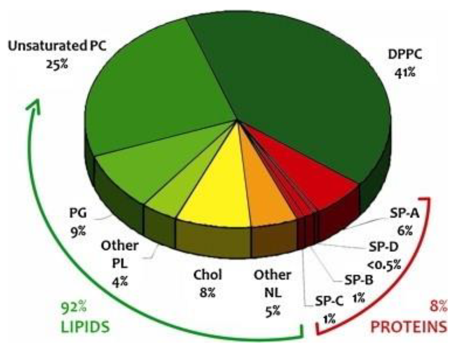
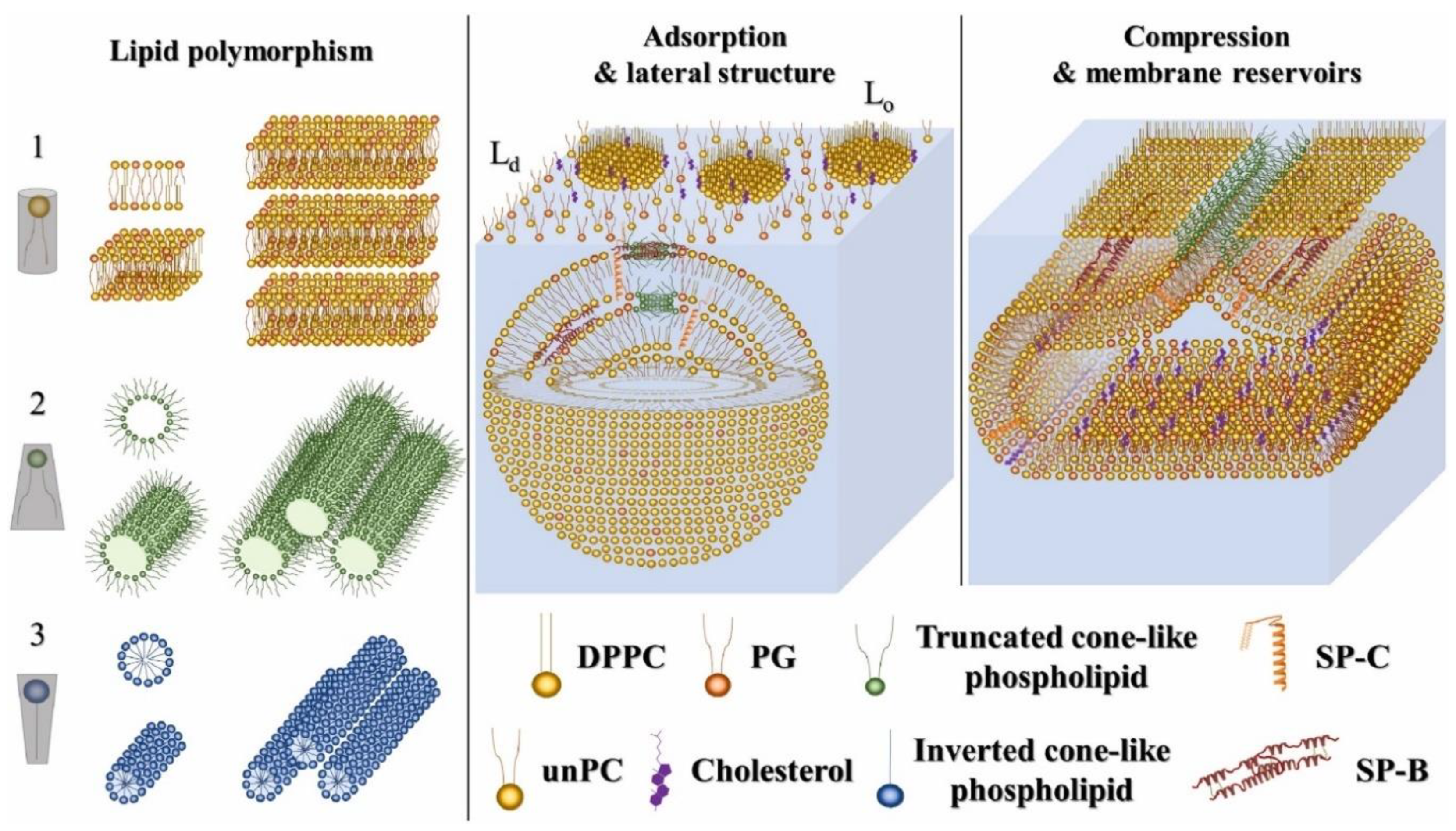
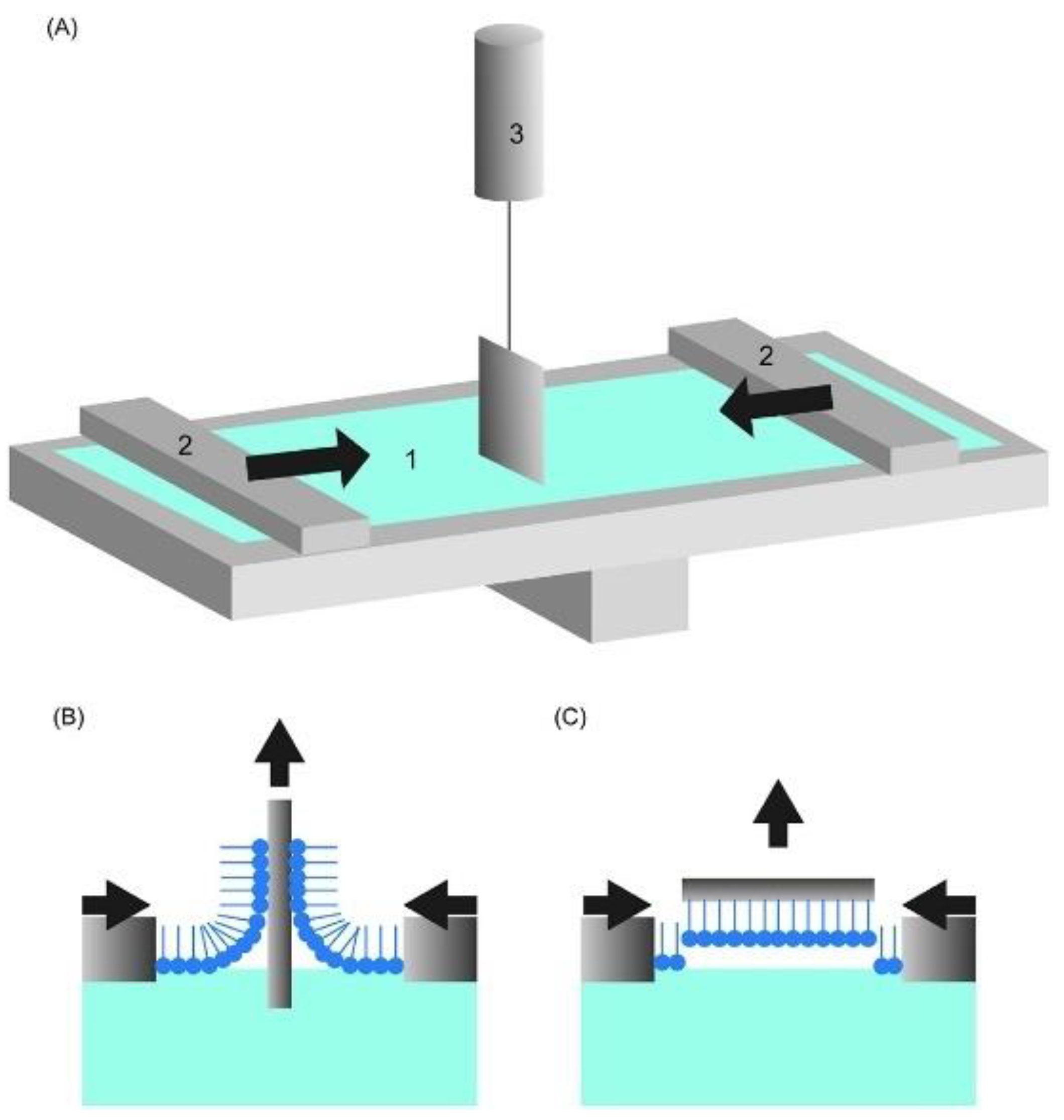
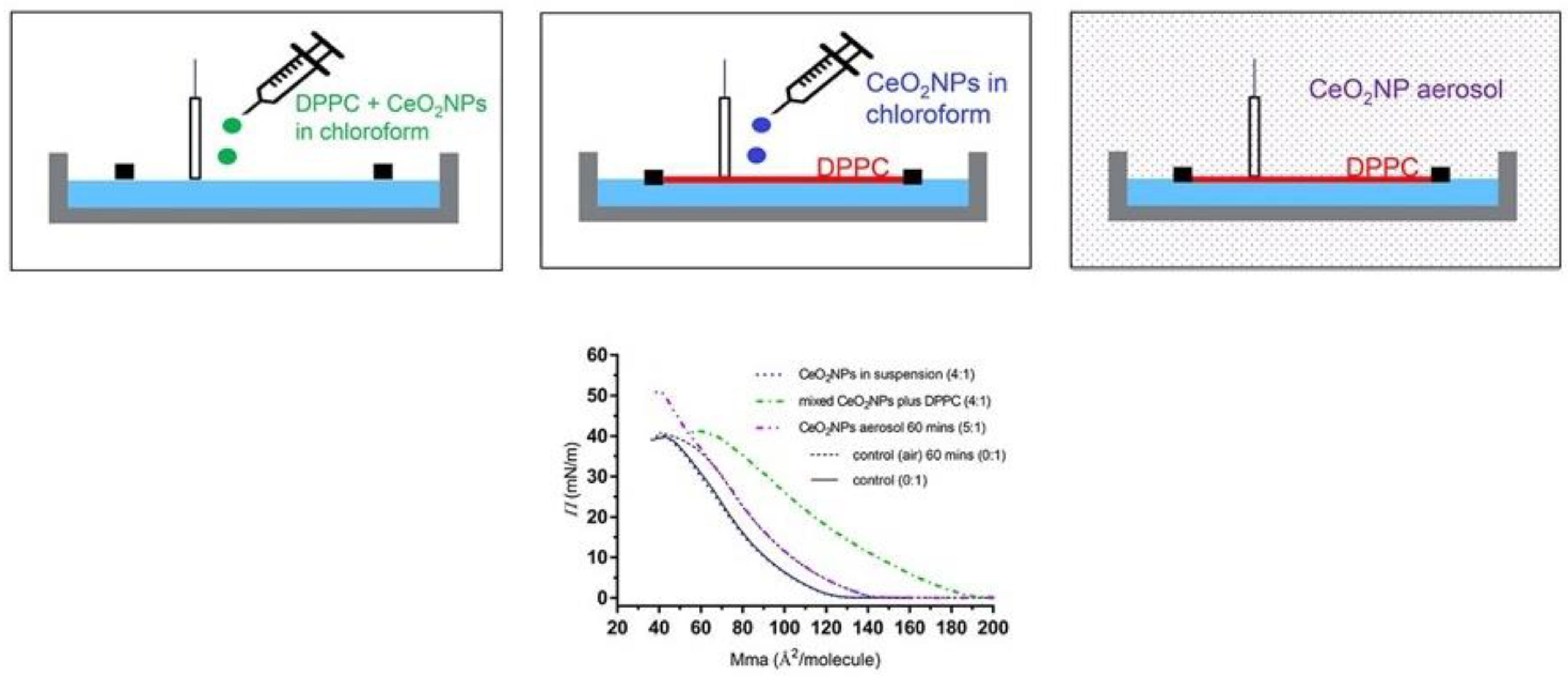
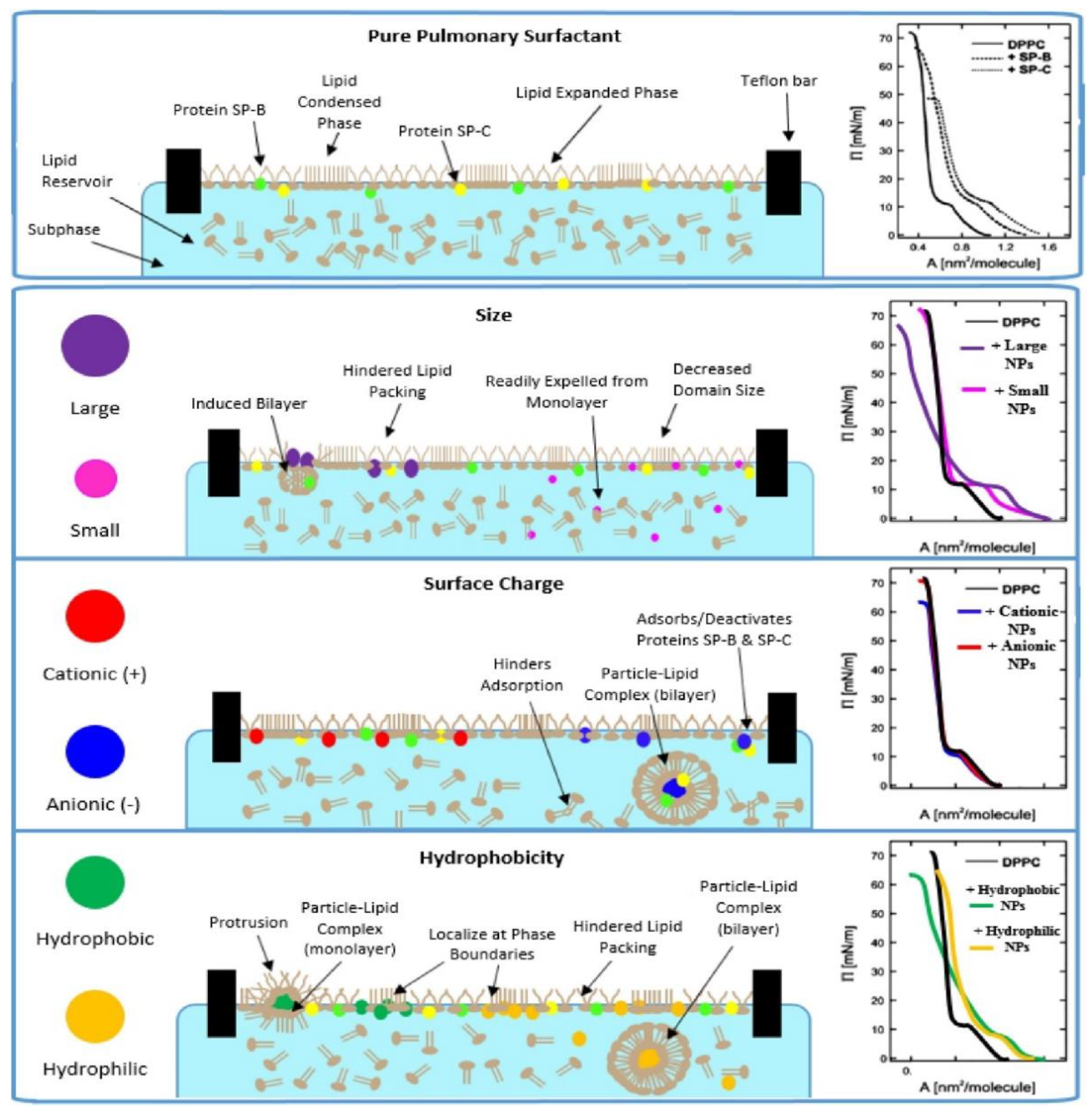
Table 1.
Summary of the most extended clinical lung surfactants, together with their type, origin, and producer.
Table 1.
Summary of the most extended clinical lung surfactants, together with their type, origin, and producer.
| Commercial Name | Origin | Producer |
|---|---|---|
| Infasurf | lavage of calf lung fluid | ONY Biotech Inc., Amherst, NY, USA |
| Curosurf | lavage of porcine lung fluid | Chiesi Farmaceutici S.p.A, Parma, Italy |
| Survanta | lavage of bovine lung fluid | AbbVie Inc., North Chicago, IL, USA |
| BLES | lavage of bovine lung fluid | BLES Biochemicals Inc., London, ON, Canada |
| Alveofact | lavage of bovine lung fluid | Lyomark Pharma, Oberhaching, Germany |
| Venticute | synthetic | Byk Gulden Pharmaceuticals, Konstanz, Germany |
| Surfaxin | synthetic | Discovery Laboratory Inc., Warrington, PA, USA |
| Exosurf | synthetic | GlaxoSmithKline, Brentford, UK |
Publisher’s Note: MDPI stays neutral with regard to jurisdictional claims in published maps and institutional affiliations. |
© 2022 by the author. Licensee MDPI, Basel, Switzerland. This article is an open access article distributed under the terms and conditions of the Creative Commons Attribution (CC BY) license (https://creativecommons.org/licenses/by/4.0/).
Share and Cite
MDPI and ACS Style
Guzmán, E. Fluid Films as Models for Understanding the Impact of Inhaled Particles in Lung Surfactant Layers. Coatings 2022, 12, 277. https://0-doi-org.brum.beds.ac.uk/10.3390/coatings12020277
AMA Style
Guzmán E. Fluid Films as Models for Understanding the Impact of Inhaled Particles in Lung Surfactant Layers. Coatings. 2022; 12(2):277. https://0-doi-org.brum.beds.ac.uk/10.3390/coatings12020277
Chicago/Turabian StyleGuzmán, Eduardo. 2022. "Fluid Films as Models for Understanding the Impact of Inhaled Particles in Lung Surfactant Layers" Coatings 12, no. 2: 277. https://0-doi-org.brum.beds.ac.uk/10.3390/coatings12020277
Note that from the first issue of 2016, this journal uses article numbers instead of page numbers. See further details here.