Review on Fabrication of Structurally Colored Fibers by Electrospinning

Institute of Textiles and Clothing, The Hong Kong Polytechnic University, Hung Hom, Kowloon, Hong Kong, China
*
Author to whom correspondence should be addressed.
Fibers 2018, 6(4), 70; https://0-doi-org.brum.beds.ac.uk/10.3390/fib6040070
Submission received: 29 June 2018
/
Revised: 19 September 2018
/
Accepted: 20 September 2018
/
Published: 26 September 2018
(This article belongs to the Special Issue Smart Coatings on Fibers and Textiles)
Abstract
:Structural color derived from the physical interactions of photons, with the specific chromatic mechanism differing from that of dyes and pigments, has brought considerable attention by the conducive virtue of being dye-free and fadeless. This has recently become a research hot-spot. Assemblies of colloidal nanoparticles enable the manufacture of periodic photonic nanostructures. In our review, the mechanism of nanoparticle assemblies into structurally colored structures by the electrospinning method was briefly introduced, followed by a comparatively comprehensive review summarizing the research related to photonic crystals with periodically aligned nanostructures constructed by the assembly of colloidal nanoparticles, and the concrete studies concerning the fabrication of well-aligned electrospun nanofibers incorporating with colloidal nanoparticles based on the investigation of relevant factors such as the sizes of colloidal nanoparticles, the weight ratio between colloidal nanoparticles, and the polymer matrix. Electrospinning is expected to be a deserving technique for the fabrication of structurally colored nanofibers while the colloidal nanoparticles can be well confined into aligned arrangement inside nanofibres during the electrospinning process after the achievement of resolving remaining challenges.
1. Introduction
Structural color arises from photonic crystals possessing a periodic modulation assembled by colloidal nanoparticles; this has invited great interest since their introduction in the late 1980s [1,2,3]. The different mechanisms for controlling and manipulating light provided by the self-assembly of photonic crystals could be analyzed on the basis of the photonic bandgap [4]. Light refraction, diffraction, and scattering exist in the periodic nanostructures that contain regularly repeating internal regions [5]. Colloidal nanoparticles used to assemble photonic crystals with closely-packed structures play an important role in generating structural color to act on the transport and manipulation of light, owing to the Bragg diffraction [6]. Photonic crystals that exhibit brilliant structural colors are usually prepared by self-assembly of inorganic colloidal nanoparticles like silicon dioxide or silica (SiO2) [7,8], titanium dioxide (TiO2), core-shell colloids containing metallic nanoparticles like gold (Au)@SiO2 colloids [9], polymeric nanoparticles including colloidal homopolymer nanoparticles [10,11], polystyrene (PS) [8,12,13], and copolymer nanoparticles like poly(styrene-methyl methacrylate-acrylic acid) [14]. Electrospinning is one of the favorable fabrication techniques that utilizes electrostatic forces to prepare polymer filament networks as scaffolds in nanometer scale, since it emerged in the early 1930s [15]. The periodically arranged nanostructures constructed by electrospinning have shown the potential of generating brilliant colors with the addition of photonic crystals, leading to a wide range of prospects in a novel biomimetic field.
In terms of the electrospinning technique, with the advantages of easy access to the adjustment of morphology of nanofibres and fast continuous preparation process [16], various kinds of electrospun nanofiber assemblies with desired properties could be prepared for myriad specific applications [17]. Unlike the dip-coating method [18], the electrospun nanofibers could be prepared without adhesion between the substrate and coating layer. No high thermal fulfillment tends to be required for the fabrication process, which is dissimilar to the melting and shear ordering method [19,20,21]. Electrospinning possesses high efficiency of manufacturing than the combined approach of microfluidic emulsification and solvent diffusion [22]. The electrophoretic method [23,24,25] has served as a fabrication technique for structurally colored fiber that has been examined on colloidal crystal growth with ordered arrangement for several decades, but the limitation of the substrate requirement with good electrical conductivity to form functional nanostructures cannot be neglected. Accordingly, electrospinning is conducted as a more effective approach for the production of structurally colored nanofibers of polymers compared with such template-directed assembly methods in which the subsequent removal of the sacrificial templates requires a troublesome and time-consuming process [26].
Even through structural color has been introduced into colloidal systems for a long time, researches regarding structurally colored electrospun nanofibers in combination with the assembly of nanoparticles are still quite sparse. Herein, this paper reviews highly relevant and significant research on structurally colored fibers with periodically aligned nanostructures by a promising electrospinning technique.
2. Mechanism of Nanoparticle Assemblies into Structurally Colored Nanofibers
While propagating the periodic structures, the wavelength of reflected light on the nanoparticle-assembled nanofibers is predicted by a Bragg equation [27].
If the light wavelength that fulfills the Bragg condition, incident light would undergo diffraction and hence the color of specific reflected light would appear on the surface of the ordered structures as demonstrated in Figure 1. The periodically ordered structures with aligned nanofibers are established by electrospinning, in which the round spheres represent the nanoparticles while the polymer matrix that surrounds the nanoparticles is mainly removed. Inserted image means cross section of part of the aligned nanofibers within a range around several microns made of nanoparticles. The periodicity and sizes of nanoparticles closely correlate to the color of reflected light, which is mainly controlled by modulating electrospinning parameters and emulsion polymerization conditions, respectively. Structural colors primarily depend on morphology of the material with certain properties, such as refraction index, playing a significant role of determining the color as well as color intensity [28]. The differences between density in amorphous and crystalline phases of the same polymer lead to differences in the refractive index, which means the ratio of the velocity of light in a vacuum to that in the polymer is closely related to the generation of structural color due to the significant role of polymers in the development of materials for photonics [6]. The width and frequency of the reflection tend to be determined by refractive index as well as the layer or film thickness in the photonic crystal film [7]. Hence refractive index has been identified as one of the fundamental optical properties of a polymer that is regarded as an input parameter for assessing many other optical properties [29,30]. A readily feasible method for changing color is to modulate the refractive indices of the nanostructures constructed by polymers [31]. Enlarging the difference between refractive index and colloidal nanoparticles and polymer binder tends to be accessible to strength the intensity of structural color. The surface polarity between the photonic crystal nanoparticles and polymer binder is shown to exert great significance in the preparation of spinning solutions for establishing electrospun nanofibers, and subsequently it would be routinely considered while producing the electrospinning solution.
A schematic representation of the preparation of nanoparticle assemblies into nanofibres with photonic crystal structures exhibiting brilliant color is given in Figure 2, which is similar to the given work [32]. Like conventional electrospinning setup, a spinneret with needle and syringe, a high voltage power supply, and a conductive collector including rotating drum (Figure 2a) or parallel collector (Figure 2b) are constructed for manufacturing well-aligned nanofibres, and the working principle of electrospinning is similar to the previous studies [33]. Syringe pump filled with spinning solution is used to provide the liquid to the tip of the needle in a certain flow rate. Due to the surface tension, a liquid droplet could form, where charges would generate while imposing a high voltage at the spinneret. A Taylor cone would exist when charge repulsion is strong enough to conquer the surface tension. Then the liquid could be ejected to the collector under the driving forces based on the electrostatic repulsion. Finally, the nanofibers align perpendicular to the axis of the drum or parallel electrode with the evaporation of solvent [34]. The mechanism that modeled and simulated the corresponding electrostatic field distributions between the needle and the collector in a three-dimensional (3D) space is shown in Figure 2c,d. The typical target collectors utilize a rotating drum and parallel collector, respectively. The electrostatic driving forces have no fixed direction that can be ascribed to the unstable electric field. In order to obtain the highly aligned nanofibres fabricated by electrospinning [34,35], it has been found to apply high rotating collectors, parallel electrode collectors [36,37], and auxiliary electrodes [38,39]. The gap width between the as-spun nanofibers reached the values ranging from centimeters to several micrometers as investigated in [40], which resulted in the uncontrollable orientation of electrospun nanofibers, and hence the deep investigation of suitable electrospinning conditions is required to build the scaffolding of aligned nanostructures that exhibits structural color. For instance, Li et al. [40] have analyzed the electrostatic forces using continuous conductive collector without and with an insulating gap, they found that a parallel collector had the priority of enabling the nanofibers in an uniaxial alignment.
3. Photonic Crystals for Fabricating Structurally Colored Fibers
The construction of photonic crystals with three-dimensional periodic geometry is a crucial part when it comes to fabricating structurally colored fibers. Coherently diffraction and scattering by the photons of specific wavelengths could occur on the periodic lattice planes of photonic crystals, which has a spacing that is similar to such wavelengths [28,41]. Prior to the preparation of three-dimensional photonic crystal structural color materials, the self-assembly of colloidal nanoparticles of spherical or cylindrical shapes into a close-packed and periodic array tends to enable the photonic crystal as an attractive candidate for generating structurally colored fibers. Colloidal nanoparticles can act as building blocks for constructing photonic crystals as a result of the superiority that includes multiple choices of chemical composition, cost-effective products, as well as tunable particle sizes [5]. The size dimension of nanoparticles mainly controlled by the adjustment of the reaction conditions during synthesis process, including the different amounts of reactants [42,43], precursor type, reaction temperature, and time [44], causes a shift of color that enlarges the potential applications of promising areas.
3.1. Inorganic Nanoparticles for Photonic Crystals
Since Iler [45] first carried out the research that multilayers assembled by colloidal nanoparticles could be formed on rigid substrates by the layer-by-layer (LBL) deposition method, many researchers have studied different nanoparticles, including inorganic nanoparticles for constructing photonic crystals, such as silicon dioxide or silica (SiO2) and titanium dioxide (TiO2), which are the two preferred materials for constructing a desired array with uniform arrangement because of the different dielectric constants they have. The controllable synthesis process of monodisperse silica spheres with micron sizes was presented by Stöber in the1960s, which has been used as a widespread reaction [46]. Wang et al. [7] adopted a new strategy to fabricate color-tunable biomimetic film with tunable structural colors in organic solvent by assembling SiO2 nanospheres, and the experimental results indicated that carbon black dopant remarkably increases the chroma and enhances the intensity of the structural colors. Structural color changes of colloidal crystal films from red to violet were determined by the assembly of SiO2 nanoparticles with different particle sizes from 207 to 350 nm using the Stöber process [43]. A recent study by Zhang [47] disclosed vivid structural color coatings from atomization deposition of SiO2 nanoparticles with poly (vinyl alcohol) additive on silk fabrics, which can possess robust mechanical properties as depicted in Figure 3.
Several conventional techniques have been employed to produce TiO2 films, including conventional sol-gel method [48,49], vapor deposition method [50,51], and liquid phase deposition [52,53]. Chen and co-workers [54] successfully used an effective method named the atomic layer deposition (ALD) technique to fabricate TiO2 coatings with vibrant and uniform structural colors using black carbon fibers as substrates, showing excellent laundering durability. These have many potential applications in the field of optical and color-display devices. Because the use of single inorganic particles is significantly limited to the formation of periodically arranged photonic structures, core-shell colloidal particles have become desirable materials. Metal nanoparticles like gold (Au) or silver (Ag) are extraordinarily efficient at strongly absorbing and scattering light to enhance the intension of reflected light for exhibiting brilliant color, due to the optical property known as localized surface plasmon resonances [55,56]. SiO2 nanoparticles incorporated with gold has been prepared to form colloidal spheres (Au@SiO2) with core-shell shapes, which can serve as the building blocks for photonic applications [9]. Yuan et al. [50] reported structural colors on polyester fabric with the coating of silver(Ag)/TiO2 films made by magnetron sputtering. The resultant samples with diverse colors of purple, light blue, blue, pink, and dark red could be displayed by controlling different thickness of TiO2 thin films. Luo et al. [57] reported electrically induced structural color alteration of core-shell (SiO2@TiO2) photonic crystals for electric field applications.
3.2. Polymeric Nanoparticles for Photonic Crystals
Since the discovery of an approach for synthesizing monodisperse polymer colloids by Vanderhoff, this synthetic method has been used to prepare diverse polymeric colloids, including homopolymer colloidal nanoparticles as well as copolymer nanoparticles, besides inorganic colloidal nanoparticles [58,59]. These monodisperse polymeric colloidal nanoparticles prepared by some mature synthesis techniques, such as emulsion polymerization, dispersion polymerization, precipitation polymerization, or seeded polymerization, can be incorporated into polymer solution for electrospinning [5]. Among the synthetic routes, emulsion and dispersion polymerization are the most commonly used.
Colloidal homopolymer nanoparticles such as polymethylmethacrylate and polystyrene monodispersed spheres, have been successfully synthesized to be used for fabricating structural color. The size of such nanoparticles influencing optical properties of electrospun nanostructures can be determined by synthesis conditions like concentration of initiator, reaction time, and temperature. The preparation of nanofibres containing polymeric nanoparticles by electrospinning has been examined [12]. Meng et al. [60] studied the mechanical stability and hydrophobicity of structural color films composed of PS microspheres, which was of great significance for potential architectural purposes in paint and decoration. According to Han et al. [61], the stability of switching performance of the tunable photonic crystals consisting of well-ordered PS colloidal arrays has been improved in long range for display applications. Tang and co-workers [62,63] presented a kind of polymer opal film with brilliant structural colors from PS nanospheres or crosslinked PS/polydimethylsiloxane (PDMS) with well-aligned structures by using the self-assembly method with thermal assistance for architectural applications. Gu et al. [8] described a kind of structural color with a lotus effect formed by mixing both PS spheres and SiO2 particles to deposit an opal film on a glass substrate. Meanwhile, researchers have made much efforts to prepare poly(methyl methacrylate) PMMA nanospheres for many promising purposes, synthesized by a commonly used method called emulsion polymerization [10,64,65]. Tang et al. [66] developed a kind of heat-resistant photonic crystal film consisting of PMMA colloidal spheres with different colors by investigating the effect of methyl methacrylate (MMA) concentration on particle size as well as the distribution of PMMA particles.
Despite colloidal homopolymer nanoparticles, a variety of copolymer nanospheres commonly exhibiting core-shell nanostructures can be synthesized with other kinds of monomers by polymerization to achieve the desired functions. Liu et al. [67] developed a kind of photonic crystal named poly(styrene-methacrylic acid) coating on the textile fabrics exhibiting brilliant and variable structural colors, possessing better hydrophobic properties. Non-iridescent brilliant structural colors (blue, green and red) from photonic structures based on assembling polystyrene/poly(N-isopropylacrylamide-co-acrylic-acid) particles with core-sheath particle structure over the full-spectrum were demonstrated by Park [68], as displayed in Figure 4a,b.
Figure 4c,d shows that a kind of core-sheath colloidal poly(styrene-methyl methacrylate-acrylic acid) nanospheres was synthesized by emulsion polymerization using three monomers including methyl methacrylate, styrene and acrylic acid from [14]. Brilliant and monochromatic colors of colloidal crystal films covering the entire visible range from red to violet color on a glass substrate were obtained by assembling such nanospheres with various diameters. Yuan et al. [69] also successfully synthesized monodispersed poly (styrene-methyl methacrylate-acrylic acid) composite nanospheres and it was designed to be blended with a measured amount of PVA to produce a dye-free electrospun fibrous membrane with tunable brilliant structural colors by colloidal electrospinning. The schematic illustration of this process and the typical scanning electron microscope (SEM) images of the nanofibres are depicted in Figure 5. The SEM results demonstrated that the colloidal spheres are well-arranged on the surfaces of nanofibres in a cylindrical shape.
4. Electrospun Nanofibers Derived from Photonic Colloidal Nanoparticles
Many researchers have successfully achieved structural color films or photonic opals or coatings on substrates [43,70,71,72], and the electrospinning technique for generating common nanofibres has been proven. Electrospinning using a high-voltage electric field to spin polymer solutions into nanofibres with nanoscale diameters, tends to be a versatile technique for successfully manufacturing polymer nanofibres from various kinds of polymer materials, like poly (vinyl alcohol), poly(ethylene terephthalate), poly(vinyl phenol), polyurethanes, polyamide, poly(methyl methacrylate), polyacrylonitrile, poly (ether imide), poly (ethylene gricol) [73], which are treated as a polymer matrix, an indispensable element and the main component of the nanostructures exhibiting brilliant colors. More importantly, the fabrication of nanofiber membranes containing SiO2 or TiO2 colloidal nanoparticles by the electrospinning method has proven to be feasible according to previous works by Zhang [74], Im [75] and Pant [76]. A discovery of present work focusing on incorporating colloidal nanoparticles into electrospun nanofibers with well aligned morphology has been found as shown in Table 1 with detailed parameters. Different morphologies of electrospun nanofibers could be achieved from random packed, necklace-like to cylindrically hexagonal patterned structures by controlling several decisive conditions, which covers the different properties of the polymer matrix, including crystallinity, glass transition temperature, molecular weight, and spinning solution properties including surface tension, viscosity, conductivity, polymer solubility as well as the electrospinning processing parameters consisting of the feeding rate, applied voltage, receiving distance, effect of collector, and needle [15]. Besides, with the addition of photonic colloidal nanoparticles, more influencing factors are thought to be considered. The sizes and shapes of colloidal nanoparticles blended in polymer matrix is one of the indispensable factors determining the morphology of the as-prepared electrospun nanofibers. In the early 20th century, Yang et al. [26] presented the concept that colloidal particles can be confined and assembled inside nanofibres during the electrospinning process and they successfully fabricated polyacrylamide/SiO2, poly(ethyleneoxide)/SiO2 and poly(acrylonitrile)/SiO2 nanofibers with close-packed SiO2 particles formed in radial direction of the nanofibres. From SEM results in Figure 6, it can be documented that with the increasing size of SiO2 particles from 100 nm to 1000 nm, the structure of polyacrylamide nanofibres transformed from less-aligned to assembled necklace-like structures.
Another affecting factor is the weight ratio between photonic colloidal nanoparticles and polymer matrix. Yuan and his group members [12] investigated the morphology of polystyrene nanosphere(PS)/poly(vinyl alcohol)(PVA) electrospun fibers with the various structures. Polystyrene nanospheres were synthesized by emulsion polymerization using the monomer (styrene) with the addition of an emulsifier. The results showed that string-on-bead and necklace-like nanofibres were obtained with certain ratios of PS and PVA, as shown in Figure 7e. A common electrospinning factor named polymer concentration was also analyzed as can be seen in Figure 7g–n.
Despite the construction for morphology structures of electrospun nanofibers, extra factors may affect the color on the nanofiber surfaces according to Table 1. To evaluate the reason that most of the resulting nanofibers with uniform ordered structures were achromatous, the researchers found that it was attributed to the low refractive index. The subsequent exploration verified that after establishing the scaffolding in which electrospun nanofibers were in alignment, the polymer matrix could be removed without the destruction of the desired nanofiber structures that was conducive to yielding color. The dissolution of the PVA matrix of poly (styrene-methyl methacrylate-acrylic acid)/PVA nanofibers increased the refractive index contrast resulted in displaying structural color [69].
5. Outlook and Conclusions
Myriad studies have successfully produced well-ordered nanostructures on electrospun nanofibers with the blending of photonic nanoparticles, however, the research on nanofibres containing photonic nanoparticles endowing homogeneous and non-iridescent structural colors remains very few, which indicates that it seems to be difficult to achieve the well-ordered structure of electrospun nanofibers incorporating photonic nanoparticles as a result of the overall consideration of a multitude of factors and thereby it remains a daunting task.
Overall, tremendous achievements in structural coloration based on photonic crystals have been visible in many application areas, especially optical fields, but so far only a relatively small number of researchers have tried to prepare nanofibres with brilliant structural colors by electrospinning because of the difficult regulation of manufacturing conditions. Major efforts are still required to further develop and improve effective manufacturing approaches to structurally colored nanofibers. Researchers have presented electrospun nanofibers exhibiting vivid structural colors with the addition of polymeric particle colloids, however, inorganic spheres with higher reflective indices are expected be used to fabricate structurally colored fibers because the high refractive index contrast is beneficial for increasing the intensity of structural colors. Other properties of structurally colored fibers such as mechanical properties are supposed to be considered and examined for more promising applications. It could be concluded that electrospinning is a versatile approach that could be used for the generation of highly aligned electrospun nanofibers, showing the promise of paving the way for a revolution in the development of structurally colored materials for various dye-free applications.
Author Contributions
Conceptualization, J.Y. and C.-W.K.; Data curation, J.Y.; Formal analysis, J.Y. and C.-W.K.; Funding acquisition, C.-W.K.; Investigation, J.Y. and C.-W.K.; Methodology, J.Y. and C.-W.K.; Project administration, C.-W.K.; Resources, C.-W.K.; Supervision, C.-W.K.; Validation, J.Y. and C.-W.K.; Visualization, J.Y.; Writing—original draft, J.Y.; Writing—review & editing, C.-W.K.
Funding
This work was funded by The Hong Kong Polytechnic University with grant numbers RHQG and G-UA9M.
Acknowledgments
The authors gratefully appreciate and acknowledge the financial support from the Hong Kong Polytechnic University.
Conflicts of Interest
The authors declare no conflict of interest.
References
- Yablonovitch, E. Inhibited spontaneous emission in solid-state physics and electronics. Phys. Rev. Lett. 1987, 58, 2059–2062. [Google Scholar] [CrossRef] [PubMed]
- John, S. Strong localization of photons in certain disordered dielectric superlattices. Phys. Rev. Lett. 1987, 58, 2486–2489. [Google Scholar] [CrossRef] [PubMed]
- Yablonovitch, E.; Gmitter, T.J. Photonic band structure: The face-centered-cubic case. Phys. Rev. Lett. 1989, 63, 1950. [Google Scholar] [CrossRef] [PubMed]
- Joannopoulos, J.D.; Villeneuve, P.R.; Fan, S. Photonic crystals: Putting a new twist on light. Nature 1997, 386, 143. [Google Scholar] [CrossRef]
- Zhang, J.; Sun, Z.; Yang, B. Self-assembly of photonic crystals from polymer colloids. Curr. Opin. Colloid Interface Sci. 2009, 14, 103–114. [Google Scholar] [CrossRef]
- Paquet, C.; Kumacheva, E. Nanostructured polymers for photonics. Mater. Today 2008, 11, 48–56. [Google Scholar] [CrossRef]
- Wang, W.; Tang, B.; Ma, W.; Zhang, J.; Ju, B.; Zhang, S. Easy approach to assembling a biomimetic color film with tunable structural colors. J. Opt. Soc. Am. A Opt. Image Sci. Vis. 2015, 32, 1109–1117. [Google Scholar] [CrossRef] [PubMed]
- Gu, Z.; Uetsuka, H.; Takahashi, K.; Nakajima, R.; Onishi, H.; Fujishima, A.; Stao, O. Structural color and the lotus effect. Angew. Chem. Int. Ed. 2003, 42, 894–897. [Google Scholar] [CrossRef] [PubMed]
- Lu, Y.; Yin, Y.; Li, Z.; Xia, Y. Synthesis and self-assembly of au@ sio2 core-shell colloids. Nano Lett. 2002, 2, 785–788. [Google Scholar] [CrossRef]
- Gu, Z.Z.; Chen, H.; Zhang, S.; Sun, L.; Xie, Z.; Ge, Y. Rapid synthesis of monodisperse polymer spheres for self-assembled photonic crystals. Colloids Surfaces A Physicochem. Eng. Asp. 2007, 302, 312–319. [Google Scholar] [CrossRef]
- Egen, M.; Zentel, R. Surfactant-free emulsion polymerization of various methacrylates: Towards monodisperse colloids for polymer opals. Macromol. Chem. Phys. 2004, 205, 1479–1488. [Google Scholar] [CrossRef]
- Yuan, W.; Zhang, K.Q. Structural evolution of electrospun composite fibers from the blend of polyvinyl alcohol and polymer nanoparticles. Langmuir 2012, 28, 15418–15424. [Google Scholar] [CrossRef] [PubMed]
- Ha, S.T.; Park, O.O.; Im, S.H. Size control of highly monodisperse polystyrene particles by modified dispersion polymerization. Macromol. Res. 2010, 18, 935–943. [Google Scholar] [CrossRef]
- Wang, J.; Wen, Y.; Ge, H.; Sun, Z.; Zheng, Y.; Song, Y.; Jiang, L. Simple fabrication of full color colloidal crystal films with tough mechanical strength. Macromol. Chem. Phys. 2006, 207, 596–604. [Google Scholar] [CrossRef]
- Ramakrishna, S. An Introduction to Electrospinning and Nanofibers; World Scientific: Singapore, 2005; p. 15. ISBN 981-256-415-2. [Google Scholar]
- Mu, Q.; Zhang, Q.; Gao, L.; Chu, Z.; Cai, Z.; Zhang, X.; Wang, K.; Wei, Y. Structural evolution and formation mechanism of the soft colloidal arrays in the core of paam nanofibers by electrospun packing. Langmuir 2017, 33, 10291–10301. [Google Scholar] [CrossRef] [PubMed]
- Bhardwaj, N.; Kundu, S.C. Electrospinning: A fascinating fiber fabrication technique. Biotechnol. Adv. 2010, 28, 325–347. [Google Scholar] [CrossRef] [PubMed]
- Zhang, J.; He, S.; Liu, L.; Guan, G.; Lu, X.; Sun, X.; Peng, H. The continuous fabrication of mechanochromic fibers. J. Mater. Chem. C 2016, 4, 2127–2133. [Google Scholar] [CrossRef]
- Ruhl, T.; Spahn, P.; Hellmann, G.P. Artificial opals prepared by melt compression. Polymer 2003, 44, 7625–7634. [Google Scholar] [CrossRef]
- Finlayson, C.E.; Spahn, P.; Snoswell, D.R.; Yates, G.; Kontogeorgos, A.; Haines, A.I.; Hellmann, G.P.; Baumberg, J.J. 3d bulk ordering in macroscopic solid opaline films by edge-induced rotational shearing. Adv. Mater. 2011, 23, 1540–1544. [Google Scholar] [CrossRef] [PubMed]
- Pursiainen, O.L.J.; Baumberg, J.J.; Winkler, H.; Viel, B.; Spahn, P.; Ruhl, T. Shear-induced organization in flexible polymer opals. Adv. Mater. 2008, 20, 1484–1487. [Google Scholar] [CrossRef]
- Kohri, M.; Yanagimoto, K.; Kawamura, A.; Hamada, K.; Imai, Y.; Watanabe, T.; Ono, T.; Taniguchi, T.; Kishikawa, K. Polydopamine-based 3d colloidal photonic materials: Structural color balls and fibers from melanin-like particles with polydopamine shell layers. ACS Appl Mater. Interfaces 2018, 10, 7640–7648. [Google Scholar] [CrossRef] [PubMed]
- Giersig, M.; Mulvaney, P. Preparation of ordered colloid monolayers by electrophoretic deposition. Langmuir 1993, 9, 3408–3413. [Google Scholar] [CrossRef]
- Yu, H.; Liao, D.; Johnston, M.B.; Li, B. All-optical full-color displays using polymer nanofibers. ACS Nano 2011, 5, 2020–2025. [Google Scholar] [CrossRef] [PubMed]
- Liu, Z.; Zhang, Q.; Wang, H.; Li, Y. Structurally colored carbon fibers with controlled optical properties prepared by a fast and continuous electrophoretic deposition method. Nanoscale 2013, 5, 6917–6922. [Google Scholar] [CrossRef] [PubMed]
- Lim, J.M.; Moon, J.H.; Yi, G.R.; Heo, C.J.; Yang, S.M. Fabrication of one-dimensional colloidal assemblies from electrospun nanofibers. Langmuir 2006, 22, 3445–3449. [Google Scholar] [CrossRef] [PubMed]
- Liu, Z.; Zhang, Q.; Wang, H.; Li, Y. Structural colored fiber fabricated by a facile colloid self-assembly method in micro-space. Chem. Commun. (Camb.) 2011, 47, 12801–12803. [Google Scholar] [CrossRef] [PubMed]
- Josephson, D.P.; Miller, M.; Stein, A. Inverse opal SiO2 photonic crystals as structurally-colored pigments with additive primary colors. Z. Anorg. Allg. Chem. 2014, 640, 655–662. [Google Scholar] [CrossRef]
- Katritzky, A.R.; Sild, S.; Karelson, M. Correlation and prediction of the refractive indices of polymers by qspr. J. Chem. Inf. Comput. Sci. 1998, 38, 1171–1176. [Google Scholar] [CrossRef]
- Jicerano, J. Prediction of Polymer Properties; CRC Press: New York, NY, USA, 2002; p. 272. ISBN 0-8247-0821-0. [Google Scholar]
- Sato, O.; Kubo, S.; Gu, Z.Z. Structural color films with lotus effects, superhydrophilicity, and tunable stop-bands. Acc. Chem. Res. 2008, 42, 1–10. [Google Scholar] [CrossRef] [PubMed]
- Jin, Y.; Yang, D.; Kang, D.; Jiang, X. Fabrication of necklace-like structures via electrospinning. Langmuir 2010, 26, 1186–1190. [Google Scholar] [CrossRef] [PubMed]
- Crespy, D.; Friedemann, K.; Popa, A.M. Colloid-electrospinning: Fabrication of multicompartment nanofibers by the electrospinning of organic or/and inorganic dispersions and emulsions. Macromol. Rapid Commun. 2012, 33, 1978–1995. [Google Scholar] [CrossRef] [PubMed]
- Zhang, C.L.; Yu, S.H. Nanoparticles meet electrospinning: Recent advances and future prospects. Chem. Soc. Rev. 2014, 43, 4423–4448. [Google Scholar] [CrossRef] [PubMed]
- Dzenis, Y. Spinning continuous fibers for nanotechnology. Science 2004, 304, 1917–1919. [Google Scholar] [CrossRef] [PubMed]
- Zhao, J.; Liu, H.; Xu, L. Preparation and formation mechanism of highly aligned electrospun nanofibers using a modified parallel electrode method. Mater. Des. 2016, 90, 1–6. [Google Scholar] [CrossRef]
- Li, D.; Wang, Y.L.; Xia, Y.N. Electrospinning of polymeric and ceramic nanofibers as uniaxially aligned arrays. Nano Lett. 2003, 3, 1167–1171. [Google Scholar] [CrossRef]
- Dersch, R.; Liu, T.; Schaper, A.K.; Greiner, A.; Wendorff, J.H. Electrospun nanofibers: Internal structure and intrinsic orientation. Polym. Chem. 2003, 41, 545–553. [Google Scholar] [CrossRef]
- Teo, W.E.; Ramakrishna, S. A review on electrospinning design and nanofibre assemblies. Nanotechnology 2006, 17, R89–R106. [Google Scholar] [CrossRef] [PubMed]
- Li, D.; Wang, Y.; Xia, Y. Electrospinning nanofibers as uniaxially aligned arrays and layer-by-layer stacked films. Adv. Mater. 2004, 16, 361–366. [Google Scholar] [CrossRef]
- Eablonovitch, E. Photonic band-gap structures. J. Opt. Soc. Am. B 1993, 10, 283–295. [Google Scholar] [CrossRef]
- Lee, C.H.; Yu, J.; Wang, Y.; Tang, A.Y.L.; Kan, C.W.; Xin, J.H. Effect of graphene oxide inclusion on the optical reflection of a silica photonic crystal film. RSC Adv. 2018, 8, 16593–16602. [Google Scholar] [CrossRef]
- Gao, W.; Rigout, M.; Owens, H. Self-assembly of silica colloidal crystal thin films with tuneable structural colours over a wide visible spectrum. Appl. Surf. Sci. 2016, 380, 12–15. [Google Scholar] [CrossRef] [Green Version]
- Mallakpour, S.; Behranvand, V. Polymeric nanoparticles: Recent development in synthesis and application. Express Polym. Lett. 2016, 10, 895–913. [Google Scholar] [CrossRef]
- Iler, R.K. Multilayers of colloidal particles. J. Colloid Interface Sci. 1996, 21, 569–594. [Google Scholar] [CrossRef]
- Stöber, W.; Fink, A.; Bohn, E. Controlled growth of monodisperse silica spheres in the micron size range. J. Colloid Interface Sci. 1968, 26, 62–69. [Google Scholar] [CrossRef]
- Li, Q.; Zhang, Y.; Shi, L.; Qiu, H.; Zhang, S.; Qi, N.; Hu, J.; Yuan, W.; Zhang, X.; Zhang, K.Q. Additive mixing and conformal coating of noniridescent structural colors with robust mechanical properties fabricated by atomization deposition. ACS Nano 2018, 12, 3095–3102. [Google Scholar] [CrossRef] [PubMed]
- Sopyan, I.; Watanabe, M.; Murasawa, S.; Hashimoto, K.; Fujishima, A. Efficient TiO2 powder and film photocatalysts with rutile crystal structure. Chem. Lett. 1996, 25, 69–70. [Google Scholar] [CrossRef]
- Chrysicopoulou, P.; Davazogloub, D.; Trapalis, C.; Kordasa, G. Optical properties of very thin (100 nm) sol–gel TiO2 films. Thin Solid Films 1998, 323, 188–193. [Google Scholar] [CrossRef]
- Yuan, X.; Xu, W.; Huang, F.; Chen, D.; Wei, Q. Structural colour of polyester fabric coated with ag/tio2 multilayer films. Surf. Eng. 2016, 33, 231–236. [Google Scholar] [CrossRef]
- Guo, D.; Ito, A.; Goto, T.; Tu, R.; Wang, C.; Shen, Q.; Zhang, L. Effect of laser power on orientation and microstructure of TiO2 films prepared by laser chemical vapor deposition method. Mater. Lett. 2013, 93, 179–182. [Google Scholar] [CrossRef]
- Sun, S.Q.S.B.; Zhang, W.Q.; Wang, D. Preparation and antibacterial activity of ag-tio 2 composite film by liquid phase deposition (lpd) method. Bull. Mater. Sci. 2008, 31, 61–66. [Google Scholar] [CrossRef]
- Herbig, B.; Löbmann, P. TiO2 photocatalysts deposited on fiber substrates by liquid phase deposition. J. Photochem. Photobiol. A Chem. 2004, 163, 359–365. [Google Scholar] [CrossRef]
- Chen, F.; Yang, H.; Li, K.; Deng, B.; Li, Q.; Liu, X.; Dong, B.; Xiao, X.; Wang, D.; Qin, Y.; et al. Facile and effective coloration of dye-inert carbon fiber fabrics with tunable colors and excellent laundering durability. ACS Nano 2017, 11, 10330–10336. [Google Scholar] [CrossRef] [PubMed]
- Zeng, J.; Huang, J.; Lu, W.; Wang, X.; Wang, B.; Zhang, S.; Hou, J. Necklace-like noble-metal hollow nanoparticle chains: Synthesis and tunable optical properties. Adv. Mater. 2007, 19, 2172–2176. [Google Scholar] [CrossRef]
- Haynes, C.L.; Van Duyne, R. Nanosphere lithography: A versatile nanofabrication tool for studies of size-dependent nanoparticle optics. J. Phys. Chem. B 2001, 105, 5599–5611. [Google Scholar] [CrossRef]
- Luo, Y.; Zhang, J.; Sun, A.; Chu, C.; Zhou, S.; Guo, J.; Chen, T.; Xu, G. Electric field induced structural color changes of sio2@tio2 core–shell colloidal suspensions. J. Mater. Chem. C 2014, 2, 1990–1994. [Google Scholar] [CrossRef]
- Vanderhoff, J.W.; Vitkuske, J.F.; Bradford, E.B.; Alfrey, T., Jr. Some factors involved in the preparation of uniform particle size latexes. J. Polym. Sci. Part A Polym. Chem. 1956, 20, 225–234. [Google Scholar] [CrossRef]
- Kim, S.H.; Lee, S.Y.; Yang, S.M.; Yi, G.R. Self-assembled colloidal structures for photonics. NPG Asia Mater. 2011, 3, 25–33. [Google Scholar] [CrossRef] [Green Version]
- Meng, Y.; Tang, B.; Xiu, J.; Zheng, X.; Ma, W.; Ju, B.; Zhang, S. Simple fabrication of colloidal crystal structural color films with good mechanical stability and high hydrophobicity. Dyes Pigments 2015, 123, 420–426. [Google Scholar] [CrossRef]
- Han, M.G.; Heo, C.-J.; Shim, H.; Shin, C.G.; Lim, S.-J.; Kim, J.W.; Jin, Y.W.; Lee, S. Structural color manipulation using tunable photonic crystals with enhanced switching reliability. Adv. Opt. Mater. 2014, 2, 535–541. [Google Scholar] [CrossRef]
- Tang, B.; Xu, Y.; Lin, T.; Zhang, S. Polymer opal with brilliant structural color under natural light and white environment. J. Mater. Res. 2015, 30, 3134–3141. [Google Scholar] [CrossRef]
- Tang, B.; Zheng, X.; Lin, T.; Zhang, S. Hydrophobic structural color films with bright color and tunable stop-bands. Dyes Pigments 2014, 104, 146–150. [Google Scholar] [CrossRef]
- Tanrisever, T.; Okay, O.; Soenmezoğlu, I.C. Kinetics of emulsifier-free emulsion polymerization of methyl methacrylate. J. Appl. Polym. Sci. 1996, 61, 485–493. [Google Scholar] [CrossRef]
- Zou, D.; Ma, S.; Guan, R.; Park, M.; Sun, L.; Aklonis, J.J.; Salovey, R. Model filled polymers. V. Synthesis of crosslinked monodisperse polymethacrylate beads. J. Polym. Sci. Part A Polym. Chem. 1992, 30, 137–144. [Google Scholar] [CrossRef]
- Tang, B.; Wu, C.; Lin, T.; Zhang, S. Heat-resistant pmma photonic crystal films with bright structural color. Dyes Pigments 2013, 99, 1022–1028. [Google Scholar] [CrossRef]
- Park, J.G.; Kim, S.H.; Magkiriadou, S.; Choi, T.M.; Kim, Y.S.; Manoharan, V.N. Full-spectrum photonic pigments with non-iridescent structural colors through colloidal assembly. Angew. Chem. Int. Ed. Engl. 2014, 53, 2899–2903. [Google Scholar] [CrossRef] [PubMed]
- Liu, G.; Zhou, L.; Wang, C.; Wu, Y.; Li, Y.; Fan, Q.; Shao, J. Study on the high hydrophobicity and its possible mechanism of textile fabric with structural colors of three-dimensional poly(styrene-methacrylic acid) photonic crystals. RSC Adv. 2015, 5, 62855–62863. [Google Scholar] [CrossRef]
- Yuan, W.; Zhou, N.; Shi, L.; Zhang, K.Q. Structural coloration of colloidal fiber by photonic band gap and resonant mie scattering. ACS Appl. Mater. Interfaces 2015, 7, 14064–14071. [Google Scholar] [CrossRef] [PubMed]
- Jia, Y.; Zhang, Y.; Zhou, Q.; Fan, Q.; Shao, J. Structural colors of the SiO2 /polyethyleneimine thin films on poly(ethylene terephthalate) substrates. Thin Solid Films 2014, 569, 10–16. [Google Scholar] [CrossRef]
- Liu, G.; Zhou, L.; Zhang, G.; Li, Y.; Chai, L.; Fan, Q.; Shao, J. Fabrication of patterned photonic crystals with brilliant structural colors on fabric substrates using ink-jet printing technology. Mater. Des. 2017, 114, 10–17. [Google Scholar] [CrossRef]
- Li, Y.; Zhou, L.; Liu, G.; Chai, L.; Fan, Q.; Shao, J. Study on the fabrication of composite photonic crystals with high structural stability by co-sedimentation self-assembly on fabric substrates. Appl. Surf. Sci. 2018, 444, 145–153. [Google Scholar] [CrossRef]
- Huang, Z.-M.; Zhang, Y.Z.; Kotaki, M.; Ramakrishna, S. A review on polymer nanofibers by electrospinning and their applications in nanocomposites. Compos. Sci. Technol. 2003, 63, 2223–2253. [Google Scholar] [CrossRef]
- Zhang, F.; Ma, X.; Cao, C.; Li, J.; Zhu, Y. Poly(vinylidene fluoride)/SiO2 composite membranes prepared by electrospinning and their excellent properties for nonwoven separators for lithium-ion batteries. J. Power Sources 2014, 251, 423–431. [Google Scholar] [CrossRef]
- Im, J.S.; Kim, M.I.; Lee, Y.S. Preparation of pan-based electrospun nanofiber webs containing TiO2 for photocatalytic degradation. Mater. Lett. 2008, 62, 3652–3655. [Google Scholar] [CrossRef]
- Pant, H.R.; Pandeya, D.R.; Nam, K.T.; Baek, W.I.; Hong, S.T.; Kim, H.Y. Photocatalytic and antibacterial properties of a TiO2/nylon-6 electrospun nanocomposite mat containing silver nanoparticles. J. Hazard. Mater. 2011, 189, 465–471. [Google Scholar] [CrossRef] [PubMed]
- Kanehata, M.; Ding, B.; Shiratori, S. Nanoporous ultra-high specific surface inorganic fibres. Nanotechnology 2007, 18, 315602. [Google Scholar] [CrossRef] [Green Version]
- Lim, J.M.; Yi, G.R.; Moon, J.H.; Heo, C.J.; Yang, S.M. Superhydrophobic films of electrospun fibers with multiple-scale surface morphology. Langmuir 2007, 23, 7981–7989. [Google Scholar] [CrossRef] [PubMed]
- Stoiljkovic, A.; Ishaque, M.; Justus, U.; Hamel, L.; Klimov, E.; Heckmann, W.; Eckhardt, B.; Wendorff, J.H.; Greiner, A. Preparation of water-stable submicron fibers from aqueous latex dispersion of water-insoluble polymers by electrospinning. Polymer 2007, 48, 3974–3981. [Google Scholar] [CrossRef]
Figure 1.
Demonstration of generating structural color according to Bragg’s law.

Figure 2.
Schematic diagram of electrospinning setup using (a) rotating drum, (b) parallel electrode; representative simulation of electrostatic field distribution between the needle and (c) rotating drum, (d) parallel electrode using COMSOL software. Red arrows refer to electrostatic forces.
Figure 2.
Schematic diagram of electrospinning setup using (a) rotating drum, (b) parallel electrode; representative simulation of electrostatic field distribution between the needle and (c) rotating drum, (d) parallel electrode using COMSOL software. Red arrows refer to electrostatic forces.

Figure 3.
Schematic illustration showing the fabrication process of amorphous SiO2 nanoparticles into photonic crystals with noniridescent colors on various substrate materials by atomization deposition. Reprinted with permission from [47] American Chemical Society, 2018.
Figure 3.
Schematic illustration showing the fabrication process of amorphous SiO2 nanoparticles into photonic crystals with noniridescent colors on various substrate materials by atomization deposition. Reprinted with permission from [47] American Chemical Society, 2018.

Figure 4.
(a,b) Three structural colors (blue, green, and red) of photonic microcapsules prepared with various shell thicknesses of core-sheath particles under optical micrographs. (a) bright-field, (b) dark-field. Reprinted with permission from [68] Wiley-VCH Verlag GmbH & Co. KGaA, Weinheim, 2014; (c) Schematic diagram of poly(St-MMA-AA) nanostructure and the typical transmission electron microscope (TEM) image of the core-sheath nanospheres with the diameter of 173 nm; (d) Pictures of prepared films with different sizes deposited on the glass substrate. Reprinted with permission from [14] Wiley-VCH Verlag GmbH & Co. KGaA, Weinheim, 2006.

Figure 5.
Schematic illustration of the fabrication process of electrospinning for preparation of colorful fibrous membranes. Reprinted with permission from [69] American Chemical Society, 2015.
Figure 5.
Schematic illustration of the fabrication process of electrospinning for preparation of colorful fibrous membranes. Reprinted with permission from [69] American Chemical Society, 2015.

Figure 6.
Scanning electron microscope (SEM) images of the structures and morphology of polyacrylamide nanofibres with the increase of SiO2 nanoparticle sizes: (a) 100 nm, (b) 300 nm, (c) 450 nm, (d) 700 nm, and (e) 1000 nm. Reprinted with permission from [26] American Chemical Society, 2006.
Figure 6.
Scanning electron microscope (SEM) images of the structures and morphology of polyacrylamide nanofibres with the increase of SiO2 nanoparticle sizes: (a) 100 nm, (b) 300 nm, (c) 450 nm, (d) 700 nm, and (e) 1000 nm. Reprinted with permission from [26] American Chemical Society, 2006.

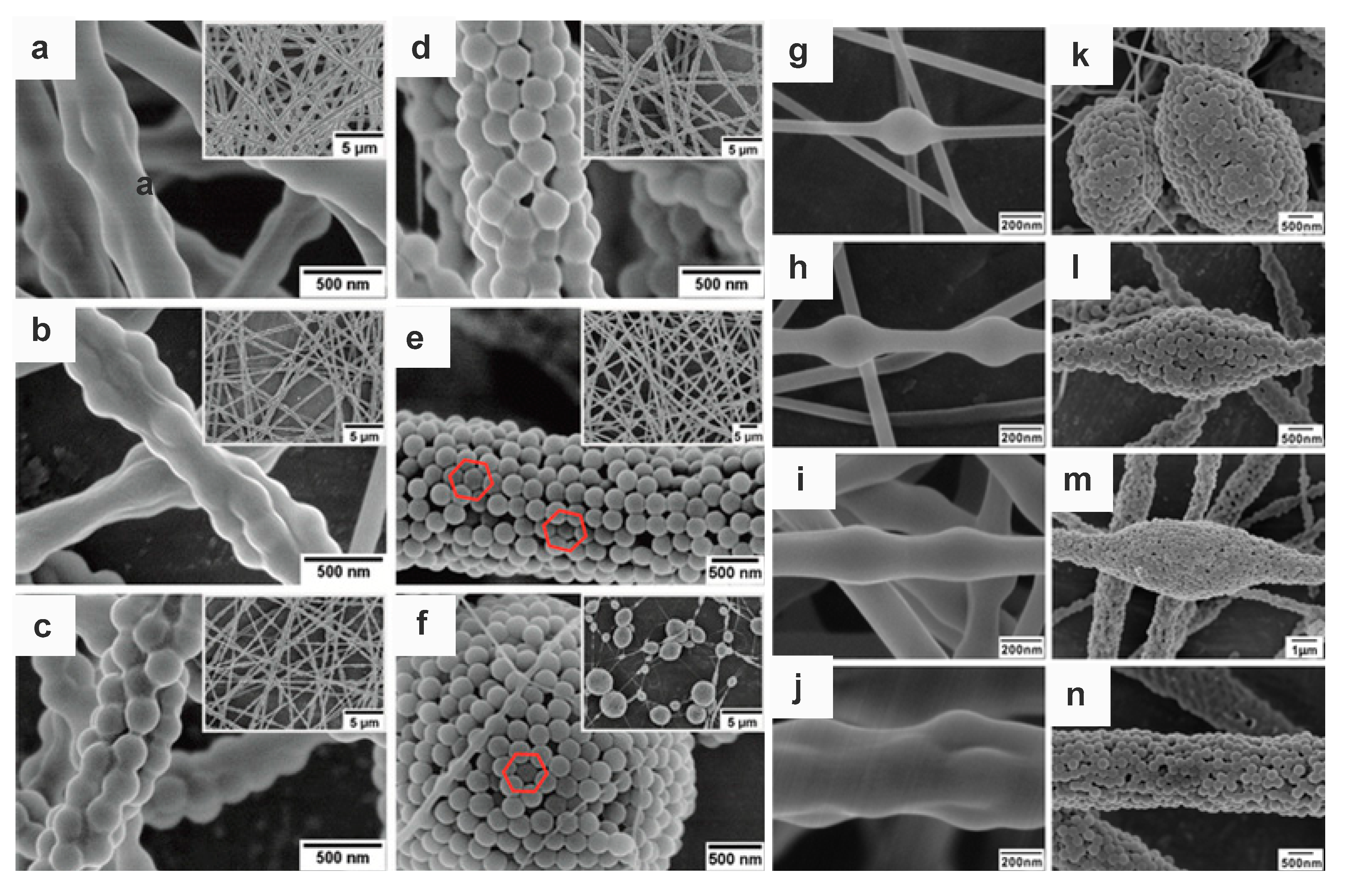
Figure 7.
SEM graphs of polystyrene nanoparticles into PVA nanofibres: (a–f) different weight ratios of PS and PVA of 1:4, 1:2, 1:1, 2:1, 4:1 and 6:1; The inserted images in each subfigure from a to f are measured under lower magnification. (g–n) different concentrations of PVA: 7, 9, 11, 13 wt%, while (g–j) PS:PVA is 1:4 and (k–n) PS:PVA is 4:1. Reprinted with permission from [12] American Chemical Society, 2012.


Table 1.
Fundamental parameters of nanoparticles contained electrospun nanofibers with well-aligned morphology.
Table 1.
Fundamental parameters of nanoparticles contained electrospun nanofibers with well-aligned morphology.
| Colloidal Nanoparticles | Sizes (nm) | C1 (wt%) | Polymer Matrix | Mw | C2 (wt%) | R | Rf (mL/h) | V (kv) | D (cm) | T; H | Morphology | Ref. |
|---|---|---|---|---|---|---|---|---|---|---|---|---|
| silica | 15, 50, 100 | 20 | polyvinyl alcohol | Mn: 66,000 | 10 | 2:3 | 1 | 10 | 10 | 25 °C 50 ± 5% | grain-like singly aligned | [77] |
| silica | 100, 300, 450, 700 | 20 | Polyacrylamide poly(ethyleneoxide) | 600,000–1,000,000 600,000 | 10 | 2:3 | 0.5–3 | 5–13 | 10 | 500 °C calcination | necklace-like | [26] |
| silica | 143, 265, 910 | 12.2 21.6 13.1 | polyvinyl alcohol | 88,000 | 12 10 12 | 500:500 600:400 200:800 300:700 | — | 10–30 | 10 | — | necklace-like blackberry-like | [32] |
| Silica polystyrene | 700, 50 237 | 34 10 | Polyacrylamide poly(ethyleneoxide) | 600,000–1,000,000 600,000 | 10 | — | 0.5 | 5–13 | 10 | calcination | stand-alone structures, superhydrophobic | [78] |
| polystyrene | 100, 200, 335 | 40 | polyvinyl alcohol | 145,000 195,000 | 6 | 80:20 | 0.7 | 0–30 | 20 | 15-18 °C 30–50% | random to relative compact packing | [79] |
| polystyrene | 225, 473 | 10, 15, 20, 30, 40 | polyvinyl alcohol | 14,500 | 13 | 1:1 2:1 4:1 | 0.2–0.8 | 10 | 15 | 25 °C 50 ± 5% | blackberry-like to uniform | [12] |
| poly (styrene-methyl methacrylate-acrylic acid) | 220, 246, 280 | 40 | polyvinyl alcohol | 14,500 | 13 | 4:1 | 0.5 | 10 | 15 | — | green, red, purplish-red color cylindrically hexagonal ordered | [69] |
| poly (N-isopropylacrylamide-co-tert-butyl acrylate) | 226 | 40 | polyacrylamide | 146,000 | 16 | 1:4 2:3 1:1 3:2 4:1 | 0.5 | 10 | 15 | 20 °C 30 ± 5% | necklace-like blackberry-like | [16] |
Where, Mw and Mn are the weight-average and number-average molecule weight of polymer, respectively; C1, C2 denote the concentration of nanoparticles and polymer matrix, respectively; R is the ratio of nanoparticle and polymer; Rf is the feed rate; V is electrospinning applied voltage; D means the distance between needle and collector; T, H represent temperature and humidity. Rf, V, D, T, H represent the parameters in the electrospinning process.
© 2018 by the authors. Licensee MDPI, Basel, Switzerland. This article is an open access article distributed under the terms and conditions of the Creative Commons Attribution (CC BY) license (http://creativecommons.org/licenses/by/4.0/).
Share and Cite
MDPI and ACS Style
Yu, J.; Kan, C.-W. Review on Fabrication of Structurally Colored Fibers by Electrospinning. Fibers 2018, 6, 70. https://0-doi-org.brum.beds.ac.uk/10.3390/fib6040070
AMA Style
Yu J, Kan C-W. Review on Fabrication of Structurally Colored Fibers by Electrospinning. Fibers. 2018; 6(4):70. https://0-doi-org.brum.beds.ac.uk/10.3390/fib6040070
Chicago/Turabian StyleYu, Jiali, and Chi-Wai Kan. 2018. "Review on Fabrication of Structurally Colored Fibers by Electrospinning" Fibers 6, no. 4: 70. https://0-doi-org.brum.beds.ac.uk/10.3390/fib6040070
Note that from the first issue of 2016, this journal uses article numbers instead of page numbers. See further details here.