Article

Adding New Pieces to the Puzzle of Karyotype Evolution in Harttia (Siluriformes, Loricariidae): Investigation of Amazonian Species

by
Francisco de M. C. Sassi
1,
Orlando Moreira-Filho
1,
Geize A. Deon
1,2,
Alexandr Sember
3,
Luiz A. C. Bertollo
1,
Thomas Liehr
4,*,
Vanessa C. S. Oliveira
1,
Patrik F. Viana
5,
Eliana Feldberg
5,
Marcelo R. Vicari
2 and
Marcelo de B. Cioffi
1
1
Laboratório de Citogenética de Peixes, Departamento de Genética e Evolução, Universidade Federal de São Carlos, São Carlos 13565-905, SP, Brazil
2
Laboratório de Biologia Cromossômica, Estrutural e Função, Departamento de Biologia Estrutural, Molecular e Genética, Universidade Estadual de Ponta Grossa, Ponta Grossa 84030-900, PR, Brazil
3
Laboratory of Fish Genetics, Institute of Animal Physiology and Genetics, Czech Academy of Sciences, Rumburská 89, 277-21 Liběchov, Czech Republic
4
Institute of Human Genetics, University Hospital Jena, Friedrich Schiller University, 07747 Jena, Germany
5
Laboratório de Genética Animal, Coordenação de Biodiversidade, Instituto Nacional de Pesquisas da Amazônia, Manaus 69067-375, AM, Brazil
*
Author to whom correspondence should be addressed.
Submission received: 25 August 2021 / Revised: 10 September 2021 / Accepted: 13 September 2021 / Published: 16 September 2021
(This article belongs to the Section Genetics and Genomics)

Abstract

:

Simple Summary

Fishes represent a useful model for evolutionary studies, given their diversity of species and habitats. In this study, we investigate the chromosomes of three unexplored Harttia fish species from the Amazonian region and compare the obtained data with previous analyses. Our data reveal that both Harttia dissidens and Harttia sp. 3 exhibit the same number of chromosomes in their cells (54), but that they differ in the karyotype organization. Harttia guianensis possesses 58 chromosomes, being thus the first representative from north Brazil to present this feature for both sexes. Although otherwise rather common in Harttia species, we observed no chromosomal differences between sexes in all but one species. Namely in Harttia sp. 3, we revealed signs of initial differentiation between homologues of one chromosome pair in males but not in females. Altogether, our data bring new evidence strengthening the view that Harttia spp. represent an informative model for studying patterns of karyotype and sex chromosome dynamics in teleost fishes.

Abstract

A remarkable morphological diversity and karyotype variability can be observed in the Neotropical armored catfish genus Harttia. These fishes offer a useful model to explore both the evolution of karyotypes and sex chromosomes, since many species possess male-heterogametic sex chromosome systems and a high rate of karyotype repatterning. Based on the karyotype organization, the chromosomal distribution of several repetitive DNA classes, and the rough estimates of genomic divergences at the intraspecific and interspecific levels via Comparative Genomic Hybridization, we identified shared diploid chromosome numbers (2n = 54) but different karyotype compositions in H. dissidens (20m + 26sm + 8a) and Harttia sp. 3 (16m + 18sm + 14st + 6a), and different 2n in H. guianensis (2n = 58; 20m + 26sm + 2st + 10a). All species further displayed similar patterns of chromosomal distribution concerning constitutive heterochromatin, 18S ribosomal DNA (rDNA) sites, and most of the surveyed microsatellite motifs. Furthermore, differences in the distribution of 5S rDNA sites and a subset of microsatellite sequences were identified. Heteromorphic sex chromosomes were lacking in H. dissidens and H. guianensis at the scale of our analysis. However, one single chromosome pair in Harttia sp. 3 males presented a remarkable accumulation of male genome-derived probe after CGH, pointing to a tentative region of early sex chromosome differentiation. Thus, our data support already previously outlined evidence that Harttia is a vital model for the investigation of teleost karyotype and sex chromosome dynamics.

1. Introduction

With more than 35,000 species [1], fishes have the richest biodiversity among vertebrates [2,3]. The evolutionary success of ray-finned and especially teleost fishes seem to be, at least partially, related to a higher plasticity of their genomes when compared to other vertebrates [4,5,6,7], going hand in hand with their propensity towards polyploidization and hybridization events [8,9]. Another factor at play is the wealth of aquatic niches fishes adapted to [2,10,11,12], and the geomorphological parameters affecting the dispersion and populational dynamics in particular species (e.g., [13,14]). In effect, at the level of genome organization, fishes present a wide range of chromosome counts and genome sizes [15,16] and the karyotype dynamics may differ considerably among lineages [17]. Finally, fishes encompass astounding variability in mechanisms of sex determination and differentiation [18,19,20,21,22].
Siluriformes (Teleostei, Ostariophysi) represent a lineage with high diversification, comprising more than 3000 species distributed among 39 families [1]. Inside Siluriformes, the family Loricariidae whose representatives are commonly known as “armored catfishes” (due to ossified plates covering their bodies; [23]) encompasses 1015 recognized species included in more than 100 genera [1]. It is hence the most species-rich siluriform family. These fishes are distributed throughout the Neotropical region, but with the greatest diversity being found from north Costa Rica to south Argentina [24]. Notably, the diversification is associated with huge karyotype variability in Loricariidae, with diploid chromosome numbers (2n) ranging from 36 in Rineloricaria latirostris to 74 in Sturisoma cf. nigrirostrum [25,26,27]. Additionally, several types of sex chromosome systems including standard, derived, and multiple ones have been described for Loricariidae, as exemplified by Hypostomus genus with different ZW and XY cases [28,29,30,31], XX/XY, XX/X0, ZZ/ZW, and Z1Z1Z2Z2/Z1Z2W1W2 in several Ancistrus species (reviewed in ref. [32]), and three male-heterogametic systems in the genus Harttia (see below).
In this context, the genus Harttia can be recognized as a remarkable model for cytogenetic studies, especially to investigate the evolutionary processes related to sex chromosomes, diversification and karyotype dynamics [33,34,35,36]. Harttia harbors 27 recognized species [1,37], of which only 14 have been cytogenetically analyzed to date, along with several other Harttia spp. waiting for proper taxonomic description (summarized in [35]). This genus also encompasses the second-largest variation in the 2n among Loricariidae, ranging from 2n = 52 (females)/53 (males) in Harttia carvalhoi [33] to 2n = 62 in H. absaberi and Harttia sp. 2 [35,38]. Furthermore, three different male-heterogametic sex chromosome systems are known or have been suggested to exist among Harttia species: XX/XY1Y2 in H. carvalhoi, H. intermontana, and Harttia sp. 1 [33,35], X1X1X2X2/X1X2Y in H. punctata, H. villasboas, and H. duriventris [34,36], in addition to a putative XX/XY in H. rondoni [36].
Harttia kronei was the first species analyzed cytogenetically in the genus [39] and, for a long time, a single representative of Harttia was studied from this perspective in the northern Brazilian region: the species H. punctata [34]. However, this gap has been progressively closed with our previous studies on Harttia duriventris, H. rondoni, and H. villasboas [36]. Here, we aim to cytogenetically investigate another three representatives from this region. For this, we applied C-banding, repetitive DNA mapping, and comparative genomic hybridization (CGH) experiments in H. dissidens, H. guianensis, and Harttia sp. 3. Our results highlight the high chromosomal dynamics within the investigated species and a putative early stage of sex chromosome differentiation, particularly in Harttia sp. 3.

2. Materials and Methods

2.1. Sampling

Three collection sites in the Brazilian Pará state (Figure 1, Table 1) were accessed, allowing the sampling of three species: Harttia dissidens, H. guianensis, and another yet undescribed taxon herein referred to as Harttia sp. 3.
Sampling was done with the authorization of the environmental agency ICMBIO/SISBIO (License 48628-14) and SISGEN (A96FF09). The species Harttia dissidens and H. guianensis were identified by Dr. Lúcia Helena Rapp-Py Daniel, curator of the fish collection of the Instituto Nacional de Pesquisa da Amazônia (INPA) and deposited there under the voucher numbers INPA-ICT 059577 and INPA-ICT 059584, respectively. The specimens of Harttia sp. 3 were scientifically described by Dr. Oswaldo Takeshi Oyakawa, from the Museu de Zoologia da Universidade de São Paulo (MZUSP).

2.2. Chromosome Obtainment and C-Banding

The living animals were anesthetized with Eugenol before the anterior kidney extraction for the obtainment of mitotic chromosomes following the classical air-drying protocol [40]. Experiments followed the ethical conducts approved by the Ethics Committee on Animal Experimentation of the Universidade Federal de São Carlos (Process number CEUA 1853260315). The detection of constitutive heterochromatin regions followed the C-banding protocol [41]. The C-banding procedure was performed on the slides previously used for the mapping of ribosomal DNAs, to allow a sequential analysis of the same metaphase plates.

2.3. Fluorescence In Situ Hybridization (FISH)-Based Experiments

Ribosomal DNA (rDNA), microsatellite, and the telomeric (TTAGGG)n sequences were used as probes for chromosomal mapping by FISH. The 5S rDNA probe was prepared according to Pendás et al. [42] from Hoplias malabaricus genome, containing a 120-base pair (bp) segment of the 5S rDNA transcribed region and 200 bp of a non-transcribed spacer (NTS). This probe was red-labeled with ATTO550-dUTP using the Nick-Translation mix kit (Jena Bioscience, Jena, Germany). The 18S rDNA probe, containing a 1400 bp-long segment of the transcribed region was also isolated from H. malabaricus following Cioffi et al. [43] and green-labeled with AF488-dUTP using the Nick-Translation mix kit (Jena Bioscience). The microsatellites (A)30, (CA)15, and (GA)15 were selected based on their abundance and patterns of chromosome distribution encountered in previous studies in different fish species [36,44], and the corresponding probes were directly labeled with Cy3 (Sigma-Aldrich, Darmstadt, Germany) during their synthesis, as described in [45]. Telomeric sequence (TTAGGG)n was in situ located using the DAKO Telomere PNA FISH Kit/FITC (DAKO, Glostrup, Denmark). High stringency conditions described in [46] were used in all FISH experiments. As a final step for all fluorescence assays, chromosomes were counterstained with 4′,6-diamidino-2-phenylindole (DAPI), and the slides were mounted in an antifade solution (VECTASHIELD; Vector Laboratories, Burlingame, CA, USA). Two sets of comparative genomic hybridization (CGH) experiments were designed to seek for the occurrence of sex chromosomes, both following the protocol of Sember et al. [47], with adaptations on probe/C0t−1 DNA ratio based on previous Harttia studies [35,36]. In the first set of experiments, male and female genomic probes were co-hybridized to the male chromosomes (i.e., the expected heterogametic sex), in each species separately, to detect regions with specific or biased hybridization of male probe which might point to regions with sex-specific repetitive DNA accumulation on sex chromosomes. For this experimental scheme, male and female-derived whole-genome DNAs (gDNAs) of all analyzed species were extracted from the liver tissue by the standard phenol-chloroform-isoamyl-alcohol method [48] and labeled with ATTO550-dUTP (male gDNA in red) and AF488-dUTP (female gDNA in green) using the Nick Translation mix kit (Jena Bioscience). To block the excess of shared repetitive sequences, an unlabeled C0t-1 DNA, obtained from each species according to Zwick et al. [49] was included in the final probe mixture. For each slide, male and female genomic probe (500 ng each) and 25 µg of female-derived C0t−1 DNA from the respective species were co-precipitated in 96% ethanol, and the dry pellets were resuspended in 20 µL of the hybridization buffer containing 50% formamide, 2×SSC, 10% dextran sulfate, and Denhardt’s buffer (pH 7.0). In the second CGH assay, the genomes of H. dissidens and H. guianensis were compared to one of H. villasboas, which possesses an X1X1X2X2/X1X2Y sex chromosome system and whose chromosome preparations were previously obtained by our research group [36]. For this purpose, male-derived gDNAs from H. dissidens, H. guianensis, and H. villasboas were extracted as described above and labeled with fluorochromes emitting green (AF488-dUTP), cyan (ATTO425-dUTP), and red (ATT0550-dUTP) fluorescence, with the Nick Translation mix kit (Jena Bioscience). The final probe mixture was composed of 500 ng of male-derived gDNA of each species and 10 µg of female-derived C0t−1 DNA of each species. This probe was then hybridized to the male metaphase plates of H. villasboas.

2.4. Image Analysis and Processing
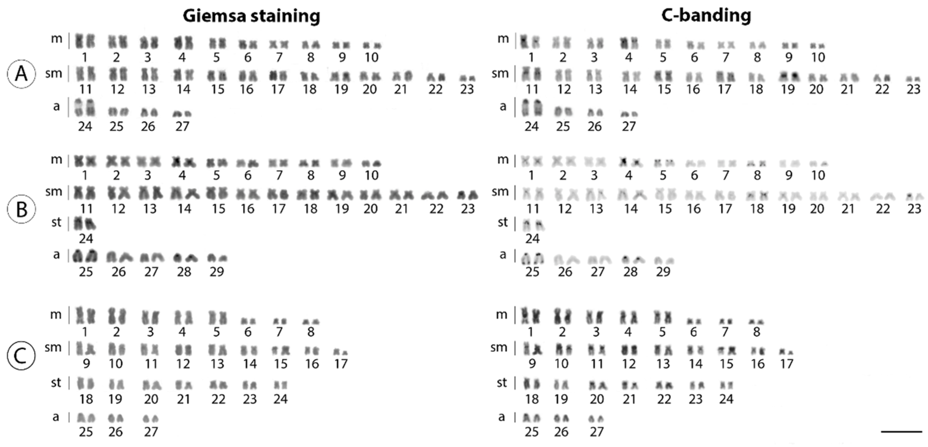
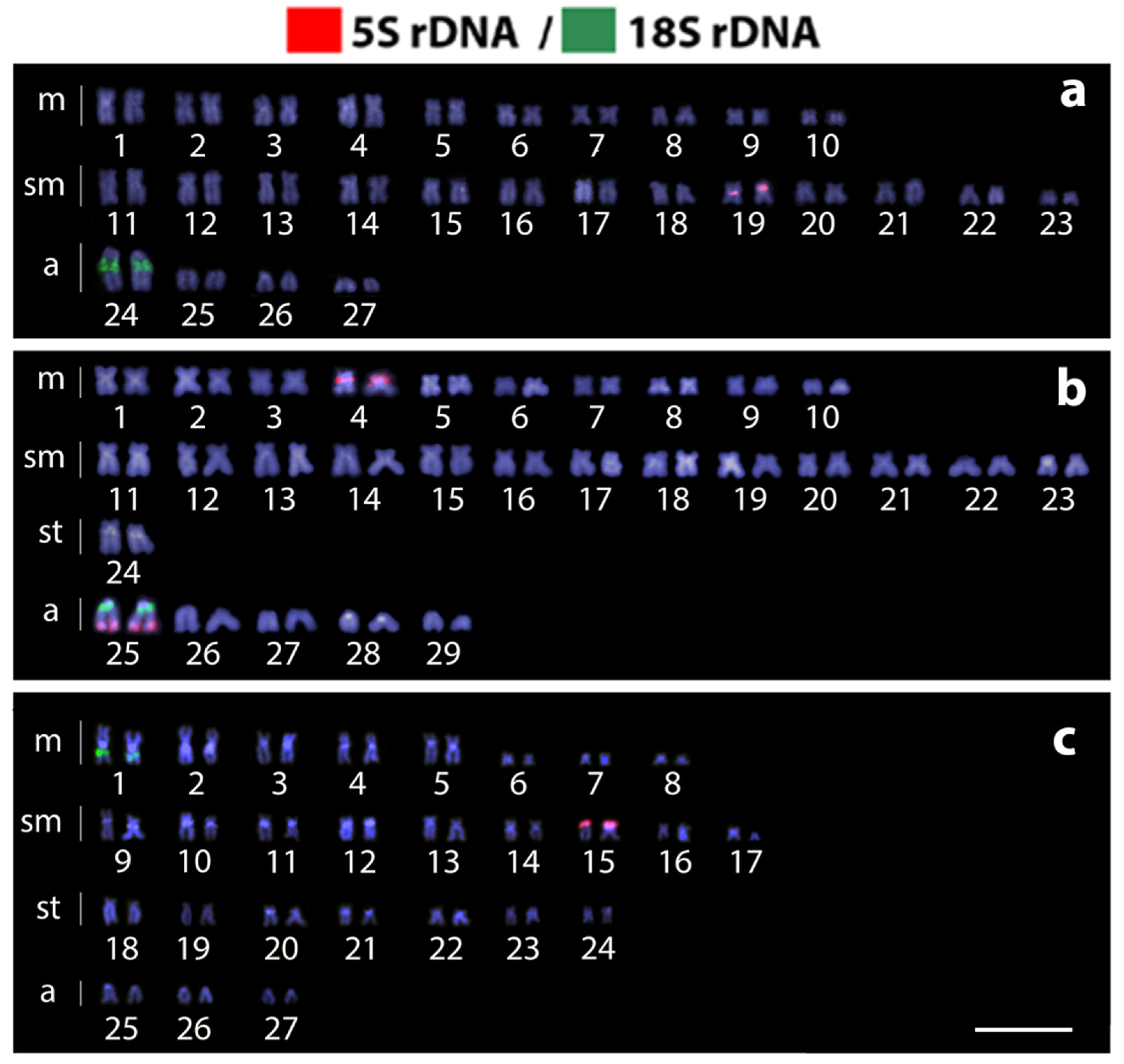
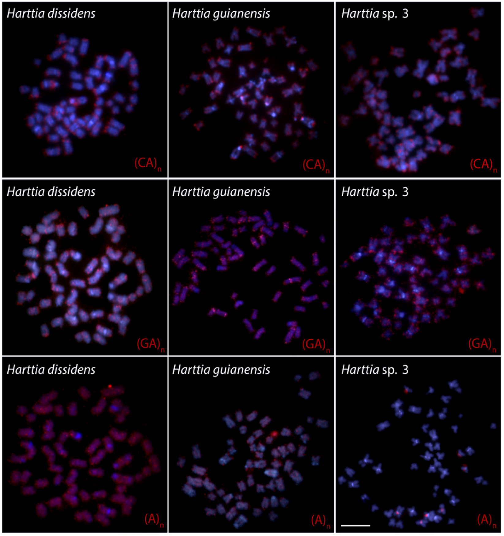
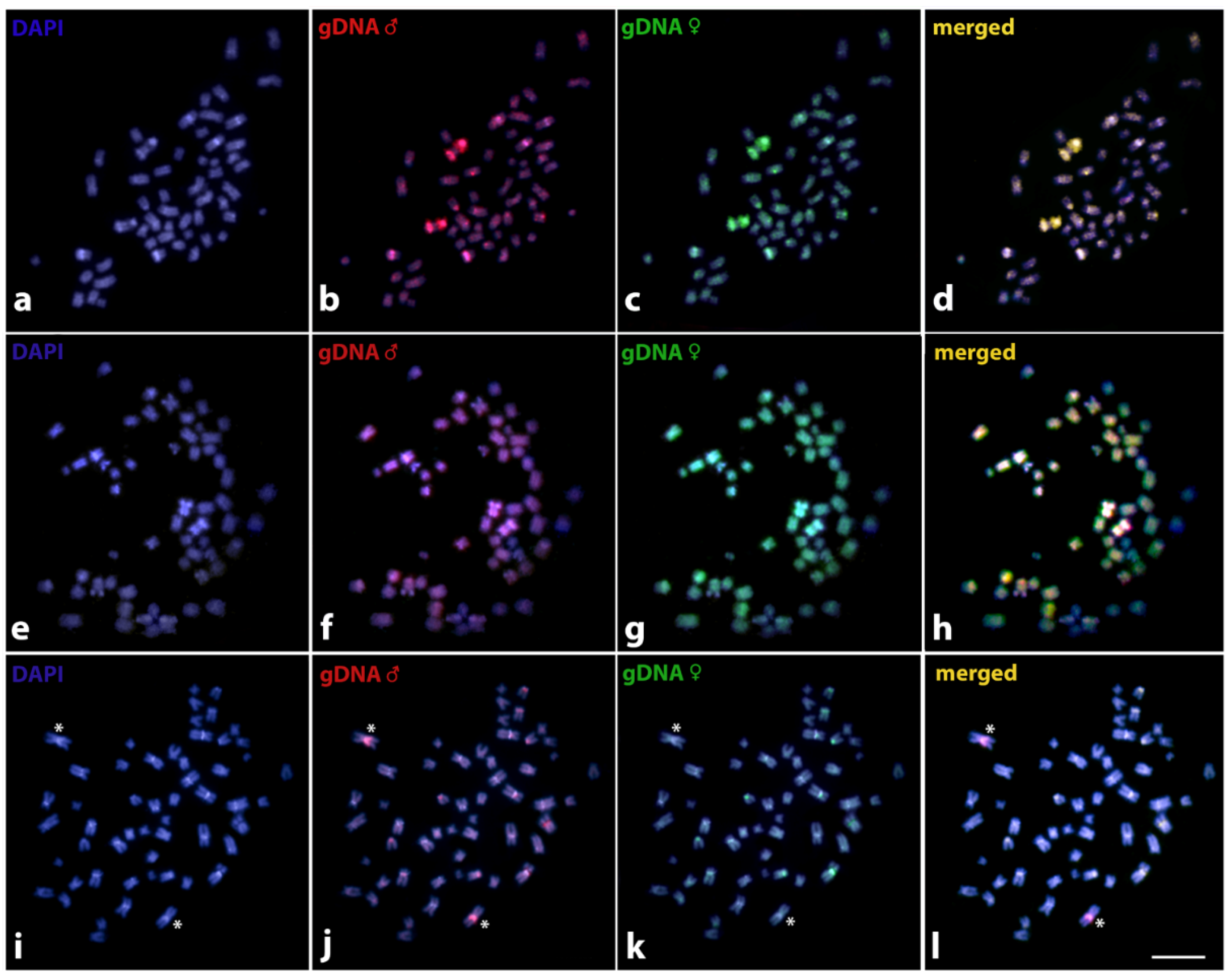
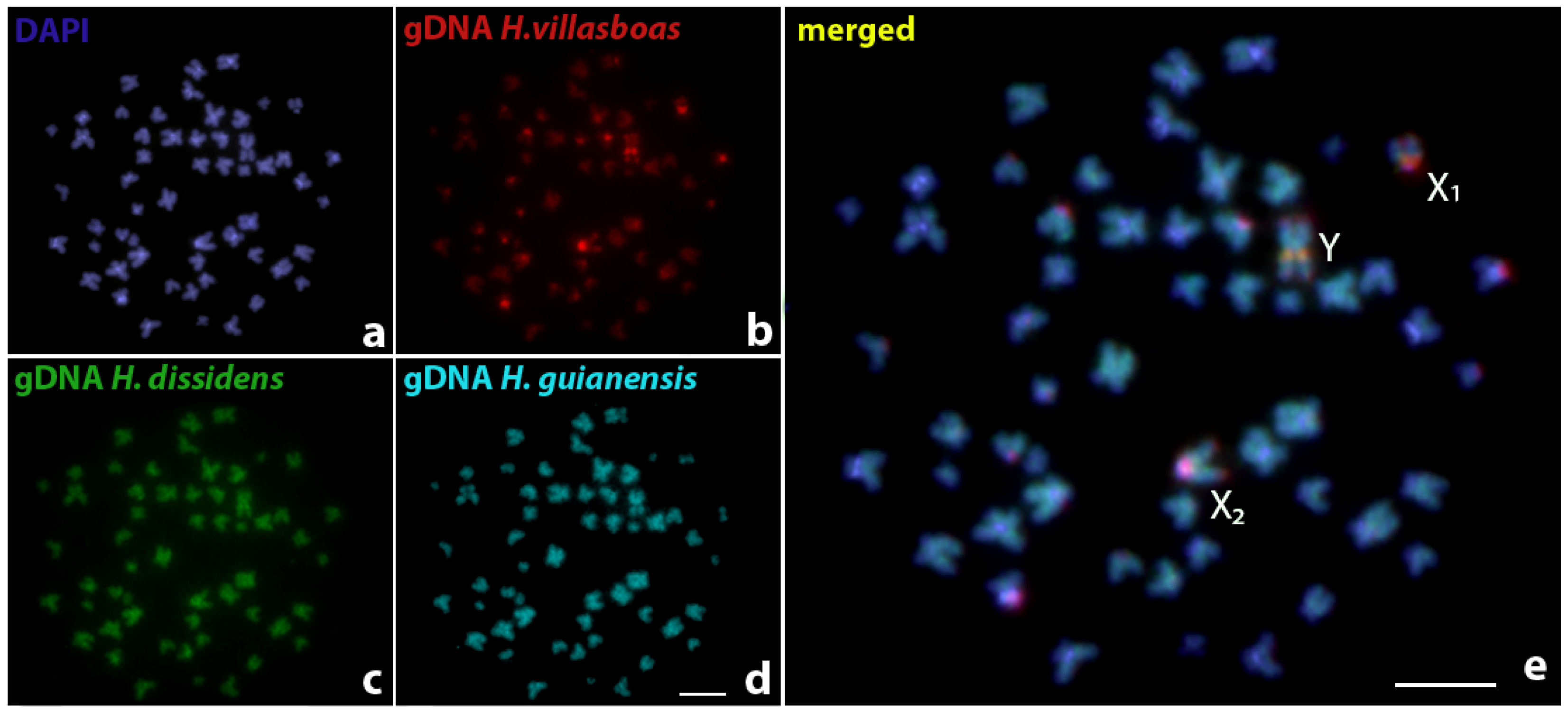
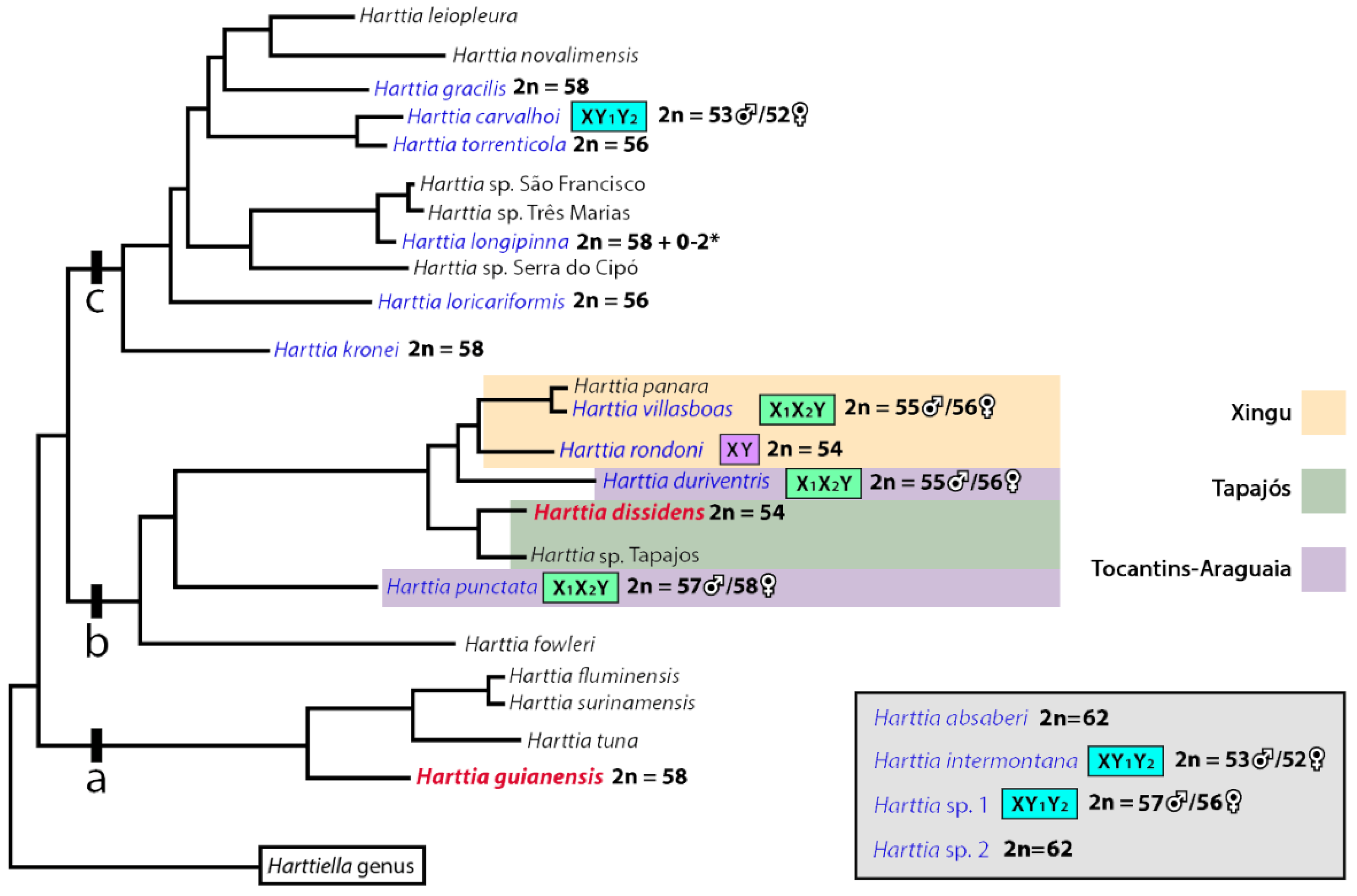
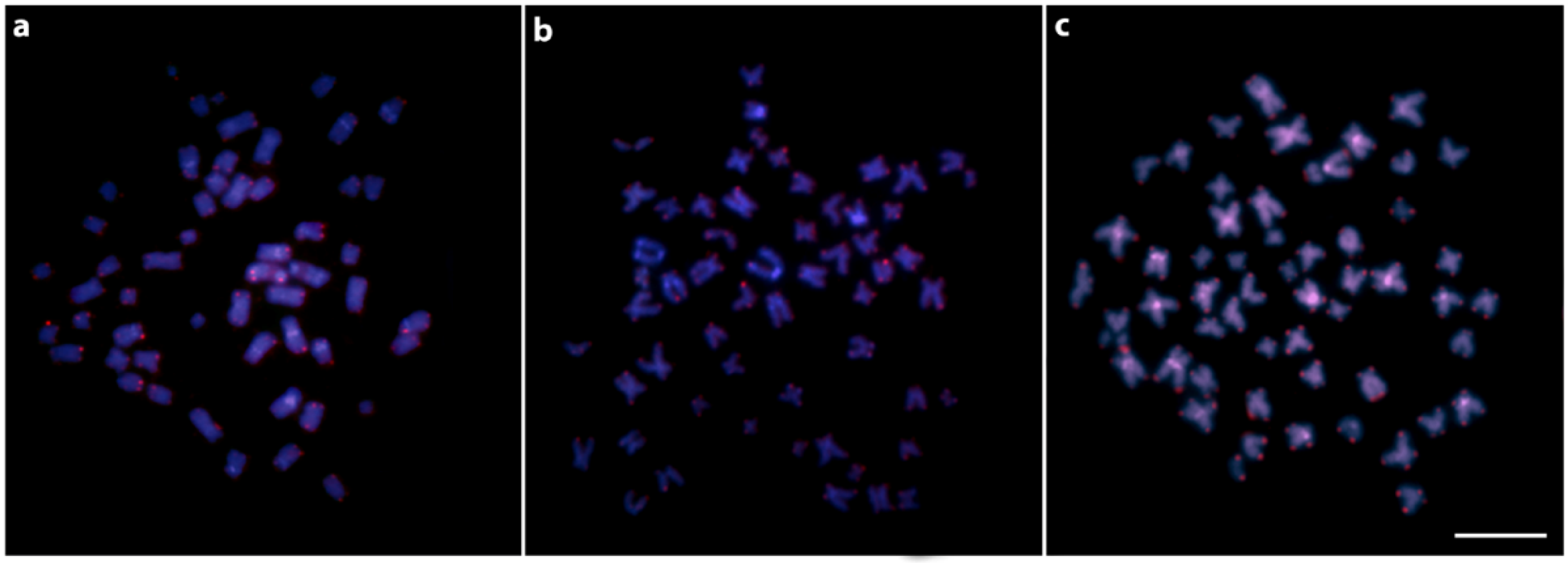
The 2n, karyotype structure, distribution of repetitive DNAs, and CGH results were confirmed by the evaluation of results in at least 30 metaphase spreads per individual. Images were captured using an Olympus BX50 microscope (Olympus Corporation, Ishikawa, Japan), coupled with a CoolSNAP camera, and processed using Image-Pro Plus 4.1 software (Media Cybernetics, Silver Spring, MD, USA). Chromosomes were classified as metacentric (m), submetacentric (sm), subtelocentric (st), or acrocentric (a) according to their arm-ratios [50]. Figures were organized with Adobe Photoshop CC 2020.

3. Results

Harttia dissidens and Harttia sp. 3 exhibited the same 2n = 54 chromosomes but differed in the karyotype composition: 20m + 26sm + 8a and 16m + 18sm + 14st + 6a, respectively. On other hand, H. guianensis differed from the other two species both in 2n and karyotype constitution: 2n = 58, and 20m + 26sm + 2st + 10a chromosomes (Figure 2). In H. dissidens and H. guianensis, constitutive heterochromatin was located preferentially in the pericentromeric and terminal regions of several chromosomes, but with distinct patterns of distribution between the species. In turn, Harttia sp. 3 differed by having larger C-positive bands, with signals distributed in the pericentromeric regions of almost all chromosomes (Figure 2). The mapping of the 18S rDNA sequence revealed a single chromosome pair bearing these sites in all three species (pair 24 in H. dissidens, pair 25 in H. guianensis, and pair 1 in Harttia sp. 3). The 5S rDNA cluster was found in a single chromosome pair in H. dissidens (pair 19) and Harttia sp. 3 (pair 15), and in two chromosome pairs (4 and 25) in H. guianensis. It is also notable that both 5S and 18S rDNA sites present a syntenic location on chromosome pair 25 in H. guianensis (Figure 3b).
The mapping of three microsatellite sequences revealed a similar pattern for (CA)15 and (GA)15, with accumulation at the terminal regions of all chromosomes. The last studied motif, (A)30, presented a dispersed distribution throughout the chromosome complement of H. dissidens and H. guianensis, while it was accumulated in the pericentromeric regions of few chromosome pairs in Harttia sp. 3 (Figure 4). The mapping of the telomeric (TTAGGG)n sequence revealed only the standard pattern in all the three species, i.e., positive signals at the physical ends of all chromosomes (Figure A1).
The CGH-based experiments between sexes in each of the three analyzed species showed approximately equal binding of both male- and female-specific probes to the vast majority of chromosomes in the male complement, with preferential location to regions with high content of repetitive DNA (yellow signals, i.e., combination of green and red fluorescence). In Harttia dissidens and H. guianensis, CGH failed to reveal any region with predominant or exclusive hybridization of the male-specific probe, thus we did not observe any signs of potential sex chromosome molecular differentiation at the level detectable by this methodological approach. In Harttia sp. 3, however, the male genomic probe predominantly (but not exclusively) marked the pericentromeric region of one large-sized metacentric chromosome pair (Figure 5).
We further performed a second CGH assay with hybridization of the male genomic probes from Harttia dissidens, H. guianensis, and H. villasboas against the chromosomal set of H. villasboas (i.e., the species previously reported to have X1X1X2X2/X1X2Y multiple sex chromosome system; see ref. [36]). We found uniform hybridization patterns of both H. dissidens and H. guianensis probes across the chromosome complement of H. villasboas males. The H. villasboas male probe seldom predominantly highlighted the regions of high repetitive content, pointing to the fact that the other two compared genomes do not (qualitatively or quantitatively) share this specific portion of H. villasboas repetitive DNA classes (Figure 6).

4. Discussion

4.1. New Pieces into the Chromosomal Evolution of Harttia

In a previous chromosomal study in Harttia species, 2n = 58 was proposed as the likely ancestral 2n for this genus [34]. A few years later, the karyotype diversity among Harttia species was described as the result of three different evolutionary pathways: (i) the maintenance of the ancestral 2n = 58 up to today; (ii) the elevation of 2n by centric fissions, and (iii) the 2n reduction by fusions [35]. Here, we add new pieces to this puzzle by analyzing the karyotypes of another three Harttia species.
In the most recent phylogenetic analysis of the subfamily Loricariinae [51], the existence of three clades within the Harttia genus was proposed. The earliest-diverging clade is composed of H. guianensis, H. tuna, H. fluminensis, and H. surinamensis which inhabit waters from the Guiana shield. The two more recently diverging clades display a well-defined and non-overlapping geographical distribution. Their species are found in southeast and south Brazilian regions, which are separated from the northern rivers. Until the present study, cytogenetical investigations only comprised fishes from the latter two clades [34,35,36].
The description of the H. guianensis karyotype in the present study reinforces the proposal of 2n = 58 as the ancestral 2n for the genus [34]. If we keep in mind that this species is basalmost in the clade, as pointed by phylogenetic reconstructions [51,52], and that it shares the same 2n with other Harttia species from other clades, such as H. gracilis, H. longipinna, and H. punctata, such hypothesis is still valid but lacks profound reconstruction analysis. Furthermore, although not a common occurrence, the syntenic organization of at least some sites of both rDNA classes is also considered as a basal karyotype feature for Loricariidae fishes [53]; this is a pattern that we also found in the karyotype of H. guianensis, in which 5S and 18S rDNA site share the location on the chromosome pair 25. Interestingly, all three Harttia species under study share one 5S rDNA site, which might be hypothetically located on homeologous chromosomes (pairs 19, 4, and 15 in H. dissidens, H. guianensis, and Harttia sp. 3, respectively). The mentioned chromosome pairs only show mild changes in shape and size between species and they differ also regarding the position of an 5S rDNA site (terminal vs. more proximal), which could be explained by paracentric inversions. The second extra 5S rDNA site found in H. guianensis only, i.e., the one found in synteny with 18S rDNA cluster on chromosome pair 25 is a karyotype trait that has been so far reported only for two other Harttia species: H. carvalhoi [34] and Harttia sp. 1 [35]. Given the position of H. guianensis at the base of Harttia phylogeny [51], it seems probable that this second 5S rDNA site has been eliminated during evolution in many Harttia species while it was retained in a subset of them.
The 5S rDNA site has been either entirely eliminated or reduced in copy number of repeats below the resolution of the standard FISH protocol. Based on tracking the location of the 18S rDNA site, we may infer that particularly this linkage group underwent a dynamic evolution (see below). Based on the comparison of 18S rDNA patterns, it is highly probable that the chromosome pair bearing the 18S rDNA site was involved in a fusion event in H. dissidens and Harttia sp. 3. This may be inferred from the terminal location of 18S rDNA site on a rather middle-sized chromosome pair in H. guianensis vs. an interstitial position of 18S rDNA cluster on large-sized chromosomes in the remaining two species. While the presence of a large metacentric pair in Harttia sp. 3 supports centric fusion as an underlying mechanism, the large 18S rDNA-bearing chromosome in H. dissidens is acrocentric and hence either tandem fusion took place here or the situation was more complex (i.e., involving more consecutive rearrangements).
For loricariid genomes, it was proposed that the evolutionary breakpoint regions occur adjacent to rDNA sites, promoting double-strand breaks and the reorganization of these sequences in Ancistrus [54], Harttia [35,36], and Rineloricaria [55]. The number of studies showing rDNA sites inside or nearby a predicted fusion points is steadily growing for fishes [56,57,58,59] and a high potential of rDNAs to facilitate chromosome rearrangements has also been shown in mice and humans (e.g., [60,61]). A possible explanation for rDNA instability might be a high transcriptional activity of tandemly arrayed rDNA genes due to the necessity to synthetize high amounts of ribosomes in the cell [62]. Long stretches of transcriptionally active euchromatin might be prone to double-strand breaks [63,64]. Interestingly, mapping of telomeric repeats did not reveal any interstitial telomeric sites (ITSs), which could point to regions of former rearrangements, in our case specifically fusions or inversions; therefore, it seems that the mechanisms of these rearrangements in Harttia usually do not preserve these sequences in the fusion points [65,66].
The two species analyzed herein, H. dissidens and Harttia sp. 3, share the same chromosome count (2n = 54) as was previously only described in H. rondoni [36], a species that also inhabits the northern Brazilian region. Indeed, H. dissidens occurs in the Tapajós river basin [67], and H. rondoni inhabits the Xingu River basin [68]. The range of distribution of Harttia sp. 3 cannot be inferred from the current data. This suspected new Harttia species was collected in a small rocky-bottom river located on the edge of Serra do Cachimbo, 700 m highlands dividing the Tapajós and Xingu basins. Therefore, it is not yet known whether it flows into the Tapajós River basin or the Xingu River basin.
Although the karyotypes of H. dissidens, H. rondoni, and Harttia sp. 3 share 2n equal to 54 chromosomes, notable differences can be found between the species at the level of karyotype composition, and distribution of repetitive DNA classes–either specific ones or the uncharacterized repetitive DNA fraction revealed by CGH. Taking an important role in the initial steps of sex chromosome differentiation, in post-zygotic reproductive isolation, or by contributing to local adaptation in certain populations, chromosomal inversions can suppress the recombination, especially around the rearrangement breakpoints [69,70,71,72,73,74,75,76,77]. In addition, repetitive DNAs may also play an important role in promoting biodiversity, in the differentiation of sex-specific chromosomal regions, and speciation of diverse eukaryotic organisms including fishes [78,79,80,81]. As evidenced in our present study, the differences in karyotype organization and number and distribution of rDNA sites are instrumental as chromosomal markers for the determination of Harttia species with 2n = 54 chromosomes. The fixation of such karyotype differences might be facilitated by vicariant events after the Serra do Cachimbo uplifting [36]. This event seems to have played a significant role in the diversification of Harttia species, as pointed out by the position of the clades in the phylogenetic reconstruction of this genus. Species from Xingu and Tocantins-Araguaia basins, which have at least two different sex chromosome systems [36], form a sister clade to the branch which encompasses species from the Tapajós basin [57]. One member of the latter group investigated herein, H. dissidens, does not present a cytologically detectable sex chromosome system. Additionally, H. punctata, a species from the Tocantins-Araguaia basin which possesses an X1X1X2X2/X1X2Y multiple sex chromosome system, corresponds to the sister group of all those above-mentioned species in their phylogenetic reconstruction ([52], Figure 7).

4.2. Sex Chromosomes in Harttia species

At least nine distinct sex chromosome systems are described among fishes, so far known to be distributed in approx. 1% of teleost species (440 cases according to [83]). Many of these systems present only subtle genetic differentiation, which is not detectable by standard cytogenetic methods [19,83].
Until now, Harttia has presented three distinct male-heterogametic sex chromosome systems: (i) X1X1X2X2/X1X2Y found in H. punctata [34], H. duriventris and H. villasboas [36], (ii) XX/XY1Y2 found in H. carvalhoi [33,34], H. intermontana and Harttia sp. 1 [35], and (iii) the putative XX/XY sex chromosome system at an early stage of differentiation proposed for H. rondoni [36]. Consequently, Harttia can be recognized as a taxon with high karyotype dynamics, which is prone to formation of multiple sex chromosome systems. Up to now, 75 cases of multiple sex chromosomes with predicted 60 independent origins have been reviewed by Sember et al. [83] and another two systems have been concomitantly described in Harttia [35]. Together with Nothobranchius, Harttia is the genus with highest number (N = 6) of multiple sex chromosomes (reviewed in ref. [83]).
Interestingly, although largely different from many viewpoints, both mentioned teleost taxa (Harttia and Nothobranchius) share the evolution in small allopatric populations with restricted gene flow and propensity to fusions and fissions. It is becoming increasingly apparent that Robertsonian rearrangements might have a significant effect on the nuclear organization of chromosomes along with gene evolution and expression (e.g., [84]). Although present in seven out of the 14 karyotyped species, differentiated sex chromosomes are still intensely discussed concerning the ancestral karyotype of Harttia. For this, two main scenarios have been already proposed [36]. The first one—“the proto-XY hypothesis”— considers both X and Y chromosomes to differ only by amplification or contraction of tandem repeats, thus representing an early stage of sex chromosome differentiation. Following this presumption, a proto-XY sex chromosome system, like the one found in H. rondoni, could be present in the ancestral karyotype and, consequently, would have originated the X1X2Y sex chromosome system found in H. duriventris, H. punctata, and H. villasboas via centric fissions [36]. A possible second hypothesis, named “the neo-XY system”, considers the fusions of ancestral X and Y chromosomes with an autosome pair, thus leading to the formation of neo-XY sex chromosomes accompanied by the reduction of 2n [36]. A third hypothesis partially overlaps with the second one and it can be considered for the southeast Brazilian clade of Harttia, which does not present any proto-sex, or heteromorphic XY sex chromosomes identified so far. In this case, similar CGH patterns and the phylogenetic relationships between H. torrenticola (without differentiated sex chromosomes) and H. carvalhoi (with an XY1Y2 sex system), point to a Robertsonian fusion originating the X-chromosome of H. carvalhoi, H. intermontana, and Harttia sp. 1, as well as the first chromosome pair of H. torrenticola [35]. Such hypotheses, especially the first two of them, were proposed assuming the scenario in which all species from the northern Brazilian region present differentiated sex chromosomes in their karyotypes. However, this is not the case, as the present CGH experiments confirmed that H. dissidens and H. guianensis do not possess differentiated sex chromosomes, at least not at the stage which could be detected by CGH [47,85]. These data suggest rather early steps of differentiation in the Harttia species with XX/XY1X2 sex chromosome system when compared to the karyotypes with homomorphic sex chromosomes in the clade from northern Brazilian region.
Recent genomic studies continue to reveal homomorphic sex chromosome systems among fishes, thus pointing to a potentially gross underestimation regarding the counts of fish sex chromosomes cases by previous cytogenetic reports [86,87]. This might especially apply to the Harttia genus, where several cryptic and undescribed species occur, in addition to the scarcity of cytogenetic (and a complete absence of genomic) data, especially for species from both northern Brazil and Guyana shield. Our CGH experiments utilizing male and female genomic probes to be compared on the male chromosome background in Harttia sp. 3, revealed a specific pericentromeric region in one metacentric pair (pair 2) highlighted more (but not exclusively) by the male genomic probe. This observation might point to a nascent region of suppressed recombination on homomorphic sex chromosomes [47], bearing in mind that specific repetitive DNA accumulations might happen on corresponding regions of both sex chromosomes [88]; however, we cannot entirely discard the possibility of intraspecific variability in the copy number and distribution of certain repetitive DNAs.

Author Contributions

Formal analysis, F.d.M.C.S., O.M.-F., G.A.D., A.S., T.L., V.C.S.O., P.F.V., M.R.V. and M.d.B.C.; Funding acquisition, M.d.B.C.; Investigation, F.d.M.C.S., O.M.-F., G.A.D., A.S., L.A.C.B., T.L., P.F.V., E.F. and M.d.B.C.; Methodology, F.d.M.C.S., G.A.D., T.L., V.C.S.O., P.F.V. and M.d.B.C.; Project administration, O.M.-F., T.L. and M.d.B.C.; Validation, F.d.M.C.S., O.M.-F., G.A.D., A.S., L.A.C.B., T.L., V.C.S.O., P.F.V., E.F., M.R.V. and M.d.B.C.; Visualization, F.d.M.C.S., A.S., L.A.C.B., T.L., V.C.S.O., E.F., M.R.V. and M.d.B.C.; Writing—original draft, F.d.M.C.S. and M.d.B.C.; Writing—review & editing, O.M.-F., G.A.D., A.S., L.A.C.B., V.C.S.O., P.F.V., E.F. and M.R.V. All authors have read and agreed to the published version of the manuscript.

Funding

This research was funded by FAPESP, grant numbers 2020/02681-9 and 2020/11772-8. A.S. was supported by RVO: 67985904 of IAPG CAS, Liběchov.

Institutional Review Board Statement

Sampling was done with the authorization of the environmental agency ICMBIO/SISBIO (License 48628-14) and SISGEN (A96FF09). Experiments followed the ethical conduct approved by the Ethics Committee on Animal Experimentation of the Universidade Federal de São Carlos (Process number CEUA 1853260315).

Informed Consent Statement

Not applicable.

Data Availability Statement

The data presented in this study are available on request from the corresponding author.

Acknowledgments

Special thanks to Alany Pedrosa Gonçalves (Mamirauá Sustainable Development Reserve) and Cárlison Silva de Oliveira for the indication of sampling sites, to Marcella Costa Radael (Federal University of West Pará-UFOPA) for the support getting chemical reagents during our fieldwork, and to Lucia Rapp-Py Daniel (National Institute of Amazonian Research–INPA) and Oswaldo Oyakawa (USP Zoology Museum) for species identification.

Conflicts of Interest

The authors declare no conflict of interest. The funders had no role in the design of the study; in the collection, analyses, or interpretation of data; in the writing of the manuscript, or in the decision to publish the results.

Appendix A

Figure A1. Distribution of the telomeric probe (TTAGGG)n directly labeled with Cy3-dUTP (red) and hybridized against the DAPI-counterstained (blue) chromosomes of Harttia dissidens (a), H. guianensis (b) and Harttia sp. 3 (c). Bar scale = 10 µm.
Biology 10 00922 g0a1

References

  1. Fricke, R.; Eschmeyer, W.N.; van der Laan, R. Eschmeyer’s Catalog of Fishes: Genera, Species, References. Available online: http://researcharchive.calacademy.org/research/ichthyology/catalog/fishcatmain.asp (accessed on 24 August 2020).
  2. Dudgeon, D.; Arthington, A.H.; Gessner, M.O.; Kawabata, Z.-I.; Knowler, D.J.; Lévêque, C.; Naiman, R.J.; Prieur-Richard, A.-H.; Soto, D.; Stiassny, M.L.J. Freshwater biodiversity: Importance, threats, status and conservation challenges. Biol. Rev. 2006, 81, 163–182. [Google Scholar] [CrossRef]
  3. Nelson, J.S.; Grande, T.C.; Wilson, M.V.H. Fishes of the World, 5th ed.; John Wiley & Sons: Hoboken, NJ, USA, 2016; ISBN 111834233X. [Google Scholar]
  4. Volff, J.N. Genome evolution and biodiversity in teleost fish. Heredity 2005, 94, 280–294. [Google Scholar] [CrossRef] [PubMed] [Green Version]
  5. Ravi, V.; Venkatesh, B. Rapidly evolving fish genomes and teleost diversity. Curr. Opin. Genet. Dev. 2008, 18, 544–550. [Google Scholar] [CrossRef] [PubMed]
  6. Lee, A.P.; Kerk, S.Y.; Tan, Y.Y.; Brenner, S.; Venkatesh, B. Ancient vertebrate conserved noncoding elements have been evolving rapidly in teleost fishes. Mol. Biol. Evol. 2011, 28, 1205–1215. [Google Scholar] [CrossRef] [Green Version]
  7. Sallan, L.C. Major issues in the origins of ray-finned fish (Actinopterygii) biodiversity. Biol. Rev. 2014, 89, 950–971. [Google Scholar] [CrossRef] [PubMed]
  8. Mable, B.K.; Alexandrou, M.A.; Taylor, M.I. Genome duplication in amphibians and fish: An extended synthesis. J. Zool. 2011, 284, 151–182. [Google Scholar] [CrossRef]
  9. Braasch, I.; Postlethwait, J.H. Polyploidy in fish and the teleost genome duplication. In Polyploidy and Genome Evolution; Springer: Berlin/Heidelberg, Germany, 2012; pp. 341–383. [Google Scholar]
  10. Bernardi, G. Speciation in fishes. Mol. Ecol. 2013, 22, 5487–5502. [Google Scholar] [CrossRef]
  11. Pörtner, H.O.; Schulte, P.M.; Wood, C.M.; Schiemer, F. Niche dimensions in fishes: An integrative view. Physiol. Biochem. Zool. 2010, 83, 808–826. [Google Scholar] [CrossRef] [Green Version]
  12. Reis, R.E.; Albert, J.S.; Di Dario, F.; Mincarone, M.M.; Petry, P.; Rocha, L.A. Fish biodiversity and conservation in South America. J. Fish Biol. 2016, 89, 12–47. [Google Scholar] [CrossRef] [Green Version]
  13. Albert, J.S.; Reis, R. Historical Biogeography of Neotropical Freshwater Fishes; Univ of California Press: Berkeley, CA, USA, 2011; ISBN 0520948505. [Google Scholar]
  14. Albert, J.S.; Tagliacollo, V.A.; Dagosta, F. Diversification of Neotropical freshwater fishes. Annu. Rev. Ecol. Evol. Syst. 2020, 51, 27–53. [Google Scholar] [CrossRef]
  15. Arai, R. Fish Karyotypes: A Check List; Springer Science & Business Media: Berlin/Heidelberg, Germany, 2011. [Google Scholar]
  16. Gregory, T.R. Animal Genome Size Database. Available online: http://www.genomesize.com (accessed on 19 August 2021).
  17. Yoshida, K.; Kitano, J. Tempo and mode in karyotype evolution revealed by a probabilistic model incorporating both chromosome number and morphology. PLoS Genet. 2021, 17, e1009502. [Google Scholar] [CrossRef]
  18. Devlin, R.H.; Nagahama, Y. Sex determination and sex differentiation in fish: An overview of genetic, physiological, and environmental influences. Aquaculture 2002, 208, 191–364. [Google Scholar] [CrossRef]
  19. Schartl, M.; Schmid, M.; Nanda, I. Dynamics of vertebrate sex chromosome evolution: From equal size to giants and dwarfs. Chromosoma 2016, 125, 553–571. [Google Scholar] [CrossRef] [PubMed]
  20. Guiguen, Y.; Fostier, A.; Herpin, A. Sex determination and differentiation in fish: Genetic, genomic, and endocrine aspects. In Sex Control in Aquaculture, 1st ed.; Wang, H.-P., Piferrer, F., Chen, S.-L., Eds.; John Wiley & Sons: Hoboken, NJ, USA, 2018; pp. 35–63. [Google Scholar]
  21. Shen, Z.; Wang, H. Environmental sex determination and sex differentiation in teleost—How sex is established. In Sex Control in Aquaculture, 1st ed.; Wang, H.-P., Piferrer, F., Chen, S.-L., Eds.; John Wiley & Sons: Hoboken, NJ, USA, 2018; pp. 85–115. [Google Scholar]
  22. Kuwamura, T.; Sunobe, T.; Sakai, Y.; Kadota, T.; Sawada, K. Hermaphroditism in fishes: An annotated list of species, phylogeny, and mating system. Ichthyol. Res. 2020, 67, 341–360. [Google Scholar] [CrossRef]
  23. Schaefer, S.A.; Lauder, G.V. Historical transformation of functional design: Evolutionary morphology of feeding mechanisms in loricarioid catfishes. Syst. Zool. 1986, 35, 489–508. [Google Scholar] [CrossRef]
  24. Reis, R.E.; Kullander, S.; Ferraris, C.J. Check List of the Freshwater Fishes of South and Central America; Edipucrs: Porto Alegre, Brazil, 2003; ISBN 8574303615. [Google Scholar]
  25. Kavalco, K.F.; Pazza, R.; Bertollo, L.A.C.; Moreira-Filho, O. Karyotypic diversity and evolution of Loricariidae (Pisces, Siluriformes). Heredity 2005, 94, 180–186. [Google Scholar] [CrossRef] [Green Version]
  26. Takagui, F.H.; Baumgärtner, L.; Venturelli, N.B.; Paiz, L.M.; Viana, P.; Dionísio, J.F.; Pompeo, L.R.S.; Margarido, V.P.; Fenocchio, A.S.; da Rosa, R. Unrevealing the karyotypic evolution and cytotaxonomy of armored catfishes (Loricariinae) with emphasis in Sturisoma, Loricariichthys, Loricaria, Proloricaria, Pyxiloricaria, and Rineloricaria. Zebrafish 2020, 17, 319–332. [Google Scholar] [CrossRef]
  27. Giuliano-Caetano, L. Polimorfismo Cromossômico Robertsoniano Em Populações de Rineloricaria latirostris (Pisces, Loricariinae); Universidade Federal de Sâo Carlos: São Carlo, Brazil, 1998. [Google Scholar]
  28. de Lara Kamei, M.C.S.; Baumgärtner, L.; Paiva, S.; Zawadzki, C.H.; Martins-Santos, I.C.; de Portela-Castro, A.L.B. Chromosomal diversity of three species of Hypostomus Lacépède, 1803 (Siluriformes, Loricariidae), from the Paraná River Basin, Brazil: A species complex in Hypostomus Ancistroides reinforced by a ZZ/ZW sex chromosome system. Zebrafish 2017, 14, 357–363. [Google Scholar] [CrossRef] [PubMed]
  29. Takagui, F.H.; de Moura, L.F.; Ferreira, D.C.; Centofante, L.; de A. Vitorino, C.; Bueno, V.; Margarido, V.P.; Venere, P.C. Karyotype diversity in Doradidae (Siluriformes, Doradoidea) and presence of the heteromorphic ZZ/ZW sex chromosome system in the family. Zebrafish 2017, 14, 236–243. [Google Scholar] [CrossRef] [PubMed]
  30. Rocha-Reis, D.A.; de Oliveira Brandão, K.; de Almeida-Toledo, L.F.; Pazza, R.; Kavalco, K.F. The persevering cytotaxonomy: Discovery of a unique XX/XY sex chromosome system in catfishes suggests the existence of a new, endemic and rare species. Cytogenet. Genome Res. 2018, 156, 45–55. [Google Scholar] [CrossRef] [PubMed]
  31. de Oliveira, L.C.; Ribeiro, M.O.; de Medeiros Costa, G.; Zawadzki, C.H.; Prizon-Nakajima, A.C.; Borin-Carvalho, L.A.; Martins-Santos, I.C.; de Brito Portela-Castro, A.L. Cytogenetic characterization of Hypostomus soniae Hollanda-Carvalho & Weber, 2004 from the Teles Pires River, southern Amazon basin: Evidence of an early stage of an XX/XY sex chromosome system. Comp. Cytogenet. 2019, 13, 411. [Google Scholar]
  32. Glugoski, L.; Deon, G.; Schott, S.; Vicari, M.R.; Nogaroto, V.; Moreira-Filho, O. Comparative cytogenetic analyses in Ancistrus species (Siluriformes: Loricariidae). Neotrop. Ichthyol. 2020, 18, e200013. [Google Scholar] [CrossRef]
  33. Centofante, L.; Bertollo, L.A.C.; Moreira-Filho, O. Cytogenetic characterization and description of an XX/XY1Y2 sex chromosome system in catfish Harttia carvalhoi (Siluriformes, Loricariidae). Cytogenet. Genome Res. 2006, 320–324. [Google Scholar] [CrossRef]
  34. Blanco, D.R.; Vicari, M.R.; Lui, R.L.; Traldi, J.B.; Bueno, V.; de Martinez, J.F.; Brandão, H.; Oyakawa, O.T.; Moreira Filho, O. Karyotype diversity and evolutionary trends in armored catfish species of the genus Harttia (Siluriformes: Loricariidae). Zebrafish 2017, 14, 169–176. [Google Scholar] [CrossRef] [PubMed]
  35. Deon, G.A.; Glugoski, L.; Vicari, M.R.; Nogaroto, V.; Sassi, F.M.C.; Cioffi, M.B.; Liehr, T.; Bertollo, L.A.C.; Moreira-Filho, O. Highly rearranged karyotypes and multiple sex chromosome systems in armored catfishes from the genus Harttia (Teleostei, Siluriformes). Genes 2020, 11, 1366. [Google Scholar] [CrossRef] [PubMed]
  36. Sassi, F.M.C.; Deon, G.A.; Moreira-Filho, O.; Vicari, M.R.; Bertollo, L.A.C.; Liehr, T.; de Oliveira, E.A.; Cioffi, M.B. Multiple sex chromosomes and evolutionary relationships in Amazonian catfishes: The outstanding model of the genus Harttia (Siluriformes: Loricariidae). Genes 2020, 11, 1179. [Google Scholar] [CrossRef]
  37. De Oliveira, J.C.; Oyakawa, O.T. New loricariid fishes from headwaters on Serra da Mantiqueira and Complexo do Espinhaço, Minas Gerais State, Brazil (Teleostei: Siluriformes: Loricariidae). Zootaxa 2019, 4586, 401–424. [Google Scholar] [CrossRef] [PubMed]
  38. Rodrigues, R.M. Estudos Cromossômicos e Moleculares em Loricariinae Com Ênfase em Espécies de Rineloricaria (Siluriformes, Loricariidae): Uma Perspectiva Evolutiva. Ph.D. Thesis, Universidade de São Paulo, São Paulo, Brazil, 2010. [Google Scholar]
  39. Alves, A.L.; Oliveira, C.; Foresti, F. Karyotype variability in eight species of the subfamilies Loricariinae and Ancistrinae (Teleostei, Siluriformes, Loricariidae). Caryologia 2003, 56, 57–63. [Google Scholar] [CrossRef] [Green Version]
  40. Bertollo, L.A.C.; Cioffi, M.B.; Moreira-Filho, O. Direct chromosome preparation from freshwater teleost fishes. In Fish Cytogenetic Techniques (Chondrichthyans and Teleosts); Ozouf-Costaz, C., Pisano, E., Foresti, F., Almeida Toledo, L.F., Eds.; CRC Press: Boca Raton, FL, USA, 2015; pp. 21–26. [Google Scholar]
  41. Sumner, A.T. A simple technique for demonstrating centromeric heterochromatin. Exp. Cell Res. 1972, 75, 304–306. [Google Scholar] [CrossRef]
  42. Pendás, A.M.; Móran, P.; Freije, J.P.; Garcia-Vásquez, E. Chromosomal location and nucleotide sequence of two tandem repeats of the Atlantic salmon 5S rDNA. Cytogenet. Cell Genet. 1994, 67, 31–36. [Google Scholar] [CrossRef]
  43. Cioffi, M.B.; Martins, C.; Centofante, L.; Jacobina, U.P.; Bertollo, L.A.C. Chromosomal variability among allopatric populations of Erythrinidae fish Hoplias malabaricus: Mapping of three classes of repetitive DNAs. Cytogenet. Genome Res. 2009, 125, 132–141. [Google Scholar] [CrossRef] [PubMed]
  44. Phimphan, S.; Chaiyasan, P.; Suwannapoom, C.; Reungsing, M.; Juntaree, S.; Tanomtong, A.; Supiwong, W. Comparative karyotype study of three Cyprinids (Cyprinidae, Cyprininae) in Thailand by classical cytogenetic and FISH techniques. Comp. Cytogenet. 2020, 14, 597. [Google Scholar] [CrossRef] [PubMed]
  45. Kubat, Z.; Hobza, R.; Vyskot, B.; Kejnovský, E. Microsatellite accumulation in the Y chromosome of Silene latifolia. Genome 2008, 51, 350–356. [Google Scholar] [CrossRef] [Green Version]
  46. Yano, C.F.; Bertollo, L.A.C.; Cioffi, M.B. Fish-FISH: Molecular cytogenetics in fish species. In Fluorescence In Situ Hybridization (FISH)—Application Guide; Liehr, T., Ed.; Springer: Berlin, Germany, 2017; pp. 429–444. [Google Scholar]
  47. Sember, A.; Bertollo, L.A.C.; Ráb, P.; Yano, C.F.; Hatanaka, T.; de Oliveira, E.A.; Cioffi, M.B. Sex chromosome evolution and genomic divergence in the fish Hoplias malabaricus (Characiformes, Erythrinidae). Front. Genet. 2018, 9, 71. [Google Scholar] [CrossRef]
  48. Sambrook, J.; Russell, D.W. Molecular Cloning: A Laboratory Manual; Cold Spring Harbor Laboratory Press: New York, NY, USA, 2001; Volume 855. [Google Scholar]
  49. Zwick, M.S.; Hanson, R.E.; Mcknight, T.D.; Islam-Faridi, M.H.; Stelly, D.M.; Wing, R.A.; Price, H.J. A rprocedure for the isolation of C0t−1 DNA from plants. Genome 1997, 40, 138–142. [Google Scholar] [CrossRef] [Green Version]
  50. Levan, A.; Fredga, K.; Sandberg, A.A. Nomenclature for centromeric position on chromosomes. Hereditas 1964, 52, 201–220. [Google Scholar] [CrossRef]
  51. Londoño-Burbano, A.; Reis, R.E. A combined molecular and morphological phylogeny of the Loricariinae (Siluriformes: Loricariidae), with emphasis on the Harttiini and Farlowellini. PLoS ONE 2021, 16, e0247747. [Google Scholar] [CrossRef] [PubMed]
  52. Covain, R.; Fisch-Muller, S.; Oliveira, C.; Mol, J.H.; Montoya-Burgos, J.I.; Dray, S. Molecular phylogeny of the highly diversified catfish subfamily Loricariinae (Siluriformes, Loricariidae) reveals incongruences with morphological classification. Mol. Phylogenet. Evol. 2016, 94, 492–517. [Google Scholar] [CrossRef]
  53. Ziemniczak, K.; Barros, A.V.; Rosa, K.O.; Nogaroto, V.; Almeida, M.C.; Cestari, M.M.; Moreira-Filho, O.; Artoni, R.F.; Vicari, M.R. Comparative cytogenetics of Loricariidae (Actinopterygii: Siluriformes): Emphasis in Neoplecostominae and Hypoptopomatinae. Ital. J. Zool. 2012, 79, 492–501. [Google Scholar] [CrossRef]
  54. Barros, A.V.; Wolski, M.A.V.; Nogaroto, V.; Almeida, M.C.; Moreira-Filho, O.; Vicari, M.R. Fragile sites, dysfunctional telomere and chromosome fusions: What is 5S rDNA role? Gene 2017, 608, 20–27. [Google Scholar] [CrossRef]
  55. Glugoski, L.; Giuliano-Caetano, L.; Moreira-Filho, O.; Vicari, M.R.; Nogaroto, V. Co-located hAT transposable element and 5S rDNA in an interstitial telomeric sequence suggest the formation of Robertsonian fusion in armored catfish. Gene 2018, 650, 49–54. [Google Scholar] [CrossRef]
  56. Molina, W.F.; Galetti-Jr, P.M. Robertsonian rearrangements in the reef fish Chromis (Perciformes, Pomacentridae) involving chromosomes bearing 5S rRNA genes. Genet. Mol. Biol. 2002, 25, 373–377. [Google Scholar] [CrossRef] [Green Version]
  57. Rosa, K.O.; Ziemniczak, K.; de Barros, A.V.; Nogaroto, V.; Almeida, M.C.; Cestari, M.M.; Artoni, R.F.; Vicari, M.R. Numeric and structural chromosome polymorphism in Rineloricaria lima (Siluriformes: Loricariidae): Fusion points carrying 5S rDNA or telomere sequence vestiges. Rev. Fish. Biol. Fish. 2012, 22, 739–749. [Google Scholar] [CrossRef]
  58. Kushwaha, B.; Baisvar, V.S.; Kumar, R. 18S rDNA mapping revealed conservation and rearrangements of chromosome segments in two Channa species. Proc. Natl. Acad. Sci. India Sect. B Biol. Sci. 2021, 1–5. [Google Scholar] [CrossRef]
  59. Getlekha, N.; Molina, W.F.; Cioffi, M.B.; Yano, C.F.; Maneechot, N.; Bertollo, L.A.C.; Supiwong, W.; Tanomtong, A. Repetitive DNAs highlight the role of chromosomal fusions in the karyotype evolution of Dascyllus species (Pomacentridae, Perciformes). Genetica 2016, 144, 203–211. [Google Scholar] [CrossRef]
  60. Cazaux, B.; Catalan, J.; Veyrunes, F.; Douzery, E.J.P.; Britton-Davidian, J. Are ribosomal DNA clusters rearrangement hotspots? A case study in the genus Mus (Rodentia, Muridae). BMC Evol. Biol. 2011, 11, 124. [Google Scholar] [CrossRef] [PubMed] [Green Version]
  61. Tchurikov, N.A.; Uroshlev, L.A.; Klushevskaya, E.S.; Alembekov, I.R.; Lagarkova, M.A.; Kravatskaya, G.I.; Makeev, V.Y.; Kravatsky, Y. V Chromosomal translocations in NK-cell lymphomas originate from inter-chromosomal contacts of active rDNA clusters possessing hot spots of DSBs. Cancers 2021, 13, 3889. [Google Scholar] [CrossRef]
  62. Schöfer, C.; Weipoltshammer, K. Nucleolus and chromatin. Histochem. Cell Biol. 2018, 150, 209–225. [Google Scholar] [CrossRef] [PubMed] [Green Version]
  63. Berthelot, C.; Muffato, M.; Abecassis, J.; Crollius, H.R. The 3D organization of chromatin explains evolutionary fragile genomic regions. Cell Rep. 2015, 10, 1913–1924. [Google Scholar] [CrossRef] [Green Version]
  64. Warmerdam, D.O.; Wolthuis, R.M.F. Keeping ribosomal DNA intact: A repeating challenge. Chromosome Res. 2019, 27, 57–72. [Google Scholar] [CrossRef] [Green Version]
  65. Slijepcevic, P. Telomeres and mechanisms of Robertsonian fusion. Chromosoma 1998, 107, 136–140. [Google Scholar] [CrossRef] [PubMed]
  66. Ocalewicz, K. Telomeres in fishes. Cytogenet. Genome Res. 2013, 141, 114–125. [Google Scholar] [CrossRef] [PubMed]
  67. Rapp Py-Daniel, L.H.; Oliveira, E.C. Seven new species of Harttia from the Amazonian-Guyana region (Siluriformes: Loricariidae). Ichthyol. Explor. Freshw. 2001, 12, 79–96. [Google Scholar]
  68. Oyakawa, O.T.; Fichberg, I.; Py-Daniel, L.R. Three new species of Harttia (Loricariidae: Loricariinae) from Serra do Cachimbo, Rio Xingu basin, Pará, Northern Brazil. Zootaxa 2018, 4387, 75–90. [Google Scholar] [CrossRef]
  69. Lahn, B.T.; Page, D.C. Retroposition of autosomal mRNA yielded testis-specific gene family on human Y chromosome. Nat. Genet. 1999, 21, 429–433. [Google Scholar] [CrossRef]
  70. Charlesworth, D.; Charlesworth, B.; Marais, G. Steps in the evolution of heteromorphic sex chromosomes. Heredity 2005, 95, 118–128. [Google Scholar] [CrossRef] [PubMed] [Green Version]
  71. Bergero, R.; Charlesworth, D. The evolution of restricted recombination in sex chromosomes. Trends Ecol. Evol. 2009, 24, 94–102. [Google Scholar] [CrossRef]
  72. Ortiz-Barrientos, D.; Engelstädter, J.; Rieseberg, L.H. Recombination rate evolution and the origin of species. Trends Ecol. Evol. 2016, 31, 226–236. [Google Scholar] [CrossRef] [Green Version]
  73. Charlesworth, D. The guppy sex chromosome system and the sexually antagonistic polymorphism hypothesis for Y chromosome recombination suppression. Genes 2018, 9, 264. [Google Scholar] [CrossRef] [Green Version]
  74. Wellenreuther, M.; Bernatchez, L. Eco-evolutionary genomics of chromosomal inversions. Trends Ecol. Evol. 2018, 33, 427–440. [Google Scholar] [CrossRef]
  75. Connallon, T.; Olito, C.; Dutoit, L.; Papoli, H.; Ruzicka, F.; Yong, L. Local adaptation and the evolution of inversions on sex chromosomes and autosomes. Philos. Trans. R. Soc. B Biol. Sci. 2018, 373, 20170423. [Google Scholar] [CrossRef] [PubMed] [Green Version]
  76. Natri, H.M.; Merilä, J.; Shikano, T. The evolution of sex determination associated with a chromosomal inversion. Nat. Commun. 2019, 10, 145. [Google Scholar] [CrossRef] [Green Version]
  77. Peichel, C.L.; McCann, S.R.; Ross, J.A.; Naftaly, A.F.S.; Urton, J.R.; Cech, J.N.; Grimwood, J.; Schmutz, J.; Myers, R.M.; Kingsley, D.M. Assembly of the threespine stickleback Y chromosome reveals convergent signatures of sex chromosome evolution. Genome Biol. 2020, 21, 177. [Google Scholar] [CrossRef] [PubMed]
  78. Cioffi, M.B.; Bertollo, L.A.C. Chromosomal distribution and evolution of repetitive DNAs in fish. In Repetitive DNA; Garrido-Ramos, M.A., Ed.; Karger: Basel, Switzerland, 2012; pp. 197–221. ISBN 9783318021509. [Google Scholar]
  79. Blommaert, J.; Riss, S.; Hecox-Lea, B.; Welch, D.B.M.; Stelzer, C.-P. Small, but surprisingly repetitive genomes: Transposon expansion and not polyploidy has driven a doubling in genome size in a metazoan species complex. BMC Genom. 2019, 20, 466. [Google Scholar] [CrossRef] [Green Version]
  80. Bracewell, R.; Chatla, K.; Nalley, M.J.; Bachtrog, D. Dynamic turnover of centromeres drives karyotype evolution in Drosophila. eLife 2019, 8, e49002. [Google Scholar] [CrossRef] [PubMed]
  81. Ávila Robledillo, L.; Neumann, P.; Koblížková, A.; Novák, P.; Vrbová, I.; Macas, J. Extraordinary sequence diversity and promiscuity of Ccentromeric Ssatellites in the Llegume tribe Fabeae. Mol. Biol. Evol. 2020, 37, 2341–2356. [Google Scholar] [CrossRef] [PubMed] [Green Version]
  82. Blanco, D.R.; Vicari, M.R.; Artoni, R.F.; Traldi, J.B.; Moreira-Filho, O. Chromosomal characterization of armored catfish Harttia longipinna (Siluriformes, Loricariidae): First report of B chromosomes in the genus. Zoolog. Sci. 2012, 29, 604–609. [Google Scholar] [CrossRef]
  83. Sember, A.; Nguyen, P.; Perez, M.F.; Altmanová, M.; Ráb, P.; Cioffi, M.B. Multiple sex chromosomes in teleost fishes from a cytogenetic perspective: State of the art and future challenges. Philos. Trans. R. Soc. B 2021, 376, 20200098. [Google Scholar] [CrossRef]
  84. Vara, C.; Paytuví-Gallart, A.; Cuartero, Y.; Álvarez-González, L.; Marín-Gual, L.; Garcia, F.; Florit-Sabater, B.; Capilla, L.; Sanchéz-Guillén, R.A.; Sarrate, Z. The impact of chromosomal fusions on 3D genome folding and recombination in the germ line. Nat. Commun. 2021, 12, 2981. [Google Scholar] [CrossRef]
  85. Traut, W.; Winking, H. Meiotic chromosomes and stages of sex chromosome evolution in fish: Zebrafish, platyfish and guppy. Chromosome Res. 2001, 9, 659–672. [Google Scholar] [CrossRef] [PubMed]
  86. Gamble, T. Using RAD-seq to recognize sex-specific markers and sex chromosome systems. Mol. Ecol. 2016, 25, 2114–2116. [Google Scholar] [CrossRef] [PubMed] [Green Version]
  87. Kuhl, H.; Guiguen, Y.; Höhne, C.; Kreuz, E.; Du, K.; Klopp, C.; Lopez-Roques, C.; Yebra-Pimentel, E.S.; Ciorpac, M.; Gessner, J. A 180 Myr-old female-specific genome region in sturgeon reveals the oldest known vertebrate sex determining system with undifferentiated sex chromosomes. Philos. Trans. R. Soc. B 2021, 376, 20200089. [Google Scholar] [CrossRef] [PubMed]
  88. Chalopin, D.; Volff, J.-N.; Galiana, D.; Anderson, J.L.; Schartl, M. Transposable elements and early evolution of sex chromosomes in fish. Chromosome Res. 2015, 23, 545–560. [Google Scholar] [CrossRef] [PubMed]
Figure 1. Brazilian territory (green), and the Harttia species cytogenetically analyzed in former reports [34,35,36,38] (white circles), including the present study (red circles). Each Harttia species is presented by a certain number; the table with the coding system is presented in the white frame on the right.
Biology 10 00922 g001
Figure 2. Karyotypes of three Harttia species arranged from mitotic metaphases after Giemsa staining, and C-banding. Harttia dissidens (A), H. guianensis (B), and Harttia sp. 3 (C). Scale bar = 10 µm.
Biology 10 00922 g002
Figure 3. Karyotypes of Harttia dissidens (a), H. guianensis (b), and Harttia sp. 3 (c) after dual-color FISH: 5S (red signals) and 18S (green signals) rDNA. Note the adjacent position of 5S and 18S rDNA signals on chromosome pair No. 25 in H. guianensis (b). Chromosomes were counterstained with DAPI (blue). Scale bar = 10 µm.
Biology 10 00922 g003
Figure 4. Chromosomal mapping of three microsatellite sequences on Harttia mitotic chromosomes. Probes (red signals): (A)30, (CA)15, and (GA)15. Chromosomes were counterstained with DAPI (blue). Scale bar = 10 µm.
Biology 10 00922 g004
Figure 5. Mitotic chromosome spreads of Harttia males after intraspecific (male-to-female) CGH procedure. Harttia dissidens (ad), H. guianensis (eh), and Harttia sp. 3. (il). First column (a,e,i): DAPI images (blue); second column (b,f,j): hybridization patterns of male gDNA probes; third column (c,g,k): hybridization patterns of female gDNA probes; fourth column (d,h,l): merged images of both genomic probes and DAPI counterstaining. Regions with excessive hybridization of both genomic probes are yellow (i.e., combination of green and red). Asterisks highlight the regions with preferential accumulation of male gDNA probe when compared to patterns of a female gDNA probe hybridization. Scale bar = 10 µm.
Biology 10 00922 g005
Figure 6. Mitotic metaphase from Harttia villasboas male after interspecific comparative genomic hybridization (CGH). Chromosome spreads were probed with male-derived genomic probes from Harttia villasboas (red) (b), H. dissidens (green) (c), and H. guianensis (light blue) (d). The composed image (e) contains all three merged genomic probes and DAPI counterstaining (a). The genomic regions with shared excessive hybridization of applied male genomic probes are yellowish. Scale bar = 10 µm.
Biology 10 00922 g006
Figure 7. Updated schematic representation of the phylogenetic relationships between Harttia species (modified from our formerly published scheme—see ref. [36], based on the molecular data (see ref. [52]), and with plotted cytogenetic characteristics based on previous cytogenetic studies (blue), and the present study (red). Differentiated sex chromosomes are indicated in boxes as follows: X1X2Y (green box), XY1Y2 (light blue box), and XY (pink box). Species listed in the gray box (placed at the bottom-right corner) were cytogenetically investigated but their position in the phylogenetic reconstruction needs yet to be determined. River basins where norther Harttia species occur are highlighted as follows: Xingu (yellow), Tapajós (green), and Tocantins-Araguaia (purple). Asterisks correspond to extra B chromosomes that can be found in H. longipinna karyotype [82].
Biology 10 00922 g007
Table 1. Geographic coordinates of the collection sites and the number of specimens per species analyzed in this study. The number codes placed before the species names correspond to the numbering system in Figure 1.
SpeciesLocalityn
3-Harttia dissidensUpper section of Grim waterfall, Tambor stream,
Rurópolis–PA, Brazil (4°5′37.8″ S 55°0′30.2″ W)
25♂, 07♀
6-Harttia guianensisParaíso stream (formerly known as Inferno stream), Alenquer–PA, Brazil (1°29′02.2″ S 54°50′31.2″ W)10♂, 06♀
17-Harttia sp. 3Rio do Peixe, Cachoeira da Serra, Altamira–PA (08°39′20.7″ S 55°09′24.1″ W) 15♂,11♀
Publisher’s Note: MDPI stays neutral with regard to jurisdictional claims in published maps and institutional affiliations.

