Article

Influence of Season, Population and Individual Characteristics on the Prevalence of Leptospira spp. in Bank Voles in North-West Germany

1
Institute of Animal Hygiene and Veterinary Public Health, University of Leipzig, 04103 Leipzig, Germany
2
Institute for Plant Protection in Horticulture and Forests, Julius Kühn-Institute (JKI), 48161 Münster, Germany
3
Institute of Novel and Emerging Infectious Diseases, Friedrich-Loeffler-Institut (FLI), Federal Research Institute for Animal Health, 17493 Greifswald-Insel Riems, Germany
4
Lower Saxony State Office for Consumer Protection and Food Safety, 26203 Wardenburg, Germany
5
Lower Saxony State Office for Consumer Protection and Food Safety, 30173 Hannover, Germany
6
German Federal Institute for Risk Assessment, 12277 Berlin, Germany
*
Author to whom correspondence should be addressed.
Submission received: 23 August 2021 / Revised: 13 September 2021 / Accepted: 15 September 2021 / Published: 18 September 2021
(This article belongs to the Special Issue Advances in the Biology of Leptospira, Borrelia and Other Spirochetes)

Abstract

:

Simple Summary

Leptospirosis is a worldwide emerging zoonotic disease. Clinical symptoms in humans range from mild flu-like symptoms to severe clinical disease with kidney failure and multiple organ dysfunction. Infections occur after contact with infected animals or through water and soil contaminated by urine of infected animals. Cases are mostly linked to occupational risk groups such as field workers or farmers, but contact with pets and recreational activities like fresh water sports also lead to a higher exposure risk. This study was conducted to evaluate the prevalence and species distribution of Leptospira in bank voles in Germany. We analyzed the DNA of 1817 kidney samples and detected a mean prevalence of 7.5% with the following pathogenic Leptospira species: L. interrogans, L. kirschneri, and L. borgpetersenii. The individual infection risk in bank voles depended on season, body weight and abundance of bank voles. Even if leptospirosis case numbers in Germany are low, our study shows that pathogenic Leptospira spp. are present and a potential source for human infection, which should be recognized by clinicians and veterinarians.

Abstract

Leptospirosis is a worldwide zoonotic disease with more than 1 million human cases annually. Infections are associated with direct contact to infected animals or indirect contact to contaminated water or soil. As not much is known about the prevalence and host specificity of Leptospira spp. in bank voles (Clethrionomys glareolus), our study aimed to evaluate Leptospira spp. prevalence and genomospecies distribution as well as the influence of season, host abundance and individual characteristics on the Leptospira prevalence. Bank voles, which are abundant and widely distributed in forest habitats, were collected in the years 2018 to 2020 in North-West Germany, covering parts of North Rhine-Westphalia and Lower Saxony. The DNA of 1817 kidney samples was analyzed by real-time PCR targeting the lipl32 gene. Positive samples were further analyzed by targeting the secY gene to determine Leptospira genomospecies and multilocus sequence typing (MLST) to determine the sequence type (ST). The overall prevalence was 7.5% (95% confidence interval: 6.4–8.9). Leptospira interrogans (83.3%), L. kirschneri (11.5%) and L. borgpetersenii (5.2%) were detected in bank voles. Increasing body weight as a proxy for age increased the individual infection probability. Only in years with high bank vole abundance was this probability significantly higher in males than in females. Even if case numbers of human leptospirosis in Germany are low, our study shows that pathogenic Leptospira spp. are present and thus a persisting potential source for human infection.

1. Introduction

The genus Leptospira comprises a large group of spirochetal bacteria, which can be genetically divided into 66 different species or serologically into 24 serogroups with more than 300 serovars [1]. Additionally, Leptospira can be assigned to different sequence types (ST) on a genetic base [1,2]. Species of the genus Leptospira can also be divided according to their pathogenicity into pathogenic (P) and saprophytic (S) clades and two subclades each (P1, P2, S1, S2). For many species the pathogenicity is not clear yet and they are listed as likely pathogenic [3].
Leptospirosis is a potentially fatal zoonosis with more than 1 million human cases worldwide annually [4]. Highly endemic areas are located in tropical and subtropical regions and are characterized by poor hygiene, heavy rainfall and flooding [5]. Most cases occur in Latin America (35.8%), followed by South Asia (12.9%) [6]. Recent studies show that leptospirosis is also prevalent in highly developed countries with moderate climate [7,8,9,10,11,12,13]. The European Centre for Disease Prevention and Control (ECDC) recorded over 1000 confirmed cases in the European Union in the year 2019 [14]. Case numbers in Europe and particularly in Germany are increasing from an incidence of 0.04 per 100,000 inhabitants in 2003 to 0.14 in 2020, with peaks in the years 2014 and 2019 (each ≥ 0.19) [15].
Nearly every mammalian species can either be a main reservoir host or an accidental host for Leptospira spp. [16,17]. In main reservoir hosts (e.g., rodents, large and small ruminants, swine, horses, dogs and cats), Leptospira spp. persist in the proximal renal tubular epithelium [18]. Mostly without exhibiting clinical symptoms, main reservoir hosts shed Leptospira spp. via urine over a long time-period. The transmission is promoted by environmental conditions such as ground moisture and host population size [19,20,21].
Infections of accidental hosts with pathogenic Leptospira spp. occur after direct contact with infected animals or indirectly through contact with water or soil, which is contaminated by the urine of infected animals. Portals of entry are mucosal membranes of conjunctival, oral or genital surfaces as well as cuts and abrasions [22]. Leptospirosis outbreaks in humans are often connected to flooding and heavy rainfall [5,23,24,25,26]. Outdoor activities such as freshwater swimming, canoeing, kayaking and triathlons lead to potential exposure to pathogenic Leptospira spp. through contaminated water [27,28,29,30,31]. Due to close contact with soil, natural water sources and farm animals, workers on dairy farms and slaughterhouses, harvesters and veterinarians are also at a higher risk of exposure [24,32,33,34]. Pet animals, especially rats, are also a potential source of infection [35].
Accidental hosts, e.g., humans, can develop serious clinical outcomes ranging from unspecific, mild flu-like symptoms to severe clinical onset with fever, jaundice and dysfunction of multiple organs [24,36,37,38,39]. Overall, 5–6.5% of confirmed leptospirosis cases in humans are fatal [4,6]. Due to the unspecific clinical symptoms, leptospirosis is suspected to be highly underdiagnosed in humans [24,40]. The similarity of leptospirosis symptoms to that of malaria, yellow fever and hantavirus disease makes their differentiation difficult and may cause misdiagnosis, especially in tropical regions [4,36].
Because of their wide distribution and potentially close contact to humans, small mammals play an important role in the transmission of Leptospira spp. [24,41,42,43,44]. Former studies showed a wide distribution of Leptospira spp. in rodents of different genera in Europe. Prevalence ranges from 5.3% in The Netherlands [45], 7.3% in Austria [46], 10.4% in Corsica, France [47], 3.1–12% in Czech Republic [48], 7.9–12% in Spain [49,50] and up to 21.5% in Croatia [51]. In Germany, a mean prevalence of 6% up to 21.3% in rodents of different genera was detected [52,53,54,55,56]. Studies including multiple rodent genera concluded that rodents of the genus Microtus, as the main reservoir of Leptospira, showed much higher Leptospira prevalences (up to over 30%) compared to bank voles (4–11%) [52,57,58]. Further, it is commonly assumed that Leptospira species or serovars are adapted to certain host species, e.g., L. interrogans serovar Icterohaemorrhagiae is often associated with Norway rats (Rattus norvegicus) [59] or L. kirschneri with field voles (Microtus agrestis) and common voles (Microtus arvalis) [54]. In contrast, multiple Leptospira genomospecies were detected in the bank vole (Clethrionomys glareolus), yellow-necked field mouse (Apodemus flavicollis) and wood mouse (Apodemus sylvaticus) [58].
The bank vole is widely spread in multiple, but mainly forest habitats in Germany. The population size fluctuates with peaks every two to three years, through bottom-up control of food resources mainly driven by the beech mast of the previous year [60]. The epidemiology of Leptospira spp. within bank vole populations is unclear. Our study was conducted to fill this knowledge gap and therefore we aimed to (1) analyze the Leptospira spp. prevalence in bank voles at the transect North Rhine-Westphalia and Lower Saxony, Germany; (2) identify Leptospira genomospecies and sequence types; and (3) investigate the influence of individual characteristics such as weight and sex as well as season and bank vole abundance on Leptospira spp. prevalence in bank voles over three consecutive years.

2. Materials and Methods

2.1. Sample Collection

Bank voles were trapped during 2018–2020 along a transect for Puumala orthohantavirus (PUUV) monitoring within the RoBoPub consortium covering parts of North Rhine-Westphalia and Lower Saxony [61]. The locations in North Rhine-Westphalia (NW1 and NW2) and Lower Saxony (LS3, LS4, LS5, LS6) were chosen because of high human hantavirus incidences and to find the border of the PUUV distribution in bank voles (Figure 1). Live and snap traps were set in small beech forests in North Rhine-Westphalia; carcasses from snap traps and animals that were found dead in live traps were included in this study (Table A1). Collection of bank voles in Lower Saxony was performed exclusively by snap trapping (Figure 1, Table A1).
At each trapping location, 49 or 100 traps were set for three consecutive nights at multiple sites (2–13 sites per location) (Table A1). Trapping success was documented once or twice a day (Table A1).
During dissection, species, weight, and sex were recorded. The obtained kidney tissue samples were stored at −20 °C until further investigation.
The relative abundance index was calculated by standardizing the number of trapped bank voles into individuals per 100 trap nights for each trapping session.

2.2. DNA Extraction

For DNA extraction, one kidney of each animal was used. Depending on the size of the kidney, 10 to 180 mg of tissue were mixed with a fourfold amount of phosphate-buffered saline (PBS, at least 160 µL) and placed in a vial with 0.6 g of 1.4 mm-sized zirconium oxide beads (Bertin Technologies SAS, Montigny-le-Bretonneux, France). Samples were homogenized by using Precellys® 24 lysis & homogenization (Bertin Technologies SAS, Montigny-le-Bretonneux, France) and two rounds of shaking at 5500 rpm with a 10 s break. In total, a 125 µL-aliquot of each sample was used for further extraction.
DNA extraction was performed using the QIAamp DNA Mini Kit (Qiagen, Hilden, Germany) following the manufacturer’s instructions.
The quality of the DNA preparations was controlled by measuring with a spectrophotometer (PEQLAB Biotechnologies GmbH, Erlangen, Germany).

2.3. PCR Methods and Multilocus Sequence Typing

Initially, all samples were tested by quantitative PCR (qPCR), targeting a fragment of the lipl32 gene (242 base pairs, bp) (Table A2) encoding an outer membrane lipoprotein. The PCR followed the protocol by Stoddard et al. [62] and was performed using the Qiagen QuantiTect Multiplex no Rox Kit (Qiagen, Hilden, Germany) and the Agilent Mx3000P qPCR System (Agilent, Santa Clara, CA, USA). The DNA of a laboratory strain of L. kirschneri serovar Grippothyphosa was used as a positive control.
Only samples with a sufficient amount of DNA were included in the typing analyses. Based on experiences from prior studies, samples showing a Cycle threshold (Ct) value below 35 in screening qPCR were analyzed by SLST to determine the Leptospira species. The DNA concentration of the samples was adjusted to 40–80 ng/µL and the PCR protocol by Victoria et al. [63] detecting the secY gene (657 bp) was used (Table A2). We slightly modified the protocol by using the HotStarTaq DNA Mastermix (Qiagen, Hilden, Germany). PCR products were visualized by electrophoresis on 2% agarose gels stained with HDGreen Plus DNA Stain (Intas Science Imaging Instruments GmbH, Göttingen, Germany). The DNA of a laboratory strain of L. interrogans serovar Icterohaemorrhagiae was used as a positive control.
The PCR products were purified using the Invisorb Fragment CleanUp Kit (Invitek, Berlin, Germany) or Macherey-Nagel Nucleo Spin Gel and PCR Clean-up (Macherey-Nagel, Düren, Germany) following the manufacturer’s instructions. Sequencing was commercially performed using forward and reverse primers of the performed PCR (Interdisziplinäres Zentrum für Klinische Forschung, Leipzig, Germany and Eurofins Genomics Germany GmbH, Ebersberg, Germany). Bionumerics (Applied Maths NV, Sint-Martens-Latem, Belgium) was used to assemble the resulting sequences.
Leptospira species were identified by comparing the resulting sequences with reference sequences of 127 Leptospira isolates using the online Basic Local Alignment Search Tool (BLAST) (https://blast.ncbi.nlm.nih.gov/Blast.cgi). The assignment of genomospecies was confirmed if the percent nucleotide sequence identity was 100%.
MLST was performed for secY-positive samples with a Ct value less than 28 in the screening qPCR. The MLST scheme by Boonsilp et al. was used, and detected the following genes: glmU (expected size of amplicon: 650 bp), pntA (621 bp), sucA (640 bp), tpiA (639 bp), pfkB (588 bp), mreA (791 bp), caiB (650 bp) (Table A2) [2]. PCR was performed in a volume of 25 µL per reaction, containing 0.625 unit GoTaq® G2 Flexi DNA Polymerase, 1 unit Green GoTaq® Flexi Buffer, 1.5 mM MgCl2 (Promega High-Performance GoTaq® G2 DNA Polymerase with Mg-Free Buffer System, Promega, Madison, WI, USA), 200µM dNTP (Thermo Fisher Scientific, Waltham, MA, USA), 5pmol of each forward and reverse primer and 2 µL DNA preparation. Amplification was performed according to Boonsilp et al., with slightly modified to 35 cycles of annealing with an annealing temperature of 50 °C. PCR products were visualized by gel electrophoresis on 1.2% agarose gels stained with GelRed® Nucleic Acid Gel Stain (Biotium Inc., Fremont, CA, USA) or HDGreen Plus DNA Stain (Intas Science Imaging Instruments GmbH, Göttingen, Germany). DNA of a laboratory strain of L. interrogans serovar icterohaemorrhagiae was used as positive control.
Bionumerics was used to analyze the allelic profile of each gene, using the trimming patterns provided by PubMLST (https://pubmlst.org, accessed on 18 August 2020). The sequence type was identified by the combination of allelic profiles using the PubMLST database (https://pubmlst.org, accessed on 20 August 2020).

2.4. Statistical Analyses

Confidence intervals (95% CI) for prevalence of Leptospira spp. in bank voles were determined by the Clopper and Pearson method with Graph Pad Software (Graph Pad Software Inc., San Diego, CA, USA).
Independence of compared sample sizes was tested with the two-tailed chi-squared test and a significance level of α = 0.05 and Yates correction for comparison of prevalence of L. interrogans versus all positive tested samples in lipl32 qPCR and L. interrogans versus all samples tested.
To analyse Leptospira spp. prevalence variations within bank vole hosts, we generated generalised linear mixed models (GLMM) using the lme4 package [64] within the R-software [65]. The infection status of individuals was used as a binary dependent variable (either Leptospira spp. positive/negative) giving the GLMM a binomial error structure. In total three separate GLMMs were generated to estimate how (1) seasonality; (2) individual demographics; and (3) direct and delayed host abundance can influence the probability of individual infection. The following independent variables were incorporated: (1) season (categorical; spring, summer, autumn) and year (categorical; 2018, 2019, 2020) as well as the interaction between season and year to estimate seasonality within each year. As the interaction term consisted of two variables with three levels each, we estimated marginal means using the emmeans package and compared within-subject contrasts for each year (post-hoc analysis) [66]; (2) Sex (categorical; male/female), weight (continuous; in gram body weight [g]); and (3) Bank vole abundance of the present season (abundance) and abundance of the previous season (delayed abundance). In addition, study site as well as season nested in year were incorporated as random factors to account for the spatially and temporally replicated study design. All analyses were performed in R base version 4 [65].

3. Results

3.1. Collection of Rodents

In total, 1817 bank voles were trapped in the transect during the years 2018 (n = 263), 2019 (n = 1116) and 2020 (n = 438) (Table 1). During dissection, all animals were considered healthy and no macroscopic lesions were documented.
There were 972 males (53.5%) and 779 females (42.8%). For 66 voles, sex could not be determined due to poor sample condition; these samples were excluded from further statistical analyses regarding the influence of sex on the individual infection probability.

3.2. Leptospira Prevalence, Genomospecies and ST Determination

Overall, 137 samples out of 1817 were Leptospira spp. positive in the lipl32 qPCR (7.5%, 95% CI: 6.4–8.9). Detection of Leptospira genomospecies by secY-SLST was possible for 96 samples (Table 1) (GenBank accession numbers: MZ678532-MZ678627).
L. interrogans (83.3%, 95% CI: 74.4–90.2) was the most frequently detected Leptospira genomospecies out of all samples tested for Leptospira spp. DNA (χ2 = 42.466, df = 1, p < 0.0001) and out of all samples tested positive in lipl32 qPCR (χ2 = 63.642, df = 1, p < 0.0001). The genomospecies L. kirschneri (11.5%, 95%CI: 5.9–19.6) and L. borgpetersenii (5.2%, 95% CI: 1.7–11.7) were less often detected.
MLST was possible for 36 samples and L. interrogans ST24 (n = 29), L. kirschneri ST110 (n = 4) and L. borgpetersenii ST197 (n = 3) (Table 1) were identified. Sequence types are related to certain serogroups and serovars as follows: ST24 is related to serogroup Australis serovar Bratislava, Jalna, Lora, Muenchen; ST110 is related to serogroup Grippotyphosa serovar Grippotyphosa, Vanderhoedeni, Valbuzzi; and ST197 is related to serogroup Sejroe [67].

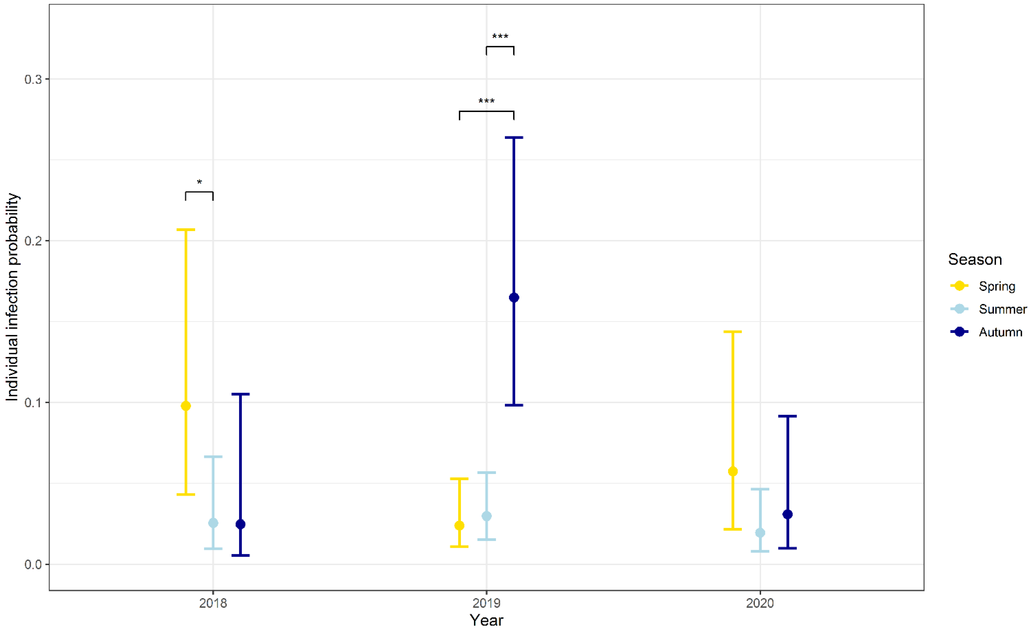
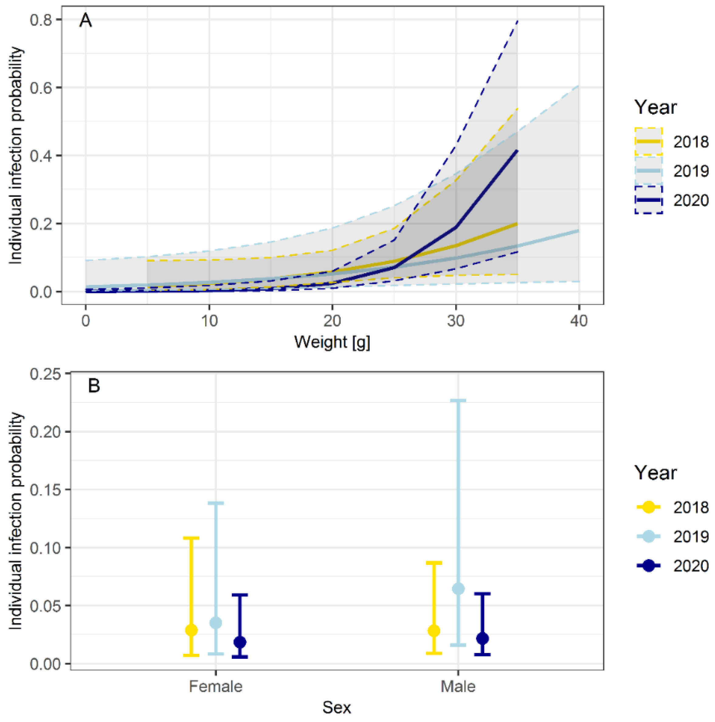
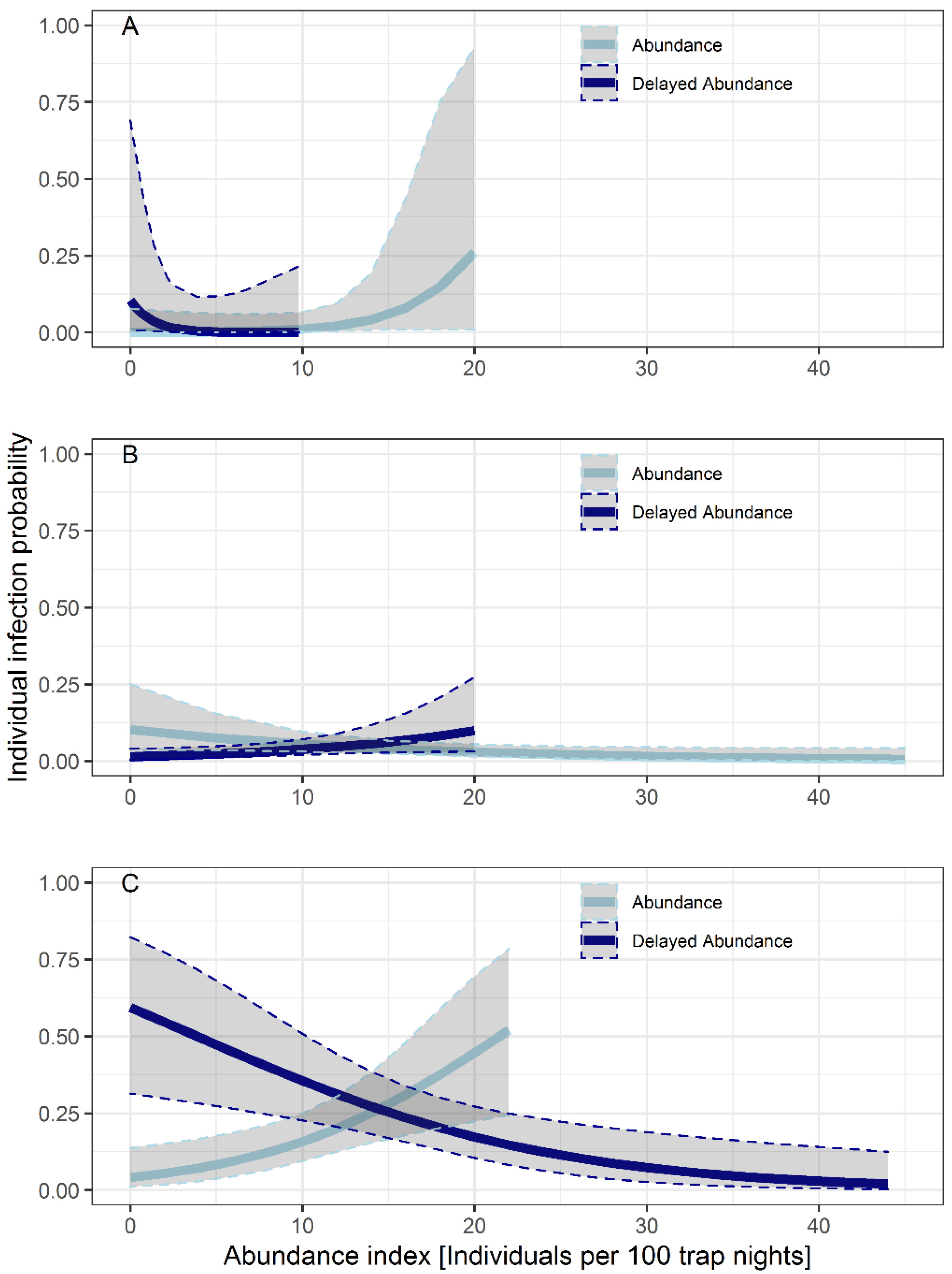
3.3. Influence of Seasonality, Body Weight, Sex and Abundance on the Probability of Infection

Overall, there were clear interannual differences with 2019 showing a significantly higher infection probability compared to 2018 and 2020 (Table 2). Different seasonal patterns were observed between the years (Table 2, Figure 2). In the year 2018, the individual infection probability was significantly higher in spring compared to summer but not autumn. The highest individual infection probability was documented in autumn 2019, which was significantly higher compared to summer 2019 (p < 0.001) and spring 2019 (p < 0.001) (Table 2). There were no significant differences between the seasons in 2020, though infection probability was highest in spring.
The predicted probability of infection was positively correlated with body weight in the years 2019 (p = 0.045) and 2020 (p < 0.001). The correlation was not significant in the year 2018 (p = 0.071) (Figure 3A, Table 3).
Sex had no influence on the individual probability of infection in 2018 (p = 0.773) and 2020 (p = 0.764). In contrast, male bank voles were more often Leptospira spp.-positive than female bank voles in the year 2019 (p = 0.015) (Figure 3B, Table 3).
There was no statistical effect of the abundance on infection probability of bank voles in the years 2018 and 2020. In summer 2019, there was a positive relationship between the individual probability of infection and abundance in the previous season (spring 2019; delayed abundance) (p = 0.038) and a negative relationship with increasing abundance of the present season (p = 0.042) (abundance) (Figure 4B, Table 4). In the following autumn (2019), the direction of this relationship switched. The infection probability was significantly negatively-related to increased abundances in the previous season (p = 0.004) and positively related to abundances in the present season (p = 0.005) (Figure 4C, Table 4).

4. Discussion

Our study detected a mean Leptospira spp. prevalence of 7.5% (95% CI: 6.4–8.9), with a range of 0% (LS6) to 15.7% (NW1) in bank voles from North-West Germany in the years 2018 to 2020 (Table 1). Results of previous studies in Germany showed mean prevalence values of 4.1% (95% CI: 2.8–5.9) up to 11.4% (95% CI: 8.7–14.6%) in bank voles [52,57], which is similar to our results
However, there are obvious regional differences. Lower values were found in studies investigating the Leptospira prevalence in South and East Germany. Here, a prevalence of 0% up to 13% was detected in bank voles, depending on the site and year [53,57]. Another study, which captured different small mammal species at multiple locations throughout Germany in the years 2002 to 2010, detected a mean prevalence of 6% (66/1016) in bank voles [54].
A similar mean prevalence (7.8%) in bank voles was detected across four locations in Germany in the years 2010 to 2014 [58]. One trapping site of the former study in North Rhine-Westphalia is in close geographical proximity to NW1 of our study, and here the authors found a slightly higher prevalence (20.4%, 95% CI: 16.1–25.2) [58] compared to our study (15.7%, 95% CI: 12.9–18.9). A higher mean prevalence in bank voles was also detected in a study in central Germany (11.4%) [52] and the Czech Republic (12%) [48]. The lower mean prevalence in our study might be explained by the extreme drought in the years 2018 to 2020 [68], which gave Leptospira a less favorable environment in which to survive [69,70]. On the other hand, it needs to be taken in account that the highest prevalence in our study was documented in 2019, also a year affected by drought (Figure 2), which is most probably due to high bank vole abundance. Further studies need to be done to predict the influence of drought on the Leptospira prevalence in bank voles. The absence of Leptospira spp. at location LS6 might be due to geographical differences, which inhibited the spread of Leptospira spp. in this region. However, the number of collected bank voles (n = 60) was rather small.
Data of the genomospecies distribution in bank voles in Europe are rare. Former studies in Germany showed that bank voles can be infected by different Leptospira species, with L. kirschneri being the most common [52,58]. Our study confirms this observation, but interestingly the most common Leptospira genomospecies in bank voles of our study was L. interrogans, followed by L. kirschneri and L. borgpetersenii. These results suggest that bank voles are susceptible to at least three Leptospira genomospecies.
In former studies of bank voles, L. interrogans was represented by ST24, L. kirschneri by ST110 and ST117 and L. borgpetersenii by ST197 and ST146 in Germany [53,58]. The findings of our study are in line with these results, except for ST117 and ST146, which were not identified in our study. Certain STs and related serogroups can be associated with a specific host, for example the ST17 serogroup Icterohaemorhagiae with Norway rats [59]. We did not observe a host specificity of serogroups and STs for bank voles in our study, as the STs detected in our study were also found in other small mammal species: ST24 was found in the yellow-necked field mouse, the wood mouse, and the Eurasian beaver (Castor fiber); ST110 in the yellow-necked field mouse, common vole, field vole and the common shrew (Sorex araneus); and ST197 in the common shrew and crowned shrew (S. coronatus) [53,58,71]. Studies from Europe and Asia also show a high ST diversity among different genera of small mammals [72,73,74,75]. Serogroups detected in our study were detected worldwide in different host species, e.g., serogroup Australis in swine, goats, cats and dogs [76,77,78,79]; serogroup Grippotyphosa in cattle, sheep, wild boars, dogs and horses [16,80,81,82,83]; and serogroup Sejroe in cattle [84,85].
Habitat seems to have an important influence on the distribution of different Leptospira genomospecies and STs. In previous studies, common voles and field voles, which have their main habitat in grassland, were primarily infected by L. kirschneri ST110 [54,58]. In contrast, bank voles, wood mice and yellow-necked field mice, mostly found in forests, were susceptible to L. kirschneri ST110, L. interrogans ST24, L. borgpetersenii ST197, and ST146 [58], which is in line with results of our study.
The STs in our study can be associated with various Leptospira serovars. ST24 is associated with serovars Bratislava, Muenchen, Lora and Jalna and ST110 with serovars Grippotyphosa, Vanderhoedenii and Valbuzzi [67]. The ST 197 is related to serogroup Sejroe, but no designated serovar is documented for ST197 [67], as it contains a non-standard length allele (caiB51) [2]. Awareness in a public health context should be raised to the pathogenic serogroups Australis, Grippotyphosa and Sejroe and related serovars Bratislava and Grippotyphosa, which can cause severe clinical symptoms in humans and reduced productivity in livestock [8,16]. The finding of serogroups, which are pathogenic for multiple species, underlies the importance of bank voles as reservoirs for Leptospira spp. and sources of infection for humans and livestock.
Individual and population-based factors are known to influence Leptospira spp. prevalence in rodents. Age is a significant driver of Leptospira prevalence in different rodent reservoirs, because the persisting infection with Leptospira spp. increases the probability of infection with the increasing age of the host [86,87,88,89]. Our study confirms this observation for bank voles. Taking body weight as a proxy for age, we showed that with increasing body weight, and therefore increasing age, the probability of infection was positively correlated (Figure 3A, Table 3). This effect was more or less consistent of the study period. Although it was not formally significant in 2018 (p = 0.071), it was significant in the years 2019 (p = 0.045) and 2020 (p < 0.001) (Table 3).
The influence of sex on the individual infection probability depended on the collection year. No difference between male and female bank voles was observed in the years 2018 and 2020. In contrast, male bank voles showed a significantly higher prevalence than female bank voles in the year 2019 (p = 0.015) (Table 3). This result suggests that sex only had an influence in years with high bank vole abundance. Higher abundance and reproduction causes an increase in male aggressive behavior and contact to multiple sexual partners during the breeding season [90,91], which increases the risk of direct or indirect contact to Leptospira spp. However, our results are in line with previous studies, which yielded mixed results as some found no effect of sex [45,47] and others did [92,93].
Overall, effects of body weight (age) were more consistent compared to sex in terms of individual infection probability for Leptospira spp. in bank voles. These results are in line with conclusions of former studies [20,58], and has led to the assumption that weight is a good indicator to predict the individual probability of infection with Leptospira spp. in bank voles. However, that weight depends on individual developmental circumstances regarding food resources, environmental influences and seasonal fluctuation, and can only be taken as a proxy for age [94,95].
In previous studies, Leptospira prevalence increased in common vole and field vole populations over the course of the year with a high prevalence in autumn [52,58]. However, our bank vole study yielded mixed results in terms of seasonality, with annual differences. Seasonal patterns could be detected in the years 2018 and 2020 when the Leptospira spp. prevalence was low in summer and high in spring and autumn (Figure 2). An explanation might be the seasonal differences in the composition of the bank vole population. In spring, the population consisted mainly of adult rodents. These overwintering adults, which were persistently infected from the previous season, may have transmitted Leptospira spp. to the following spring population and caused high prevalences in spring. During the breeding season in summer, uninfected juveniles entered the population, which lead to lower prevalences in summer. This “juvenile dilution effect” was also described for PUUV infections in bank voles [96]. Additionally, maternal antibodies transmitted from infected females to their offspring might lead to lower prevalences [97]. In autumn, at the end of the breeding season, adults that dominated [98] were more likely to be infected (Figure 3A, Table 3). Furthermore, seasonal weather conditions can have an impact on Leptospira prevalence. The survival of Leptospira spp. outside the host is dependent on humidity and water availability, temperature and pH [21]. In summer, higher temperatures and dry conditions may have led to decreased indirect transmission and therefore a lower risk of infection compared to spring and autumn.
In contrast to the described seasonal patterns in the years 2018 and 2020, a different course was documented in the year 2019, when prevalence increased throughout the year, with the highest prevalence in autumn (23.0%, 95% CI: 18.3–28.3). The prevalence in autumn 2019 was the highest documented in the three years of our study (Figure 2, Table 2). This might have been due to high bank vole abundance in 2019, which was caused by the beech mast in the year 2018 [99]. This result was also reflected in high PUUV prevalence in bank voles in North-West Germany in the year 2019 [99]. High bank vole abundance should have promoted high Leptospira prevalence in autumn 2019. In summer, the prevalence increased in regions which already showed a high abundance in spring. However, even more significant was the influence of abundance in autumn 2019, as the prevalence increased at all locations with high abundance at the present season (p = 0.005) (Table 4). As population density rose, direct transmission was promoted, because of more aggressive encounters between conspecifics for food, territory and mates [90,91]. Additionally, more leptospires were potentially shed into the environment via the urine of persistently infected bank voles that frequently mark their home range by shedding urine [91], which can promote indirect transmission through contact with contaminated water and soil.
The influence of abundance to the infection probability differed between season and year. No statistical effect could be documented in the years 2018 and 2020. This result led to the assumption that abundance has an effect on the individual infection probability for Leptospira spp. in bank voles, but only during certain seasons and years with high abundance and intense reproduction.

5. Conclusions

Our study showed that Leptospira spp. are widely spread in bank voles in North-West Germany. The individual infection probability of bank voles was influenced by season, body weight, and year.
This is relevant for public health, as the three detected Leptospira species were classified as pathogenic [3], and the detected STs were related to pathogenic serovars. People working in agriculture and forestry are at a particularly higher risk because of their close contact to small mammals, natural water sources and soil.
The human incidence in Germany was low (0.06–0.2 per 100,000 inhabitants) in the years 2010 to 2020, but leptospirosis should be on the differential diagnosis list of every clinician and veterinarian. Incidence may rise due to increasing popularity of outdoor activities, which lead to a higher risk of exposure in the human population. Furthermore, climate change might be an additional influencing factor as it will lead to more extreme weather events in moderate climate zones, like drought, storm, heavy rainfalls and flooding [100], as recently seen in Germany in the summer of 2021.

Author Contributions

Conceptualization, M.P., J.J., R.G.U., J.F. and M.R.; methodology, E.S., A.O. and A.M.-S.; validation, E.S., A.O. and M.P.; formal analysis, C.I. and A.O.; investigation, E.S.; resources, R.G.U., S.D., J.J., M.S. and M.P.; data curation, E.S.; writing—original draft preparation, E.S.; writing—review and editing, A.O., R.G.U., S.D., J.J., C.I., A.M.-S., M.S. and M.P.; visualization, E.S. and C.I.; supervision, M.P.; project administration, M.P. and R.G.U.; funding acquisition, M.P., R.G.U., J.J., M.R., M.S. and A.M.-S. All authors have read and agreed to the published version of the manuscript.

Funding

This research was funded by the German Federal Ministry of Education and Research within the Research Network of Zoonotic Infectious Diseases, grant number 01KI1903A and 01KI1903B to M.P. and R.G.U. and grant number 01KI1721A and 01KI2004A to R.G.U, 01KI1721E and 01KI2004E to J.J, 01KI1721H to M.R. and M.S., 01KI1721B to A.M.-S.

Institutional Review Board Statement

All procedures involving animals were conducted according to relevant legislation and by permission of the federal authorities. Permits for snap trapping were approved by Kreis Coesfeld (Abteilung 70-Umwelt/Natur- und Bodenschutz; Untere Naturschutzbehörde) (protocol code: 70.2.2.27-2018/0006, date of approval: 9 May 2018) and Kreis Warendorf (Amt für Planung und Naturschutz; Untere Landschaftsbehörde) (protocol code: 613904, date of approval: 13 August 2018; protocol code: 613901, date of approval: 13 February 2020). Permits for live trapping were approved by Landesamt für Natur, Umwelt und Verbraucherschutz Nordrhein-Westfalen (protocol code: 81-02.04.2018.A150, date of approval: 9 April 2018).

Informed Consent Statement

Not applicable.

Data Availability Statement

The data presented in this study are available in the article.

Acknowledgments

We want to thank the working group RoBoPub (Rodent-Borne-Pathogens-and-Public-Health) for providing the samples for this study. We gratefully acknowledge the German Federal Institute for Risk Assessment, Berlin for technical support, implementation of MLST and for providing sequence data of 127 Leptospira reference strains. Many thanks go to Dana Rüster and Cornelia Göllner for excellent technical support in the laboratory and Maja Haut, Dörte Kaufmann, Sina Nippert, Stefan Fischer, René Ryll, Elisa Heuser, Kathrin Jeske, Maysaa Dafalla, Patrick Slowikowski, Paul Pieper, Jana Hey, Anastasia Tavakina, Kim Usko, Maximilian Rüttermann, Christine Jelinek, Georgina Bredel, Vanessa Schulze, Florian Binder, Elias Bendl, Diana A. Below, Hendrik Ennen, Maya Braun, Felicitas Bujnoch, Yusuff Scholer, Orestis Kazazidis, Teresa Budde, Mechthild Budde, Martha Göbel, Philipp Harpering, Florian Hüls, Christin Hesse, Britta Oltmann, Helmut Jacobs, and Sönke Röhrs for their help during sampling and/or dissection. We acknowledge support from Leipzig University for Open Access Publishing.

Conflicts of Interest

The authors declare no conflict of interest.

Appendix A

Table A1. Details of rodent trapping in transect North-Rhine Westphalia and Lower Saxony, Germany in the years 2018 to 2020.
Federal StateNorth Rhine-WestphaliaLower Saxony
Trapping location
(Number of trapping sites per
location)
NW1
(13 trapping sites)
LS3
(3 trapping sites)
NW2
(3 trapping sites)
LS4
(6 trapping sites)
LS5
(2 trapping sites)
LS6
(2 trapping sites)
Trapping frequencyThree times yearly in spring (March to Mai), summer (July, August)
and autumn (October to November)
Three times yearly in spring (April), summer (July)
and autumn (October)
Type of trapUgglan multiple capture live traps (Grahnab®, Gnosjö, Sweden)Metal snap traps
(Deufa, Neuburg, Germany)
Metal snap traps
(Deufa, Neuburg, Germany)
Traps per night and trapping site/Number of nights/Control
frequency
49 traps/
3 nights/
Checked twice a day
49 traps/
3 nights/
Checked once a day
100 traps/
3 nights/
Checked once a day
Baitapple, rodent pellets, rolled oats, peanut curlspeanut butter with rolled oatspeanut butter with rolled oats
Additional informationRodents captured alive were sampled and released, only rodents found dead were used in this study
PublicationTrapping according to [101]Trapping according to [99]Trapping according to [99]
Table A2. Gene loci, primer sequences and probe used for qPCR [62], SLST [63] and MLST [2].
LocusPrimer/BREAKProbeSequence (5′ to 3′)
lipl32lipl32-FAAG CAT TAC CGC TTG TGG TG
lipl32-RGAA CTC CCA TTT CAG CGA TT
probe6FAM-AA AGC CAG GAC AAG CGC CG BHQ1
secYsecY-FGAA TTT CTC TTT TGA TCT TCG
secY-RGAG TTA GAG CTC AAA TCT AAG
glmUglmU-FAGG ATA AGG TCG CTG TGG TA
glmU-RAGT TTT TTT CCG GAG TTT CT
pntApntA-FTAG GAA ARA TGA AAC CRG GAA C
pntA-RAAG AAG CAA GAT CCA CAA YTA C
sucAsucA-FTCA TTC CAC TTY TAG ATA CGA T
sucA-RTCT TTT TTG AAT TTT TGA CG
tpiAtpiA-FTTG CAG GAA ACT GGA AAA TGA AT
tpiA-RGTT TTA CRG AAC CHC CGT AGA GAA T
pfkBpfkB-FCGG AGA GTT TTA TAA RAA GGA CAT
pfkB-RAGA ACA CCC GCC GCA AAA CAA T
mreAmreA-FGGC TCG CTC TYG ACG GAA A
mreA-RTCC RTA ACT CAT AAA MGA CAA AGG
caiBcaiB-FCAA CTT GCG GAY ATA GGA GGA G
caiB-RATT ATG TTC CCC GTG AYT CG
F = forward primer, R = reverse primer.

References

  1. Caimi, K.; Ruybal, P. Leptospira spp., a genus in the stage of diversity and genomic data expansion. Infect. Genet. Evol. 2020, 81, 104241. [Google Scholar] [CrossRef]
  2. Boonsilp, S.; Thaipadungpanit, J.; Amornchai, P.; Wuthiekanun, V.; Bailey, M.S.; Holden, M.T.G.; Zhang, C.; Jiang, X.; Koizumi, N.; Taylor, K.; et al. A single multilocus sequence typing (MLST) scheme for seven pathogenic Leptospira species. PLoS Negl. Trop. Dis. 2013, 7, e1954. [Google Scholar] [CrossRef] [PubMed] [Green Version]
  3. Picardeau, M. Leptospira and leptospirosis. Methods Mol. Biol. 2020, 2134, 271–275. [Google Scholar] [CrossRef] [PubMed]
  4. Costa, F.; Hagan, J.E.; Calcagno, J.; Kane, M.; Torgerson, P.; Martinez-Silveira, M.S.; Stein, C.; Abela-Ridder, B.; Ko, A.I. Global morbidity and mortality of leptospirosis: A systematic review. PLOS Negl. Trop. Dis. 2015, 9, e0003898. [Google Scholar] [CrossRef] [Green Version]
  5. Lau, C.L.; Smythe, L.D.; Craig, S.B.; Weinstein, P. Climate change, flooding, urbanisation and leptospirosis: Fuelling the fire? Trans. R. Soc. Trop. Med. Hyg. 2010, 104, 631–638. [Google Scholar] [CrossRef] [PubMed]
  6. Munoz-Zanzi, C.; Groene, E.; Morawski, B.M.; Bonner, K.; Costa, F.; Bertherat, E.; Schneider, M.C. A systematic literature review of leptospirosis outbreaks worldwide, 1970–2012. Rev. Panam. Salud Públ. 2020, 44, e78. [Google Scholar] [CrossRef]
  7. Aviat, F.; Blanchard, B.; Michel, V.; Blanchet, B.; Branger, C.; Hars, J.; Mansotte, F.; Brasme, L.; de Champs, C.; Bolut, P.; et al. Leptospira exposure in the human environment in France: A survey in feral rodents and in fresh water. Comp. Immunol. Microbiol. Infect. Dis. 2009, 32, 463–476. [Google Scholar] [CrossRef]
  8. Desai, S.; Van Treeck, U.; Lierz, M.; Espelage, W.; Zota, L.; Sarbu, A.; Czerwinski, M.; Sadkowska-Todys, M.; Avdicová, M.; Reetz, J.; et al. Resurgence of field fever in a temperate country: An epidemic of leptospirosis among seasonal strawberry harvesters in Germany in 2007. Clin. Infect. Dis. 2009, 48, 691–697. [Google Scholar] [CrossRef]
  9. Habus, J.; Persic, Z.; Spicic, S.; Vince, S.; Stritof, Z.; Milas, Z.; Cvetnic, Z.; Perharic, M.; Turk, N. New trends in human and animal leptospirosis in Croatia, 2009–2014. Acta Trop. 2017, 168, 1–8. [Google Scholar] [CrossRef] [PubMed]
  10. Vieira, M.L.; Gama-Simões, M.J.; Collares-Pereira, M. Human leptospirosis in Portugal: A retrospective study of eighteen years. Int. J. Infect. Dis. 2006, 10, 378–386. [Google Scholar] [CrossRef] [PubMed] [Green Version]
  11. Garvey, P.; Connell, J.; O’Flanagan, D.; McKeown, P. Leptospirosis in Ireland: Annual incidence and exposures associated with infection. Epidemiol. Infect. 2014, 142, 847–855. [Google Scholar] [CrossRef]
  12. Dupouey, J.; Faucher, B.; Edouard, S.; Richet, H.; Kodjo, A.; Drancourt, M.; Davoust, B. Human leptospirosis: An emerging risk in Europe? Comp. Immunol. Microbiol. Infect. Dis. 2014, 37, 77–83. [Google Scholar] [CrossRef] [PubMed]
  13. Van Alphen, L.B.; Kunøe, A.L.; Ceper, T.; Kähler, J.; Kjelsø, C.; Ethelberg, S.; Krogfelt, K.A. Trends in human leptospirosis in Denmark, 1980 to 2012. Eurosurveillance 2015, 20, 21019. [Google Scholar] [CrossRef] [PubMed]
  14. European Centre for Disease Prevention and Control. Available online: https://www.ecdc.europa.eu/en (accessed on 11 March 2021).
  15. Robert Koch-Institut. SurvStat@RKI 2.0. Available online: https://survstat.rki.de (accessed on 1 March 2021).
  16. Ellis, W.A. Animal leptospirosis. In Leptospira and Leptospirosis, 1st ed.; Adler, B., Ed.; Springer: Berlin/Heidelberg, Germany, 2015; pp. 99–137. ISBN 009783662523995. [Google Scholar]
  17. Collings, D.F. Leptospira interrogans infection in domestic and wild animals in Fiji. N. Z. Vet. J. 1984, 32, 21–24. [Google Scholar] [CrossRef] [PubMed]
  18. Sterling, C.R.; Thiermann, A.B. Urban rats as chronic carriers of leptospirosis: An ultrastructural investigation. Vet. Pathol. 1981, 18, 628–637. [Google Scholar] [CrossRef] [PubMed] [Green Version]
  19. Davis, S.; Calvet, E.; Leirs, H. Fluctuating rodent populations and risk to humans from rodent-borne zoonoses. Vector Borne Zoonotic Dis. 2005, 5, 305–314. [Google Scholar] [CrossRef]
  20. Perez, J.; Brescia, F.; Becam, J.; Mauron, C.; Goarant, C. Rodent abundance dynamics and leptospirosis carriage in an area of hyper-endemicity in New Caledonia. PLoS Negl. Trop. Dis. 2011, 5, e1361. [Google Scholar] [CrossRef] [Green Version]
  21. Bierque, E.; Thibeaux, R.; Girault, D.; Soupé-Gilbert, M.-E.; Goarant, C. A systematic review of Leptospira in water and soil environments. PLoS ONE 2020, 15, e0227055. [Google Scholar] [CrossRef] [PubMed]
  22. Haake, D.A.; Levett, P.N. Leptospirosis in humans. In Leptospira and Leptospirosis, 1st ed.; Adler, B., Ed.; Springer: Berlin/Heidelberg, Germany, 2015; pp. 65–88. ISBN 009783662523995. [Google Scholar]
  23. Dadon, Y.; Haas, E.J.; Kaliner, E.; Anis, E.; Singer, S.R.; Atiya-Nasagi, Y.; Cohen-Dar, M.; Avramovich, E.; King, R.; Sued, O.; et al. Outbreak of human leptospirosis linked to contaminated water bodies in Northern Israel, June to August 2018. Eurosurveillance 2018, 23, 1800486. [Google Scholar] [CrossRef] [Green Version]
  24. Bharti, A.R.; Nally, J.E.; Ricaldi, J.N.; Matthias, M.A.; Diaz, M.M.; Lovett, M.A.; Levett, P.N.; Gilman, R.H.; Willig, M.R.; Gotuzzo, E.; et al. Leptospirosis: A zoonotic disease of global importance. Lancet Infect. Dis. 2003, 757–771. [Google Scholar] [CrossRef]
  25. Dechet, A.M.; Parsons, M.; Rambaran, M.; Mohamed-Rambaran, P.; Florendo-Cumbermack, A.; Persaud, S.; Baboolal, S.; Ari, M.D.; Shadomy, S.V.; Zaki, S.R.; et al. Leptospirosis outbreak following severe flooding: A rapid assessment and mass prophylaxis campaign; Guyana, January-February 2005. PLoS ONE 2012, 7, e39672. [Google Scholar] [CrossRef] [Green Version]
  26. Gaynor, K.; Katz, A.R.; Park, S.Y.; Nakata, M.; Clark, T.A.; Effler, P.V. Leptospirosis on Oahu: An outbreak associated with flooding of a university campus. Am. J. Trop. Med. Hyg. 2007, 76, 882–885. [Google Scholar] [CrossRef] [PubMed]
  27. Brockmann, S.; Piechotowski, I.; Bock-Hensley, O.; Winter, C.; Oehme, R.; Zimmermann, S.; Hartelt, K.; Luge, E.; Nöckler, K.; Schneider, T.; et al. Outbreak of leptospirosis among triathlon participants in Germany, 2006. BMC Infect. Dis. 2010, 10, 91. [Google Scholar] [CrossRef] [PubMed] [Green Version]
  28. Lau, C.L.; Townell, N.; Stephenson, E.; Van den Berg, D.; Craig, S.B. Leptospirosis: An important zoonosis acquired through work, play and travel. Aust. J. Gen. Pract. 2018, 47, 105–110. [Google Scholar] [CrossRef] [PubMed] [Green Version]
  29. Radl, C.; Müller, M.; Revilla-Fernandez, S.; Karner-Zuser, S.; De Martin, A.; Schauer, U.; Karner, F.; Stanek, G.; Balcke, P.; Hallas, A.; et al. Outbreak of leptospirosis among triathlon participants in Langau, Austria, 2010. Wien. Klin. Wochenschr. 2011, 123, 751–755. [Google Scholar] [CrossRef] [PubMed]
  30. Guillois, Y.; Bourhy, P.; Ayral, F.; Pivette, M.; Decors, A.; Grau, J.H.A.; Champenois, B.; Malhère, C.; Combes, B.; Richomme, C.; et al. An outbreak of leptospirosis among kayakers in Brittany, North-West France, 2016. Eurosurveillance 2018, 23. [Google Scholar] [CrossRef]
  31. Morgan, J.; Bornstein, S.L.; Karpati, A.M.; Bruce, M.; Bolin, C.A.; Austin, C.C.; Woods, C.W.; Lingappa, J.; Langkop, C.; Davis, B.; et al. Outbreak of leptospirosis among triathlon participants and community residents in Springfield, Illinois, 1998. Clin. Infect. Dis. 2002, 34, 1593–1599. [Google Scholar] [CrossRef] [PubMed] [Green Version]
  32. Jansen, A.; Schöneberg, I.; Frank, C.; Alpers, K.; Schneider, T.; Stark, K. Leptospirosis in Germany, 1962–2003. Emerg. Infect. Dis. 2005, 11, 1048–1054. [Google Scholar] [CrossRef]
  33. Narkkul, U.; Thaipadungpanit, J.; Srisawat, N.; Rudge, J.W.; Thongdee, M.; Pawarana, R.; Pan-Ngum, W. Human, animal, water source interactions and leptospirosis in Thailand. Sci. Rep. 2021, 11, 3215. [Google Scholar] [CrossRef]
  34. Meny, P.; Menéndez, C.; Ashfield, N.; Quintero, J.; Rios, C.; Iglesias, T.; Schelotto, F.; Varela, G. Seroprevalence of leptospirosis in human groups at risk due to environmental, labor or social conditions. Rev. Argent. Microbiol. 2019, 51, 324–333. [Google Scholar] [CrossRef]
  35. Mori, M.; Bourhy, P.; Le Guyader, M.; Van Esbroeck, M.; Djelouadji, Z.; Septfons, A.; Kodjo, A.; Picardeau, M. Pet rodents as possible risk for leptospirosis, Belgium and France, 2009 to 2016. Eurosurveillance 2017, 22, 30553. [Google Scholar] [CrossRef] [Green Version]
  36. Karpagam, K.B.; Ganesh, B. Leptospirosis: A neglected tropical zoonotic infection of public health importance—An updated review. Eur. J. Clin. Microbiol. Infect. Dis. 2020, 39, 835–846. [Google Scholar] [CrossRef]
  37. Yang, H.-Y.; Yen, T.-H.; Lin, C.-Y.; Chen, Y.-C.; Pan, M.-J.; Lee, C.-H.; Yu, C.-C.; Wu, M.-S.; Wu, S.-S.; Weng, C.-H.; et al. Early identification of leptospirosis as an ignored cause of multiple organ dysfunction syndrome. Shock 2012, 38, 24–29. [Google Scholar] [CrossRef]
  38. Maier, A.; Kaeser, R.; Thimme, R.; Boettler, T. Acute pancreatitis and vasoplegic shock associated with leptospirosis—A case report and review of the literature. BMC Infect. Dis. 2019, 19, 395. [Google Scholar] [CrossRef]
  39. Fernandez, P.S.; Kodjo, A.; Medkour, H.; Laidoudi, Y.; Dubourg, G.; Eldin, C.; Parola, P.; Davoust, B.; Lagier, J.-C. Autochthonous human and animal leptospirosis, Marseille, France. IDCases 2020, 21, e00899. [Google Scholar] [CrossRef] [PubMed]
  40. Ellis, T.; Imrie, A.; Katz, A.R.; Effler, P.V. Underrecognition of leptospirosis during a dengue fever outbreak in Hawaii, 2001–2002. Vector Borne Zoonotic Dis. 2008, 8, 541–547. [Google Scholar] [CrossRef]
  41. Briskin, E.A.; Casanovas-Massana, A.; Ryff, K.R.; Morales-Estrada, S.; Hamond, C.; Perez-Rodriguez, N.M.; Benavidez, K.M.; Weinberger, D.M.; Castro-Arellano, I.; Wunder, E.A.; et al. Seroprevalence, Risk Factors, and Rodent Reservoirs of Leptospirosis in an Urban Community of Puerto Rico, 2015. J. Infect. Dis. 2019, 220, 1489–1497. [Google Scholar] [CrossRef]
  42. Thiermann, A.B. The Norway rat as a selective chronic carrier of Leptospira icterohaemorrhagiae. J. Wildl. Dis. 1981, 17, 39–43. [Google Scholar] [CrossRef] [PubMed] [Green Version]
  43. Koizumi, N.; Muto, M.; Tanikawa, T.; Mizutani, H.; Sohmura, Y.; Hayashi, E.; Akao, N.; Hoshino, M.; Kawabata, H.; Watanabe, H. Human leptospirosis cases and the prevalence of rats harbouring Leptospira interrogans in urban areas of Tokyo, Japan. J. Med Microbiol. 2009, 58, 1227–1230. [Google Scholar] [CrossRef] [PubMed]
  44. Holt, J.; Davis, S.; Leirs, H. A model of Leptospirosis infection in an African rodent to determine risk to humans: Seasonal fluctuations and the impact of rodent control. Acta Trop. 2006, 99, 218–225. [Google Scholar] [CrossRef] [PubMed]
  45. Krijger, I.M.; Ahmed, A.A.A.; Goris, M.G.A.; Cornelissen, J.B.W.J.; Koerkamp, P.W.G.G.; Meerburg, B.G. Wild rodents and insectivores as carriers of pathogenic Leptospira and Toxoplasma gondii in The Netherlands. Vet. Med. Sci. 2020, 6, 623–630. [Google Scholar] [CrossRef] [PubMed]
  46. Schmidt, S.; Essbauer, S.S.; Mayer-Scholl, A.; Poppert, S.; Schmidt-Chanasit, J.; Klempa, B.; Henning, K.; Schares, G.; Groschup, M.H.; Spitzenberger, F.; et al. Multiple infections of rodents with zoonotic pathogens in Austria. Vector Borne Zoonotic Dis. 2014, 14, 467–475. [Google Scholar] [CrossRef] [Green Version]
  47. Izquierdo-Rodríguez, E.; Fernández-Álvarez, Á.; Martín-Carrillo, N.; Marchand, B.; Feliu, C.; Miquel, J.; Foronda, P.; Quilichini, Y. Pathogenic Leptospira species in rodents from Corsica (France). PLoS ONE 2020, 15, e0233776. [Google Scholar] [CrossRef] [PubMed]
  48. Balážová, A.; Nosková, E.; Široký, P.; Durrant, C.; Baláž, V. Diversity and dynamics of zoonotic pathogens within a local community of small mammals. Biologia 2021, 76. [Google Scholar] [CrossRef]
  49. Millán, J.; Cevidanes, A.; Chirife, A.D.; Candela, M.G.; León-Vizcaíno, L. Risk factors of Leptospira infection in Mediterranean periurban micromammals. Zoonoses Public Health 2018, 65, e79–e85. [Google Scholar] [CrossRef]
  50. Jeske, K.; Emirhar, D.; García, J.T.; González-Barrio, D.; Olea, P.P.; Fons, F.R.; Schulz, J.; Mayer-Scholl, A.; Heckel, G.; Ulrich, R.G.; et al. Frequent Leptospira spp. detection, but absence of Tula orthohantavirus in Microtus voles, Northwestern Spain. J. Wildl. Dis. 2021, 57. [Google Scholar] [CrossRef]
  51. Tadin, A.; Tokarz, R.; Markotić, A.; Margaletić, J.; Turk, N.; Habuš, J.; Svoboda, P.; Vucelja, M.; Desai, A.; Jain, K.; et al. Molecular survey of zoonotic agents in rodents and other small mammals in Croatia. Am. J. Trop. Med. Hyg. 2016, 94, 466–473. [Google Scholar] [CrossRef]
  52. Jeske, K.; Jacob, J.; Drewes, S.; Pfeffer, M.; Heckel, G.; Ulrich, R.G.; Imholt, C. Hantavirus-Leptospira coinfections in small mammals from central Germany. Epidemiol. Infect. 2021, 149, 1–25. [Google Scholar] [CrossRef]
  53. Obiegala, A.; Woll, D.; Karnath, C.; Silaghi, C.; Schex, S.; Eßbauer, S.; Pfeffer, M. Prevalence and genotype allocation of pathogenic Leptospira species in small mammals from various habitat types in Germany. PLoS Negl. Trop. Dis. 2016, 10, e0004501. [Google Scholar] [CrossRef] [Green Version]
  54. Mayer-Scholl, A.; Hammerl, J.A.; Schmidt, S.; Ulrich, R.G.; Pfeffer, M.; Woll, D.; Scholz, H.C.; Thomas, A.; Nöckler, K. Leptospira spp. in rodents and shrews in Germany. Int. J. Environ. Res. Public Health 2014, 11, 7562–7574. [Google Scholar] [CrossRef]
  55. Heuser, E.; Fischer, S.; Ryll, R.; Mayer-Scholl, A.; Hoffmann, D.; Spahr, C.; Imholt, C.; Alfa, D.M.; Fröhlich, A.; Lüschow, D.; et al. Survey for zoonotic pathogens in Norway rat populations from Europe. Pest Manag. Sci. 2017, 73, 341–348. [Google Scholar] [CrossRef]
  56. Kocianová, E.; Kozuch, O.; Bakoss, P.; Rehácek, J.; Kovácová, E. The prevalence of small terrstrial mammals infected with tick-borne encephalitis virus and leptospirae in the foothills of the Bavarian forest, Germany. Appl. Parasitol. 1993, 34, 283–290. [Google Scholar]
  57. Obiegala, A.; Albrecht, C.; Dafalla, M.; Drewes, S.; Oltersdorf, C.; Turni, H.; Imholt, C.; Jacob, J.; Wagner-Wiening, C.; Ulrich, R.G.; et al. Leptospira spp. in small mammals from areas with low and high human hantavirus incidences in south-west Germany. Vector Borne Zoonotic Dis. 2017, 17, 312–318. [Google Scholar] [CrossRef]
  58. Fischer, S.; Mayer-Scholl, A.; Imholt, C.; Spierling, N.G.; Heuser, E.; Schmidt, S.; Reil, D.; Rosenfeld, U.M.; Jacob, J.; Nöckler, K.; et al. Leptospira genomospecies and sequence type prevalence in small mammal populations in Germany. Vector Borne Zoonotic Dis. 2018, 18, 188–199. [Google Scholar] [CrossRef]
  59. Boey, K.; Shiokawa, K.; Rajeev, S. Leptospira infection in rats: A literature review of global prevalence and distribution. PLoS Negl. Trop. Dis. 2019, 13, e0007499. [Google Scholar] [CrossRef] [PubMed]
  60. Piechotowski, I.; Brockmann, S.O.; Schwarz, C.; Winter, C.H.; Ranft, U.; Pfaff, G. Emergence of hantavirus in South Germany: Rodents, climate and human infections. Parasitol. Res. 2008, 103, 131–137. [Google Scholar] [CrossRef]
  61. Nationale Forschungsplattform für Zoonosen. RoBoPub. Available online: https://zoonosen.net/en/forschungsnetz/verbunde-nachwuchsgruppen/robopub (accessed on 3 May 2021).
  62. Stoddard, R.A.; Gee, J.E.; Wilkins, P.P.; McCaustland, K.; Hoffmaster, A.R. Detection of pathogenic Leptospira spp. through TaqMan polymerase chain reaction targeting the LipL32 gene. Diagn. Microbiol. Infect. Dis. 2009, 64, 247–255. [Google Scholar] [CrossRef] [PubMed]
  63. Victoria, B.; Ahmed, A.; Zuerner, R.L.; Ahmed, N.; Bulach, D.M.; Quinteiro, J.; Hartskeerl, R.A. Conservation of the S10-spc-α locus within otherwise highly plastic genomes provides phylogenetic insight into the genus Leptospira. PLoS ONE 2008, 3, e2752. [Google Scholar] [CrossRef] [PubMed] [Green Version]
  64. Bates, D.; Mächler, M.; Bolker, B.; Walker, S. Fitting linear mixed-effects models using lme4. J. Stat. Softw. 2015, 67, 1–48. [Google Scholar] [CrossRef]
  65. R Core Team. R: A Language and Environment for Statistical Computing; R Foundation for Statistical Computing: Vienna, Austria, 2020. [Google Scholar]
  66. Lenth, R.V. Estimated marginal means, aka least-squares means. R Package Version 2018, 1, 3. [Google Scholar]
  67. University of Oxford. PubMLST. Available online: https://pubmlst.org/ (accessed on 27 April 2021).
  68. Helmholtz Zentrum für Umweltforschung. Dürremonitor Deutschland: Dürre 1952–2020 (Jährlich). Available online: https://www.ufz.de/index.php?de=47252 (accessed on 14 June 2021).
  69. Karaseva, E.V.; Chernukha, Y.G.; Piskunova, L.A. Results of studying the time of survival of pathogenic leptospira under natural conditions. J. Hyg. Epidemiol. Microbiol. Immunol 1973, 17, 339–345. [Google Scholar]
  70. Nau, L.H.; Obiegala, A.; Król, N.; Mayer-Scholl, A.; Pfeffer, M. Survival time of Leptospira kirschneri serovar Grippotyphosa under different environmental conditions. PLoS ONE 2020, 15, e0236007. [Google Scholar] [CrossRef]
  71. Woll, D.; Karnath, C.; Pfeffer, M.; Allgöwer, R. Genetic characterization of Leptospira spp. from beavers found dead in south-west Germany. Vet. Microbiol. 2012, 158, 232–234. [Google Scholar] [CrossRef]
  72. Ferreira, A.S.; Ahmed, A.; Rocha, T.; Vieira, M.L.; Paiva-Cardoso, M.D.N.; Mesquita, J.R.; Van der Linden, H.; Goris, M.; Thompson, G.; Hartskeerl, R.A.; et al. Genetic diversity of pathogenic leptospires from wild, domestic and captive host species in Portugal. Transbound. Emerg. Dis. 2020, 67, 852–864. [Google Scholar] [CrossRef]
  73. Piredda, I.; Ponti, M.N.; Palmas, B.; Noworol, M.; Pedditzi, A.; Rebechesu, L.; Chisu, V. Molecular typing of pathogenic Leptospira species isolated from wild mammal reservoirs in Sardinia. Animals 2021, 11, 1109. [Google Scholar] [CrossRef]
  74. Azhari, N.N.; Ramli, S.N.A.; Joseph, N.; Philip, N.; Mustapha, N.F.; Ishak, S.N.; Mohd-Taib, F.S.; Nor, S.M.; Yusof, M.A.; Sah, S.A.M.; et al. Molecular characterization of pathogenic Leptospira spp. in small mammals captured from the human leptospirosis suspected areas of Selangor state, Malaysia. Acta Trop. 2018, 188, 68–77. [Google Scholar] [CrossRef] [PubMed]
  75. Zhang, C.; Xu, J.; Zhang, T.; Qiu, H.; Li, Z.; Zhang, E.; Li, S.; Chang, Y.-F.; Guo, X.; Jiang, X.; et al. Genetic characteristics of pathogenic Leptospira in wild small animals and livestock in Jiangxi Province, China, 2002–2015. PLoS Negl. Trop. Dis. 2019, 13, e0007513. [Google Scholar] [CrossRef] [PubMed]
  76. Balboni, A.; Zamagni, S.; Bertasio, C.; Boniotti, M.B.; Troìa, R.; Battilani, M.; Dondi, F. Identification of serogroups Australis and Icterohaemorrhagiae in two dogs with a severe form of acute leptospirosis in Italy. Pathogens 2020, 9, 351. [Google Scholar] [CrossRef] [PubMed]
  77. Moreno, L.Z.; Kremer, F.S.; Jaeger, L.H.; Loureiro, A.P.; Miraglia, F.; Eslabao, M.R.; Dellagostin, O.A.; Lilenbaum, W.; Moreno, A.M. Genomic characterization and comparative analysis of Leptospira interrogans serogroup Australis isolated from swine. Pathog. Dis. 2017, 75, ftx119. [Google Scholar] [CrossRef]
  78. Czopowicz, M.; Kaba, J.; Smith, L.; Szalus-Jordanow, O.; Nowicki, M.; Witkowski, L.; Frymus, T. Leptospiral antibodies in the breeding goat population of Poland. Vet. Rec. 2011, 169, 230. [Google Scholar] [CrossRef] [PubMed]
  79. Weis, S.; Rettinger, A.; Bergmann, M.; Llewellyn, J.R.; Pantchev, N.; Straubinger, R.K.; Hartmann, K. Detection of Leptospira DNA in urine and presence of specific antibodies in outdoor cats in Germany. J. Feline Med. Surg. 2017, 19, 470–476. [Google Scholar] [CrossRef] [PubMed]
  80. Jaeger, L.H.; Moreno, L.Z.; Kremer, F.S.; Dellagostin, O.A.; Moreno, A.M.; Lilenbaum, W. Genomic characterization and comparative analysis of Leptospira kirschneri serogroup Grippotyphosa UC5/2011, a strain isolated after mare abortion: Implications for genital animal leptospirosis. Comp. Immunol. Microbiol. Infect. Dis. 2019, 64, 7–9. [Google Scholar] [CrossRef] [PubMed]
  81. Geisen, V.; Stengel, C.; Brem, S.; Müller, W.; Greene, C.; Hartmann, K. Canine leptospirosis infections—Clinical signs and outcome with different suspected Leptospira serogroups (42 cases). J. Small Anim. Pract. 2007, 48, 324–328. [Google Scholar] [CrossRef] [PubMed]
  82. Hamond, C.; Martins, G.; Bremont, S.; Medeiros, M.A.; Bourhy, P.; Lilenbaum, W. Molecular characterization and serology of Leptospira kirschneri (Serogroup Grippotyphosa) isolated from urine of a mare post-abortion in Brazil. Zoonoses Public Health 2016, 63, 191–195. [Google Scholar] [CrossRef] [Green Version]
  83. Cilia, G.; Bertelloni, F.; Piredda, I.; Ponti, M.N.; Turchi, B.; Cantile, C.; Parisi, F.; Pinzauti, P.; Armani, A.; Palmas, B.; et al. Presence of pathogenic Leptospira spp. in the reproductive system and fetuses of wild boars (Sus scrofa) in Italy. PLoS Negl. Trop. Dis. 2020, 14, e0008982. [Google Scholar] [CrossRef]
  84. Grippi, F.; Giudice, E.; Di Pietro, S.; Sciacca, C.; Santangelo, F.; Galluzzo, P.; Barreca, S.; Guercio, A. Leptospira interrogans serogroup Sejroe serovar Hardjo in aborting cows: Two herd cases in Sicily (Italy). J. Vet. Res. 2020, 64, 73–78. [Google Scholar] [CrossRef]
  85. Pinna, M.H.; Martins, G.; Loureiro, A.P.; Lilenbaum, W. Detection of bovine carriers of Leptospira by serological, bacteriological, and molecular tools. Trop. Anim. Health Prod. 2018, 50, 883–888. [Google Scholar] [CrossRef]
  86. Ivanova, S.; Herbreteau, V.; Blasdell, K.; Chaval, Y.; Buchy, P.; Guillard, B.; Morand, S. Leptospira and rodents in Cambodia: Environmental determinants of infection. Am. J. Trop. Med. Hyg. 2012, 86, 1032–1038. [Google Scholar] [CrossRef] [Green Version]
  87. Krøjgaard, L.H.; Villumsen, S.; Markussen, M.D.K.; Jensen, J.S.; Leirs, H.; Heiberg, A.-C. High prevalence of Leptospira spp. in sewer rats (Rattus norvegicus). Epidemiol. Infect. 2009, 137, 1586–1592. [Google Scholar] [CrossRef]
  88. Himsworth, C.G.; Bidulka, J.; Parsons, K.L.; Feng, A.Y.T.; Tang, P.; Jardine, C.M.; Kerr, T.; Mak, S.; Robinson, J.; Patrick, D.M. Ecology of Leptospira interrogans in Norway rats (Rattus norvegicus) in an inner-city neighborhood of Vancouver, Canada. PLoS Negl. Trop. Dis. 2013, 7, e2270. [Google Scholar] [CrossRef] [PubMed]
  89. Vanasco, N.B.; Sequeira, M.D.; Sequeira, G.; Tarabla, H.D. Associations between leptospiral infection and seropositivity in rodents and environmental characteristics in Argentina. Prev. Vet. Med. 2003, 60, 227–235. [Google Scholar] [CrossRef]
  90. Marchlewska-Koj, A.; Kolodziej, B.; Filimowska, A. Aggressive behavior of adult bank voles (Clethrionomys glareolus) towards conspecifics. Aggr. Behav. 1989, 15, 381–387. [Google Scholar] [CrossRef]
  91. Tamarin, R.H.; Ostfeld, R.S.; Pugh, S.R.; Bujalska, G. Social Systems and Population Cycles in Voles; Springer: Basel, Switzerland, 1990; ISBN 009783764324377. [Google Scholar]
  92. Gomes, C.K.; Guedes, M.; Potula, H.-H.; Dellagostin, O.A.; Gomes-Solecki, M. Sex matters: Male hamsters are more susceptible to lethal infection with lower doses of pathogenic Leptospira than female hamsters. Infect. Immun. 2018, 86. [Google Scholar] [CrossRef] [PubMed] [Green Version]
  93. Krijger, I.M.; Ahmed, A.A.A.; Goris, M.G.A.; Koerkamp, P.W.G.G.; Meerburg, B.G. Prevalence of Leptospira infection in rodents from Bangladesh. Int. J. Environ. Res. Public Health 2019, 16, 2113. [Google Scholar] [CrossRef] [PubMed] [Green Version]
  94. Morris, P. A review of mammalian age determination methods. Mammal Rev. 1972, 2, 69–104. [Google Scholar] [CrossRef]
  95. Mazurkiewicz, M. Density and weight structure of populations of the bank vole in open and enclosed areas. Acta Theriol. 1972, 17, 455–465. [Google Scholar] [CrossRef] [Green Version]
  96. Mills, J.N.; Ksiazek, T.G.; Peters, C.J.; Childs, J.E. Long-term studies of hantavirus reservoir populations in the southwestern United States: A synthesis. Emerg. Infect. Dis. 1999, 5, 135–142. [Google Scholar] [CrossRef] [PubMed]
  97. Birnbaum, S.; Shenberg, E.; Torten, M. The influence of maternal antibodies on the epidemiology of leptospiral carrier state in mice. Am. J. Epidemiol. 1972, 96, 313–317. [Google Scholar] [CrossRef] [PubMed]
  98. Korslund, L.; Steen, H. Small rodent winter survival: Snow conditions limit access to food resources. J. Anim. Ecol. 2006, 75, 156–166. [Google Scholar] [CrossRef] [PubMed]
  99. Binder, F.; Drewes, S.; Imholt, C.; Saathoff, M.; Below, D.A.; Bendl, E.; Conraths, F.J.; Tenhaken, P.; Mylius, M.; Brockmann, S.; et al. Heterogeneous Puumala orthohantavirus situation in endemic regions in Germany in summer 2019. Transbound. Emerg. Dis. 2020, 67, 502–509. [Google Scholar] [CrossRef] [Green Version]
  100. McMichael, A.J.; Woodruff, R.E.; Hales, S. Climate change and human health: Present and future risks. Lancet 2006, 367, 859–869. [Google Scholar] [CrossRef]
  101. Reil, D.; Rosenfeld, U.M.; Imholt, C.; Schmidt, S.; Ulrich, R.G.; Eccard, J.A.; Jacob, J. Puumala hantavirus infections in bank vole populations: Host and virus dynamics in Central Europe. BMC Ecol. 2017, 17, 9. [Google Scholar] [CrossRef] [PubMed] [Green Version]
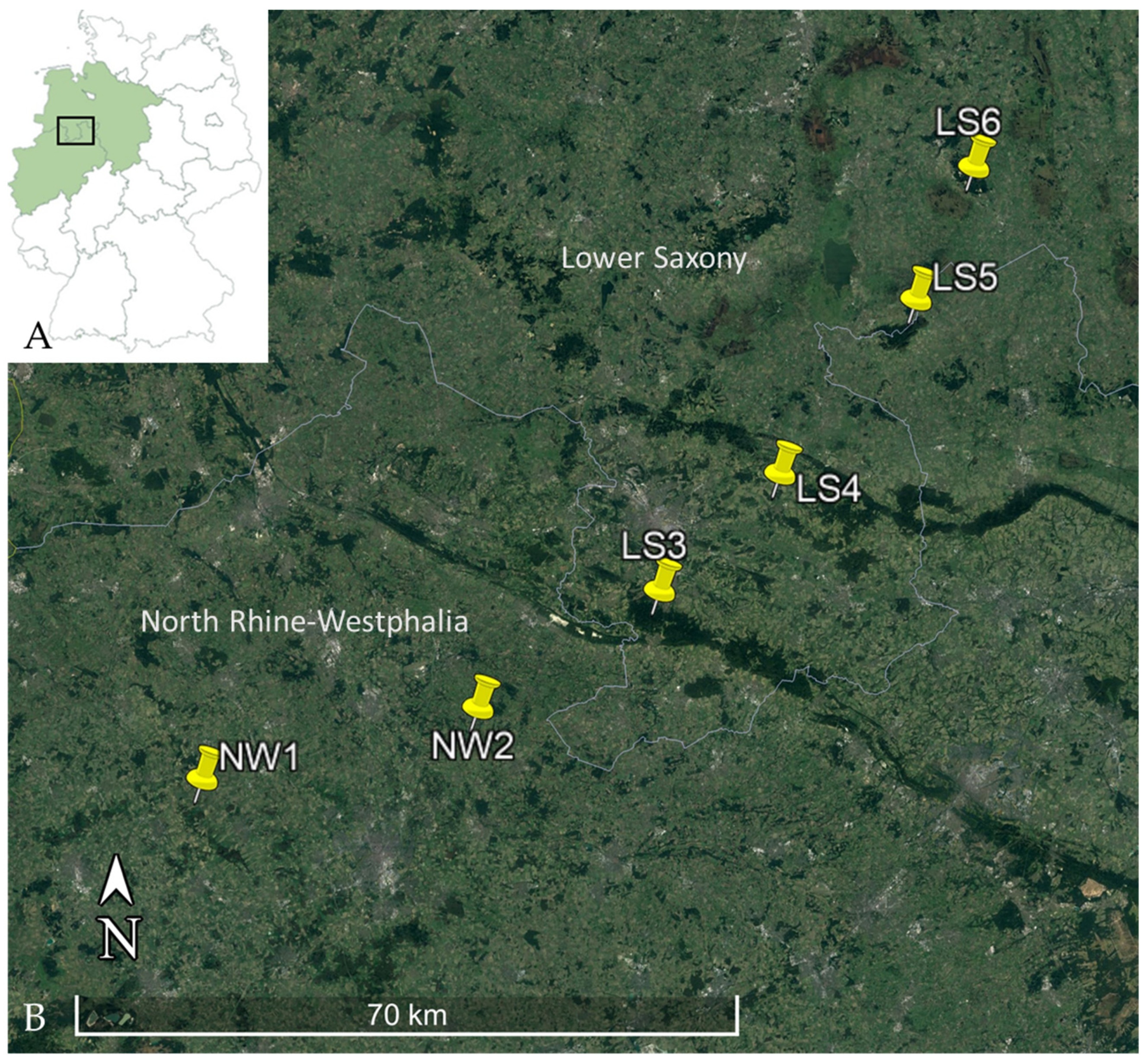
Figure 1. (A) Map of Germany with involved federal states highlighted in green and location of the transect marked by a square, (B) Trapping locations within the transect, NW = North Rhine-Westphalia, LS = Lower Saxony. Image was created by using Google Earth Pro, Map: Google Earth ©2021 Google, Image Landsat/Copernicus ©2021 GeoBasis-DE/BKG.
Biology 10 00933 g001
Figure 2. Potential influence of season on the individual infection probability in the years 2018 to 2020. Results are based on lipl32-qPCR-positive animals, significant differences between seasons are highlighted by stars: * = significant (p < 0.05), *** = highly significant (p < 0.001).
Biology 10 00933 g002
Figure 3. Potential influence of (A) body weight and (B) sex on individual infection probability. Results are based on lipl32-qPCR-positive animals. Only animals with sex determination (n = 1751) were analyzed for influence of sex on the individual infection probability.
Biology 10 00933 g003
Figure 4. Potential influence of abundance and delayed abundance in (A) spring, (B) summer and (C) autumn in the year 2019 on individual infection probability (Abundance = bank vole abundance of the present season, Delayed Abundance = bank vole abundance of the previous season). Results are based on lipl32-qPCR-positive animals. (Years 2018 and 2020 are not shown, because no significant influences were documented).
Biology 10 00933 g004
Table 1. Leptospira spp. prevalence and genomospecies distribution in bank voles captured in the years 2018 to 2020.
Federal StateTotal
Number of Bank Voles
Trapping Location
(see Figure 1)
Number of Leptospira DNA Positive/Total Number of Bank Voles Tested (lipl32-qPCR)
(Percentage, 95% CI)
Number of secY-SLST/MLST Positive Bank Voles *
L. interrogans/
ST24
L. kirschneri/
ST110
L. borgpetersenii/
ST197
North Rhine-Westphalia782NW195/605
(15.7%, 12.9—18.9)
55/158/32/1
NW212/177
(6.8%, 3.6—11.5)
7/51/0N/A
Lower
Saxony
1035LS31/160
(0.6, 0—3.4)
N/AN/AN/A
LS426/770
(4.3%, 2.2—4.9)
17/92/13/2
LS53/45
(6.7%, 1.4—18.3)
1N/AN/A
LS60/60
(0%, 0—6%)
N/AN/AN/A
total1817 137/1817 *
(7.5%, 95% CI: 6.4—8.9)
80/2911/45/3
NW = North Rhine-Westphalia, LS = Lower Saxony, CI = confidence interval, SLST = single locus sequence typing, MLST = multi locus sequence typing, ST = sequence type, N/A = not available, * Determination of genomospecies and ST was not possible for all samples tested positive in lipl32-qPCR.
Table 2. Results of generalized linear mixed modelling with binomial error distribution showing the influence of season, year and their interaction on the individual infection probability.
FactorEstimateStd. Errorz-Valuep-Value
Intercept−3.6760.783−4.696<0.001
Season [spring]1.4540.8231.7670.077
Season [summer]0.0320.8570.0370.971
Year [2019]2.0540.7572.7120.007
Year [2020]0.2250.9090.2480.804
Year:seasonEstimateStd. Errorz-ratiop-value
Year 2018
Autumn v. spring−1.4540.823−1.7670.181
Autumn v. summer−0.0320.857−0.0370.999
Spring v. summer1.4230.5672.5110.032
Year 2019
Autumn v. spring2.0870.3645.732<0.001
Autumn v. summer1.8680.2856.554<0.001
Spring v. summer−0.2190.400−0.5460.848
Year 2020
Autumn v. spring−0.6490.691−0.9390.616
Autumn v. summer0.4730.6430.7350.743
Spring v. summer1.1220.5821.9280.131
Random effectsVarianceStd.Dev.
Site1.4601.208
Year0.0000.000
Season:year0.0000.000
For categorical factors the reference categories are: Season—autumn, Year—2018. For the interaction term Year:season the results of a post-hoc (Tukey) analysis on the estimated marginal means are presented to highlight seasonal differences within each year. Bold values represent significant factors. Std. Error = Standard Error; Std.Dev. = Standard Deviation, v. = versus, z-value/ratio = Wald statistics.
Table 3. Results of generalized linear mixed modelling with binomial error distribution showing the impact of demographic factors on individual infection probability for each year.
FactorEstimate Std. Errorz-Valuep-Value
Year 2018
Intercept−4.7461.277−3.717<0.001
Sex [m]0.1500.5180.2890.773
Weight0.0930.0521.8080.071
Random effectsVariance Std.Dev.
Site0.7090.842
Season0.1370.370
Year 2019
Intercept−4.6391.039−4.466<0.001
Sex [m]0.6370.2622.4330.015
Weight0.0690.0352.0010.045
Random effectsVariance Std.Dev.
Site1.6141.271
Season1.3231.150
Year 2020
Intercept−8.2841.680−4.931<0.001
Sex [m]0.1610.5350.3010.764
Weight0.2240.0653.441<0.001
Random effectsVariance Std.Dev.
Site1.5251.235
Season0.0000.000
For categorical factors the reference categories are: Sex—female. Bold values represent significant factors. Std. Error = Standard Error; Std.Dev = Standard Deviation, z value/ratio = Wald statistics.
Table 4. Results of generalized linear mixed modelling with binomial error distribution showing the impact of abundance and delayed abundance on individual infection probability for the year 2019.
FactorEstimate Std. Errorz-Valuep-Value
Spring 2019
Intercept−5.5191.714−3.2200.001
Delayed abundance−0.8500.553−1.5380.124
Abundance0.3520.2191.6050.109
Random effectsVariance Std.Dev.
Site2.8021.674
Summer 2019
Intercept−2.8450.602−4.7270.000
Delayed abundance0.1020.0492.0780.038
Abundance−0.0680.033−2.0300.042
Random effectsVariance Std.Dev.
Site0.3980.631
Autumn 2019
Intercept−1.2840.576−2.2300.026
Delayed abundance−0.0970.034−2.8650.004
Abundance0.1470.0522.8020.005
Random effectsVariance Std.Dev.
Site0.6110.782
Abundance = Bank vole abundance of the present season, Delayed abundance = abundance of the previous season. Bold values represent significant factors. Std. Error = Standard Error; Std.Dev = Standard Deviation, z value/ratio = Wald statistics.
Publisher’s Note: MDPI stays neutral with regard to jurisdictional claims in published maps and institutional affiliations.

