Review

Regulated DNA Methylation and the Circadian Clock: Implications in Cancer

Department of Animal Sciences, School of Environmental and Biological Sciences, Rutgers, The State University of New Jersey, New Brunswick, NJ 08904, USA
*
Author to whom correspondence should be addressed.
Submission received: 7 May 2014 / Revised: 12 August 2014 / Accepted: 15 August 2014 / Published: 5 September 2014
(This article belongs to the Special Issue DNA Methylation)

Abstract

:
Since the cloning and discovery of DNA methyltransferases (DNMT), there has been a growing interest in DNA methylation, its role as an epigenetic modification, how it is established and removed, along with the implications in development and disease. In recent years, it has become evident that dynamic DNA methylation accompanies the circadian clock and is found at clock genes in Neurospora, mice and cancer cells. The relationship among the circadian clock, cancer and DNA methylation at clock genes suggests a correlative indication that improper DNA methylation may influence clock gene expression, contributing to the etiology of cancer. The molecular mechanism underlying DNA methylation at clock loci is best studied in the filamentous fungi, Neurospora crassa, and recent data indicate a mechanism analogous to the RNA-dependent DNA methylation (RdDM) or RNAi-mediated facultative heterochromatin. Although it is still unclear, DNA methylation at clock genes may function as a terminal modification that serves to prevent the regulated removal of histone modifications. In this capacity, aberrant DNA methylation may serve as a readout of misregulated clock genes and not as the causative agent. This review explores the implications of DNA methylation at clock loci and describes what is currently known regarding the molecular mechanism underlying DNA methylation at circadian clock genes.

1. DNA Methylation

Methylation on the number 5 carbon of cytosine (5mC) in DNA is found at silenced retrotransposons, imprinted loci and the inactive X chromosome in females [1,2,3,4,5]. DNA methylation is implicated in the etiology of numerous diseases, most notably cancer, and a recent PubMed search revealed over 3000 reviews. Despite this, the role of DNA methylation in cancer remains enigmatic, with hyper-, hypo- or hemi-methylated sequences all potentially contributing to, or being a consequence of, misregulated expression [6,7]. This is due in part to the different regulatory mechanisms by which the methyl group is added to cytosine and further complicated by the discovery that the Tet (ten-eleven translocation) family of proteins can convert 5mC to 5-hydroxymethylcytosine (5hmC) [8]. Maintenance methylation occurs during semiconservative DNA replication that preserves 5mC through mitosis and meiosis, supporting the notion that DNA methylation is the premier epigenetic modification involved in silencing. In contrast, noncoding RNAs (ncRNAs) are involved in de novo DNA methylation [9,10], and altered expression of these ncRNAs may impact methylation.
DNA methylation is essential for normal vertebrate development, with genome-wide DNA methylation patterns changing during differentiation [11,12]. Interestingly, although DNA methylation is prevalent in many organisms, it is extremely rare or absent in some metazoans, like Drosophila and C. elegans, indicating that transcription factors and underlying histone modification(s) are sufficient for the developmental program [13,14]. DNA methylation only appears to be essential for viability in higher vertebrates, providing the counter argument that the complexity of the organism’s genome and the need for carefully controlled and timed differentiation dictates the need for DNA methylation-mediated regulatory function. This ongoing debate only serves to pique curiosity and provide continued interest in a modification that now appears to be more dynamic than originally thought. Thus, DNA methylation, once considered a static permanent modification, is dynamic and may have context-dependent roles.

2. Genome-Wide Analyses

Genome-wide analysis of DNA methylation has revealed a landscape far more involuted than once imagined. In mammals, 5mC was originally thought to occur predominantly at CpG dinucleotides, and studies in embryonic stems (ES) cells using reduced representation bisulfite sequencing (RRBS) revealed a bimodal distribution that correlates with the underlying histone modifications [12]. Later genome-wide studies in ES cells at base-pair (bp) resolution demonstrated that approximately one-quarter of all 5mC occurs in a non-CpG context, but is lost upon differentiation [15]. However, the mammalian brain appears to be the notable exception, where non-CpG methylation persists [16]. How this impacts the association of MeCP2 or other methyl DNA binding proteins with chromatin needs additional work. These, as well as other genome-wide studies indicate wide-spread distribution of 5mC and demonstrate the dynamic nature of DNA methylation. The metamorphic nature of DNA methylation is not restricted to development, and transient, dynamic 5mC has been observed at circadian clock genes in Neurospora and mammals [17,18]. DNA methylation is also detected at clock genes in Arabidopsis, but its role in the clock has not been extensively examined [19]. In Drosophila, DNA methylation is extremely rare, and there are no reports at clock genes [13]. Therefore, this review is mostly limited to the Neurospora and mammalian circadian systems.

3. The Circadian Clock

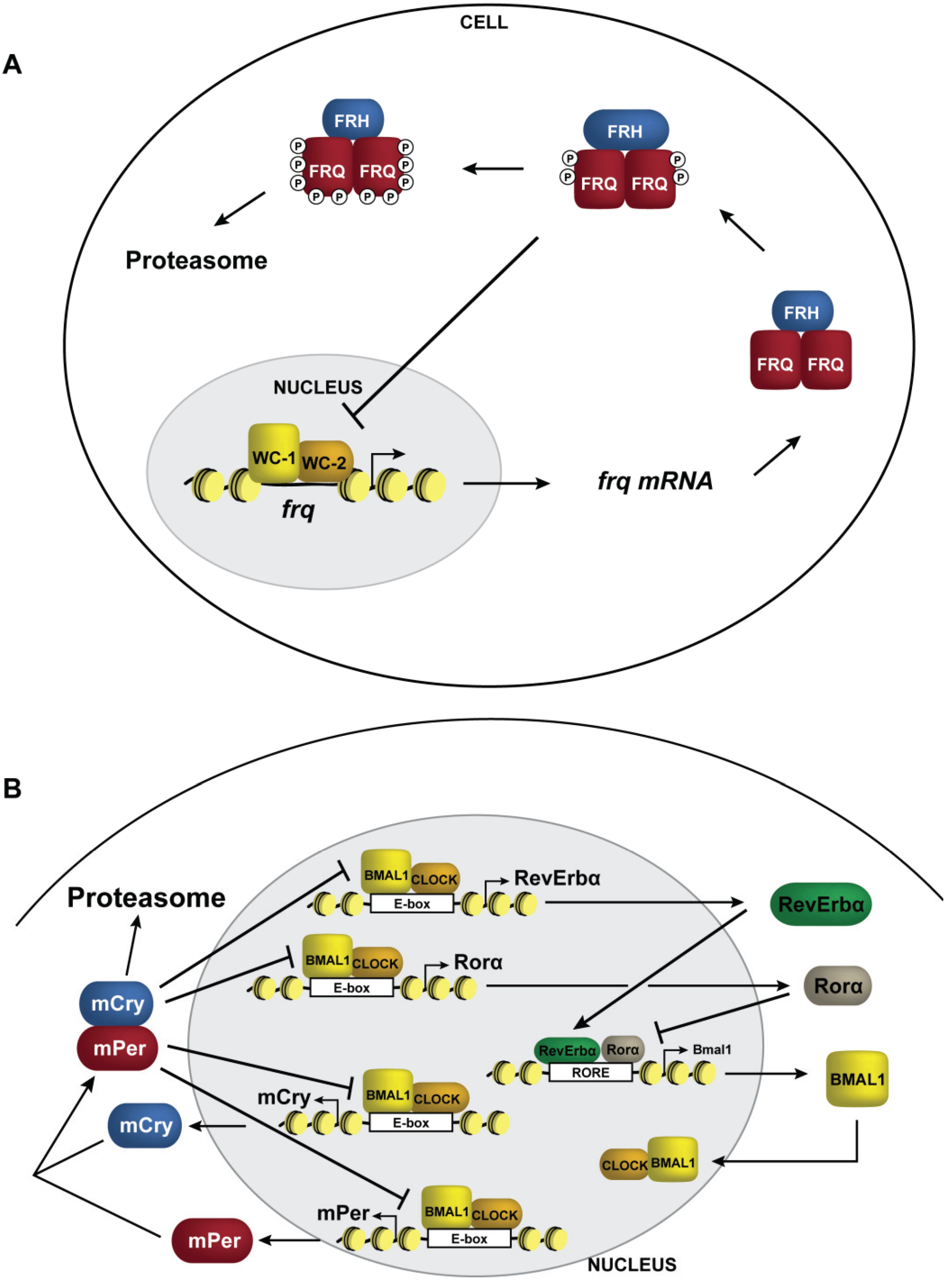
The circadian clock is an entrainable, free-running, temperature-compensated, anticipatory system that allows organisms to prepare for daily changes in the environment that arise from the Earth’s rotation [20,21,22,23,24,25]. In this capacity, it controls, among others, daily metabolism and subsequent activity rhythms in mammals and insects, asexual spore development in Neurospora and leaf movement in Arabidopsis [26,27,28,29,30,31]. The clock rhythm in gene expression persists in part due to time-of-day chromatin-associated events that facilitate the core transcriptional negative feedback loop. The physiological output of the circadian rhythm results from oscillations in genome-wide expression of clock controlled genes (ccg) that range from greater than 10% of the genome in mammals to as much as a third of the genome in Arabidopsis [32,33,34,35]. Moreover, oscillating transcripts differ based on cell/organ type with a subset exhibiting one-quarter (6 h) and one-half (12 h) harmonics [34,36]. There is an extensive number of molecular events that control global changes in ccg expression, and a large focus in the molecular biology era has centered on understanding the interconnected transcription/translational feedback loops (TTFL) [37,38]. As our knowledge continues to grow, it is clear that the circadian clocks from fungi to mammals consist of a transcriptional negative feedback loop, where heterodimeric, PAS domain-containing transcription factors drive the expression of negative elements that feedback, with delays, to inhibit their own expression (Figure 1). In two widely-studied clock systems, the activators consist of WHITE COLLAR (WC)-1:WC-2 in Neurospora and CLOCK:BMAL1 in mammals [39,40,41,42]. The negative elements are FREQUENCY (FRQ) and FRQ-interacting RNA helicase (FRH) in Neurospora and PERIOD (Per) 1, 2 and 3 and cryptochrome (Cry) 1 and 2 in mammals [43,44,45,46,47,48,49]. In addition, mammals have a secondary loop where Rev-Erbα and Rorα coordinate to control rhythms in BMAL1 expression [50,51]. Once the negative elements are translated, they receive extensive post-translational modifications that serve to regulate nucleo-cytoplasmic shuttling, which allows them to block their own expression [52,53,54,55,56,57]. Eventually, they become terminally modified with extensive phosphorylation, causing them to be destabilized, ubiquitinated and degraded by the proteasome. Many of the numerous reviews referenced above describe the molecular mechanism of the circadian clock, the corresponding covalent modifications and how they impact the central oscillator and output. The focus of this review is on DNA methylation and chromatin in the circadian clock.

4. The Clock and Chromatin

The combined nature of the transcriptional negative feedback loop and genome-wide oscillations in ccg expression requires coordinated control of chromatin and the genome structure [58,59,60]. In this capacity, it should not be surprising that the circadian field has seen a boom in discoveries involving chromatin-associated enzymes involved in clock function. Moreover, Per complexes contain a variety of interacting partners with known catalytic activity toward chromatin [61,62,63]. The chromatin enzymes identified thus far can be grouped into four classes; activating, repressive, amplitude-modulating or phase-modulating, based on their occurrence and the resulting phenotype. Moreover, the enzymes only tell a partial tale, because circadian-regulated metabolism provides substrates in the form of S-adenosyl methionine (SAM/AdoMet), nicotinamide adenine dinucleotide (NAD+) and acetyl co-enzyme A (Acetyl-CoA) utilized by methyltransferases, histone deacetylases and acetyltransferases, respectively, providing an additional interconnected feedback network that helps controls the amplitude, phasing and underlying rhythm [64,65,66,67].
Figure 1. Transcriptional negative feedback in the clock. Schematic representation of circadian negative feedback in (A) Neurospora and (B) mammals. The negative elements are shown in red (FRQ and mPer) and blue (FRH and mCry), and the positive elements are shown in yellow (WC-1 and BMAL1) and orange (WC-2 and CLOCK). For simplicity, only a single Per gene and single Cry gene were included. Rorα and Rev-Erbα, which form a second interconnect loop in mammals regulating rhythms in Bmal1 expression, are shown in beige and green, respectively.
Biology 03 00560 g001
There are a significant number of coincident histone modifications that change over the circadian cycle, and in some cases, the corresponding histone-modifying enzymes have been identified. Histone H3 serine 10 (H3S10) phosphorylation was the first chromatin mark implicated in circadian-regulated gene expression, and it increases in the mouse SCN neurons when exposed to light at night [68]. Later studies documented rhythms in acetylation of histone H3 in mPer1, mPer2 and Cry1 promoters, with the peaks occurring during the transcriptionally active phase [69,70]. Rhythms in acetylated histones presumably occur as a direct result of the transcriptional activator, CLOCK. Affinity purification of CLOCK indicates that it associates with the ubiquitous KAT3B, p300 [69] and possesses its own catalytic acetyltransferase activity [71]. In mouse embryonic fibroblasts (MEFs), CLOCK has also been shown to interact with the KMT2, MLL1 (mixed-lineage leukemia) and MLL1 and catalyzes the methylation of H3K4 [72]. Collectively, these data indicate that components of the biological clock can direct chromatin modifications to help maintain rhythms. The observations of oscillations in histone acetylation naturally lead to the identification of a growing list of HDACs that include HDAC1 and HDAC2 (part of the Sin3B complex) [73] and the NAD-dependent HDAC, SIRT1 [74,75]. In addition, polypyrimidine tract-bind protein-associated factor (PSF) is in a complex with PER and recruits HDACs associated with Sin3A [62].
Methylation and demethylation of histones also occurs coincident with circadian-regulated gene expression. However, some of the details surrounding many of the enzymes are still underdeveloped. It has been demonstrated that during the repressive phase, there is di- and tri-methylation of H3K27 at mPer1 and mPer2, which is dependent on the Polycomb group protein, EZH2 (KMT6) [76]. In studies performed on mouse livers, H3K9Ac is rhythmic in the D-element binding protein (Dbp) gene (a high-amplitude ccg), and studies in MEFs demonstrate a rhythm in H3K4me3 that is catalyzed by MLL1 (mixed-lineage leukemia) [72,77]. However, ChIP-seq data examining genome-wide oscillations in H3K4me3 indicate that it is prevalent during the repressive phase of the circadian cycle, and this is consistent with studies in Neurospora, indicating that the KMT2, SET1, is needed for efficient negative feedback inhibition [78]. Interestingly, JARID1a, a known H3K4 lysine demethylases (KMD), does not appear to have any effect on H3K4me3, but instead inhibits HDAC1 recruitment [79]. However, overexpression of JARID1b and JARID1c can potentially reverse H3K4 methylation, but this has not been examined in great detail [79].
In addition to methylation of lysine 4, there exists rhythmic facultative heterochromatin consisting of H3K9me2 and HP1 binding at Dbp, Per1 and Per2 during the repressive phase, and presumably, this occurs through the association of Suv39h and HP1γ with PER2 [63,77]. Lysine-specific demethylase (LSD1), which can remove methyl groups from H3K4 and/or H3K9, associates with CLOCK and BMAL1 and could potentially serve to reverse H3K9 methylation. However, the mutant Lsd1 used in this study did not affect H3K9me3 levels [80]. In addition, both the mammalian and Arabidopsis circadian clocks utilize the histone demethylase JMJD5 to demethylate H3K36 [81]. Overall, there appears to be ordered recruitment of activating and repressive modifications that ultimately generate facultative heterochromatin on the circadian time-scale.
In Neurospora, much of the research has been focused on ATP-dependent chromatin-remodeling enzymes that coordinately control the chromatin architecture at frq. CLOCKSWITCH (CSW-1) and CATP (CLOCK ATPase) remodel chromatin at the C-box element, functioning during the repressive and activation phases, respectively [82,83]. A chromodomain helicase DNA binding family member, CHD1, appears to be required for activation and has the interesting phenotype of causing an increase in heterochromatic spreading at frq [17]. The cause and implications of this DNA hypermethylation phenotype are still unknown, but possible reasons are discussed below.
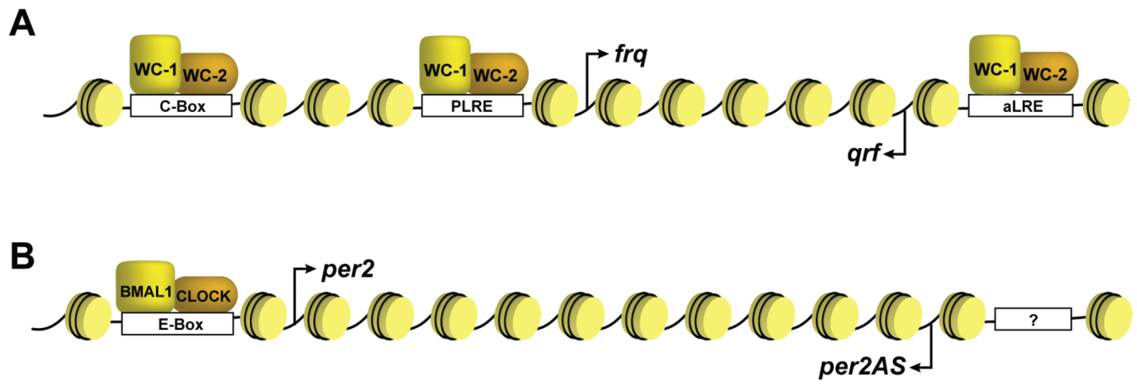
A paradoxical observation in circadian research is the finding of antisense transcripts that originate from the frq gene in Neurospora and all three Period genes in mammals [58,59,84] (Figure 2). Moreover, overlapping sense/antisense transcripts have been observed in Arabidopsis with a specific pair displaying antagonistic circadian expression patterns [85]. In general, these circadian natural antisense transcripts (NATs) are expressed antiphasic to the endogenous protein coding clock genes and in the case with the frq antisense qrf, expression is driven by the same transcriptional activators [84,86]. This incongruous expression confounds simple linear models of negative feedback and necessitates a molecular switch directing transcript synthesis. In doing so, there must also be amplitude modulation, since the NATs are lower abundance relative to their sense counterparts. Thus far, our findings and those of others indicate that the antisense transcript is involved in DNA methylation and rhythmic facultative heterochromatin formation at frq; both of which are needed for proper phasing [17,87].
Figure 2. Schematic of the clock genes. Illustration of (A) the frq gene in Neurospora and (B) the Per2 gene in mammals, shown as the typical “beads on string” with nucleosomes shaded yellow. The figure illustrates the sense and antisense transcripts that originate from both loci. The cis-acting sequences are shown as boxes on the DNA and labeled accordingly. The “?” within the Per2AS promoter indicates that the transcription factors responsible for expression are still unknown. Note that these representations are not drawn to scale.
Biology 03 00560 g002

5. DNA Methylation in Neurospora

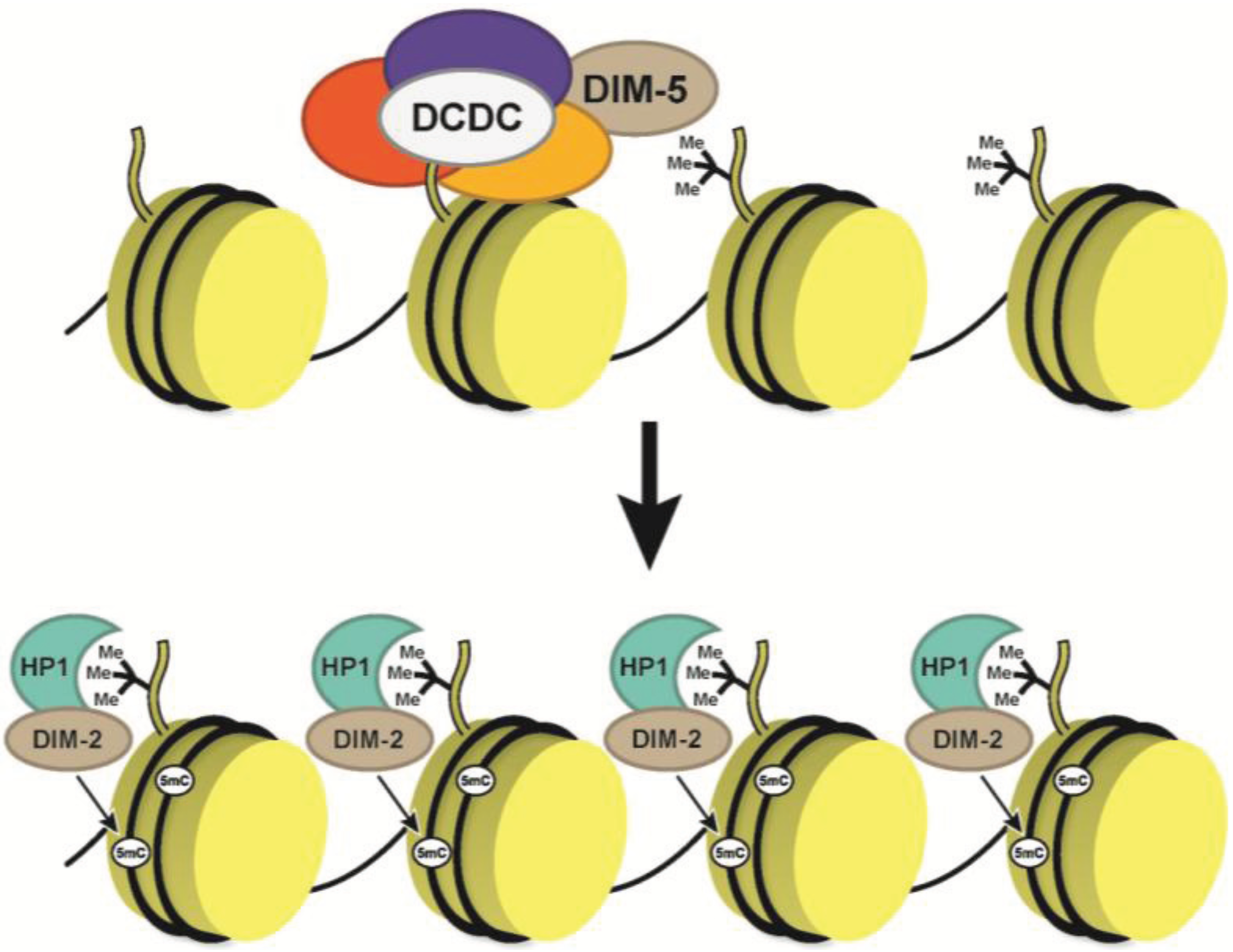
Before exploring RNA-induced DNA methylation at clock genes that is mediated in part by the convergent transcripts, it is useful to review general DNA methylation in Neurospora (Figure 3). Neurospora has little, if any, gene redundancy, forcing many of the methylation enzymes to function both at repetitive regions and at promoters. Methylation in Neurospora occurs largely at relics of repeated-induced point mutations (RIP’ed regions), whose origins were likely transposable elements [88]. DNA methylation requires the DNMT, DIM-2 (defective in methylation), heterochromatin protein 1 (HP1) and a multisubunit complex, DCDC (DIM-5,/-7/-9, CUL4/DDB1 complex) [89,90,91,92,93]. A core subunit of DCDC is the Histone H3 lysine 9 (H3K9) methyltransferase DIM-5 (KMT1) that is needed for mono- (H3K9me1), di- (H3K9me2) and trimethylation (H3K9me3) [94,95]. Current models suggest that DCDC is recruited to RIP’ed regions by DIM-7, although how this recruitment occurs is still unknown. Then, DIM-5, in association with CUL4/DDB1/DIM-9, catalyzes H3K9me3 [91]. DIM-2 is associated with HP1 and HP1 binds to H3K9me3 via its chromodomain, facilitating the subsequent DNA methylation [96]. Interestingly, DIM-2 has little to no effect on growth and only minor phenotypes, compared to DIM-5 or the hpo mutant (gene encoding HP1), suggesting that DNA methylation is likely ancillary relative to the H3K9me3 and HP1 binding.
Figure 3. DNA methylation in Neurospora. The current model for DNA methylation in Neurospora suggests a multistep process that starts with DIM-5 recruitment and H3K9 methylation mediated by the DCDC complex. Once the H3K9me3 is established, HP1 in association with DIM-2 and binds to H3K9me3 via its chromodomain, and then, DIM-2 catalyzes 5mC.
Biology 03 00560 g003

6. Dynamic DNA Methylation at Circadian Clock Genes

It is now unequivocal that circadian disruption results in substantial health consequences, and numerous studies indicate shift workers suffer a higher incidence of cancer [97,98,99]. Because anomalous DNA methylation correlates with cancer, it was natural to explore DNA methylation at clock genes. Some early studies found changes in promoter 5mC at Per1, Per2 and Per3 in breast cancerous tissues relative to surrounding non-cancerous tissue [100]. Since then, aberrant DNA methylation patterns have been found in every core clock gene in a variety of malignancies, and in most instances, the methylation status correlates with expression [101,102,103,104,105]. Methylation defects are also found at Clock (hypomethylated) and Cry2 (hypermethylated), as well as pathways involved in DNA recombination and repair in shift-workers [106]. Whether or not these changes in methylation are a cause or an effect is still unknown. However, data from Neurospora suggest that DNA methylation differences are an effect of clock misregulation, because cells that contain a dysfunctional clock have aberrant 5mC in frq.
The molecular mechanism underlying DNA methylation at clock genes in mammals and why DNA methylation at these loci is altered in cancer cells is largely unknown. In contrast, the molecular mechanism of clock gene DNA methylation is better understood in the Neurospora circadian system, in part due to Neurospora’s unique characteristics. Unlike mammals, where all the DNMT are essential for viability, loss of dim-2 in Neurospora has no discernable effect on cell growth and only minor effects on the clock [17]. Part of this may reside in the simple genomic makeup of Neurospora, because it only has a small subset of genes that contain DNA methylation and substantially less repetitive DNA where 5mC is normally prevalent [87,88].
The discovery of DNA methylation at the central clock gene frq arose serendipitously while characterizing the ATP-dependent chromatin-remodeling enzyme, CHD1 [17]. Common assays used to examine chromatin remodeling are restriction site accessibility experiments or MNaseI assays, both of which are reliant on restriction endonuclease cleavage followed by southern blot analysis. In MNaseI experiments designed to unravel the remodeling activity of CHD1, methyl-sensitive restriction enzymes had a stark reduction in activity. Further analysis revealed that the strains lacking chd1 had a hyper DNA methylation defect that caused spreading of the methylated regions beyond what was normally observed in an isogenic wild-type strain. Characterization of the DNA methylation indicated that it requires a functional clock (including both FRQ and FRH), the NAT qrf and WCC-mediated gene expression [17]. This supports the claim that changes to DNA methylation at clock genes often observed in cancer cells may arise because the underling clock is no longer functioning properly.
Figure 4. DNA methylation at the frq. The molecular mechanism of de novo DNA methylation in Neurospora at frq. The sense antisense pair gives rise to disiRNA that promote H3K9me3. Once the H3K9me3 is established, there is subsequent binding of HP1 and DNA methylation mediated by DIM-2.
Biology 03 00560 g004
The requirement of qrf for normal DNA methylation at frq is a strong indication of a mechanism analogous to RdDM/RNAi-mediated heterochromatin formation. This notion was further supported by the findings that convergent transcription of frq/qrf produces dicer-independent siRNA (disiRNA), and DNA methylation is present at all of the approximate 50 disiRNA loci [87,107]. Work recently submitted by our laboratory indicates that qrf expression supports frq expression by creating a more permissible chromatin state. However, once the level of qrf reaches a threshold, it stimulates DIM-5-mediated facultative heterochromatin formation, which includes H3K9me3 and HP1 binding, and this is needed for the terminal DNA methylation generating a repressive environment (Figure 4) [108,109]. Of special note, alluding to the possible function of DNA methylation, is the observation that H3K9me3 levels at frq are not preserved in the DNMT mutant dim-2 [87]. This suggests that DNA methylation may serve as a terminal modification that inhibits H3K9-specific demethylation. In other words, in the absence of the terminal DNA methylation modification, the underlying histone modifications are either actively removed or not maintained as effectively. Moreover, this finding is not unique to Neurospora and has been observed in Arabidopsis, leading us to speculate that in some instances, DNA methylation may serve as a terminal modification to prevent the removal of the underlying H3K9me3 [110,111,112]. This hypothesis can be easily examined in Neurospora (or Arabidopsis) through close examination of a double mutant containing the both H3K9 KDM and DNMT.

7. DNA Methylation, the Circadian Clock and Cancer

The discovery that the clock can impart control on some DNA methylation piques curiosity underlying the connections between the clock, DNA methylation and cancer. There are many ccg in key regulatory processes connected with cancer progression, including DNA repair, cell cycle, apoptosis and the developmental program [34]. In fact, many tumor suppressors and oncogenes are under circadian control, and Per genes function as tumor suppressors [113]. Once such ccg is cMyc, which is misregulated in the absence of Per2, and mice lacking Per2 expression are cancer prone [113]. Reciprocally, major tumor suppressors, like p53, are under circadian control, and in the case with p53, it directly regulates Per2 expression [114]. Paradoxically, overall clock gene expression is typically impaired in cancers, creating a scientific “catch-22” where cause-and-effect is blurred, but cellular homeostatic interconnected feedback among the clock genes (including antisense transcripts), key regulators (tumor suppressor and oncogenes), neuroendocrine hormones and epigenetics reign supreme. Support for this notion comes from the anti-proliferative effect of both melatonin and β-endorphin on breast cancer [115,116].
Ultimately, defects in the clock likely contribute to cancer due to misregulation of a variety of critical cell-physiological processes, and oncogenic mutations affect cross-talk with clock expression. This notion likely underlies at least some of the DNA methylation alterations observed in cancer and is supported by a comparison of two independent reports. First, circadian transcriptional profiling revealed oscillations in lincRNA and NATs that coincided with rhythmic histone modifications, but DNA methylation remained relatively stable [59]. Second, and in contrast, altered light reprogramming to 22-h day caused global changes to DNA methylation and increased expression of Dnmt3l [18]. These reports, combined with studies in Neurospora, collectively indicate that DNA methylation is dynamic, but relatively stable, unless the clock is perturbed or broken. Considering Period NATs, other rhythmic NATs and lincRNA that comprise the >95,000 lncRNA transcripts in humans [117], all of which have the potential to alter chromatin and DNA methylation throughout the genome, then it is not surprising that misregulation of clock genes found in cancers may cause changes in DNA methylation. Ergo, the ncRNA transcriptome that has a prominent role in the RdDM is most certainly altered when the circadian rhythm is disrupted, and this is potentially responsible for the changes in DNA methylation.

8. Misregulated Genes and DNA Methylation: The Chicken or the Egg?

The notion that small ncRNAs, such as piRNA, or in the case with Neurospora, disiRNA, are involved in de novo DNA methylation raises a circular question that may be at the heart of our understanding of DNA methylation. If the genetic units that give rise to the small noncoding are somehow misregulated, then this most certainly will cause aberrant DNA methylation. Consequently, DNA methylation may be a terminal readout of misregulated gene expression, and this may account for the changes in 5mC with cell passage [12]. Concrete support for this is limited, but data showing DNA methylation at frq is affected in every clock mutant tested, and that H3K9me3 is not maintained at frq in the absence of the DNMT, DIM-2, suggests that DNA methylation is needed, at least in this instance, to prevent reversion to a more transcriptionally permissive state. This question is inherently difficult in other systems, due to either the lack of DNA methylation, as is the case with Drosophila or C. elegans, or the viability issues that occur in mammals. However, non-circadian day lengths do change DNA methylation states in mice [18]. Contrarily, if DNA methylation is a pinnacle modification in gene regulation, either hypo- or hyper-methylation would, by default, cause changes in gene expression. In addition, because DNMT is essential for viability in vertebrates, there are likely enormous pleiotropic effects that arise in silencing experiments, further complicating an already perplexing biological phenomenon. When considering both arguments, one must also consider the notion of context-dependent events, especially in the circadian system, where gene expression may persist, even in methylated promoters [17,59].
Thus, results indicating defects in DNA methylation at clock genes in cancer cells may be more indicative of defective clock gene expression, because any time you perturb the clock, the methylation status will naturally be different than in normal cells. Thus, DNA methylation may not be the causative agent of defective regulation, but instead a readout.

Acknowledgments

The authors thank Gerben Zylstra for critically reading this review. We apologize in advance to our colleagues whose work was not cited due to space limitations. This work is supported by Nation Institute of General Medical Sciences (NIGMS) (GM101378) to William J. Belden and by the New Jersey Agriculture Experiment Station (NJAES) to Tammy M. Joska and William J. Belden.

Author Contributions

Tammy M. Joska and William J. Belden wrote the manuscript, and Riasat Zaman created the figures.

Conflicts of Interest

The authors declare no conflict of interest.

References

  1. Bestor, T.H. Cloning of a mammalian DNA methyltransferase. Gene 1988, 74, 9–12. [Google Scholar] [CrossRef]
  2. Bernstein, B.E.; Meissner, A.; Lander, E.S. The mammalian epigenome. Cell 2007, 128, 669–681. [Google Scholar] [CrossRef]
  3. Goll, M.G.; Bestor, T.H. Eukaryotic cytosine methyltransferases. Annu. Rev. Biochem. 2005, 74, 481–514. [Google Scholar] [CrossRef] [PubMed]
  4. Ooi, S.K.; O’Donnell, A.H.; Bestor, T.H. Mammalian cytosine methylation at a glance. J. Cell Sci. 2009, 122, 2787–2791. [Google Scholar] [CrossRef]
  5. Bird, A. DNA methylation patterns and epigenetic memory. Genes Dev. 2002, 16, 6–21. [Google Scholar] [CrossRef]
  6. Bestor, T.H. Unanswered questions about the role of promoter methylation in carcinogenesis. Ann. N. Y. Acad. Sci. 2003, 983, 22–27. [Google Scholar] [CrossRef]
  7. Ehrlich, M.; Lacey, M. DNA hypomethylation and hemimethylation in cancer. Adv. Exp. Med. Biol. 2013, 754, 31–56. [Google Scholar]
  8. Tahiliani, M.; Koh, K.P.; Shen, Y.; Pastor, W.A.; Bandukwala, H.; Brudno, Y.; Agarwal, S.; Iyer, L.M.; Liu, D.R.; Aravind, L.; et al. Conversion of 5-methylcytosine to 5-hydroxymethylcytosine in mammalian DNA by MLL partner TET1. Science 2009, 324, 930–935. [Google Scholar] [CrossRef]
  9. Mathieu, O.; Bender, J. RNA-directed DNA methylation. J. Cell Sci. 2004, 117, 4881–4888. [Google Scholar] [CrossRef]
  10. Klose, R.J.; Bird, A.P. Genomic DNA methylation: The mark and its mediators. Trends Biochem. Sci. 2006, 31, 89–97. [Google Scholar] [CrossRef]
  11. Li, E.; Bestor, T.H.; Jaenisch, R. Targeted mutation of the DNA methyltransferase gene results in embryonic lethality. Cell 1992, 69, 915–926. [Google Scholar] [CrossRef]
  12. Meissner, A.; Mikkelsen, T.S.; Gu, H.; Wernig, M.; Hanna, J.; Sivachenko, A.; Zhang, X.; Bernstein, B.E.; Nusbaum, C.; Jaffe, D.B.; et al. Genome-scale DNA methylation maps of pluripotent and differentiated cells. Nature 2008, 454, 766–770. [Google Scholar]
  13. Capuano, F.; Mulleder, M.; Kok, R.; Blom, H.J.; Ralser, M. Cytosine DNA methylation is found in Drosophila melanogaster but absent in saccharomyces cerevisiae, schizosaccharomyces pombe, and other yeast species. Anal. Chem. 2014, 86, 3697–3702. [Google Scholar] [CrossRef]
  14. Walsh, C.P.; Bestor, T.H. Cytosine methylation and mammalian development. Genes Dev. 1999, 13, 26–34. [Google Scholar] [CrossRef]
  15. Lister, R.; Pelizzola, M.; Dowen, R.H.; Hawkins, R.D.; Hon, G.; Tonti-Filippini, J.; Nery, J.R.; Lee, L.; Ye, Z.; Ngo, Q.M.; et al. Human DNA methylomes at base resolution show widespread epigenomic differences. Nature 2009, 462, 315–322. [Google Scholar] [CrossRef]
  16. Lister, R.; Mukamel, E.A.; Nery, J.R.; Urich, M.; Puddifoot, C.A.; Johnson, N.D.; Lucero, J.; Huang, Y.; Dwork, A.J.; Schultz, M.D.; et al. Global epigenomic reconfiguration during mammalian brain development. Science 2013, 341, 1237905. [Google Scholar] [CrossRef]
  17. Belden, W.J.; Lewis, Z.A.; Selker, E.U.; Loros, J.J.; Dunlap, J.C. CHD1 remodels chromatin and influences transient DNA methylation at the clock gene frequency. PLoS Genet. 2011, 7, e1002166. [Google Scholar] [CrossRef]
  18. Azzi, A.; Dallmann, R.; Casserly, A.; Rehrauer, H.; Patrignani, A.; Maier, B.; Kramer, A.; Brown, S.A. Circadian behavior is light-reprogrammed by plastic DNA methylation. Nat. Neurosci. 2014, 17, 377–382. [Google Scholar] [CrossRef]
  19. Shen, H.; He, H.; Li, J.; Chen, W.; Wang, X.; Guo, L.; Peng, Z.; He, G.; Zhong, S.; Qi, Y.; et al. Genome-wide analysis of DNA methylation and gene expression changes in two Arabidopsis ecotypes and their reciprocal hybrids. Plant Cell 2012, 24, 875–892. [Google Scholar] [CrossRef]
  20. Bell-Pedersen, D.; Cassone, V.M.; Earnest, D.J.; Golden, S.S.; Hardin, P.E.; Thomas, T.L.; Zoran, M.J. Circadian rhythms from multiple oscillators: Lessons from diverse organisms. Nat. Rev. Genet. 2005, 6, 544–556. [Google Scholar]
  21. Hardin, P.E.; Panda, S. Circadian timekeeping and output mechanisms in animals. Curr. Opin. Neurobiol. 2013, 23, 724–731. [Google Scholar] [CrossRef]
  22. Schibler, U.; Sassone-Corsi, P. A web of circadian pacemakers. Cell 2002, 111, 919–922. [Google Scholar] [CrossRef]
  23. Dunlap, J.C. Molecular bases for circadian clocks. Cell 1999, 96, 271–290. [Google Scholar] [CrossRef]
  24. Ko, C.H.; Takahashi, J.S. Molecular components of the mammalian circadian clock. Hum. Mol. Genet. 2006, 15, R271–R277. [Google Scholar] [CrossRef]
  25. Reppert, S.M.; Weaver, D.R. Coordination of circadian timing in mammals. Nature 2002, 418, 935–941. [Google Scholar] [CrossRef]
  26. Bass, J.; Takahashi, J.S. Circadian integration of metabolism and energetics. Science 2010, 330, 1349–1354. [Google Scholar] [CrossRef]
  27. Loros, J.J.; Dunlap, J.C.; Larrondo, L.F.; Shi, M.; Belden, W.J.; Gooch, V.D.; Chen, C.H.; Baker, C.L.; Mehra, A.; Colot, H.V.; et al. Circadian output, input, and intracellular oscillators: Insights into the circadian systems of single cells. Cold Spring Harb. Symp. Quant. Biol. 2007, 72, 201–214. [Google Scholar] [CrossRef]
  28. Sahar, S.; Sassone-Corsi, P. Metabolism and cancer: The circadian clock connection. Nat. Rev. Cancer 2009, 9, 886–896. [Google Scholar] [CrossRef]
  29. McClung, C.R. The genetics of plant clocks. Adv. Genet. 2011, 74, 105–139. [Google Scholar]
  30. Harmer, S.L. The circadian system in higher plants. Annu. Rev. Plant Biol. 2009, 60, 357–377. [Google Scholar] [CrossRef]
  31. Young, M.W. The molecular control of circadian behavioral rhythms and their entrainment in Drosophila. Annu. Rev. Biochem. 1998, 67, 135–152. [Google Scholar] [CrossRef]
  32. Panda, S.; Antoch, M.P.; Miller, B.H.; Su, A.I.; Schook, A.B.; Straume, M.; Schultz, P.G.; Kay, S.A.; Takahashi, J.S.; Hogenesch, J.B. Coordinated transcription of key pathways in the mouse by the circadian clock. Cell 2002, 109, 307–320. [Google Scholar] [CrossRef]
  33. Harmer, S.L.; Hogenesch, J.B.; Straume, M.; Chang, H.S.; Han, B.; Zhu, T.; Wang, X.; Kreps, J.A.; Kay, S.A. Orchestrated transcription of key pathways in Arabidopsis by the circadian clock. Science 2000, 290, 2110–2113. [Google Scholar] [CrossRef]
  34. Storch, K.F.; Lipan, O.; Leykin, I.; Viswanathan, N.; Davis, F.C.; Wong, W.H.; Weitz, C.J. Extensive and divergent circadian gene expression in liver and heart. Nature 2002, 417, 78–83. [Google Scholar] [CrossRef]
  35. Covington, M.F.; Maloof, J.N.; Straume, M.; Kay, S.A.; Harmer, S.L. Global transcriptome analysis reveals circadian regulation of key pathways in plant growth and development. Genome Biol. 2008, 9, R130. [Google Scholar] [CrossRef]
  36. Hughes, M.E.; DiTacchio, L.; Hayes, K.R.; Vollmers, C.; Pulivarthy, S.; Baggs, J.E.; Panda, S.; Hogenesch, J.B. Harmonics of circadian gene transcription in mammals. PLoS Genet. 2009, 5, e1000442. [Google Scholar] [CrossRef]
  37. Brunner, M.; Kaldi, K. Interlocked feedback loops of the circadian clock of Neurospora crassa. Mol. Microbiol. 2008, 68, 255–262. [Google Scholar] [CrossRef]
  38. Liu, Y.; Bell-Pedersen, D. Circadian rhythms in Neurospora crassa and other filamentous fungi. Eukaryot. Cell 2006, 5, 1184–1193. [Google Scholar] [CrossRef]
  39. Crosthwaite, S.K.; Dunlap, J.C.; Loros, J.J. Neurospora wc-1 and wc-2: Transcription, photoresponses, and the origins of circadian rhythmicity. Science 1997, 276, 763–769. [Google Scholar] [CrossRef]
  40. Bunger, M.K.; Wilsbacher, L.D.; Moran, S.M.; Clendenin, C.; Radcliffe, L.A.; Hogenesch, J.B.; Simon, M.C.; Takahashi, J.S.; Bradfield, C.A. Mop3 is an essential component of the master circadian pacemaker in mammals. Cell 2000, 103, 1009–1017. [Google Scholar] [CrossRef]
  41. Hogenesch, J.B.; Gu, Y.Z.; Jain, S.; Bradfield, C.A. The basic-helix-loop-helix-PAS orphan MOP3 forms transcriptionally active complexes with circadian and hypoxia factors. Proc. Natl. Acad. Sci. USA 1998, 95, 5474–5479. [Google Scholar] [CrossRef]
  42. Antoch, M.P.; Song, E.J.; Chang, A.M.; Vitaterna, M.H.; Zhao, Y.; Wilsbacher, L.D.; Sangoram, A.M.; King, D.P.; Pinto, L.H.; Takahashi, J.S. Functional identification of the mouse circadian clock gene by transgenic BAC rescue. Cell 1997, 89, 655–667. [Google Scholar] [CrossRef]
  43. Cheng, P.; He, Q.; Wang, L.; Liu, Y. Regulation of the Neurospora circadian clock by an RNA helicase. Genes Dev. 2005, 19, 234–241. [Google Scholar] [CrossRef]
  44. Aronson, B.D.; Johnson, K.A.; Dunlap, J.C. Circadian clock locus frequency: Protein encoded by a single open reading frame defines period length and temperature compensation. Proc. Natl. Acad. Sci. USA 1994, 91, 7683–7687. [Google Scholar] [CrossRef]
  45. Sato, T.K.; Yamada, R.G.; Ukai, H.; Baggs, J.E.; Miraglia, L.J.; Kobayashi, T.J.; Welsh, D.K.; Kay, S.A.; Ueda, H.R.; Hogenesch, J.B. Feedback repression is required for mammalian circadian clock function. Nat. Genet. 2006, 38, 312–319. [Google Scholar] [CrossRef]
  46. Zylka, M.J.; Shearman, L.P.; Weaver, D.R.; Reppert, S.M. Three period homologs in mammals: Differential light responses in the suprachiasmatic circadian clock and oscillating transcripts outside of brain. Neuron 1998, 20, 1103–1110. [Google Scholar] [CrossRef]
  47. Darlington, T.K.; Wager-Smith, K.; Ceriani, M.F.; Staknis, D.; Gekakis, N.; Steeves, T.D.; Weitz, C.J.; Takahashi, J.S.; Kay, S.A. Closing the circadian loop: CLOCK-induced transcription of its own inhibitors per and time. Science 1998, 280, 1599–1603. [Google Scholar] [CrossRef]
  48. Tei, H.; Okamura, H.; Shigeyoshi, Y.; Fukuhara, C.; Ozawa, R.; Hirose, M.; Sakaki, Y. Circadian oscillation of a mammalian homologue of the Drosophila period gene. Nature 1997, 389, 512–516. [Google Scholar] [CrossRef]
  49. Kume, K.; Zylka, M.J.; Sriram, S.; Shearman, L.P.; Weaver, D.R.; Jin, X.; Maywood, E.S.; Hastings, M.H.; Reppert, S.M. MCRY1 and mCRY2 are essential components of the negative limb of the circadian clock feedback loop. Cell 1999, 98, 193–205. [Google Scholar] [CrossRef]
  50. Sato, T.K.; Panda, S.; Miraglia, L.J.; Reyes, T.M.; Rudic, R.D.; McNamara, P.; Naik, K.A.; FitzGerald, G.A.; Kay, S.A.; Hogenesch, J.B. A functional genomics strategy reveals rora as a component of the mammalian circadian clock. Neuron 2004, 43, 527–537. [Google Scholar] [CrossRef]
  51. Preitner, N.; Damiola, F.; Lopez-Molina, L.; Zakany, J.; Duboule, D.; Albrecht, U.; Schibler, U. The orphan nuclear receptor REV-ERBalpha controls circadian transcription within the positive limb of the mammalian circadian oscillator. Cell 2002, 110, 251–260. [Google Scholar] [CrossRef]
  52. Baker, C.L.; Kettenbach, A.N.; Loros, J.J.; Gerber, S.A.; Dunlap, J.C. Quantitative proteomics reveals a dynamic interactome and phase-specific phosphorylation in the Neurospora circadian clock. Mol. Cell 2009, 34, 354–363. [Google Scholar] [CrossRef]
  53. Cha, J.; Yuan, H.; Liu, Y. Regulation of the activity and cellular localization of the circadian clock protein FRQ. J. Biol. Chem. 2011, 286, 11469–11478. [Google Scholar] [CrossRef]
  54. Diernfellner, A.C.; Querfurth, C.; Salazar, C.; Hofer, T.; Brunner, M. Phosphorylation modulates rapid nucleocytoplasmic shuttling and cytoplasmic accumulation of Neurospora clock protein FRQ on a circadian time scale. Genes Dev. 2009, 23, 2192–2200. [Google Scholar] [CrossRef]
  55. Nawathean, P.; Stoleru, D.; Rosbash, M. A small conserved domain of Drosophila PERIOD is important for circadian phosphorylation, nuclear localization, and transcriptional repressor activity. Mol. Cell. Biol. 2007, 27, 5002–5013. [Google Scholar] [CrossRef]
  56. Ko, H.W.; Kim, E.Y.; Chiu, J.; Vanselow, J.T.; Kramer, A.; Edery, I. A hierarchical phosphorylation cascade that regulates the timing of PERIOD nuclear entry reveals novel roles for proline-directed kinases and GSK-3beta/SGG in circadian clocks. J. Neurosci. 2010, 30, 12664–12675. [Google Scholar] [CrossRef]
  57. St John, P.C.; Hirota, T.; Kay, S.A.; Doyle, F.J., 3rd. Spatiotemporal separation of PER and CRY posttranslational regulation in the mammalian circadian clock. Proc. Natl. Acad. Sci. USA 2014, 111, 2040–2045. [Google Scholar]
  58. Koike, N.; Yoo, S.H.; Huang, H.C.; Kumar, V.; Lee, C.; Kim, T.K.; Takahashi, J.S. Transcriptional architecture and chromatin landscape of the core circadian clock in mammals. Science 2012, 338, 349–354. [Google Scholar] [CrossRef]
  59. Vollmers, C.; Schmitz, R.J.; Nathanson, J.; Yeo, G.; Ecker, J.R.; Panda, S. Circadian oscillations of protein-coding and regulatory RNAs in a highly dynamic mammalian liver epigenome. Cell Metab. 2012, 16, 833–845. [Google Scholar] [CrossRef]
  60. Aguilar-Arnal, L.; Hakim, O.; Patel, V.R.; Baldi, P.; Hager, G.L.; Sassone-Corsi, P. Cycles in spatial and temporal chromosomal organization driven by the circadian clock. Nat. Struct. Mol. Biol. 2013, 20, 1206–1213. [Google Scholar]
  61. Brown, S.A.; Ripperger, J.; Kadener, S.; Fleury-Olela, F.; Vilbois, F.; Rosbash, M.; Schibler, U. PERIOD1-associated proteins modulate the negative limb of the mammalian circadian oscillator. Science 2005, 308, 693–696. [Google Scholar] [CrossRef]
  62. Duong, H.A.; Robles, M.S.; Knutti, D.; Weitz, C.J. A molecular mechanism for circadian clock negative feedback. Science 2011, 332, 1436–1439. [Google Scholar] [CrossRef]
  63. Duong, H.A.; Weitz, C.J. Temporal orchestration of repressive chromatin modifiers by circadian clock Period complexes. Nat. Struct. Mol. Biol. 2014, 21, 126–132. [Google Scholar] [CrossRef]
  64. Masri, S.; Zocchi, L.; Katada, S.; Mora, E.; Sassone-Corsi, P. The circadian clock transcriptional complex: Metabolic feedback intersects with epigenetic control. Ann. N. Y. Acad. Sci. 2012, 1264, 103–109. [Google Scholar] [CrossRef]
  65. Ramsey, K.M.; Yoshino, J.; Brace, C.S.; Abrassart, D.; Kobayashi, Y.; Marcheva, B.; Hong, H.K.; Chong, J.L.; Buhr, E.D.; Lee, C.; et al. Circadian clock feedback cycle through NAMPT-mediated NAD+ biosynthesis. Science 2009, 324, 651–654. [Google Scholar] [CrossRef]
  66. Nakahata, Y.; Sahar, S.; Astarita, G.; Kaluzova, M.; Sassone-Corsi, P. Circadian control of the NAD+ salvage pathway by CLOCK-SIRT1. Science 2009, 324, 654–657. [Google Scholar] [CrossRef]
  67. Kim, J.S.; Coon, S.L.; Blackshaw, S.; Cepko, C.L.; Moller, M.; Mukda, S.; Zhao, W.Q.; Charlton, C.G.; Klein, D.C. Methionine adenosyltransferase:adrenergic-cAMP mechanism regulates a daily rhythm in pineal expression. J. Biol. Chem. 2005, 280, 677–684. [Google Scholar] [CrossRef]
  68. Crosio, C.; Cermakian, N.; Allis, C.D.; Sassone-Corsi, P. Light induces chromatin modification in cells of the mammalian circadian clock. Nat. Neurosci. 2000, 3, 1241–1247. [Google Scholar] [CrossRef]
  69. Etchegaray, J.P.; Lee, C.; Wade, P.A.; Reppert, S.M. Rhythmic histone acetylation underlies transcription in the mammalian circadian clock. Nature 2003, 421, 177–182. [Google Scholar] [CrossRef]
  70. Curtis, A.M.; Seo, S.B.; Westgate, E.J.; Rudic, R.D.; Smyth, E.M.; Chakravarti, D.; FitzGerald, G.A.; McNamara, P. Histone acetyltransferase-dependent chromatin remodeling and the vascular clock. J. Biol. Chem. 2004, 279, 7091–7097. [Google Scholar] [CrossRef]
  71. Doi, M.; Hirayama, J.; Sassone-Corsi, P. Circadian regulator CLOCK is a histone acetyltransferase. Cell 2006, 125, 497–508. [Google Scholar] [CrossRef]
  72. Katada, S.; Sassone-Corsi, P. The histone methyltransferase MLL1 permits the oscillation of circadian gene expression. Nat. Struct. Mol. Biol. 2010, 17, 1414–1421. [Google Scholar] [CrossRef]
  73. Naruse, Y.; Oh-hashi, K.; Iijima, N.; Naruse, M.; Yoshioka, H.; Tanaka, M. Circadian and light-induced transcription of clock gene Per1 depends on histone acetylation and deacetylation. Mol. Cell. Biol. 2004, 24, 6278–6287. [Google Scholar] [CrossRef]
  74. Nakahata, Y.; Kaluzova, M.; Grimaldi, B.; Sahar, S.; Hirayama, J.; Chen, D.; Guarente, L.P.; Sassone-Corsi, P. The NAD+-dependent deacetylase SIRT1 modulates CLOCK-mediated chromatin remodeling and circadian control. Cell 2008, 134, 329–340. [Google Scholar] [CrossRef]
  75. Asher, G.; Gatfield, D.; Stratmann, M.; Reinke, H.; Dibner, C.; Kreppel, F.; Mostoslavsky, R.; Alt, F.W.; Schibler, U. SIRT1 regulates circadian clock gene expression through PER2 deacetylation. Cell 2008, 134, 317–328. [Google Scholar] [CrossRef]
  76. Etchegaray, J.P.; Yang, X.; DeBruyne, J.P.; Peters, A.H.; Weaver, D.R.; Jenuwein, T.; Reppert, S.M. The polycomb group protein EZH2 is required for mammalian circadian clock function. J. Biol. Chem. 2006, 281, 21209–21215. [Google Scholar] [CrossRef]
  77. Ripperger, J.A.; Schibler, U. Rhythmic CLOCK-BMAL1 binding to multiple E-box motifs drives circadian Dbp transcription and chromatin transitions. Nat. Genet. 2006, 38, 369–374. [Google Scholar] [CrossRef]
  78. Raduwan, H.; Isola, A.L.; Belden, W.J. Methylation of histone H3 on lysine 4 by the lysine methyltransferase SET1 protein is needed for normal clock gene expression. J. Biol. Chem. 2013, 288, 8380–8390. [Google Scholar] [CrossRef]
  79. DiTacchio, L.; Le, H.D.; Vollmers, C.; Hatori, M.; Witcher, M.; Secombe, J.; Panda, S. Histone lysine demethylase JARID1a activates CLOCK-BMAL1 and influences the circadian clock. Science 2011, 333, 1881–1885. [Google Scholar] [CrossRef]
  80. Nam, H.J.; Boo, K.; Kim, D.; Han, D.H.; Choe, H.K.; Kim, C.R.; Sun, W.; Kim, H.; Kim, K.; Lee, H.; et al. Phosphorylation of LSD1 by PKCalpha is crucial for circadian rhythmicity and phase resetting. Mol. Cell 2014, 53, 791–805. [Google Scholar] [CrossRef]
  81. Jones, M.A.; Covington, M.F.; Ditacchio, L.; Vollmers, C.; Panda, S.; Harmer, S.L. Jumonji domain protein JMJD5 functions in both the plant and human circadian systems. Proc. Natl. Acad. Sci. USA 2010, 107, 21623–21628. [Google Scholar] [CrossRef]
  82. Belden, W.J.; Loros, J.J.; Dunlap, J.C. Execution of the circadian negative feedback loop in Neurospora requires the ATP-dependent chromatin-remodeling enzyme CLOCKSWITCH. Mol. Cell 2007, 25, 587–600. [Google Scholar] [CrossRef]
  83. Cha, J.; Zhou, M.; Liu, Y. CATP is a critical component of the Neurospora circadian clock by regulating the nucleosome occupancy rhythm at the frequency locus. EMBO Rep. 2013, 14, 923–930. [Google Scholar] [CrossRef]
  84. Kramer, C.; Loros, J.J.; Dunlap, J.C.; Crosthwaite, S.K. Role for antisense RNA in regulating circadian clock function in Neurospora crassa. Nature 2003, 421, 948–952. [Google Scholar] [CrossRef]
  85. Kunova, A.; Zubko, E.; Meyer, P. A pair of partially overlapping Arabidopsis genes with antagonistic circadian expression. Int. J. Plant Genomics 2012, 2012, 349527. [Google Scholar]
  86. Smith, K.M.; Sancar, G.; Dekhang, R.; Sullivan, C.M.; Li, S.; Tag, A.G.; Sancar, C.; Bredeweg, E.L.; Priest, H.D.; McCormick, R.F.; et al. Transcription factors in light and circadian clock signaling networks revealed by genomewide mapping of direct targets for neurospora white collar complex. Eukaryot. Cell 2010, 9, 1549–1556. [Google Scholar] [CrossRef]
  87. Dang, Y.; Li, L.; Guo, W.; Xue, Z.; Liu, Y. Convergent transcription induces dynamic DNA methylation at disiRNA loci. PLoS Genet. 2013, 9, e1003761. [Google Scholar] [CrossRef]
  88. Lewis, Z.A.; Honda, S.; Khlafallah, T.K.; Jeffress, J.K.; Freitag, M.; Mohn, F.; Schubeler, D.; Selker, E.U. Relics of repeat-induced point mutation direct heterochromatin formation in Neurospora crassa. Genome Res. 2009, 19, 427–437. [Google Scholar] [CrossRef] [PubMed]
  89. Kouzminova, E.; Selker, E.U. Dim-2 encodes a DNA methyltransferase responsible for all known cytosine methylation in Neurospora. EMBO J. 2001, 20, 4309–4323. [Google Scholar] [CrossRef]
  90. Freitag, M.; Hickey, P.C.; Khlafallah, T.K.; Read, N.D.; Selker, E.U. HP1 is essential for DNA methylation in neurospora. Mol. Cell 2004, 13, 427–434. [Google Scholar] [CrossRef]
  91. Lewis, Z.A.; Adhvaryu, K.K.; Honda, S.; Shiver, A.L.; Knip, M.; Sack, R.; Selker, E.U. DNA methylation and normal chromosome behavior in Neurospora depend on five components of a histone methyltransferase complex, DCDC. PLoS Genet. 2010, 6, e1001196. [Google Scholar] [CrossRef]
  92. Xu, H.; Wang, J.; Hu, Q.; Quan, Y.; Chen, H.; Cao, Y.; Li, C.; Wang, Y.; He, Q. DCAF26, an adaptor protein of Cul4-based E3, is essential for DNA methylation in Neurospora crassa. PLoS Genet. 2010, 6, e1001132. [Google Scholar] [CrossRef]
  93. Zhao, Y.; Shen, Y.; Yang, S.; Wang, J.; Hu, Q.; Wang, Y.; He, Q. Ubiquitin ligase components Cullin4 and DDB1 are essential for DNA methylation in Neurospora crassa. J. Biol. Chem. 2010, 285, 4355–4365. [Google Scholar] [CrossRef]
  94. Tamaru, H.; Selker, E.U. A histone H3 methyltransferase controls DNA methylation in Neurospora crassa. Nature 2001, 414, 277–283. [Google Scholar] [CrossRef]
  95. Tamaru, H.; Zhang, X.; McMillen, D.; Singh, P.B.; Nakayama, J.; Grewal, S.I.; Allis, C.D.; Cheng, X.; Selker, E.U. Trimethylated lysine 9 of histone H3 is a mark for DNA methylation in Neurospora crassa. Nat. Genet. 2003, 34, 75–79. [Google Scholar] [CrossRef]
  96. Honda, S.; Selker, E.U. Direct interaction between DNA methyltransferase DIM-2 and HP1 is required for DNA methylation in Neurospora crassa. Mol. Cell. Biol. 2008, 28, 6044–6055. [Google Scholar] [CrossRef]
  97. Hansen, J. Increased breast cancer risk among women who work predominantly at night. Epidemiology 2001, 12, 74–77. [Google Scholar] [CrossRef]
  98. Schernhammer, E.S.; Laden, F.; Speizer, F.E.; Willett, W.C.; Hunter, D.J.; Kawachi, I.; Fuchs, C.S.; Colditz, G.A. Night-shift work and risk of colorectal cancer in the nurses' health study. J. Natl. Cancer Inst. 2003, 95, 825–828. [Google Scholar] [CrossRef]
  99. Davis, S.; Mirick, D.K.; Stevens, R.G. Night shift work, light at night, and risk of breast cancer. J. Natl. Cancer Inst. 2001, 93, 1557–1562. [Google Scholar] [CrossRef]
  100. Chen, S.T.; Choo, K.B.; Hou, M.F.; Yeh, K.T.; Kuo, S.J.; Chang, J.G. Deregulated expression of the PER1, PER2 and PER3 genes in breast cancers. Carcinogenesis 2005, 26, 1241–1246. [Google Scholar] [CrossRef]
  101. Taniguchi, H.; Fernandez, A.F.; Setien, F.; Ropero, S.; Ballestar, E.; Villanueva, A.; Yamamoto, H.; Imai, K.; Shinomura, Y.; Esteller, M. Epigenetic inactivation of the circadian clock gene BMAL1 in hematologic malignancies. Cancer Res. 2009, 69, 8447–8454. [Google Scholar] [CrossRef]
  102. Hsu, M.C.; Huang, C.C.; Choo, K.B.; Huang, C.J. Uncoupling of promoter methylation and expression of Period1 in cervical cancer cells. Biochem. Biophys. Res. Commun. 2007, 360, 257–262. [Google Scholar] [CrossRef]
  103. Yang, M.Y.; Chang, J.G.; Lin, P.M.; Tang, K.P.; Chen, Y.H.; Lin, H.Y.; Liu, T.C.; Hsiao, H.H.; Liu, Y.C.; Lin, S.F. Downregulation of circadian clock genes in chronic myeloid leukemia: Alternative methylation pattern of hPER3. Cancer Sci. 2006, 97, 1298–1307. [Google Scholar] [CrossRef]
  104. Shih, M.C.; Yeh, K.T.; Tang, K.P.; Chen, J.C.; Chang, J.G. Promoter methylation in circadian genes of endometrial cancers detected by methylation-specific PCR. Mol. Carcinog. 2006, 45, 732–740. [Google Scholar] [CrossRef]
  105. Yeh, K.T.; Yang, M.Y.; Liu, T.C.; Chen, J.C.; Chan, W.L.; Lin, S.F.; Chang, J.G. Abnormal expression of period 1 (PER1) in endometrial carcinoma. J. Pathol. 2005, 206, 111–120. [Google Scholar] [CrossRef]
  106. Zhu, Y.; Stevens, R.G.; Hoffman, A.E.; Tjonneland, A.; Vogel, U.B.; Zheng, T.; Hansen, J. Epigenetic impact of long-term shiftwork: Pilot evidence from circadian genes and whole-genome methylation analysis. Chronobiol. Int. 2011, 28, 852–861. [Google Scholar] [CrossRef]
  107. Lee, H.C.; Li, L.; Gu, W.; Xue, Z.; Crosthwaite, S.K.; Pertsemlidis, A.; Lewis, Z.A.; Freitag, M.; Selker, E.U.; Mello, C.C.; et al. Diverse pathways generate microRNA-like RNAs and dicer-independent small interfering RNAs in fungi. Mol. Cell 2010, 38, 803–814. [Google Scholar] [CrossRef]
  108. Li, N.; Joska, T.M.; Ruesch, C.E.; Coster, S.J.; Belden, W.J. The frequency natural antisense transcript first promotes, then represses, frequency gene expression via facultative heterochromatin. Proc. Natl. Acad. Sci. USA 2014. submitted. [Google Scholar]
  109. Ruesch, C.E.; Chong, H.S.; Park, J.; Li, N.; Joska, T.M.; Belden, W.J. The histone H3 lysine 9 methylastransferase DIM-5 modifies chromatin at frequency and represses light-activated gene expression. G3 2014. submitted. [Google Scholar]
  110. Lippman, Z.; May, B.; Yordan, C.; Singer, T.; Martienssen, R. Distinct mechanisms determine transposon inheritance and methylation via small interfering RNA and histone modification. PLoS Biol. 2003, 1, E67. [Google Scholar] [CrossRef] [Green Version]
  111. Soppe, W.J.; Jasencakova, Z.; Houben, A.; Kakutani, T.; Meister, A.; Huang, M.S.; Jacobsen, S.E.; Schubert, I.; Fransz, P.F. DNA methylation controls histone H3 lysine 9 methylation and heterochromatin assembly in Arabidopsis. EMBO J. 2002, 21, 6549–6559. [Google Scholar] [CrossRef]
  112. Tariq, M.; Saze, H.; Probst, A.V.; Lichota, J.; Habu, Y.; Paszkowski, J. Erasure of CpG methylation in Arabidopsis alters patterns of histone H3 methylation in heterochromatin. Proc. Natl. Acad. Sci. USA 2003, 100, 8823–8827. [Google Scholar] [CrossRef]
  113. Fu, L.; Pelicano, H.; Liu, J.; Huang, P.; Lee, C. The circadian gene Period2 plays an important role in tumor suppression and DNA damage response in vivo. Cell 2002, 111, 41–50. [Google Scholar] [CrossRef]
  114. Miki, T.; Matsumoto, T.; Zhao, Z.; Lee, C.C. P53 regulates Period2 expression and the circadian clock. Nat. Commun. 2013, 4, 2444. [Google Scholar] [CrossRef]
  115. Hill, S.M.; Blask, D.E.; Xiang, S.; Yuan, L.; Mao, L.; Dauchy, R.T.; Dauchy, E.M.; Frasch, T.; Duplesis, T. Melatonin and associated signaling pathways that control normal breast epithelium and breast cancer. J. Mammary Gland Biol. Neoplasia 2011, 16, 235–245. [Google Scholar] [CrossRef]
  116. Sarkar, D.K.; Zhang, C.; Murugan, S.; Dokur, M.; Boyadjieva, N.I.; Ortiguela, M.; Reuhl, K.R.; Mojtehedzadeh, S. Transplantation of beta-endorphin neurons into the hypothalamus promotes immune function and restricts the growth and metastasis of mammary carcinoma. Cancer Res. 2011, 71, 6282–6291. [Google Scholar] [CrossRef]
  117. Xie, C.; Yuan, J.; Li, H.; Li, M.; Zhao, G.; Bu, D.; Zhu, W.; Wu, W.; Chen, R.; Zhao, Y. NONCODEv4: Exploring the world of long non-coding RNA genes. Nucleic Acids Res. 2014, 42, D98–D103. [Google Scholar] [CrossRef]