Article

Skin Brightening Efficacy of Exosomes Derived from Human Adipose Tissue-Derived Stem/Stromal Cells: A Prospective, Split-Face, Randomized Placebo-Controlled Study

1
ExoCoBio Exosome Institute (EEI), ExoCoBio Inc., Seoul 08594, Korea
2
Department of Plastic Surgery, Plastic Surgical Associates of Fort Collins, P.C., Fort Collins, CO 80525, USA
3
RJ Clinical Institute, Mission Viejo, CA 92691, USA
4
School of Chemical and Biological Engineering, Seoul National University, Seoul 08826, Korea
5
Oaro Dermatology Institute, Seoul 05269, Korea
6
Department of Dermatology, Dongtan Sacred Heart Hospital, Hallym University College of Medicine, Hwaseong 18450, Gyeonggi-do, Korea
7
Guam Dermatology Institute, Tamuning, GU 96913, USA
8
Department of Dermatology, College of Medicine, Dankook University, Cheonan 31116, Korea
9
Department of Dermatology, College of Medicine, Chung-Ang University, Seoul 156755, Korea
10
Biosensor Laboratories, Inc., Seoul 08826, Korea
*
Authors to whom correspondence should be addressed.
Submission received: 9 October 2020 / Revised: 11 November 2020 / Accepted: 11 November 2020 / Published: 13 November 2020
(This article belongs to the Special Issue Feature Papers in Cosmetics in 2020)

Abstract

:
Studies have shown that stem cells and their derivatives, including conditioned media (CM), have inhibitory effects on skin pigmentation. However, evidence supporting the skin brightening effect of exosomes derived from stem cells is lacking. We studied the antipigmentation effect in vitro and skin brightening efficacy in vivo of exosomes derived from human adipose tissue-derived mesenchymal stem/stromal cells (ASC-exosomes). Exosomes were isolated from the CM of ASCs using the tangential flow filtration method. ASC-exosomes reduced intracellular melanin levels in B16F10 melanoma cells regardless of the presence of the α-melanocyte-stimulating hormone (α-MSH). The skin brightening efficacy of a cosmetic formulation containing ASC-exosomes was assessed in human volunteers with hyperpigmentation in a prospective, split-face, double-blind, randomized placebo-controlled study. The ASC-exosome-containing formulation statistically decreased the melanin contents compared to the placebo control. However, the melanin-reduction activity was limited and diminished along with time. A further improvement in efficient transdermal delivery of ASC-exosomes will be helpful for more profound efficacy. In summary, these results suggest that ASC-exosomes can be used as a cosmeceutical for skin brightening.

1. Introduction

Hyperpigmentation or hypermelanosis is a skin disorder caused by excessive accumulation of melanin in skin, leaving the skin aesthetically undesirable [1]. It is characterized by the presence of darker patches mostly in the cheeks, upper lip, chin, and forehead. Many skin brightening agents, such as kojic acid, hydroquinone, retinoids, and vitamin C, have been widely used as topical treatments but with limited efficacy and side effects [2]. Although hyperpigmentation is not a life-threatening dermatologic condition, it has psychosocial effects, which may reduce the quality of life for both women and men [1,3]. Treating hyperpigmentation remains challenging, leaving an unmet demand for a safe and efficacious alternative for skin brightening [2].
Previous studies have reported that stem-cell conditioned media (CM), mostly from mesenchymal stem/stromal cell (MSC) cultures, have beneficial effects on skin such as antiaging, antiwrinkle, and antipigmentation [4,5,6,7]. The secretome in MSC conditioned media (MSC-CM) contains soluble proteins and extracellular vesicles (EVs) including exosomes [3]. Exosomes are produced from cells through the endosomal pathway as a result of fusion of the plasma membrane and multivesicular bodies that contain intraluminal vesicles (ILVs) [8,9]. Exosomes are released ILVs in the extracellular space. They are enveloped in a lipid bilayer of 30–200 nm in diameter and contain proteins, genetic material such as messenger RNAs (mRNAs) and microRNAs (miRNAs), and metabolites [10]. Exosomes derived from MSCs (MSC-exosomes) primarily mediate the paracrine effects of MSC therapy and are a promising next-generation cell-free therapeutic option [8,9]. MSC-exosomes have the potential to regenerate or rejuvenate the skin [3]. Specifically, exosomes derived from adipose tissue-derived MSCs (ASC-exosomes) have been reported to reduce the severity of atopic dermatitis through skin barrier restoration and anti-inflammatory effects [11,12]. ASC-exosomes have also been shown to induce the synthesis of type I collagen and elastin in human dermal fibroblasts in vitro [13,14]. Although exosomes derived from keratinocytes have been reported to regulate the homeostasis of melanogenesis [15,16], evidence supporting the skin brightening effects of ASC- or other MSC-exosomes is lacking. In this report, we reveal the antipigmentation effects of ASC-exosomes in vitro and the skin brightening effects of an ASC-exosome-containing formulation in vivo in a prospective, split-face, double-blind, randomized placebo-controlled study.

2. Materials and Methods

2.1. Isolation and Characterization of ASC-Exosomes

Isolation and characterization of ASC-exosomes were performed with minor modifications to previously described methods [13]. In brief, after establishing a cryopreserved bank of ASCs from the adipose tissue of a healthy donor according to the guidelines of Korea Ministry of Food and Drug Safety (MFDS), ASCs were cultured and the CM were collected from ASCs cultured with serum-free and Xeno-free CEFOgroTM XF-MSC media (CEFO Co., Ltd., Seoul, Korea). The CM were centrifuged at 1500 rpm for 5 min and filtered through a 0.22 μm membrane filter (Merck Millipore, Billerica, MA, USA). Then, the CM were concentrated and diafiltrated by tangential flow filtration (TFF) using a 100 kDa molecular weight cutoff membrane cartridge (GE Healthcare, Chicago, IL, USA) with phosphate-buffered saline (PBS). The final solution of ASC-exosomes was stored at −80 °C in small aliquots for further use.
The ASC-exosomes were analyzed using a NanoSight NS300 Nanoparticle Tracking Analysis (NTA) Instrument (Malvern Panalytical, Amesbury, UK) equipped with a 642 nm laser as described previously [11,12,13,17]. Exosome surface markers, including cluster of differentiation 9 (CD9), CD63, and CD81, were also identified using bead-based flow cytometry as previously described [11,17]. The absence of calnexin in ASC-exosomes was confirmed using ELISA (LSBio, Seattle, WA, USA) [17].

2.2. Cell-Based Assay

The mouse melanoma cell line B16F10 was purchased from the American Type Culture Collection (ATCC; Manassas, VA, USA) and maintained with Dulbecco′s modified Eagle′s medium (DMEM; Cat. No. 11995040; purchased from ThermoFisher Scientific, Waltham, MA, USA) containing 10% fetal bovine serum (FBS; ThermoFisher Scientific, Waltham, MA, USA) and 1× penicillin–streptomycin (ThermoFisher Scientific, Waltham, MA, USA). The cell number and viability were monitored using automated cell counting after trypan blue staining [18].
The cytotoxicity assay was performed by the OATC Skin Clinical Trial Center (Seoul, Korea). To assess the cytotoxicity of ASC-exosomes, B16F10 cells were treated with increasing concentrations of ASC-exosomes for 72 h, and the numbers of viable cells were determined using the Cell Viability Assay Kit (EZ-Cytox, DoGenBio, Seoul, Korea) with the Synergy H1 Hybrid Multimode Microplate Reader (BioTek, Winooski, VT, USA). The percentage of viable cells was calculated as follows: (OD450 of test)/(OD450 of control) × 100, where OD450 is the optical density at 450 nm.
For melanin content analysis, B16F10 cells were cultured for 24 h and then treated with ASC-CM or ASC-exosomes in the absence or presence of 100 nM of α-melanocyte-stimulating hormone (α-MSH; purchased from Sigma-Aldrich, St. Louis, MO, USA) and further cultured for 48 h, while 1 mM of arbutin (Sigma-Aldrich, St. Louis, MO, USA) was used as a positive control. After removing the culture media, the cells were washed with Dulbecco’s phosphate-buffered saline (DPBS; ThermoFisher Scientific, Waltham, MA, USA) and supplemented with culture media containing Cell Counting Kit-8 (CCK-8) reagent (Dojindo Molecular Technologies, Rockville, MD, USA). The cells were incubated for 90 min at 37 °C, with 5% CO2, and the supernatants were transferred into a 96-well plate. The OD405 was determined using a SpectraMax i3x multimode detection system (Molecular Devices, San Jose, CA, USA). The remaining cells were washed with DPBS and lysed using 1 N NaOH (Merck Millipore, Burlington, MA, USA) containing 10% dimethyl sulfoxide for 30 min at 80 °C. The melanin levels were measured at OD405 using a SpectraMax i3x multimode detection system (Molecular Devices, San Jose, CA, USA), and the melanin concentration was determined using the standard curve of melanin solutions with known concentrations. The effect of ASC-exosomes on the melanin levels in B16F10 cells was also confirmed by the OATC Skin Clinical Trial Center (Seoul, Korea), an independent external organization. The cell-based assays were performed in accordance with the Korean Functional Cosmetics Codex by the Korea MFDS.

2.3. Preparation of the ASC-Exosome-Containing Formulation

The reagents were purchased from the following sources: glycerin from Proctor and Gamble (Cincinnati, OH, USA); 1,3-hexandiol from Cosbon Cosmetics (Seoul, Korea); l-arginine from Daesang (Seoul, Korea); xanthan gum from Jungbunzlauer (Basel, Switzerland); Carbopol 344 from Guangzhou Tinci Materials Technology (Guangzhou, China); water for injection (WFI) from Dai Han Pharm (Seoul, Korea). The placebo control solution was prepared with 5.0% glycerin, 0.2% xanthan gum, 0.2% Carbopol 344, 2.0% 1,2-hexanediol, 0.005% fragrance, and 0.2% l-arginine in WFI. The test solution further contained 2.0 × 10¹⁰ particles/mL of ASC-exosomes.

2.4. Clinical Evalution

The clinical study was conducted according to the Helsinki Declaration and the Good Clinical Practice (GCP) guidelines of the Korea Dermatology Research Institute (KDRI; Seongnam-si, Gyeonggi-do, Korea). The study was designed and performed as an 8 week prospective, split-face, double-blind, randomized placebo-controlled study according to the Korea MFDS regulation on the evaluation of functional cosmetics and related guidelines. Twenty-one female volunteers with hyperpigmentation, aged 39–55 years, participated in this study. All volunteers were informed about the details of the study and signed an informed consent form approved by the Institutional Review Board of the KDRI (approval #: KDRI-IRB-19599). Exclusion criteria included pregnancy or lactation, photo-allergy or photosensitization, >1 month use of a steroid-containing cream, sensitive or hypersensitive skin, participation in a similar study within the last 6 months, skin abnormalities in the test area such as spots, acne, erythema, and capillary expansion, use of the same or similar products within the last 3 months, and active use of food items or drugs for skin brightening. Approximately 0.2 g of the placebo (without ASC-exosomes) or test (with ASC-exosomes) formulation was applied twice a day for 8 weeks. Skin evaluation was performed before treatment and 2, 4, and 8 weeks after treatment. Before evaluation, the volunteer’s face was washed with a facial wash, and the residual water was removed using a paper towel. All volunteers had to rest for 30 min in a controlled environment at 20–24 °C and 40–60% relative humidity. Melanin levels were measured using a Mexameter (Courage + Khazaka Electronic GmbH, Köln, Germany). The average of five measurements was used to determine the improvement rate (%), calculated as follows: ((value before treatment) − (value after treatment))/(value before treatment) × 100.

2.5. Statistical Analysis

Minitab 19 software (Ver 19.2, Minitab Inc., State College, PA, USA) was used for statistical analyses of clinical evaluation. The Ryan–Joiner normality test was used to ascertain the normality of values. When the normality was accepted, statistical significance was determined using parametric statistics; the paired t-test was used for intragroup comparisons, and the Welch t-test was used for intergroup comparisons. When the normality was rejected, nonparametric statistics were used; the Wilcoxon signed-rank test was used for intragroup comparisons, and the Mann–Whitney U test was used for intergroup comparisons. Values were presented as the mean ± standard error of the mean (SEM). Statistical significance was considered for * p < 0.05 and ** p < 0.01.
Prism 8.4.2 (GraphPad Software Inc., San Diego, CA, USA) was used to analyze data from cell-based assays. The two-tailed paired t-test was used for comparisons. Values were presented as mean ± SD. Statistical significance was considered for p-values of * p < 0.05 and ** p < 0.01.

3. Results

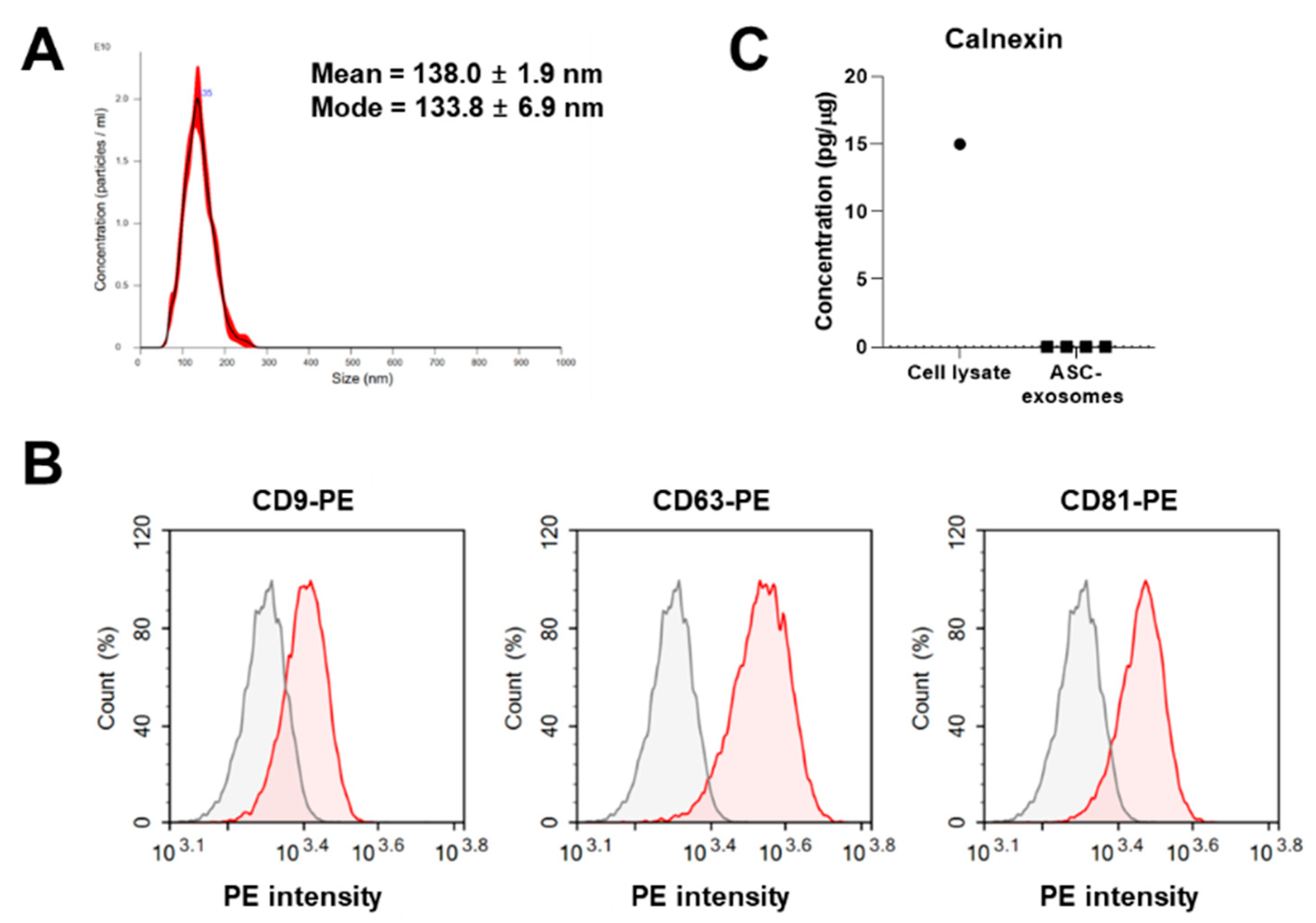
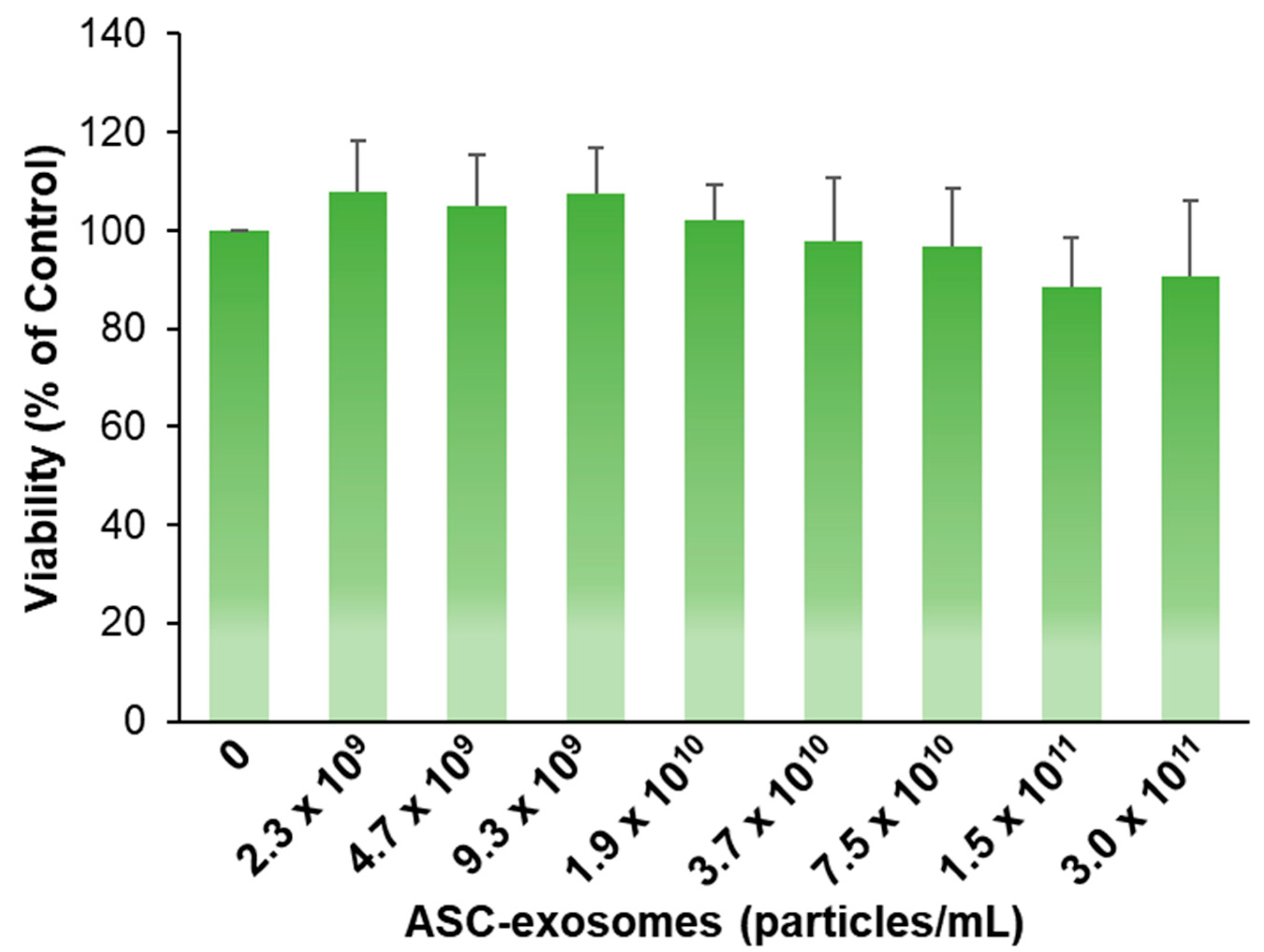
3.1. Isolation and Characterization of ASC-Exosomes

MSC-CM have been reported to have antipigmentation effects on melanocytes [6,7]. Given that exosomes in the secretome of MSCs are a major paracrine mediator, we studied the effect of exosomes from ASCs on the melanin contents in vitro. First, we isolated exosomes from the CM of ASCs using ultrafiltration followed by diafiltration with tangential flow filtration (TFF) systems, as described previously [13]. ASC-exosomes were characterized using NTA and flow cytometry. ASC-exosomes range from 30 to 200 nm in size, with a mean and mode diameter of 138.0 ± 1.9 and 133.8 ± 6.9 nm, respectively [11,12,13,17], and they contain distinct surface marker proteins (Figure 1A,B). The concentration of isolated ASC-exosomes was 1.47 × 10¹² particles/mL. Calnexin was not detected in ASC-exosomes (Figure 1C).

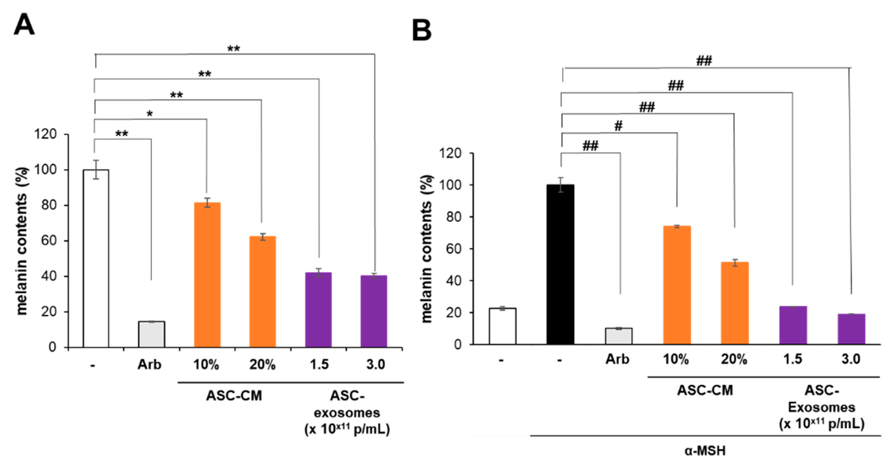
3.2. Antipigmentation Effect of ASC-Exosomes In Vitro

The effect of ASC-exosomes on the production of melanin was assessed in B16F10 mouse melanoma cells in vitro. First, the effect of ASC-exosomes on the viability of B16F10 cells was analyzed. B16F10 melanoma cells were treated with ASC-exosomes for 72 h, and the viability of the cells was assessed. As shown in Figure 2, no distinct reduction in cell viability by ASC-exosomes was observed up to a concentration of 3.0 × 10¹¹ particles/mL.
Second, the effects of ASC-CM and ASC-exosomes on the production of melanin in B16F10 cells were studied in the absence of α-MSH. ASC-CM reduced the concentration of intracellular melanin in a dose-dependent fashion (Figure 3A). ASC-exosomes also reduced the levels of intracellular melanin in B16F10 cells, and this effect saturated at a concentration of 1.5 × 10¹¹ particles/mL. These results suggest that ASC-exosomes negatively regulate intracellular melanin in B16F10 cells in the absence of α-MSH.
Third, the effects of both ASC-CM and ASC-exosomes on the melanin levels in B16F10 cells were evaluated in the presence of 100 nM α-MSH. The antipigmentation effects of both ASC-CM and ASC-exosomes were more pronounced in the presence of α-MSH (Figure 3B). Specifically, intracellular melanin levels in B16F10 cells were reduced by ASC-CM up to approximately 50% in a dose-dependent manner (Figure 3B). The ASC-exosomes showed a greater reduction in intracellular melanin levels in B16F10 cells than ASC-CM, reducing the melanin levels to those measured in B16F10 cells in the absence of α-MSH. Conclusively, ASC-exosomes negatively regulated intracellular melanin levels in B16F10 cells successfully.

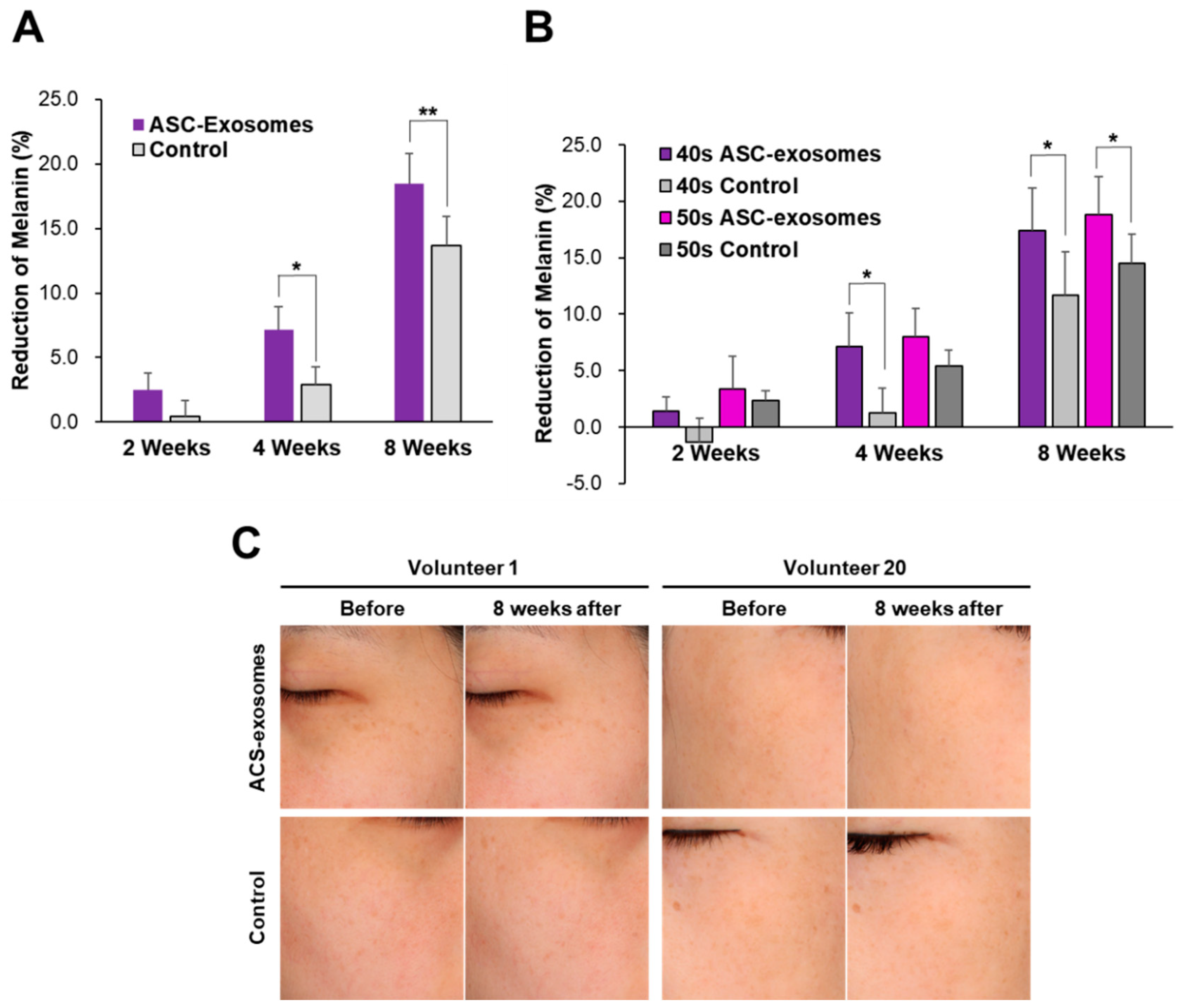
3.3. Skin Brightening Efficacy of ASC-Exosomes

We conducted a clinical study with an ASC-exosome-containing formulation in a prospective, split-face, double-blind, randomized placebo-controlled study to assess its skin brightening efficacy. The placebo (without ASC-exosomes) or test (with ASC-exosomes) formulation was applied twice a day for 8 weeks on the face of 21 female volunteers. Melanin levels were measured using a Mexameter before and 2, 4, and 8 weeks after treatment (Table 1). The reduction in melanin levels began 2 weeks after treatment, and a statistically significant reduction was observed 4 weeks after treatment compared to the placebo-treated areas (Figure 4). However, the melanin-reduction activity was diminished along with time (at week 8). The reduction in melanin levels was more pronounced in women aged <50 years, as observed 4 weeks after treatment (Figure 4B). No adverse effects were observed in any volunteer during or after the study.

4. Discussion

Skin brightening cosmetic products help improve skin tone and lessen skin pigmentation. They are increasingly being used to obtain an even skin tone, alleviate blemishes, and reduce age spots. The global market value for skin brightening products is expected to reach USD 13.7 billion by 2025 and is growing with a compound annual growth rate of 7.4% over the forecast period [19]. This positive trend is because of the increasing awareness among consumers about the aesthetic needs of the modern society, skin pigmentation, and aging.
Melanin deposition is induced by ultraviolet (UV) radiation; it protects the skin from UV rays. However, hyperactivation of melanocytes causes pigmentation of keratinocytes in the skin [20]. Therefore, reducing melanin levels is necessary for skin brightening. Melanocytes are located in the basal layer of the epidermis and contain melanosomes; they synthesize the pigment melanin via melanogenesis [21]. Melanogenesis is a complex process involving three proteins that are important mediators of melanogenesis: tyrosinase, tyrosinase-related protein-1 (TYRP-1), and TYRP-2. Melanogenesis includes catalytic reactions and a series of enzyme reactions. Among those, tyrosinase plays a critical role in melanogenesis by promoting the formation of 3,4-dihydroxy-l-phenylalanine (l-DOPA) from l-tyrosine and, subsequently, in the production of l-dopaquinone from l-DOPA through enzymatic reactions and autoxidation [22].
In the present study, we showed that ASC-exosomes reduced intracellular melanin levels in B16F10 cells without significant cytotoxicity whether in the presence or the absence of α-MSH. In addition, the ASC-exosome-containing topical formulation reduced hyperpigmentation in a prospective, split-face, double-blind, randomized placebo-controlled study. No statistically significant differences in the reduction of melanin levels between test-treated and placebo-controlled areas were noted until 2 weeks of treatment. The differences were, however, noticeable after 4 weeks of treatment and sustained until 8 weeks after treatment. Although instrumental measurement of the melanin contents resulted in statistically significant differences between test-treated and placebo-controlled areas, ASC-exosomes have worked in vitro but clinically relevant brightening effects were not evident in volunteers, probably due to inefficient delivery of exosomes into the deep dermis [23]. Further clinical studies should be followed with enhanced transdermal delivery of ASC-exosomes. Even though exosomes, by topical treatment, have been reported to be absorbed by human skin and reached the epidermis in an ex vivo experiment [24], additional improvement of transdermal delivery will be helpful for more profound melanin-reduction activity by ASC-exosomes, which was observed in the in vitro cell-based assay (Figure 3). Various transdermal delivery systems have been developed for either cosmetic or medical purposes, which include chemical boosters, ultrasound, iontophoresis, microneedles, thermal ablation, microdermabrasion, electroporation, and cavitation ultrasound [25]. Efficacious transdermal delivery of exosomes has been reported with the keratin hydrogel-based microneedle patch [26], needle-free jet injector [27], or iontophoresis [28]. There were no skin abnormalities (erythema, edema, scaling, itching, stinging, burning, tightness, and prickling) during and after the study (data now shown).
Another factor, which may affect the efficacy of exosomes, is their engulfment by phagocytic immune cells. Phagocytic cells such as tissue-resident macrophages and Kupffer cells are responsible for clearance of foreign materials by phagocytosis [29]. “Don’t eat me” signals on the membrane surface, mainly determining the escape from this clearance, include CD47 [30], β2-microglobulin (B2M) [31], programmed death-ligand 1 (PD-L1, also known as CD274 or B7-H1) [32], and CD24 [33]. These “don’t eat me” signals are also reported on the surface of exosomes from bone marrow-derived MSCs [34]. Importantly, our previous study revealed that ASC-exosomes contain CD47 and B2M [12]. It would be important to analyze the contribution of these molecules to the efficacy of ASC-exosomes in skin healthiness in future study.
The key purpose of the current study was to determine whether ASC-exosomes have an antimelanin effect or not, but we cannot exclude the additional effects by other soluble factors in the secretome of ASC-CM such as soluble cytokines, growth factor, or extracellular RNAs. Although these soluble factors may contribute additional antimelanin effects by ASC-CM, their effects may be limited because the naked soluble factors are more vulnerable to degradation by protease or RNase compared to exosome-associated cargo. In addition, the delivery efficiency of these soluble factors into cells is expected to be lower than that of exosomes, natural-born delivery vehicles.
ASC-exosomes contain various cargo including proteins, RNAs, and lipids. The complexity of exosomal cargo is demonstrated as 9769 proteins, 3408 mRNAs, 2838 miRNAs, and 1116 lipid entries from 286 studies registered in the ExoCarta database [35,36,37,38]. For examples, proteomics analysis revealed a total of 1008 proteins and common 471 proteins in three batches of ASC-exosomes in the previous study [12]. In addition, 358 lipid species were identified in ASC-exosomes [12]. These cargo molecules contribute to the function of ASC-exosomes in various biological process.
Previously, our group found that ASC-exosomes increased synthesis of ceramides, dihydroceramides, sphingosine, and sphingosine 1-phosphate (S1P) in mouse skin with oxazolone-induced barrier defects [12]. In addition, increased activity of sphingosine kinase 1 (SPHK1) and reduced activity of S1P lyase were observed in the lesional skin of ASC-exosome-treated mice [12]. Interestingly, ceramides and S1P have been reported to negatively regulate melanin synthesis in melanocytes [39,40,41]. Microphthalmia-associated transcription factor (MITF), a key transcription factor in melanogenesis, is degraded by S1P-induced activation of the extracellular signal-regulated kinase (ERK) [41]. Further study is needed to understand whether and how ASC-exosomes induce ceramide or S1P synthesis in relation to melanogenesis in melanocytes.
Another potential factor in ASC-exosomes constitutes microRNAs (miRNAs) such as hsa-miR-137, hsa-miR-145, and hsa-miR-330. In a pilot study, we identified these miRNAs among 598 miRNAs associated with ASC-exosomes using small RNA-sequencing (unpublished observation). Previous studies showed that miR-137 targets MITF and reduces pigmentation in melanoma cells [42,43]. The overexpression of miR-145 inhibits melanosome transport by targeting myosin-5a [44]. Furthermore, miRNA-330 targets tyrosinase and reduces melanin levels in melanoma cells [43].
A limitation of current study is the use of murine melanoma cell line B16F10 cultured in DMEM. The excess of l-tyrosine in DMEM (120 mg/L as disodium hydrate) compared to others such as Roswell Park Memorial Institute (RPMI) 1640 (28.8 mg/L as disodium hydrate) is able to induce pigmentation in melanoma cells even in the absence of α-MSH stimulation [45,46]. Since the excess of l-tyrosine induces tyrosinase activity [45] and ASC-exosomes reduced the intracellular melanin contents of B16F10 cells in the absence of α-MSH (Figure 3A), ASC-exosomes may regulate, at least partially, the downstream factors of tyrosinase including TYRP-1 and TYRP-2. The significance of this finding should be further confirmed in normal human melanocytes or a human melanoma cell line SK-MEL-2, which can be maintained Eagle’s Minimum Essential Medium (EMEM) containing 51.9 mg/L of l-tyrosine 2Na 2H2O (www.atcc.org).
Another limitation is that the disorder of each volunteer was not diagnosed exactly since the purpose of this study was the evaluation of cosmetic formulation, not therapeutic formulation. Clinically, hyperpigmentation disorders include solar lentigines, postinflammatory hyperpigmentation, melasma, ephelides, and café au lait macules with different pathologies and involvement of melanocytes [47,48]. Further studies are needed to decide the therapeutic potential of ASC-exosomes in different hyperpigmentation disorders.
Since the long-term topical use of phenolic compounds such as hydroquinone causes chronic adverse effects [2], there is an unmet need for safer and more effective new materials for skin brightening. Although the molecular mechanisms underlying the antipigmentation effects of ASC-exosomes remain unknown, this study shows that they effectively reduce intracellular melanin levels in vitro and that a cosmetic formulation containing ASC-exosomes is helpful in skin brightening. ASC-exosomes isolated using the ExoSCRT™ technology have been registered as a cosmetic ingredient in the International Cosmetic Ingredient Dictionary (ICID) [3]. Moreover, the good laboratory practice (GLP) toxicological tests showed that ASC-exosomes are safe for use, with no skin sensitization, photosensitization, eye and skin irritation, or acute oral toxicity [13]. Additionally, previous results suggested that ASC-exosomes have little or no immunogenicity in allogeneic application because of no detectable expression of human leukocyte antigens (HLA-ABC and HLA-DRDPDQ) on them [12,17]. A further improvement of transdermal delivery of ASC-exosomes will be helpful for their more profound efficacy as a cosmeceutical for skin brightening.

Author Contributions

Conceptualization, B.S.C. and Y.W.Y.; methodology, J.L. (Jinah Lee), Y.W., J.Y., J.C., M.J. and Y.W.Y.; investigation, B.S.C., J.L. (Jinah Lee), Y.W., J.Y. and Y.W.Y.; resources, B.S.C.; data curation, J.L. (Jinah Lee), Y.W. and Y.W.Y.; data analysis, J.L. (Jinah Lee), Y.W., D.I.D., R.C.J., J.L. (Joon Lee), H.H.K., G.-H.P., S.H.Y., B.C.P., K.Y.P., J.Y., J.C., M.J. and Y.W.Y.; writing—original draft preparation, B.S.C., J.L. (Jinah Lee), Y.W., J.Y. and Y.W.Y.; writing—review and editing, B.S.C., J.L. (Jinah Lee), Y.W., D.I.D., R.C.J., J.L. (Joon Lee), H.H.K., G.-H.P., S.H.Y., B.C.P., K.Y.P., J.C., M.J. and Y.W.Y.; visualization, J.L. (Jinah Lee), Y.W., J.Y. and Y.W.Y.; supervision, B.S.C. and Y.W.Y.; project administration, J.L. (Jinah Lee) and Y.W.Y.; funding acquisition, M.J. and Y.W.Y. All authors have read and agreed to the published version of the manuscript.

Funding

This research was partially supported by ExoCoBio Inc. This research was also supported by the Korea Ministry of Trade, Industry, and Energy (MOTIE) R&D Program of Development of Material and Component Technology Grant (#10079925). The funders had no involvement in data collection, study design, analyses, data interpretation, manuscript preparation, or publication.

Conflicts of Interest

B.S.C. and Y.W.Y. are founders and stockholders of ExoCoBio Inc. B.S.C., J.L. (Jinah Lee), Y.W., J.Y., and Y.W.Y. are employees of ExoCoBio Inc. J.C. and M.J. are employees of Biosensor Laboratories, Inc. The other authors declare no conflicts of interest.

References

  1. Berlitz, S.J.; De Villa, D.; Inácio, L.A.M.; Davies, S.; Zatta, K.C.; Guterres, S.S.; Külkamp-Guerreiro, C. Azelaic acid-loaded nanoemulsion with hyaluronic acid—A new strategy to treat hyperpigmentary skin disorders. Drug Dev. Ind. Pharm. 2019, 45, 642–650. [Google Scholar] [CrossRef] [PubMed]
  2. Sarkar, R.; Arora, P.; Garg, K.V. Cosmeceuticals for hyperpigmentation: What is available? J. Cutan. Aesthet. Surg. 2013, 6, 4–11. [Google Scholar] [CrossRef] [PubMed]
  3. Ha, D.H.; Kim, H.-k.; Lee, J.; Kwon, H.H.; Park, G.-H.; Yang, S.H.; Jung, J.Y.; Choi, H.; Lee, J.H.; Sung, S.; et al. Mesenchymal stem/stromal cell-derived exosomes for immunomodulatory therapeutics and skin regeneration. Cells 2020, 9, 1157. [Google Scholar] [CrossRef] [PubMed]
  4. Hwang, I.; Park, J.-H.; Park, H.-S.; Choi, K.-A.; Seol, K.-C.; Oh, S.-I.; Kang, S.; Hong, S. Neural stem cells inhibit melanin production by activation of Wnt inhibitors. J. Dermatol. Sci. 2013, 72, 274–283. [Google Scholar] [CrossRef]
  5. Kim, D.-W.; Jeon, B.-J.; Hwang, N.-H.; Kim, M.-S.; Park, S.-H.; Dhong, E.-S.; Yoon, E.-S.; Lee, B.-I. Adipose-derived stem cells inhibit epidermal melanocytes through an interleukin-6-mediated mechanism. Plasti. Reconstr. Surg. 2014, 134, 470–480. [Google Scholar] [CrossRef]
  6. Kim, E.S.; Jeon, H.B.; Lim, H.; Shin, J.H.; Park, S.J.; Jo, Y.K.; Oh, W.; Yang, Y.S.; Cho, D.-H.; Kim, J.-Y. Conditioned media from human umbilical cord blood-derived mesenchymal stem cells inhibits melanogenesis by promoting proteasomal degradation of MITF. PLoS ONE 2015, 10, e0128078. [Google Scholar] [CrossRef] [Green Version]
  7. Wang, X.; Shu, X.; Huo, W.; Zou, L.; Li, L. Efficacy of protein extracts from medium of adipose-derived stem cells via microneedles on Asian skin. J. Cosmet. Laser Ther. 2018, 20, 237–244. [Google Scholar] [CrossRef]
  8. Kalluri, R.; LeBleu, V.S. The biology, function, and biomedical applications of exosomes. Science 2020, 367, eaau6977. [Google Scholar] [CrossRef]
  9. Zhang, Y.; Liu, Y.; Liu, H.; Tang, W.H. Exosomes: Biogenesis, biologic function and clinical potential. Cell Biosci. 2019, 9, 19. [Google Scholar] [CrossRef]
  10. Yi, Y.W.; Lee, J.H.; Kim, S.-Y.; Pack, C.-G.; Ha, D.H.; Park, S.R.; Youn, J.; Cho, B.S. Advances in analysis of biodistribution of exosomes by molecular imaging. Int. J. Mol. Sci. 2020, 21, 665. [Google Scholar] [CrossRef] [Green Version]
  11. Cho, B.S.; Kim, J.O.; Ha, D.H.; Yi, Y.W. Exosomes derived from human adipose tissue-derived mesenchymal stem cells alleviate atopic dermatitis. Stem Cell Res. Ther. 2018, 9, 187. [Google Scholar] [CrossRef] [PubMed] [Green Version]
  12. Shin, K.-O.; Ha, D.H.; Kim, J.O.; Crumrine, D.A.; Meyer, J.M.; Wakefield, J.S.; Lee, Y.; Kim, B.; Kim, S.; Kim, H.-K.; et al. Exosomes from human adipose tissue-derived mesenchymal stem cells promote epidermal barrier repair by inducing de novo synthesis of ceramides in atopic dermatitis. Cells 2020, 9, 680. [Google Scholar] [CrossRef] [PubMed] [Green Version]
  13. Ha, D.H.; Kim, S.-D.; Lee, J.; Kwon, H.H.; Park, G.-H.; Yang, S.H.; Jung, J.Y.; Lee, J.H.; Park, S.R.; Youn, J.; et al. Toxicological evaluation of exosomes derived from human adipose tissue-derived mesenchymal stem/stromal cells. Regul. Toxicol. Pharmacol. 2020, 115, 104686. [Google Scholar] [CrossRef] [PubMed]
  14. Kwon, H.H.; Yang, S.H.; Lee, J.; Park, B.C.; Park, K.Y.; Jung, J.Y.; Bae, Y.; Park, G.-H. Combination treatment with human adipose tissue stem cell-derived exosomes and fractional CO2 laser for acne scars: A 12-week prospective, double-blind, randomized, split-face study. ACTA Derm. Venereol. 2020, 100, adv00310. [Google Scholar] [CrossRef] [PubMed]
  15. Cicero, A.L.; Delevoye, C.; Gilles-Marsens, F.; Loew, D.; Dingli, F.; Guéré, C.; André, N.; Vié, K.; van Niel, G.; Raposo, G. Exosomes released by keratinocytes modulate melanocyte pigmentation. Nat. Commun. 2015, 6, 7506. [Google Scholar] [CrossRef] [PubMed]
  16. Takano, K.; Hachiya, A.; Murase, D.; Tanabe, H.; Kasamatsu, S.; Takahashi, Y.; Moriwaki, S.; Hase, T. Quantitative changes in the secretion of exosomes from keratinocytes homeostatically regulate skin pigmentation in a paracrine manner. J. Dermatol. 2006, 28, 269–276. [Google Scholar] [CrossRef]
  17. Lee, J.H.; Ha, D.H.; Go, H.-k.; Youn, J.; Kim, H.-k.; Jin, R.C.; Miller, R.B.; Kim, D.-h.; Cho, B.S.; Yi, Y.W. Reproducible large-scale isolation of exosomes derived from adipose tissue-derived mesenchymal stem/stromal cells and their application in acute kidney injury. Int. J. Mol. Sci. 2020, 21, 4774. [Google Scholar] [CrossRef]
  18. Kim, S.I.; Kim, H.J.; Lee, H.J.; Lee, K.; Hong, D.; Lim, H.; Cho, K.; Jung, N.; Yi, Y.W. Application of a non-hazardous vital dye for cell counting with automated cell counters. Anal. Biochem. 2016, 492, 8–12. [Google Scholar] [CrossRef] [Green Version]
  19. Grand View Research, Skin Lighting Products Market Size worth $13.7 Billion by 2025. Available online: https://www.grandviewresearch.com/press-release/global-skin-lightening-products-market (accessed on 19 August 2020).
  20. Pillaiyer, T.; Manickam, M.; Jung, S.-H. Recent development of signaling pathways inhibitors of melanogenesis. Cell Signal. 2017, 40, 99–115. [Google Scholar] [CrossRef]
  21. Bonaventure, J.; Domingues, M.J.; Larue, L. Cellular and molecular mechanisms controlling the migration of melanocytes and melanoma cells. Pigment Cell Melanoma Res. 2013, 26, 316–325. [Google Scholar] [CrossRef]
  22. Jimenez-Cervantes, C.; Garcia-Borron, J.C.; Valverde, P.; Solano, F.; Lozano, J.A. Tyrosinase isoenzymes in mammalian melanocytes: 1. biochemical characterization of two melanosomal tyrosinases from B16 mouse melanoma. Eur. J. Biochem. 1993, 217, 549–556. [Google Scholar] [CrossRef]
  23. Ng, K.W. Penetration enhancement of topical formulations. Pharmaceutics 2018, 10, 51. [Google Scholar] [CrossRef] [Green Version]
  24. Kim, Y.-J.; Yoo, S.M.; Park, H.H.; Lim, H.J.; Kim, Y.-L.; Lee, S.; Seo, K.-W.; Kang, K.-S. Exosomes derived from human umbilical cord blood mesenchymal stem cells stimulates rejuvenation of human skin. Biochem. Biophys. Res. Commun. 2017, 493, 1102–1108. [Google Scholar] [CrossRef]
  25. Elkhoury, K.; Kocak, P.; Kang, A.; Arab-Tehrany, E.; Ward, J.E.; Shin, S.R. Engineering smart targeting nanovesicles and their combination with hydrogels for controlled drug delivery. Pharmaceutics 2020, 12, 849. [Google Scholar] [CrossRef]
  26. Yang, G.; Chen, Q.; Wen, D.; Chen, Z.; Wang, J.; Chen, G.; Wang, Z.; Zhang, X.; Zhang, Y.; Hu, Q.; et al. A therapeutic microneedle patch made from hair-derived keratin for promoting hair regrowth. ACS Nano 2019, 13, 4354–4360. [Google Scholar] [CrossRef]
  27. Hu, S.; Li, Z.; Cores, J.; Huang, K.; Su, T.; Dinh, P.-Y.; Cheng, K. Needle-free injection of exosomes derived from human dermal fibroblast spheroids ameliorates skin photoaging. ASC Nano 2019, 13, 11273–11282. [Google Scholar] [CrossRef]
  28. Yi, Y.W.; Cho, B.S. Exosome Kit and Method for Enhancing Transdermal Permeation of Exosomes by Using Same. Patent WO2019088545A1, 9 May 2019. [Google Scholar]
  29. Belhadj, Z.; He, B.; Deng, H.; Song, S.; Zhang, H.; Wang, X.; Dai, W.; Zhang, Q. A combined “eat me/don’t eat me” strategy based on extracellular vesicles for anticancer nanomedicine. J. Extracell. Vesicles 2020, 9, 1806444. [Google Scholar] [CrossRef]
  30. Oldernborg, P.-A.; Zheleznyak, A.; Fang, Y.-F.; Lagenaur, C.F.; Gresham, H.D.; Lindberg, F.P. Role of CD47 as a marker of self on red blood cells. Science 2000, 288, 2051–2054. [Google Scholar] [CrossRef]
  31. Barkal, A.A.; Weiskopf, K.; Kao, K.S.; Gordon, S.R.; Rosental, B.; Yiu, Y.Y.; Geroge, B.M.; Markovic, M.; Ring, N.G.; Tsai, J.M.; et al. Engagement of MHC class I by the inhibitory receptor LILRB1 suppresses macrophages and is a target of cancer immunotherapy. Nat. Immunol. 2017, 19, 76–84. [Google Scholar] [CrossRef]
  32. Gordon, S.R.; Maute, R.L.; Dulken, B.W.; Hutter, G.; George, B.M.; McCracken, M.N.; Gupta, R.; Tsai, J.M.; Sinha, R.; Corey, D.; et al. PD-1 expression by tumour-associated macrophages inhibits phagocytosis and tumour immunity. Nature 2017, 545, 495–499. [Google Scholar] [CrossRef]
  33. Kristiansen, G.; Winzer, K.-J.; Mayordomo, E.; Bellach, J.; Schluns, K.; Denkert, C.; Dahl, E.; Pilarsky, C.; Altevogt, P.; Guski, H.; et al. CD24 expression is a new prognostic marker in breast cancer. Clin. Cancer Res. 2003, 9, 4906–4913. [Google Scholar]
  34. Mendt, M.; Kamerkar, S.; Sugimoto, H.; McAndrews, K.M.; Wu, C.-C.; Gagea, M.; Yang, S.; Blanko, E.V.R.; Peng, Q.; Ma, X.; et al. Generation and testing of clinical-grade exosomes for pancreatic cancer. JCI Insight. 2018, 3, e99263. [Google Scholar] [CrossRef]
  35. Mathivanan, S.; Simpson, R.J. ExoCarta: A compendium of exosomal proteins and RNA. Proteomics 2009, 21, 4997–5000. [Google Scholar] [CrossRef]
  36. Mathivanan, S.; Fahner, C.J.; Reid, G.E.; Simpson, R.J. ExoCarta 2012: Database of exosomal protein, RNA and lipids. Nuc. Acids Res. 2012, 40, D1241–D1244. [Google Scholar] [CrossRef] [Green Version]
  37. Simpson, R.J.; Kalra, H.; Mathivanan, S. ExoCarta as a resource for exosomal research. J. Extracell. Vesicles 2012, 1, 18374. [Google Scholar] [CrossRef]
  38. Keerthikumar, S.; Chisanga, D.; Ariyaratne, D.; Al Saffar, H.; Anand, S.; Zhao, K.; Samuel, M.; Pathan, M.; Jois, M.; Chilamkurti, N. ExoCarta: A web-based compendium of exosomal cargo. J. Mol. Biol. 2016, 428, 688–692. [Google Scholar] [CrossRef] [Green Version]
  39. Kim, D.-S.; Kim, S.-Y.; Moon, S.-J.; Chung, J.-H.; Kim, K.-H.; Cho, K.-H.; Park, K.-C. Ceramide inhibits cell proliferation through Atk/PKB inactivation and decreases melanin synthesis in Mel-Ab cells. Pigment Cell Res. 2001, 14, 110–115. [Google Scholar] [CrossRef]
  40. Kim, D.S.; Kim, S.-Y.; Chung, J.-H.; Kim, K.-H.; Eun, H.-C.; Park, K.-C. Delayed ERK activation by ceramide reduced melanin synthesis in human melanocytes. Cell. Signal. 2002, 14, 779–875. [Google Scholar] [CrossRef]
  41. Kim, D.-S.; Hwang, E.-S.; Lee, J.-E.; Kim, S.-Y.; Kwon, S.-B.; Park, K.-C. Sphigosine-1-phosphate decreases melanin synthesis via sustained ERK activation and subsequent MITF degradation. J. Cell Sci. 2003, 116, 1699–1706. [Google Scholar] [CrossRef] [Green Version]
  42. Bemis, L.T.; Chen, R.; Amato, C.M.; Classen, E.H.; Robinson, S.E.; Coffey, D.G.; Erickson, P.F.; Shellman, Y.G.; Robinson, W.A. MicroRNA-137 targets microphthalmia-associated transcription factor in melanoma cell lines. Cancer Res. 2008, 68, 1362–1368. [Google Scholar] [CrossRef] [Green Version]
  43. Rambow, F.; Bechadergue, A.; Saintigny, G.; Morizot, F.; Mahe, C.; Larue, L. miR-330-5p targets tyrosinase and induces depigmentation. J. Invest. Dermatol. 2014, 134, 2846–2849. [Google Scholar] [CrossRef] [Green Version]
  44. Dynoodt, P.; Mestdagh, P.; Van Peer, G.; Vandesompele, J.; Goossens, K.; Peelman, L.J.; Geusens, B.; Speeckaert, R.M.; Lambert, J.L.W.; Van Gele, M.J.L. Identification of miR-145 as a key regulator of the pigmentary process. J. Invest. Dermatol. 2013, 133, 201–209. [Google Scholar] [CrossRef] [PubMed] [Green Version]
  45. Slominski, A.; Moellmann, G.; Kuklinska, E.; Bomirski, A.; Pawelek, J. Positive regulation of melanin pigmentation by two key substrates of the melanogenic pathway, L-tyrosine and L-dopa. J. Cell Sci. 1988, 89, 287–296. [Google Scholar] [PubMed]
  46. Wolnicka-Glubisz, A.; Nogal, K.; Zadlo, A.; Plonka, P.M. Curcumin does not switch melanin synthesis towards pheomelanin in B16F10 cells. Arch. Dermatol. Res. 2015, 307, 89–98. [Google Scholar] [CrossRef] [PubMed]
  47. Plensdorf, S.; Livieratos, M.; Dada, N. Pigmentation disorders: Diagnosis and management. Am. Fam. Physician. 2017, 96, 797–804. [Google Scholar] [PubMed]
  48. Kwon, S.H.; Na, J.-I.; Choi, J.-Y.; Park, K.-C. Melasma: Updates and perspectives. Exp. Dermatol. 2019, 28, 704–708. [Google Scholar] [CrossRef] [PubMed] [Green Version]
Figure 1. Characteristics of human adipose tissue-derived mesenchymal stem/stromal cells (ASC-exosomes). (A) A representative Nanoparticle Tracking Analysis (NTA) histogram of ASC-exosomes. (B) Bead-based flow cytometric analysis of ASC-exosomes with antibodies for indicated proteins. ASC-exosomes were captured by Dynabeads coated with antibodies for exosome surface markers including cluster of differentiation 9 (CD9), CD63, or CD81. Then, the phycoerythrin (PE)-conjugated antibodies were reacted with captured ASC-exosomes as indicated. The mouse immunoglobulin G1 (IgG1)-PE was used as an isotype negative control. Gray peaks, PE fluorescence count from isotype control antibody; red peaks, PE fluorescence count from antibodies for indicated proteins (CD9, CD63, and CD81); X-axis, signal intensity of PE. (C) Concentration of calnexin measured by ELISA. n = 4 for ASC-exosomes and n = 1 for cell lysates performed in duplicate.
Cosmetics 07 00090 g001
Figure 2. No cytotoxicity of ASC-exosomes on B16F10 melanoma cells. The cytotoxic effect of ASC-exosomes was determined by measuring cell viability after incubation of the cells with increasing concentrations of ASC-exosomes for 72 h. Data are presented as the mean ± SD performed in triplicate.
Cosmetics 07 00090 g002
Figure 3. The antipigmentation effect of ASC-exosomes in the absence and presence of α-melanocyte-stimulating hormone (α-MSH). The melanin levels in B16F10 melanoma cells were measured after treatment with ASC-exosomes in the (A) absence or (B) presence of α-MSH for 48 h. Representative data are presented as the mean ± SD from multiple experiments performed in triplicate. (A) * p < 0.05 and ** p < 0.01 vs. the negative control group were considered statistically significant. (B) # p < 0.05 and ## p < 0.01 vs. the α-MSH control group were considered statistically significant. Arb: arbutin.
Cosmetics 07 00090 g003
Figure 4. Clinical evaluation of the skin brightening effect of ASC-exosomes. Melanin levels were measured using a Mexameter on the faces of 21 female volunteers before and 2, 4, and 8 weeks after treatment with a placebo (without ASC-exosomes) or test (with ASC-exosome) formulation twice daily for 8 weeks. (A) Reduction of melanin over time. (B) Reduction of melanin in different age groups. n = 11 for 40s and n = 8 for 50s. * p < 0.05 and ** p < 0.01 vs. placebo-treated area was considered statistically significant. (C) Representative images of two volunteers.
Cosmetics 07 00090 g004
Table 1. Melanin index of each volunteer.
AgeVolunteer #1ASC-ExosomesPlacebo Control
0 Weeks2 Weeks4 Weeks8 Weeks0 Weeks2 Weeks4 Weeks8 Weeks
30s1207.7198.5200.0186.5170.8165.7169.2147.0
2195.5195.4195.4170.6193.5193.5191.0175.1
40s3185.9185.9185.0165.4185.6186.0188.0172.8
4211.1207.7198.4177.6209.1205.1196.8178.0
5180.0183.1180.2159.9187.9188.0187.1166.2
6214.4211.0209.6210.1220.4239.0234.6234.2
7210.5203.4193.9180.9225.5223.0215.5217.1
8200.4206.4199.4204.7197.0207.1200.9199.9
9168.5168.4168.6163.3186.6183.4186.6180.5
10190.5190.1190.1165.5204.0202.0204.6180.4
11175.6174.9174.6159.8158.8159.1159.0144.8
12195.8186.6184.9187.0190.0190.1183.5183.0
13212.6212.5181.9180.1223.4220.0217.7202.9
50s14155.0148.0146.1140.6155.4155.2143.0140.6
15184.7184.5184.4160.0202.9196.4196.0187.8
16229.9229.7221.0216.0213.4213.3210.4200.8
17121.9126.1123.4108.1130.8130.4130.4124.8
18232.0211.9219.4194.5204.6199.8203.0176.2
19170.7170.4149.6147.0187.1187.2180.6166.0
20170.0161.8164.6163.3180.8176.7171.8171.1
21130.7135.3122.3115.0123.4120.6120.0115.4
Mean187.78185.31180.61169.33188.14187.70185.22174.50
SD28.8826.6727.2227.4327.7329.1628.7028.95
SEM6.305.825.945.996.056.366.266.32
#1: identification number of each volunteer.
Publisher’s Note: MDPI stays neutral with regard to jurisdictional claims in published maps and institutional affiliations.

