Article

4′,7-Isoflavandiol (Equol) Enhances Human Dermal Fibroblast Renewal and Has Effects Similar to 17β-Estradiol in Stimulating Collagen and Elastin Expression. Cell Cycle and RT-PCR Analysis without Phenol Red

Department of Physiology, Developmental Biology and The Neuroscience Center, College of Life Sciences, LS 4005, Brigham Young University, Provo, UT 84602, USA
Submission received: 16 December 2020 / Revised: 30 December 2020 / Accepted: 30 December 2020 / Published: 4 January 2021

Abstract

:
Polyphenols have general health benefits including anti-photoaging influences to counter the negative effects of ultra-violet (UV) rays from solar light (via the generation of reactive oxygen species (ROS) and oxidative stress (OS)), which leads to the stimulation of matrix metalloproteinases (MMPs) that break down collagen and elastin. The changes in elastin and collagen represent major factors in dermal aging along with a decrease in skin fibroblast number and function. The purpose of this study was to determine the influence of a polyphenolic molecule, 4′,7-Isoflavandiol (Equol) at 10 nM on: (1) fibroblast number and function via cell cycle testing (including apoptosis) and collagen protein expression (types I and III) using long-term (eight-week) 3D human fibroblast cultures by intracellular fluorescent-activated cell sorting (FACS) analysis, and (2) quantifying elastin gene expression levels in short-term (four day) cultures using human monolayer fibroblasts by RT-PCR. In both in vitro testing methods, the presence of phenol red (tissue culture indicator) interfered with the parameter results. Therefore, all experiments were performed without phenol red. Using FACS analysis in the long-term 3D cultures exposure to 10 nM of equol for four days significantly increased the percentage of cycling fibroblasts (rejuvenation) above vehicle control (dimethyl sulfoxide (DMSO)) or 17β-estradiol levels, while apoptosis was not altered by any treatment. In addition, in the long-term cultures, collagen levels were significantly increased in the equol and 17β-estradiol treatments above vehicle control values. In short-term cultures, 10 nM of equol or 17β-estradiol significantly increased elastin gene expression levels above vehicle control values. In summary: (a) phenol red may interfere with tissue culture parameter results and (b) the polyphenolic equol compound, derived from plants, may provide protection against photoaging in cosmetic formulations by stimulating collagen, elastin, and enhancing fibroblast renewal.

1. Introduction

Polyphenols found in plants have general health benefits, including use in skin care products [1,2,3]. This is due to their anti-photoaging influences that counteract the negative effects of ultra-violet (UV) rays from solar light, which generate reactive oxygen species (ROS) and oxidative stress (OS) [1,2,3,4]. Associated with photoaging is the stimulation of inflammatory agents along with the increase in skin enzymes, namely matrix metalloproteinases (MMPs) that break down collagen and elastin [2,3,4,5,6]. The changes in elastin and collagen represent major factors in dermal aging, which are paramount in how skin properties enhance a youthful appearance. For example, UV exposure in photoaging causes a collagen deficit by shifting homeostasis from production/deposition to degradation along with degeneration in the elastin fiber network where disorganized and non-functional deposition of elastin fibers occurs in the upper and middle dermis [6,7,8,9]. Additionally, botanicals that have anti-inflammatory and antioxidant properties along with maintaining skin fibroblast number and function are also important in slowing down dermal aging [1,2,3,4]. This is not surprising, since plants are rich in antioxidants because they must survive continual ultraviolet radiation exposure [10,11].
One of the polyphenolic compounds with promising human skin care applications is the isoflavonoid equol, which is contained in plants and food products such as beans, cabbage, lettuces, tofu, and animal products such as eggs and cow’s milk, which have been reviewed elsewhere [4,5]. Equol has powerful antioxidant and anti-inflammatory properties along with beneficial topical dermal effects that have been reported in in vitro and clinical studies demonstrating its skin enhancing properties, especially in estrogen-deficient skin [4,5,12]. Among the studies that have examined equol’s properties, in vitro testing using cell and tissue culture represents some of the primary investigative techniques to ascertain further scientific information on this polyphenolic isoflavonoid compound that has a chemical structural and molecular weight similar to 17β-estradiol, see Figure 1 [5]. In fact, both equol and 17β-estradiol have estrogenic actions, however, equol appears to act as a selective estrogen receptors modulator (SERM) compared to 17β-estradiol where equol has a higher affinity for estrogen receptor (ER) beta compared to ER alpha [5].
Therefore, the purpose of this study was to determine the influence of a polyphenolic molecule, 4′,7-Isoflavandiol (Equol) at 10 nM on: (1) fibroblast number and function via cell cycle testing (including apoptosis) and collagen protein expression (types I and III) using long-term (eight-week) 3D human fibroblast cultures by intracellular fluorescent-activated cell sorting (FACS) analysis, and (2) quantifying elastin gene expression levels in short-term (four days) cultures using human monolayer fibroblasts by RT-PCR.

2. Materials and Methods

2.1. Test Material Stock Solution Preparation

Racemic Equol was purchased from LC Labs. ≥ 99% (Woburn, MA, USA, CAS # 94105-90-5). All other reagents and materials were purchased from Sigma-Aldrich (St. Louis, MO, USA, e.g., 17β-estradiol ≥ 98% CAS # 50-28-2) unless otherwise noted. The equol and 17β-estradiol test materials were dissolved in 100% dimethyl sulfoxide (DMSO) and then diluted to a final concentration of 10 nM racemic equol, 10 nM 17β-estradiol and DMSO at 10 nM as the control vehicle treatment. Notably, since it was determined that the phenol red tissue indicator interfered with the parameter results, no phenol red dye was added. In the reported results in the present study, only the elastin RT-PCR results display the presence or absence of phenol red dye for comparison.

2.2. Monolayer Cell Culture

Primary human dermal fibroblasts from neonatal foreskin were sub-cultured (or the recorded number of times this was sub-cultured was 10 to 11 times, or in other words, at a passage of 10 to 11 times) that were cultured in medium, which consisted of Dulbecco’s Modified Eagle Medium (DMEM) with 1X non-essential amino acids, 1X antibiotic/antimycotic, and 2% bovine calf serum. For experiments conducted without phenol red, the same medium formulation was used, but no phenol red dye was added. Approximately 5 × 10⁵ cells per 10 cm dish were cultured for 16 to 24 h in a 37 °C, humidified incubator with 5% CO2, then the medium was changed and test materials and vehicle controls were added. Cells were cultured for four days in the presence of test materials 10 nM racemic equol or 10 nM 17β-estradiol or 10 nM of DMSO that served as the control vehicle.

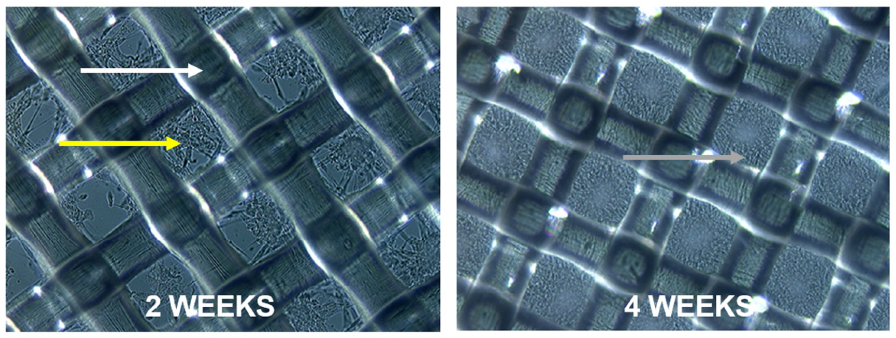
2.3. Organotypic 3D Dermal Cultures

To produce the organotypic, 3D cultures, dermal fibroblasts were seeded onto nylon mesh and allowed to grow for approximately eight weeks essentially as described [13,14]. This in vitro model closely mimics the development of the dermis, offering a system for study with organotypic properties such as the ability to support epidermal differentiation [13,14] and collagen fibrillogenesis [15] (see Figure 2). After 2 weeks, all 3D cultures were supplemented with 20 μg/mL ascorbate while monolayer cultures were not treated with ascorbate. Otherwise, all materials and procedures were essentially equivalent between monolayer and 3D experiments.

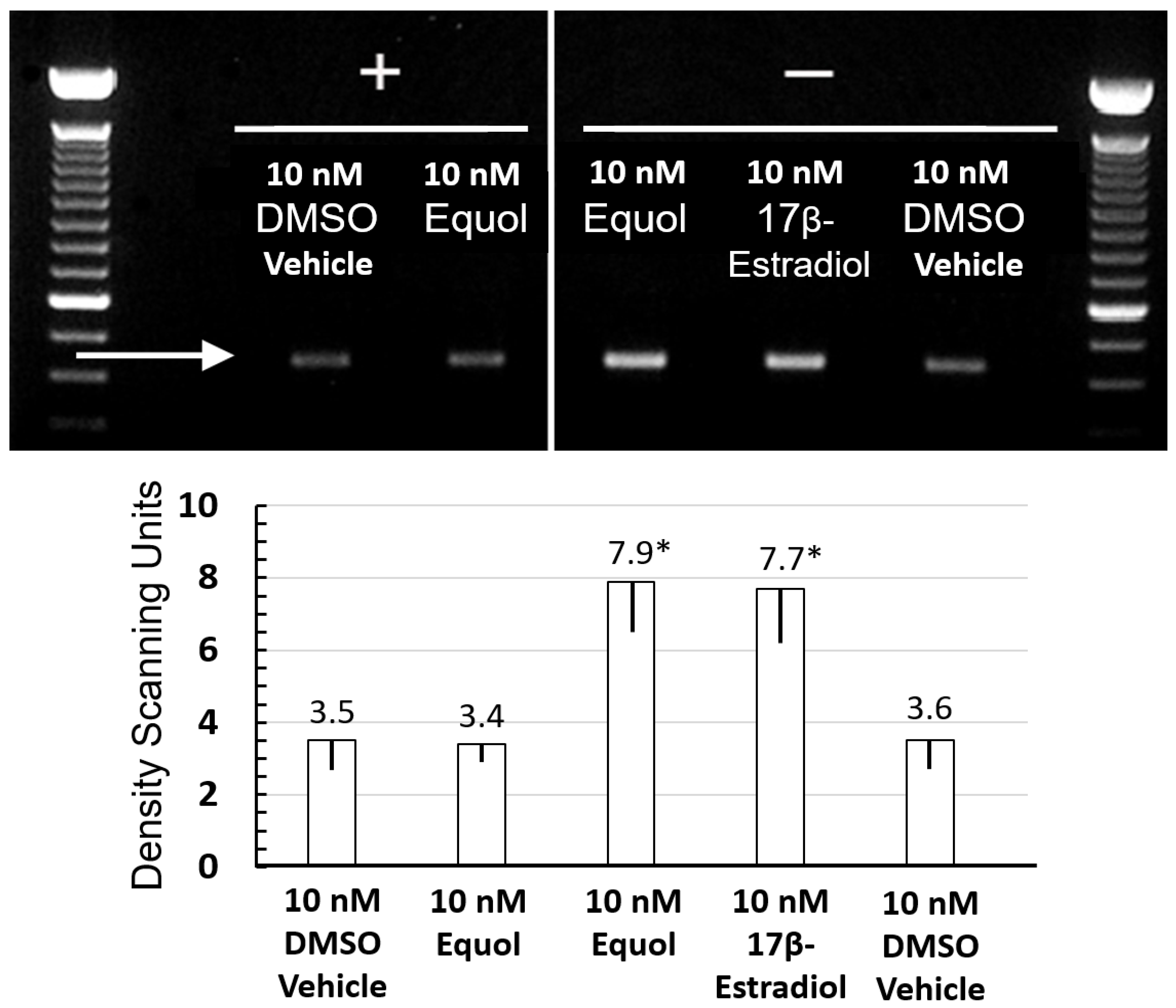
2.4. RT-PCR Analysis of Elastin Gene Expression

Duplicate fibroblast cultures in 10 cm dishes were cultured for four days in the presence of 10 nM equol or 10 nM 17β-estradiol or a vehicle control (10 nM DMSO) and were then prepared for elastin gene expression analysis by RT-PCR. RNA was isolated using a commercial kit, RNEasy Mini kit (QIAGEN Inc., Hilden, Germany) according to the manufacturer’s instructions. First-strand cDNA synthesis using random hexamer primers and 5 µgs total RNA was performed with a commercial kit, SuperScript III (Invitrogen Inc., Carlsbad, CA, USA) according to the manufacturer’s instructions. Control reactions without the reverse transcriptase enzyme added were performed to ensure amplification from mRNA only. PCR amplification was performed for 30 cycles using the following primers: ELAS-F1: 5′-cggagtgaagcctgggaaagtgccgggtgt-3′, and ELAS-R1: 5′-caccagggaggactccggctgctccagc-3′, which correspond to a 431 bp fragment of the human elastin gene (ELN: National Center for Biotechnology Information (NCBI), National Library of Medicine (NLM), National Institutes of Health (NIH), USA). Equal amounts of cDNA and PCR reactions were loaded in each lane, electrophoresed on a 1.8% agarose gel and visualized by ethidium bromide staining. Stained gels were illuminated with UV light, imaged with a digital camera, and band density quantified by NIH image analysis software (Bethesda, MD, USA) via reverse light/dark field contrast.

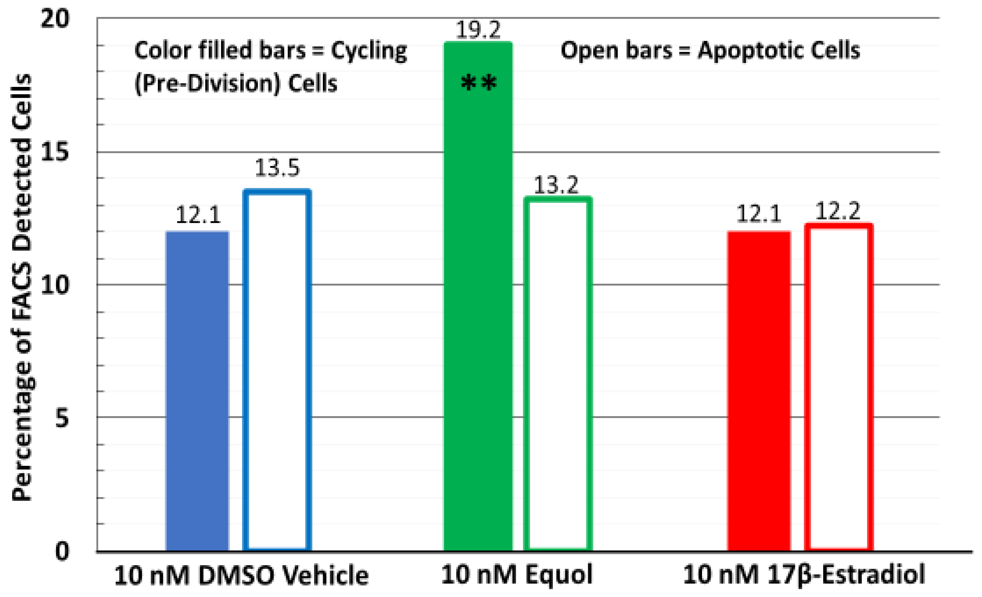
2.5. Intracellular FACS Analysis and Cell Cycle Determinations

Single-cell suspensions were produced by gentle trypsinization of monolayers, or extensive digestion in 1 mg/mL collagenase from 3D cultures. A commercial kit was utilized for the preparation of cells for intracellular detection by flow cytometry (IntraCyte, Orion BioSolutions, Inc., San Diego, CA, USA) according to the manufacturer’s instructions. In brief, cells were fixed with formaldehyde, permeabilized with non-ionic detergents, and non-specific protein binding was blocked. The following primary antibodies were used at 1 to 2 μg/mL: affinity-purified, anti-human collagen type I (Chemicon-Fisher Scientific, Waltham, MA, USA), affinity-purified anti-human type III collagen (Southern Biotech, Birmingham, AL, USA), and monoclonal anti-human elastin (Sigma-Aldrich, St. Louis, MO, USA). Negative controls included irrelevant immunoglobulins from the same species as each primary antibody and at the same concentration as well as unstained cells, and cells without primary antibody. Primary antibody binding was detected using affinity-purified, species-specific, fluorochrome-conjugated secondary antibodies. For cell cycle analysis, cells were stained with propidium iodide (fluorescent DNA dye) to quantify simultaneous levels of the cell cycle and apoptotic cell death. Finally, for all intracellular fluorescent-activated cell sorting (FACS) analysis, a Coulter EPICS Elite cytometer equipped with 488 nm argon laser was used, and between 10,000 and 20,000 cells per file were analyzed using Coulter ELITE software. At 10,000 to 20,000 cells per file analyzed the parameter variance of the obtained data was 1 to 2%. All FACS analysis data displayed in the present study had 20,000 cells per file with a variance of 1% among the treatment groups.

2.6. Statistical Analysis

For comparison between the treatment groups (DMSO vs. racemic equol or 17β-estradiol and especially the equol versus the 17β-estradiol results) the data were analyzed using analysis of variance (ANOVA) followed by Tukey’s honestly significant difference (HSD) post-hoc tests [16]. Treatment groups were compared to the DMSO levels, and a p value of less than or equal to 0.05 was reported as statistically significant results (p ≤ 0.05). All results were expressed as mean ± standard error of the mean in all figures, except the FACS analysis results, which had a variance of 1%, which was too small to display graphically in print form.
For the 3D long-term culture experiments using human dermal fibroblasts via FACS analysis of the collagens, cell cycle and apoptosis biomarkers, a binomial regression model was fitted with PROC GENMOD using SAS (Institute, Cary, NC, USA). The P-values of pair-wise comparisons were considered significant (all values were p < 0.001) where the DMSO vehicle served as the controls.

3. Results

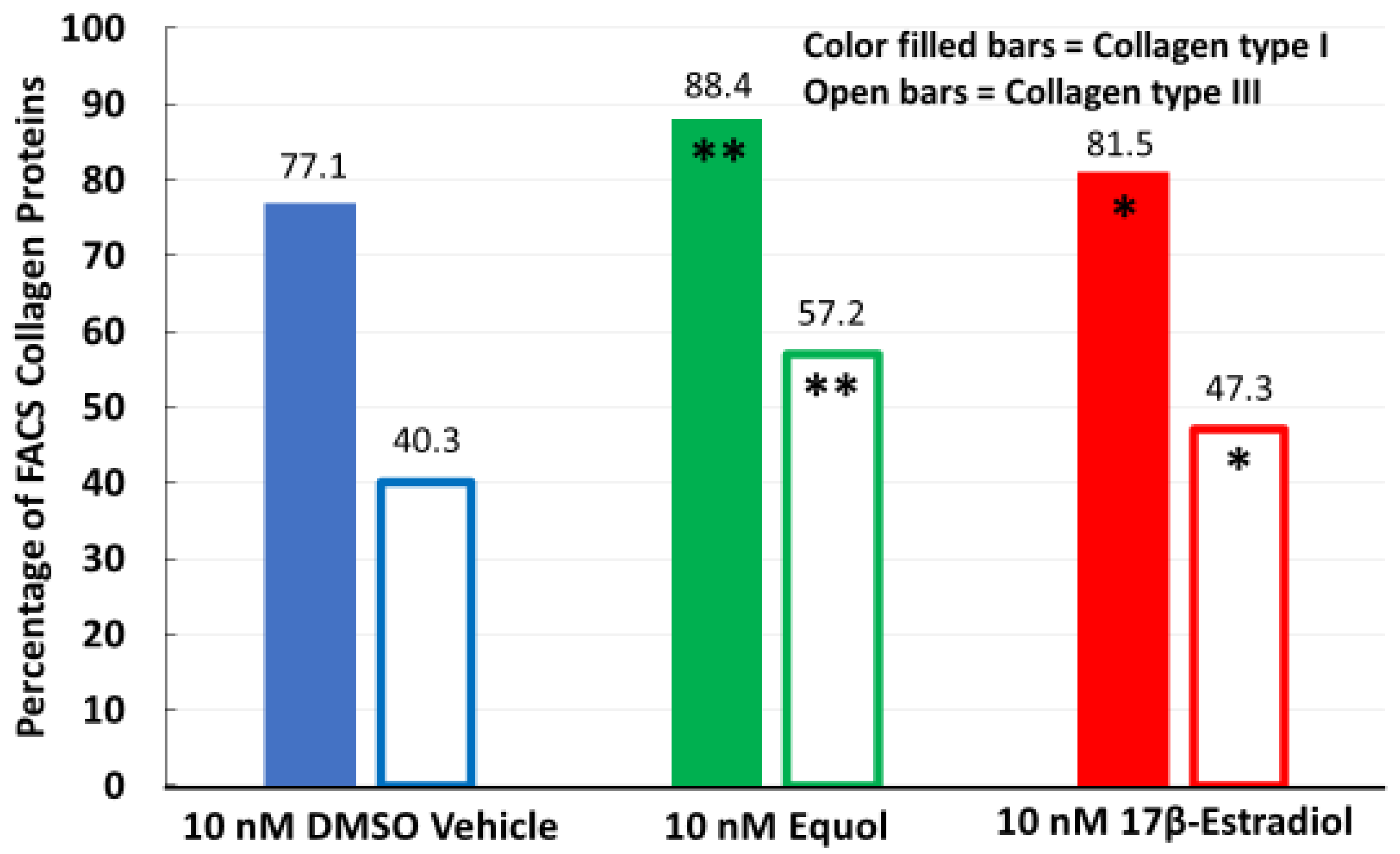
3.1. Equol and 17β-Estradiol Stimulated Collagen Types I and III Protein Expression via FACS Analysis in Long-Term 3D Human Fibroblast Cultures

To examine how equol may influence collagen protein expression, intracellular fluorescent-activated cell sorting (FACS) analysis was employed in long-term eight weeks 3D human fibroblast tissue cultures to quantify the percentage of cells among the 20,000 cells tested along with 17β-estradiol, which served as the natural steroid hormone positive control versus the DMSO vehicle controls. As shown in Figure 3, exposure to either 10 nM equol or 10 nM 17β-estradiol for four days significantly increased the percentage of cells for collagen types I and III (extracellular matrix proteins) above that of the DMSO control values (p < 0.001). Remarkably, the equol treatment also displayed significantly higher levels of the percentage of cells for collagen proteins compared to the 17β-estradiol values. For example, the equol treated cultures had 88.4% of the cells that expressed collagen type I protein compared to 81.5% for the 17β-estradiol group and both were significantly greater when tested against the DMSO level of 77.1% (see Figure 3). Furthermore, since collagen type III protein is expressed at lower levels compared to collagen type I, the FACS analysis in long-term tissue cultures was appropriate to detect any differences among the treatments tested. In fact, the number of cells expressing the collagen type III in the equol group was 57.2% compared to 47.3% for the 17β-estradiol treatment and again, both treatment groups displayed significantly greater levels when tested against the DMSO control value of 40.3%.

3.2. Equol and 17β-Estradiol Stimulated Elastin Gene Expression via RT-PCR Analysis. Phenol Red Tissue Indicator Inhibits Gene Expression

To examine how equol may influence elastin gene expression in short-term (four days) human monolayer fibroblast cultures along with 17β-estradiol, which served as the natural steroid hormone positive control versus the DMSO vehicle controls, expression levels were determined by RT-PCR and then quantified by density scanning analysis. As determined in prior cell/tissue culture experiments, the phenol red tissue indicator interfered with the results of the test materials. However, for this data set, the presence and absence of phenol red were tested and displayed in Figure 4. In the presence of phenol red, there were no differences between the DMSO control versus the racemic equol treatment group for the levels of the elastin gene product, which were both very low. However, in the absence of phenol red, the 10 nM racemic equol and 10 nM 17β-estradiol treatments significantly stimulated elastin gene product levels above DMSO control values by approximately two-fold (p < 0.05). Thus, equol stimulated elastin levels similar to the natural steroid hormone 17β-estradiol in this experiment, while the presence of the phenol red tissue indicator apparently interfered with the result.

3.3. Only Equol Stimulated Human Fibroblast Cell Renewal in Long-Term Tissue Cultures via FACS Analysis While Apoptosis Was Not Altered Among Any of the Treatments

When the cells were stained with propidium iodide (fluorescent DNA dye) in the long-term cultures to determine the simultaneous quantification of cell cycle and apoptotic cell death, only the 10 nM equol treatment significantly increased the percentage of cycling (pre-division) cells to 19.2% (among the 20,000 cells tested, p < 0.001) compared to 12.1% for the 10 nM 17β-estradiol group, which was not significantly different when tested against the 10 nM DMSO control vehicle value of 12.1% (see Figure 5). The significant increase in fibroblast cell cycle renewal by equol was approximately 58% greater compared to 17β-estradiol or DMSO control levels.
Cell cycle analysis by intracellular FACS methodology that examined the level of apoptosis in the long-term human dermal fibroblast cultures did not detect any significant alterations among the treatment groups where DMSO vehicle controls were at 12.1%, while the equol group displayed 13.2% and 17β-estradiol was at 12.2% (see Figure 5).

4. Discussion

Plant-derived polyphenolic compounds represent many of the active therapeutic agents in cosmetics that have been developed and marketed to function as skin protectants against ultra-violet (UV) light, reactive oxygen species (ROS) and oxidative stress (OS), air pollution, and act as anti-inflammatory, antioxidants, and support anti-aging claims in topical and oral personal care products [1,2,3,4,5]. This is not surprising since over 10,000 phytochemicals have been identified to date [17]. For example, a few notable polyphenolic compounds include the stilbene (resveratrol), the flavonoid (quercetin), and the isoflavonoid (equol), where the use of equol in cosmetics has been relatively recent compared to other polyphenols. However, beneficial skin properties of equol have been reported in various in vitro and clinical studies [4,5,12,18] and, in fact, equol was reported to have significantly greater efficacy compared to astaxanthin for antioxidants, extracellular matrix integrity, growth factors, and inflammatory biomarkers via human skin gene expression analysis [19]. Therefore, the purpose of the present study was to confirm and extend the data base in understanding the skin-related properties of equol by in vitro techniques and point out the potential challenges associated with cell/tissue culture conditions in this examination.
For instance, challenges associated with cell/tissue culture were first reported by Welshons et al. in the late 1980s, where they reported that the phenol red tissue indicator apparently contained an estrogenic contaminant, which may alter or interfere with experimental results [20]. Later, several journal reports indicated the concern of the estrogenic action of phenol red and/or that its lipophilic contaminants were a non-issue, suggesting that this was a “red herring” [21]. In the present in vitro studies it was found that the phenol red tissue indicator did impact the outcome of the experimental results in either short-term (four days) or long-term (eight-week) human fibroblast cell/tissue cultures, where the natural steroid hormone, 17β-estradiol, did not produce the expected results as a positive control for estrogenic actions (which were weak, lower than expected or no different compared to control values). This factor, along with an awareness that bovine calf serum and especially fetal calf serum contains various steroids that may influence the outcome of experimental results should be kept in mind, which was published in the 1970s [22]. Of course, suppliers have published the levels of steroid hormones in calf serum, which are generally low, but in trouble-shooting poor experimental results these factors may be investigated if parameter endpoints have estrogenic and hormone sensitive biochemical and molecular mechanisms in in vitro studies. While in the present study any interference from the bovine calf serum was ruled out, it was apparent from our several preliminary in vitro conditions tested that the phenol red tissue indicator did alter the parameter outcomes, which resulted in conducting the experiments without it.
Collagen is the most abundant protein in the human body, where is it responsible for structure, stability, and strength, especially within the dermal layers [23,24]. Dermal collagen (composed mainly of types I and III fibers) has a two-fold action in the skin where: (a) it first provides the building block components for collagen (and elastin) and, (b) it binds receptors in dermal fibroblasts to stimulate the synthesis of collagen and elastin as well as hyaluronic acid [23,24]. In the present study, collagen types I and III were significantly increased by 10 nM equol along with the positive control, 10 nM 17β-estradiol above DMSO control values by intracellular fluorescent-activated cell sorting (FACS) analysis in long-term eight-week cultures (see Figure 3). While collagen type I is easily detected in short-term cultures due to its abundance, collagen type III is less abundant and more readily detected in long-term culture because of its slow turnover rate [23,24]. In general, the present collagen results confirm and extend previous reports on the ability of equol to stimulate both collagen types via in vitro investigations [4].
Elastin is an essential extracellular matrix (ECM) protein that is paramount for good dermal health. It forms the elastic fiber network responsible for recoil and elasticity of the skin, but it also plays a role in tissue repair [4,23,24]. The interconnection between collagen (with thick fibers) and elastin (with thinner filaments) was thought to have an orientation at right-angles to each other. While this may still be the case, recent analysis using combined multiphoton imaging of the human dermis showed that collagen appears as a basket weave-like structure whereas elastin fibers are interspersed among the bundle fibers, forming a mess-like structure [25]. Moreover, while other investigators demonstrated the detection of elastin stimulation in vitro by RT-PCR methods [26], our laboratory had to (previously) employ long-term (eight-week) fibroblast cultures to quantify changes in elastin levels with various treatments due to the low abundance of this ECM protein. In the present study, (see Figure 4), in the absence of the phenol red tissue indicator, 10 nM of equol or 10 nM of 17β-estradiol significantly stimulated elastin levels by approximately two-fold over DMSO control values in short-term (four day) cultures. However, in the presence of phenol red it apparently interfered with the parameter outcomes. Thus, the importance of defining the experimental in vitro culture conditions is highlighted and the present results confirm and extend previous study findings of the beneficial influence of equol in dermal proteins [4,5,26].
Fibroblasts are the most abundant cells in the dermis, and play an important role in the regeneration of the dermis, including wound healing [27]. The feature characteristic of these cells is the ability to synthesize and remodel the ECM by the production of collagen type I [27,28]. This explains the high percentage of cells expressing collagen type I levels regardless of treatment in long-term cultures in the present study. While the differences in papillary and reticular fibroblasts has been reviewed [29], in culture the functional activities of the papillary fibroblasts are higher for proliferation and synthetic activities compared to reticular fibroblasts [27,30]. In the present study, when the long-term human fibroblast cultures were used to examine the cycle cell and apoptosis characteristics via FACS analysis, only the 10 nM equol treatment enhanced fibroblast renewal by approximately 58% compared to 10 nM 17β-estradiol or the DMSO control. The dye staining of DNA allowed the simultaneous differentiation of cells into the G0/G1, S phase, and G2/M, as well as the identification of which cells were apoptotic [31]. The enhancement of fibroblast rejuvenation may explain, in part, the increase in collagen types I and III levels with the 10 nM equol treatment in the long-term fibroblast cultures. However, this potential correlation was not examined in the present study or in previous investigations. Although, the effect of antioxidants on fibroblast replicative lifespan (RLS) in vitro has been reported recently, and were found to extend the RLS of fibroblasts [32]. It is intriguing to speculate that the powerful antioxidant properties of equol may have contributed to the enhanced fibroblast renewal results in the present study. As noted, there were no significant differences in the levels of apoptotic cells among the treatments at just above 10%. Finally, telomere shortening apparently limits the cellular doubling capacity, at least in human cells and a clinical study examining topical equol administration showed a significant increase in telomere length, skin texture, smoothness, firmness, and elasticity [18], which may also support the anti-aging effects of equol, especially in estrogen-deficient skin, as reported in another clinical study [12]. Notably, both clinical studies examining equol’s topical effects on dermal parameters were performed independently of our (Lephart) laboratory.

5. Conclusions

Human dermal fibroblasts provide a useful tool to study ECM protein and gene expression parameters along with predicting age since the skin is the conspicuous mirror of good dermal health [33,34]. In addition, estrogens are known to enhance skin characteristics and polyphenolic compounds derived from plants that have similar chemical structural and molecular weight properties to 17β-estradiol, where many of these natural compounds are now used in cosmetic formulations due to their selective estrogen receptor modulator (SERM) activities [12,34,35], especially in skin photoaging [4,5,12]. Equol is such a polyphenolic compound, with many beneficial skin properties that are described in the present study such as increasing collagen, elastin, and fibroblast renewal via in vitro experimental analysis. However, caution and awareness of in vitro cell/tissue culture conditions should be exercised in planning, trouble shooting, and analyzing results in order to reveal the actual significance of investigational studies with hormone sensitive endpoints.

Funding

This research was funded, in part, by a grant from the LS College/TTO # 19-2215 from Brigham Young University.

Institutional Review Board Statement

Ethical review and approve were waived for this study, due to the use of in vitro human cell cultures that were commercially obtained.

Informed Consent Statement

The human dermal fibroblast cell line was obtained from Orion BioSolutions, San Diego, CA, USA.

Data Availability Statement

The data presented in this study are available on request from the corresponding author. The data are not publicly available due to funding sponsor intellectual property agreement.

Acknowledgments

The author expresses thanks to Frank Zeigler for assistance with the in vitro cultures and FACS analysis.

Conflicts of Interest

Edwin D. Lephart has no funding or sponsor conflict of interest in the data collection, analysis, and interpretation of the research presented in this report and regarding the publication of this manuscript. Edwin D. Lephart is an inventor on equol patents (US and worldwide) on various human health applications.

References

  1. Lui, T.; Li, N.; Yan, Y.-Q.; Liu, Y.; Xiong, K.; Liu, Y.; Xia, Q.-M.; Zhang, H.; Liu, Z.-D. Recent advances in the anti-aging effects of phytoestrogens on collagen, water content, and oxidative stress. Phytother. Res. 2020, 34, 435–447. [Google Scholar]
  2. Wen, S.; Zhang, J.C.; Yang, B.; Elias, P.M.; Man, M.Q. Role of resveratrol in regulating cutaneous functions. Evid. Bases Compl. Alternat. Med. 2020, 2020, 2416837. [Google Scholar] [CrossRef] [Green Version]
  3. Ronsisvalle, S.; Panarello, F.; Longhitano, G.; Siciliano, E.A.; Montenegro, L.; Panico, A. Natural Flavones and Flavonols: Relatio ships among antioxidant activity, glycation, and metalloproteinase inhibition. Cosmetics 2020, 7, 71. [Google Scholar] [CrossRef]
  4. Lephart, E.D. Skin aging and oxidative stress: Equol’s anti-aging effects via biochemical and molecular mechanisms. Ageing Res. Rev. 2016, 31, 36–54. [Google Scholar] [CrossRef] [PubMed]
  5. Lephart, E.D. Resveratrol, 4′ Acetoxy Resveratrol, R-Equol, Racemic Equol or S-Equol as cosmeceuticals to Improve Dermal Health. Int. J. Mol. Sci. 2017, 18, 1193. [Google Scholar] [CrossRef] [Green Version]
  6. Pavida, P.; Meephansan, J.; Prapapan, O.; Komine, M.; Ohtsuki, M. Role of matrix metalloproteinases in photoaging and photocarcinogenesis. Int. J. Mol. Sci. 2016, 17, 868. [Google Scholar] [CrossRef] [Green Version]
  7. Weihermann, A.C.; Lorencini, M.; Brohem, C.A.; de Cavalho, C.M. Elastin structure and its involvement in skin photoaging. Internat. J. Cosmet. Sci. 2017, 39, 241–247. [Google Scholar] [CrossRef]
  8. Uitto, J. The role of elastin and collagen in cutaneous aging: Intrinsic aging versus photoexposure. J. Drugs Dermatol. 2008, 7 (Suppl. S2), s12–s16. [Google Scholar]
  9. Rittie, L.; Fisher, G.J. Natural and sun-induced aging of human skin. Cold Spring Harb. Perspect. Med. 2015, 5, a015370. [Google Scholar] [CrossRef]
  10. Carlsen, M.H.; Halvorsen, B.L.; Holte, K.; Bohn, S.K.; Dragland, S.; Sampson, L.; Willey, C.; Senoo, H.; Umezono, Y.; Sanada, C.; et al. The Total Antioxidant Content of More than 3100 Foods, Beverages, Spices, Herbs, and Supplements Used Worldwide. Nutr. J. 2010, 9, 3. Available online: http://www.nutritionj.com/content/9/1/3 (accessed on 28 December 2020). [CrossRef]
  11. Son, M.; Pinnola, A.; Gordon, S.C.; Bassi, R.; Schlau-Cohen, G.S. Observation of dissipative chlorophyll-to-carotenoid energy transfer in light-harvesting complex II in membrane nanodiscs. Nat. Commun. 2020, 11, 1295. [Google Scholar] [CrossRef] [PubMed] [Green Version]
  12. Lephart, E.D.; Naftolin, F. Menopause and the skin: Old favorites and new innovations in cosmeceuticals for estrogen-deficient skin. Dermatol. Ther. 2020, in press. [Google Scholar] [CrossRef]
  13. Fleischmajer, R.; Contard, P.; Schwartz, E.; MacDonald, E.D., 2nd; Jacobs, L., 2nd; Sakai, L.Y. Elastin-associated microfibrils (10 nm) in a three-dimensional fibroblast culture. J. Investig. Dermatol. 1991, 97, 638–643. [Google Scholar] [CrossRef] [PubMed] [Green Version]
  14. Slivka, S.R.; Landeen, L.K.; Zeigler, F.; Zimber, M.P.; Bartel, R.L. Characterization, barrier function, and drug metabolism of an in vitro skin model. J. Investig. Dermatol. 1993, 100, 40–46. [Google Scholar] [CrossRef] [PubMed] [Green Version]
  15. Contard, P.; Jacobs, L., II; Perlish, J.S.; Fleischmajer, R. Collagen fibrillogenesis in a three-dimensional fibroblast cell culture system. Cell Tissue Res. 1993, 273, 571–575. [Google Scholar] [CrossRef] [PubMed]
  16. Kim, N.; Fischer, A.H.; Dyring-Andersen, B.; Rosner, B.; Okoye, G.A. Research techniques made simple: Choosing appropriate statistical methods for clinical research. J. Investig. Dermatol. 2017, 137, e173–e178. [Google Scholar] [CrossRef] [Green Version]
  17. Zhang, Y.J.; Gan, R.Y.; Li, S.; Zhou, Y.; Li, A.N.; Xu, D.P.; Li, H.B. Antioxidant phytochemicals for the prevention and treatment of chronic diseases. Molecules 2015, 20, 21138–21156. [Google Scholar] [CrossRef] [PubMed]
  18. Magnet, U.; Urbanek, C.; Gaisberger, D.; Tomeva, E.; Dunn, E.; Pointner, A.; Haslberger, A.G. Topical equol preparation improves structural and molecular skin parameters. Internat. J. Cosmet. Sci. 2017, 39, 535–542. [Google Scholar] [CrossRef]
  19. Lephart, E.D. Equol’s efficacy is greater than astaxanthin for antioxidants, extracellular matrix integrity & breakdown, growth factors and inflammatory biomarkers via human skin gene expression analysis. J. Funct. Foods 2019, 59, 380–393. [Google Scholar] [CrossRef]
  20. Welshons, W.V.; Wolf, M.F.; Murphy, C.S.; Jordan, V.C. Estrogenic activity of phenol red. Mol. Cell. Endocrinol. 1988, 57, 169–178. [Google Scholar] [CrossRef]
  21. Moreno-Cuevas, J.E.; Sirbasku, D.A. Estrogen mitogenic action. III. Is phenol red a “red herring”? In Vitro Cell. Dev. Biol. Anim. 2000, 36, 447–464. [Google Scholar] [CrossRef]
  22. Milo, G.E.; Malarky, W.B.; Powell, J.E.; Blakeslee, J.R.; Yohn, D.S. Effects of steroid hormones in fetal bovine serum on plating and cloning of human cell in vitro. In Vitro 1976, 12, 23–30. [Google Scholar] [CrossRef] [PubMed]
  23. Vollmer, D.L.; West, V.A.; Lephart, E.D. Enhancing skin health: By oral administration of natural compounds and minerals with implications to the dermal microbiome. Int. J. Mol. Sci. 2018, 19, 3059. [Google Scholar] [CrossRef] [Green Version]
  24. Kristensen, J.H.; Karsdal, M.A. Elastin. In Biochemistry of Collagens, Laminins and Elastin (Structure, Function and Biomarkers); Karsdal, M.A., Ed.; Academic Press: Cambridge, MA, USA; Elsevier Press: Amsterdam, The Netherlands, 2016; pp. 197–201. Available online: https://0-www-sciencedirect-com.brum.beds.ac.uk/science/article/pii/B9780128098479000301 (accessed on 28 December 2020).
  25. Ueda, M.; Saito, S.; Terusasa, M.; Hirano, T.; Bise, R.; Kabashima, K.; Suzuki, S. Combined multiphoton imaging and biaxail tissue extension for quantitative analysis of geometric fiber organization in human reticular dermis. Sci. Rep. 2019, 9, 10644. [Google Scholar] [CrossRef] [PubMed] [Green Version]
  26. Attia-Vigneau, J.; Terryn, C.; Lorimier, S.; Sandre, J.; Antonicelli, F.; Hornbeck, W. Regeneration of human dermis by a multi-headed peptide. J. Investig. Dermatol. 2014, 134, 58–67. [Google Scholar] [CrossRef] [Green Version]
  27. Rippa, A.L.; Kalabusheva, E.P.; Vorotelyak, E.A. Regeneration of the dermis: Scarring and cells involved. Cells 2019, 8, 607. [Google Scholar] [CrossRef] [PubMed] [Green Version]
  28. Sorrell, J.M.; Caplan, A.I. Fibroblasts—A diverse population at the center of it all. Internat. Rev. Cell Mol. Biol. 2009, 276, 161–214. [Google Scholar]
  29. Harper, R.A.; Grove, G. Human skin fibroblasts derived from papillary and reticular dermis: Differences in growth potential in vitro. Science 1979, 204, 526–527. [Google Scholar] [CrossRef]
  30. Janson, D.G.; Saintigny, G.; van Adrichem, A.; Mahé, C.; El Ghalbzouri, A. Different gene expression patterns in human papillary and reticular fibroblasts. J. Investig. Dermatol. 2012, 132, 2565–2572. [Google Scholar] [CrossRef] [Green Version]
  31. Moore, A.; Donahue, C.J.; Bauer, K.D.; Mather, J.P. Simultaneous measurement of cell cycle and apoptotic cell death. Methods Cell Biol. 1998, 57, 265–278. [Google Scholar]
  32. Sadowska-Bartosz, I.; Bartosz, G. Effect of antioxidants on the fibroblast replicative lifespan in vitro. Oxidative Med. Cell. Longev. 2020, 2020, 6423783. [Google Scholar] [CrossRef] [PubMed]
  33. Fleischer, J.G.; Schulte, R.; Tsai, H.H.; Tyagi, S.; Barra, I.A.; Shokhirev, M.V.; Navlakha, S. Predicting age from the transcriptome of human dermal fibroblasts. BMC Genome Biol. 2018, 19, 221. [Google Scholar] [CrossRef] [PubMed]
  34. Lephart, E.D. A review of the role of estrogen in dermal aging and facial attractiveness in women. J. Cosmet. Dermatol. 2018, 17, 282–288. [Google Scholar] [CrossRef] [PubMed]
  35. Thompson, A.; Maibach, H.I. Chapter 35: Biological effects of estrogen on skin. In Textbook of Aging Skin; Farage, M.A., Miller, K.W., Maibach, H.I., Eds.; Springer: Berlin/Heidelberg, Germany, 2010; pp. 361–367. [Google Scholar] [CrossRef]
Figure 1. Comparison of the chemical structures, molecular formulas, molecular weights and CLogP values between 17β-Estradiol and equol. CLogP = the logP value of a compound representing its partition coefficient and lipophilicity. The purple circles at carbon 3 and 17 for 17β-estradiol represent the functional positions, and the green circles at carbon 7 and 4’ represent the functional position for equol. The equol structure shown above may represent, S-equol, racemic equol or R-equol. Reproduced with permission from MDPI Journals—Int. J. Mol. Sci. [5].
Cosmetics 08 00005 g001
Figure 2. Organotypic 3D dermal model. Phase contrast photomicrograph (100X) of 3D dermal model after two and four weeks in culture (white arrow: woven nylon scaffold; yellow arrow: dermal fibroblasts depositing an immature extracellular matrix; gray arrow: extracellular matrix deposited by dermal fibroblasts). After seeding to nylon scaffolding, dermal fibroblasts were cultured for approximately eight weeks until a mature dermal extracellular matrix formed; the nylon provides a meshwork with pores which are conducive to the deposition and maturation of a structurally complete 3D extracellular matrix. This relatively lengthy timeframe allows for the development and maturation of a dermal-equivalent with organotypic properties that closely resemble skin [13,14,15]; therefore, the effects of racemic equol could be tested in an environment that closely resembles the intact dermis.
Cosmetics 08 00005 g002
Figure 3. Intracellular Fluorescent-activated Cell Sorting (FACS) analysis in long-term (eight-week) tissue cultures quantifying collagen types I and Type III levels as the percentage of cells expressing each collagen type by treatment (after four days of exposure). Each bar represents 20,000 cells examined per data file. ** = Significantly greater compared to DMSO or 17β-Estradiol levels (p < 0.001). * = Significantly greater compared to DMSO vehicle control values (p < 0.001).
Cosmetics 08 00005 g003
Figure 4. RT-PCR analysis of elastin gene expression in short-term (four days) human monolayer fibroblast cultures in the presence (+) or absence (−) of phenol red dye after treatment with 10 nM DMSO vehicle (control) or 10 nM Equol or 10 nM 17β-Estradiol (top portion). The white arrow indicates the position of the 431 bp predicted elastin gene product. Molecular size ladders are shown on the far left and far right lanes. The bands by treatment were quantified by density scanning and shown in the lower portion of this figure (n = 3) per treatment. * = Significantly greater compared to DMSO control vehicle values in the absence of phenol red tissue indicator (p < 0.05).
Cosmetics 08 00005 g004
Figure 5. FACS analysis in long-term (eight-week) tissue cultures quantifying cell cycle and apoptosis as the percentage of cells by treatment (after four days of exposure). Each bar represents 20,000 cells examined per data file. ** = Significantly greater compared to DMSO or 17β-Estradiol levels (p < 0.001) There was no significant difference in apoptotic cells among the treatments at the threshold level of p < 0.001.
Cosmetics 08 00005 g005
Publisher’s Note: MDPI stays neutral with regard to jurisdictional claims in published maps and institutional affiliations.

