Article

Age-Defying and Photoprotective Potential of Geranium/Calendula Essential Oil Encapsulated Vesicular Cream on Biochemical Parameters against UVB Radiation Induced Skin Aging in Rat

by
Alka Lohani
1,* and
Pierfrancesco Morganti
2
1
School of Pharmaceutical Sciences, IFTM University, Moradabad 244001, India
2
Academy of History of Healthcare Art, 00186 Rome, Italy
*
Author to whom correspondence should be addressed.
Submission received: 24 March 2022 / Revised: 10 April 2022 / Accepted: 16 April 2022 / Published: 18 April 2022

Abstract

:
UVB irradiation promotes the production of reactive oxygen species, which can lead to an increase in oxidative stress in the cell and the generation of toxic components, resulting in photoaging. Essential oils (EOs) are well-known in the cosmetics sector for their beneficial effects, as they have a wide range of biological activities. Considering this fact, the current study investigates the photoprotective potential of geranium essential oil (GEO)/calendula essential oil (CEO) encapsulated vesicular cream on the biochemical parameters of the skin of albino rats exposed to UVB radiation. After 30 days of treatment with cream formulations and UVB irradiation, the skin tissue was assayed for several biochemical parameters and histopathology analysis. The results of biochemical study revealed that, in comparison to non-vesicular creams, vesicular cream formulations were able to protect the endogenous skin natural antioxidant system by maintaining superoxide dismutase, catalase, total protein, ascorbic acid, and hydroxyproline levels and by decreasing malondialdehyde levels in the skin after UVB exposure. Changes in various cellular structures along with the change in the epidermis and dermis of the skin after UVB exposure in the treated group were observed by a histopathology of skin tissue and compared to the non-treated group, which revealed the skin damaging effect of UVB radiation and the protective effect of vesicular creams. The results suggest that the GEO/CEO-encapsulated vesicular creams have the potential to protect the skin against harmful UVB radiation by maintaining the natural antioxidant defence mechanism of the skin. In conclusion, this research presents novel herbal cosmetic formulations with improved antioxidant capacity and photoprotective potential that may help to slow down the skin aging process.

1. Introduction

The skin is a vital organ that protects the body from harmful stimuli by covering the whole outside of the body. Besides shielding us from light, heat, and injury, it also protects us from microbes and regulates body temperature, and allows a sense of touch, warmth, and cold [1]. The growing concern regarding skin care and nourishment have increased the demand of skin care cosmetics. As we age, our skin is affected by several intrinsic (cellular metabolism, genetics, hormones, and metabolism) and external factors (like pollution, UV light exposure for a long time, ionising radiation, toxins, and chemicals). Prolonged sun exposure is linked to cutaneous aging symptoms such as dull and rough skin, wrinkles, and dark patches. Photoaging is a process marked by histologic alterations such as damaged collagen fibres and an excessive build-up of aberrant elastic fibres [2]. UV radiation is classified as UVA, UVB, or UVC, based on its electrophysical properties. UVC photons have the smallest wavelengths (100–280 nm), whereas UVA photons have the largest wavelengths (320–400 nm), and UVB photons are in the middle (280–320 nm) [3]. Because UVC rays are efficiently blocked by the atmosphere, ambient sunlight is primarily UVA (90–95%) and UVB (5–10%). UVA radiation permeates deep into the dermis and causes damage of blood vessels, endothelial cellular necrosis, and collagen degradation [4]. UVB radiation reaching the Earth’s surface has grown in recent years due to the ozone layer’s depletion [5]. UVB radiations are almost completely absorbed by the epidermis. Inflammation, sunburn, photoaging, hyperpigmentation, and skin cancer are all caused by UVB radiation, which triggers the production of cytokines, vasoactive, and neuroactive mediators in the skin. UVB light induces the production of the reactive oxygen species (ROS) such as superoxide anion, hydrogen peroxide, and the hydroxyl radical, which can lead to an increase in oxidative stress in the cell, resulting in photoaging [6,7,8]. In photoaged skin, the epidermis thickens and elastic fibers degenerate, contributing to the formation of deep wrinkles [9].
Increasing awareness of adverse effects due to continuous exposure to sun has led to increased demand for antiaging and sunscreen cosmetics. Due to increased demand of herbal cosmetics, formulators are focusing on using plant-based active constituents. Use of essential oils (EOs) is one of them. EOs are a form of plant extract that is commonly used in cosmetics. EO is a complex mixture consisting of hundreds of different compounds with varying chemical compositions and amounts. They are an important part of the perfume and cosmetic industries, but their uses are no longer limited to fragrances. EOs contain antifungal, antibacterial, and antiviral properties, and most of them also have high antioxidant properties, which means they may scavenge free radicals and protect the skin from damage [10]. However, their constituents are volatile, and oxidation, heating, volatilization, or chemical interactions can affect sensory perception. By encapsulating the EOs, these chemical and physical impacts that can affect product quality can be successfully minimised.
The EOs we have used in the study are geranium essential oil (GEO) and calendula essential oil (CEO). GEO is a major floral component in cosmeceuticals and aromatherapy, with a lovely uplifting, calming, and flowery scent. It’s commonly used to heal wounds and has antiseptic, antifungal, and antibacterial characteristics [11,12,13]. It may also be used to kill acne-causing bacteria. GEO’s skin cleaning effect aids in the removal of toxins, grime, excess sebum, and dead cells from the pores of the skin. GEO’s anti-inflammatory and skin-soothing characteristics help to soothe skin rashes and relieve inflammatory skin disorders. It is said to have anti-oxidant properties. CEO is often used in cosmetic formulations for skin care since it helps with skin inflammation, cell renewal, smoothing and softening the skin, and wound healing and has a high antioxidant capacity [14,15]. GEO and CEO are high in antioxidants, and their ability to eliminate free radicals makes them valuable antioxidants in cosmetics. The goal of this study was to assess the photoprotective potential of GEO/CEO-encapsulated vesicular cream on biochemical markers in rat skin exposed to UVB radiation.

2. Material and Methods

Ascorbic acid, Bovine serum albumin, Folin-Ciocalteau reagent, n-Butanol, Pyridine, Pyrogallol, Thiourea, Trichloroacetic acid, and Tris buffer were purchased from CDH (P) LTD. New Delhi, India. Sigma-Aldrich in Bangalore, India, provided the hydroxyproline, and HiMedia Laboratories Pvt Ltd. in Mumbai, India, provided the thiobarbituric acid. All of the chemicals utilised were of the highest quality.

2.1. Ethics Declaration

The present investigation was conducted according to ethical principles and was approved by the Institutional Animal Ethical Committee, School of Pharmaceutical Sciences, IFTM University Moradabad, India (Registration No. 837/ac/04/CPCSEA).

2.2. Previous Findings

We previously reported that GEO and CEO have the ability to reduce or prevent oxidative stress and can be used in skincare regimens to slow skin aging due to their antioxidant properties [15]. GEO was extracted from the leaves of Pelargonium graveolens and CEO from Calendula officinalis flowers using a Clevenger apparatus. Gas Chromatography-Mass Spectrometry (GCMS) analysis of both the EOs reveals that citronellol and geraniol were the major chemical constituents present in GEO, and α-pinene, trans-β-ocimene, dihydrotagetone, cis-tagetone, neo-allo-ocimene, and 1,8 cineole were among the abundant chemical constituents found in CEO [15]. The main chemical constituents of GEO were oxygenated monoterpenes (citronellol and geraniol), which had previously been identified as powerful antioxidants [16,17]. According to certain studies, oxygenated monoterpenes are primarily responsible for the antioxidant capacity of plant-derived EOs [18,19]. Monoterpenes were the most prominent chemical components discovered in CEO and they have also been found to have antioxidant properties [20,21].
To evaluate the age-defying potential of GEO/CEO, two type of topical cream formulations were prepared: 1. Vesicular cream, i.e., cream containing GEO/CEO-encapsulated ethanolic lipid vesicles (ELVs); 2. Conventional cream or non-vesicular cream, i.e., cream loaded with free GEO/CEO. The antioxidant potential, ability to inhibit collagenase and elastase enzymes, and photoprotective properties of GEO/CEO vesicular cream were all validated by the research findings. ELVs were found to be capable of preserving the efficacy of EOs while also having the potential to distribute actives deeper into the skin. When compared, free essential oil-loaded non-vesicular creams and essential oil-encapsulated vesicular creams were able to provide an antioxidant defence mechanism with a high SPF to protect the skin. The combination of ELVs, cream content, and EO was found to have a protective impact in the battle against skin ageing [22].

2.3. Cream Formulations

Phase inversion technique [23] was used to prepare the cream formulations. To begin, oily components such as cetyl alcohol, stearic acid, coconut oil, olive oil, and span-60 were blended at 60 °C. Aloe vera gel and tween 60 were used in the aqueous phase. At 60 °C, the aqueous phase was mixed continuously with the oil phase. When the temperature of the mixture was dropped to 50 °C, phase inversion occurred, and the viscosity of the prepared emulsion was increased. To prepare ethanolic lipid vesicular cream, EO-loaded vesicles were added to the emulsion when the temperature went down to 30 °C. In the case of non-vesicular cream formulations, EO was added without encapsulation.
Table 1 shows the composition of various cream formulations [22], out of which GEO- and CEO-based optimized ethanolic lipid vesicular cream formulations, i.e., GC3 and CC3 and non-vesicular cream formulations i.e., GEO6 and CEO6, were selected in this work to study the photoprotective effect on biochemical parameters against UVB radiation-induced photoaging in rat skin.

2.4. Evaluation of Photoprotective Potential of Cream Formulation

Exposing human skin to UV radiation over long periods of time in order to cause photoaging is immoral. Researchers developed a model in which photoaging of skin is caused by repeatedly irradiating hairless animal skin (mouse) with low doses of UVB radiation, because the UV-induced alterations in the skin of the animals were identical to those seen in sun-exposed human skin [24]. Humans and hairless mice have been found to have similar action spectra and time courses when it comes to the acute impact of UV light [25]. According to photoaging research, UV-induced connective tissue damage in humans is largely analogous to that seen in animals (mice) [26]. Many investigations have been conducted since then using the animal photoaging paradigm.

2.5. Animals and Treatments

After gaining authorization from IAEC, the current investigation was carried out. In this investigation, albino rats (Wistar breed) weighing 175–250 gm were used. The animals were kept in polypropylene cages at a regulated temperature (25 ± 2 °C) with alternate light and dark conditions and relative humidity (60 ± 5%). Ad libitum access to standard food pellets and drinking water were provided. Hair was shaved from the back side of rats three days prior to the study, and a 5 cm2 area was marked. The skin was observed for any reaction or sensitivity. Five groups of animals (n = 6) were used to test the photoprotective efficacy of GEO/CEO-loaded cream formulations on oxidative stress indicators (Table 2).

2.6. Exposure to UVB Radiation

To induce photo stress on the rat skin surface, a UV lamp (Toshiba Electric, Tokyo, Japan) with a wavelength range of 280–320 nm was utilized. The spectral emission ranged from 280–320 nm. Using a UV-Radiometer (UVR-300, Topcon, Tokyo, Japan), the UVB irradiation intensity was set at 0.5 mW/cm2. According to the OECD-432 test guidelines, the time of UVB sun exposure was determined as follows [27]:
T i m e   min = i r r a d i a t i o n   d o s e   J cm 2   × 1000   i r r a d i a n c e   mW cm 2   × 60
The dose of 0.1 g/cm2 cream formulation was applied to the hairless back skin of animals in groups 3, 4, 5, 6, and 7, thirty minutes prior to UVB irradiation. In the present investigation, all the tested animal groups (except group 1) were subjected to a UVB radiation exposure dose of 5 J/cm2 for 30 days with an exposure time of 5 min every day. The distance between the UVB lamp and the rat’s back was 20 cm.

2.7. Tissue Preparation for Biochemical Study

After 30 days of study, rats were sacrificed by decapitation. Under aseptic conditions, the shaved dorsal skin samples were surgically dissected out. Collected skin tissue samples from all the groups were thoroughly washed with chilled physiological saline, diluted ten times with distilled water, and kept at −80 °C. One gram of frozen skin tissue samples from each group was weighed and minced on a glass plate over ice bags to form a colloid, which was then homogenized. For the investigation of biochemical parameters, the tissue homogenate was centrifuged (Microcentrifuge-Remi Co., Mumbai, India) at 8000 rpm for 10 min, and the supernatant liquid was collected and placed in deepfreeze at −80 °C in a deep freezer [28,29].

2.8. Evaluation of Biochemical Parameters

The following biochemical parameters were evaluated in the rat skin.

2.8.1. Estimation of Total Protein Content

Protein is an important building block of the skin. Lowry’s assay was performed to estimate total protein (TP) in skin tissue [30]. In water, various dilutions of bovine serum albumin (BSA) were prepared. A total of 2 mL alkaline copper sulphate reagent was added to 0.2 mL of each dilution, stirred, and incubated at room temperature for 10 min. Then, 0.2 mL of Folin-Ciocalteau reagent was added to each test tube and incubated again for 30 min. After incubation, the absorbance of the blue–purple colour solution was measured at 660 nm against blank solution. A standard calibration curve was created by plotting absorbance against concentration. The absorbance of the test sample was likewise measured in the same way, and the concentration of protein in the sample was calculated using the standard curve plotted above.

2.8.2. Estimation of Ascorbic Acid Content

A total of 0.5 mL ice cold 10% trichloroacetic acid (TCA) was added to 0.5 mL tissue homogenate, mixed completely, and centrifuged for 20 min at 3500 rpm. Supernatant (0.5 mL) was combined with 0.1 mL DTC reagent (2,4-dinitrophenyl hydrazinethiourea-CuSO4 reagent) and incubated for 3 h (37 °C). Then, 0.75 mL of ice cold H2SO4 (65%) was added and left to rest for 30 min at room temperature. In a UV-visible spectrophotometer, the generated yellow colour was measured at 520 nm [31].

2.8.3. Estimation of Hydroxyproline Content

A total of 1.0 mL each of tissue homogenate (sample), double distilled water (blank), and standard solution of hydroxyproline (HyP) was taken in test tubes, separately. After that, freshly prepared 1.0 mL copper sulphate solution (0.01 M), 1.0 mL sodium hydroxide (2.5 N), and 1.0 mL hydrogen peroxide (6%) was added in each test tube and thoroughly mixed before being heated for 5 min in a water bath at 80 °C. The tubes were then chilled, and 4.0 mL of 3 N sulphuric acid was added to each with stirring. After that, 2.0 mL of Ehrlich reagent was added to each test tube, and the solution was heated for 15 min at 70 °C in a water bath. The UV-visible spectrophotometer was used to measure the absorbance at 540 nm [32,33].

2.8.4. Estimation of Malondialdehyde (Lipid Peroxidation Assay)

A total of 0.2 mL sodium lauryl sulphate (8.1%) and 1.5 mL acetic acid solution (20%, pH 3.5) were added to 0.2 mL tissue homogenate and centrifuged at 4000 rpm for 5 min. To the supernatant, 1.5 mL thiobarbituric acid (0.8%) and 1 mL distilled water were added and incubated for 1 h at 90 °C. After cooling with tap water, 1.0 mL distilled water and 5.0 mL n-butanol and pyridine mixture (15:1 v/v) were added and again centrifuged for 10 min at 4000 rpm. In a UV-visible spectrophotometer, the absorbance of the organic layer (upper layer) was measured at 532 nm. For the control solution, 1.5 mL distilled water was used in place of thiobarbituric acid.

2.8.5. Estimation of Superoxide Dismutase (SOD)

In a reaction mixture containing 300 µL tissue homogenate, 4.5 mL Tris–HCl buffer (100 nM, pH 8.2), and 4.2 mL distilled water, the oxidation rate of pyrogallol was monitored at 420 nm. After a 10 min incubation period at 25 °C, 0.3 mL of pyrogallol (3 mM) was added immediately for self-oxidation in 10 mM HCl [34,35].

2.8.6. Estimation of Catalase

First, 1.9 mL phosphate buffer (50 mM, pH 7.0) and 0.4 mL distilled water was added to 100 µL of tissue homogenate. A further 1 mL H2O2 solution (30 mM, freshly prepared) was added to the above mixture. In the control, the H2O2 solution was left out. The solution mixture was incubated at 37 °C for 1 min and then dichromate-acetic acid reagent (2.0 mL, 5% potassium dichromate and glacial acetic acid in 1:3 ratio) was added to stop the reaction. The samples were placed in a boiling water bath for 15 min, then cooled, and the absorbance was measured in a UV-visible spectrophotometer at 570 nm against a control [36,37,38].

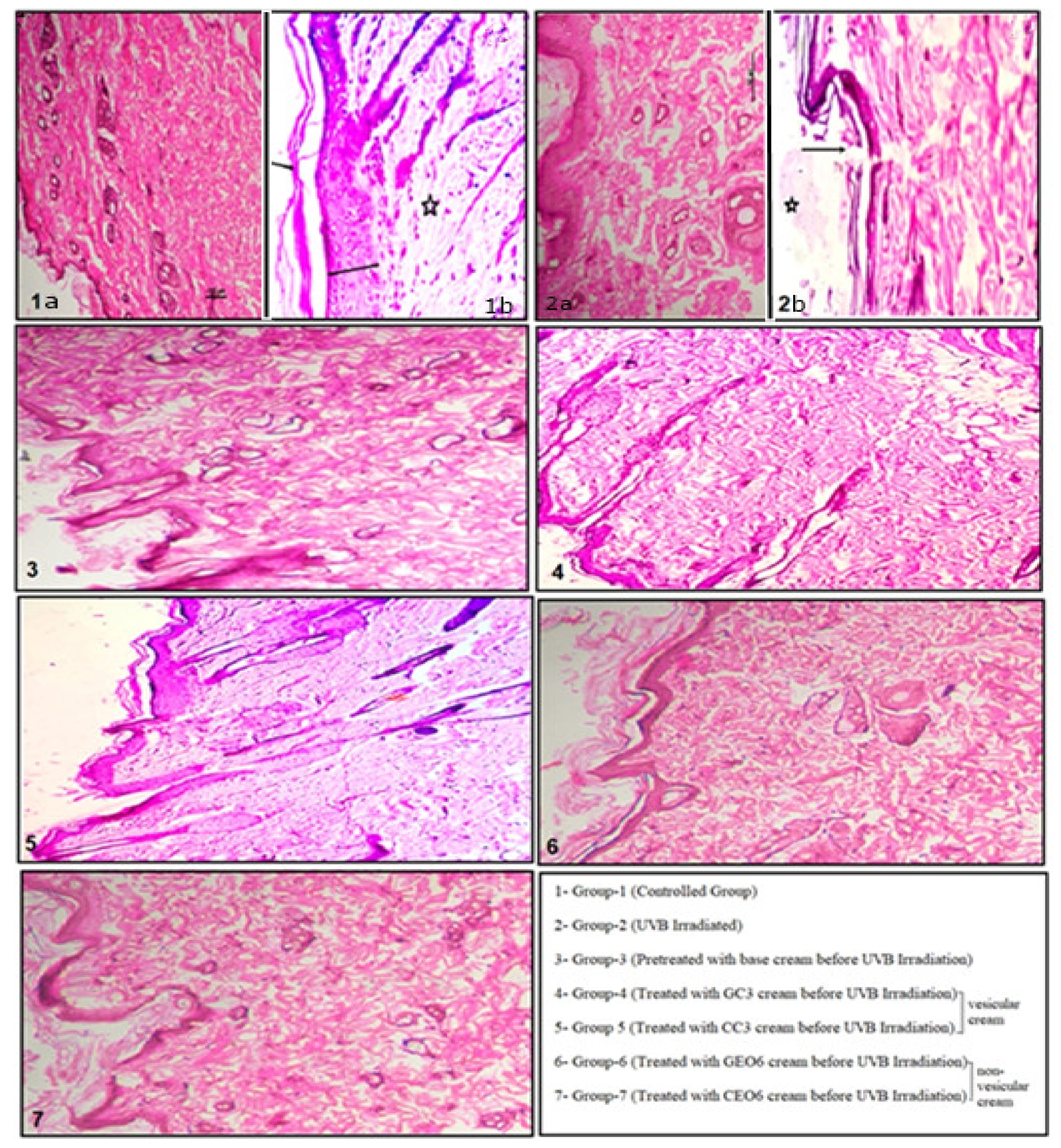
2.9. Histopathology of Skin Tissue

Skin tissue samples from all groups were fixed in 10% chilled neutral formalin (freshly produced) for 12 h at 40 °C for histopathological examinations. The skin tissue samples were then washed thoroughly with double distilled water and dehydrated in a graded alcohol series before being cleared in xylene and embedded in paraffin wax to make tissue blocks. The tissue blocks were labelled with the appropriate information. The tissue was cut into 5 μm cross-sections using a rotator microtome. The ultra-structural dermal and epidermal transformations were detected using an inverted microscope after staining the tissue with hematoxylin and eosin stains [39,40].

2.10. Statistical Analysis

All the values are expressed as the mean ± S.E. The statistical analysis was conducted using MaxStat Statistical Analysis Software (MaxStat Software, Jever-OT Cleverns, Germany). Differences with values of p < 0.01 were deemed significant.

3. Results and Discussion

The estimation of biochemical parameters and histological studies are the in-vivo parameters for the validation of photo-protective action of prepared cream formulations. The effect of UVB radiation and the photo-protective action of developed cream formulations on biochemical parameters and histological investigations of skin tissues were investigated in this study.

3.1. Estimation of Biochemical Parameters

The results of several biochemical parameters of rat skin is discussed below.

3.1.1. Total Protein Estimation

Every cell needs protein to maintain its life. Our body uses protein to repair or maintain skin tissues and also for the construction of new tissue [41]. The TP level in group-1 (no treatment) was found to be 690.02 ± 0.36 µg/mL. The diminished level was found (224.73 ± 0.12 µg/mL) in group 2, which were exposed to UVB radiation only. A decrease in TP showed that the skin tissue was vulnerable to UVB-induced oxidation of protein content. The increase in TP level in group 3, which were treated with base cream, was not very significant. The TP content in groups 4 and 5 was found to be 650.95 ± 0.32 µg/mL and 684.30 ± 0.48 µg/mL, which is high in comparison to group 2, which was irradiated with UVB radiation only (Table 3). The TP content in groups treated with non-vesicular creams—group 6 (494.02 ± 0.20 µg/mL) and group 7 (528.89 ± 0.22 µg/mL)—was not significant in comparison to the groups treated with GEO/CEO-loaded vesicular cream formulations.

3.1.2. Estimation of Ascorbic Acid

AA is one of the primary protectants of the skin and serves as the major nonenzymic antioxidant in human tissue [42]. Because it does not absorb UVA or UVB rays, it is not a sunscreen, but it has a strong antioxidant activity that protects the skin against UV-induced damage caused by ROS (generated as singlet oxygen, superoxide ion, peroxide) [43]. AA neutralizes free radicals by the consecutive donation of electrons and protects the skin from oxidative stress. However, the oxidized forms of AA are relatively unreactive.
In the present study, the AA level in group 1 (controlled group) was found to be 6.315 ± 0.157 mg/100 mL, and a significantly decreased AA level (2.358 ± 0.149 mg/100 mL) was observed in group 2, which was irradiated to UVB radiation only without any treatment. In group 3 (base cream), the AA content was slightly high (3.570 ± 0.123 mg/100 mL) in comparison to group 2. Significantly higher levels of AA were found in group 3 (5.590 ± 0.088 mg/100 mL) and group 4 (5.940 ± 0.053 mg/100 mL), both of which were pre-treated with vesicular cream GC3 and CC3, respectively. The AA level was found to be low in groups treated with non-vesicular cream formulations (groups 6 and 7), in comparison to the vesicular creams-treated group. In this investigation, it was found that UVB irradiation causes significant (p < 0.01) depletion in AA levels when compared with control group 1 (Table 3). It has been reported that the level of AA in skin tissues diminished during injury caused by UV radiation [44]. The highest level of AA in animal groups treated with GEO/CEO-encapsulated vesicular cream formulations (group 4 & 5) state that cream formulations protect the skin against UVB-induced skin damage.

3.1.3. Estimation of Hydroxyproline

HyP is a critical component of collagen synthesis, and it also inhibits the enzymes that cause collagen and elastin degradation, which is connected to skin aging, therefore minimising the indications of skin aging. The defect in collagen synthesis due to deficient HyP level leads to the breakdown of skin connective tissue, which causes sagging and wrinkles.
The HyP concentration was found (Table 3) to be 4.08 ± 0.224 mg/gm in group 1 (controlled group). In group 2 (UVB-treated), the concentration of HyP was significantly reduced to 1.25 ± 0.048 mg/gm. The concentration of HyP was found to be significantly high, i.e., 3.02 ± 0.122 mg/gm and 3.97 ± 0.179 mg/gm in groups 4 and 5, which were pre-treated with GEO- and CEO-encapsulated vesicular cream formulations, respectively. HyP concentration was found to be 2.49 ± 0.139 mg/gm and 2.84 ± 0.023 mg/gm in groups 6 and 7, which were treated with free GEO- and CEO-loaded non-vesicular cream formulations before UVB exposure, respectively.
The lower concentration of HyP in group 2 (UVB-treated) indicates that UVB exposure causes degradation of HyP, resulting in low collagen levels in the skin. The findings revealed that, when compared to vesicular cream formulations, non-vesicular cream formulations were not as effective in maintaining the HyP content of UVB exposure. The higher concentration of HyP in groups 4 and 5 indicates that HyP content was maintained even after exposure to UVB radiation. This confirms the efficacy of GEO/CEO-encapsulated vesicular cream formulations.

3.1.4. Estimation of Malondialdehyde (Lipid Peroxidation Assay)

Lipids are the key element of the skin and are found in the cell membrane and lipid bilayers of the stratum corneum [45]. Oxidative degradation of lipids is called lipid peroxidation. In this process, free radicals target lipids with carbon–carbon double bonds (particularly polyunsaturated fatty acids) that involve oxygen insertion in carbon with the abstraction of hydrogen, resulting in hydroperoxide and lipid peroxyl radicals, which cause destruction to the skin. Lipid peroxidation of the skin lipids leads to cell membrane damage and disturbs the barrier function of the skin [46]. Malondialdehyde (MDA) is a lipid peroxidation end product that is used as a marker to determine the degree of lipid peroxidation [47,48]. The presence of higher levels of MDA in skin tissues suggests increased lipid peroxidation, which promotes tissue damage and results in the breakdown of antioxidant defence mechanisms that prevent excessive free radical production.
For the control group (group 1), the value of MDA was 0.282 ± 0.14 nmoles/mg Protein. In group 2, after UVB radiation, the MDA level was increased to 0.961 ± 0.28 nmoles/mg Protein. In the group treated with base cream before UVB irradiation (group 3), the MDA level became 0.704 ± 0.03 nmoles/mg Protein. In the group treated with GEO- loaded vesicular cream (GC3) before UVB irradiation (group 4), the MDA level became 0.407 ± 0.08 nmoles/mg Protein. The MDA level was found to be 0.310 ± 0.15 nmoles/mg Protein in group 5, which was treated with CC3 (CEO loaded vesicular cream) before UVB irradiation. The increment of MDA level in groups 6 and 7, which were treated with free EO-loaded (non-vesicular) creams, was high in comparison to vesicular cream formulations. In the present investigation, a higher MDA level was observed in group 2, irradiated with UVB radiation without any treatment. The decreased level of MDA in group 4 and group 5, which was treated with GEO- and CEO-loaded vesicular cream, respectively, before UVB irradiation, indicates the protective effects of EO-loaded vesicular cream formulations against lipid peroxidation-induced skin tissue damage (Table 3).

3.1.5. Estimation of Superoxide Dismutase (SOD)

The accumulation of oxidative damage is one of the most important factors in skin aging. SOD is a naturally occurring anti-aging enzyme that plays an important function in the antioxidant defence of the body against oxidative stress. SOD is an antioxidant that catalyses the conversion of superoxide to oxygen and hydrogen peroxide in the body [49]. Studies have shown that faster aging and different age-related signs are associated with decreased concentration of SOD [50].
The level of SOD in the controlled group (group 1) was found to be 1.51 ± 0.003 U/mg protein. In group 2, which was irradiated with UVB only, the SOD level was significantly reduced (p < 0.01) to 0.662 ± 0.01 U/mg protein, in comparison to group 1. This clearly shows the repercussion of oxidative stress due to UV Radiation. In group 3, which was treated with base cream before UVB irradiation, the SOD level was found to be 0.947 ± 0.01 U/mg protein. In group 4, which was pre-treated with GEO-based vesicular cream (GC3), the SOD level was significantly enhanced to 1.42 ± 0.44 U/mg protein, and in group 5, which was pre-treated with CEO-based vesicular cream (CC3), the SOD level was significantly enhanced to be 1.28 ± 0.04 U/mg protein, in comparison to group 2. The SOD level was found to be lower in groups treated with free EO-loaded non-vesicular cream formulations (Table 3).

3.1.6. Estimation of Catalase

Catalase is an antioxidant enzyme that aids in the reduction of oxidative stress by removing cellular hydrogen peroxide and generating water and oxygen. Skin ageing is hypothesised to be associated with catalase deficiency or dysfunction [51]. In this investigation, the level of catalase in the controlled group (group 1) was found to be 37.07 ± 0.52 µmole/min/mg, and significantly dropped to 18.72 ± 0.24 µmole/min/mg in group 2, which was irradiated with UVB radiation only. It was observed that the catalase level was increased to 22.08 ± 0.18 µmole/min/mg in group 3, which was treated with base cream before UVB irradiation. A significantly high level of catalase was found in group 4 (33.22 ± 0.40 µmole/min/mg) and group 5 (31.28 ± 0.45 µmole/min/mg), which were pre-treated with vesicular cream GC3 and CC3, respectively (Table 3). No significant difference was observed in groups 6 and 7, which were pre-treated with non-vesicular cream formulations, in comparison to group 3.

3.2. Histopathology of Skin Tissue

Aging of the skin has a specific importance in humans since our appearance has become an importance aspect in our social lives. Understanding the reasons or causes behind skin aging, as well as how to address and perhaps reverse it, requires knowledge of the morphologic changes that occur as a result of the aging process of the skin. The histopathologic changes of the skin tissue in exposed and non-exposed skin needs to be examined in order to observe the effect of oxidative stress due to UV radiation. To study this, the rat skin slides were prepared and the changes in various cellular structures along with the change in epidermis and dermis of the skin were studied.
Figure 1(1a) shows the histopathological changes of group 1 (controlled). The cellular structures were readily evident in this skin section, which had an intact epidermis and dermis outer layer. At higher magnification [Figure 1(1b)], comparatively normal epidermis (dark line), stratum corneum (arrow), and healthy dermis (asterisk) can be seen. The dermis revealed the presence of collagen and elastin-rich dense irregular connective tissue. In the skin section of group 2 (only UVB-irradiated) [Figure 1(2a,b)], moderate epidermal thickness, termed as ‘hyperkeratosis’, was observed. UVB exposure leads to increased keratinocytes cell division which ultimately increases the epidermal thickness. The destruction of connective tissue integrity, congested blood vessels, and loss of hair follicles were clearly seen. This could be due to the formation of free radicals as a result of UVB exposure, which causes skin deterioration. Higher magnification of the skin section clearly shows the epidermal erosion and loss of stratum corneum. In histopathological section of group 3 [Figure 1(3)], slight changes in epidermal structure were observed with mild hyperkeratosis and increase in hair shaft. Destruction of the integrity of the connective tissues was also observed. In the skin sections of group 4 [Figure 1(4)] and group 5 [Figure 1(5)], which were treated with EO-loaded vesicular creams, GC3 and CC3 respectively, significant reduction of damaging effects was observed, in comparison to group 2. The outer layer of epidermis and the inner dermis were both intact. The integrity of connective tissues was maintained in these skin sections. Increase in hair follicles and hair shaft was observed, which had disappeared in group 2. In the skin section treated with free EO-loaded non-vesicular cream formulations of GEO6 and CEO6 (groups 6 and 7, respectively), loosened connective tissues and mild infiltration of inflammatory cells was observed [Figure 1(6,7)], in comparison to group-2, where less harm was observed.
Overall, it could be concluded that minimum damage was observed in the skin sections of groups 4 and 5 due to the prior application of EO-loaded vesicular creams. Better photoprotective effect was obtained in CEO-loaded vesicular cream (CC3) in comparison to GEO-loaded vesicular cream (GC3). It was confirmed by the results that the photoprotective potential of EO(s) enhanced significantly by incorporation into the vesicles, as they permeate deeper into the skin layers.

4. Conclusions

Extrinsic skin aging can be slowed by using cosmetic formulas that have been professionally developed and tested. Cutaneous aging symptoms such as dull and rough skin, wrinkles, and dark spots are associated with prolonged sun exposure. The greater knowledge of the negative consequences of prolonged sun exposure has resulted in increased demand for sunscreen and antiaging cosmetics. In the present research, the photoprotective potential of GEO/CEO-encapsulated vesicular cream on biochemical parameters against UVB radiation induced damage in rat skin was studied. In comparison to free EO-loaded non-vesicular creams, results demonstrated that pre-treatment with GEO/CEO-encapsulated vesicular cream formulations effectively reversed the negative biochemical changes and were able to protect the skin from the damaging effects of UVB radiation. The age-defying potential of GEO and CEO, as well as their photoprotective benefits, were validated by the findings of the study.

Author Contributions

Conceptualization: A.L.; Writing—original draft preparation: A.L.; Writing—review and editing: A.L. and P.M.; Author contribution and supervision: A.L. and P.M. All authors have read and agreed to the published version of the manuscript.

Funding

This research received no external funding.

Institutional Review Board Statement

The present investigation was conducted according to ethical principles and was approved by the Institutional Animal Ethical Committee, School of Pharmaceutical Sciences, IFTM University Moradabad, India (Registration No. 837/ac/04/CPCSEA).

Informed Consent Statement

Not applicable.

Data Availability Statement

Data is contained within the article.

Acknowledgments

The author (Alka Lohani) is grateful to IFTM University, Moradabad, for providing necessary facilities for this research work.

Conflicts of Interest

The authors declare that they have no conflict of interest.

References

  1. Lohani, A.; Verma, A.; Joshi, H.; Yadav, N.; Karki, N. Nanotechnology-Based Cosmeceuticals. Int. Sch. Res. Not. 2014, 2014, 843687. [Google Scholar] [CrossRef] [PubMed]
  2. Sams, W.M.; Smith, J.G. The Histochemistry of Chronically Sun-Damaged Skin. J. Investig. Dermatol. 1961, 37, 447–453. [Google Scholar] [CrossRef] [PubMed] [Green Version]
  3. D’Orazio, J.; Jarrett, S.; Amaro-Ortiz, A.; Scott, T. UV Radiation and the Skin. Int. J. Mol. Sci. 2013, 14, 12222–12248. [Google Scholar] [CrossRef] [PubMed] [Green Version]
  4. Mishra, A.K.; Mishra, A.; Verma, A.; Chattopadhyay, P. Effects of calendula essential oil-based cream on biochemical parameters of skin of albino rats against ultraviolet B radiation. Sci. Pharm. 2012, 80, 669–683. [Google Scholar] [CrossRef] [Green Version]
  5. Yoshimura, T.; Manabe, C.; Inokuchi, Y.; Mutou, C.; Nagahama, T.; Murakami, S. Protective effect of taurine on UVB-induced skin aging in hairless mice. Biomed. Pharmacother. 2021, 141, 111898. [Google Scholar] [CrossRef]
  6. Meyskens, J.; Farmer, F.L.; Fruehauf, P. Redox regulation in human melanocytes and melanoma. Pigment. Cell. Res. 2001, 14, 148–154. [Google Scholar] [CrossRef] [Green Version]
  7. Bickers, R.; Athar, M. Oxidative stress in the pathogenesis of skin disease. J. Investig. Dermatol. 2006, 126, 2565–2575. [Google Scholar] [CrossRef] [Green Version]
  8. Pillai, S.; Oresajo, C.; Hayward, J. Ultraviolet radiation and skin aging: Roles of reactive oxygen species, inflammation and protease activation, and strategies for prevention of inflammation-induced matrix degradation—A review. Int. J. Cosmet. Sci. 2005, 27, 17–34. [Google Scholar] [CrossRef]
  9. Gilchrest, A. Barbara. Photoaging. J. Investig. Dermatol. 2013, 133, E2–E6. [Google Scholar] [CrossRef] [Green Version]
  10. Baratta, M.T.; Dorman, H.D.; Deans, S.G.; Figueiredo, A.C.; Barroso, J.G.; Ruberto, G. Antimicrobial and antioxidant properties of some commercial essential oils. Flavour. Fragr. J. 1998, 13, 235–244. [Google Scholar] [CrossRef]
  11. Lalli, J.; Van Zyl, R.; Van Vuuren, S.; Viljoen, A. In vitro biological activities of South African Pelargonium (Geraniaceae) species. S. Afr. J. Bot. 2008, 74, 153–157. [Google Scholar] [CrossRef] [Green Version]
  12. Dorman, H.J.D.; Deans, S.G. Antimicrobial agents from plants: Antibacterial activity of plant volatile oils. J. Appl. Microbiol. 2000, 88, 308–316. [Google Scholar] [CrossRef] [PubMed]
  13. Mahboubi, M.; Feizabadi, M.M.; Khamechian, T.; Kazempour, N.; Zadeh, M.R.; Sasani, F.; Bekhradi, M. The Effect of Oliveria Decumbens and Pelargonium Graveolens on Healing of Infected Skin Wounds in Mice. World J. Plast. Surg. 2016, 5, 259–264. [Google Scholar] [PubMed]
  14. Okoye, E.I. Preliminary phytochemical analysis and antimicrobial activity of seeds of carica papaya. J. Basic Phy. Res. 2011, 1, 66–69. [Google Scholar]
  15. Lohani, A.; Mishra, A.K.; Verma, A. Cosmeceutical potential of geranium and calendula essential oil: Determination of antioxidant activity and in vitro sun protection factor. J. Cosmet. Dermatol. 2019, 18, 550–557. [Google Scholar] [CrossRef]
  16. Singh, H.P.; Kaur, S.; Negi, K.; Kumari, S.; Saini, V.; Batish, D.R.; Kohli, R.K. Assessment of in vitro antioxidant activity of essential oil of Eucalyptus citriodora (lemon-scented Eucalypt; Myrtaceae) and its major constituents. LWT-Food Sci. Technol. 2012, 48, 237–241. [Google Scholar] [CrossRef]
  17. Brito, R.G.; Guimarães, A.G.; Quintans, J.S.S.; Santos, M.R.V.; De Sousa, D.P.; Badaue-Passos, D.; De Lucca, W.; Brito, F.A.; Barreto, E.O.; Oliveira, A.P.; et al. Citronellol, a monoterpene alcohol, reduces nociceptive and inflammatory activities in rodents. J. Nat. Med. 2012, 66, 637–644. [Google Scholar] [CrossRef]
  18. Maestri, D.M.; Nepote, V.; Lamarque, A.L.; Zygadlo, J.A. Natural products as antioxidants. In Phytochemistry: Advances in Research; Imperato, F., Ed.; Research Signpost: Kerala, India, 2006; pp. 105–135. [Google Scholar]
  19. Miguel, M.G. Antioxidant and Anti-Inflammatory Activities of Essential Oils: A Short Review. Molecules 2010, 15, 9252–9287. [Google Scholar] [CrossRef] [Green Version]
  20. Shirazi, M.T.; Gholami, H.; Kavoosi, G.; Rowshan, V.; Tafsiry, A. Chemical composition, antioxidant, antimicrobial and cytotoxic activities of Tagetes minuta and Ocimum basilicum essential oils. Food. Sci. Nutr. 2014, 2, 146–155. [Google Scholar] [CrossRef]
  21. Martins, M.D.R.; Arantes, S.; Candeias, F.; Tinoco, M.T.; Cruz-Morais, J. Antioxidant, antimicrobial and toxicological properties of Schinus molle L. essential oils. J. Ethnopharmacol. 2014, 151, 485–492. [Google Scholar] [CrossRef] [Green Version]
  22. Lohani, A.; Verma, A.; Hema, G.; Pathak, K. Topical Delivery of Geranium/Calendula Essential Oil-Entrapped Ethanolic Lipid Vesicular Cream to Combat Skin Aging. BioMed Res. Int. 2021, 2021, 4593759. [Google Scholar] [CrossRef] [PubMed]
  23. Kaur, C.D.; Saraf, S. Topical vesicular formulations of Curcuma longa extract on recuperating the ultraviolet radiation-damaged skin. J. Cosmet. Dermatol. 2011, 10, 260–265. [Google Scholar] [CrossRef] [PubMed]
  24. Lorraine, H.K. The ultraviolet-irradiated hairless mouse: A model for photoaging. J. Am. Acad. Dermatol. 1989, 21, 623–631. [Google Scholar]
  25. Cole, C.A.; Davies, R.E.; Forbes, P.D.; D’Aloisio, L.C. Comparison of Action Spectra for Acute Cutaneous Responses to Ultraviolet Radiation: Man and Albino Hairless Mouse. Photochem. Photobiol. 1983, 37, 623–631. [Google Scholar] [CrossRef]
  26. Kligman, L.H.; Akin, F.J.; Kligman, A.M. Prevention of Ultraviolet Damage to the Dermis of Hairless Mice by Sunscreens. J. Investig. Dermatol. 1982, 78, 181–189. [Google Scholar] [CrossRef] [Green Version]
  27. OECD Guidelines for Testing of Chemicals. In-Vitro 3T3 NRU Phototoxicity Test. 432 Adopted 13 April 2014. Available online: https://www.oecd.org/env/ehs/testing/Test%20No.432-2004%20English.pdf (accessed on 21 October 2021).
  28. Meenakshi, J.; Jayaraman, V.; Ramakrishnan, K.M.; Babu, M. Ultrastructural differentiation of abnormal scars. Ann. Burn. Fire Disasters 2005, 18, 83–88. [Google Scholar]
  29. Nandi, A.; Chatterjee, I.B. Assay of superoxide dismutase activity in animal tissues. J. Biosci. 1988, 13, 305–315. [Google Scholar] [CrossRef]
  30. Lowry, O.H.; Rosebrough, N.J.; Farr, A.L.; Randall, R.J. Protein measurement with the Folin phenol reagent. J. Biol. Chem. 1951, 193, 265–275. [Google Scholar] [CrossRef]
  31. Eberlein-König, B.; Placzek, M.; Przybilla, B. Protective effect against sunburn of combined systemic ascorbic acid (vitamin C) and d-α-tocopherol (vitamin E). J. Am. Acad. Dermatol. 1998, 38, 45–48. [Google Scholar] [CrossRef]
  32. Shrivastav, A.; Mishra, A.K.; Ali, S.S.; Abuzinadah, A.A.M.F.; Khan, N.A. In vivo models for assessment of wound healing potential: A systematic review. Wound Med. 2018, 31, 43–53. [Google Scholar] [CrossRef]
  33. Dische, Z.; Borenfreund, E. A spectrophotometric method for the micro determination of hexosamines. J. Biol. Chem. 1950, 184, 517–522. [Google Scholar] [CrossRef]
  34. Marklund, S. Involvement of Superoxide anion radical in the autooxidation of pyrogallol and a convenient assay of super oxide dismutase. Eur. J. Biochem. 1974, 47, 469–472. [Google Scholar] [CrossRef] [PubMed]
  35. Gao, R.; Yuan, Z.; Zhao, Z.; Gao, X. Mechanism of pyrogallol autoxidation and determination of superoxide dismutase enzyme activity. Bioelectrochem. Bioenerg. 1998, 45, 41–45. [Google Scholar] [CrossRef]
  36. Sinha, A.K. Colorimetric assay of catalase. Anal. Biochem. 1972, 47, 389–394. [Google Scholar] [CrossRef]
  37. Rukkumani, R.; Aruna, K.; Varma, P.S.; Rajasekaran, K.N.; Menon, V.P. Comparative effects of curcumin and an analog of curcumin on alcohol and PUFA induced oxidative stress. J. Pharm. Pharm. Sci. 2004, 7, 274–283. [Google Scholar] [PubMed]
  38. Hadwan, M.H. Simple spectrophotometric assay for measuring catalase activity in biological tissues. BMC Biochem. 2018, 19, 7. [Google Scholar] [CrossRef] [PubMed]
  39. Rasik, A.M.; Shukla, A. Antioxidant status in delayed healing type of wounds. Int. J. Exp. Pathol. 2000, 81, 257–263. [Google Scholar] [CrossRef] [PubMed]
  40. Panchatcharam, M.; Miriyala, S.; Gayathri, V.S.; Suguna, L. Curcumin improves wound healing by modulating collagen and decreasing reactive oxygen species. Mol. Cell. Biochem. 2006, 290, 87–96. [Google Scholar] [CrossRef]
  41. Rukkumani, R.L.; Thomson, J.D. Buckley. Protein hydrolysates and tissue repair. Nutr. Res. Rev. 2011, 24, 191–197. [Google Scholar]
  42. Ravetti, S.; Clemente, C.; Brignone, S.; Hergert, L.; Allemandi, D.; Palma, S. Ascorbic Acid in Skin Health. Cosmetics 2019, 6, 58. [Google Scholar] [CrossRef] [Green Version]
  43. Darr, D.; Combs, S.; Dunston, S.; Manning, T.; Pinnell, S. Topical vitamin C protects porcine skin from ultraviolet radiation-induced damage. Br. J. Dermatol. 1992, 127, 247–253. [Google Scholar] [CrossRef] [PubMed]
  44. Pullar, J.M.; Carr, A.C.; Vissers, M.C.M. The Roles of Vitamin C in Skin Health. Nutrients 2017, 9, 866. [Google Scholar] [CrossRef] [PubMed] [Green Version]
  45. Das, C.; Olmsted, P.D. The physics of stratum corneum lipid membranes. The physics of stratum corneum lipid membranes. Philos. Trans. R. Soc. 2016, 374, 20150126. [Google Scholar] [CrossRef] [PubMed] [Green Version]
  46. Ayala, A.; Muñoz, M.F.; Argüelles, S. Lipid peroxidation: Production, metabolism, and signaling mechanisms of malondialdehyde and 4-hydroxy-2-nonenal. Oxid. Med. Cell. Longev. 2014, 2014, 360438. [Google Scholar] [CrossRef]
  47. Gaschler, M.M.; Stockwell, B.R. Lipid peroxidation in cell death. Biochem. Biophys. Res. Commun. 2017, 482, 419–425. [Google Scholar] [CrossRef]
  48. Grotto, D.; Maria, L.S.; Valentini, J.; Paniz, C.; Schmitt, G.; Garcia, S.; Pomblum, V.J.; da Rocha, J.B.T.; Farina, M. Importance of the lipid peroxidation biomarkers and methodological aspects FOR malondialdehyde quantification. Química Nova 2009, 32, 169–174. [Google Scholar] [CrossRef] [Green Version]
  49. Younus, H. Therapeutic potentials of superoxide dismutase. Int. J. Health Sci. 2018, 12, 88–93. [Google Scholar]
  50. Ighodaro, O.M.; Akinloye, O.A. First line defence antioxidants-superoxide dismutase (SOD), catalase (CAT) and glutathione peroxidase (GPX): Their fundamental role in the entire antioxidant defence grid. Alex. J. Med. 2018, 54, 287–293. [Google Scholar] [CrossRef] [Green Version]
  51. Nandi, A.; Yan, L.-J.; Jana, C.K.; Das, N. Role of Catalase in Oxidative Stress- and Age-Associated Degenerative Diseases. Oxidative Med. Cell. Longev. 2019, 2019, 9613090. [Google Scholar] [CrossRef] [Green Version]
Figure 1. Histopathology of skin sections of all groups; (1(a,b)) Skin section of group 1 (Neither irradiated nor treated with test formulation) [in 1b ― line represents epidermis, arrow represents stratum corneum, asterisk represents healthy dermis]; (2(a,b)) Skin section of group 2 (UVB-irradiated); [― in 2b represents epidermis, arrow represents stratum corneum, asterisk represents healthy dermis]; (3) Skin section of Group 3 (pre-treated with Base cream + UVB-irradiated); (4) Skin section of group 4 (pretreated with GC3 formulation + UVB-irradiated); (5) Skin section of group 5 (pretreated with CC3 Formulation + UVB-irradiated); (6) Skin section of group 6 (pretreated with GEO6 Formulation + UVB-irradiated); (7) Skin section of group 7 (pretreated with GEO6 Formulation + UVB-irradiated).
Cosmetics 09 00043 g001
Table 1. Composition of various cream formulations, i.e., base cream; vesicular cream (GEO loaded: GC1, GC2, and GC3 and CEO loaded: CC1, CC2, and CC3); non-vesicular cream GEO6 and CEO6 for GEO and CEO, respectively [22].
Ingredients (% w/w)Base CreamGC1GC2GC3 *GEO6 *CC1CC2CC3 *CEO6 *
Bees wax888888888
Stearic acid444444444
Cetyl alcohol333333333
Olive oil121212121212121212
Coconut oil161616161616161616
Span 601.41.41.41.41.41.41.41.41.4
Tween 601.51.51.51.51.51.51.51.51.5
Aloe vera gelq.s.q.s.q.s.q.s.q.s.q.s.q.s.q.s.q.s.
GEO loaded in vesicles----246---------------
CEO loaded in vesicles---------------246---
Free GEO------------6------------
Free CEO------------------------6
* Optimized cream formulations selected for the present research work.
Table 2. Grouping of Animals.
GroupsTreatment
Group 1Neither irradiated nor treated with test formulation
(maintained at similar conditions and diet)
Group 2UVB irradiated group
Group 3Pretreated with Base cream + UVB irradiated
Group 4Pretreated with GC3 formulation + UVB irradiated
Group 5Pretreated with CC3 Formulation + UVB irradiated
Group 6Pretreated with GEO6 Formulation + UVB irradiated
Group 7Pretreated with CEO6 Formulation + UVB irradiated
Table 3. Effect of UVB radiation and cream formulations on Total Protein (TP), Ascorbic Acid (AA), Hydroxyproline (HyP), Malondialdehyde, Catalase and SOD levels in rat skin.
GroupTreatmentTP
(µg/mL)
AA (mg/100mL)HyP (mg/gm)MDA
(nmoles/mg Protein)
Catalase
(µmole/min/mg)
SOD
(U/mg Protein)
1Control Group690.02 ± 0.366.315 ± 0.1574.08 ± 0.2240.282 ± 0.1437.07 ± 0.521.51 ± 0.003
2UVB irradiated group224.73 ± 0.122.358 ± 0.1491.25 ± 0.0480.961 ± 0.2818.72 ± 0.240.662 ± 0.01
3Pretreated with
base cream +
UVB irradiated
371.40 ± 0.263.570 ± 0.1232.18 ± 0.1370.704 ± 0.0322.08 ± 0.180.947 ± 0.01
4Pretreated with
GC3 cream +
UVB irradiated
650.95 ± 0.325.590 ± 0.0883.02 ± 0.1220.407 ± 0.0833.22 ± 0.401.42 ± 0.44
5Pretreated with
CC3 cream +
UVB irradiated
684.30 ± 0.485.940 ± 0.0533.97 ± 0.1790.310 ± 0.1531.28 ± 0.451.28 ± 0.04
6Pretreated with
GEO6 cream +
UVB irradiated
494.02 ± 0.204.590 ± 0.2262.49 ± 0.1390.615 ± 0.2028.20 ± 0.281.16 ± 0.03
7Pretreated with
CEO6 cream +
UVB irradiated
528.89 ± 0.223.820 ± 0.0602.84 ± 0.0230.564 ± 0.2425.12 ± 0.200.985 ± 0.01
Values are in Mean ± SEM of n = 3.
Publisher’s Note: MDPI stays neutral with regard to jurisdictional claims in published maps and institutional affiliations.

