
Sensitivity Improvement of Surface Plasmon Resonance Biosensors with GeS-Metal Layers

1
Key Laboratory of Optoelectronic Devices and Systems of Ministry of Education and Guangdong Province, College of Physics and Optoelectronic Engineering, Shenzhen University, Shenzhen 518060, China
2
International Collaborative Laboratory of 2D Materials for Optoelectronic Science & Technoloy of Ministry of Education, Shenzhen University, Shenzhen 518060, China
*
Author to whom correspondence should be addressed.
Electronics 2022, 11(3), 332; https://doi.org/10.3390/electronics11030332
Submission received: 1 December 2021
/
Revised: 6 January 2022
/
Accepted: 19 January 2022
/
Published: 21 January 2022
(This article belongs to the Special Issue Digital and Optical Security Algorithms via Machine Learning)
Abstract
:Surface plasmon resonance (SPR) biosensors, with germanium sulfide (GeS) as a sensitive medium and Al/Ag/Au as the metal layers, are reported as we aim to improve the sensitivities of the biosensors. The sensitivities in conventional SPR biosensors, consisting of only metal Al, Ag, and Au layers, are 111°/RIU, 117°/RIU, 139°/RIU, respectively. Additionally, these sensitivities of the SPR biosensors based on the GeS-Al, GeS-Ag, and GeS-Au layers have an obvious improvement, resultant of 320°/RIU, 295°/RIU, and 260°/RIU, respectively. We also discuss the changing sensing medium GeS thickness using layer number to describe the scenario which brought about the diversification on the figure of merit (FOM) and optical absorption (OA) performance of the biosensors. These biosensors show obvious improvement of sensitivity and have strong SPR excitation to analytes; we believe that these kind biosensors could find potential applications in biological detection, chemical examination, and medical diagnosis.
1. Introduction
Sensing has been considered one of the most promising applications in plasma [1,2] and photonic [3] technology. Plasma sensing was even recently selected by the scientific community in the United States as one of the top ten emerging technologies (2018). At the surface of positive permittivity materials and negative material, metals usually have the resonance excitation of surface plasmon polaritons (SPSs). This is the surface plasmon resonance (SPR) phenomenon [4], and this kind of phenomenon can be used for real-time monitoring and label-free sensing [5]. Biochemical sensors based on SPR technology have many advantageous properties, such as high operation sensitivities, detection accuracy, and biocompatibilities [6,7,8]. These kinds of sensors have been studied and used for many biological and chemical applications [9], such as medical diagnosis, enzyme detection, and pharmaceuticals [10]. The effective ways to fabricate sensors are to select large specific surface area combined/adsorption materials such as the popular two-dimensional (2D) materials graphene. After graphene was found, 2D materials fascinated much attention due to their special structural and unique physical/chemical properties [11]. These types of characteristics give 2D materials broad application perspective for different sensor fields [12,13]. Fortunately, germanium monosulfide (GeS) which is a phosphorene analogue compound was favorably forecasted and manufactured [11]. Furthermore, GeS exhibits higher air stability and electron mobility than phosphorene [14]. In addition to black phosphorus, layered group IV with a phosphorene-like structure-mono-chalcogenides MX (M = Ge, Sn; X = S, Se) are a class of emerging 2D materials [15,16,17,18]. Due to the characteristics of MX series compounds such as excellent mechanical flexibility, high light absorption, high thermoelectric coefficient, high carrier mobility, earth abundance, low toxicity, and dynamic stability, they are considered prospective optoelectronic and electronic device materials [19,20,21,22,23,24]. Li et al. revealed that the high carrier mobility of the GeS monolayer is approximately 3680 cm2 V−1, which is much higher than that of the MoS2 monolayer [25]. Xue et al. revealed a process for the preparation of GeS film which has a high absorption coefficient (105 cm−1), a suitable band gap (1.14 eV), and p conductivity by in situ self-regulation, which can be used for thin-film photovoltaic devices.
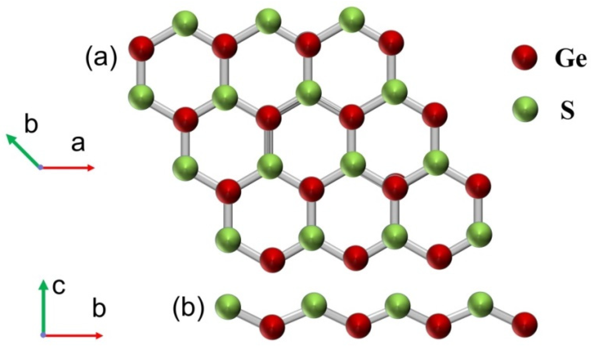
As shown in Figure 1, the single layer GeS geometries, including the top view and side view, are similar to silene and blue phosphorene. Ge atoms and S atoms are combined and arranged alternately to form a hexagonal buckled. The optimal lattice constant and Ge-S bond length of single-layer GeS is 3.495 Å after structural relaxation, which agrees well with previously reported data.
Two dimensional material GeS used as sensing medium in biosensors are reported in this paper. A comparison of three metals, Al/Ag/Au, as metal layers are also investigated in these GeS sensors. The sensitivities of the biosensors derived from the GeS-Al, GeS-Ag, and GeS-Au layers are 320°/RIU, 295°/RIU, and 260°/RIU, respectively. Additionally, the sensitivities of conventional SPR sensors which do not have GeS sensing medium, i.e., only based on Al, Ag, and Au films, are 111°/RIU, 117°/RIU, and 139°/RIU, respectively. The sensitivities have a clear enhancement in these GeS based biosensors. We also discussed the changing of the sensing medium GeS thickness using layer number to describe brought about the diversification on the figure of merit (FOM) and optical absorption (OA) performance of the biosensors. These types of biosensors have doughty SPR excitation, high detection accuracy, and high sensitivity to analytes; they will play a central role in photoelectron applications.
2. Design Considerations and Theoretical Model
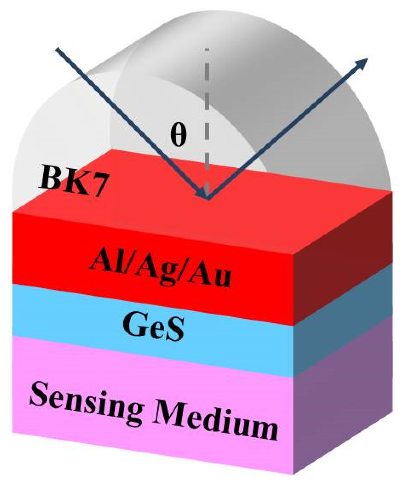
Z. Lou et al. have synthesized nanoparticles-organic clusters (NOCs) and used them as signal amplification reagents, gaining four times higher SPR signal compared to the sandwich format [26,27]. A highly sensitive sensor with a separate GeS layer is proposed in our design. In our SPR sensors, we put an Al/Ag/Au film on the surface of a BK7 coupling prism, and then put the GeS layer as a biological sensing layer on the top of the Al/Ag/Au layer, seen in Figure 2. BK7 glass is coupling prism, and the GeS monolayer thickness is 0.524 nm [28,29]. As an SPP primary metal, an Al/Ag/Au film of 50 nm is selected [30]. The BK7 glass refractive index can be expressed [30] using the next equation:
The refractive index of Ag/Al/Au can be calculated using the Drude–Lorentz model [31]:
Here, λp and λc are the plasma and collision wavelength. For Al layer sensor, the data of λc and λp are 4.0852 × 10⁻⁵ m and 1.3617 × 10⁻⁷ m; for Ag they are 1.7614 × 10⁻⁵ m and 1.4541 × 10⁻⁷ m; and for Au are 8.9342 × 10⁻⁶ m and 1.6826 × 10⁻⁷ m, respectively. Additionally, in our sensor structures, we induced 2D material GeS to put on the surface of Al, Ag, or Au to prevent oxidation in order to improve the sensitivities of our sensors.
The GeS refractive index is composed of real and imaginary parts [32], and it is expressed ns = 1.33 + Δn in the wavelength of 633 nm. The expression represents the refractive index of the target medium, where Δn indicates the refractive index variation caused by biological action or chemical reaction.
The reflectivity is studied by the transfer matrix method. The TM polarized light N-layer model which using matrix method is effective and accurate. The SPR sensor simulation is calculated using Matrix&Laboratory (MATLAB) software. In our GeS based biosensors, GeS and Al/Ag/Au layers are stacked in the BK7 glass-coupling prism vertically, and each layer is named for the dielectric constant (εk), refractive index (nk), and thickness (dk). The resonance angle is the incident angle corresponding to the minimum reflectivity. The tangential field at the first boundary Z = Z1 = 0 is related to the tangential field at the final boundary Z = ZN−1:
where V and U indicate the magnetic fields and electric fields components of the boundary surface, respectively. M represents the feature matrix of the composite architecture and k represents the kth layer in the N-layer model. p-polarized light corresponds to:
Through mathematical simplifications, reflection coefficient rp of the p-polarized light N-layer complex was obtained, and the corresponding amplitude reflection coefficient (Rp) can be achieved by the square of rp:
The sensitivities, expressed as SR1 = Δθ/Δn, cause the refractive index of the sensing medium (Δn) to change, leading to the shift in the resonance angle (Δθ) [33]. The detection accuracy (DA) is the ratio of the resonance angle shift in (∆θres) in the reflectance curve to the full width at half-maximum value (FWHM):
3. Results and Discussion
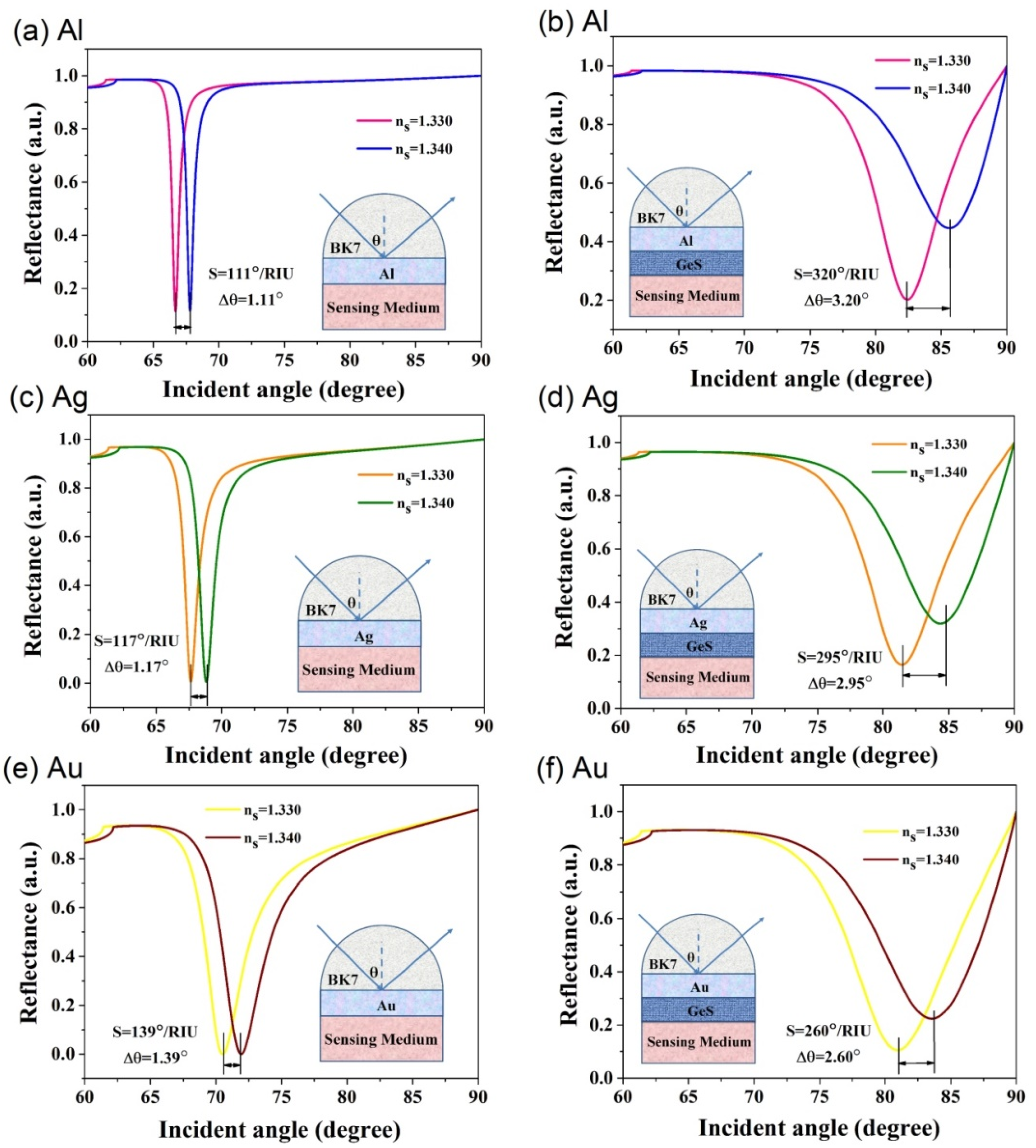
We selected BK7 glass as the coupling prism for our designed GeS biochemical sensors, due to its low refractive index [35]. Traditional SPR sensors, such as the Kretschmann geometry biochemical sensors, mainly include a simple metal layer to measure the SPPs, as shown in Figure 3. The sensitivities of conventional Al, Ag, and Au metals are 111°/RIU, 117°/RIU, and 139°/RIU, respectively. When using the BK7 glass prism, they are shown in Figure 3a,c,e, respectively. However, these values are not ideal for the sensitivity of biochemical sensors. To improve the sensitivity of the sensor, we developed an SPR biochemical sensor using GeS material. We put the addition of the GeS material on the surface of metallic Al/Ag/Au layer, and then the detection target flow past the 2D material surface in the structure, using this way to increase the sensitivity of the biosensors. In Figure 3b,d,f, the charts show that the sensitivities are greatly improved when the GeS material is introduced in the previous sensor structure. The sensitivities of these sensors obtained for the Al, Ag, and Au metal layers are 320°/RIU, 295°/RIU, and 260°/RIU when the GeS layers accumulating to 33, 28, and 18 layers, respectively.
In the following experiments, 2D GeS material can use the liquid phase stripping (LPE) method to prepare and then use the spin coating method to coat on the Al/Ag/Au layers (~50 nm) of the BK7 coupling prism. Using a refractive index matching solution, the GeS-Al/Ag/Au multilayer film can be connected with to BK7 prism to construct the SPR sensor.
Table 1 shows the changes in the number of GeS layers (L), maximum sensitivity (S), FOM, FWHM, resonance angle change (Δθ), and DA data for the GeS based on Al/Ag/Au sensors. The thickness of Al, Ag, and Au was set to 50 nm. The incident angles were set from 0° to 90°. The thickness of the GeS layer was obtained from reference [36], and the number of layers was calculated based on the single-layer thickness of GeS, which was 0.375 nm. With the increasing number of GeS layers, the reflectivity curve presents a larger peak sensitivity angle. The maximum sensitivity is achieved when the optimal numbers of GeS layers are added. For GeS on Al, Ag, and Au, the sensitivity reaches its maximum when the number of layers is 33, 28, and 18, respectively. Due to the limitation of a certain angle range, the change of resonance angle is directly related to the sensitivity; for example, the sensitivity decreases after the optimized GeS layer number is exceeded with the decrease in the resonance angle. Finally, we also realized that the SPR line width increases and that FOM decrease will improve the sensor sensitivities [37]. The refractive index of BK7 at λ= 633 nm is calculated as 1.515. The refractive index of the sensing medium can change, increasing if the prism reflection angle has a lower refractive index; this indicates that higher sensitivity will be apparent with a lower prism refractive index. Therefore, a BK7 prism is selected as the coupling prism in our GeS sensor structures to improve the sensitivities.
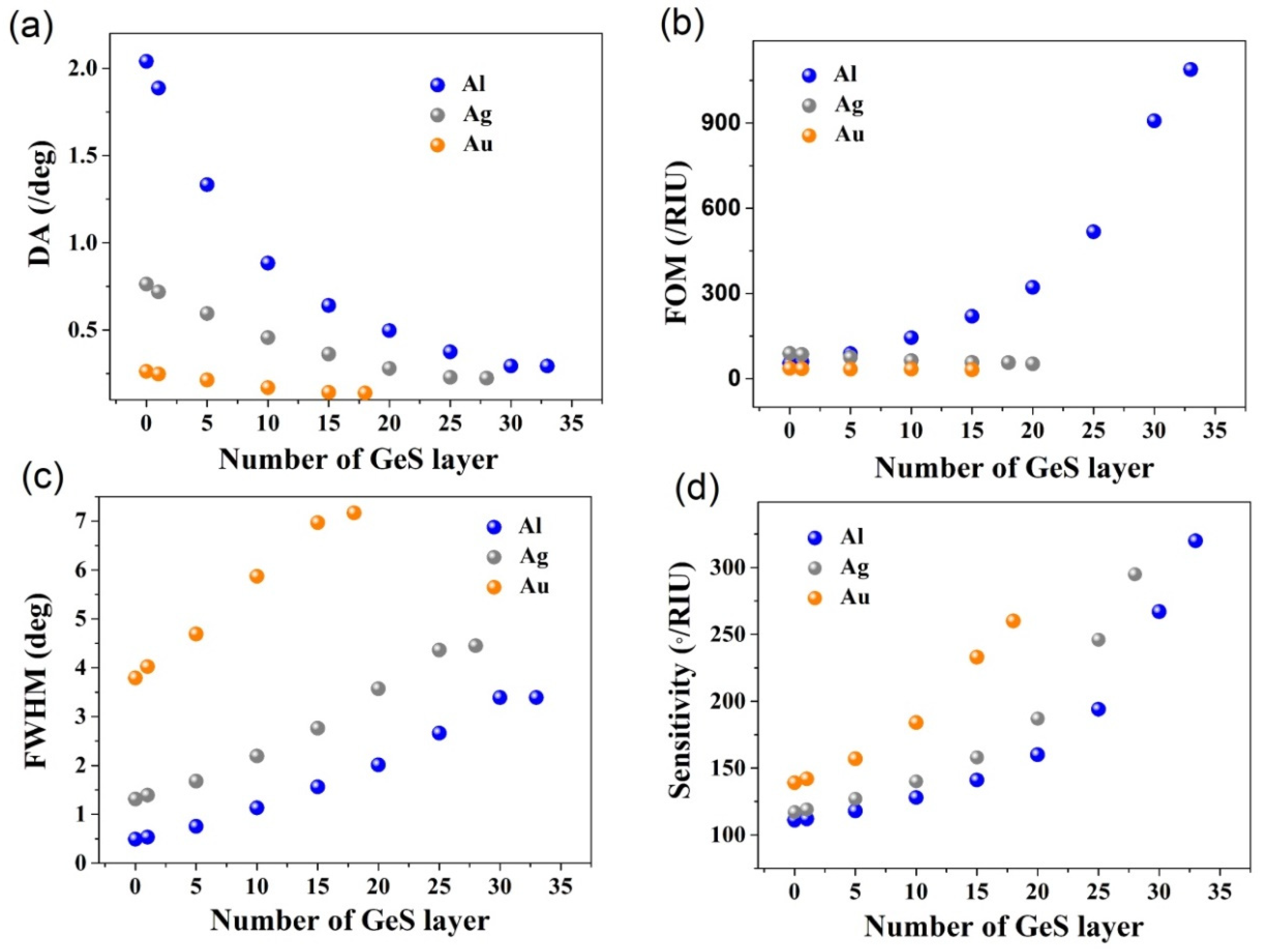
The properties of the sensors are also impacted by the GeS layer numbers. Figure 4 shows the changes in DA, FOM, and FWHM, and sensitivity with the number of GeS layers at 633 nm, with the incident angle ranging from 34.65° to 34.85°. DA is a key factor to reflect the functions of biosensors. As can be seen from the curves in Figure 4a, as the number of GeS layers changes between 1 and 35, DA presents a downward trend. This is due to the application of a germanium sulfide layer to widen the reflection curve. Although DA decreases with the increase in GeS layers, DA remains high. In Figure 4b, FOM is the specific value of the sensitivity to the FWHM in the reflectance curve. GeS-Au based biosensor FOM augments as the number of GeS layers increase, and the GeS-Al and GeS-Ag based biosensors do not exhibit significant change. Figure 4c shows that GeS layer numbers have an important effect on reflectivity. With increasing GeS layer numbers, the inclination of the SPR reflectivity curve becomes shallower and the FWHM widens due to the absorption of the GeS layers. Figure 4d indicates the change in the peak sensitivities of the SPR biosensors based on GeS-Al, GeS-Ag, and GeS-Au at the wavelength of 633 nm with the number of GeS layers (L). The SPR biosensors based on GeS-Au have higher sensitivity than GeS-Al and GeS-Ag SPR biosensors when they are at the same layers. The SPR response of Al and Ag layers is more sensitive than that of Au layers, but the chemical stability is lower. The GeS layer is an important part of the SPR sensor, which can prevent the oxidation of the oxidizable Al/Ag/Au metal, enhance the absorption of the sensing medium, and improve the stability and biocompatibility of the biosensor structure.
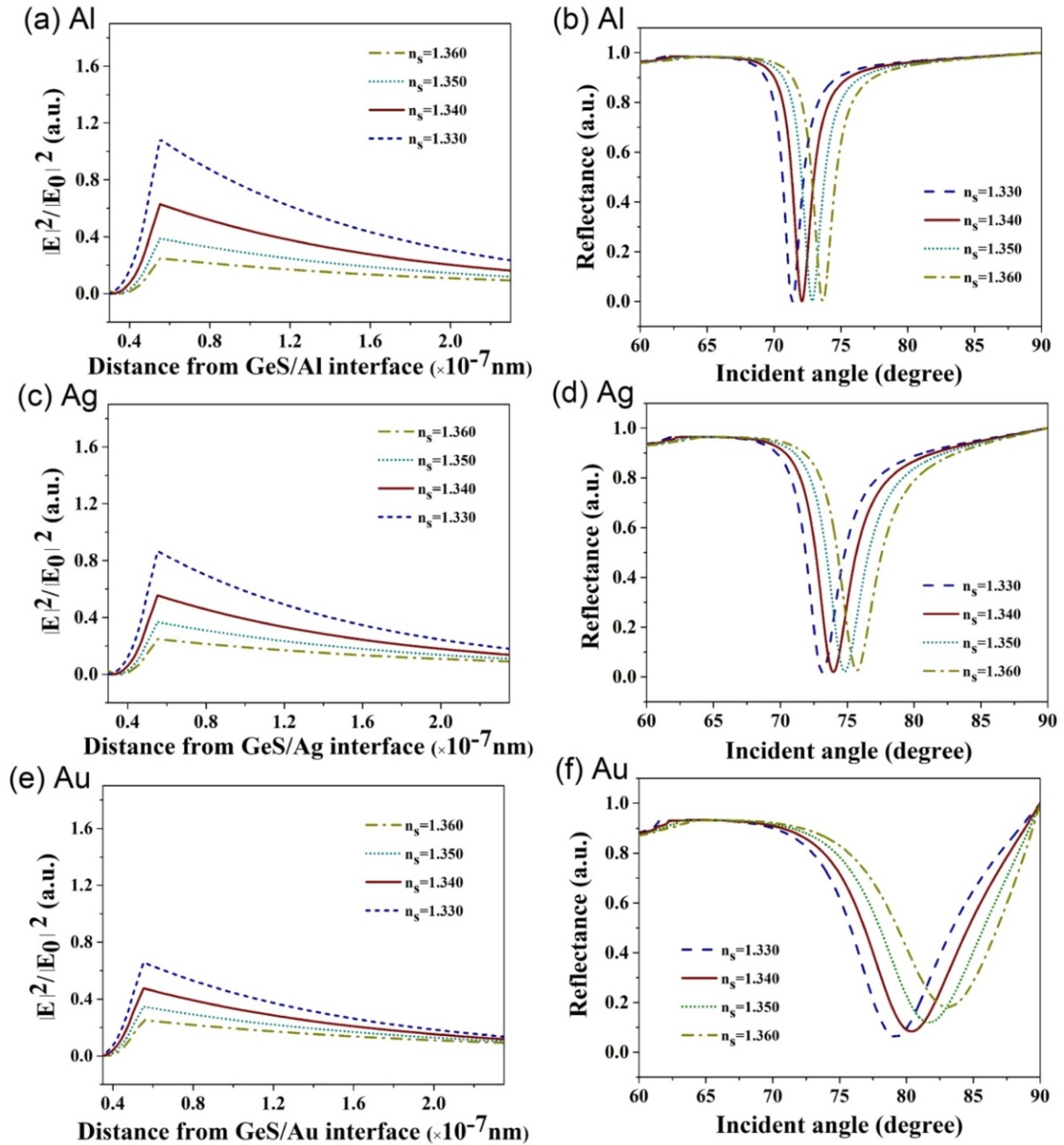
The electric field distributions corresponding to the resonance angles are shown in Figure 5a,c,e. When the detection target refractive index is from 1.33 to 1.36, the electric field at the GeS/sending medium interface obviously varies dramatically. This indicates that the GeS based sensors are sensitive to small changes when checking detection target. When the refractive index of the detection medium on the contact surface changes a little bit, the surface wave property will have a large change, and then it will lead to a change in the electric field. Figure 5b,d,f shows that the changes in reflectance with the incident angles increase with increasing detection target refractive index, and the incident angles corresponding to the resonance angles increase. From ns = 1.33 to ns = 1.36, the resonance angles are 75.205°, 77.145°, 79.395°, and 82.150°, respectively.
4. Conclusions
In this study, different kinds of GeS based sensors, designed to enhance sensitivity, are reported. Three kinds of metal, Al/Ag/Au, were used as metal layers for comparison in these GeS sensors. Compared with the conventional biosensor, which only has metal layers, the sensitivities of proposed GeS based biosensors show a large improvement. The highest sensitivities for our sensors, based on GeS-Al, GeS-Ag, and GeS-Au films, are 320°/RIU, 295°/RIU, and 260°/RIU, respectively; conventional SPR sensors, only based on Al/Ag/Au films, exhibited sensitivities of 111°/RIU, 117°/RIU, and 139°/RIU, respectively. The two dimensional material GeS layer not only prevented the oxidizable metals from oxidizing, enhancing the surface plasmons of metal, but also enhanced the absorption of biomolecules. The changing of the sensing medium GeS thickness, using numbers of layers, brought about the diversification on the figure of merit (FOM) and optical absorption (OA) performance of the biosensors. From these simulation results, the present sensors display high sensitivities and strong SPR excitations analytes. With these outstanding characteristics, these GeS based structure sensors will have a remarkable effect in the field of electrical optical sensing technology.
Author Contributions
Conceptualization and Writing Original Draft, Y.J.; Investigation, Y.L.; Project administration, H.C. All authors have read and agreed to the published version of the manuscript.
Funding
This work is supported by National Natural Science Foundation of China (NSFC) (11775147); Guangdong Basic and Applied Basic Research Foundation (Nos. 2019A1515011474 and 2019A1515110130); Shenzhen Science and Technology Program (Nos. JCYJ20210324095007020, JCYJ20200109105201936 and JCYJ20190808115605501).
Conflicts of Interest
The authors declare no conflict of interest.
References
- Mayer, K.M.; Hafner, J.H. Localized Surface Plasmon Resonance Sensors. Chem. Rev. 2011, 111, 3828–3857. [Google Scholar] [CrossRef] [PubMed]
- Špačková, B.; Wrobel, P.; Bocková, M.; Homola, J. Optical Biosensors Based on Plasmonic Nanostructures: A Review. Proc. IEEE 2016, 104, 2380–2408. [Google Scholar] [CrossRef]
- Xu, D.; Xiong, X.; Wu, L.; Ren, X.-F.; Png, C.E.; Guo, G.-C.; Gong, Q.; Xiao, Y.-F. Quantum plasmonics: New opportunity in fundamental and applied photonics. Adv. Opt. Photon. 2018, 10, 703–756. [Google Scholar] [CrossRef]
- Ouyang, Q.; Zeng, S.; Jiang, L.; Qu, J.; Dinh, X.-Q.; Qian, J.; He, S.; Coquet, P.; Yong, K.-T. Two-Dimensional Transition Metal Dichalcogenide Enhanced Phase-Sensitive Plasmonic Biosensors: Theoretical Insight. J. Phys. Chem. C 2017, 121, 6282–6289. [Google Scholar] [CrossRef]
- Srivastava, S.K.; Verma, R.; Gupta, B.D. Theoretical modeling of a self-referenced dual mode SPR sensor utilizing indium tin oxide film. Opt. Commun. 2016, 369, 131–137. [Google Scholar] [CrossRef]
- Sajad, P.; Fardin, M.N. Simultaneous analysis of some volatile compounds in food samples by array gas sensors based on polypyrrole nano-composites. Sens. Rev. 2017, 37, 155–164. [Google Scholar]
- Mohammad, A.S.-M.; Sajad, P. Nanostructured Conducting Polymer/Copper Oxide as a Modifier for Fabrication of L-DOPA and Uric Acid Electrochemical Sensor. Electroanalysis 2016, 28, 2075–2080. [Google Scholar]
- Anooshe, G.-D.; Mohammad, T.A.; Ashkan, G.-D. Quick, Single-Frequency Dielectric Characterization of Blood Samples of Pediatric Cancer Patients by a Cylindrical Capacitor: Pilot Study. Electronics 2020, 9, 1–16. [Google Scholar]
- Liedberg, B.; Nylander, C.; Lunström, I. Surface plasmon resonance for gas detection and biosensing. Sens. Actuators 1983, 4, 299–304. [Google Scholar] [CrossRef]
- Zeng, S.; Baillargeat, D.; Ho, H.-P.; Yong, K.-T. Nanomaterials enhanced surface plasmon resonance for biological and chemical sensing applications. Chem. Soc. Rev. 2014, 43, 3426–3452. [Google Scholar] [CrossRef]
- Zhang, S.; Yan, Z.; Shengli, Z.; Chen, Z.; Zeng, H. Atomically Thin Arsenene and Antimonene: Semimetal-Semiconductor and Indirect-Direct Band-Gap Transitions. Angew. Chem. Int. Ed. 2015, 54, 3112–3115. [Google Scholar] [CrossRef] [PubMed]
- Wang, S.-G.; Tan, C.-J.; Yang, Q.; Xu, Y.-X.; Li, S.-L.; Chen, X.-P. A Novel Ultra-Sensitive Nitrogen Dioxide Sensor Based on Germanium Monosulfide Monolayer. IEEE Electron. Device Lett. 2017, 38, 1590–1593. [Google Scholar] [CrossRef]
- Cui, S.; Pu, H.; Wells, S.A.; Wen, Z.; Mao, S.; Chang, J.; Hersam, M.C.; Chen, J. Ultrahigh sensitivity and layer-dependent sensing performance of phosphorene-based gas sensors. Nat. Commun. 2015, 6, 8632. [Google Scholar] [CrossRef] [PubMed] [Green Version]
- Zhang, S.; Zhou, W.; Ma, Y.; Ji, J.; Cai, B.; Yang, S.; Zhu, Z.; Chen, Z.; Zeng, H. Antimonene Oxides: Emerging Tunable Direct Bandgap Semiconductor and Novel Topological Insulator. Nano Lett. 2017, 17, 3434–3440. [Google Scholar] [CrossRef] [PubMed]
- Zhu, Z.; Tománek, D. Semiconducting Layered Blue Phosphorus: A Computational Study. Phys. Rev. Lett. 2014, 112, 176802. [Google Scholar] [CrossRef]
- Zhu, Z.; Guan, J.; Liu, D.; Tománek, D. Designing Isoelectronic Counterparts to Layered Group V Semiconductors. ACS Nano 2015, 9, 8284–8290. [Google Scholar] [CrossRef] [Green Version]
- Liu, H.; Neal, A.T.; Zhu, Z.; Luo, Z.; Xu, X.; Tomanek, D.; Ye, P.D. Phosphorene: An Unexplored 2D Semiconductor with a High Hole Mobility. ACS Nano 2014, 8, 4033–4041. [Google Scholar] [CrossRef] [Green Version]
- Zhao, H.; Mao, Y.; Mao, X.; Shi, X.; Xu, C.; Wang, C.; Zhang, S.; Zhou, D. Band Structure and Photoelectric Characterization of GeSe Monolayers. Adv. Funct. Mater. 2018, 28, 1704855. [Google Scholar] [CrossRef]
- Gu, D.; Tao, X.; Chen, H.; Zhu, W.; Ouyang, Y.; Peng, Q. Enhanced photocatalytic activity for water splitting of blue-phase GeS and GeSe monolayers via biaxial straining. Nanoscale 2019, 11, 2335–2342. [Google Scholar] [CrossRef]
- Yap, W.C.; Yang, Z.; Mehboudi, M.; Yan, J.-A.; Barraza-Lopez, S.; Zhu, W. Layered material GeSe and vertical GeSe/MoS2 p-n heterojunctions. Nano Res. 2018, 11, 420–430. [Google Scholar] [CrossRef] [Green Version]
- Rikta, K.A.; Anower, M.S.; Rahman, M.S.; Rahman, M.M. SPR biosensor using SnSe-phosphorene heterostructure. Sens. Bio-Sens. Res. 2021, 33, 100442. [Google Scholar] [CrossRef]
- Devi, T.N.; Chiu, N.-F. A Review of Graphene-Based Surface Plasmon Resonance and Surface-Enhanced Raman Scattering Biosensors: Current Status and Future Prospects. Nanomaterials 2021, 11, 216. [Google Scholar]
- Singh, A.K.; Hennig, R.G. Computational prediction of two-dimensional group-IV mono-chalcogenides. Appl. Phys. Lett. 2014, 105, 042103. [Google Scholar] [CrossRef]
- Zhao, P.; Yang, H.; Li, J.; Jin, H.; Wei, W.; Yu, L.; Huang, B.; Dai, Y. Design of new photovoltaic systems based on two-dimensional group-IV monochalcogenides for high performance solar cells. J. Mater. Chem. A 2017, 5, 24145–24152. [Google Scholar] [CrossRef]
- Li, F.; Liu, X.; Wang, Y.; Li, Y. Germanium monosulfide monolayer: A novel two-dimensional semiconductor with a high carrier mobility. J. Mater. Chem. C 2016, 4, 2155–2159. [Google Scholar] [CrossRef]
- Yuan, C.; Lou, Z.; Wang, W.; Yang, L.; Li, Y. Synthesis of Fe3C@C from Pyrolysis of Fe3O4-Lignin Clusters and Its Application for Quick and Sensitive Detection of PrPSc through a Sandwich SPR Detection Assay. Int. J. Mol. Sci. 2019, 20, 741. [Google Scholar] [CrossRef] [Green Version]
- Lou, Z.; Han, H.; Zhou, M.; Wan, J.; Sun, Q.; Zhou, X.; Gu, N. Fabrication of Magnetic Conjugation Clusters via Intermolecular Assembling for Ultrasensitive Surface Plasmon Resonance (SPR) Detection in a Wide Range of Concentrations. Anal. Chem. 2017, 89, 13472–13479. [Google Scholar] [CrossRef]
- Hess, P. Thickness of elemental and binary single atomic monolayers. Nanoscale Horiz. 2020, 5, 385–399. [Google Scholar] [CrossRef]
- Fan, X.; Su, L.; Zhang, F.; Huang, D.; Sang, D.K.; Chen, Y.; Li, Y.; Liu, F.; Li, J.; Zhang, H.; et al. Layer-Dependent Properties of Ultrathin GeS Nanosheets and Application in UV–Vis Photodetectors. ACS Appl. Mater. Interfaces 2019, 11, 47197–47206. [Google Scholar] [CrossRef]
- Dai, X.; Liang, Y.; Zhao, Y.; Gan, S.; Jia, Y.; Xiang, Y. Sensitivity Enhancement of a Surface Plasmon Resonance with Tin Selenide (SnSe) Allotropes. Sensors 2019, 19, 173. [Google Scholar] [CrossRef] [Green Version]
- Gupta, B.D.; Sharma, A.K. Sensitivity evaluation of a multi-layered surface plasmon resonance-based fiber optic sensor: A theoretical study. Sens. Actuators B Chem. 2005, 107, 40–46. [Google Scholar] [CrossRef]
- Xie, J.; Zhang, D.; Yan, X.-Q.; Ren, M.; Zhao, X.; Liu, F.; Sun, R.; Li, X.; Li, Z.; Chen, S.; et al. Optical properties of chemical vapor deposition-grown PtSe2 characterized by spectroscopic ellipsometry. 2D Mater. 2019, 6, 035011. [Google Scholar] [CrossRef]
- Maharana, P.K.; Jha, R. Chalcogenide prism and graphene multilayer based surface plasmon resonance affinity biosensor for high performance. Sens. Actuators B Chem. 2012, 169, 161–166. [Google Scholar] [CrossRef]
- Homola, J. Surface Plasmon Resonance Sensors for Detection of Chemical and Biological Species. Chem. Rev. 2008, 108, 462–493. [Google Scholar] [CrossRef]
- Wu, L.; Jia, Y.; Jiang, L.; Guo, J.; Dai, X.; Xiang, Y.; Fan, D. Sensitivity Improved SPR Biosensor Based on the MoS2/Graphene–Aluminum Hybrid Structure. J. Lightwave Technol. 2017, 35, 82–87. [Google Scholar] [CrossRef]
- Wiedemeier, H.; Georg, H.; Schnering, G.V. Refinement of the structures of GeS, GeSe, SnS and SnSe. Z. Für Krist.-Cryst. Mater. 1978, 148, 295–304. [Google Scholar] [CrossRef]
- Jia, Y.; Li, Z.; Wang, H.; Saeed, M.; Cai, H. Sensitivity Enhancement of a Surface Plasmon Resonance Sensor with Platinum Diselenide. Sensors 2020, 20, 131. [Google Scholar] [CrossRef] [Green Version]
Figure 1.
Three-dimensional representation of the structure of GeS: (a) top view (b) side view.

Figure 2.
Diagrammatic presentation of the GeS combined on Al/Ag/Au SPR biochemical sensors for improved sensitivity.
Figure 2.
Diagrammatic presentation of the GeS combined on Al/Ag/Au SPR biochemical sensors for improved sensitivity.

Figure 3.
(a,c,e) The incident angle reflectance change of the conventional sensors with single Al/Ag/Au metal layer, and (b,d,f) The incident angle reflectance change of the sensors with GeS on top of Al/Ag/Au metal layer.
Figure 3.
(a,c,e) The incident angle reflectance change of the conventional sensors with single Al/Ag/Au metal layer, and (b,d,f) The incident angle reflectance change of the sensors with GeS on top of Al/Ag/Au metal layer.

Figure 4.
Variation in (a) DA, (b) FOM, (c) FWHM, and (d) sensitivities changing with numbers of GeS layers for the proposed SPR biosensors.
Figure 4.
Variation in (a) DA, (b) FOM, (c) FWHM, and (d) sensitivities changing with numbers of GeS layers for the proposed SPR biosensors.

Figure 5.
(a,c,e) The electric field distribution of the GeS based sensor and (b,d,f) variation in reflectance with the incident angle of the sensor based on few-layer GeS for ns = 1.33 to ns = 1.36.
Figure 5.
(a,c,e) The electric field distribution of the GeS based sensor and (b,d,f) variation in reflectance with the incident angle of the sensor based on few-layer GeS for ns = 1.33 to ns = 1.36.


Table 1.
Performances of the Proposed GeS-Based Sensors.
| Type of Metal Layer | Optimized Layer Number (L) | Sensitivity (S) (°/RIU) | FOM | FWHM | ∆θ | DA |
|---|---|---|---|---|---|---|
| Al | 0 | 111 | 54.4 | 0.49 | 1.11 | 2.040 |
| 15 | 141 | 219.9 | 1.56 | 1.41 | 0.641 | |
| 33 | 320 | 1088.4 | 3.39 | 3.20 | 0.294 | |
| Ag | 0 | 117 | 89.3 | 1.31 | 1.17 | 0.763 |
| 15 | 158 | 75.5 | 1.68 | 1.58 | 0.595 | |
| 28 | 295 | 66.2 | 4.45 | 2.95 | 0.224 | |
| Au | 0 | 139 | 36.6 | 3.79 | 1.39 | 0.263 |
| 10 | 233 | 35.3 | 4.02 | 1.84 | 0.248 | |
| 18 | 260 | 33.4 | 6.97 | 2.60 | 0.143 |
Publisher’s Note: MDPI stays neutral with regard to jurisdictional claims in published maps and institutional affiliations. |
© 2022 by the authors. Licensee MDPI, Basel, Switzerland. This article is an open access article distributed under the terms and conditions of the Creative Commons Attribution (CC BY) license (https://creativecommons.org/licenses/by/4.0/).
Share and Cite
MDPI and ACS Style
Jia, Y.; Liao, Y.; Cai, H. Sensitivity Improvement of Surface Plasmon Resonance Biosensors with GeS-Metal Layers. Electronics 2022, 11, 332. https://0-doi-org.brum.beds.ac.uk/10.3390/electronics11030332
AMA Style
Jia Y, Liao Y, Cai H. Sensitivity Improvement of Surface Plasmon Resonance Biosensors with GeS-Metal Layers. Electronics. 2022; 11(3):332. https://0-doi-org.brum.beds.ac.uk/10.3390/electronics11030332
Chicago/Turabian StyleJia, Yue, Yunlong Liao, and Houzhi Cai. 2022. "Sensitivity Improvement of Surface Plasmon Resonance Biosensors with GeS-Metal Layers" Electronics 11, no. 3: 332. https://0-doi-org.brum.beds.ac.uk/10.3390/electronics11030332
Note that from the first issue of 2016, this journal uses article numbers instead of page numbers. See further details here.