Review

Plasmodium falciparum Histidine-Rich Protein 2 and 3 Gene Deletions and Their Implications in Malaria Control

by
Josphat Nyataya
1,
John Waitumbi
1,*,
Victor A. Mobegi
2,
Ayman Noreddin
3,4 and
Mohamed E. El Zowalaty
3,5,*
1
Basic Science Laboratory, US Army Medical Research Directorate-Africa/Kenya Medical Research Institute, P.O. Box 54-40100, Kisumu, Kenya
2
Department of Biochemistry, School of Medicine, University of Nairobi, P.O. Box 30197-00100, Nairobi, Kenya
3
Infectious Diseases and Anti-Infective Research Group, College of Pharmacy and Sharjah Medical Research Institute, University of Sharjah, Sharjah 27272, UAE
4
Department of Medicine, School of Medicine, University of California, Irvine, CA 92868, USA
5
Zoonosis Science Center, Department of Medical Biochemistry and Microbiology, Uppsala University, SE-75123 Uppsala, Sweden
*
Authors to whom correspondence should be addressed.
Submission received: 7 March 2020 / Revised: 25 April 2020 / Accepted: 5 May 2020 / Published: 20 May 2020
(This article belongs to the Section Infectious Disease)

Abstract

:
Malaria remains the biggest threat to public health, especially among pregnant women and young children in sub-Saharan Africa. Prompt and accurate diagnosis is critical for effective case management and detection of drug resistance. Conventionally, microscopy and rapid diagnostic tests (RDTs) are the tools of choice for malaria diagnosis. RDTs are simple to use and have been extensively used in the diagnosis of malaria among travelers to malaria-endemic regions, routine case management, and surveillance studies. Most RDTs target the histidine-rich protein (PfHRP) which is exclusively found in Plasmodium falciparum and a metabolic enzyme Plasmodium lactate dehydrogenase (pLDH) which is common among all Plasmodium species. Other RDTs incorporate the enzyme aldolase that is produced by all Plasmodium species. Recently, studies have reported false-negative RDTs primarily due to the deletion of the histidine-rich protein (pfhrp2 and pfhrp3) genes in field isolates of P. falciparum. Herein, we review published literature to establish pfhrp2/pfhrp3 deletions, the extent of these deletions in different geographical regions, and the implication in malaria control. We searched for publications on pfhrp2/pfhrp3 deletions and retrieved all publications that reported on this subject. Overall, 20 publications reported on pfhrp2/pfhrp3 deletions, and most of these studies were done in Central and South America, with very few in Asia and Africa. The few studies in Africa that reported on the occurrence of pfhrp2/pfhrp3 deletions rarely evaluated deletions on the flanking genes. More studies are required to evaluate the existence and extent of these gene deletions, whose presence may lead to delayed or missed treatment. This information will guide appropriate diagnostic approaches in the respective areas.

1. Introduction

The previous two decade was characterized by remarkable progress in the fight against malaria, with more than seven million lives having been saved since 2001 [1], thanks to the wide-scale deployment of malaria control interventions, including but not limited to accurate diagnosis, effective antimalarial therapy, and use of insecticide-treated nets [2]. However, recent reports suggest that these gains are slowing down, and this may reverse all the investments and efforts that have been utilized so far [3]. In 2015, an estimated 211 million cases of malaria were reported, and by 2018, the cases had increased to 228 million. More than 90% of these cases were reported in the African region. Globally, 405,000 deaths from malaria were reported in 2018, with 17 African countries accounting for 91% of all malaria deaths [4]. Children under five years of age and pregnant women remain the population at the highest risk of malaria.
Diagnosis of malaria requires the identification of Plasmodium parasites, which is usually done by the use of microscopy or rapid diagnostic tests (RDTs) that target antigens/enzymes of the parasite in the patient’s blood. Although molecular techniques such as Polymerase Chain Reaction (PCR) provide high sensitivity and specificity for malaria diagnosis, the high cost, limited availability, and required expertise make them unsuitable as a diagnostic tool, especially in resource-limited settings [5,6].
Recently, the use of non-invasive specimens such as saliva and urine has been proposed as an alternative to blood samples. However, the utility of these approaches has not been validated [7,8]. Microscopy and RDTs, therefore, form a core component of malaria diagnosis and case management. For effective case management, diagnosis of malaria has to be prompt and accurate. Accurate diagnosis ensures that antimalarial therapy is used rationally and correctly, malaria does not progress to complications such as severe anemia, metabolic acidosis, and cerebral involvement, and reduces the chance of transmission of the disease to other people. However, the performance of malaria RDTs is influenced by several factors—the existence of different species of Plasmodium, which requires expert microscopists to differentiate them, deletions of the antigens targeted by RDTs, transmission intensity, parasite density, immunity, and the manifestation of clinical symptoms that are similar to other tropical diseases [9,10]. Routinely, the diagnosis of malaria is based on both signs (clinical presentations/complains, fever measured by body temperature) and parasitological confirmation of parasites presence in blood [5].
Since Laveran discovered the malaria parasite and Romanowsky’s improvements on the staining techniques in the late 1800s, malaria has conventionally been diagnosed by the observation of Plasmodium parasites in slides stained with Giemsa or Field’s stain [11]. Microscopic examination of Giemsa-stained thick films (for detection of the presence of the parasites) and thin films (for species identification) is the gold standard approach for diagnosis [5]. Malaria microscopy has low direct costs, is simple, allows differentiation of parasite species and developmental stages (e.g., gametocytes), and allows determination of parasite density. These characteristics of microscopy make it an attractive tool for malaria diagnosis and drug efficacy studies [12]. However, despite its widespread use in malaria diagnosis, microscopy has several inherent limitations, including its dependence on the availability of a competent microscopists, electricity, good quality staining reagents, and enough parasite density (>100 parasites/µL) [13,14,15]. Microscopy is also labor-intensive, unsuitable for high-throughput use, and has a high error rate. The errors lead to biased estimates on measures of protective efficacy or misclassification of treatment outcomes, especially in antimalarial drug or vaccine trials. Studies comparing field-based and expert microscopy to PCR for detection of Plasmodium have shown that mixed species are common and are rarely detected by expert microscopists [16]. In rural settings with limited resources such as electricity and sources of good-quality water to make the stains, microscopy may not be reliable [17], and therefore, there is a need for other alternative diagnostic tools to complement microscopy.
To overcome the apparent limitations of microscopy, the World Health Organization (WHO) has emphasized the urgent need for new, simple, quick, accurate, and cost-effective diagnostic tools to complement malaria microscopy [6]. The advent of RDTs has greatly enhanced the quality of malaria diagnosis, especially in peripheral health centers where microscopy would not be reliable. Through global fund financing, over 148 million RDTs have been procured in 81 malaria-endemic countries, thereby improving case management, especially in children, who are the most vulnerable group [18]. The WHO Africa region, where the disease burden is highest, reported an increase in RDT sales up to 269 million from 240 million in 2015 [2]. RDTs are fast, easy to perform and interpret, do not need for electricity or equipment, and this has contributed to their widespread use.
Currently, there are about 60 manufacturers and 200 types of RDTs that have been tested and deployed in field settings [19]. All RDTs detect malaria antigen in blood sample placed on a lateral flow chromatographic membrane containing immobilized antimalaria antibodies. The dye-labeled antibody binds the target parasite antigen, and the resulting complex is subsequently captured by another antibody, which is immobilized at the test line. The color appears at the test line. Excess labeled antibody conjugate continues to flow along the strip and is captured at the control line by a secondary antibody. When a sample without target antigens is used, color appears only on the control line.
Malaria RDTs target three Plasmodium antigens that are detected in the same RDT. The histidine-rich proteins 2 and 3 (PfHRP2/3) are specific for P. falciparum, while Plasmodium lactate dehydrogenase (pLDH) and Plasmodium aldolase are made by all malaria species, hence the name pan-pLDH and pan-aldolase. Thus, in general, RDTs are sold as combinations of pan-pLDH-PfHRP2/3 or pan-aldolase-PfHRP2/3 [5].
The pLDH is a terminal enzyme in the glycolytic pathway of both the sexual and asexual stages of the parasite, and each of the four Plasmodium species has its specific isomer of this enzyme, while aldolase is conserved in all the species and is an example of a pan-malarial antigen target [20]. pLDH catalyzes (using NADH, a reduced form of nicotinamide adenine dinucleotide, as a coenzyme) the conversion of pyruvate into lactate and NAD+, (oxidised form of NAD) which is important for parasite survival in red blood cells [21,22]. Since pLDH and aldolase are only produced in living Plasmodium cells and have a short half-life (2–4 days), the presence of these enzymes is an indication of current infection [23]. RDTs that detect pLDH either detect a pan-pLDH which is common to all human infecting species or specific regions unique to P. falciparum or P. vivax-specific pLDH [24]. Although previous studies showed reduced sensitivity of most pLDH-detecting RDTs at extreme temperatures [25], a study in India demonstrated RDT stability and high sensitivity of up to 98% for P. falciparum [26].
Three histidine-rich proteins (HRPs) have been described, namely, HRP1 or the knob-associated histidine-rich protein (KAHRP), HRP2, and HRP3, also known as small histidine–alanine-rich proteins (SHARP) containing 8%, 35%, and 30% histidine repeats, respectively [27,28]. The pfhrp1 gene is associated with the expression of knob-like protrusions on the surface membrane of infected erythrocytes [29]. These knobs are not seen in erythrocytes infected with the ring stages of the parasites, but they develop with the maturation of the parasites to other developmental stages, such as late trophozoites and schizonts [30]. Studies have revealed that the knobs are the sites of attachment to the venous endothelium and are thus responsible for sequestration of infected erythrocytes to deep vasculature, a phenomenon that is common in cerebral malaria [31].
PfHRP2 is a water-soluble protein produced by the asexual stages and young gametocytes of P. falciparum and can be detected in serum, plasma, cerebrospinal fluid, and urine of infected patients and in the medium of in vitro cultured parasites [32,33,34]. Secretion and synthesis of PfHRP2 have been shown to begin with immature rings and increase gradually as the asexual parasites mature such that, more than 90% is secreted during schizogony [35,36], and its presence in plasma has been exploited in the design of RDTs for malaria diagnosis [37]. Unlike PfHRP1, which is found only in knobby (K+) variants of P. falciparum, PfHRP2 is found in both the knobby and knobless (K-) strain variants [38].
The pfhrp3 gene codes for a homologous protein histidine-rich protein-3 (PfHRP3). Both PfHRP2 and PfHRP3 are very similar in structure, although PfHRP3 has less histidine content [27]. The structural similarity between the two genes is responsible for cross-reaction of monoclonal antibodies against PfHRP2 with those of PfHRP3. Unlike PfHRP1 and PfHRP3, PfHRP2 is abundantly produced and is continuously expressed throughout the parasite life cycle.
The use of PfHRP2-based RDTs for malaria diagnosis has emerging limitations due to the existence of parasites with pfhrp2 gene deletions which, depending on the extent of the deletions, has led to false-negative results [39,40,41,42]. Initially, experimental studies demonstrated the deletions of pfhrp2 and pfhrp3 genes in laboratory lines [43,44]. Whole-genome scanning and DNA sequence analysis have demonstrated partial or complete deletions of pfhrp2/pfhrp3 genes and/or the flanking genes [40,45]. The deletions involve breakage and rejoining at the unstable sub-telomeric regions of chromosome 8 and 13 for pfhrp2 and pfhrp3 genes, respectively (Figure 1).
Recently, studies have confirmed the existence of deletions in field isolates, thereby threatening the utility of PfHRP2-based RDTs for malaria diagnosis. Depending on the extent of the deletions, the expression level of PfHRP2 and PfHRP3 proteins could cause false-negative results, leading to delay in therapy, and ultimately contribute to increased morbidity and mortality and promote disease transmission. Parasites with pfhrp2 gene deletion have been shown to be reactive to PfHRP2-based RDTs due to cross-reactivity with PfHRP3 [46,47]. Few studies have evaluated the presence and extent of these deletions in Africa. In this review, we evaluate the current literature on pfhrp2 and pfhrp3 gene deletions and their potential impact on disease diagnosis, management, control and the eradication goals.

2. Methods

2.1. Systematic Review Protocols

The guidelines and procedures of the Preferred Reporting Items for Systematic Reviews and Meta-Analyses (PRISMA) [48] were followed in the current study (Figure 1).

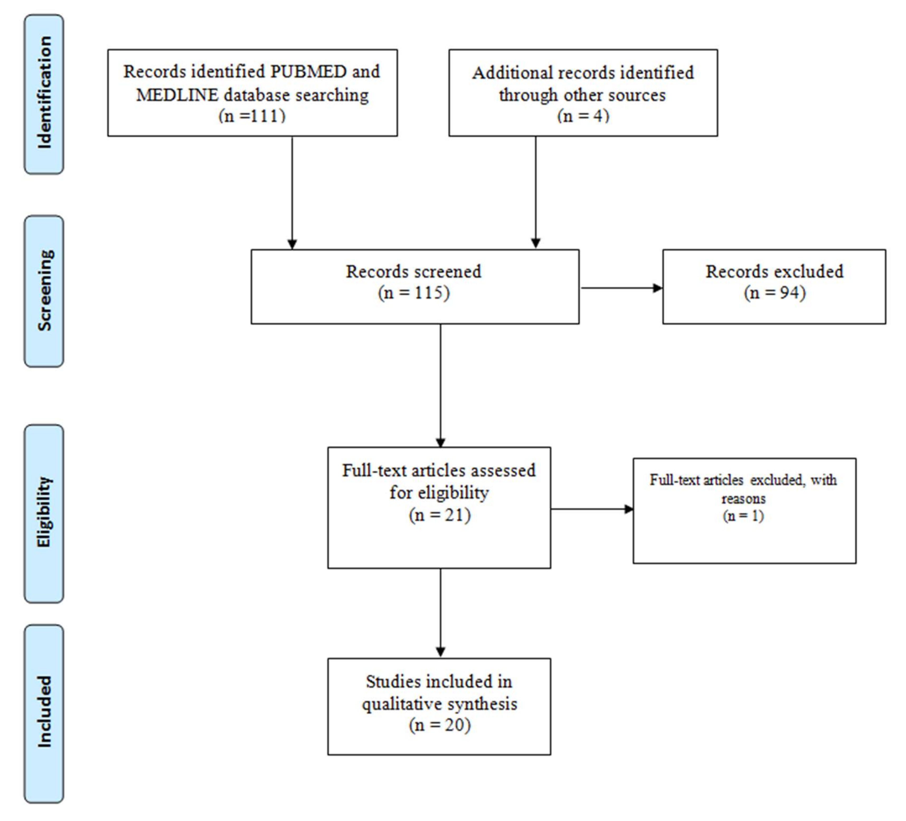
2.2. Search Strategy and Inclusion/Exclusion Criteria

Published scientific articles were retrieved from PUBMED, MEDLINE, and Science Direct. We searched the scientific literature for articles published in English from January 2010 when the first case of P. falciparum parasites lacking pfhrp2 and/ or pfhrp3 deletions was first reported [40], to December 2019.
Keywords used for the search included: PfHRP2, PfHRP3, pfhrp2/pfhrp3 gene deletions, and PfHRP2-RDTs. To filter out articles that were out of scope, we screened only for publications that were reporting on pfhrp2/pfhrp3 deletions. To be included in the analysis plan, a study had to fulfill stipulations as previously suggested [49], i.e., have one initial evidence (microscopy or RDT or both) of the parasite and include a quality control procedure such as co-amplification of a single copy gene, in addition to amplification of pfhrp2 and/or pfhrp3 genes. This is crucial to allow differentiation between lack of amplification due to gene deletions versus no amplification due to PCR failure.

2.3. Statistical Analysis

Data were entered in a Microsoft Excel database (Microsoft, Redmond, WA, USA). The meta-analysis was conducted using the random effects model of analysis since it minimizes heterogeneity of the included studies. All of the analyses were implemented using R-3.6.2 metaprop and forest packages in order to estimate the pulled proportion, to investigate publication and other bias and to summarize information on individual studies, respectively. Descriptive statistics, such as bar charts, were used to summarize the distribution of reported deletions using the Statistical Package for Social Sciences (SPSS), version 25.

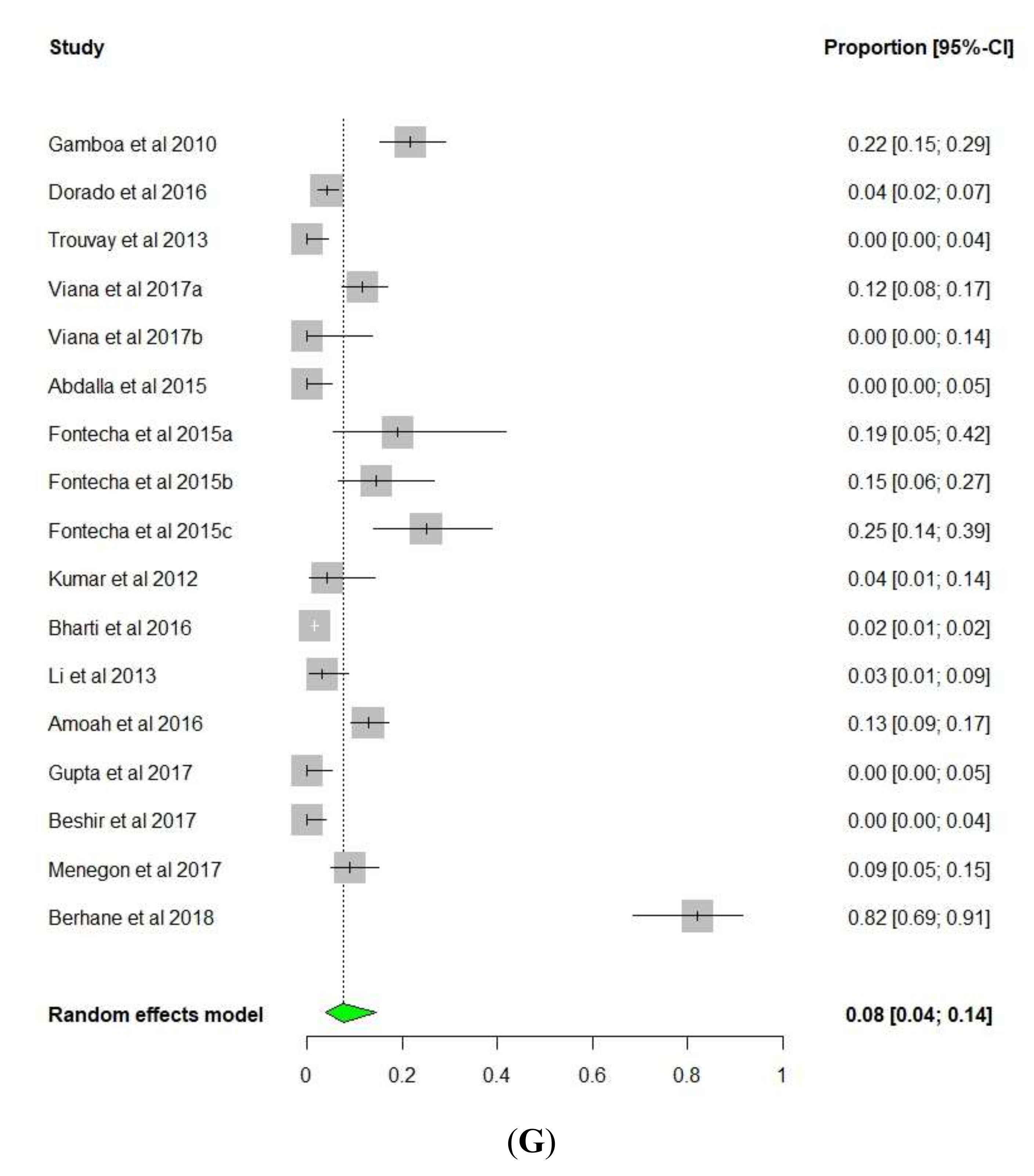
3. Results and Discussion

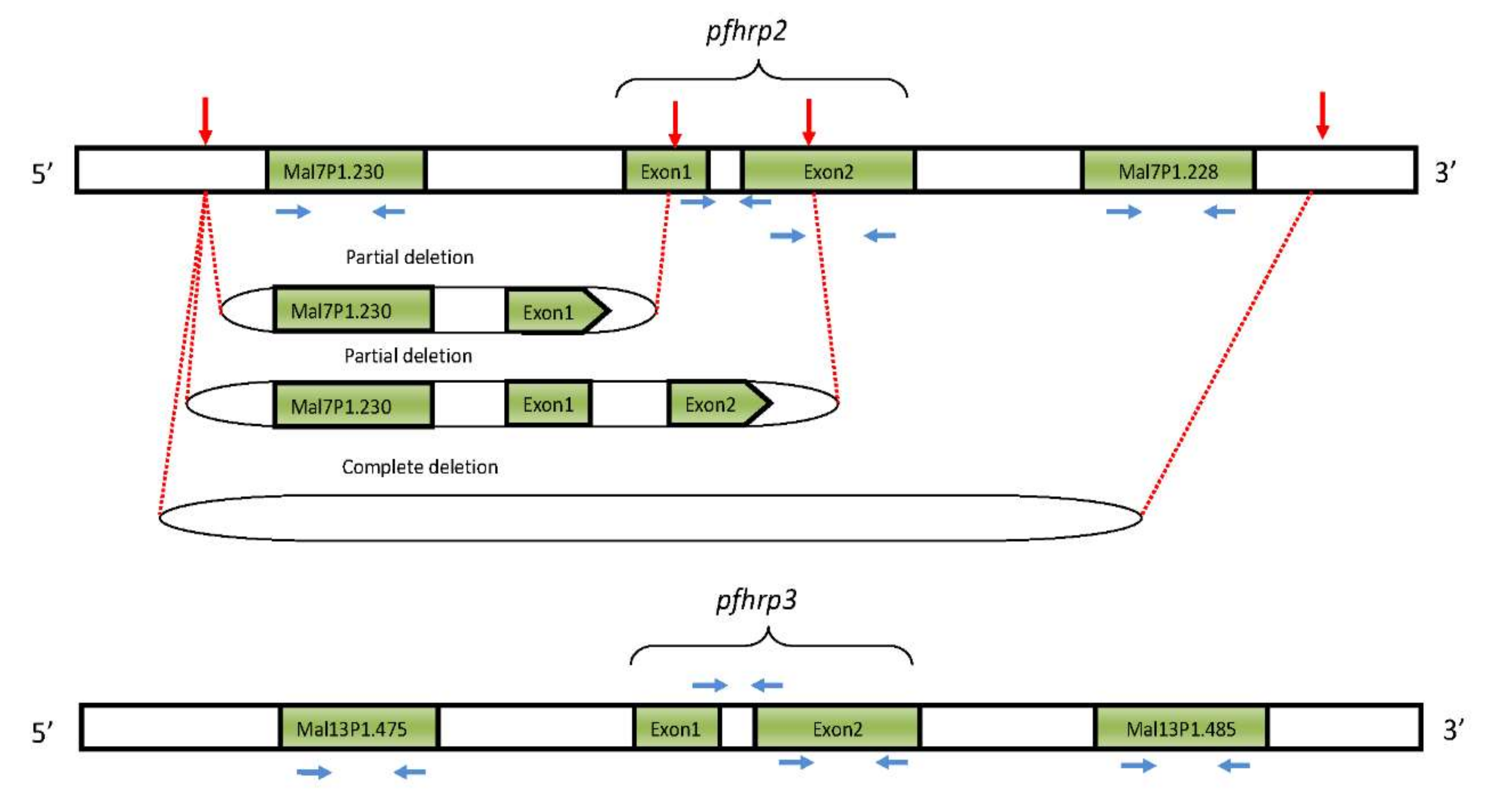
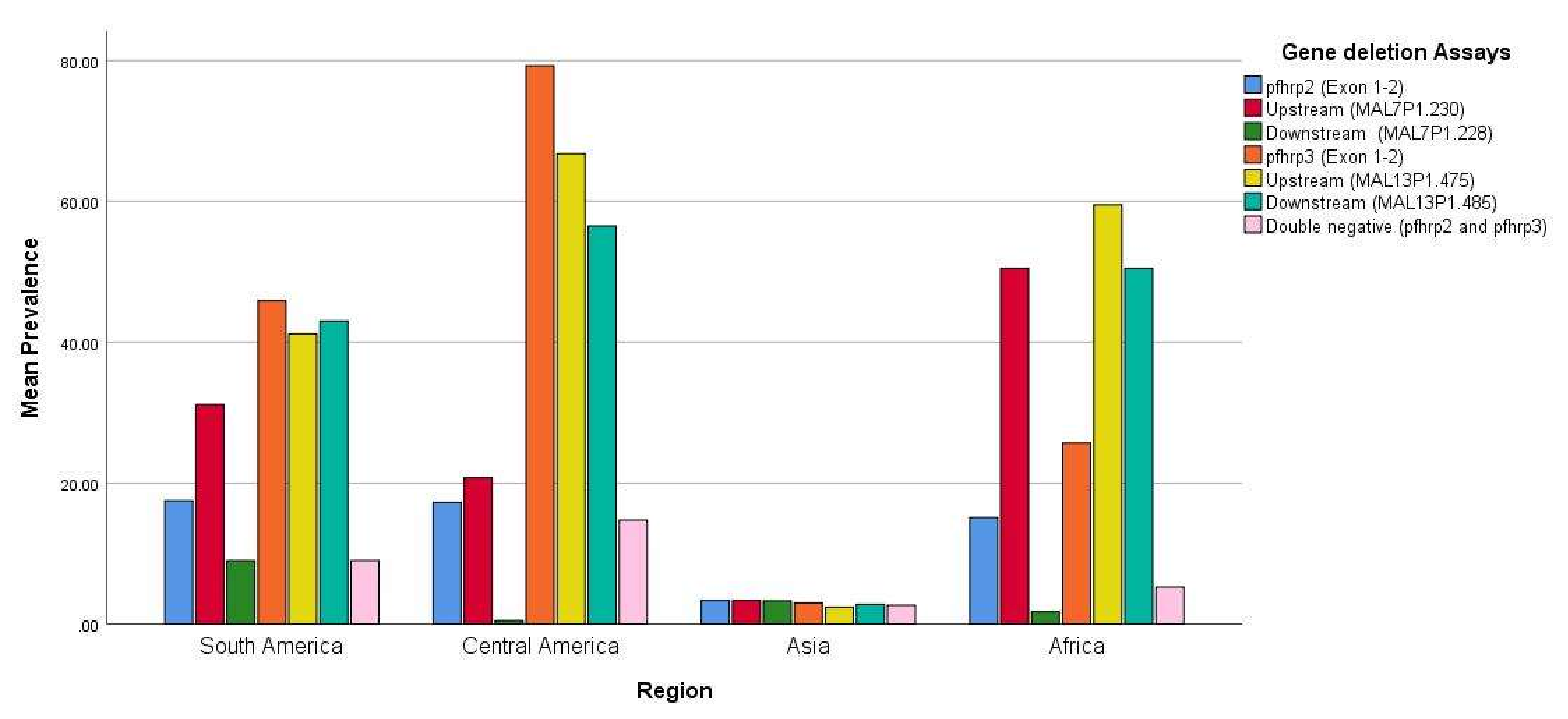
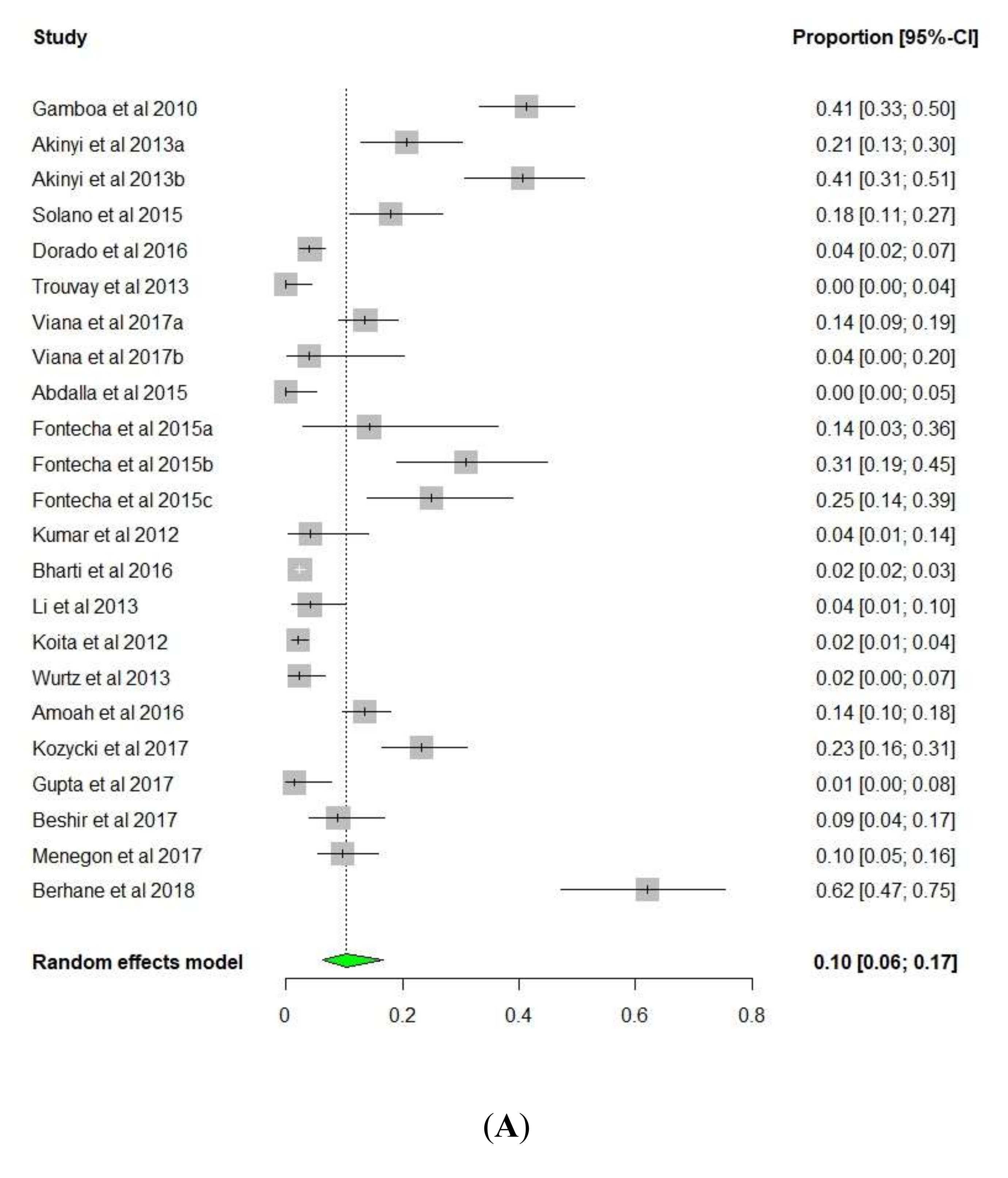
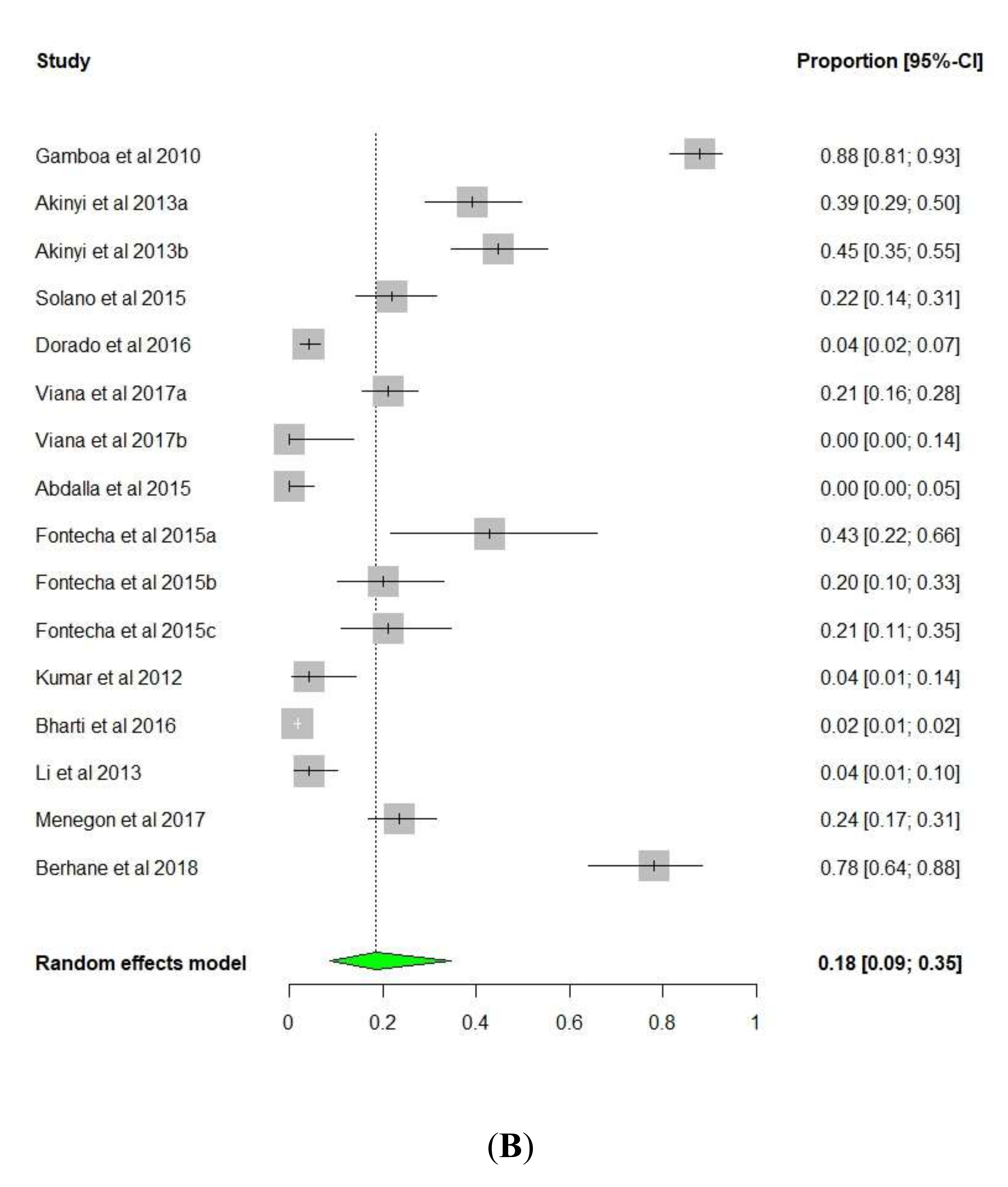
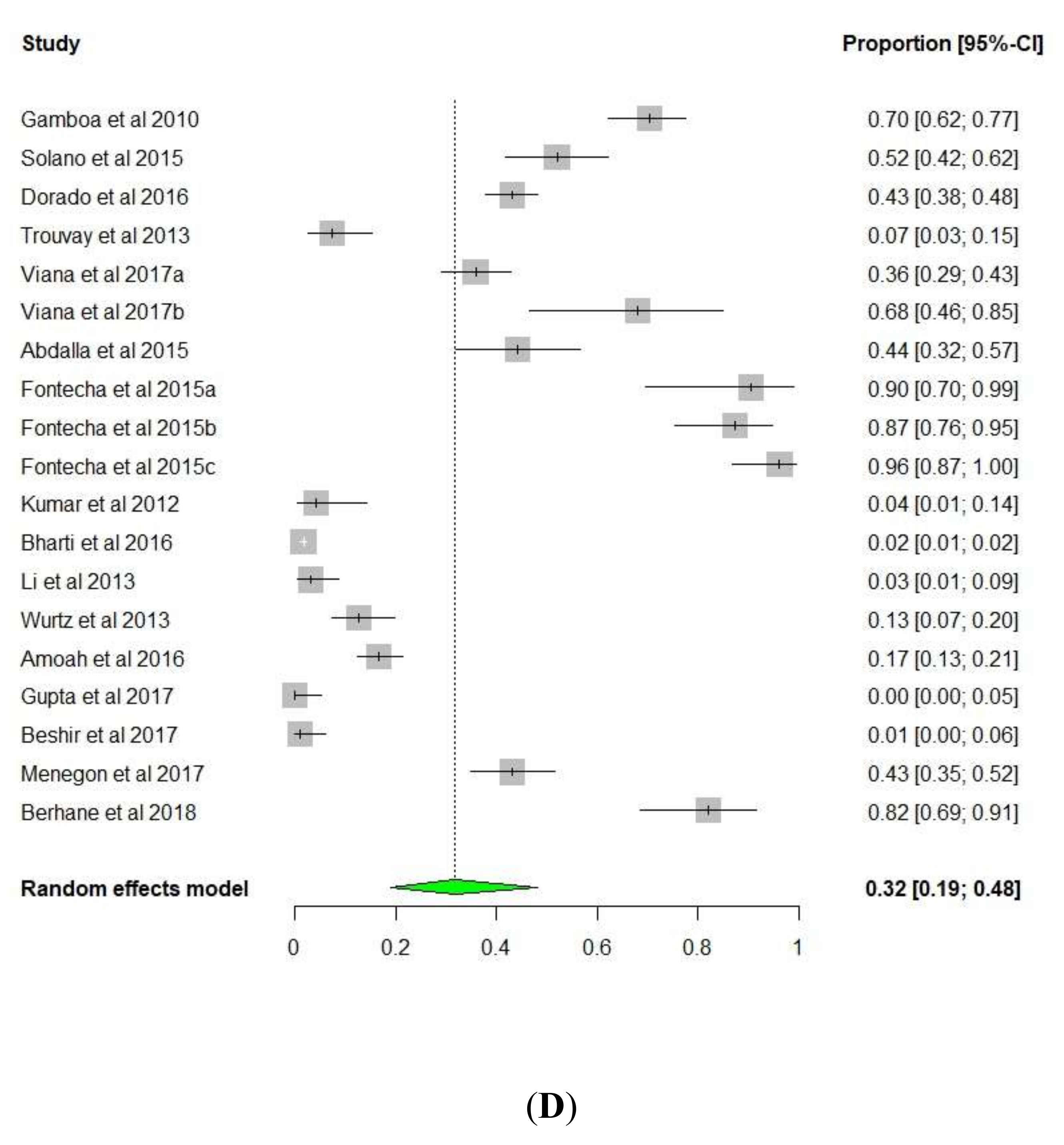
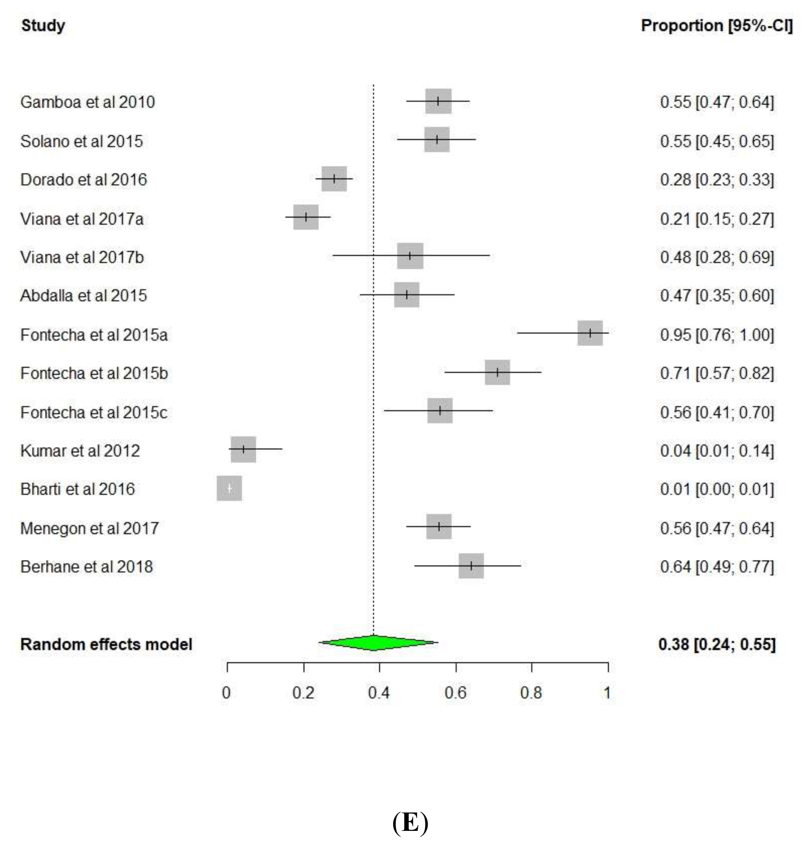
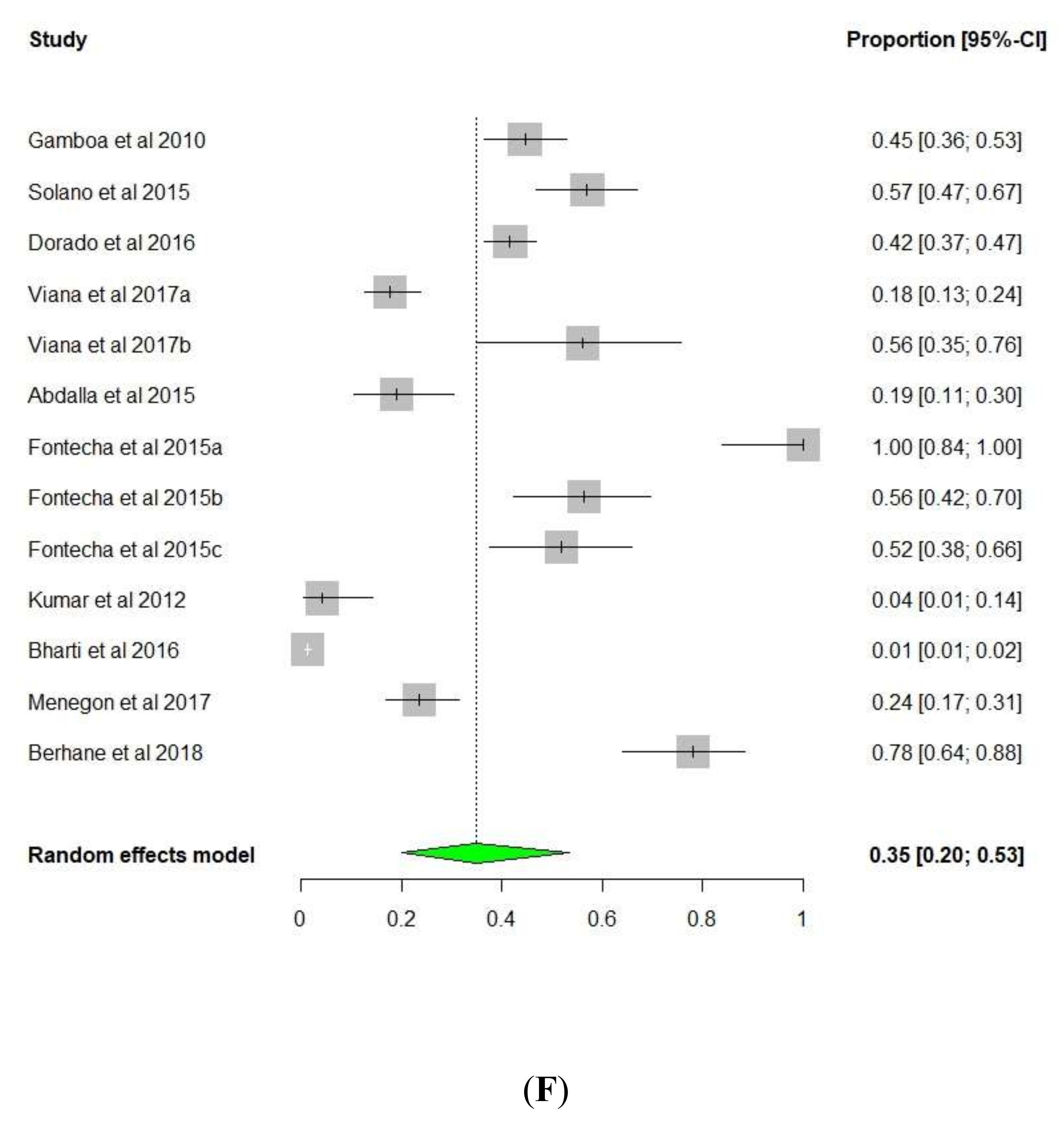
3.1. pfhrp2/pfhrp3 Deletions

Figure 2 summarizes the screening and retrieval process for the publications reporting pfhrp2 and pfhrp3 deletions. The final number of 20 publications that were reviewed is summarized in Table 1.
Gamboa et al. (2010) reported the first evidence of field isolates lacking pfhrp2 and pfhrp3 genes in Peru and demonstrated that these parasites were widespread across the Peruvian Amazon [40]. A huge proportion of the retrospectively collected samples were found to lack the pfhrp2 (41%), pfhrp3 (70%) or both (21.6%) genes. The loss of these genes was confirmed by inability to detect the proteins in an immunological assay [40]. A similar study by Akinyi et al. (2013) in the same region of Peru demonstrated a 20% to 40% increase in the frequency of malaria parasites lacking pfhrp2 over a seven year period [50].
Pfhrp2 and pfhrp3 gene deletions have also been reported in P. falciparum isolates from Colombia within the Amazon River region, like in Peru [51,52], where the prevalence of pfhrp2 deletions was lower than that of pfhrp3 (18% and 52%, respectively). This is not surprising considering that the majority of parasites with pfhrp2 deletions were collected at the Peru border, where pfhrp2 deletions had been reported before. A study that evaluated the population structure of Colombian P. falciparum at the Colombian pacific coast revealed four different subpopulations (Col-1, Col-2, Col-3, and Col-4) [67]. Similarly, the study by Solano et al. (2015) showed that the parasite isolates segregated into four clusters according to geography (Cluster 2 consisted 100% of the isolates from the Amazonas department, 59% of the isolates from Nariño department segregated into Cluster 4, 68% of isolates collected from Valle were assigned to Cluster 3, and 75% from Cordoba were assigned to Cluster 1). A total of 67% of the pfhrp2 deleted parasites and 61.5% of the pfhrp2/ pfhrp3 double negatives were found to segregate in Cluster 2, which consisted of samples mostly collected from the Amazonas department that borders Peru and Colombia within the Amazon ecological zone, indicating expansion and a common genetic origin of pfhrp2 and pfhrp3 deleted parasites [51].
Although earlier studies in Honduras only reported pfhrp3 and not pfhrp2 deletions, recent reports have documented extensive deletions of both genes [39,54]. The frequency of parasites lacking pfhrp2 and pfhrp3 in South and Central Americas has increased over time, and there is a need to evaluate the selective pressures that favor the survival of parasites that lack these genes [41].
Studies in India reported similar frequencies of parasites lacking pfhrp2 and pfhrp3 and their corresponding flanking regions [55,56,57]. Figure 3 summarizes the prevalence and distribution of pfhrp2, pfhrp3 and the flanking gene deletions in different continents. As shown in Figure 3, the lowest prevalence of pfhrp2/pfhrp3 and the respective flanking gene deletions was reported in Asia. In one of the studies, deletions of the two genes were observed in symptomatic individuals with high parasitemia [56], contrary to previous observations of pfhrp2 and pfhrp3 deletions in asymptomatic individuals with low parasitemia [60].
In Africa, where malaria is endemic and the use of RDTs is widespread, there is scarce information on pfhrp2/3 deletions, although a few surveillance studies have reported on their existence [60,61,63,68,69].
The first cases of pfhrp2 gene deleted parasites were reported in Mali, where deletions were identified in parasitemic individuals whose blood was unreactive to PfHRP2-based RDTs [60]. In that study, the participants were asymptomatic and the parasites had low multiplicity of infection, a term used to describe strain diversity in an infected individual. Although parasite density may have influenced the variability observed in PfHRP2-based RDTs, Wurtz et al. (2013) did not find any difference in parasitemia between groups with pfhrp/pfhrp3 deletions and those without deletions, suggesting that, in addition to parasite density, lack of expression of the PfHRP2 may greatly influence the performance of RDTs. A recent study in Eritrea reported the emergence of false-negative PfHRP2-based RDTs that was not related to poor quality of RDTs, storage, handling or operator errors [70]. A subsequent study in the same population revealed that most patients were infected with parasites that were lacking pfhrp2 (62%) and pfhrp3 (82%), and that these parasites may have emerged due to local selective pressure since the parasite clusters obtained were not related to South American strains [66].
In Kenya, one study has reported the presence of pfhrp2 deleted parasites in samples collected from asymptomatic children [12]. Curiously, these pfhrp2 deleted samples were positive by PfHRP2-based RDTs. Although this phenomenon was not reported in the South and Central America studies, it may be due to close reaction of PfHRP2 monoclonal antibodies with PfHRP3. In vitro studies have confirmed the detection of parasites with deleted pfhrp2 on PfHRP2-based RDTs, but this has not been reported widely in field samples [46]. Because of potential failures of RDTs, studies have recommended the performance of two RDTs in parallel as an initial screening test when evaluating pfhrp2/pfhrp3 deletion [49].
Overall, pfhrp2/pfhrp3 deletions have been widely reported in Central and South America, where most studies reported a higher prevalence of pfhrp3 (Lowest = 7.4% in French Guiana and Highest = 96.2% in Honduras) than of pfhrp2 (Lowest = 4.0% in Bolivia and Highest = 40.6% in Peru). Similar deletions were also observed for the upstream and downstream regions flanking pfhrp2 and pfhrp3 genes (Table 1 and Figure 3). The studies included in the current analysis showed extensive heterogeneity in reporting the prevalence of pfhrp2/pfhrp3 and the flanking genes (Figure 4). This could partly be attributed to the small sample size in these studies. We did not find any publication that reported a lack of deletion in all the genes of interest.
The results of the meta-analysis with random effects model are presented in Figure 4. Based on the estimates, we noted that the proportion of pfhrp2 exon1-2 among the included studies ranges from a minimum of 0.01 (95% CI: 0.00, 0.04) [53] to a maximum of 0.62 (95% CI: 0.47, 0.75) [66]. Furthermore, the pooled proportion of pfhrp2 exon1-2 was 0.10 (95% CI; 0.06, 0.17) (Figure 4A). The heterogeneity test showed presence of lack of uniformity, p-value = < 0.001. Similarly, the pooled prevalence of Upstream MALP1.230, Downstream MALP1.228, pfhrp3 exon1-2, Upstream MAL13P1.475, Downstream MAL13P1.485 and Double negative for pfhrp2 and pfhrp3 were 18.00%, 3.00%, 32.00%, 38.00%, 35.00% and 8.00%, respectively (Figure 4B-G).
Cheng et al.(2014) published a set of recommendations for reporting pfhrp2/pfhrp3 deletions [49]. These recommendations include an initial screening whereby the sample is confirmed positive for P. falciparum by two expert microscopists and PCR. This is followed by a confirmatory evidence of deletion through PCR amplification of exon 2 of pfhrp2 and pfhrp3 and the region across exon 1 and 2 of both genes.
For quality control, it is also recommended to include PCR amplification of single copy genes such as msp1 and msp2 to rule out negative results due to low-quality DNA or PCR failure. We used these recommendations to evaluate which studies conformed to this protocol. All studies conducted in South America, Central America, and Asia conformed to these recommendations for reporting pfhrp2/pfhrp3 deletions including PCR analysis of the flanking genes, performing RDTs on the suspected samples, and functional analysis through PfHRP2 ELISA. These recommendations are also in agreement with the recently published WHO guidelines on reporting pfhrp2/pfhrp3 deletions [71] and as such, should be adhered to for the validity of any study on pfhrp2/pfhrp3 gene deletions.
Unfortunately, of the eight studies conducted in Africa, only two followed these guidelines. Most reported on the prevalence of pfhrp2 (Lowest = 1.5% in Mozambique and Highest = 62.0% in Eritrea) and pfhrp3 (Lowest = 1.1% in Kenya and Highest = 82.0% in Eritrea [12,62,63,64]. The two studies that included analysis of flanking genes in the detection of pfhrp2/pfhrp3 deletion were from Eritrea. However, the study by Menegon et al. (2017) did not include RDT results [65].
Although the upstream and downstream flanking genes for pfhrp2 and pfhrp3 are not well characterized, they have been observed [49], and as such, it is recommended to include these genes when evaluating pfhrp2 and pfhrp3 gene deletions. As shown in Table 1, most studies did not conform to the WHO recommendation for accurate reporting of pfhrp2/pfhrp3 deletions. This may affect the validity of the claims made [49,71].
Apart from gene deletions, sequencing of the pfhrp2 and pfhrp3 genes has revealed extensive variability and different haplotype profiles in parasites from different geographical areas, and this may influence the performance of RDTs [72,73]. For example, the sequencing of these genes has revealed an extensive variation in the number of histidine repeats, copy numbers, and single nucleotide polymorphisms [58,74,75]. The functional utility of these variations needs further investigations, since this variability has not been shown to affect the amount of PfHRP2 in plasma, which is the basis for parasite detection on RDTs [74,76].

3.2. Implication of pfhrp2/pfhrp3 Deletions on RDT Use

The extensive decline in malaria witnessed over the last two decades is attributed to improved diagnosis, effective treatment, and monitoring. The details of the decline are captured in the WHO Global Initiative for malaria control referred to as T3 (Test, Treat, and Track) which provided a framework for malaria control and elimination in malaria-endemic countries [77]. This initiative has contributed to the increased use of RDTs for diagnostic testing, especially in areas where microscopy is not available.
Recent evidence of false-negative RDTs due to the emergence of parasites lacking pfhrp2 and/or pfhrp3 may negatively impact the success of RDTs in malaria diagnosis and could present a major threat to the progress made in the fight against malaria [40,49], and may delay eventual malaria eradication goals. Although the selective pressure-favoring survival of pfhrp2 depleted parasites is not clearly defined, mathematical modeling has suggested that the use of PfHRP2-based RDTs over the last decade favored the survival of parasites lacking pfhrp2 genes. The model also suggests that selection pressure is high in areas where the prevalence of malaria is low, and the treatment of cases is based on RDT diagnosis [78]. This is because, in low transmission settings, most infections are asymptomatic and are characterized by low-density parasitemia, which is below the detection limit of the RDTs. This means that these infections are not treated and thereby enhance parasite survival [79,80]. Because of the structural homology of PfHRP2 and PfHRP3 antigens and the observed cross-reaction of monoclonal antibodies used in the RDTs, it would be expected that PfHRP3 would compensate for the deletion of pfhrp2. However, because PfHRP3 is not abundantly produced, this is likely to be beneficial only at high parasitemia [46].
The decline in malaria transmission that has been witnessed in most areas means that regions previously considered endemic may not experience as high a number of malaria cases as before, and most infections will likely be characterized by low-density parasitemia. If the model proposed by Watson is correct, this will increase the number of missed diagnosis by both microscopy and RDT; therefore, individuals harboring pfhrp2 depleted parasites are likely to go undetected [78]. Consequently, this will likely amplify the population of malaria parasites with the these deletions [49]. In peripheral health care facilities where microscopy is not available or among travelers to malaria-endemic areas, a common practice is to rule out malaria in patients that test negative with an RDT. This practice could also aid in the selection of pfhrp2 deleted malaria parasites.
This review demonstrates the emerging threat of pfhrp2/pfhrp3 deleted parasites mostly in areas that have had a history of P. falciparum malaria. This could become a big challenge, especially with the increased use of PfHRP2-based RDTs. In sub-Saharan Africa, where malaria is still endemic, the extent of pfhrp2/pfhrp3 gene deletions is underreported [71,81]. There is, therefore, a need for more extensive studies to evaluate the true prevalence of pfhrp2/pfhrp3 deleted parasites and the impact they have on malaria diagnosis.

Funding

Salary support for Josphat Nyataya comes from the Armed Forces Health Surveillance Branch (AFHSB) and it’s GEIS (Global Emerging Infections Surveillance and Response) Section (grant number P152_20_KY_02).

Acknowledgments

Authors would like to thank staff members of the Basic Science Laboratory and Information Technology department of the US Army Medical Research Directorate-Africa/Kenya, for their support in this project. We are grateful to the South African National Research Foundation (NRF) for supporting Prof. Mohamed Ezzat El Zowalaty through Africa Knowledge, Interchange and Collaboration (KIC) grant number (KIC170928263699) awarded to Prof. ME El Zowalaty for his research collaboration and visit to the Basic Science Laboratory of the US Army Medical Research Directorate-Africa/Kenya Medical Research Institute. We acknowledge Zelalem G. Dessie from the School of Mathematics, Statistics and Computer Science, University of KwaZulu-Natal, Durban, South Africa for his assistance in analysis. We appreciate the three anonymous reviewers for their efforts and valuable comments which significantly improved the manuscript.

Conflicts of Interest

The authors declare that they have no competing interest.

References

  1. Centers for Dises Control and Prevention. World Malaria Day 2018. Features; Centers for Dises Control and Prevention: Atlanta, GA, USA, 2018; Available online: https://www.cdc.gov/features/worldmalariaday/index.html (accessed on 12 October 2018).
  2. WHO. World Malaria Report 2016; World Health Organization: Geneva, Switzerland, 2016; Available online: https://www.who.int/malaria/publications/world-malaria-report-2016/report/en/ (accessed on 12 October 2018).
  3. WHO. WHO News Release. Available online: https://www.who.int/mediacentre/news/releases/2011/malaria_report_20111213/en/ (accessed on 22 October 2018).
  4. WHO. World Malaria Report 2019; World Health Organization: Geneva, Switzerland, 2019; Available online: https://www.who.int/publications-detail/world-malaria-report-2019 (accessed on 12 October 2018).
  5. WHO. Guidlines for the Treatment of Malaria; World Health Organization: Geneva, Switzerland, 2015; Available online: https://www.who.int/malaria/publications/atoz/9789241549127/en/ (accessed on 6 July 2019).
  6. Tangpukdee, N.; Duangdee, C.; Wilairatana, P.; Krudsood, S. Malaria diagnosis: A brief review. Korean J. Parasitol. 2009, 47, 93–102. [Google Scholar] [CrossRef] [PubMed]
  7. Jirk, M.; Petrželková, K.J.; Zuzana, H.; Modrý, D.; Lukeš, J. Detection of Plasmodium spp. in Human Feces. Emerg. Infect. Dis. 2012, 18, 634–636. [Google Scholar]
  8. Putaporntip, C.; Jongwutiwes, S.; Sakihama, N.; Ferreira, M.U.; Kho, W.G.; Kaneko, A.; Kanbara, H.; Hattori, T.; Tanabe, K. Mosaic organization and heterogeneity in frequency of allelic recombination of the Plasmodium vivax merozoite surface protein-1 locus. Proc. Natl. Acad. Sci. USA 2002, 99, 16348–16353. [Google Scholar] [CrossRef] [PubMed] [Green Version]
  9. Wongsrichanalai, C.; Barcus, M.J.; Muth, S.; Sutamihardja, A.; Wernsdorfer, W.H. A Review of Malaria Diagnostic Tools: Microscopy and Rapid Diagnostic Test (RDT). Am. J. Trop. Med. Hyg. 2007, 77, 119–127. Available online: https://0-www-ncbi-nlm-nih-gov.brum.beds.ac.uk/books/NBK1695/ (accessed on 22 October 2018). [CrossRef] [PubMed] [Green Version]
  10. Murphy, S.C.; Shott, J.P.; Parikh, S.; Etter, P.; Prescott, W.R.; Stewart, V.A. Review article: Malaria diagnostics in clinical trials. Am. J. Trop. Med. Hyg. 2013, 89, 824–839. [Google Scholar] [CrossRef] [PubMed] [Green Version]
  11. Mirdha, B.R.; Samantaray, J.C.; Mishra, B. Laboratory diagnosis of malaria. J. Clin. Pathol. 1997, 50, 356. [Google Scholar] [CrossRef] [Green Version]
  12. Beshir, K.B.; Sepúlveda, N.; Bharmal, J.; Robinson, A.; Mwanguzi, J.; Busula, A.O.; De Boer, J.G.; Sutherland, C.; Cunningham, J.; Hopkins, H. Plasmodium falciparum parasites with histidine-rich protein 2 (pfhrp2) and pfhrp3 gene deletions in two endemic regions of Kenya. Sci. Rep. 2017, 7, 14718. [Google Scholar] [CrossRef]
  13. Khairnar, K.; Martin, D.; Lau, R.; Ralevski, F.; Pillai, D.R. Multiplex real-time quantitative PCR, microscopy and rapid diagnostic immuno-chromatographic tests for the detection of Plasmodium spp: Performance, limit of detection analysis and quality assurance. Malar. J. 2009, 8, 284. [Google Scholar] [CrossRef] [Green Version]
  14. Ohrt, C.; Sutamihardja, M.A.; Tang, D.; Kain, K.C. Impact of Microscopy Error on Estimates of Protective Efficacy in Malaria-Prevention Trials. J. Infect. Dis. 2002, 186, 540–546. [Google Scholar] [CrossRef]
  15. Payne, D. Use and limitations of light microscopy for diagnosing malaria at the primary health care level. Bull. World Health Organ. 1988, 66, 621–626. [Google Scholar]
  16. Snounou, G.; Viriyakosol, S.; Jarra, W.; Thaithong, S.; Brown, K.N. Identification of the four human malaria parasite species in field samples by the polymerase chain reaction and detection of a high prevalence of mixed infections. Mol. Biochem. Parasitol. 1993, 58, 283–292. [Google Scholar] [CrossRef]
  17. Erdman, L.K.; Kain, K.C. Molecular diagnostic and surveillance tools for global malaria control. Travel Med. Infect. Dis. 2008, 6, 82–99. [Google Scholar] [CrossRef]
  18. Zhao, J.; Lama, M.; Korenromp, E.; Aylward, P.; Shargie, E.; Filler, S.; Komatsu, R.; Atun, R. Adoption of rapid diagnostic tests for the diagnosis of malaria, a preliminary analysis of the global fund program data, 2005 to 2010. PLoS ONE 2012, 7, e43549. [Google Scholar] [CrossRef] [PubMed]
  19. WHO. Malaria Rapid Diagnostic Test Performance: Summary Results of WHO Product Testing of Malaria RDTs: Round 1-5 (2008–2013); WHO: Geneva, Switzerland, 2014; Available online: https://www.who.int/malaria/publications/atoz/9789241507554/en/ (accessed on 6 July 2019).
  20. Meier, B.; Döbeli, H.; Certa, U. Stage-specific expression of aldolase isoenzymes in the rodent malaria parasite Plasmodium berghei. Mol. Biochem. Parasitol. 1992, 52, 15–27. [Google Scholar] [CrossRef]
  21. Makler, M.T.; Hinrichs, D.J. Measurement of the lactate dehydrogenase activity of Plasmodium falciparum as an assessment of parasitemia. Am. J. Trop. Med. Hyg. 1993, 48, 205–210. [Google Scholar] [CrossRef]
  22. Wiwanitkit, V. Plasmodium and Host Lactate Dehydrogenase Molecular Function and Biological Pathways: Implication for Antimalarial Drug Discovery. Chem. Biol. Drug Des. 2007, 69, 280–283. [Google Scholar] [CrossRef]
  23. Ouattara, A.; Doumbo, S.; Saye, R.; Beavogui, A.H.; Traoré, B.; Djimdé, A.; Niangaly, A.; Kayentao, K.; Diallo, M.; Doumbo, O.K.; et al. Use of a pLDH-based dipstick in the diagnostic and therapeutic follow-up of malaria patients in Mali. Malar. J. 2011, 10, 345. [Google Scholar] [CrossRef] [Green Version]
  24. Dalrymple, U.; Arambepola, R.; Gething, P.W.; Cameron, E. How long do rapid diagnostic tests remain positive after anti-malarial treatment? Malar. J. 2018, 17, 228. [Google Scholar] [CrossRef] [Green Version]
  25. Chiodini, P.L.; Bowers, K.; Jorgensen, P.; Barnwell, J.W.; Grady, K.K.; Luchavez, J.; Moody, A.H.; Cenizal, A.; Bell, D. The heat stability of Plasmodium lactate dehydrogenase-based and histidine-rich protein 2-based malaria rapid diagnostic tests. Trans. R. Soc. Trop. Med. Hyg. 2007, 101, 331–337. [Google Scholar] [CrossRef]
  26. Singh, N.; Bharti, P.K.; Singh, M.P.; Mishra, S.; Shukla, M.M.; Sharma, R.K.; Singh, R.K. Comparative Evaluation of Bivalent Malaria Rapid Diagnostic Tests versus Traditional Methods in Field with Special Reference to Heat Stability Testing in Central India. PLoS ONE 2013, 8, e58080. [Google Scholar] [CrossRef] [Green Version]
  27. Rock, E.P.; Marsh, K.; Saul, A.J.; Wellems, T.E.; Taylor, D.W.; Maloy, W.L.; Howard, R.J. Comparative analysis of the Plasmodium falciparum histidine-rich proteins HRP-I, HRP-II and HRP-III in malaria parasites of diverse origin. Parasitology 1987, 95, 209–227. [Google Scholar] [CrossRef] [PubMed]
  28. Sharma, Y.D. Genomic organization, structure and possible function of histidine-rich proteins of malaria parasites. Int. J. Biochem. 1988, 20, 471–477. [Google Scholar] [CrossRef]
  29. Gruenberg, J.; Allred, D.R.; Sherman, I.W. Scanning electron microscope-analysis of the protrusions (knobs) present on the surface of Plasmodium falciparum-infected erythrocytes. J. Cell Biol. 1983, 97, 795–802. [Google Scholar] [CrossRef] [PubMed]
  30. Kilejian, A. Characterization of a protein correlated with the production of knob-like protrusions on membranes of erythrocytes infected with Plasmodium falciparum. Proc. Natl. Acad. Sci. USA 1979, 76, 4650–4653. [Google Scholar] [CrossRef] [Green Version]
  31. Luse, S.A.; Miller, L.H. Plasmodium falciparum malaria. Ultrastructure of parasitized erythrocytes in cardiac vessels. Am. J. Trop. Med. Hyg. 1971, 20, 655–660. [Google Scholar] [CrossRef]
  32. Manning, L.; Laman, M.; Stanisic, D.; Rosanas-Urgell, A.; Bona, C.; Teine, D.; Siba, P.; Mueller, I.; Davis, T.M.E. Plasma Plasmodium falciparum histidine-rich protein-2 concentrations do not reflect severity of malaria in Papua New Guinean children. Clin. Infect. Dis. 2011, 52, 440–446. [Google Scholar] [CrossRef] [Green Version]
  33. Parra, M.E.; Evans, C.B.; Taylor, D.W. Identification of Plasmodium falciparum Histidine-Rich Protein 2 in the Plasma of Humans with Malaria. J. Clin. Microbiol. 1991, 29, 1629–1634. [Google Scholar] [CrossRef] [Green Version]
  34. Rodriguez-del Valle, M.; Quakyi, I.A.; Amuesi, J.; Quaye, J.T.; Nkrumah, F.; Taylor, D.W. Detection of antigen and antibodies and in urine of humans with Plasmodium falciparum malaria. J. Clin. Microbiol. 1991, 29, 1236–1242. [Google Scholar] [CrossRef] [Green Version]
  35. Howard, R.; Uni, S.; Aley, S.B.; Taylorll, W.; Diseases, I.; Francisco, S.; Hospital, G.; Francisco, S. Plasmodium falciparum-infected. J. Cell Biol. 1986, 103, 1269–1277. [Google Scholar] [CrossRef] [Green Version]
  36. Desakorn, V.; Dondorp, A.M.; Silamut, K.; Pongtavornpinyo, W.; Sahassananda, D.; Chotivanich, K.; Pitisuttithum, P.; Smithyman, A.M.; Day, N.P.J.; White, N.J. Stage-dependent production and release of histidine-rich protein 2 by Plasmodium falciparum. Trans. R. Soc. Trop. Med. Hyg. 2005, 99, 517–524. [Google Scholar] [CrossRef]
  37. Moody, A. Rapid diagnostic tests for malaria parasites. Clin. Microbiol. Rev. 2002, 15, 66–78. [Google Scholar] [CrossRef] [PubMed] [Green Version]
  38. Leech, J.H.; Aley, S.B.; Miller, L.H.; Howard, R.J. Plasmodium falciparum malaria: Cytoadherence of infected erythrocytes to endothelial cells and associated changes in the erythrocyte membrane. Prog. Clin. Biol. Res. 1984, 155, 63–77. [Google Scholar] [PubMed]
  39. Abdallah, J.F.; Okoth, S.A.; Fontecha, G.A.; Mejia Torres, R.E.; Banegas, E.I.; Matute, M.L.; Bucheli, S.T.M.; Goldman, I.F.; De Oliveira, A.M.; Barnwell, J.W.; et al. Prevalence of pfhrp2 and pfhrp3 gene deletions in Puerto Lempira, Honduras. Malar. J. 2015, 14, 19. [Google Scholar] [CrossRef] [Green Version]
  40. Gamboa, D.; Ho, M.F.; Bendezu, J.; Torres, K.; Chiodini, P.L.; Barnwell, J.W.; Incardona, S.; Perkins, M.; Bell, D.; McCarthy, J.; et al. A large proportion of P. falciparum isolates in the Amazon region of Peru lack pfhrp2 and pfhrp3: Implications for malaria rapid diagnostic tests. PLoS ONE 2010, 5. [Google Scholar] [CrossRef] [PubMed] [Green Version]
  41. Okoth, S.A.; Abdallah, J.F.; Ceron, N.; Adhin, M.R.; Chandrabose, J.; Krishnalall, K.; Huber, C.S.; Goldman, I.F.; De Oliveira, A.M.; Barnwell, J.W.; et al. Variation in Plasmodium falciparum histidine-rich protein 2 (Pfhrp2) and Plasmodium falciparum histidine-rich protein 3 (Pfhrp3) gene deletions in Guyana and Suriname. PLoS ONE 2015, 10, e0126805. [Google Scholar]
  42. Viana, G.M.R.; Okoth, S.A.; Silva-Flannery, L.; Barbosa, D.R.L.; De Oliveira, A.M.; Goldman, I.F.; Morton, L.C.; Huber, C.; Anez, A.; Machado, R.L.D.; et al. Histidine-rich protein 2 (pfhrp2) and pfhrp3 gene deletions in Plasmodium falciparum isolates from select sites in Brazil and Bolivia. PLoS ONE 2017, 12, e0171150. [Google Scholar]
  43. Walker-Jonah, A.; Dolan, S.A.; Gwadz, R.W.; Panton, L.J.; Wellems, T.E. An RFLP map of the Plasmodium falciparum genome, recombination rates and favored linkage groups in a genetic cross. Mol. Biochem. Parasitol. 1992, 51, 313–320. [Google Scholar] [CrossRef]
  44. Pologe, L.G.; Ravetch, J.V. Large deletions result from breakage and healing of P. falciparum chromosomes. Cell 1988, 55, 869–874. [Google Scholar] [CrossRef]
  45. Dharia, N.V.; Plouffe, D.; Bopp, S.E.R.; González-Páez, G.E.; Lucas, C.; Salas, C.; Soberon, V.; Bursulaya, B.; Kochel, T.J.; Bacon, D.J.; et al. Genome scanning of Amazonian Plasmodium falciparum shows subtelomeric instability and clindamycin-resistant parasites. Genome Res. 2010, 20, 1534–1544. [Google Scholar] [CrossRef] [Green Version]
  46. Baker, J.; McCarthy, J.; Gatton, M.; Kyle, D.E.; Belizario, V.; Luchavez, J.; Bell, D.; Cheng, Q. Genetic Diversity of Plasmodium falciparum Histidine-Rich Protein 2 (PfHRP2) and Its Effect on the Performance of PfHRP2-Based Rapid Diagnostic Tests. J. Infect. Dis. 2005, 192, 870–877. [Google Scholar] [CrossRef] [Green Version]
  47. Bharti, P.K.; Chandel, H.S.; Krishna, S.; Nema, S. Sequence variation in Plasmodium falciparum Histidine Rich Proteins 2 and 3 in Indian isolates: Implications for Malaria Rapid Diagnostic Test Performance. Sci. Rep. 2017, 7, 1308. [Google Scholar]
  48. Liberati, A.; Altman, D.G.; Tetzlaff, J.; Mulrow, C.; Gotzsche, P.C.; Ioannidis, J.P.; Clarke, M.; Devereaux, P.J.; Kleijnen, J.; Moher, D. The PRISMA statement for reporting systematic reviews and meta-analyses of studies that evaluate health care interventions: Explanation and elaboration. PLoS Med. 2009, 6, e1000100. [Google Scholar] [CrossRef] [PubMed]
  49. Cheng, Q.; Gatton, M.L.; Barnwell, J.; Chiodini, P.; McCarthy, J.; Bell, D.; Cunningham, J. Plasmodium falciparum parasites lacking histidine-rich protein 2 and 3: A review and recommendations for accurate reporting. Malar. J. 2014, 13, 283. [Google Scholar] [CrossRef] [PubMed] [Green Version]
  50. Akinyi, S.; Hayden, T.; Gamboa, D.; Torres, K.; Bendezu, J.; Abdallah, J.F.; Griffing, S.M.; Quezada, W.M.; Arrospide, N.; De Oliveira, A.M.E.; et al. Multiple genetic origins of histidine-rich protein 2 gene deletion in Plasmodium falciparum parasites from Peru. Sci. Rep. 2013, 3, 2797. [Google Scholar] [CrossRef] [PubMed] [Green Version]
  51. Murillo Solano, C.; Akinyi Okoth, S.; Abdallah, J.F.; Pava, Z.; Dorado, E.; Incardona, S.; Huber, C.S.; Macedo de Oliveira, A.; Bell, D.; Udhayakumar, V.; et al. Deletion of Plasmodium falciparum Histidine-Rich Protein 2 (pfhrp2) and Histidine-Rich Protein 3 (pfhrp3) Genes in Colombian Parasites. PLoS ONE 2015, 10, e0131576. [Google Scholar] [CrossRef] [PubMed] [Green Version]
  52. Dorado, E.J.; Okoth, S.A.; Montenegro, L.M.; Diaz, G.; Barnwell, J.W.; Udhayakumar, V.; Solano, C.M. Genetic characterisation of Plasmodium falciparum isolates with deletion of the pfhrp2 and/or pfhrp3 genes in Colombia: The amazon region, a challenge for malaria diagnosis and control. PLoS ONE 2016, 11, e0163137. [Google Scholar] [CrossRef]
  53. Trouvay, M.; Palazon, G.; Berger, F.; Volney, B.; Blanchet, D.; Faway, E.; Donato, D.; Legrand, E.; Carme, B.; Musset, L. High Performance of Histidine-Rich Protein 2 Based Rapid Diagnostic Tests in French Guiana are Explained by the Absence of pfhrp2 Gene Deletion in P. falciparum. PLoS ONE 2013, 8, e74269. [Google Scholar] [CrossRef]
  54. Fontecha, G.; Mejía, R.E.; Banegas, E.; Ade, M.P.; Mendoza, L.; Ortiz, B.; Sabillón, I.; Alvarado, G.; Matamoros, G.; Pinto, A. Deletions of pfhrp2 and pfhrp3 genes of Plasmodium falciparum from Honduras, Guatemala and Nicaragua. Malar. J. 2018, 17, 320. [Google Scholar] [CrossRef]
  55. Kumar, N.; Pande, V.; Bhatt, R.M.; Shah, N.K.; Mishra, N.; Srivastava, B.; Valecha, N.; Anvikar, A.R. Genetic deletion of hrp2 and hrp3 in Indian Plasmodium falciparum population and false negative malaria rapid diagnostic test. Acta Trop. 2013, 125, 119–121. [Google Scholar] [CrossRef]
  56. Bharti, P.K.; Chandel, H.S.; Ahmad, A.; Krishna, S.; Udhayakumar, V.; Singh, N. Prevalence of pfhrp2 and/or pfhrp3 gene deletion in Plasmodium falciparum population in eight highly endemic states in India. PLoS ONE 2016, 11, e0157949. [Google Scholar] [CrossRef]
  57. Pati, P.; Dhangadamajhi, G.; Bal, M.; Ranjit, M. High proportions of pfhrp2 gene deletion and performance of HRP2-based rapid diagnostic test in Plasmodium falciparum field isolates of Odisha. Malar. J. 2018, 17, 394. [Google Scholar] [CrossRef] [PubMed] [Green Version]
  58. Li, P.; Xing, H.; Zhao, Z.; Yang, Z.; Cao, Y.; Yan, G.; Sattabongkot, J.; Cui, L.; Fan, Q. Genetic diversity of Plasmodium falciparum histidine-rich protein 2 in the China-Myanmar border area. Acta Trop. 2015, 152, 26–31. [Google Scholar] [PubMed] [Green Version]
  59. Nima, M.K.; Hougard, T.; Hossain, M.E.; Kibria, M.G.; Mohon, A.N.; Johora, F.T.; Rahman, R.; Haque, R.; Alam, M.S. Case Report: A Case of Plasmodium falciparum hrp2 and hrp3 Gene Mutation in Bangladesh. Am. J. Trop. Med. Hyg. 2017, 97, 1155–1158. [Google Scholar] [CrossRef] [PubMed]
  60. Koita, O.A.; Doumbo, O.K.; Ouattara, A.; Tall, L.K.; Konaré, A.; Diakité, M.; Diallo, M.; Sagara, I.; Masinde, G.L.; Doumbo, S.N.; et al. False-negative rapid diagnostic tests for malaria and deletion of the histidine-rich repeat region of the hrp2 gene. Am. J. Trop. Med. Hyg. 2012, 86, 194–198. [Google Scholar] [CrossRef] [PubMed] [Green Version]
  61. Wurtz, N.; Fall, B.; Bui, K.; Pascual, A.; Fall, M.; Camara, C.; Diatta, B.; Fall, K.B.; Mbaye, P.S.; Diémé, Y.; et al. Pfhrp2 and pfhrp3 polymorphisms in Plasmodium falciparum isolates from Dakar, Senegal: Impact on rapid malaria diagnostic tests. Malar. J. 2013, 12, 34. [Google Scholar] [CrossRef] [PubMed] [Green Version]
  62. Amoah, L.E.; Abankwa, J.; Oppong, A. Plasmodium falciparum histidine rich protein-2 diversity and the implications for PfHRP 2: Based malaria rapid diagnostic tests in Ghana. Malar. J. 2016, 15, 101. [Google Scholar] [CrossRef] [PubMed] [Green Version]
  63. Kozycki, C.T.; Umulisa, N.; Rulisa, S.; Mwikarago, E.I.; Musabyimana, J.P.; Habimana, J.P.; Karema, C.; Krogstad, D.J. False-negative malaria rapid diagnostic tests in Rwanda: Impact of Plasmodium falciparum isolates lacking hrp2 and declining malaria transmission. Malar. J. 2017, 16, 123. [Google Scholar] [CrossRef] [Green Version]
  64. Gupta, H.; Matambisso, G.; Galatas, B.; Cisteró, P.; Nhamussua, L.; Simone, W.; Cunningham, J.; Rabinovitch, N.R.; Alonso, P.; Saute, F.; et al. Molecular surveillance of pfhrp2 and pfhrp3 deletions in Plasmodium falciparum isolates from Mozambique. Malar. J. 2017, 16, 416. [Google Scholar] [CrossRef] [Green Version]
  65. Menegon, M.; L’Episcopia, M.; Nurahmed, A.M.; Talha, A.A.; Nour, B.Y.M.; Severini, C. Identification of Plasmodium falciparum isolates lacking histidine-rich protein 2 and 3 in Eritrea. Infect. Genet. Evol. 2017, 55, 131–134. [Google Scholar] [CrossRef]
  66. Berhane, A.; Anderson, K.; Mihreteab, S.; Gresty, K.; Rogier, E.; Mohamed, S.; Hagos, F.; Embaye, G.; Chinorumba, A.; Zehaie, A.; et al. Major threat to malaria control programs by Plasmodium falciparum lacking histidine-rich protein 2, Eritrea. Emerg. Infect. Dis. 2018, 24, 462–470. [Google Scholar] [CrossRef] [Green Version]
  67. Echeverry, D.F.; Nair, S.; Osorio, L.; Menon, S.; Murillo, C.; Anderson, T.J.C. Long term persistence of clonal malaria parasite Plasmodium falciparum lineages in the Colombian Pacific region. BMC Genet. 2013, 14. [Google Scholar] [CrossRef] [PubMed] [Green Version]
  68. Laban, N.M.; Kobayashi, T.; Hamapumbu, H.; Sullivan, D.; Mharakurwa, S.; Thuma, P.E.; Shiff, C.J.; Moss, W.J. Comparison of a PfHRP2-based rapid diagnostic test and PCR for malaria in a low prevalence setting in rural southern Zambia: Implications for elimination. Malar. J. 2015, 14, 3–9. [Google Scholar] [CrossRef] [PubMed] [Green Version]
  69. Parr, J.B.; Anderson, O.; Juliano, J.J.; Meshnick, S.R. Streamlined, PCR-based testing for pfhrp2- and pfhrp3-negative Plasmodium falciparum. Malar. J. 2018, 17, 137. [Google Scholar] [CrossRef]
  70. Berhane, A.; Russom, M.; Bahta, I.; Hagos, F.; Ghirmai, M.; Uqubay, S. Rapid diagnostic tests failing to detect Plasmodium falciparum infections in Eritrea: An investigation of reported false negative RDT results. Malar. J. 2017, 16, 105. [Google Scholar] [CrossRef] [PubMed] [Green Version]
  71. WHO. Protocol for Estimating the Prevalence of pfhrp2/pfhrp3 Gene Deletions among Symptomatic Falciparum Patients with False-Negative RDT Results; World Health Organization: Geneva, Switzerland, 2018; Available online: http://www.who.int/malaria/publications/atoz/hrp2-deletion-protocol/en/ (accessed on 12 October 2018).
  72. Fontecha, G.; Pinto, A.; Escobar, D.; Matamoros, G.; Ortiz, B. Genetic variability of Plasmodium falciparum histidine-rich proteins 2 and 3 in Central America. Malar. J. 2019, 18, 31. [Google Scholar] [CrossRef] [PubMed] [Green Version]
  73. Lee, N.; Baker, J.; Andrews, K.T.; Gatton, M.L.; Bell, D.; Cheng, Q.; McCarthy, J. Effect of sequence variation in Plasmodium falciparum histidine-rich protein 2 on binding of specific monoclonal antibodies: Implications for rapid diagnostic tests for malaria. J. Clin. Microbiol. 2006, 44, 2773–2778. [Google Scholar] [CrossRef] [Green Version]
  74. Baker, J.; Ho, M.; Pelecanos, A.; Gatton, M.; Chen, N.; Abdullah, S.; Albertini, A.; Ariey, F.; Barnwell, J.; Bell, D.; et al. Global sequence variation in the histidine-rich proteins 2 and 3 of Plasmodium falciparum: Implications for the performance of malaria rapid diagnostic tests. Malar. J. 2010, 9, 129. [Google Scholar] [CrossRef] [Green Version]
  75. Gendrot, M.; Fawaz, R.; Dormoi, J.; Madamet, M.; Pradines, B. Genetic diversity and deletion of Plasmodium falciparum histidine-rich protein 2 and 3: A threat to diagnosis of P. falciparum malaria. Clin. Microbiol. Infect. 2018, 25, 580–585. [Google Scholar] [CrossRef]
  76. Ramutton, T.; Hendriksen, I.C.; Mwanga-Amumpaire, J.; Mtove, G.; Olaosebikan, R.; Tshefu, A.K.; Onyamboko, M.A.; Karema, C.; Maitland, K.; Gomes, E.; et al. Sequence variation does not confound the measurement of plasma PfHRP2 concentration in African children presenting with severe malaria. Malar. J. 2012, 11, 276. [Google Scholar] [CrossRef] [Green Version]
  77. WHO. T3: Test. Treat. Track. Scaling up Diagnostic Testing, Treatment and Surveillance for Malaria; WHO: Geneva, Switzerland, 2013; Available online: http://www.who.int/malaria/publications/atoz/t3_brochure/en/ (accessed on 12 October 2018).
  78. Watson, O.J.; Slater, H.C.; Verity, R.; Parr, J.B.; Mwandagalirwa, M.K.; Tshefu, A.; Meshnick, S.R.; Ghani, A.C. Modelling the drivers of the spread of Plasmodium falciparum hrp2 gene deletions in sub-Saharan Africa. Elife 2017, 6, e25008. [Google Scholar] [CrossRef]
  79. Shakely, D.; Elfving, K.; Aydin-Schmidt, B.; Msellem, M.I.; Morris, U.; Omar, R.; Weiping, X.; Petzold, M.; Greenhouse, B.; Baltzell, K.A.; et al. The usefulness of rapid diagnostic tests in the new context of low malaria transmission in Zanzibar. PLoS ONE 2013. [Google Scholar] [CrossRef] [PubMed] [Green Version]
  80. Ranadive, N.; Kunene, S.; Darteh, S.; Ntshalintshali, N.; Nhlabathi, N.; Dlamini, N.; Chitundu, S.; Saini, M.; Murphy, M.; Soble, A.; et al. Limitations of rapid diagnostic testing in patients with suspected malaria: A diagnostic accuracy evaluation from Swaziland, a low-endemicity country aiming for malaria elimination. Clin. Infect. Dis. 2017, 64, 1221–1227. [Google Scholar] [CrossRef] [PubMed]
  81. Agaba, B.B.; Yeka, A.; Nsobya, S.; Arinaitwe, E.; Nankabirwa, J.; Opigo, J.; Mbaka, P.; Lim, C.S.; Kalyango, J.N.; Karamagi, C.; et al. Systematic review of the status of pfhrp2 and pfhrp3 gene deletion, approaches and methods used for its estimation and reporting in Plasmodium falciparum populations in Africa: Review of published studies 2010–2019. Malar. J. 2019, 18, 355. [Google Scholar] [CrossRef] [PubMed]
Figure 1. Illustration of pfhrp2 and pfhrp3 genes and their flanking regions (green color), chromosome breakage and rejoining points (in red arrows) and the primer locations for the target genes (blue arrows).
Diseases 08 00015 g001
Figure 2. Preferred Reporting Items for Systematic Reviews and Meta-Analyses (PRISMA) flow chart showing the screening and retrieval process of publications that were eventually used in the analysis.
Diseases 08 00015 g002
Figure 3. Bar chart showing the mean prevalence and distribution of pfhrp2, pfhrp3, and the corresponding flanking gene deletions in South America, Central America, Asia, and Africa.
Diseases 08 00015 g003
Figure 4. Forest plot showing the prevalence of (A): (pfhrp2 exon1-2) (B): (Upstream MALP1.230) (C): (Downstream MALP1.228) (D): (pfhrp3 exon1-2), (E): (Upstream MAL13P1.475) (F): (Downstream MAL13P1.485), and (G): (Double negative for pfhrp2 and pfhrp3).
Diseases 08 00015 g004aDiseases 08 00015 g004bDiseases 08 00015 g004cDiseases 08 00015 g004dDiseases 08 00015 g004eDiseases 08 00015 g004fDiseases 08 00015 g004g
Table 1. Summary of studies reporting pfhrp2/hrp3 deletions.
RegionCountry (Year of Sample Collection)Initial EvidenceGene Deletion Assays
RDTMicroscopypfhrp2-(Exon 1–2)Upstream MAL7P1.230Downstream MAL7P1.228pfhrp3-(Exon 1–2)Upstream MAL13P1.475Downstream MAL13P1.485Double Negative (pfhrp2 and pfhrp3)Reference
South AmericaPeru (2003–2007)DD41% (61/148)88% (8/9)070% (103/148)55.6% (5/9)44.4% (4/9)21.6% (32/148)[40]
Peru (1998–2001)NDND20.7% (19/92)39.1% (36/92)14.1% (13/92)NDNDNDND[50]
Peru (2003–2005)NDND40.6(36/96)44.8(43/96)11.5% (11/96)NDNDNDND
Colombia (1999–2009)DD18% (18/100)22% (22/100)1% (1/100)52% (52/100)55% (55/100)57% (57/100)ND[51]
Colombia (2003–2012)DD4.1% (15/365)4.1% (15/365)043.0% (157/365)28.0% (102/365)41.6% (152/365)4.1% (15/365)[52]
French Guiana (2010–2011)DD0NDND7.4% (6/81)NDND0[53]
Brazil (2010–2012)DD13.6% (27/198)21.2% (42/198)0.5% (1/198)35.9% (71/198)20.7% (41/198)17.7% (35/198)11.6% (23/198)[42]
Bolivia (2010)DD4.0% (1/25)036.0% (9/25)68.0% (17/25)48.0% (12/25)56.0% (14/25)NS
Central AmericaHonduras (2008–2009)NDD00044.1% (30/68)47.1% (32/68)19.1% (13/68)0[39]
Guatemala, 2015NDD14.3% (3/21)42.8% (9/21)090.5% (19/21)95.2% (20/21)100% (21/21)20% (11/55)[54]
Nicaragua, 2015NDD30.9% (17/55)20.0% (11/55)087.3% (48/55)70.9% (39/55)56.4% (31/55)14.3% (3/21)
Honduras (2011–2017)NDD25.0% (13/52)21.1% (11/52)1.9% (1/52)96.2% (50/52)55.8% (29/52)51.9% (27/52)25% (13/52)
AsiaIndia 2010DD4.2% (2/48)4.2% (2/48)4.2% (2/48)4.2% (2/48)4.2% (2/48)4.2% (2/48)4.2% (2/48)[55]
India 2014DD2.4% (36/1571)1.7% (28/1571)1.6% (26/1571)1.7% (27/1571)0.6% (10/1571)1.4% (22/1571)1.6% (25/1571)[56]
India (2018)DD3.6% (38/1058)0.5% (5/1058)0.4% (4/1058)2.3% (24/1058)0.1% (1/1058)0.3% (3/1058)1.6% (17/1058)[57]
China–Myanmar (2011–2012)DD4.1% (4/97)4.1% (4/97)4.1% (4/97)3.1% (3/97)NDND3.1% (3/97)[58]
Bangladesh 2017DNSCSCSCSCSCSCSCS[59]
AfricaMali (1996)DD2.1% (10/480)NDNDNDNDNDND[60]
Senegal (2009–2012)DD2.4% (3/125)NDND12.8% (16/125)NDNDNS[61]
Ghana (2015)DD13.5% (29/288)CND16.7% (48/288)NDND12.9% (37/288)[62]
Rwanda (2014–2015)DD23.1% (32/138)NDNDNDNDNDND[63]
Mozambique (2010–2016)DD1.5% (1/69)NDND0NDND0[64]
KenyaDD9% (8/89)NDND1.1% (1/89)NDND0[12]
Eritrea (2013–2014)NDD9.7% (914/144)23.6% (34/144)3.5% (5/144)43% (62/144)55.6% (80/155)23.6% (34/144)9% (13/144)[65]
Eritrea (2016)DD62% (31/50)78% (39/50)082% (41/50)64% (32/50)78% (39/50)82% (41/50)[66]

