Methods to Evaluate Skin Penetration In Vitro


Institute of Pharmaceutical Technology and Regulatory Affairs, Faculty of Pharmacy, University of Szeged, Eötvös u. 6, H-6720 Szeged, Hungary
*
Author to whom correspondence should be addressed.
Sci. Pharm. 2019, 87(3), 19; https://0-doi-org.brum.beds.ac.uk/10.3390/scipharm87030019
Submission received: 21 May 2019
/
Revised: 24 June 2019
/
Accepted: 31 July 2019
/
Published: 8 August 2019
(This article belongs to the Special Issue New Insights into Drug Delivery and Absorption)
Abstract
:Dermal and transdermal drug therapy is increasing in importance nowadays in drug development. To completely utilize the potential of this administration route, it is necessary to optimize the drug release and skin penetration measurements. This review covers the most well-known and up-to-date methods for evaluating the cutaneous penetration of drugs in vitro as a supporting tool for pharmaceutical research scientists in the early stage of drug development. The aim of this article is to present various experimental models used in dermal/transdermal research and summarize the novel knowledge about the main in vitro methods available to study skin penetration. These techniques are: Diffusion cell, skin-PAMPA, tape stripping, two-photon microscopy, confocal laser scanning microscopy, and confocal Raman microscopic method.
1. Introduction
The number of dermal formulations has grown in recent decades. According to a market analysis conducted in 2016, profits from transdermal preparations will increase significantly by 2024 [1]. The basic reason for their success is that they have numerous benefits. These include, for example, non-invasive treatment, gastrointestinal tract protection, and avoiding the first pass metabolism of the liver. Topical semisolid preparations are complex formulations. The physical characteristics of the product depend on numerous factors, including the particle size of dispersed particles, the surface tension between the phases, the fractional distribution of the drug between the phases, and rheological behavior. These attributes collectively determine the in vitro release profile, together with additional characteristics. The quantity of Active Pharmaceutical Ingredients (API) released in vitro is an essential characteristic of a product.
Topical and transdermal formulations are commonly used for carrying drugs to the skin and the underlying tissue, or through the skin for systemic action. The modelling of penetration into a skin layer and permeation through the skin is a complex challenge. The device with the attributes of the formulation influence how the system can be examined the most effectively. There are several types of methods with which the release and penetration of drug carrier systems can be examined.
Semisolid products, including creams, ointments, and gels are regularly well-accepted by patients. Topical formulations deliver the active substance into different layers of the skin, therefore, various diseases can be prevented and/or cured. The start, duration, and depth of the therapeutic effect depend on the efficacy of three consecutive processes: (1) The liberation of the API from the carrier system; (2) the penetration/permeation of the API into the stratum corneum (SC) or other skin layers; (3) the effect at the target point. All these processes determine the safety-efficacy profile of a product [2].
The investigational method is greatly influenced by the predictive ability, time, and labor requirements of the given method and its cost. Human skin examinations give the most appropriate information but, because of the high cost, it is a commonly accepted way to choose simpler methods in the early stages of formulation development [3]. These investigations also help other industries in addition to pharmaceutical research. In agrochemistry, pesticides and insect repellents are involved, and the veterinary and cosmetic industries also rely on these methods [4].
1.1. Structure and Function of the Skin
The skin is the largest organ of the human body and has a surface area of about 2 m2 in healthy grown-ups [5]. The human skin is a complex organ that acts as the first protecting barrier of the body. The clarification of skin structure, mainly in relation to its barrier function, has been studied by many researchers since the 1960s [6,7,8,9,10]. It is a multilayer tissue, and its main function is to guard the body against external circumstances by functioning as an effective barrier to the absorption of exogenous particles [11,12,13]. The skin is an important target as well as a main barrier for dermal drug delivery [14].
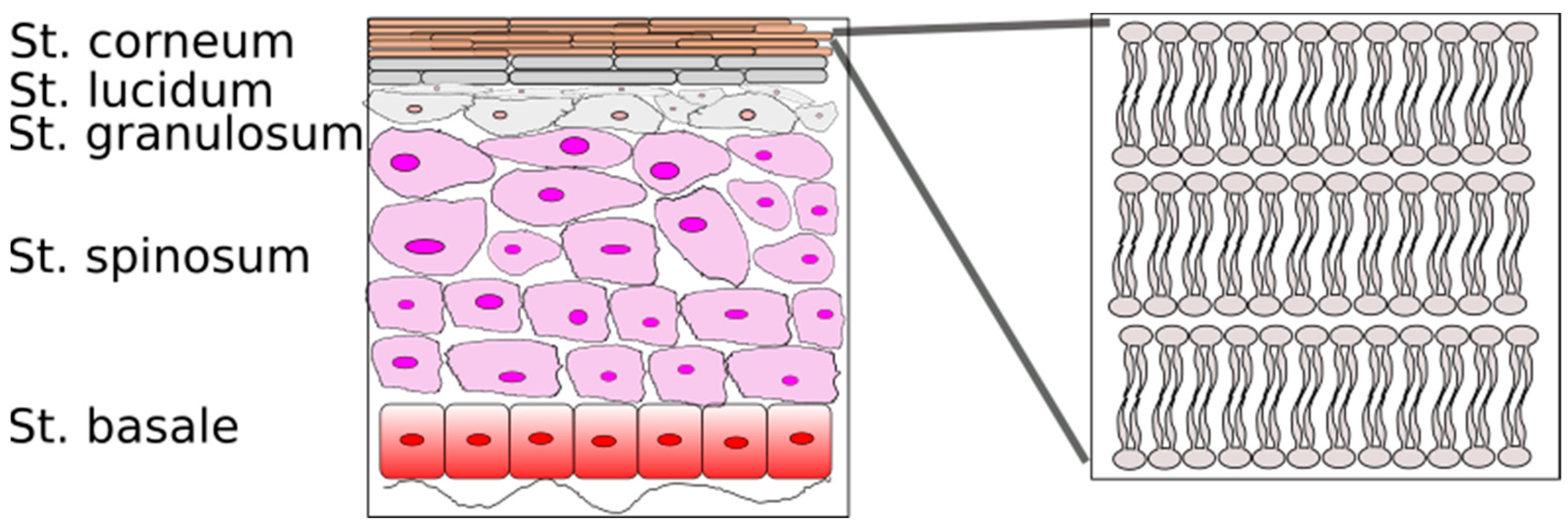
The skin consists of three main layers: The epidermis, the dermis, and the subcutaneous tissue. The epidermis, excluding the stratum corneum, which is its outside layer, is a viable tissue. The epidermis does not have vascularization, and nutrients diffuse from the dermo-epidermal junction to maintain its viability. There are five layers that describe the different steps of cell life in the epidermis (Figure 1) [15]. These sublayers are the following, beginning from the non-viable stratum corneum: Stratum lucidum (clear layer), stratum granulosum (granular layer), stratum spinosum (spinous or prickle layer), and stratum germinativum (basal layer). Providing mechanical protection, the cutaneous barrier protects the body from drying as well as from the penetration of dangerous substances and microorganisms. The stratum corneum acts as a critical part in the barrier function for topical drug penetration. Various models have been recommended for mimicking the SC. The most simplistic model is defined as a “brick and mortar” structure; the stratum corneum includes corneocytes (the “bricks”) and an intercellular lipid matrix (the “mortar”), which is essentially responsible for the barrier function [9]. The SC cells are named corneocytes. There are 15 to 20 layers of corneocytes with a thickness of 10 to 15 μm, but when hydrated, the stratum corneum considerably grows, and its thickness may reach up to 40 μm, showing extended permeability. These cells are dense, functionally dead and anucleated. The lipids form bilayers surrounding the corneocytes. The intercellular lipid consists of a mixture of fatty acids, ceramides, cholesterol, cholesterol esters and a small fraction of cholesterol sulfate [11,16,17]. Considering its barrier qualities and water resistance, the stratum corneum is the main layer that limits drug permeation through the skin [15].
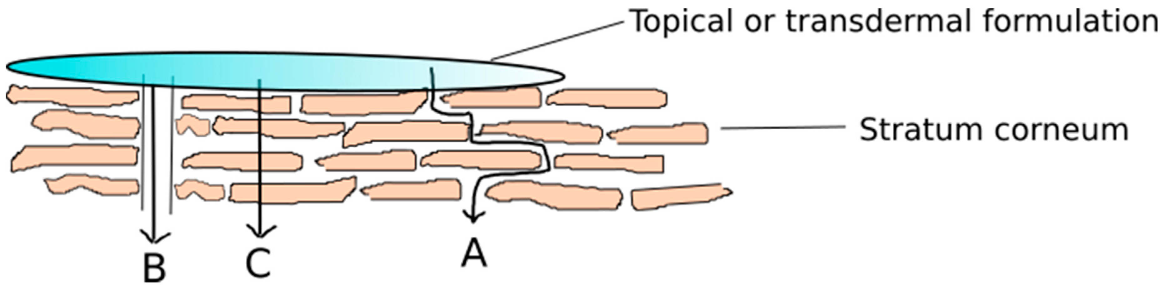
There are three different pathways allowing substances to pass through the cutaneous barrier [18]. There are three major routes of penetration for the passive diffusion of a molecule across the SC (Figure 2). The first pathway is the intercellular penetration pathway, which has come to be the most important one for many years. Approximately 15 years ago, the follicular penetration pathway was also found to be a second essential pathway. The transcellular penetration pathway, where the materials pass through both the corneocytes and the lipid layers, seems to be unimportant at present [16,19].
The difference between skin penetration and permeation is important. Buist and co-workers have defined them as follows [20]. Dermal penetration: The movement of a chemical from the outer surface of the skin into the epidermis, but not necessarily into the circulatory system. Dermal permeation: The penetration through one layer into another, which is both functionally and structurally different from the first layer.
1.2. Modifying Factors of Skin Penetration
The interactions between the drug, skin, and vehicle determine: (1) The drug release, (2) the penetration through the SC, (3) the penetration through the viable skin layers.
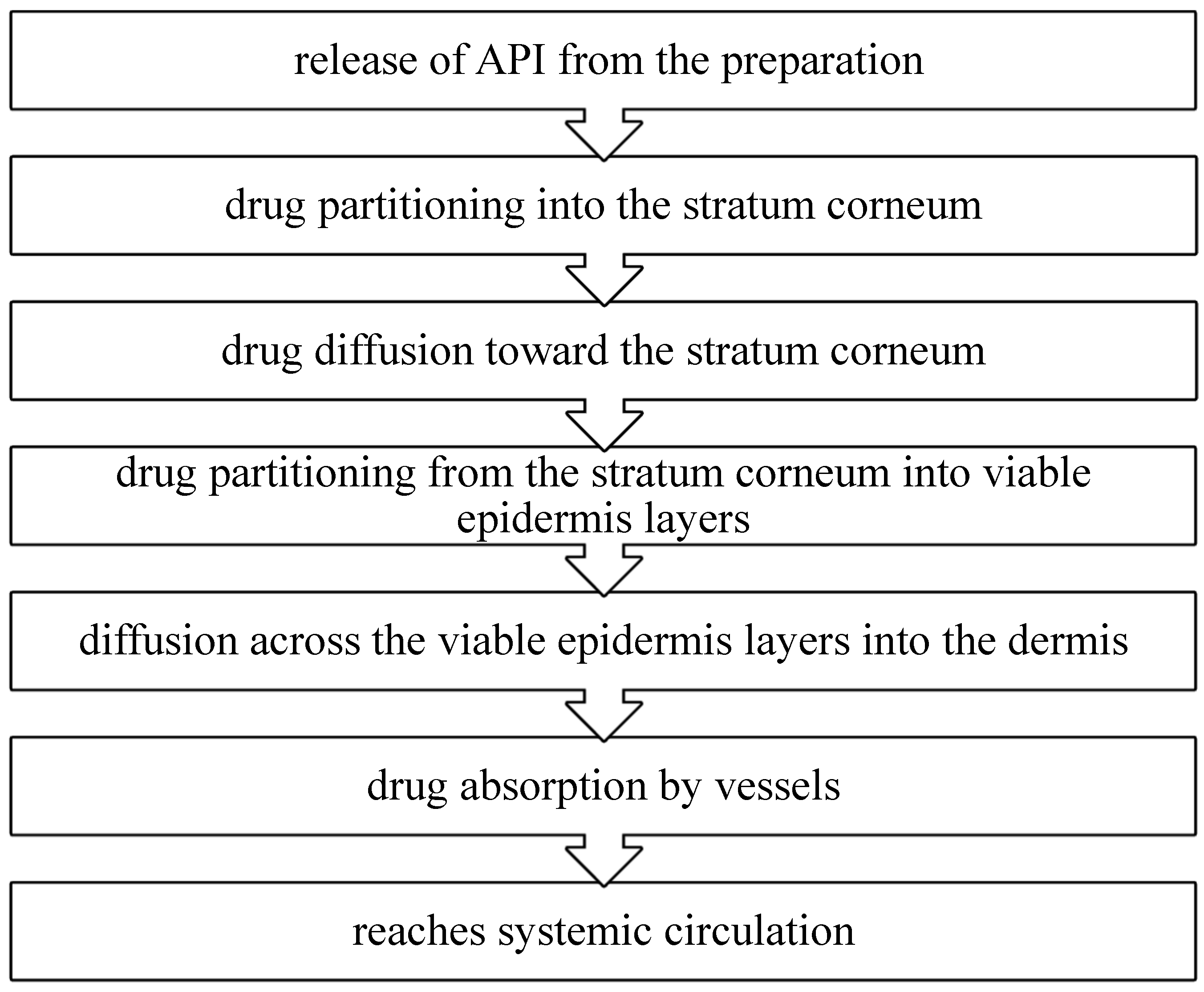
The liberation of an API from a pharmaceutical product applied to the skin and its transport to the systemic circulation is a multistep process which includes (a) the release of API from the preparation, (b) drug partitioning into the stratum corneum, (c) drug diffusion toward the stratum corneum, (d) drug partitioning from the stratum corneum into viable epidermis layers, (e) diffusion across the viable epidermis layers into the dermis, (f) drug absorption by vessels, (g) reaching systemic circulation (Figure 3).
The selection of APIs for dermal delivery must be based on many factors, including physicochemical characteristics, drug interactions with the membrane, and pharmacokinetic aspects. The ideal physicochemical characteristics of a drug chosen for cutaneous administration are low molecular weight (<600 Da); low melting point (<200 ℃), which is related to appropriate solubility; a high but stable partition coefficient because very high partition coefficients may increase drug retention, thus inhibiting drug clearance from the skin, and of course, solubility in water and oils to achieve a proper concentration gradient and increase the diffusion force over the skin [21,22,23,24].
Percutaneous penetration is affected by biological factors, too. There is basic variability in the skin barrier, due to factors such as skin hydration level, age, gender, site of the skin surface, malformations due to illness or damage and prior treatment [25,26,27,28,29]. To provide the skin with a healthy barrier function, the hydration level of the skin needs to be balanced, and enough volume of water is needed. If hydration increases, permeability may be improved [30]. Age has an impact on penetration into the skin. Baby skin and damaged skin have higher permeability.
The permeability of the drug through the stratum corneum is modulated by the carrier/vehicle, too. A vehicle can improve the physical state and permeability of the skin by the hydration effect or an alteration of the lipid bilayer structure.
1.3. General Guidelines of Skin Penetration Testing
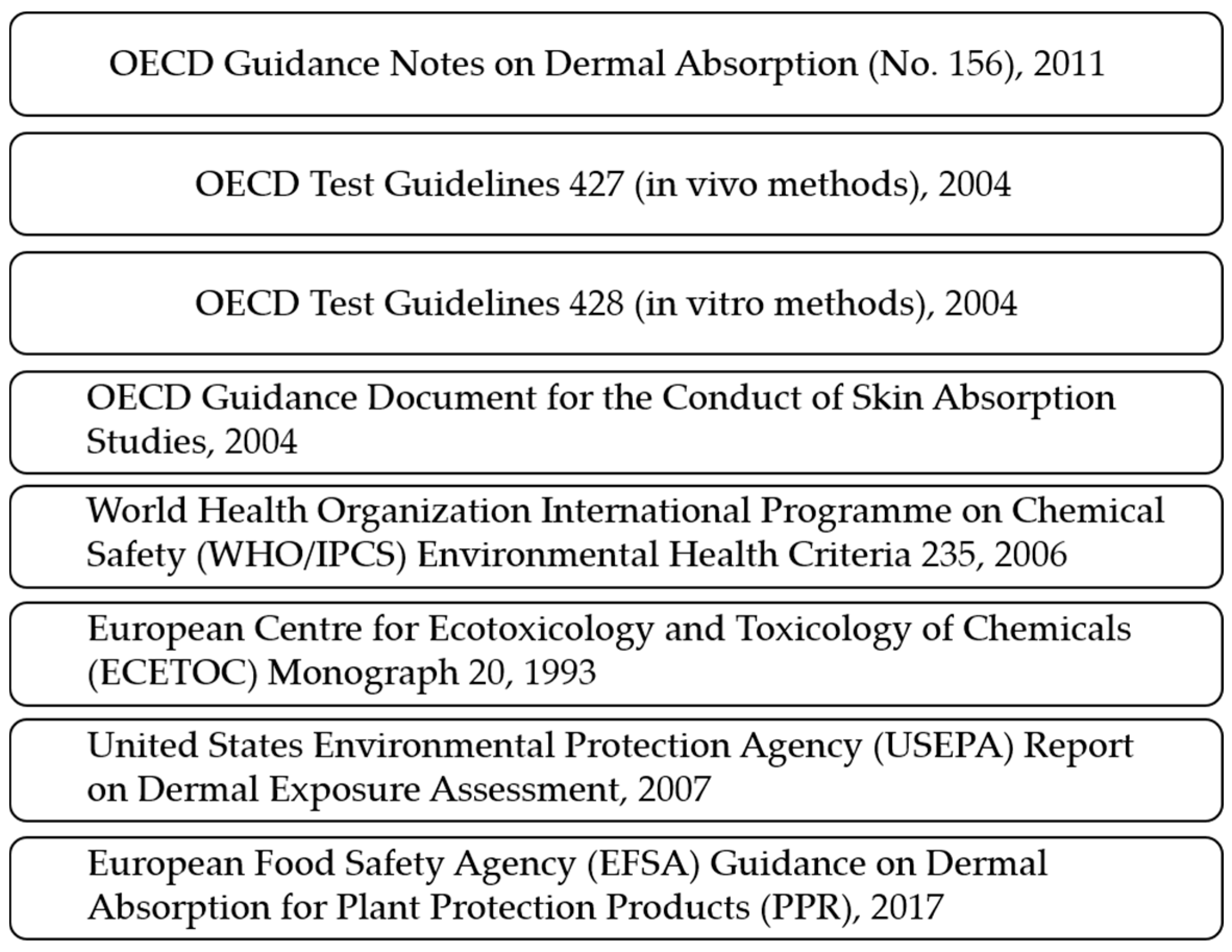
Recently, the regulation of dermal and transdermal preparations has received increasing attention. More and more documents are available for dermal absorption studies from Europe and the United States (Figure 4). These documents promote a harmonized road to conducting dermal and transdermal studies. The Organization for Economic Cooperation and Development (OECD) published several issues about this topic, including the Guidance Notes on Dermal Absorption (No. 156) [20], Test Guidelines 427 (in vivo methods) [31] and 428 (in vitro methods) [32], and the Guidance Document for the Conduct of Skin Absorption Studies [33]. There are some other documents such as the World Health Organization International Programme on Chemical Safety (WHO/IPCS) Environmental Health Criteria 235 [34], the European Centre for Ecotoxicology and Toxicology of Chemicals (ECETOC) Monograph 20 [35], the United States Environmental Protection Agency (USEPA) report on dermal exposure assessment [36], and the European Food Safety Agency (EFSA) Guidance on dermal absorption for plant protection products [20,37]. These documents present rules on and descriptions of how to perform dermal absorption assays, however, the measurements are not properly regulated. There are two suggested methods. One is the widely used diffusion cell and the tape stripping methods. However, in the scientific literature there are a lot of other types of measurements, such as spectroscopic and microscopic methods, to complete the suggested tests.
2. Techniques for Modelling Penetration/Permeation through Human Skin
There are two main types of techniques. Quantitative techniques include the use of diffusion cells and skin-PAMPA. Qualitative or semiquantitative techniques are different microscopic and spectroscopic methods and the combinations thereof. Figure 5 shows the quantitative and qualitative methods for following up skin penetration.
Quantitative in vitro tests are regularly performed to measure the amount of API permeated through a membrane over time in relation to the diffusion area related to the collected quantity of API in an acceptor chamber [11,38,39,40,41,42,43]. In the case of qualitative techniques, the aim is usually to follow the active substance. The presence or the relative amount of the active ingredient in the different skin layers can be determined. The use of multiple methods together can perfectly complement each other, making the regulatory authorization process easier.
2.1. Diffusion Cells
The skin can be considered as a selectively permeable biological membrane of drugs. Diffusion tests working with diffusion cells have become the “gold-standard” model due to the pioneering work of Dr. Thomas J. Franz, who developed the “Franz cell” in 1970. It determines important relationships between skin, API, and formulation [44,45,46]. The diffusion test models the method of medicine applied to the skin. This tool consists of a cell that holds a chamber for drug application, a membrane within which the drug may diffuse, and an acceptor media chamber from which samples may be investigated [47].
2.1.1. Types and Properties of Diffusion Cells
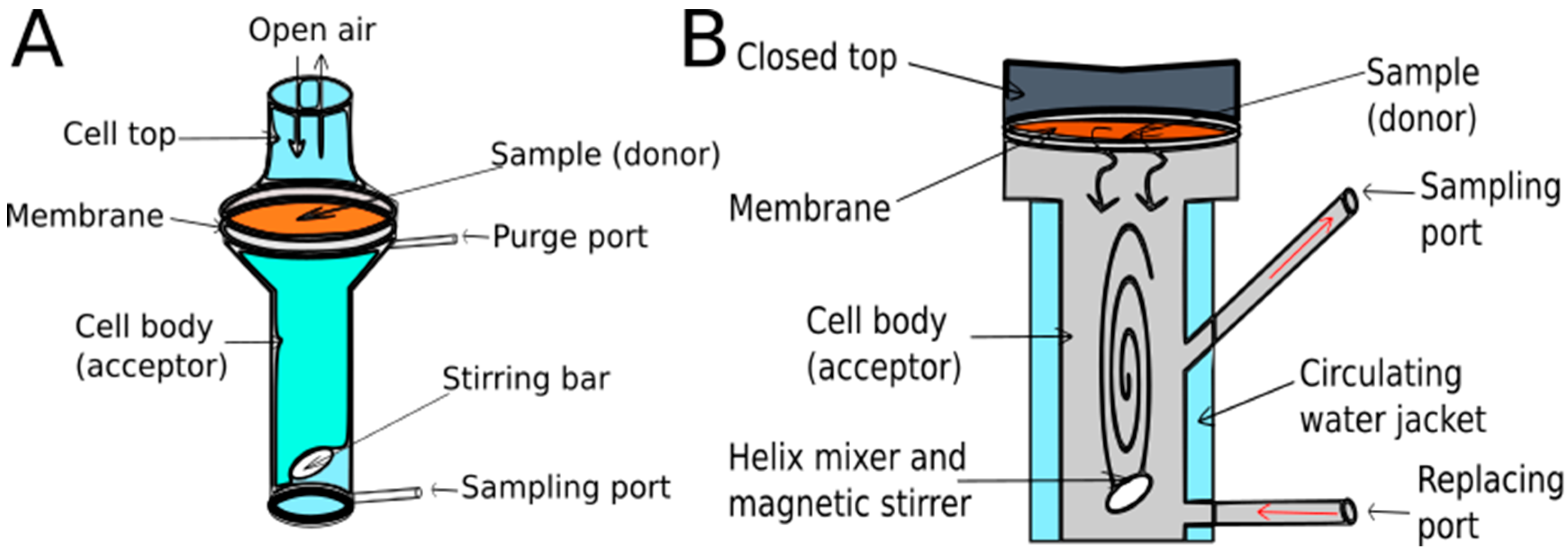
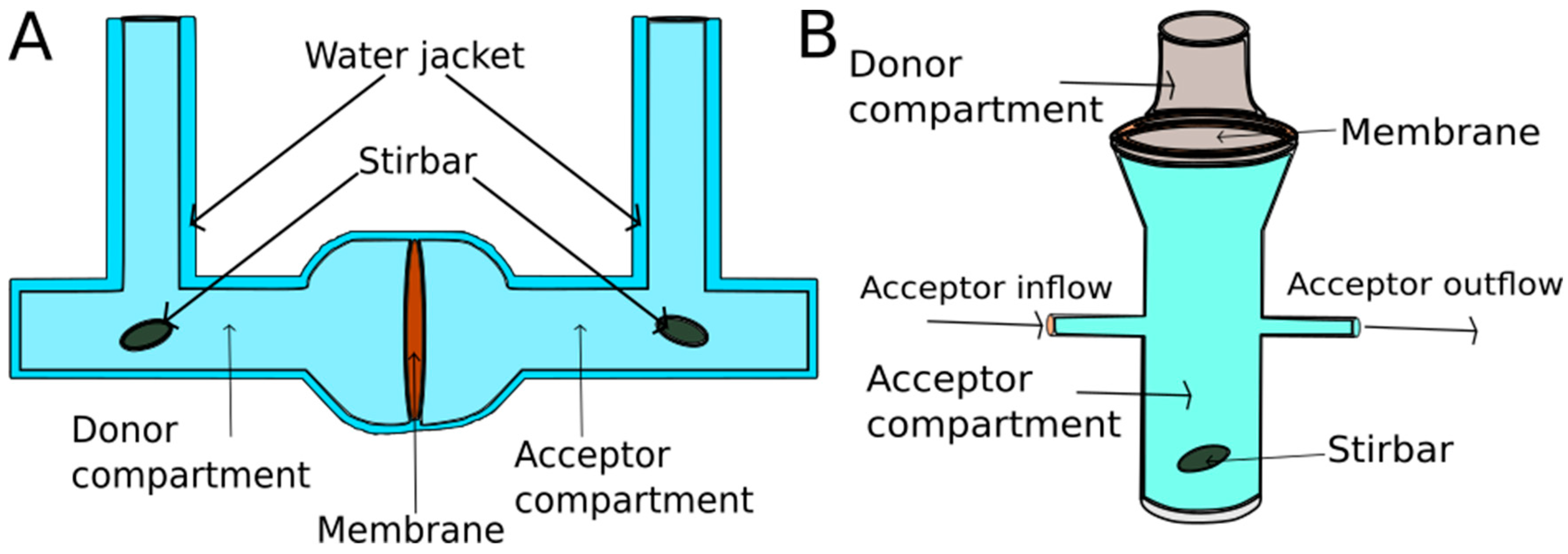
Diffusion cells can be categorized into two main classes, static and flow-through cells. The cells are normally made of glass. In static cells, the donor, the membrane, and the acceptor may be placed either vertically, as in the popular Franz diffusion cell (Figure 6) [44], or horizontally (Figure 7) [48]. There are Franz cells which open from above, therefore the measurement runs on atmospheric pressure. However, most of the cells are closed from the top, and because of this, the pressure is higher, predicting higher penetration values. Nowadays, the “hand-sampler” Franz diffusion cells have been replaced by systems connected to an automated sampler. Sampling systems facilitate the work of researchers and reduce error from human sampling.
2.1.2. Diffusion Test Types
In the EMA (2018) document (Draft Guideline on Quality and Equivalence of Topical Products) diffusion tests are grouped as follows. There are in vitro release tests (IVRT) and in vitro skin permeation studies (IVPT).
In the case of IVRT, synthetic membrane (lipid-based or non-lipid based model membranes) should be used. The sample dose must be occluded and infinite. The IVRT study shows the release rate. The detected quantities are in the µg to mg range. In the case of IVPT, human skin should be used in unoccluded circumstances. The sample dose must be finite. The IVPT study shows the flux profile and the detected quantities are in the pg to ng range. In the early stages of development, IVRT should be used, thereafter, IVPT can be used for promising formulations [49]. The main differences are summarized in Table 1.
Every year, several papers are published involving the use of a vertical Franz diffusion cell. In the last four months, “Franz Diffusion” as a keyword can be found in 13 articles. The specifications of the publications are summarized in Table 2. Many different APIs and membranes were used.
2.2. Skin-PAMPA
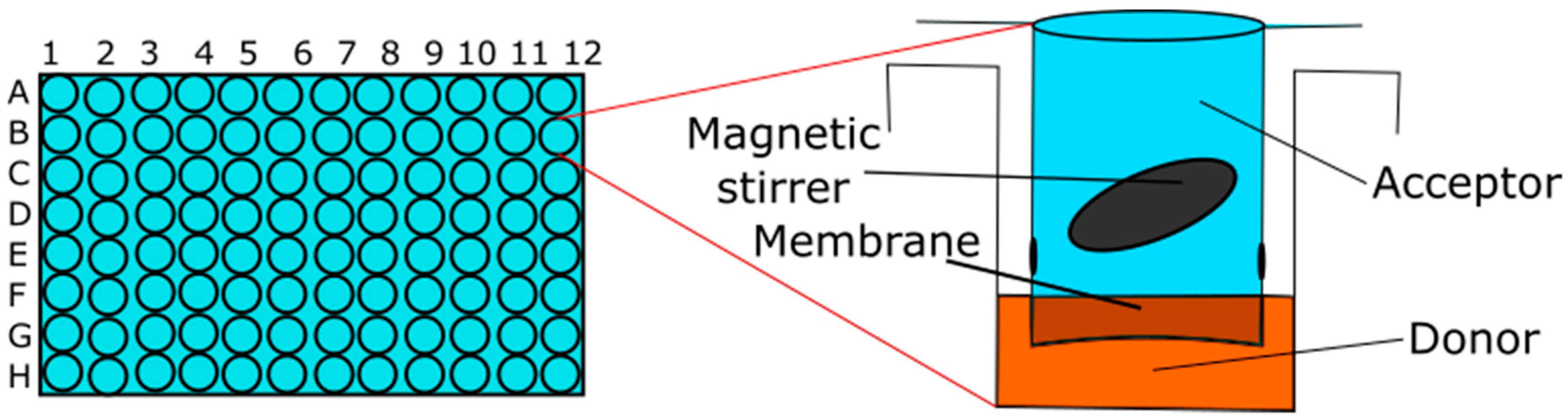
The Parallel Artificial Membrane Permeability Assay (PAMPA) is a 96-well plate-based method for the fast determination of the passive membrane permeability of molecules [62]. It is a promising method because of its low cost and high throughput. There are different PAMPA models and one for skin penetration. The earlier published skin model combines silicone oil and isopropyl myristate [63].
Sinkó et al. have developed the skin-PAMPA method for the prediction of skin penetration in vitro [64]. Skin-PAMPA has been created to imitate the characteristics of the stratum corneum [25]. Using this new plate, the donor phase contacts the hydrated artificial membrane (approximately 60–70 µL). This amount was ~180–200 μL using a conventional bottom plate. When examining the cross-sectional image of the donor cell of the new plate, the amount of material that is in direct contact with the membrane is about 30 μL. This results in a clear penetration surface (0.3 cm2), leading to 100 µL/cm2 of donor per unit area. Compared to the OECD recommendation, this is still one order of magnitude higher, but it is closer to the concept of finite dose [65]. Skin-PAMPA membrane was produced by using cholesterol, free fatty acid, and a ceramide-analogue compound that imitates the features of lipid matrix [66]. The donor plate of the conventional skin-PAMPA procedure requires the use of a large amount of sample, which does not meet the concept of finite dose. The experimental design is shown in Figure 8.
2.3. The Most Important Experimental Considerations in the Case of Quantitative Methods
In order to obtain reliable dermal permeability data, several parameters have to be considered for the design of the test system, which are influenced by the solubility of the compounds:
- the sink condition
- the incubation time
- the incubation temperature
- the mixing
- the hydration of the membrane
- the amount of dose [73].
In the case of molecules with low solubility, it is not so easy to create the sink condition. Ueda and colleagues have determined the sink condition: To achieve sink conditions, the receptor medium must have a relatively high capacity to dissolve or carry away the drug and the receptor media should not exceed 10% of drug solubility in the releasing matrix at the end of the test [74]. By changing the composition and the pH of the acceptor and donor phases, the suitable sink condition can be created. This means that under certain experimental conditions, the backflow from the acceptor phase can be considered as zero. Alternatively, the sink condition can be achieved by the use of surfactants in the acceptor phase. A further solution is the use of serum albumin because it is capable of binding lipophilic components, thereby keeping the free concentration of the molecule lower. The advantage of using albumin is that it is a sufficiently large molecule to not permeate through the skin.
In the case of the incubation time, it is important to consider the fact that the structure of the skin or the artificial membrane remains unchanged until the end of the experiment. To avoid the overestimation of dermal penetration, different skin integrity tests are needed to eliminate the use of damaged skin preparations. This test should ensure the exclusive use of skin-derived data with an appropriate barrier function in the assays. Several methods can be used to examine skin integrity. Each has advantages and disadvantages. Widely used methods are the measurement of tritium water permeability, the measurement of transepidermal water loss, and the determination of transepidermal electrical resistance. A relatively new method is the use of the tritium-labeled internal reference standard, where integrity testing is performed simultaneously with the measurement [60,61,62,63,64,66,67]. For each method, it can be stated that the measurements are different and not properly regulated. Different labs use different limits and measure under different conditions. In the future, the proper regulation of integrity testing and the clear definition of limit values for the proper evaluation of dermal formulations will be essential.
The incubation temperature affects both the permeability of the compounds and the rheological properties of the product studied. The selected mixing speed should guarantee enough mixing of the acceptor phase during the test [74]. Hydration of the skin affects, among other things, the barrier function of the stratum corneum, therefore, the creation of an optimal hydration state is essential [75].
Depending on the amount of sample in the test cells, two types of experiments can be distinguished: Finite and infinite dose measurements. In the case of the infinite dose, the amount of sample used is in large excess, hence the donor phase (the sample itself) cannot empty under normal circumstances. As a result, when examining a permeability-time profile in this case, the straight line usually rises with a constant slope without experiencing a plateau phase. The opposite of the infinite dose is the finite dose. When finite doses are used, a limited amount of sample (donor phase) is applied to the skin or to the surface of the artificial membrane. It should be noted that the latter experimental circumstance is much closer to the condition when the patient applies a given preparation onto his or her own skin. Based on the recommendation of the OECD (Organization for Economic Co-Operation and Development), a finite dose for a solution-phase sample is recommended if the applied dose does not exceed 10 μL/cm2 or is in the range of 1 to 10 mg/cm2 for a semisolid sample [75]. Certainly, it is more advantageous to design the measurement conditions to approach the finite dose requirements as much as possible [64].
There are some measurement parameters which affect drug penetration, such as occlusive or non-occlusive application [76,77]. Dermal and transdermal drug delivery can be performed after occlusive or non-occlusive (open) application. Occlusion can be achieved by covering the skin by impermeable films or others, such as strips, gloves, diapers, textiles garments, wound dressings or transdermal therapeutic systems [78].
2.4. Tape Stripping
The quantification of APIs within the skin is crucial for topical and transdermal delivery examinations. Nowadays, horizontal sectioning, consisting of tape completely stripping the stratum corneum, has become one of the conventional investigation procedures [79]. The method may be quantitative or semiquantitative depending on the analysis.
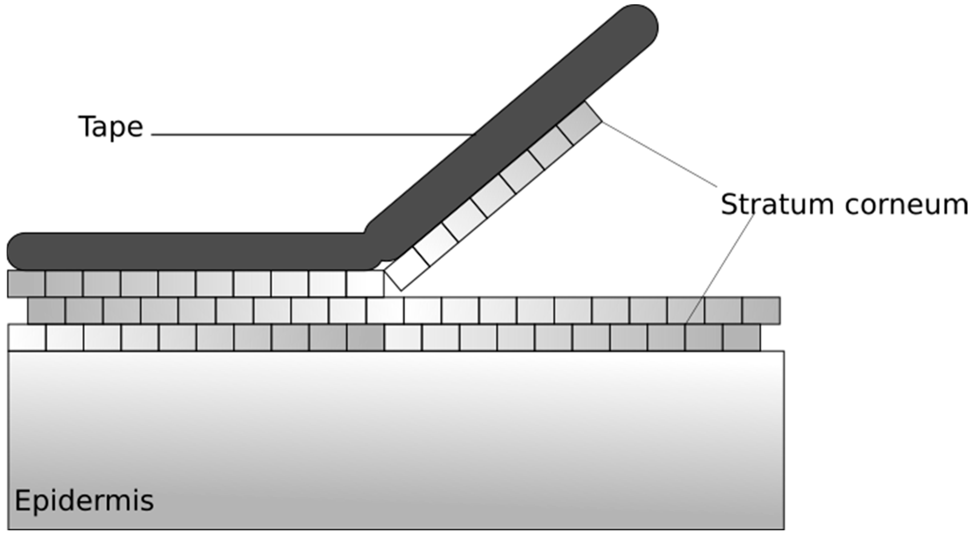
Tape stripping is a commonly used, minimally invasive method for testing the penetration of topically applied formulations through the stratum corneum, whereby layers of the stratum corneum are removed by an adhesive tape and skin layers residing on the adhesive tape are examined (Figure 9). On the one hand, the protein content of the different layers can be determined by the method, and on the other hand—after the topical application of the composition to be tested—the amount of the active ingredient in the selected layer can be determined. In addition to drug penetration testing, the effect of the substance on skin hydration and skin proteins can be observed, e.g., to modify the secondary structure of keratin. Tape stripping can also be used in vitro and in vivo on human and animal skin, as well as on appropriate skin models [15,25,80]. The Organization for Economic Co-operation and Development Test Guideline (OECD TG) 427 describes the use of tape stripping for the removal of upper skin layers and the in vivo penetration of active substances [31], while the OECD TG 428 has been performed with the in vitro application of the method for experiments [32].
2.4.1. Method Description
The tape stripping process begins after the topical application of the test composition and waiting for an appropriate incubation time. The composition can be removed or left on the skin in order to provide the original amount of the composition used during the measurement. This is of particular importance, for example, when examining sunscreen preparations, where the active substance on the skin surface will exert an effect, so we need to analyze it. The adhesive tape is placed on the skin surface and removed, always from the same selected area. It is important that the adhesive tape is always flattened with the same force as the roller, which eliminates the influence of wrinkles and recesses on tape stripping. In addition, the speed of removal is also an important factor: The slower the removal of the adhesive tape, the greater the adhesion of the stratum corneum to the patch, thus increasing the amount of skin removed from the patch. Removed adhesive tapes will contain both the layers of the SC and the active ingredient of the composition used [25,81,82].
2.4.2. Possibility of Sample Analysis
Several methods can be used to test the sample on the adhesive tape. The HPLC analysis generates a quantitative result, while the spectroscopic method yields semiquantitative results. During the HPLC (High-Pressure Liquid Chromatography) analysis, the test material on the adhesive tape is extracted by a suitable method and chromatographed. In addition, atomic absorption spectrometry may also be used to detect certain active substances, e.g., titanium dioxide. Furthermore, the active ingredients containing chromophore may also be useful for testing vitamin E [83]. However, the most widespread method is Attenuated Total Reflectance Fourier transform infrared spectroscopy (ATR-FTIR) [83]. Spectroscopy measurements are based on the irradiation of the sample, vibrations, and bond angle between the atoms in it, due to absorption or scattering of the infrared radiation. The changes in the radiation passing through the sample are measured by plotting the wavelength/wavenumber to obtain the spectrum that can be analyzed by qualitative and quantitative information. The penetration depth is thus determined by the wavelength of the infrared light, the refractive index of the ATR crystal and the measured material, and the angle of reflection [15,25]. Tape stripping combined with ATR-FTIR spectroscopy may be suitable for detecting various exogenous substances in certain layers of the SC. However, the difficulty of the method is that the characteristic peaks of the substances to be detected are often present and overlap with skin-specific peaks.
2.4.3. Possibilities for Optimizing the Experimental Protocol
The following options may be suitable for standardizing the experimental protocol. The composition should preferably be applied with a latex glove in a defined amount of time. Before applying, it is advisable to cut one finger of the rubber gloves and dip them into the preparation to fill the holes on the latex. Then the glove should be cleaned. This minimizes the amount of drug lost in the rubber glove, thus allowing the same quantities to be applied [84]. It is advisable that parallel measurements should always be carried out by the same person, as this may exclude the potential impact of different application techniques. The intact barrier function of the skin should be checked before starting the experiments. A TEWL test can be used to measure skin integrity. The intact barrier function, suitable for starting tape stripping experiments, indicates a TEWL value of 15–20 g m−2 h−1 [80]. It is recommended that the adhesive tape be smoothed onto the skin using a roller to eliminate the effects of wrinkles and recesses on the skin [82].
2.5. Microscopic and Spectroscopic Methods Utilised for the Percutaneous Penetration of APIs
Microscopic techniques also give important information about the spatial distribution of the drug inside the different skin layers or explain the mechanism of penetration. Furthermore, these are not destructive in vitro techniques [85,86,87,88]. Fluorescence microscopy is a frequently used method, as well. The evaluation of skin treated with fluorescently labeled materials by fluorescence microscopy has shown that the fluorescent labeled material resided in the SC or penetrated deeper in the epidermis [89,90].
Confocal microscopy is a specialized form of standard fluorescence microscopy. It has developed over the last five to six decades and now it is a necessary tool for scientists. The major confocal techniques are two-photon microscopy (2-PFM), confocal laser scanning microscopy (CLSM), and the youngest, Raman microscopic methods. These methods have the possibility to image deeper into a three-dimensional biological sample like skin at high resolution, high speed, in in vitro and in vivo circumstances [15,25,91,92].
2.5.1. Two-Photon Microscopy Method
Two-photon scanning fluorescence microscopy has become an important tool for imaging skin cells. It employs a Ti-sapphire laser excitation source. In single-photon fluorescence, the fluorescence photon is created when a high-energy photon is incident on the fluorophore and increases the energy level of one of the electrons to an excited state. In two-photon excitation, the combined energy shift of the two low-energy photons is enough to raise the same electron to a higher energy level. The setup of a two-photon microscope is quite similar to that of a confocal scanning microscope with two major differences. Two-photon microscopes work with a tunable Ti-sapphire high-frequency pulsed laser. These lasers emit red and near-infrared light in the range from 650 to 1100 nanometers. The other big difference is that there are no pinholes in front of the detector [93].
The most relevant advantages of 2-PFM are that the total energy transferred to the specimen is much lower as compared to other techniques. Furthermore, the two-photon excitation phenomenon occurs only at a very small focal volume and thanks to the fluorophores, a small volume of the specimen is excited, which decreases the possibility of photo-bleaching and photo-damage. The skin samples can be studied without cryofixation and cutting. In the case of imaging of UV absorbing fluorophores, deep tissue imaging is possible with infrared excitation because of less scattering and less absorption. The limitations of a two-photon microscope include the relatively high price of the lasers and the fact that they require a complex cooling system. Additionally, it has a lower lateral resolution compared to other techniques, but in practice, the difference in their resolution is not significant [25,94].
2.5.2. Confocal Laser Scanning Microscopy Method
Confocal laser scanning microscopy (CLSM) is a non-invasive method developed from fluorescence microscopy. In the last years, CLSM has been widely accepted as a device to visualize the fluorescent model compounds in the skin. CLSM can be used to examine the skin structure without cutting tissue as well as to evaluate the influences of physical and chemical enhancers on skin permeability. It has been used in vivo and in vitro, too. CLSM is used to diagnose general skin dysfunctions and to identify malignant lesions, and it can characterize keratinization and pigmentation disorders as well [100,101,102].
CLSM can be applied to explain the mechanism by which nanoparticulate formulations promote skin transport. Fluorescent markers (e.g., fluorescein, Nile red, 5-bromodeoxyuridine) may be incorporated in nanostructured formulations, in which they are encapsulated. The therapeutic effect of these formulations can be examined by CLSM to identify the penetration profiles of these fluorescent markers across the skin or skin appendages [15,25]. Table 5 summarizes the most important recent results.
2.5.3. Confocal Raman Microscopic Method
Spectroscopic methods can produce molecular information about the structure of the skin. Raman spectroscopy is a promising spectroscopic method based on discovering the characteristic vibrational energy levels of a molecule excited by a laser ray and it gives information about the molecular structure of tissue components without the use of fluorescent labels or chemical stains [110,111,112,113]. Therefore, this method is promising for discovering modifications in the structure of skin components. In addition, it is good for following up the penetration/permeation of APIs [114,115]. Nowadays, Raman microscopy is a powerful technique to properly understand skin structure and percutaneous drug delivery [111,116,117,118].
Traditionally, the tape stripping method is used to model the penetration into the stratum corneum. This method is time-consuming, semi-destructive, and difficult to reproduce, and only the stratum corneum can be examined. Important technical developments are custom setting and the confocal microscope combination. This enables chemically selective and non-destructive sample analysis with high spatial resolution in three dimensions and allows multiple components to be monitored at the same time. Confocal Raman microscopy can be used to investigate topical formulations for both penetration and penetration depth in vitro and in vivo [119,120].
Permeation through the skin can be explored by chemical mapping (Figure 10). Chemical mapping is one of the methods of vibration spectrometry; the suitable test apparatus is a vibration spectrometer (e.g., Raman) and a suitable optical unit (e.g., microscope). Raman Spectrometry is highly selective, with a fingerprint spectrum that allows clear molecular identification. The irradiating light can be focused on a very small point (~1 µm2) with a suitable microscope lens, so reliable chemical information can also be obtained from a microscopic area of the sample. The spatial distribution of the fundamental components of the sample can be determined by the spectra that make up the map. Chemical maps contain a huge quantity of data. Although these can be controlled by methods commonly used in spectroscopy, the huge amount of data can be utilized more efficiently by using appropriate mathematical (multivariate data analysis) techniques. Thanks to these advantages, the technology has enormous potential in various applications: From the analysis of the distribution of the physiological components of the skin and tissues through the diagnosis of pathological conditions to biopharmaceutical studies, such as drug penetration kinetics [15,25,120,121]. Table 6 summarizes the most important recent results.
3. Conclusions
The modelling of penetration into a skin layer and permeation through the skin is a complex challenge. Although there are many quantitative and qualitative methods for following up skin penetration/permeation (in vitro and in vivo), the different techniques are not fully equivalent but complement each other. The advantages and disadvantages of the different methods are summarized in Table 7. For the evaluation of drug penetration, pharmaceutical technologists must decide those properly suited to their examinations. The models give a possibility for rapid screening and faster optimization of products. The selection of the most suitable in vitro model should be based on availability, facility of use, cost, and the respective limitations [30]. The success of topical and transdermal therapy is correlated to the techniques used for the evaluation of the preparations, which facilitate the optimization of the skin penetration of the API so that it can reach sufficient API penetration at the therapeutic site.
Funding
This research was funded by GINOP, grant number 2.2.1-15-2016-00023.
Conflicts of Interest
The authors declare no conflict of interest.
References
- Transdermal Drug Delivery Systems Market-Industry Analysis, Market Size, Share, Trends, Application Analysis, Growth and Forecast 2019–2024. Available online: https://industryarc.com/Research/Transdermal-Drug-Delivery-Systems-Market-Research (accessed on 1 April 2019).
- Shah, V.P.; Yacobi, A.; Rădulescu, F.Ş.; Miron, D.S.; Lane, M.E. A science based approach to topical drug classification system (TCS). Int. J. Pharm. 2015, 491, 21–25. [Google Scholar] [CrossRef] [PubMed]
- Wiedersberg, S.; Guy, R.H. Transdermal drug delivery: 30+ years of war and still fighting! J. Control. Release 2014, 190, 150–156. [Google Scholar] [CrossRef] [PubMed]
- So, J.; Ahn, J.; Lee, T.-H.; Park, K.-H.; Paik, M.-K.; Jeong, M.; Cho, M.-H.; Jeong, S.-H. Comparison of International Guidelines of Dermal Absorption Tests Used in Pesticides Exposure Assessment for Operators. Toxicol. Res. 2014, 30, 251–260. [Google Scholar] [CrossRef] [PubMed]
- Groeber, F.; Holeiter, M.; Hampel, M.; Hinderer, S.; Schenke-Layland, K. Skin tissue engineering—in vivo and in vitro applications. Adv. Drug Deliv. Rev. 2011, 63, 352–366. [Google Scholar] [CrossRef] [PubMed]
- Feldmann, R.J.; Maibach, H.I. Absorption of Some Organic Compounds through the Skin in Man. J. Investig. Derm. 1970, 54, 399–404. [Google Scholar] [CrossRef] [PubMed]
- Blank, I.H. Factors Which Influence the Water Content of the Stratum Corneum. J. Investig. Derm. 1952, 18, 433–440. [Google Scholar] [CrossRef] [PubMed] [Green Version]
- Vinson, L.J.; Singer, E.J.; Koehler, W.R.; Lehman, M.D.; Masurat, T. The nature of the epidermal barrier and some factors influencing skin permeability. Toxicol. Appl. Pharm. 1965, 7, 7–19. [Google Scholar] [CrossRef]
- Elias, P.M. The permeability barrier in mammalian epidermis. J. Cell Biol. 1975, 65, 180–191. [Google Scholar] [CrossRef] [PubMed]
- Sweeney, T.M.; Downing, D.T. The Role of Lipids in the Epidermal Barrier to Water Diffusion. J. Investig. Derm. 1970, 55, 135–140. [Google Scholar] [CrossRef]
- Anissimov, Y.G.; Jepps, O.G.; Dancik, Y.; Roberts, M.S. Mathematical and pharmacokinetic modelling of epidermal and dermal transport processes. Adv. Drug Deliv. Rev. 2013, 65, 169–190. [Google Scholar] [CrossRef]
- Bo Forslind. A domain mosaic model of the skin barrier. Acta Derm. Venereol. 1994, 74, 1–6. [Google Scholar]
- Godin, B.; Touitou, E. Transdermal skin delivery: Predictions for humans from in vivo, ex vivo and animal models. Adv. Drug Deliv. Rev. 2007, 59, 1152–1161. [Google Scholar] [CrossRef] [PubMed]
- Hadgraft, J. Skin deep. Eur. J. Pharm. Biopharm. 2004, 58, 291–299. [Google Scholar] [CrossRef] [PubMed]
- Ruela, A.L.M.; Perissinato, A.G.; de Lino, M.E.S.; Mudrik, P.S.; Pereira, G.R. Evaluation of skin absorption of drugs from topical and transdermal formulations. Braz. J. Pharm. Sci. 2016, 52, 527–544. [Google Scholar] [CrossRef] [Green Version]
- Jepps, O.G.; Dancik, Y.; Anissimov, Y.G.; Roberts, M.S. Modeling the human skin barrier—Towards a better understanding of dermal absorption. Adv. Drug Deliv. Rev. 2013, 65, 152–168. [Google Scholar] [CrossRef] [PubMed]
- Barry, B.W. Novel mechanisms and devices to enable successful transdermal drug delivery. Eur. J. Pharm. Sci. 2001, 14, 101–114. [Google Scholar] [CrossRef]
- Blume-Peytavi, U.; Massoudy, L.; Patzelt, A.; Lademann, J.; Dietz, E.; Rasulev, U.; Garcia Bartels, N. Follicular and percutaneous penetration pathways of topically applied minoxidil foam. Eur. J. Pharm. Biopharm. 2010, 76, 450–453. [Google Scholar] [CrossRef]
- Patzelt, A.; Richter, H.; Knorr, F.; Schäfer, U.; Lehr, C.-M.; Dähne, L.; Sterry, W.; Lademann, J. Selective follicular targeting by modification of the particle sizes. J. Control. Release 2011, 150, 45–48. [Google Scholar] [CrossRef]
- European Food Safety Authority (EFSA); Buist, H.; Craig, P.; Dewhurst, I.; Hougaard Bennekou, S.; Kneuer, C.; Machera, K.; Pieper, C.; Court Marques, D.; Guillot, G.; et al. Guidance on dermal absorption. EFSA J. 2017, 15, e04873. [Google Scholar] [Green Version]
- Kalia, Y.N.; Guy, R.H. Modeling transdermal drug release. Adv. Drug Deliv. Rev. 2001, 48, 159–172. [Google Scholar] [CrossRef]
- Machado, A.C.H.R.; Lopes, P.S.; Raffier, C.P.; Haridass, I.N.; Roberts, M.; Grice, J.; Leite-Silva, V.R. Skin Penetration. In Cosmetic Science and Technology; Elsevier: Amsterdam, The Netherlands, 2017; pp. 741–755. ISBN 978-0-12-802005-0. [Google Scholar]
- Moser, K.; Kriwet, K.; Naik, A.; Kalia, Y.N.; Guy, R.H. Passive skin penetration enhancement and its quantification in vitro. Eur. J. Pharm. Biopharm. 2001, 10, 103–112. [Google Scholar] [CrossRef]
- Delgado-Charro, M.B.; Guy, R.H. Effective use of transdermal drug delivery in children. Adv. Drug Deliv. Rev. 2014, 73, 63–82. [Google Scholar] [CrossRef] [PubMed] [Green Version]
- Dragicevic, N.; Maibach, H.I. Percutaneous Penetration Enhancers Drug Penetration Into/Through the Skin; Springer: Berlin/Heidelberg, Germany, 2017; ISBN 978-3-662-53268-3. [Google Scholar]
- Machado, M.; Hadgraft, J.; Lane, M.E. Assessment of the variation of skin barrier function with anatomic site, age, gender and ethnicity: Assessment of the variation of skin barrier function. Int. J. Cosmet. Sci. 2010, 32, 397–409. [Google Scholar] [CrossRef] [PubMed]
- Waller, J.M.; Maibach, H.I. Age and skin structure and function, a quantitative approach (I): Blood flow, pH, thickness, and ultrasound echogenicity. Skin Res. Technol. 2005, 11, 221–235. [Google Scholar] [CrossRef] [PubMed]
- Behl, C.R.; Flynn, G.L.; Barrett, M.; Walters, K.A.; Linn, E.E.; Mohamed, Z.; Kurihara, T.; Ho, N.F.H.; Higuchi, W.I.; Pierson, C.L. Permeability of thermally damaged skin II: Immediate influences of branding at 60 °C on hairless mouse skin permeability. Burns 1981, 7, 389–399. [Google Scholar] [CrossRef]
- Behl, C.R.; Flynn, G.L.; Kurihara, T.; Smith, W.; Gatmaitan, O.; Higuchi, W.I.; Ho, N.F.H.; Pierson, C.L. Permeability of Thermally Damaged Skin: I. Immediate Influences of 60 °C Scalding on Hairless Mouse Skin. J. Investig. Derm. 1980, 75, 340–345. [Google Scholar] [CrossRef] [PubMed]
- Flaten, G.E.; Palac, Z.; Engesland, A.; Filipović-Grčić, J.; Vanić, Ž.; Škalko-Basnet, N. In vitro skin models as a tool in optimization of drug formulation. Eur. J. Pharm. Sci. 2015, 75, 10–24. [Google Scholar] [CrossRef] [PubMed] [Green Version]
- OECD. Test Guideline 427: Skin absorption: In Vivo Method; OECD: Paris, France, 2004. [Google Scholar]
- OECD. Test Guideline 428: Skin absorption: In Vitro Method; OECD: Paris, France, 2004. [Google Scholar]
- OECD. Guidance Document for the Conduct of Skin Absorption Studies; OECD Series on Testing and Assessment; OECD: Paris, France, 2004; ISBN 978-92-64-07879-6. [Google Scholar]
- Kielhorn, J. International Programme on Chemical Safety Dermal Absorption; Environmental Health Criteria; WHO: Geneva, Switzerland, 2006; ISBN 978-92-4-157235-4. [Google Scholar]
- European Centre for Ecotoxicology and Toxicology of Chemicals. Percutaneous absorption, Monograph; No. 20; European Centre for Ecotoxicology and Toxicology of Chemicals: Bruxelles, Belgium, 1993; ISSN 0773-6347-20. [Google Scholar]
- EPAU.S. Environmental Protection Agency (EPA). Dermal Exposure Assessment: A Summary of EPA Approaches. Available online: http://www.epa.gov/ncea (accessed on 1 April 2019).
- Dumont, C.; Prieto, P.; Asturiol, D.; Worth, A. Review of the Availability of In Vitro and In Silico Methods for Assessing Dermal Bioavailability. Appl. Vitro. Toxicol. 2015, 1, 147–164. [Google Scholar] [CrossRef]
- Anissimov, Y.G.; Roberts, M.S. Diffusion Modelling of Percutaneous Absorption Kinetics: 4. Effects of a Slow Equilibration Process Within Stratum Corneum on Absorption and Desorption Kinetics. J. Pharm. Sci. 2009, 98, 772–781. [Google Scholar] [CrossRef] [PubMed] [Green Version]
- Anissimov, Y.G.; Roberts, M.S. Diffusion Modeling of Percutaneous Absorption Kinetics: 3. Variable Diffusion and Partition Coefficients, Consequences for Stratum Corneum Depth Profiles and Desorption Kinetics. J. Pharm. Sci. 2004, 93, 470–487. [Google Scholar] [CrossRef] [PubMed]
- Anissimov, Y.G.; Roberts, M.S. Diffusion modeling of percutaneous absorption kinetics: 2. Finite vehicle volume and solvent deposited solids. J. Pharm. Sci. 2001, 90, 504–520. [Google Scholar] [CrossRef]
- Anissimov, Y.G.; Roberts, M.S. Diffusion modeling of percutaneous absorption kinetics. 1. Effects of flow rate, receptor sampling rate, and viable epidermal resistance for a constant donor concentration. J. Pharm. Sci. 1999, 88, 1201–1209. [Google Scholar] [CrossRef] [PubMed]
- Todo, H.; Oshizaka, T.; Kadhum, W.; Sugibayashi, K. Mathematical Model to Predict Skin Concentration after Topical Application of Drugs. Pharmaceutics 2013, 5, 634–651. [Google Scholar] [CrossRef] [PubMed] [Green Version]
- Crank, J. The Mathematics of Diffusion, 2nd ed.; Clarendon Press: Oxford, UK, 1975; ISBN 978-0-19-853344-3. [Google Scholar]
- Franz, T.J. Percutaneous Absorption. On the Relevance of in Vitro Data. J. Investig. Derm. 1975, 64, 190–195. [Google Scholar] [CrossRef] [PubMed] [Green Version]
- Franz, T.J. The Finite Dose Technique as a Valid in vitro Model for the Study of Percutaneous Absorption in Man. In Current Problems in Dermatology; Simon, G.A., Paster, Z., Klingberg, M.A., Kaye, M., Eds.; S. Karger AG: Basel, Switzerland, 1979; Volume 7, pp. 58–68. ISBN 978-3-8055-2797-2. [Google Scholar]
- Sesto Cabral, M.E.; Ramos, A.N.; Cabrera, C.A.; Valdez, J.C.; González, S.N. Equipment and method for in vitro release measurements on topical dosage forms. Pharm. Dev. Technol. 2015, 20, 619–625. [Google Scholar] [CrossRef] [PubMed]
- Diffusion Measurements. Available online: www.hansonresearch.com (accessed on 1 April 2019).
- Bronaugh, R.L.; Stewart, R.F. Methods for In Vitro Percutaneous Absorption Studies IV: The Flow-Through Diffusion Cell. J. Pharm. Sci. 1985, 74, 64–67. [Google Scholar] [CrossRef] [PubMed]
- European Medicines Agency. FDA Draft Guideline on Quality and Equivalence of Topical Products EMA/CHMP/QWP/708282/2018; European Medicines Agency: Amsterdam, The Netherlands, 2018.
- Lee, J.D.; Kim, J.Y.; Jang, H.J.; Lee, B.M.; Kim, K.B. Percutaneous permeability of 1-phenoxy-2-propanol, a preservative in cosmetics. Regul. Toxicol. Pharm. 2019, 103, 56–62. [Google Scholar] [CrossRef]
- Zhang, Y.; Lane, M.E.; Hadgraft, J.; Heinrich, M.; Chen, T.; Lian, G.; Sinko, B. A comparison of the in vitro permeation of niacinamide in mammalian skin and in the Parallel Artificial Membrane Permeation Assay (PAMPA) model. Int. J. Pharm. 2019, 556, 142–149. [Google Scholar] [CrossRef]
- Trombino, S.; Russo, R.; Mellace, S.; Varano, G.P.; Laganà, A.S.; Marcucci, F.; Cassano, R. Solid lipid nanoparticles made of trehalose monooleate for cyclosporin-A topic release. J. Drug Deliv. Sci. Technol. 2019, 49, 563–569. [Google Scholar] [CrossRef]
- Taofiq, O.; Rodrigues, F.; Barros, L.; Barreiro, M.F.; Ferreira, I.C.; Oliveira, M.B. Mushroom ethanolic extracts as cosmeceuticals ingredients: Safety and ex vivo skin permeation studies. Food Chem. Toxicol. 2019, 127, 228–236. [Google Scholar] [CrossRef] [Green Version]
- Ameen, D.; Michniak-Kohn, B. Development and in vitro evaluation of pressure sensitive adhesive patch for the transdermal delivery of galantamine: Effect of penetration enhancers and crystallization inhibition. Eur. J. Pharm. Biopharm. 2019, 139, 262–271. [Google Scholar] [CrossRef] [PubMed]
- Silva-Abreu, M.; Gonzalez-Pizarro, R.; Espinoza, L.C.; Rodríguez-Lagunas, M.J.; Espina, M.; García, M.L.; Calpena, A.C. Thiazolidinedione as an alternative to facilitate oral administration in geriatric patients with Alzheimer’s disease. Eur. J. Pharm. Sci. 2019, 129, 173–180. [Google Scholar] [CrossRef] [PubMed]
- Intarakumhaeng, R.; Alsheddi, L.; Wanasathop, A.; Shi, Z.; Li, S.K. Skin Permeation of Urea Under Finite Dose Condition. J. Pharm. Sci. 2019, 108, 987–995. [Google Scholar] [CrossRef] [PubMed]
- Rajithaa, P.; Shammika, P.; Aiswarya, S.; Gopikrishnan, A.; Jayakumar, R.; Sabitha, M. Chaulmoogra oil based methotrexate loaded topical nanoemulsion for the treatment of psoriasis. J. Drug Deliv. Sci. Technol. 2019, 49, 463–476. [Google Scholar] [CrossRef]
- Salehi, S.; Boddohi, S. Design and optimization of kollicoat ® IR based mucoadhesive buccal film for co-delivery of rizatriptan benzoate and propranolol hydrochloride. Mater. Sci. Eng. C 2019, 97, 230–244. [Google Scholar] [CrossRef] [PubMed]
- Soriano-Ruiz, J.L.; Suñer-Carbó, J.; Calpena-Campmany, A.C.; Bozal-de Febrer, N.; Halbaut-Bellowa, L.; Boix-Montañés, A.; Souto, E.B.; Clares-Naveros, B. Clotrimazole multiple W/O/W emulsion as anticandidal agent: Characterization and evaluation on skin and mucosae. Coll. Surf. B Biointerfaces 2019, 175, 166–174. [Google Scholar] [CrossRef] [PubMed]
- Serpe, L.; Muniz, B.V. Full-Thickness Intraoral Mucosa Barrier Models for In Vitro Drug- Permeation Studies Using Microneedles. J. Pharm. Sci. 2019, 108, 1756–1764. [Google Scholar] [CrossRef]
- Lee, W.-R.; Hsiao, C.-Y.; Huang, T.-H.; Wang, C.-L.; Alalaiwe, A.; Chen, E.-L.; Fang, J.-Y. Post-irradiation recovery time strongly influences fractional laser-facilitated skin absorption. Int. J. Pharm. 2019, 564, 48–58. [Google Scholar] [CrossRef]
- Kansy, M.; Senner, F.; Gubernator, K. Physicochemical High Throughput Screening: Parallel Artificial Membrane Permeation Assay in the Description of Passive Absorption Processes. J. Med. Chem. 1998, 41, 1007–1010. [Google Scholar] [CrossRef]
- Ottaviani, G.; Martel, S.; Carrupt, P.-A. Parallel Artificial Membrane Permeability Assay: A New Membrane for the Fast Prediction of Passive Human Skin Permeability. J. Med. Chem. 2006, 49, 3948–3954. [Google Scholar] [CrossRef]
- Sinkó, B.; Garrigues, T.M.; Balogh, G.T.; Nagy, Z.K.; Tsinman, O.; Avdeef, A.; Takács-Novák, K. Skin–PAMPA: A new method for fast prediction of skin penetration. Eur. J. Pharm. Sci. 2012, 45, 698–707. [Google Scholar] [CrossRef] [PubMed]
- Karadzovska, D.; Riviere, J.E. Assessing vehicle effects on skin absorption using artificial membrane assays. Eur. J. Pharm. Sci. 2013, 50, 569–576. [Google Scholar]
- Sinkó, B.; Pálfi, M.; Béni, S.; Kökösi, J.; Takács-Novák, K. Synthesis and Characterization of Long-Chain Tartaric Acid Diamides as Novel Ceramide-Like Compounds. Molecules 2010, 15, 824–833. [Google Scholar] [CrossRef]
- Sinkó, B.; Vizserálek, G.; Takács-Novák, K. Skin PAMPA: Application in practice. Admet Dmpk 2015, 2, 191–198. [Google Scholar] [CrossRef] [Green Version]
- Lee, P.H.; Conradi, R.; Shanmugasundaram, V. Development of an in silico model for human skin permeation based on a Franz cell skin permeability assay. Bioorg. Med. Chem. Lett. 2010, 20, 69–73. [Google Scholar] [CrossRef] [PubMed]
- Tsinman, K.; Tsinman, O.; Schalau, G.; Aliyar, H.; Huber, R.; Loubert, G. Application of Skin PAMPA to Differentiate between Topical Pharmaceutical Formulations of Ibuprofen. (R6058) in AAPS Annual Meeting and Exposition, Chicago, IL, USA, 2012. [Google Scholar]
- Vizserálek, G.; Balogh, T.; Takács-Novák, K.; Sinkó, B. PAMPA study of the temperature effect on permeability. Eur. J. Pharm. 2014, 53, 45–49. [Google Scholar] [CrossRef]
- Clough, M.; Richardson, N.; Langley, N.; Tsinman, K.; Tsinman, O. Assessment of Transdermal Penetration Enhancement by Topical Pharmaceutical Excipients Using Skin PAMPA Method. (T2267) in AAPS Annual Meeting and Exposition, San Antonio, TX, USA, 2013. [Google Scholar]
- Luo, L.; Sinkó, B.; Tsinman, K.; Abdalghafor, H.; Hadgraft, J.; Lane, M. A Comparison of Drug Permeation in the Skin PAMPA Model and the Franz Cell Model. (W5104) in AAPS Annual Meeting and Exposition, San Diego, CA, USA, 2014. [Google Scholar]
- Vizserálek, G.; Vizserálek, G. Examination of Permeability of Drugs by PAMPA Method in Theoretical and Practical Aspects. Ph.D. Thesis, Semmelweis University, Budapest, Hungary, 13 December 2016. [Google Scholar]
- Ueda, C.T.; Shah, V.P.; Derdzinski, K.; Ewing, G.; Flynn, G.; Maibach, H.; Marques, M.; Rytting, H.; Shaw, S.; Thakker, K.; et al. Topical and Transdermal Drug Products. Dissolution Technol. 2010, 17, 12–25. [Google Scholar] [CrossRef]
- Selzer, D.; Abdel-Mottaleb, M.M.A.; Hahn, T.; Schaefer, U.F.; Neumann, D. Finite and infinite dosing: Difficulties in measurements, evaluations and predictions. Adv. Drug Deliv. Rev. 2013, 65, 278–294. [Google Scholar] [CrossRef] [PubMed]
- Zhai, H.; Maibach, H.I. Effects of Skin Occlusion on Percutaneous Absorption: An Overview. Skin Pharm. Physiol. 2001, 14, 1–10. [Google Scholar] [CrossRef] [PubMed]
- Treffel, P.; Muret, P.; Muret-D’Aniello, P.; Coumes-Marquet, S.; Agache, P. Effect of occlusion on in vitro percutaneus absorption of two compounds with different physicochemical properties. Skin Pharm. Physiol. 1992, 5, 108–113. [Google Scholar] [CrossRef]
- Zhai, H.; Maibach, H.I. Occlusion vs. skin barrier function: Occlusion versus skin barrier function. Skin Res. Technol. 2002, 8, 1–6. [Google Scholar] [CrossRef] [PubMed]
- Escobar-Chavez, J.J.; Merino-Sanjuán, V.; López-Cervantes, M.; Urban-Morlan, Z.; Piñón-Segundo, E.; Quintanar-Guerrero, D.; Ganem-Quintanar, A. The Tape-Stripping Technique as a Method for Drug Quantification in Skin. J. Pharm. Pharm. Sci. 2008, 11, 104. [Google Scholar] [CrossRef] [PubMed]
- Klang, V.; Schwarz, J.C.; Lenobel, B.; Nadj, M.; Auböck, J.; Wolzt, M.; Valenta, C. In vitro vs. in vivo tape stripping: Validation of the porcine ear model and penetration assessment of novel sucrose stearate emulsions. Eur. J. Pharm. Biopharm. 2012, 80, 604–614. [Google Scholar] [CrossRef] [PubMed]
- Pailler-Mattei, C.; Guerret-Piecourt, C.; Zahouani, H.; Nicoli, S. Interpretation of the human skin biotribological behaviour after tape stripping. J. R. Soc. Interface 2011, 8, 934–941. [Google Scholar] [CrossRef] [PubMed]
- Lademann, J.; Jacobi, U.; Surber, C.; Weigmann, H.-J.; Fluhr, J.W. The tape stripping procedure–evaluation of some critical parameters. Eur. J. Pharm. Biopharm. 2009, 72, 317–323. [Google Scholar] [CrossRef]
- DB ALM-Tape Stripping. Available online: https://ecvam-dbalm.jrc.ec.europa.eu/ (accessed on 2 April 2019).
- Nagelreiter, C.; Mahrhauser, D.; Wiatschka, K.; Skipiol, S.; Valenta, C. Importance of a suitable working protocol for tape stripping experiments on porcine ear skin: Influence of lipophilic formulations and strip adhesion impairment. Int. J. Pharm. 2015, 491, 162–169. [Google Scholar] [CrossRef]
- Zhang, L.W.; Monteiro-Riviere, N.A. Use of confocal microscopy for nanoparticle drug delivery through skin. J. Biomed. Opt. 2012, 18, 061214. [Google Scholar] [CrossRef]
- Förster, M.; Bolzinger, M.-A.; Montagnac, G.; Briançon, S. Confocal Raman microspectroscopy of the skin. Eur. J. Dermatol. 2011, 851–863. [Google Scholar] [CrossRef] [PubMed]
- Schreiner, V.; Pfeiffer, S.; Lanzendörfer, G.; Wenck, H.; Diembeck, W.; Gooris, G.S.; Proksch, E.; Bouwstra, J. Barrier Characteristics of Different Human Skin Types Investigated with X-Ray Diffraction, Lipid Analysis, and Electron Microscopy Imaging. J. Investig. Derm. 2000, 114, 654–660. [Google Scholar] [CrossRef] [PubMed] [Green Version]
- Hofland, H.E.J.; Bouwstra, J.A.; Boddé, H.E.; Spies, F.; Junginger, H.E. Interactions between liposomes and human stratum corneum in vitro: Freeze fracture electron microscopical visualization and small angle X-ray scattering studies. Br. J. Derm. 2010, 132, 853–866. [Google Scholar] [CrossRef] [PubMed]
- Wang, P.; An, Y.; Liao, Y. A novel peptide-based fluorescent chemosensor for Cd(II) ions and its applications in bioimaging. Spectrochim. Acta Part A Mol. Biomol. Spectrosc. 2019, 216, 61–68. [Google Scholar] [CrossRef] [PubMed]
- König, K.; Ehlers, A.; Stracke, F.; Riemann, I. In vivo Drug Screening in Human Skin Using Femtosecond Laser Multiphoton Tomography. Skin Pharm. Physiol. 2006, 19, 78–88. [Google Scholar] [CrossRef] [PubMed]
- Ashtikar, M.; Matthäus, C.; Schmitt, M.; Krafft, C.; Fahr, A.; Popp, J. Non-invasive depth profile imaging of the stratum corneum using confocal Raman microscopy: First insights into the method. Eur. J. Pharm. Sci. 2013, 50, 601–608. [Google Scholar] [CrossRef] [PubMed]
- Caspers, P.J.; Lucassen, G.W.; Carter, E.A.; Bruining, H.A.; Puppels, G.J. In Vivo Confocal Raman Microspectroscopy of the Skin: Noninvasive Determination of Molecular Concentration Profiles. J. Investig. Derm. 2001, 116, 434–442. [Google Scholar] [CrossRef] [PubMed] [Green Version]
- Dunn, K.W.; Young, P.A. Principles of Multiphoton Microscopy. Nephron Exp. Nephrol. 2006, 103, e33–e40. [Google Scholar] [CrossRef] [PubMed]
- Imanishi, Y.; Lodowski, K.H.; Koutalos, Y. Two-Photon Microscopy: Shedding Light on the Chemistry of Vision. Biochemistry 2007, 46, 9674–9684. [Google Scholar] [CrossRef] [Green Version]
- Plasencia, I.; Norlén, L.; Bagatolli, L.A. Direct Visualization of Lipid Domains in Human Skin Stratum Corneum’s Lipid Membranes: Effect of pH and Temperature. Biophys. J. 2007, 93, 3142–3155. [Google Scholar] [CrossRef]
- Umino, Y.; Ipponjima, S.; Denda, M. Modulation of lipid fluidity likely contributes to the fructose/xylitol-induced acceleration of epidermal permeability barrier recovery. Arch. Derm. Res. 2019, 311, 317–324. [Google Scholar] [CrossRef] [PubMed]
- Batista, A.; Breunig, H.G.; Uchugonova, A.; Morgado, A.M.; König, K. Two-photon spectral fluorescence lifetime and second-harmonic generation imaging of the porcine cornea with a 12-femtosecond laser microscope. J. Biomed. Opt. 2016, 21, 036002. [Google Scholar] [CrossRef] [PubMed]
- Carrer, D.C.; Vermehren, C.; Bagatolli, L.A. Pig skin structure and transdermal delivery of liposomes: A two photon microscopy study. J. Control. Release 2008, 132, 12–20. [Google Scholar] [CrossRef] [PubMed]
- Hanson, K.M.; Behne, M.J.; Barry, N.P.; Mauro, T.M.; Gratton, E.; Clegg, R.M. Two-Photon Fluorescence Lifetime Imaging of the Skin Stratum Corneum pH Gradient. Biophys. J. 2002, 83, 1682–1690. [Google Scholar] [CrossRef] [Green Version]
- Verma, D.D.; Verma, S.; Blume, G.; Fahr, A. Liposomes increase skin penetration of entrapped and non-entrapped hydrophilic substances into human skin: A skin penetration and confocal laser scanning microscopy study. Eur. J. Pharm. Biopharm. 2003, 55, 271–277. [Google Scholar] [CrossRef]
- Alvarez-Román, R.; Naik, A.; Kalia, Y.N.; Fessi, H.; Guy, R.H. Visualization of skin penetration using confocal laser scanning microscopy. Eur. J. Pharm. Biopharm. 2004, 58, 301–316. [Google Scholar] [CrossRef] [PubMed]
- Vardaxis, N.J.; Brans, T.A.; Boon, M.E.; Kreis, R.W.; Marres, L.M. Confocal laser scanning microscopy of porcine skin: Implications for human wound healing studies. J. Anat. 1997, 190, 601–611. [Google Scholar] [CrossRef] [PubMed]
- Simonetti, O.; Kempenaar, J.A.; Ponec, M.; Hoogstraate, A.J.; Bialik, W.; Schrijvers, A.H.G.J.; Boddé, H.E. Visualization of diffusion pathways across the stratum corneum of native and in-vitro-reconstructed epidermis by confocal laser scanning microscopy. Arch. Derm. Res. 1995, 287, 465–473. [Google Scholar] [CrossRef] [PubMed]
- Zellmer, S.; Reissig, D.; Lasch, J. Reconstructed human skin as model for liposome–skin interaction. J. Control. Release 1998, 55, 271–279. [Google Scholar] [CrossRef]
- van Kuijk-Meuwissen, M.E.M.J.; Mougin, L.; Junginger, H.E.; Bouwstra, J.A. Application of vesicles to rat skin in vivo: A confocal laser scanning microscopy study. J. Control. Release 1998, 56, 189–196. [Google Scholar] [CrossRef]
- van Kuijk-Meuwissen, M.E.M.J.; Junginger, H.E.; Bouwstra, J.A. Interactions between liposomes and human skin in vitro, a confocal laser scanning microscopy study. Biochim. Et Biophys. Acta (Bba)-Biomembr. 1998, 1371, 31–39. [Google Scholar] [CrossRef] [Green Version]
- Touitou, E.; Godin, B.; Dayan, N.; Weiss, C.; Piliponsky, A.; Levi-Schaffer, F. Intracellular delivery mediated by an ethosomal carrier. Biomaterials 2001, 22, 3053–3059. [Google Scholar] [CrossRef]
- Grams, Y.Y.; Alaruikka, S.; Lashley, L.; Caussin, J.; Whitehead, L.; Bouwstra, J.A. Permeant lipophilicity and vehicle composition influence accumulation of dyes in hair follicles of human skin. Eur. J. Pharm. Sci. 2003, 18, 329–336. [Google Scholar] [CrossRef]
- Grams, Y.Y.; Whitehead, L.; Cornwell, P.; Bouwstra, J.A. Time and depth resolved visualisation of the diffusion of a lipophilic dye into the hair follicle of fresh unfixed human scalp skin. J. Control. Release 2004, 98, 367–378. [Google Scholar] [CrossRef] [PubMed]
- Chen, G.; Ji, C.; Miao, M.; Yang, K.; Luo, Y.; Hoptroff, M.; Collins, L.Z.; Janssen, H.-G. Ex-vivo measurement of scalp follicular infundibulum delivery of zinc pyrithione and climbazole from an anti-dandruff shampoo. J. Pharm. Biomed. Anal. 2017, 143, 26–31. [Google Scholar] [CrossRef] [PubMed]
- Binder, L.; SheikhRezaei, S.; Baierl, A.; Gruber, L.; Wolzt, M.; Valenta, C. Confocal Raman spectroscopy: In vivo measurement of physiological skin parameters–A pilot study. J. Derm. Sci. 2017, 88, 280–288. [Google Scholar] [CrossRef] [PubMed]
- Nakagawa, N.; Matsumoto, M.; Sakai, S. In vivo measurement of the water content in the dermis by confocal Raman spectroscopy. Skin Res. Technol. 2010, 16, 137–141. [Google Scholar] [CrossRef]
- Sigurdsson, S.; Philipsen, P.A.; Hansen, L.K.; Larsen, J.; Gniadecka, M.; Wulf, H.C. Detection of Skin Cancer by Classification of Raman Spectra. IEEE Trans. Biomed. Eng. 2004, 51, 1784–1793. [Google Scholar] [CrossRef] [Green Version]
- Bakonyi, M.; Gácsi, A.; Kovács, A.; Szűcs, M.-B.; Berkó, S.; Csányi, E. Following-up skin penetration of lidocaine from different vehicles by Raman spectroscopic mapping. J. Pharm. Biomed. Anal. 2018, 154, 1–6. [Google Scholar] [CrossRef]
- Berkó, S.; Zsikó, S.; Deák, G.; Gácsi, A.; Kovács, A.; Budai-Szűcs, M.; Pajor, L.; Bajory, Z.; Csányi, E. Papaverine hydrochloride containing nanostructured lyotropic liquid crystal formulation as a potential drug delivery system for the treatment of erectile dysfunction. Drug Des. Dev. 2018, 12, 2923–2931. [Google Scholar] [CrossRef]
- Ilchenko, O.; Pilgun, Y.; Makhnii, T.; Slipets, R.; Reynt, A.; Kutsyk, A.; Slobodianiuk, D.; Koliada, A.; Krasnenkov, D.; Kukharskyy, V. High-speed line-focus Raman microscopy with spectral decomposition of mouse skin. Vib. Spectrosc. 2016, 83, 180–190. [Google Scholar] [CrossRef]
- Pyatski, Y.; Zhang, Q.; Mendelsohn, R.; Flach, C.R. Effects of permeation enhancers on flufenamic acid delivery in Ex vivo human skin by confocal Raman microscopy. Int. J. Pharm. 2016, 505, 319–328. [Google Scholar] [CrossRef] [PubMed]
- dos Santos, L.; Téllez, S.C.A.; Sousa, M.P.J.; Azoia, N.G.; Cavaco-Paulo, A.M.; Martin, A.A.; Favero, P.P. In vivo confocal Raman spectroscopy and molecular dynamics analysis of penetration of retinyl acetate into stratum corneum. Spectrochim. Acta Part A Mol. Biomol. Spectrosc. 2017, 174, 279–285. [Google Scholar] [CrossRef] [PubMed] [Green Version]
- Smith, G.P.S.; McGoverin, C.M.; Fraser, S.J.; Gordon, K.C. Raman imaging of drug delivery systems. Adv. Drug Deliv. Rev. 2015, 89, 21–41. [Google Scholar] [CrossRef] [PubMed]
- Franzen, L.; Windbergs, M. Applications of Raman spectroscopy in skin research—From skin physiology and diagnosis up to risk assessment and dermal drug delivery. Adv. Drug Deliv. Rev. 2015, 89, 91–104. [Google Scholar] [CrossRef] [PubMed]
- Vajna, B. Multivariate Curve Resolution and Regression Methods in Raman Chemical Imaging. Ph.D. Thesis, Budapest University of Technology and Economics, Budapest, Hungary, 30 November 2012. [Google Scholar]
- Zhang, G.; Moore, D.J.; Sloan, K.B.; Flach, C.R.; Mendelsohn, R. Imaging the Prodrug-to-Drug Transformation of a 5-Fluorouracil Derivative in Skin by Confocal Raman Microscopy. J. Investig. Derm. 2007, 127, 1205–1209. [Google Scholar] [CrossRef] [Green Version]
- Zhang, G.; Flach, C.R.; Mendelsohn, R. Tracking the dephosphorylation of resveratrol triphosphate in skin by confocal Raman microscopy. J. Control. Release 2007, 123, 141–147. [Google Scholar] [CrossRef] [Green Version]
- Freudiger, C.W.; Min, W.; Saar, B.G.; Lu, S.; Holtom, G.R.; He, C.; Tsai, J.C.; Kang, J.X.; Xie, X.S. Label-Free Biomedical Imaging with High Sensitivity by Stimulated Raman Scattering Microscopy. Science 2008, 322, 1857–1861. [Google Scholar] [CrossRef] [Green Version]
- Mélot, M.; Pudney, P.D.A.; Williamson, A.-M.; Caspers, P.J.; Van Der Pol, A.; Puppels, G.J. Studying the effectiveness of penetration enhancers to deliver retinol through the stratum cornum by in vivo confocal Raman spectroscopy. J. Control. Release 2009, 138, 32–39. [Google Scholar] [CrossRef]
- Gotter, B.; Faubel, W.; Neubert, R.H.H. FTIR microscopy and confocal Raman microscopy for studying lateral drug diffusion from a semisolid formulation. Eur. J. Pharm. Biopharm. 2010, 74, 14–20. [Google Scholar] [CrossRef]
- Saar, B.G.; Contreras-Rojas, L.R.; Xie, X.S.; Guy, R.H. Imaging Drug Delivery to Skin with Stimulated Raman Scattering Microscopy. Mol. Pharm. 2011, 8, 969–975. [Google Scholar] [CrossRef] [PubMed] [Green Version]
- Franzen, L.; Selzer, D.; Fluhr, J.W.; Schaefer, U.F.; Windbergs, M. Towards drug quantification in human skin with confocal Raman microscopy. Eur. J. Pharm. Biopharm. 2013, 84, 437–444. [Google Scholar] [CrossRef] [PubMed]
Figure 1.
Structure of the epidermis and the intercellular lipid matrix.

Figure 2.
Routes of penetration. A: Intercellular penetration pathway, B: Follicular penetration pathway, C: Transcellular penetration pathway.
Figure 2.
Routes of penetration. A: Intercellular penetration pathway, B: Follicular penetration pathway, C: Transcellular penetration pathway.

Figure 3.
Drug transport across the skin.

Figure 4.
Guidelines for modelling dermal penetration/permeation.

Figure 5.
The main quantitative and qualitative methods for following up skin penetration.

Figure 6.
Open (A) and closed (B) vertical Franz diffusion cells.

Figure 7.
Side-by-side cell (A) and flow-through cell (B).

Figure 8.
Experimental design of the skin-PAMPA method.

Figure 9.
Layers of the stratum corneum are removed by an adhesive tape.

Figure 10.
Raman chemical mapping.


Table 1.
In vitro release tests (IVRT) and in vitro skin permeation studies (IVPT).
| IVRT | IVPT |
|---|---|
| Synthetic membrane | Human skin |
| Occluded dose | Unoccluded dose |
| Infinite dose | Finite dose |
| Release rate | Flux profile |
| µg to mg range | pg to ng range |
| Relative consistency | Donor variability |
Table 2.
Some Franz diffusion measurements (last 4 months).
| Researcher | Membrane/Skin Model | Main Topic |
|---|---|---|
| Jung Dae Lee et al. [50] | full-thickness Sprague-Dawley rat skin | permeability of 1-phenoxy-2-propanol in a shampoo and a cream |
| Yanling Zhang et al. [51] | heat separated human epidermis and full-thickness porcine ear skin; skin-PAMPA membrane | comparison of the Franz cell methods with different membranes and different doses to the skin-PAMPA method |
| Sonia Trombino et al. [52] | dialysis membranes and rabbit ear skin | cyclosporin-A incorporated in SLN for the topical treatment of psoriasis |
| Oludemi Taofiq et al. [53] | pig ear skin | studied mushroom ethanolic extracts |
| Dina Ameen et al. [54] | dermatomed human cadaver skin | matrix–type patches for the transdermal delivery of galantamine for the treatment of Alzheimer’s disease |
| Marcelle Silva-Abreu et al. [55] | dialysis membrane and porcine mucosa (buccal, sublingual, nasal and intestinal) | oral solution of pioglitazone for the treatment of Alzheimer’s disease |
| Rattikorn Intarakumhaeng et al. [56] | heat-separated torso split-thickness cadaver skin | skin permeation of urea at finite dose and comparison to infinite dose condition |
| Panonnummal Rajitha et al. [57] | cellophane membrane, isolated pig ear skin and full-thickness pig ear skin | methotrexate loaded topical nanoemulsion for the treatment of psoriasis |
| Sahar Salehi et al. [58] | rat buccal mucosa | mucoadhesive buccal film containing rizatriptan benzoate and propranolol hydrochloride |
| José L. Soriano-Ruiz et al. [59] | nylon, cellulose and polysulfone membranes; porcine buccal mucosa, porcine sublingual, vaginal mucosa | multiple emulsion for the topical application of clotrimazole |
| Luciano Serpe et al. [60] | porcine palatal mucosa | permeation profiles of lidocaine and prilocaine across the palatal mucosa without bone |
| Woan-Ruoh Lee et al. [61] | nude mouse skin | fractional CO2 laser effect on drug penetration and absorption |
| Ahmed O.H. El-Nezhawy et al. | cellophane membrane | amidoalkylating agent and SLNs with antimicrobial activity |
Table 3.
Posters and articles related to the skin-PAMPA method [67].
Table 3.
Posters and articles related to the skin-PAMPA method [67].
| Researcher | Year | Main Topic |
|---|---|---|
| Lee et al. [68] | 2010 |
|
| Tsinman et al. [69] | 2012 |
|
| Karadzovska and Riviere [65] | 2013 |
|
| Vizserálek et al. [70] | 2013 |
|
| Clough et al. [71] | 2013 |
|
| Luo et al. [72] | 2014 |
|
Table 4.
Two-photon microscopy results.
| Researcher | Year | Main Topic |
|---|---|---|
| Hanson et al. [99] | 2002 |
|
| Plasencia et al. [95] | 2007 |
|
| Carrer et al. [98] | 2008 |
|
| Batista et al. [97] | 2016 |
|
| Umino et al. [96] | 2019 |
|
Table 5.
Confocal laser scanning microscopy results.
| Researcher | Year | Main Topic |
|---|---|---|
| Simonetti et al. [103] | 1995 |
|
| Zellmer et al. [104] | 1998 |
|
| Kuijk-Meuwissen et al. [105,106] | 1998 |
|
| Touitou et al. [107] | 2001 |
|
| Grams et al. [108,109] | 2003 2004 |
|
| Alvarez-Roman et al. [101] | 2004 |
|
| Patzelt et al. [19] | 2011 |
|
Table 6.
Confocal Raman microscopic results.
| Researcher | Year | Main Topic |
|---|---|---|
| Zhang et al. [122,123] | 2007 |
|
| Freudiger et al. [124] | 2008 |
|
| Melot et al. [125] | 2009 |
|
| Gotter et al. [126] | 2010 |
|
| Saar et al. [127] | 2011 |
|
| Franzen et al. [128] | 2013 |
|
| Smith et al. [119] | 2015 |
|
| Ilchenko et al. [116] | 2016 |
|
| Berkó et al. [115] | 2018 |
|
| Bakonyi et al. [114] | 2018 |
|
Table 7.
Advantages and disadvantages of previously discussed methods.
| Method | Advantages | Disadvantages |
|---|---|---|
| Franz Diffusion |
|
|
| Skin-PAMPA |
|
|
| Tape-Stripping |
|
|
| Microscopic and Spectroscopic Methods |
|
|
© 2019 by the authors. Licensee MDPI, Basel, Switzerland. This article is an open access article distributed under the terms and conditions of the Creative Commons Attribution (CC BY) license (http://creativecommons.org/licenses/by/4.0/).
Share and Cite
MDPI and ACS Style
Zsikó, S.; Csányi, E.; Kovács, A.; Budai-Szűcs, M.; Gácsi, A.; Berkó, S. Methods to Evaluate Skin Penetration In Vitro. Sci. Pharm. 2019, 87, 19. https://0-doi-org.brum.beds.ac.uk/10.3390/scipharm87030019
AMA Style
Zsikó S, Csányi E, Kovács A, Budai-Szűcs M, Gácsi A, Berkó S. Methods to Evaluate Skin Penetration In Vitro. Scientia Pharmaceutica. 2019; 87(3):19. https://0-doi-org.brum.beds.ac.uk/10.3390/scipharm87030019
Chicago/Turabian StyleZsikó, Stella, Erzsébet Csányi, Anita Kovács, Mária Budai-Szűcs, Attila Gácsi, and Szilvia Berkó. 2019. "Methods to Evaluate Skin Penetration In Vitro" Scientia Pharmaceutica 87, no. 3: 19. https://0-doi-org.brum.beds.ac.uk/10.3390/scipharm87030019