Article

Promising Anti-MRSA Activity of Brevibacillus sp. Isolated from Soil and Strain Improvement by UV Mutagenesis

by
Nuttapon Songnaka
1,
Monthon Lertcanawanichakul
2 and
Apichart Atipairin
1,3,*
1
School of Pharmacy, Walailak University, Nakhon Si Thammarat 80160, Thailand
2
School of Allied Health Sciences, Walailak University, Nakhon Si Thammarat 80160, Thailand
3
Drugs and Cosmetic Excellence Center, Walailak University, Nakhon Si Thammarat 80160, Thailand
*
Author to whom correspondence should be addressed.
Submission received: 24 November 2020 / Revised: 7 December 2020 / Accepted: 10 December 2020 / Published: 23 December 2020

Abstract

:
Antibiotic-resistant infection is a major health problem, and a limited number of drugs are currently approved as antibiotics. Soil bacteria are promising sources in the search for novel antibiotics. The aim of the present study is to isolate and assess soil bacteria with anti-MRSA activity and improve their capabilities by UV mutagenesis. Soil samples from the upper south of Thailand were screened for antibacterial activity using the cross-streak method. Agar well diffusion was used to examine the activity of isolates against a spectrum of human pathogens. The most active isolate was identified by 16S rRNA sequencing, and the production kinetics and stability were investigated. The most promising isolate was mutated by UV radiation, and the resulting activity and strain stability were studied. The results show that isolates from the cross-streak method could inhibit Staphylococcus aureus TISTR 517 (94 isolates) and Escherichia coli TISTR 887 (67 isolates). Nine isolates remained active against S. aureus TISTR 517 and MRSA, and eight isolates inhibited the growth of E. coli TISTR 887 as assessed using agar well diffusion. The most active strain was Brevibacillus sp. SPR-20, which had the highest activity at 24 h of incubation. The active substances in culture supernatants exhibited more than 90% activity when subjected to treatments involving various heat, enzymes, surfactants, and pH conditions. The mutant M201 showed significantly higher activity (109.88–120.22%) and strain stability compared to the wild-type strain. In conclusion, we demonstrate that soil Brevibacillus sp. is a potential resource that can be subjected to UV mutagenesis as a useful approach for improving the production of anti-MRSA in the era of antibiotic resistance.

1. Introduction

Antibacterial resistance is a major public health problem worldwide, and is responsible for the mortality of 1 million people annually [1]. Staphylococcus aureus is a pathogen that can cause serious infection, and several antibacterial drugs have become resistant by this pathogenic bacteria, especially multidrug-resistant S. aureus. It is recognized by World Health Organization (WHO) as high priority in terms of urgency in the search for new antibiotics required for clinical situations [2,3,4]. Methicillin-resistant S. aureus (MRSA) is a clinically important pathogen that can tolerate many effective agents such as cephalosporin and fluoroquinolone. The situation regarding antibiotic resistance is deteriorating as a result of antibiotic misuse in agriculture, veterinary, and human medication, and the acquisition of bacterial resistance genes [5,6]. Meanwhile, the number of new antibiotics that are being launched in the market is limited. Most antibiotic drugs are derived from microorganisms, and are produced as a defense mechanism against external stimuli. Novel classes of drugs have been reported from soil bacteria, which are promising sources for drug discovery. This raises interest in the need to create different strategies for resolving this problem, and the discovery of new antibiotics is one of the effective approaches.
Soil is the most advantageous source for antibiotic discovery because of the high abundance of microbial communities. A number of antibacterial agents are produced from bacteria, and they are secondary metabolites for defense against other microorganisms or cell signaling within and between microbial communities. Soil bacteria are a promising resource that accounts for about 70% of antibiotics [7,8]. A number of these substances predominantly originated from soil Streptomyces sp., Bacillus sp., and Brevibacillus sp., which contain many biosynthetic gene clusters for streptomycin, daptomycin, tetracycline, and polymyxin [9,10]. Generally, the production of antibacterial substances is limited by their gene clusters. There are several approaches to improve the amount and potency of antibacterial activity through physical (gamma rays or UV radiation) and chemical (nitrous acid or N-methyl-N′-nitro-N-nitrosoguanidine) mutagenesis. UV radiation is a convenient and readily applied method for increasing the capabilities of bioactive compounds via random mutagenesis [11,12]. The present study aimed to isolate and search for bacteria with anti-MRSA activity from soil samples in Thailand. Potential strains were studied in terms of the kinetics of antibacterial substance production, and its stability was assessed under deleterious conditions. The candidate isolate was mutated by UV radiation, and the resulting activity against S. aureus and its antibiotic-resistant strains (MRSA) was evaluated. Drug susceptibility and the strain stability of the mutant were also determined.

2. Materials and Methods

2.1. Sample Collection and Bacterial Isolation

Soil samples were collected from 10 botanical and national parks in the upper south of Thailand. The samples were randomly picked from a 10–15 cm depth with the collected upper soil surface (1–2 cm). They were placed in clean polyethylene bags and subsequently dried at 45–50 °C in a hot air oven for 2 days. The dried soil (10 g) was transferred into a sterile 250 mL Erlenmeyer flask and diluted with 90 mL of sterile 0.85% NaCl solution (RCI Labscan Ltd., Bangkok, Thailand). The soil suspension was agitated in a shaking incubator at 150 rpm for 30 min in ambient air and then incubated at 60 °C for 30 min. They were then subjected to 10-fold serial dilutions to 10−6. Each dilution (100 µL) was spread on Mueller Hinton (MH) agar (Titan Biotech Ltd., Rajasthan, India). The plates were incubated at 30 °C for a week before restreaking to obtain pure isolates [13].

2.2. Determination of Antibacterial Activity

2.2.1. Cross-Streak Method

Bacterial isolates were screened for antibacterial activity using the cross-streak method according to a previous study [14]. Briefly, each isolate was placed on MH agar and incubated at 37 °C for 4 days. The indicator microorganisms (S. aureus TISTR 517 and Escherichia coli TISTR 887) were precultured, and the turbidity was adjusted according to the 0.5 McFarland standard (Titan Biotech Ltd., Rajasthan, India) before streaking perpendicularly to the line of test isolates. They were incubated at 37 °C for 18 h, and the active isolates were maintained in 40% glycerol stock (Ajax Finechem Pty Ltd., Taren Point, NSW, Australia) at −80 °C.

2.2.2. Agar Well Diffusion Assay

The agar well diffusion method was used to verify the antibacterial activity in the potential isolates [15]. S. aureus TISTR 517, E. coli TISTR 887, Pseudomonas aeruginosa TISTR 357, Candida albicans TISTR 5554, and MRSA isolates 142, 1096, and 2468 were incubated at 37 °C for 18 h on MH agar. Active isolates from the cross-streak screening were inoculated in half-strength (0.5×) Luria Bertani (half LB) broth (Titan Biotech Ltd., Rajasthan, India) and incubated at 30 °C with shaking at 150 rpm for 18 h. The turbidity of the starter culture was adjusted according to the 0.5 McFarland standard, and 1 mL was transferred to fresh half LB broth (49 mL). Samples were then incubated at 30 °C at 150 rpm for 4 days before centrifugation at 10,000× g at 4 °C for 15 min to collect the crude supernatant. Indicator microorganisms were prepared equivalently to the 0.5 McFarland standard before spreading on MH agar. The cell-free supernatant (CFS) (100 µL) was aseptically transferred to 9 mm diameter wells and incubated at 37 °C for 18 h. Cefoxitin (30 µg), oxacillin (1 µg), and vancomycin (30 µg) antibiotics (Sigma-Aldrich Co., St. Louis, MO, USA) were used as positive controls. The experiment was performed in triplicate, and the diameter of the zone of inhibition was measured and reported as mean ± SD. The result was analyzed by Student’s t-test with p < 0.05 indicating significant differences compared to the wild-type strain.

2.3. Bacterial Identification by 16S rRNA Sequencing

The three most active isolates (Pr-18, Pr-82, and SPR-20) were grown on MH agar. The bacterial genome was extracted, and 16S rDNA sequencing was then performed at the Thailand Institute of Scientific and Technological Research (TISTR, Bangkok, Thailand). PCR products were amplified using universal primers: 27F (5′-AGAGTTTGATCCTGGCTCAG-3′) and 1492R (5′-GGTTACCTTGTTACGACTT-3′) as forward and reverse primers, respectively. Partial nucleotide sequences were deposited in GenBank, and sequence similarities were compared via BLASTN in the NCBI database [16]. Phylogenetic trees were constructed via molecular evolutionary genetic analysis (MEGA X version 10.1.8) using the neighboring-joining method with 1000 bootstraps [17].

2.4. Production Kinetics of Anti-MRSA Substances

The preculture of the most active isolate (SPR-20) was prepared with an equivalent turbidity to the 0.5 McFarland standard and then inoculated (2%) in 50 mL of half LB broth. It was incubated at 30 °C with shaking at 150 rpm for 18 h. The inoculum was aseptically taken at 4, 8, 16, and 24 h of the first day and at every 24 h from Days 2–7. The bacterial growth was monitored at 600 nm using a UV-vis spectrophotometer (JASCO Corporation, Tokyo, Japan). The supernatant was centrifuged at 10,000× g at 4 °C for 15 min, and an agar well diffusion assay was carried out using S. aureus TISTR 517 and MRSA isolate 2468 as indicator strains [18].

2.5. Stability Study of Antibacterial Substances

The starter culture of SPR-20 (2%) equivalent to the 0.5 McFarland standard was transferred to the half LB broth and shaken at 150 rpm, 30 °C, for 18 h. The sample was incubated at 30 °C with shaking at 150 rpm for 24 h. The cultured supernatant was collected by centrifugation at 10,000× g at 4 °C for 15 min. The stability of the antibacterial substances was analyzed in triplicate via agar well diffusion against S. aureus TISR 517 and MRSA isolate 2468 under the following conditions. Nisin (200 µg) (Sigma-Aldrich Co., St. Louis, MO, USA) was used as a standard antibiotic. The results are presented as the activity (mean ± SD) relative to an untreated sample and were analyzed by Student’s t-test with p < 0.05 indicating significant differences.

2.5.1. Thermal Stability

The supernatant was incubated at 60, 80, and 100 °C for 1 h and at 121 °C, 15 psi, for 15 min (autoclave). The heat-treated samples were left at room temperature to cool down before assessing the antibacterial activity. The untreated samples and fresh medium broth were used as positive and negative controls, respectively.

2.5.2. pH Sensitivity

The pH of the culture supernatant was adjusted from 2 to 14 by with addition of 1 M NaOH or 1 M HCl. The samples were incubated at room temperature for 1 h, neutralized to pH 8, and subsequently studied via an antibacterial activity assay [19].

2.5.3. Catalytic Enzyme and Surfactant Stability

Proteolytic enzymes such as proteinase K, trypsin, and α-chymotrypsin (Vivantis Technologies Sdn. Bhd., Selangor Darul Ehsan, Malaysia) were introduced into the cultured supernatant, for a 1 mg/mL final concentration, and incubated at 37 °C for 1 h [19]. Sodium dodecyl sulphate (SDS) and Triton X-100 (AppliChem GmbH, Darmstadt, Germany) were also added to the supernatant to 1% final concentration and then incubated at 37 °C for 1 h, whereas the half LB broth containing SDS or Triton X-100 was used as a negative control.

2.6. Strain Improvement by UV Mutagenesis

The strain SPR-20 was cultured in half LB broth at 30 °C until the OD 600 nm reached 0.5, and the absorbance was then adjusted to 0.2 using 0.85% NaCl solution. The culture (10 mL) was exposed to UV radiation (Philips UVC lamp 254 nm and 30 W, Pila, Poland) at a 20 cm distance for 0, 10, 15, 30, 60, 90, 120, 180, 240, and 300 s. The UV-treated cells were kept in a dark chamber for 2 h before spreading (100 µL) onto the MH agar followed by incubation at 30 °C for 24 h [20]. The number of mutant colonies under each condition was recorded, and the activity was examined in triplicate using an agar overlay assay against MRSA isolate 2468. The culture of MRSA isolate 2468 (1 mL) was dispersed in melted MH soft agar (9 mL). A single colony of the mutant was placed on MH agar and incubated at 30 °C for 24 h before pouring soft agar containing MRSA isolate 2468. The overlaid agar was allowed to settle and was incubated at 37 °C for 24 h. The antibacterial activity was expressed as the mean ± SD of the ratio between the diameter of the colony and the inhibition zone. The activity enhancement of the mutant was analyzed by Student’s t-test with p < 0.05 indicating significant differences compared to the wild-type strain. The significantly active mutants were further evaluated for their activity against S. aureus TISTR 517 and MRSA isolates 142, 1096, and 2468 using the agar well diffusion method. Briefly, the turbidity of the starter culture of the mutant strains was adjusted according to the 0.5 McFarland standard, and 1 mL was added to fresh half LB broth (49 mL). Samples were then incubated at 30 °C with shaking at 150 rpm for 1 day. CFS (100 µL) was transferred to the wells and incubated at 37 °C for 18 h. The results of the inhibition zones from three experiments are presented as mean ± SD.

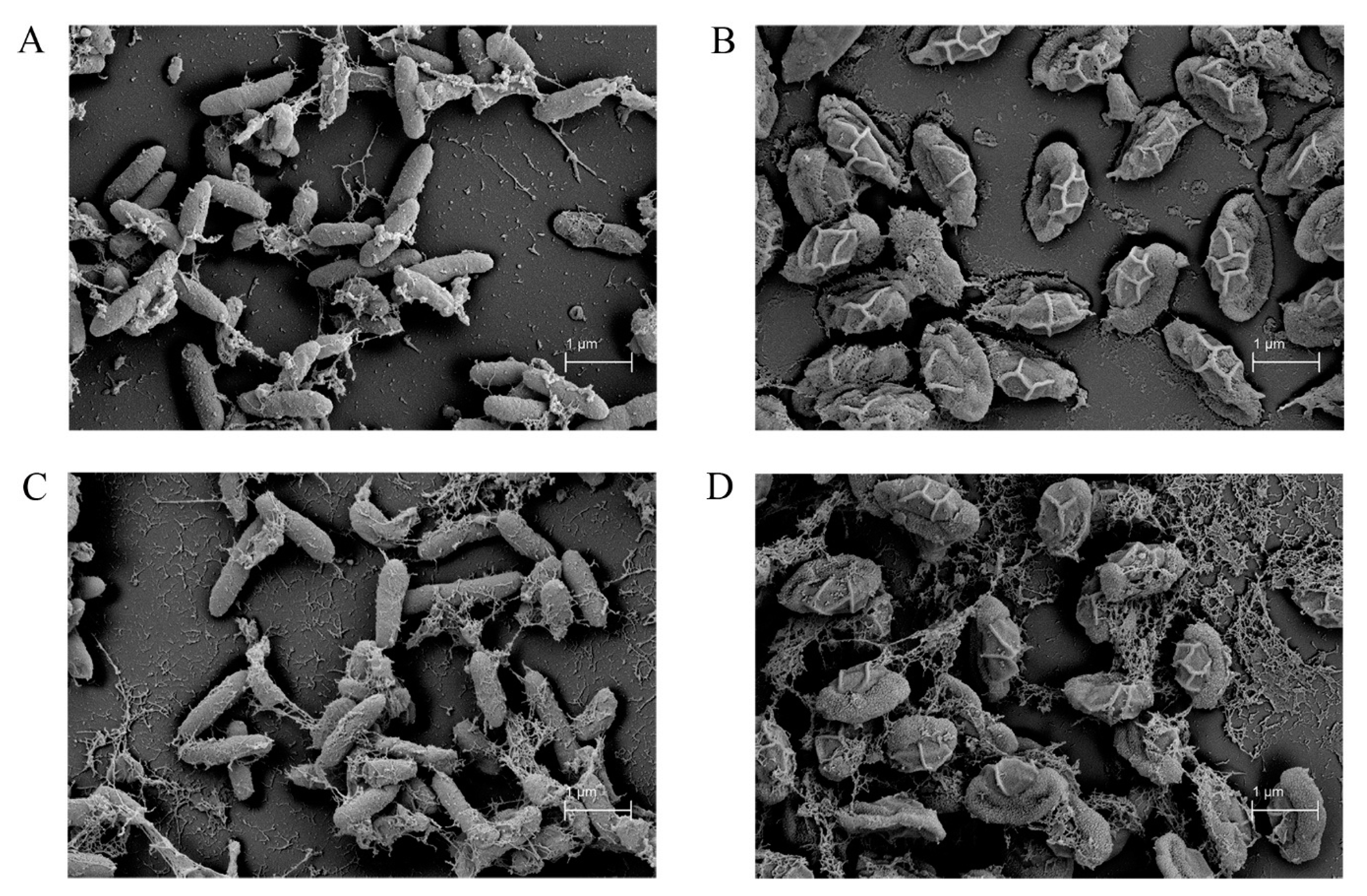
2.7. Scanning Electron Microscope (SEM)

The morphological characteristics between the wild-type and mutant (M201) strain were visualized by scanning electron microscopy [21]. The vegetative cells and spores of both strains were cultured on MH agar and incubated at 30 °C for 1 and 7 days, respectively, before applying onto a glass slide. The bacterial cells were fixed by soaking with 2.5% glutaraldehyde in 0.1 M phosphate buffer, pH 7.2, for 48 h. The fixed samples were washed twice with 0.1 M phosphate buffer, pH 7.2, and dehydrated with increasing concentrations of ethanol (20%, 40%, 60%, 80%, and 100%). The samples were incubated at room temperature for 30 min upon changing the ethanol concentration. Sample drying was accomplished using the critical point drying technique (EMS Quorum, PA, USA) followed by coating of samples with gold (Cressington Scientific Intrusment Ltd., Watford, UK). SEM micrographs were taken at a magnification of 50,000× using a scanning electron microscope (Carl Zeiss, Oberkochen, Germany).

2.8. Strain Stability of the Mutant on the Antibacterial Activity

The UV mutant strain was studied for strain stability by monitoring the antibacterial activity over five generations. The single colony of the mutant strain was suspended, and the turbidity was adjusted according to the 0.5 McFarland standard with 0.85% NaCl solution. Cell suspension (2%) was transferred to half LB broth and incubated at 30 °C with shaking at 150 rpm for 24 h. The supernatant of the first culture was collected by centrifugation at 10,000× g at 4 °C for 15 min, while the turbidity of the cell suspension was adjusted to the 0.5 McFarland standard with 0.85% NaCl for the preparation of next generations (5 passages). The strain stability of each generation was evaluated for antibacterial activity by the agar well diffusion technique and was statistically analyzed by Student’s t-test with p < 0.05 indicating significant differences.

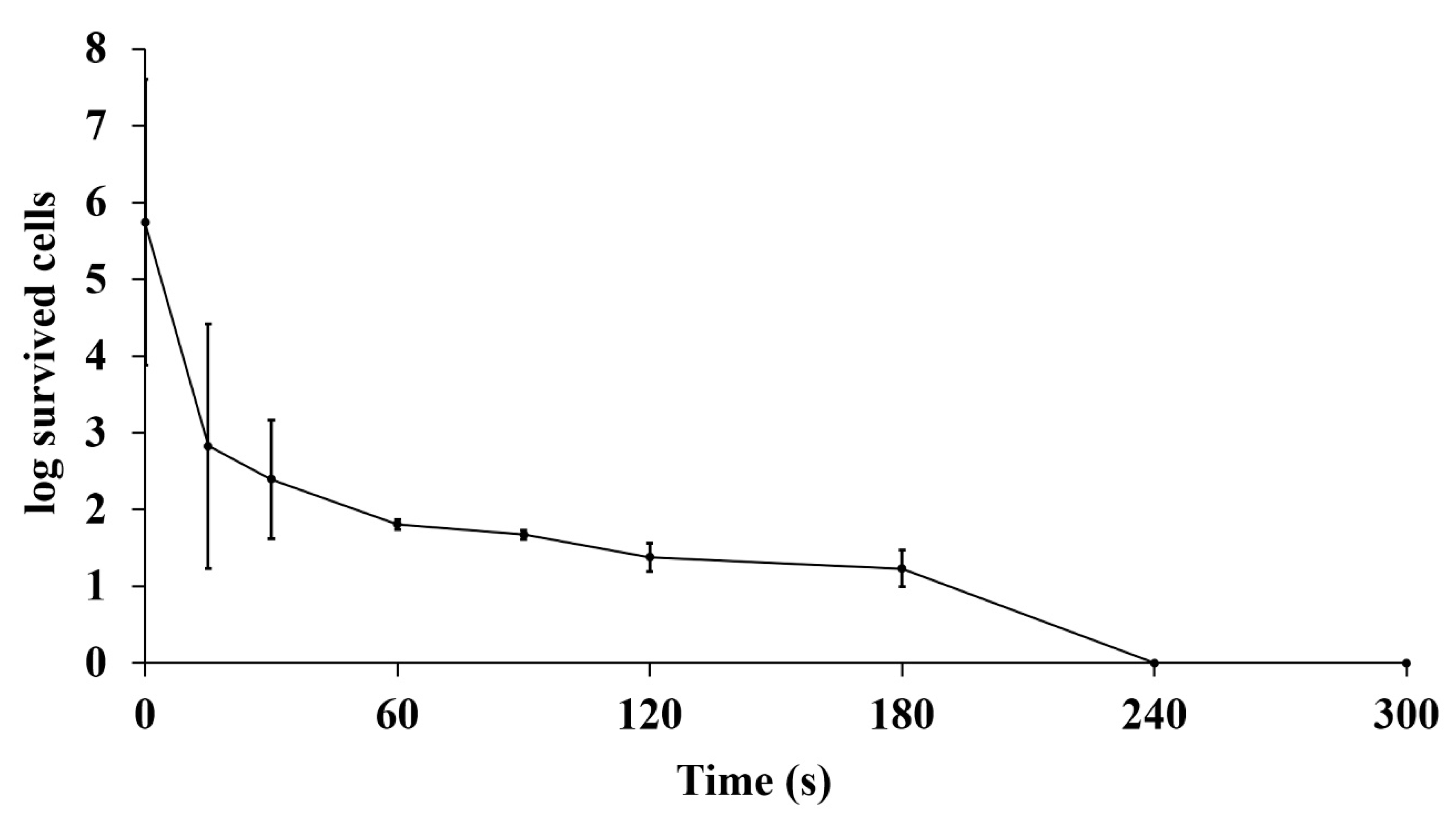
2.9. Susceptibility Study of the Mutant Strain

The susceptibility of the mutant SPR-20 to the standard antibiotics was investigated by disc diffusion assay [22]. The wild-type and mutant SPR-20 strains were inoculated at 30 °C with shaking at 150 rpm for 24 h in half LB broth, and the culture was adjusted according to the 0.5 McFarland standard before spreading onto the MH agar. The antibiotic discs (Oxoid Ltd., Hampshire, UK) cefoxitin (30 µg), ceftriaxone (30 µg), ciprofloxacin (5 µg), erythromycin (15 µg), gentamicin (10 µg), imipenem (10 µg), tetracycline (30 µg), and vancomycin (30 µg) were placed on the culture-seeded agar and then incubated at 30 °C for 24 h. The susceptibility was performed in triplicate, and the inhibition zone was presented as mean ± SD. The result was analyzed by Student’s t-test with p < 0.05 indicating significant differences compared with the wild-type strain.

3. Results

3.1. Bacterial Isolation and Antibacterial Activity

Soil samples in the south of Thailand have characteristic properties influenced by their surroundings, as they are covered by tropical rain forests and surrounded by the Gulf of Thailand and the Andaman Sea. Hence, the geographical and environmental factors structure the soil microbial biodiversity. Terrestrial soil was collected from 10 different locations in botanical and national parks. Soil suspensions were heated at 60 °C for 30 min to kill the majority of heat-sensitive bacterial cells before serial dilution to isolate a single colony. All isolates were picked up and screened for antibacterial activity. There were 94 and 67 isolates found to be active against Gram-positive S. aureus TISTR 517 and Gram-negative E. coli TISTR 887 indicator strains, respectively (Table 1). The activity of isolates, including determination of the spectrum of antibacterial activity, was assessed using the agar well diffusion assay. Nine isolates were active against S. aureus TISTR 517 and its resistant strains (MRSA isolate 142, 1096, and 2468), whereas eight isolates exhibited activity against E. coli TISTR 887 (Table 2). None of the isolates demonstrated any activity on P. aeruginosa TISTR 357 or C. albicans TISTR 5554. The three most active isolates (Pr-18, Pr-82, and SPR-20) showed the highest zone of inhibition against S. aureus TISTR 517 and its resistant strains (MRSA isolate 142, 1096, and 2468). In addition, Pr-82 and SPR-20 also exhibited broad-spectrum activity that extended to E. coli TISTR 887. Furthermore, the antibacterial substances from these strains showed activity against the same studied pathogens, similar to the activity of vancomycin, whereas other antibiotics (cefoxitin and oxacillin) were inactive on MRSA (Table 2). The results indicate that the soil samples had high microbial biodiversity, and the isolated strains could be targets for the discovery of anti-MRSA agents.

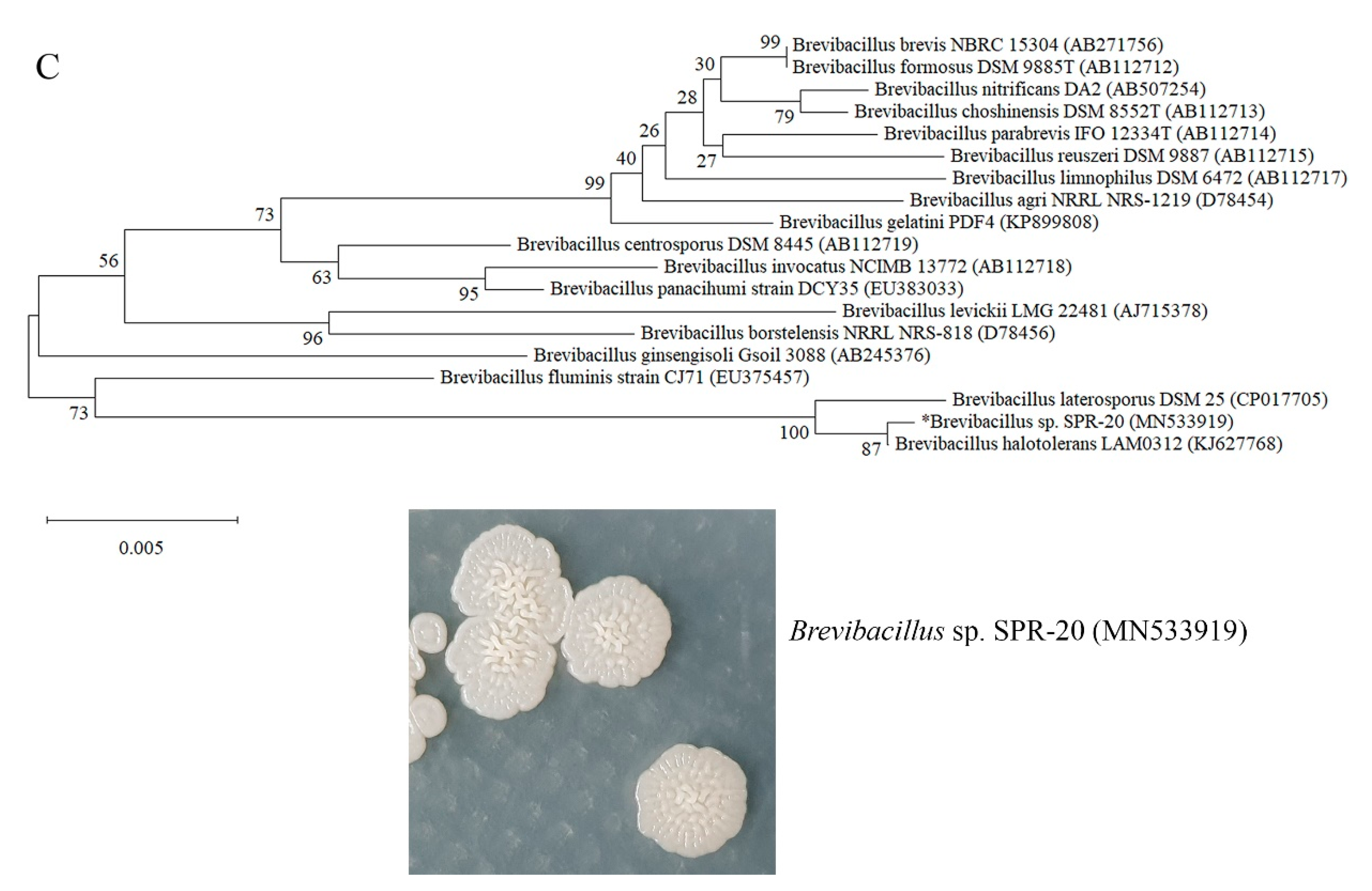
3.2. Bacterial Identification and Phylogenetic Tree Construction

The three most potentially active isolates (Pr-18, Pr-82, and SPR-20) that were cultured on MH agar at 30 °C for 4 days were Gram-positive, spore-forming bacteria. The strain Pr-18 appeared as a brown and filamentous colony with a powdery surface, whereas Pr-82 and SPR-20 formed cream, circular colonies with a smooth and wrinkled surface, respectively (Figure 1). Subsequently, they were analyzed by 16S rRNA gene sequencing at TISTR. The partial nucleotides were deposited to NCBI GenBank, and multiple sequence alignment comparisons with other sequences from the NCBI gene database was conducted using BLASTN. Phylogenetic trees were constructed using MEGA X version 10.1.8 and the neighboring-joining method with 1000 bootstrap values. The Pr-18 strain belonged to the genus Bacillus and was assigned as Bacillus sp. Pr-18 (accession numbers MN533920) because it exhibited a sequence similarity of 99.01% with Bacillus sonorensis strain NBRC 101234. The isolates Pr-82 and SPR-20 were in the genus Brevibacillus and defined as Brevibacillus sp. Pr-82 (accession numbers MN533921) and Brevibacillus sp. SPR-20 (accession numbers MN533919) with a close phylogenetic relationship to Brevibacillus brevis strain NBRC 15304 (99.78%) and Brevibacillus halotolerans strain LAM0312 (99.86%), respectively (Figure 1).

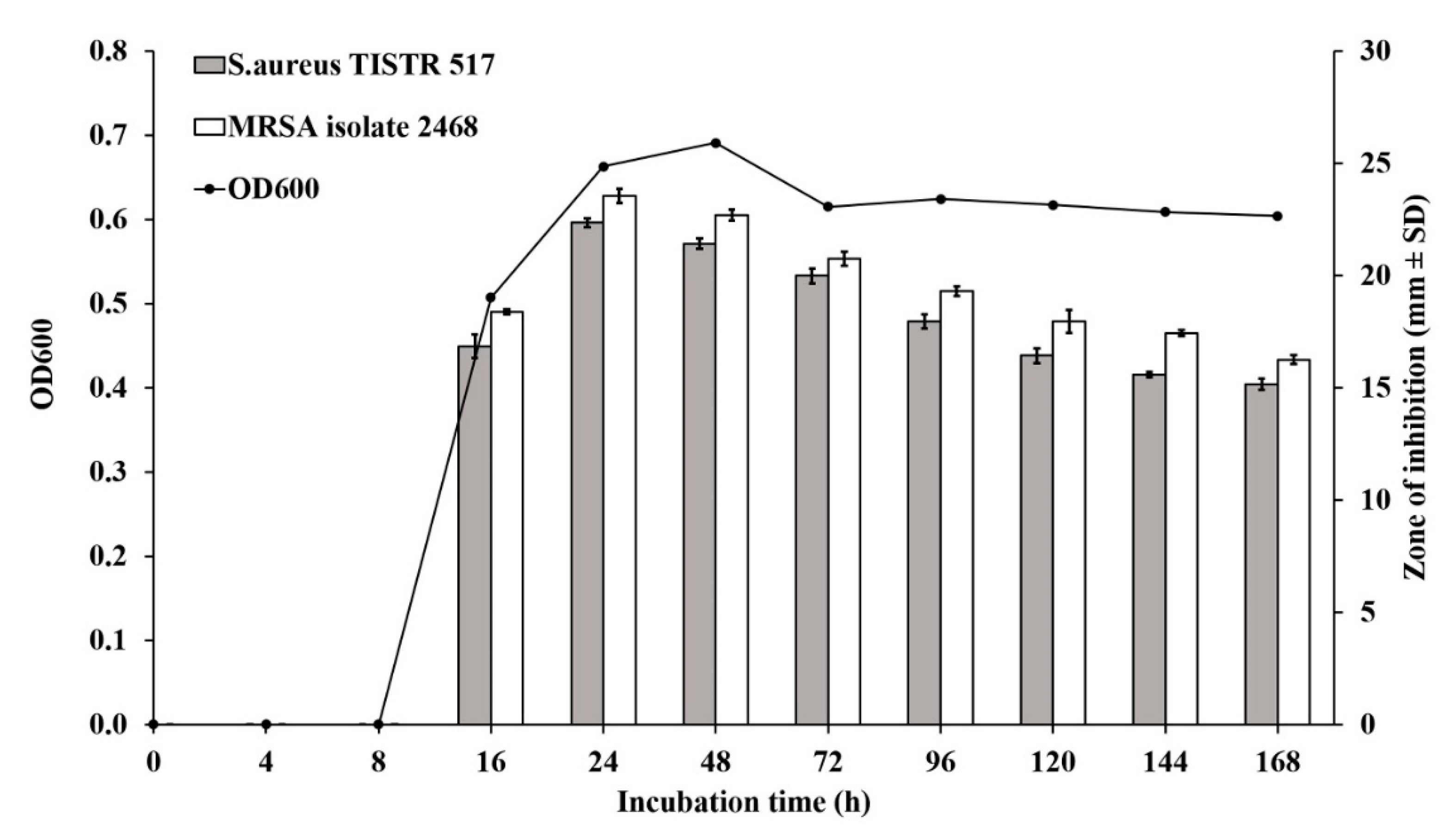
3.3. Production Kinetics of Anti-MRSA Substances from Brevibacillus sp. SPR-20

Brevibacillus sp. SPR-20 had the highest activity and a broad spectrum against bacterial pathogens. It was selected for investigating the kinetics of antibacterial production. This isolate exhibited activity related to the logarithmic phase at 16 h. It had maximum activity at 24 h (early stationary phase) with an inhibition zone of 22.35 ± 0.21 and 23.54 ± 0.32 mm against S. aureus TISTR 517 and MRSA isolate 2468, respectively. Furthermore, the inhibition activity gradually declined until 168 h of incubation (Figure 2).

3.4. Stability Study of Antibacterial Substances

The active substances contained in the cultured supernatant of Brevibacillus sp. SPR-20 and nisin peptide antibiotic were studied for their stability with respect to various conditions using S. aureus TISTR 517 and MRSA isolate 2468 as the indicator microorganisms. Antibacterial activity was comparably decreased in both pathogens with the following described conditions. The inhibition effect of SPR-20 was diminished when the temperature was increased (Table 3). More than 90% activity remained against S. aureus TISTR 517 and MRSA isolate 2468 after sterilization conditions (121 °C, 15 min, 15 psi), indicating that the active substances of SPR-20 were thermostable. Conversely, the activity of nisin (a peptide antibiotic) dramatically decreased to below 90% at 60 °C. Proteinase K, trypsin, and α-chymotrypsin are catalytic enzymes that degrade antibacterial peptides. Treatment with these enzymes resulted in slight degradation of the active substances of SPR-20, decreasing the activity to 94.89 ± 3.34% and 98.00 ± 1.83% against the indicator pathogens. In addition, the antibacterial activity of nisin was completely lost under all enzymatic treatments, except for the α-chymotrypsin-treated conditions on MRSA isolate 2468 (68.95 ± 3.20%). SDS (anionic surfactant) alone showed antibacterial activity, whereas Triton X-100 (non-ionic surfactant) had no effect on the indicator microorganisms. The mixture of SPR-20 and surfactant (SDS or Triton X-100) could synergize the inhibition zone, supporting the stability of active substances on the presence of the surfactants. Interestingly, the combination of nisin with Triton X-100 showed synergistic activity against both indicator bacteria (121.05 ± 4.27% and 148.00 ± 1.98%), but mixtures with SDS had less activity (95.14 ± 1.40% and 130.86 ± 0.99%, respectively) when compared to each surfactant. The antibacterial substances of SPR-20 were tolerant to a wide pH range (pH 2–14), with the activity varying from 99.16 ± 2.17% to 102.93 ± 1.26%.The activity of nisin was apparently lost at pH > 11. Taken together, it is implied that the anti-MRSA substances from Brevibacillus sp. SPR-20 were antimicrobial peptides with high stability in relation to temperature, proteolytic enzymes, surfactants, and pH.

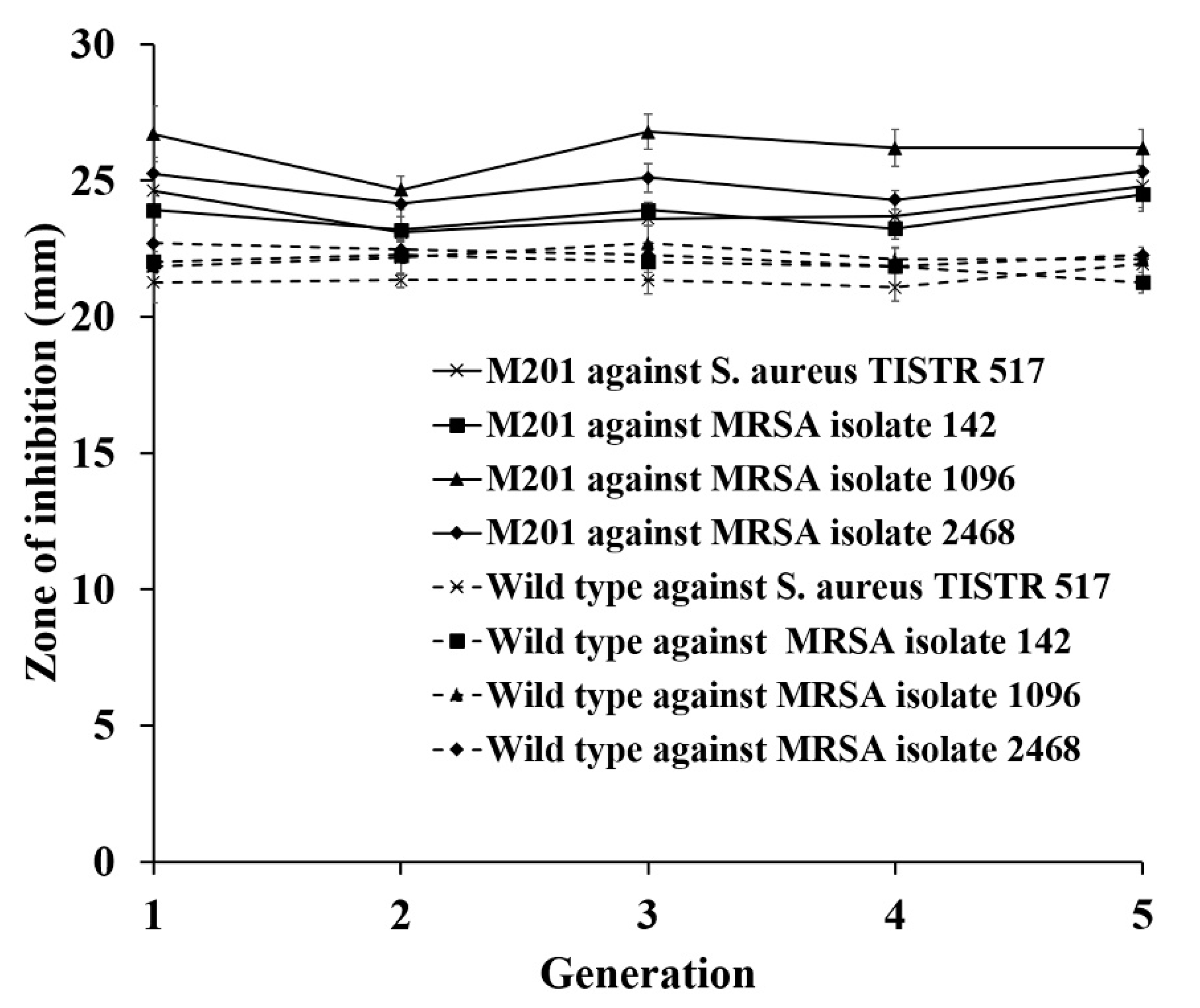
3.5. Strain Improvement of SPR-20 by UV Mutagenesis

Brevibacillus sp. SPR-20 produced the highest antibacterial activity among all isolates in this study. Generally, the production capability of antibacterial substances is regulated by the part of the genome that is involved in the metabolic machinery, especially the rate-limiting step of biosynthetic enzymes. Several approaches have been utilized to overcome this limitation through mutagenesis and genetic engineering. UV radiation generates random mutagenesis for the enhanced production of biological substances. The strain SPR-20 was exposed to UV radiation, and it was found that more than 99% of the bacterial cells were killed when the exposure time was prolonged (Figure 3). The mutants of 364 isolates were evaluated for antibacterial activity using agar overlay. Twenty-two mutants showed a significant inhibition ratio against MRSA isolate 2468 (ratio of 7.98 ± 0.29 to 12.52 ± 1.03) when compared to the wild-type strain (ratio of 6.86 ± 2.08) (p < 0.05). These were then confirmed by assaying activity by agar well diffusion. It was found that only one mutant (M201) exhibited significantly increased inhibitory activity against all indicator strains when compared to the wild-type strain (24.62 ± 0.67 and 21.23 ± 0.69 mm for S. aureus TISTR 517, 23.91 ± 0.52 and 21.76 ± 0.82 mm for MRSA isolate 142, 26.70 ± 1.02 and 22.21 ± 0.83 mm for MRSA isolate 1096, and 25.24 ± 0.61 and 22.27 ± 0.83 mm for MRSA isolate 2468, respectively) (Table 4). SEM revealed that the vegetative cells of M201 and wild-type strains had similar morphological characteristics (a rod shape), with a size of 1.50–1.75 µm (Figure 4A,C). The spores of both strains (1.00–1.50 µm) also had rough and folded envelopes on the surface (Figure 4B,D). Their growth curves were also observed as having the same characteristics. The susceptibility of the mutant M201 and wild-type strains was also determined using various standard antibiotic discs. The results show that M201 was more sensitive to antibiotics than the wild-type strain. Ceftriaxone displayed a significant inhibition zone of 36.91 ± 0.96 mm, while that of the wild-type strain was 32.34 ± 0.24 mm (Table 5). The mutant strain seemed to be more susceptible to other antibiotics (ciprofloxacin, imipenem, tetracycline, and vancomycin) as they showed a higher average inhibition zone without any statistical significance, indicating increased drug susceptibility after UV mutagenesis.
Furthermore, strain stability was studied to ensure the genetic consistency of the selected mutant by measuring for constant antibacterial production. Previous studies have evaluated the strain stability after subculture production in a fermentation medium for 5–10 successive generations [23]. Both M201 and wild-type strains could produce invariable antibacterial activity, and M201 yielded a significantly higher range of inhibition zone (23.09 ± 0.33 to 24.79 ± 0.78 mm for S. aureus TISTR 517, 23.19 ± 0.78 to 24.48 ± 0.62 mm for MRSA isolate 142, 24.65 ± 0.51 to 26.78 ± 0.64 mm for MRSA isolate 1096, and 24.15 ± 0.46 to 25.32 ± 0.88 mm for MRSA isolate 2468) when compared to the wild-type strain (21.08 ± 0.51 to 21.93 ± 0.64 mm for S. aureus TISTR 517, 21.25 ± 0.39 to 22.27 ± 0.15 mm for MRSA isolate 142, 21.84 ± 0.25 to 22.69 ± 0.64 mm for MRSA isolate 1096, and 21.84 ± 0.67 to 22.96 ± 0.64 mm for MRSA isolate 2468, respectively) (Figure 5). This implies that both the mutant and wild-type strains were stably producing anti-MRSA substances.

4. Discussion

Heat treatment was performed on the terrestrial soil for the reduction of heat-labile microbes. Heat-tolerant bacteria were isolated in the search for antibacterial activity. Antibacterial properties exist in several types of bacteria as a result of nutrient competition between species under environments inadequate for survival [24]. Predominant antibacterial producers are of the genera Streptomyces, Bacilli, and Brevibacillus, which are heat-stable microorganisms. In this study, three potential isolates (Pr-18, Pr-82, and SPR-20) that exhibited high activity against indicator pathogens (S. aureus TISTR 517, MRSA isolate 142, 1096, and 2468) were assigned to the Bacillus and Brevibacillus genera. Several studies have reported that Bacillus sp. and Brevibacillus sp. can be isolated from soil environments, and most of them are non-pathogenic agents that produce active peptide substances against the human pathogenic bacteria (S. aureus and MRSA) [24]. Loloathins, laterocidin, and bogorols are non-ribosomal synthesized antibacterial peptides from Brevibacillus laterosporus, whereas bac-GM100 is a ribosomal-synthesized antimicrobial peptide from B. brevis that showed broad-spectrum activity against clinical pathogens of S. aureus, P. aeruginosa, and Candida tropicalis [25,26,27,28,29]. The most potential isolate in this study, Brevibacillus sp. SPR-20, was shown to be a promising target for inhibiting S. aureus and MRSA. It produced the maximum anti-MRSA activity after 24 h of incubation, which corresponded to the stationary phase of the growth curve. The culture supernatant had a broad spectrum of activity with comparable potency to vancomycin, whereas cefoxitin and oxacillin were ineffective in treating MRSA strains. These inhibitory effects were also demonstrated in other Brevibacillus sp., such as crude extract and brevibacillin prepared from B. laterosporus strain SA14 and B. laterosporus strain OSY-I, respectively [27,28]. B. halotolerans strain LAM0312 is a new species in this genus, and it was shown to be highly similar to Brevibacillus sp. SPR-20 based on its 16S rRNA sequence [29]. However, there is no scientific literature on the activity of B. halotolerans, whereas B. laterosporus strain DSM25 was demonstrated to have a close phylogenetic relation to SPR-20 and can produce antimicrobial compounds with predicted biosynthetic gene clusters of brevibacillin, paenibacterin, and petrobactin [30]. Although the active substances from SPR-20 have not been fully investigated, these results provide an opportunity to search for drug candidates to combat antibiotic-resistant pathogens. Bacillus sp. Pr-18, found in this study, has a close relationship with B. sonorensis strain NBRC 101234. Previous studies have presented the isolation of B. sonorensis MT93 and identified an antimicrobial peptide named sonorensin with a molecular mass of 6.27 kDa. It was demonstrated to be a potent antibacterial agent against S. aureus, P. aeruginosa, and Listeria monocytogenes [31]. Brevibacillus sp. Pr-82 was adjacent to B. brevis strain NBRC 15304, and several studies have demonstrated small antibacterial peptides from those strains. Tostadin from B. brevis strain XDH was isolated as a novel substance with a broad spectrum against S. aureus and E. coli [31,32].
The anti-MRSA substances from Brevibacillus sp. SPR-20 are antimicrobial peptides which the remaining activity was more than 90% under various conditions, such as involving temperature (60–121 °C), proteolytic enzymes (proteinase K, trypsin, and α-chymotrypsin), surfactants (SDS and Triton X-100), and a wide pH range (2–14). Conversely, the peptide antibiotic nisin was more sensitive to temperature, proteolytic enzymes, and a basic pH. This result is consistent with previous stability studies, demonstrating that nisin was stable at −18 °C but substantially degraded at temperatures above 25 °C [33]. The antibacterial activity of nisin was also shown to be diminished when treated with proteases or a pH above 10 because of the presence of enzymatic and chemical cleavage sites in the peptide antibiotic [34,35]. The activity of SPR-20 and nisin was enhanced when combined with surfactants. However, the addition of SDS to nisin reduced antibacterial activity when compared to SDS alone. This might result from the charge incompatibility between cationic nisin and anionic SDS complexes [36]. The stability profile of SPR-20 was consistent with other studies, showing that some bacteriocin-like inhibitory substances (bacillocin Bb, brevicin AF01, entomocin 9, paenibacterin, and pumilicin) were tolerant to those conditions [37,38,39,40,41]. These results show that the strain SPR-20 could produce anti-MRSA substances with a stable and broad spectrum of antibacterial activity.
Generally, UV mutagenesis is used to enhance the production of bioactive substances through dimerization of thymine bases, forming crosslinks in DNA that might result in base transversion, deletion, frameshifts, or transitions from GC to AT [12]. Tolerant bacterial cells would use repair mechanisms to relieve genetic abnormalities through potentially generating mutagenesis, leading to selection and cell survival. UV-mediated mutagenesis is a useful, classical approach which is advantageous when genetic information of the microbial strains or biosynthetic pathway is lacking. It is assumed that random genetic alteration can bypass regulatory controls and checkpoints, contributing to the overproduction of active substances [42]. In this study, the application of UV mutagenesis led to an increase in the overall production of antibacterial activity. Mutagenesis might lead to enhanced production of the same active substances. Alternatively, it might also activate the cryptic biosynthetic gene cluster of other antibacterial compounds that are not expressed under normal conditions. The mutant strains might then produce multiple active compounds. These two possibilities result in an increased zone of inhibition on the agar growth medium. The drawback of this method is the random selection of strains, and beneficial mutations may depend on several factors such as radiation power, frequency, and exposure time of mutagen treatment, type of bacterial culture, or conditions of treatment and post-treatment. The UV-treated Brevibacillus sp. SPR-20 showed that the number of surviving cells decreased after prolonged UV exposure time, and they were sterilized within 4 min. The cell morphology and growth rate between mutant and wild-type strains did not differ, and both spores appeared to have unique characteristics, with rough and fold envelopes on the surface.
Brevibacillus sp. SPR-20 was more sensitive to UV irradiation than other Brevibacillus sp., such as Brevibacillus sp. strain AS-S10-IIM201 and Brevibacillus borstelensis R1, which were more tolerant to UV irradiation for several minutes [43,44]. The agar overlay method was used as the pre-selection technique to screen several mutated isolates with higher antibacterial activity. An agar diffusion assay was carried out to determine antagonist effects against indicator pathogens for the remaining desired strains. The mutant M201 exhibited significantly higher activity (109.88–120.22%) when compared to the wild-type strain. This emphasizes UV mutagenesis as a potential approach for strain improvement. Similar observations were also reported in a number of strain improvement studies: Bacillus licheniformis was irradiated by UV, and could produce bacitracin with 1.85-fold higher activity [45]. A previous study also presented that the UV-treated B. brevis strain FJAT-0809-GLX yielded a positive mutant rate of 7.8%, and enhanced the production of ethylparaben with higher activity toward pathogens [46].
Furthermore, the mutant M201 was a stable strain as judged by the consistent antibacterial activity throughout many generations. This mutated isolate was more susceptible to some antibiotics (ceftriaxone) than the wild-type strain. This implies that UV radiation activated adaptive or protective responses and altered the bacterial cell wall or membrane. This resulted in an increase in the secretion of active substances into the culture medium and a higher sensitivity to antibiotic drugs that target cell wall synthesis [47]. However, further experiments are required to elucidate the susceptible consequences and mechanism of action of these anti-MRSA substances for drug development in the antibiotic-resistant era.

5. Conclusions

MRSA infection is a public health concern that requires researchers to search for new antibacterial agents. Soil bacteria are useful for this purpose as producers of antibiotics against resistant pathogens. In this study, nine isolates from soil samples in Thailand exhibited activity against S. aureus and MRSA strains, whereas eight isolates showed inhibition to E. coli. Brevibacillus sp. SPR-20 presented the highest activity against pathogens (S. aureus and MRSA). The active substances were stable under several stress conditions of temperature, proteolytic enzymes, surfactants, and pH. Its mutant strain (M201), produced by UV mutagenesis, had enhanced activity compared to the parental strain. The mutated isolate appeared to demonstrate strain stability and antibiotic susceptibility. However, the bioactive substances from Brevibacillus sp. SPR-20 and its mutant strains might be promising candidates for the treatment of antibiotic-resistant infection.

Author Contributions

Conceptualization, N.S., M.L., and A.A.; methodology, N.S., M.L., and A.A.; investigation, N.S., M.L., and A.A.; resources, M.L. and A.A.; writing—original draft preparation, N.S., M.L., and A.A.; writing—review and editing, A.A.; supervision, A.A.; project administration, A.A.; funding acquisition, A.A. All authors have read and agreed to the published version of the manuscript.

Funding

This research was funded by Walailak University (Grant No. WU-IRG-62-015) and Walailak University Graduate Research Fund (Grant No. CGS-RF-2020/05).

Institutional Review Board Statement

Not applicable.

Informed Consent Statement

Not applicable.

Data Availability Statement

Data is contained within the article or supplementary material.

Acknowledgments

The work was approved by the Institutional Biosafety Committee (WU-IBC-62-004; 27 June 2019). In addition, we would like to thank the Center of Scientific and Technological Equipments, Walailak University for research facilities.

Conflicts of Interest

The authors declare no conflict of interest.

References

  1. O’Neill, J. Review on Antimicrobial Resistance, Tackling Drug-resistant Infections Globally: Final Report and Recommendations; Wellcome Trust and HM Government: London, UK, 2016; pp. 10–16.
  2. Antimicrobial Resistance Division, World Health Organization. 2019 Antibacterial Agents in Clinical Development: An Analysis of the Antibacterial Clinical Development Pipeline; Weiss, G., Ed.; World Health Organization: Geneva, Switzerland, 2019; pp. 1–24. [Google Scholar]
  3. Hiramatsu, K.; Katayama, Y.; Matsuo, M.; Sasaki, T.; Morimoto, Y.; Sekiguchi, A.; Baba, T. Multi-drug-resistant Staphylococcus aureus and future chemotherapy. J. Infect. Chemother. 2014, 20, 593–601. [Google Scholar] [CrossRef] [Green Version]
  4. Kapoor, G.; Saigal, S.; Elongavan, A. Action and resistance mechanisms of antibiotics: A guide for clinicians. J. Anaesthesiol. Clin. Pharmacol. 2017, 33, 300–305. [Google Scholar] [CrossRef]
  5. Bin Zaman, S.; Hussain, M.A.; Nye, R.; Mehta, V.; Mamun, K.T.; Hossain, N. A Review on antibiotic resistance: Alarm Bells are ringing. Cureus 2017, 9, e1403. [Google Scholar] [CrossRef] [Green Version]
  6. Zulkeflle, S.N.M.; Yusaimi, Y.A.; Sugiura, N.; Iwamoto, K.; Goto, M.; Utsumi, M.; Othman, N.B.; Zakaria, Z.; Hara, H. Phenotypic and genetic characterization of multidrug-resistant Staphylococcus aureus in the tropics of Southeast Asia. Microbiology 2016, 162, 2064–2074. [Google Scholar] [CrossRef]
  7. Hug, J.J.; Bader, C.D.; Remškar, M.; Cirnski, K.; Müller, R. Concepts and methods to access novel antibiotics from actinomycetes. Antibiot 2018, 7, 44. [Google Scholar] [CrossRef] [Green Version]
  8. Andayani, D.G.S.; Sukandar, U.; Sukandar, E.Y.; Adnyana, I.K. Antibacterial, antifungal and anticancer activity of five strains of soil microorganisms isolated from Tangkuban Perahu mountain by fermentation. HAYATI J. Biosci. 2015, 22, 186–190. [Google Scholar] [CrossRef] [Green Version]
  9. Sumi, C.D.; Yang, B.W.; Yeo, I.-C.; Hahm, Y.T. Antimicrobial peptides of the genus Bacillus: A new era for antibiotics. Can. J. Microbiol. 2015, 61, 93–103. [Google Scholar] [CrossRef]
  10. Ahmad, M.S.; El-Gendy, A.O.; Ahmed, R.R.; Hassan, H.M.; El-Kabbany, H.M.; Merdash, A.G. Exploring the antimicrobial and antitumor potentials of Streptomyces sp. AGM12-1 isolated from egyptian soil. Front. Microbiol. 2017, 8, 438. [Google Scholar] [CrossRef] [PubMed] [Green Version]
  11. Saxena, S. Strategies of Strain Improvement of Industrial Microbes. In Applied Microbiology; Springer: New Delhi, India, 2015; pp. 155–171. [Google Scholar] [CrossRef]
  12. Ikehata, H.; Ono, T. The mechanisms of UV mutagenesis. J. Radiat. Res. 2011, 52, 115–125. [Google Scholar] [CrossRef] [PubMed] [Green Version]
  13. Nasfi, Z.; Busch, H.; Kehraus, S.; Linares-Otoya, L.; König, G.M.; Schäberle, T.F.; Bachoual, R. Soil bacteria isolated from tunisian arid areas show promising antimicrobial activities against gram-negatives. Front. Microbiol. 2018, 9, 2742. [Google Scholar] [CrossRef] [PubMed] [Green Version]
  14. Selvin, J.; Shanmughapriya, S.; Gandhimathi, R.; Kiran, G.S.; Ravji, T.R.; Natarajaseenivasan, K.; Hema, T.A. Optimization and production of novel antimicrobial agents from sponge associated marine actinomycetes Nocardiopsis dassonvillei MAD08. Appl. Microbiol. Biotechnol. 2009, 83, 435–445. [Google Scholar] [CrossRef] [PubMed]
  15. Valgas, C.; De Souza, S.M.; Smânia, E.F.A.; Smânian, A.S., Jr. Screening methods to determine antibacterial activity of natural products. Braz. J. Microbiol. 2007, 38, 369–380. [Google Scholar] [CrossRef] [Green Version]
  16. Altschul, S.F.; Gish, W.; Miller, W.; Myers, E.W.; Lipman, D.J. Basic local alignment search tool. J. Mol. Biol. 1990, 215, 403–410. [Google Scholar] [CrossRef]
  17. Kumar, S.; Stecher, G.; Li, M.; Knyaz, C.; Tamura, K. MEGA X: Molecular evolutionary genetics analysis across computing platforms. Mol. Biol. Evol. 2018, 35, 1547–1549. [Google Scholar] [CrossRef]
  18. Goh, H.F.; Philip, K. Purification and characterization of bacteriocin produced by Weissella confusa A3 of dairy origin. PLoS ONE 2015, 10, e0140434. [Google Scholar] [CrossRef]
  19. Chalasani, A.G.; Dhanarajan, G.; Nema, S.; Sen, R.; Roy, U. An antimicrobial metabolite from Bacillus sp.: Significant activity against pathogenic bacteria including multidrug-resistant clinical strains. Front. Microbiol. 2015, 6, 1335. [Google Scholar] [CrossRef] [Green Version]
  20. Hmani, M.; Boukedi, H.; Ben Khedher, S.; Elleuch, A.; Tounsi, S.; Abdelkefi-Mesrati, L. Improvement of Vip3Aa16 toxin production and efficiency through nitrous acid and UV mutagenesis of Bacillus thuringiensis (Bacillales: Bacillaceae). J. Econ. Entomol. 2018, 111, 108–111. [Google Scholar] [CrossRef]
  21. Chatterjee, S.; Shekhawat, K.; Gupta, N. Bioreduction of toxic hexavalent chromium by novel indigenous microbe Brevibacillus agri isolated from tannery wastewater. Int. J. Environ. Sci. Technol. 2019, 16, 3549–3556. [Google Scholar] [CrossRef]
  22. CLSI. M100 Performance Standards for Antimicrobial Susceptibility Testing, 13th ed.; Clinical and Laboratory Standards Institute: Wayne, PA, USA, 2020; pp. 58–66. [Google Scholar]
  23. Ma, X.-J.; Zhang, H.-M.; Lu, X.-F.; Han, J.; Zhu, H.-X.; Wang, H.; Yao, R.-S. Mutant breeding of Starmerella bombicola by atmospheric and room-temperature plasma (ARTP) for improved production of specific or total sophorolipids. Bioprocess Biosyst. Eng. 2020, 43, 1869–1883. [Google Scholar] [CrossRef]
  24. Bérdy, J. Bioactive microbial metabolites. J. Antibiot. 2005, 58, 1–26. [Google Scholar] [CrossRef] [Green Version]
  25. Yang, X.; Yousef, A.E. Antimicrobial peptides produced by Brevibacillus spp.: Structure, classification and bioactivity: A mini review. World J. Microbiol. Biotechnol. 2018, 34, 57. [Google Scholar] [CrossRef] [PubMed]
  26. Ghadbane, M.; Harzallah, D.; Ibn Laribi, A.; Jaouadi, B.; Belhadj, H. Purification and biochemical characterization of a highly thermostable bacteriocin isolated from Brevibacillus brevis strain GM100. Biosci. Biotechnol. Biochem. 2013, 77, 151–160. [Google Scholar] [CrossRef] [PubMed] [Green Version]
  27. Yang, X.; Huang, E.; Yuan, C.; Zhang, L.; Yousef, A.E. Isolation and structural elucidation of brevibacillin, an antimicrobial lipopeptide from Brevibacillus laterosporus that combats drug-resistant gram-positive bacteria. Appl. Environ. Microbiol. 2016, 82, 2763–2772. [Google Scholar] [CrossRef] [PubMed] [Green Version]
  28. Chawawisit, K.; Lertcanawanichakul, M. Minimum inhibitory concentration (MIC) of crude preparations of Brevibacillus laterosporus SA14 bioactive material compared to vancomycin and oxacillin, against clinical isolates of methicillin-resistant Staphylococcus aureus. World J. Microbiol. Biotechnol. 2008, 24, 2199–2204. [Google Scholar] [CrossRef]
  29. Song, J.; Wang, Y.; Song, Y.; Zhao, B.; Wang, H.; Zhou, S.; Kong, D.; Guo, X.; Li, Y.; He, M.; et al. Brevibacillus halotolerans sp. nov., isolated from saline soil of a paddy field. Int. J. Syst. Evol. Microbiol. 2017, 67, 772–777. [Google Scholar] [CrossRef]
  30. Glare, T.; Durrant, A.; Berry, C.; Palma, L.; Ormskirk, M.M.; Cox, M.P. Phylogenetic determinants of toxin gene distribution in genomes of Brevibacillus laterosporus. Genomics 2019, 112, 1042–1053. [Google Scholar] [CrossRef]
  31. Chopra, L.; Singh, G.; Choudhary, V.; Sahoo, D.K. Sonorensin: An antimicrobial peptide, belonging to the heterocycloanthracin subfamily of bacteriocins, from a new marine isolate, Bacillus sonorensis MT93. Appl. Environ. Microbiol. 2014, 80, 2981–2990. [Google Scholar] [CrossRef] [Green Version]
  32. Song, Z.; Liu, Q.; Guo, H.; Ju, R.; Zhao, Y.; Li, J.; Liu, X. Tostadin, a novel antibacterial peptide from an antagonistic microorganism Brevibacillus brevis XDH. Bioresour. Technol. 2012, 111, 504–506. [Google Scholar] [CrossRef]
  33. Holcapkova, P.; Raskova, Z.K.; Hrabalikova, M.; Salakova, A.; Drbohlav, J.; Sedlarik, V. Isolation and thermal stabilization of bacteriocin nisin derived from whey for antimicrobial modifications of polymers. Int. J. Polym. Sci. 2017, 2017, 3072582. [Google Scholar] [CrossRef] [Green Version]
  34. Moreno, I.; Lerayer, A.L.S.; Baldini, V.L.S.; Leitão, M.F. Characterization of bacteriocins produced by Lactococcus lactis strains. Braz. J. Microbiol. 2000, 31, 183–191. [Google Scholar] [CrossRef] [Green Version]
  35. Slootweg, J.C.; Liskamp, R.M.J.; Rijkers, D.T.S. Scalable purification of the lantibiotic nisin and isolation of chemical/enzymatic cleavage fragments suitable for semi-synthesis. J. Pept. Sci. 2013, 19, 692–699. [Google Scholar] [CrossRef]
  36. Liu, K.; Yang, L.; Peng, X.; Gong, H.; Wang, J.; Lu, J.R.; Xu, H. Effects of conventional surfactants on the activity of designed antimicrobial peptide. Langmuir 2020, 36, 3531–3539. [Google Scholar] [CrossRef]
  37. Saleem, F.; Ahmad, S.; Yaqoob, Z.; Rasool, S.A. Comparative study of two bacteriocins produced by representative indigenous soil bacteria. Pak. J. Pharm. Sci. 2009, 22, 252–258. [Google Scholar]
  38. Faheem, F.; Saeed, S.; Rasool, S.A. Studies on Brevicin AF01: A bacteriocin like inhibitory substance active against methicillin resistant Staphylococcus aureus. Pak. J. Bot. 2007, 39, 1293–1302. [Google Scholar]
  39. Cherif, A.J.A.A.; Chehimi, S.; Limem, F.; Hansen, B.; Hendriksen, N.; Daffonchio, D.; Boudabous, A. Detection and characterization of the novel bacteriocin entomocin 9, and safety evaluation of its producer, Bacillus thuringiensis ssp. entomocidus HD9. J. Appl. Microbiol. 2003, 95, 990–1000. [Google Scholar] [CrossRef] [PubMed]
  40. Aunpad, R.; Na-Bangchang, K. Pumilicin 4, A Novel Bacteriocin with anti-MRSA and Anti-VRE activity produced by newly isolated bacteria Bacillus pumilus strain WAPB4. Curr. Microbiol. 2007, 55, 308–313. [Google Scholar] [CrossRef] [PubMed]
  41. Guo, Y.; Huang, E.; Yuan, C.; Zhang, L.; Yousef, A.E. Isolation of a Paenibacillus sp. strain and structural elucidation of its broad-spectrum lipopeptide antibiotic. Appl. Environ. Microbiol. 2012, 78, 3156–3165. [Google Scholar] [CrossRef] [PubMed] [Green Version]
  42. Parekh, S.; Vinci, V.A.; Strobel, R.J. Improvement of microbial strains and fermentation processes. Appl. Microbiol. Biotechnol. 2000, 54, 287–301. [Google Scholar] [CrossRef]
  43. Mukherjee, A.K.; Rai, S.K.; Bordoloi, N.K. Biodegradation of waste chicken-feathers by an alkaline β-keratinase (Mukartinase) purified from a mutant Brevibacillus sp. strain AS-S10-II. Int. Biodeterior. Biodegrad. 2011, 65, 1229–1237. [Google Scholar] [CrossRef]
  44. Suribabu, K.; Lalitha Govardhan, T.; Hemalatha, K.P.J. Strain improvement of Brevibacillus borostelensis R1 for optimization of α-amylase production by mutagens. J. Microb. Biochem. Technol. 2014, 6, 123–127. [Google Scholar] [CrossRef] [Green Version]
  45. Ali, S.; Nelofer, R.; Andleeb, S.; Baig, S.; Shakir, H.A.; Qazi, J.I. Biosynthesis and optimization of bacitracin by mutant Bacillus licheniformis using submerged fermentation. Indian J. Biotechnol. 2018, 17, 251–260. [Google Scholar]
  46. Che, J.; Liu, B.; Liu, G.; Chen, Q.; Huang, D. Induced mutation breeding of Brevibacillus brevis FJAT-0809-GLX for improving ethylparaben production and its application in the biocontrol of Lasiodiplodia theobromae. Postharvest Biol. Technol. 2018, 146, 60–67. [Google Scholar] [CrossRef]
  47. Gamalier, J.P.; Silva, T.P.; Zarantonello, V.; Dias, F.F.; Melo, R.C. Increased production of outer membrane vesicles by cultured freshwater bacteria in response to ultraviolet radiation. Microbiol. Res. 2017, 194, 38–46. [Google Scholar] [CrossRef] [PubMed]
Figure 1. Phylogenetic tree based on 16S rRNA gene sequence and colony morphology of (A) Bacillus sp. Pr-18, (B) Brevibacillus sp. Pr-82 and (C) Brevibacillus sp. SPR-20. The trees were constructed via MEGA X version 10.1.8 using the neighboring-joining method with 1000 bootstrap values. * Indicated the testing isolates.
Scipharm 89 00001 g001aScipharm 89 00001 g001b
Figure 2. The growth curve and production kinetics of anti-MRSA substances from Brevibacillus sp. SPR-20. The growth curve was measured at optical density (OD) 600 nm, while the antibacterial activity against S. aureus TISTR 517 and MRSA isolate 2468 was determined by the zone of inhibition using the agar well diffusion technique. The experiments were performed in triplicate, and the results are expressed as mean ± SD.
Scipharm 89 00001 g002
Figure 3. The survival curve of Brevibacillus sp. SRP-20 at different time intervals when exposed to UV radiation. The results are expressed as mean ± SD (n = 3).
Scipharm 89 00001 g003
Figure 4. SEM micrographs for (A) vegetative cells of Brevibacillus sp. SPR-20, (B) spores of Brevibacillus sp. SPR-20, (C) vegetative cells of its UV-treated mutant (M201), and (D) spores of its UV-treated mutant (M201). The vegetative cells and spores were obtained by culture on the MH agar for 1 and 7 days, respectively, at 30 °C.
Scipharm 89 00001 g004
Figure 5. Strain stability between the wild-type strain of Brevibacillus sp. SPR-20 and its mutant strain (M201) for five generations. Agar well diffusion was performed against a number of indicator pathogens. The results for the inhibition zone are represented as mean ± SD (n = 3).
Scipharm 89 00001 g005
Table 1. Sampling locations and the number of soil isolates that exhibited antibacterial activity from the cross-streak method.
Sampling LocationS. aureus TISTR 517E. coli TISTR 887
8°33′55.4″ N, 99°31′12.1″ E10
8°46′03.3″ N, 99°48′09.3″ E52
12°25′07.3″ N, 99°58′58.3″ E2920
9°05′22.6″ N, 99°53′39.9″ E45
9°00′50.3″ N, 99°46′18.0″ E71
8°33′33.4″ N, 99°46′55.6″ E76
8°38′31.7″ N, 99°53′38.7″ E138
8°38′55.6″ N, 99°52′51.4″ E60
8°38′49.7″ N, 54°45′50″ E58
8°38′18.3″ N, 54°45′48.5″ E1717
Total9467
Table 2. Agar well diffusion assay of the active isolates against human pathogens (means ± SD; n = 3).
IsolatesS. aureus
TISTR 517
(mm)
MRSA
Isolate 142
(mm)
MRSA
Isolate 1096
(mm)
MRSA
Isolate 2468
(mm)
E. coli
TISTR 887
(mm)
P. aeruginosa
TISTR 357
(mm)
C. albicans
TISTR 5554
(mm)
AK-740.00 ± 0.000.00 ± 0.000.00 ± 0.000.00 ± 0.0010.89 ± 0.160.00 ± 0.000.00 ± 0.00
B6-280.00 ± 0.000.00 ± 0.000.00 ± 0.000.00 ± 0.0011.95 ± 0.270.00 ± 0.000.00 ± 0.00
Play-880.00 ± 0.000.00 ± 0.000.00 ± 0.000.00 ± 0.0011.26 ± 0.530.00 ± 0.000.00 ± 0.00
Pr-1815.41 ± 0.6116.01 ± 0.4216.12 ± 0.3516.07 ± 0.240.00 ± 0.000.00 ± 0.000.00 ± 0.00
Pr-8215.75 ± 0.3316.38 ± 0.1317.14 ± 0.3217.09 ± 0.2712.65 ± 0.390.00 ± 0.000.00 ± 0.00
Pr-8313.12 ± 0.9210.99 ± 0.2613.09 ± 0.3511.82 ± 0.570.00 ± 0.000.00 ± 0.000.00 ± 0.00
Pr-11714.02 ± 0.1113.70 ± 0.5514.60 ± 0.2714.02 ± 0.470.00 ± 0.000.00 ± 0.000.00 ± 0.00
Pr-12215.08 ± 0.0515.46 ± 0.1614.00 ± 0.4415.97 ± 0.260.00 ± 0.000.00 ± 0.000.00 ± 0.00
SPR-020.00 ± 0.000.00 ± 0.000.00 ± 0.000.00 ± 0.0013.34 ± 0.340.00 ± 0.000.00 ± 0.00
SPR-140.00 ± 0.000.00 ± 0.000.00 ± 0.000.00 ± 0.0010.42 ± 0.500.00 ± 0.000.00 ± 0.00
SPR-2017.52 ± 0.6018.28 ± 0.5818.03 ± 0.3619.05 ± 0.8413.29 ± 0.120.00 ± 0.000.00 ± 0.00
S-STD-10.00 ± 0.000.00 ± 0.000.00 ± 0.000.00 ± 0.0011.18 ± 0.440.00 ± 0.000.00 ± 0.00
Su-711.80 ± 0.7613.62 ± 0.1914.25 ± 0.5713.60 ± 1.080.00 ± 0.000.00 ± 0.000.00 ± 0.00
WUL-0211.49 ± 0.8112.10 ± 0.050.00 ± 0.0012.06 ± 0.110.00 ± 0.000.00 ± 0.000.00 ± 0.00
WUL-1014.59 ± 0.0615.91 ± 0.5815.11 ± 0.4314.66 ± 0.060.00 ± 0.000.00 ± 0.000.00 ± 0.00
Cefoxitin (30 µg)35.98 ± 0.530.00 ± 0.000.00 ± 0.000.00 ± 0.0034.71 ± 0.730.00 ± 0.000.00 ± 0.00
Oxacillin (1 µg)30.82 ± 1.400.00 ± 0.000.00 ± 0.000.00 ± 0.000.00 ± 0.000.00 ± 0.000.00 ± 0.00
Vancomycin (30 µg)25.48 ± 0.1525.74 ± 0.1525.74 ± 0.2925.99 ± 0.290.00 ± 0.000.00 ± 0.000.00 ± 0.00
Table 3. Stability study of antibacterial substances from Brevibacillus sp. SPR-20 and nisin (standard peptide antibiotic; 200 µg). The percent activity is expressed by the ratio between the inhibition zone of treated and untreated samples (mean ± SD, n = 3).
Conditions % Activity
S. aureus TISTR 517MRSA Isolate 2468
SPR-20NisinSPR-20Nisin
Untreated Sample100.00 ± 4.46100.00 ± 3.78100.00 ± 4.93100.00 ± 1.95
Thermal Stability
Sample 60 °C, 1 h100.00 ± 1.3789.13 ± 3.77 *99.31 ± 3.1792.43 ± 2.49 *
Sample 80 °C, 1 h96.44 ± 1.81 *73.91 ± 2.49 *97.92 ± 1.5988.05 ± 1.38 *
Sample 100 °C, 1 h95.65 ± 1.81 *0.00 ± 0.00 *96.54 ± 1.04 *69.72 ± 1.38 *
Sample 121 °C, 15 psi, 15 min (autoclave)91.30 ± 3.14 *72.28 ± 3.39 *92.39 ± 1.80 *92.43 ± 0.69 *
Enzyme Stability
Sample + Proteinase K (1 mg/mL)96.08 ± 0.68 *0.00 ± 0.00 *95.90 ± 2.330.00 ± 0.00 *
Sample + Trypsin (1 mg/mL)98.00 ± 1.830.00 ± 0.00 *96.72 ± 0.63 *0.00 ± 0.00 *
Sample + α-Chymotrypsin (1 mg/mL)95.20 ± 1.83 *0.00 ± 0.00 *94.89 ± 3.3468.95 ± 3.20 *
Proteinase K (1 mg/mL)0.00 ± 0.00 *0.00 ± 0.00 *0.00 ± 0.00 *0.00 ± 0.00 *
Trypsin (1 mg/mL)0.00 ± 0.00 *0.00 ± 0.00 *0.00 ± 0.00 *0.00 ± 0.00 *
α-Chymotrypsin (1 mg/mL)0.00 ± 0.00 *0.00 ± 0.00 *0.00 ± 0.00 *0.00 ± 0.00 *
Surfactant Stability
Sample + 1% SDS108.81 ± 2.76 *130.86 ± 0.99 *102.95 ± 1.28 *95.14 ± 1.40 *
Sample + 1% Triton X-100109.24 ± 0.75 *148.00 ± 1.98 *106.36 ± 1.94 *121.05 ± 4.27 *
1% SDS105.29 ± 0.75 *137.71 ± 1.98 *102.95 ± 2.5399.60 ± 1.21
1% Triton X-1000.00 ± 0.00 *0.00 ± 0.00 *0.00 ± 0.00 *0.00 ± 0.00 *
pH Stability
pH 2102.51 ± 1.9298.86 ± 0.99102.33 ± 1.1697.98 ± 2.42
pH 4100.84 ± 2.90100.57 ± 0.99102.33 ± 0.0097.18 ± 1.85
pH 5100.84 ± 3.1697.71 ± 1.71102.33 ± 2.0198.39 ± 1.85
pH 6100.00 ± 2.9096.57 ± 1.98101.55 ± 2.93100.00 ± 1.85
pH 7100.42 ± 2.1797.71 ± 1.71100.39 ± 0.6797.98 ± 1.21
pH 8100.00 ± 1.45101.14 ± 2.97100.00 ± 1.1699.19 ± 1.21
pH 9101.67 ± 1.2698.29 ± 1.98101.57 ± 0.0099.18 ± 0.71
pH 11101.26 ± 0.7296.00 ± 1.71 *100.00 ± 0.6897.95 ± 1.42
pH 12102.93 ± 1.260.00 ± 0.00 *100.39 ± 1.1878.69 ± 2.13 *
pH 13102.51 ± 1.450.00 ± 0.00 *101.97 ± 1.360.00 ± 0.00 *
pH 1499.16 ± 2.170.00 ± 0.00 *101.97 ± 0.630.00 ± 0.00 *
* Significance according to Student’s t-test at p < 0.05 compared to the untreated samples.
Table 4. Comparison of antibacterial activity between Brevibacillus sp. SPR-20 and its mutant strains by agar well diffusion (means ± SD; (n = 3).
IsolatesS. aureus
TISTR 517
(mm)
MRSA
Isolate 142
(mm)
MRSA
Isolate 1096
(mm)
MRSA
Isolate 2468
(mm)
M520.56 ± 0.25 *20.85 ± 0.7322.02 ± 1.2121.93 ± 0.26
M621.41 ± 0.4021.76 ± 0.2721.70 ± 0.6423.26 ± 0.56
M721.32 ± 0.2521.67 ± 0.3722.46 ± 0.3022.53 ± 0.39
M822.27 ± 0.52 *22.20 ± 0.4122.46 ± 0.2923.53 ± 1.21
M1020.99 ± 0.6220.70 ± 0.9319.89 ± 0.9722.02 ± 1.07
M1121.32 ± 0.5321.00 ± 0.14 *21.54 ± 0.3821.68 ± 0.53
M1921.57 ± 0.5022.01 ± 0.1822.22 ± 0.4222.70 ± 0.26
M5620.90 ± 0.3921.59 ± 0.4321.96 ± 0.5222.10 ± 0.44
M7921.57 ± 0.2921.76 ± 0.1422.21 ± 0.2922.70 ± 0.73
M8320.73 ± 0.6721.34 ± 0.00 *21.21 ± 0.5221.34 ± 0.51
M14221.92 ± 0.4021.93 ± 0.7721.61 ± 0.25 *22.99 ± 0.31 *
M14921.57 ± 1.0921.88 ± 0.7323.17 ± 1.3623.04 ± 1.03
M15120.87 ± 0.4221.47 ± 0.5822.67 ± 0.16 *23.03 ± 0.16 *
M15921.82 ± 0.5022.13 ± 0.1822.53 ± 0.2422.45 ± 0.15
M17821.42 ± 0.1622.41 ± 0.28 *22.67 ± 0.16 *23.40 ± 0.75
M20124.62 ± 0.67 *23.91 ± 0.52 *26.70 ± 1.02 *25.24 ± 0.61 *
M20321.49 ± 0.2521.50 ± 0.5222.12 ± 0.6622.01 ± 0.52
M21221.82 ± 0.6421.67 ± 0.2922.87 ± 0.6622.79 ± 0.52
M21421.51 ± 0.2821.38 ± 0.16 *22.21 ± 0.16 *22.27 ± 0.00
M22021.15 ± 0.5822.09 ± 0.8722.62 ± 0.4321.93 ± 0.40
M22821.57 ± 0.6621.63 ± 0.5421.80 ± 0.2422.53 ± 0.15
M23121.40 ± 0.2922.00 ± 0.4321.56 ± 0.3723.14 ± 0.45
Wild-type21.23 ± 0.6921.76 ± 0.8222.21 ± 0.8322.27 ± 0.83
* Significance according to Student’s t-test at p < 0.05 compared to the wild-type strain.
Table 5. Antibiotic susceptibility of Brevibacillus sp. SPR-20 and its mutant strain (means ± SD; n = 3).
AntibioticsWild-Type Strain (mm)Mutant 201 Strain (mm)
Cefoxitin (30 µg)36.24 ± 0.9636.58 ± 1.10
Ceftriaxone (30 µg)32.34 ± 0.2436.91 ± 0.96 *
Ciprofloxacin (5 µg)34.71 ± 0.6335.90 ± 0.86
Erythromycin (15 µg)37.25 ± 0.8638.78 ± 1.27
Gentamicin (10 µg)26.42 ± 1.1026.25 ± 1.46
Imipenem (10 µg)36.58 ± 0.8338.10 ± 0.41
Tetracycline (30 µg)28.79 ± 0.2430.31 ± 1.04
Vancomycin (30 µg)19.64 ± 0.2421.67 ± 0.86
* Significance according to Student’s t-test at p < 0.05 compared to the wild-type strain.
Publisher’s Note: MDPI stays neutral with regard to jurisdictional claims in published maps and institutional affiliations.

