Communication

Preliminary Protocol Development of a HPLC-TBARS-EVSC (Ex Vivo Stratum Corneum) Assay for Skin Research: Application in a Sunscreen System

by
Rafael Sauce
1,
Claudinéia Aparecida Sales de Oliveira Pinto
1,
Carmen Ayala-Jara
2,
Zulita Adriana Prieto
2,
Maria Valéria Robles Velasco
1 and
André Rolim Baby
1,*
1
Department of Pharmacy, Faculty of Pharmaceutical Sciences, University of São Paulo, São Paulo 05508-900, Brazil
2
Universidad Nacional de Trujillo Juan Pablo II Av. s/n, Trujillo 13011, Peru
*
Author to whom correspondence should be addressed.
Submission received: 15 March 2021 / Revised: 6 April 2021 / Accepted: 25 April 2021 / Published: 27 April 2021
(This article belongs to the Special Issue Functional Ingredients in Topical and Transdermal Delivery Systems)

Abstract

:
Considering the importance of the cutaneous tissue investigation and the need for the development of new protocols to non-invasively establish the safety and efficacy of dermocosmetics and topical products, we aimed at developing an HPLC-TBARS-EVSC (high performance liquid chromatography–thiobarbituric acid reactive species–ex vivo stratum corneum) assay for the lipid peroxidation measurement on subjects’ stratum corneum (SC) obtained by tape stripping; additionally, we applied the HPLC-TBARS-EVSC assay in an emulsified sunscreen system containing ethylhexyl triazone and bemotrizinol as UV filters. HPLC analysis was performed in isocratic mode with 35% methanol/65% phosphate buffer (pH 7.0) as the mobile phase. The diode detector was set at 532 nm to quantify the malondialdehyde (MDA)-TBA adduct. An ex vivo tape stripping method was applied in 10 volunteers in three pre-defined regions of the volar forearms: the control; the irradiated; and the site containing the sunscreen (2.0 mg·cm−2). Ten adhesive tapes per region were used for SC removal. An exclusive ex vivo protocol to measure SC lipid peroxidation was preliminarily developed with linearity and selectivity. The protocol suggested the use of an artificial irradiation dose (5506 KJ·m−2) to improve the assay response from the SC. The sunscreen system had a significative decrease in SC lipoperoxidative damage compared to the control. Our protocol can aid in the efficacy establishment of anti-UV and antioxidant agents, for instance, in studies that aim at elucidating the level of SC lipid peroxidation and even in carrying out baseline investigations characterizing different ethnicities and genders.

1. Introduction

Lipid peroxidation, one of the processes generated by oxidative stress, occurs when free radicals react with polyunsaturated fatty acids from the phospholipids of the cell membranes, thus disrupting them and allowing the penetration of radicals into the intracellular structures [1,2,3,4,5,6,7]. As a reaction result, there is the formation of lipid peroxides, leading to the deleterious actions in cells, such as the rupture of cell membranes; DNA mutations; oxidation of unsaturated lipids; formation of chemical residues; and involvement of extracellular matrix components (proteoglycans, collagen and elastin), among others [1]. The chemical reactions involving lipid peroxides can cause the production of reactive nitrogen species (RNS) and/or reactive oxygen species (ROS), which are associated with many serious diseases [2,3,4,5]. It is a complex process mediated by activation of inflammatory or immune cells. Macrophages, part of the immune system, can overproduce pro-inflammatory cytokines and mediators (ROS, RNS and prostaglandin E2) generated by activated inducible cyclooxygenase and nitric oxide synthase (iNOS) [8].
Considering the importance of the cutaneous tissue investigation (and healthy skin maintenance) and the need for the development of new protocols to non-invasively establish the safety and efficacy of dermocosmetics and topical products—not only against UV radiation, but also to cover the protective aspects versus the oxidative stress—this research was justified and challenged by a sunscreen system prototype. We preliminarily developed an HPLC-TBARS-EVSC (high performance liquid chromatography–thiobarbituric acid reactive species–ex vivo stratum corneum) assay for the lipid peroxidation measurement of the malondialdehyde (MDA) production on the subjects’ stratum corneum obtained by the tape stripping technique; additionally, we applied the HPLC-TBARS-EVSC assay with an emulsified sunscreen system containing ethylhexyl triazone and bemotrizinol as UV filters.

2. Materials and Methods

2.1. Chemicals

Thiobarbituric acid, 1,1,3,3-tetramethoxypropane (TEP), tetrahydrofuran, methanol, H2SO4, H3PO4 and BHT were obtained from Sigma–Aldrich® (St. Louis, MO, USA). N-butanol and 1,4-dioxan were obtained from Merck® (Darmstadt, Hesse, Germany). All reagents were of analytical grade, used as received, without any further purification. Purified water (Milli-Q® Simplicity UV, Millipore®, St. Louis, MO, USA) was used for all assays. The adhesive tape was Scotch® Transparent 750 polypropylene (3M®, Sumaré, São Paulo, Brazil) and the syringe filter was 0.22 µm (Millipore®, Burlington, MA, USA).

2.2. Prototype Formulation

An emulsified oil-in-water (O/W) sunscreen system containing 5.0% (w/w) ethylhexyl triazone and 10.0% (w/w) bemotrizinol, developed by Peres et al., 2018, was used. The vehicle qualitative composition was the following: C12-C15 alkyl benzoate; butylene glycol cocoate; isopropyl myristate; cyclomethicone; cyclomethicone (and) dimethicone crosspolymer; hydroxyethyl acrylate (and) sodium acryloyldimethyl taurate copolymer (and) isohexadecane (and) polysorbate 60; glycerin; phenoxyethanol (and) methylparaben (and) ethylparaben (and) butylparaben (and) propylparaben (and) isobutylparaben; disodium EDTA; acrylates (and) C10–30 alkyl acrylate crosspolymer; purified water; and triethanolamine [9,10].

2.3. Instrumentation

HPLC was a Shimadzu® Prominence (Kyoto, Japan) composed of an SPD-M20A diode array spectrophotometric detector and a CTO-20A column oven. The C18 column (250 × 4.6 mm, 5 mm particle size, Shimadzu, Kyoto, Japan) was preceded by a pre-column (10 × 4.6 mm). The artificial UV simulator chamber was an Atlas Suntest® CPS + (Atlas Material Testing Technology, Mount Prospect, IL, USA) with a xenon lamp (1500 W) and a filter responsible for simulating solar radiation, allowing the passage of wavelengths above 290 nm. Analytical balance (AUY 220, Shimadzu®, Kyoto, Japan), thermostatic bath (Nova Ética N480, Ethik®, Vargem Grande Paulista, São Paulo, Brazil), ultrasonic bath (UltraCleaner 1600A, Unique®, Cotia, São Paulo, Brazil), centrifuge (RX2, Hitachi®, São Paulo, Brazil) and single-channel micropipettes (100–1000 μL and 1.0–10.0 mL, Eppendorf®, ão Paulo, Brazil) were used.

2.4. Determination of Stratum Corneum Lipid Peroxidation by TBARS-HPLC Assay

Chromatographic conditions
HPLC analysis was performed in isocratic mode [11] with a mobile phase composed of 35% methanol and 65% phosphate buffer (50 mM, pH 7.0), filtered through a syringe filter. The flow rate was performed at 1 mL·min−1 for 10 min at 30 °C, with 40 μL of sample injection. The diode detector was set at 532 nm to quantify the MDA-TBA adduct formed by the reaction.

2.5. MDA Stock Solution

The method was adapted from Bastos et al., 2012 [12]. A solution was prepared with the addition of 22.0 μL of 1,1,3,3-tetramethoxypropane (TEP) to 10.0 mL of H2SO4 (1%), and maintained at room temperature for 2 h, protected from light, generating MDA by the acid hydrolysis of TEP. Then, 10.0 μL of this solution was added to 3.0 mL of H2SO4 (1%), forming the stock solution of MDA. Its concentration was determined by reading the absorbance through the spectrophotometer at 245 nm (ε245 nm = 13,700 L·mol−1·cm−1).

2.6. Preparation of the Reaction

The method of Hong et al., 2000, with modifications, was used [13]. One mL aliquots of the stock solution/stratum corneum sample was added to 144 μL of 0.2% BHT (in methanol) and 400 μL of H3PO4 (0.44 M) in a glass tube, capped. Tubes were vortexed for 10 min and kept at room temperature. Then, 600 μL of 0.6% TBA (in 0.44 M H3PO4) was added to the tubes, with subsequent mixing with the vortex. The tubes were heated in a thermostated bath for 45 min at 90 °C. So, 600 μL of n-butanol was added, with vortex mixing for 1 min and centrifugation at 3000× g for 10 min. After syringe filtration, the supernatant was analyzed by the HPLC system.

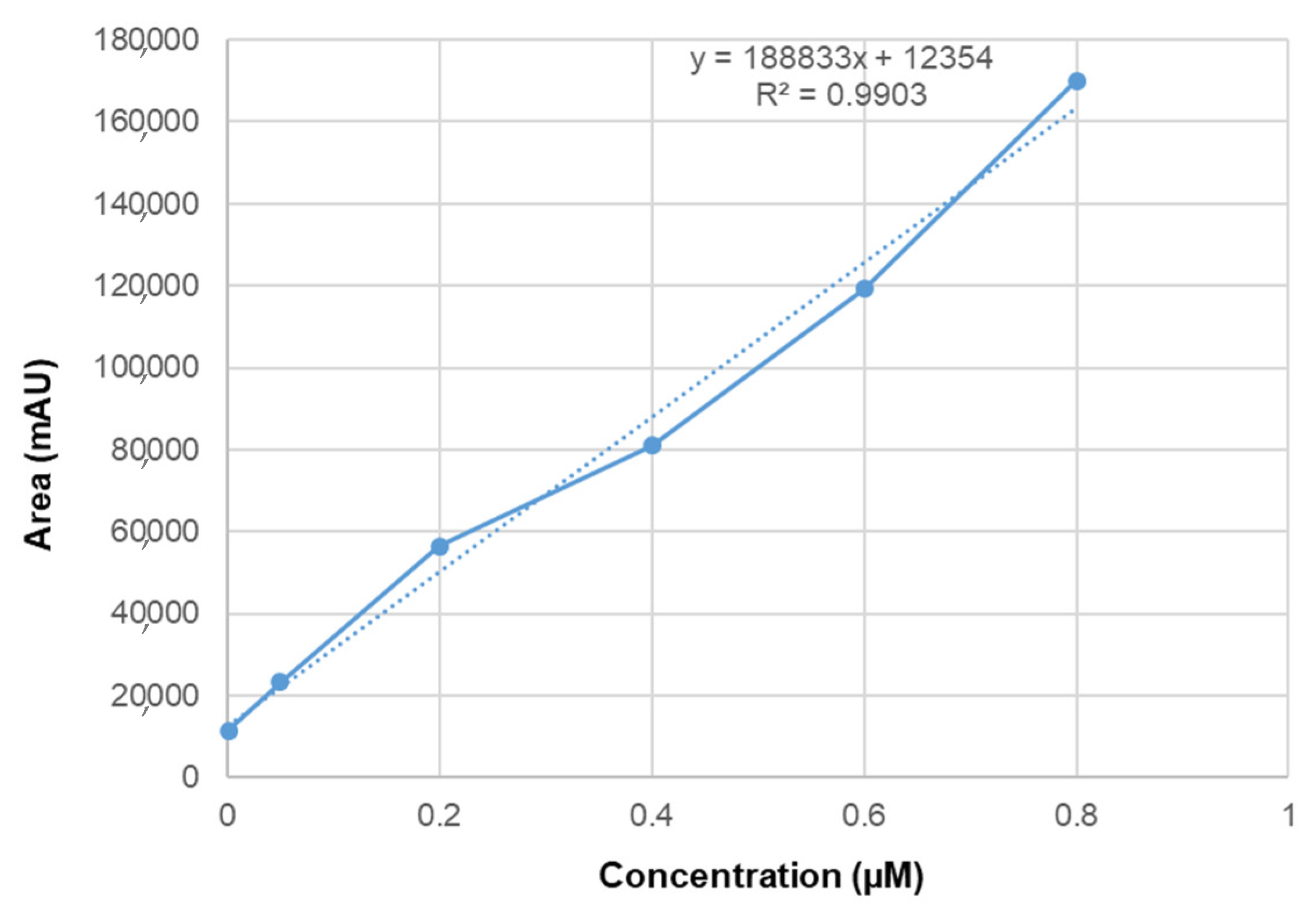
2.7. Linearity and Selectivity

The analytical curve was prepared by diluting the known concentrations of MDA with PBS, with the subsequent method reaction. Increasing concentrations were used (0.001, 0.05, 0.2, 0.4, 0.6, 0.8 μM). All reagents that were used in the TBARS experiment and the adhesive tapes used in the ex vivo assay were examined individually. The tapes were first extracted with 10 mL of methanol with 15 min of ultrasonic bath, followed by the TBARS method. Therefore, the supernatant of the reaction and all reagents were individually inserted into vials after syringe filtration, with HPLC readings through the same chromatographic conditions as the TBARS assay.

2.8. Ethical Issue

The volunteers were provided with the necessary information and clarification regarding the trial. The consent was given by means of the signature through the responsible committee document, maintaining the anonymity of the participant and guaranteeing the departure from the study, voluntarily, at any time. The project was approved by the Ethics Committee by the Faculty of Pharmaceutical Sciences of University of São Paulo, number CAAE: 31583814.0.0000.0067, protocol number 735.493. Volunteers were maintained at a temperature of 21 ± 2 °C and relative humidity of 40–60%. Volunteers were admitted with healthy skin and aging from 18 to 70 years old, with skin type II-V and with no additional exclusion criteria. It was recommended not to apply cosmetic products for 24 h before the experiment in the tested area.

2.9. Ex Vivo Protocol and Stratum Corneum Sample

The ex vivo tape stripping method was applied in 10 volunteers. The volunteer’s volar forearm was cleansed once with purified water and the stratum corneum was removed by the tape stripping technique [14,15,16,17]. Three regions were defined in the volar forearms, one as the control, one irradiated and the third site containing the sunscreen system applied at a ratio of 2.0 mg·cm−2. After 2 h of application, 10 adhesive tapes per region were used for stratum corneum removal [14,15,16,17]. Adapted from Alonso et al., 2009 [15], the tapes were irradiated (except for the control region) in a solar simulator chamber at 2753 or 5506 KJ·m−2. The adhesive tapes were transferred to a glass beaker and the stratum corneum extraction from the tapes was performed by adding 5 mL of methanol followed by an ultrasonic bath for 15 min [15,17]. After being mixed in the vortex and filtrated in the syringe filter, the samples were assayed by HPLC. The adduct quantification was performed according to the reaction method already described.

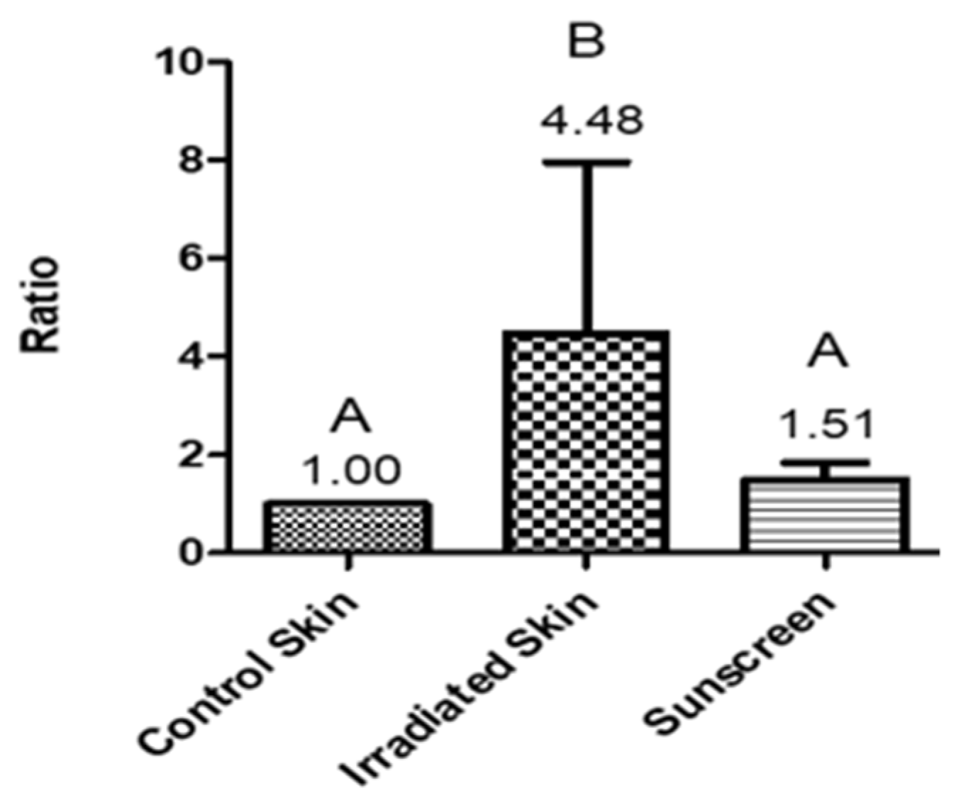
3. Results and Discussion

Thiobarbituric acid reactive species (TBARS) assay—a method to measure lipid peroxidation by the production of malondialdehyde (MDA), which is a product resulting from the peroxidation of polyunsaturated fatty acids—was used as an indicator of the oxidative stress [18], in our particular research, from the stratum corneum obtained by tape stripping. However, until the present literature, there was no data on the measurement of lipid peroxidation by the TBARS method by HPLC in the stratum corneum of participants withdrawn by a tape stripping (ex vivo) technique. This method allows the evaluation of the lipid peroxide species from the stratum corneum formed through artificial UV irradiation, in order to measure the antioxidant activity of several types of samples, from active ingredients to complex formulations.
Considering the characteristics of the tape stripping process being non-invasive [19], biological information from this part of the epidermis could be assessed to develop new protocols to establish—in association with the liquid chromatography [10], which in this investigation was the HPLC—innovative and robust assays for the safety and efficacy of dermocosmetics and topical products.
When using HPLC, an analytical method that admits the identification and quantification of different substances in a sample variety, it is fundamental to prove that it produces reliable and appropriate results for the intended purpose [20]. The linearity of all points was obtained in triplicate with r2 equal to 0.99, as illustrated in Figure 1. It was necessary to know if there was any interfering molecule at the same retention time that the TBARS adduct read 532 nm; the method showed no interferences (Figure 2). Such a result highlighted that this technique, as proposed by our research group, can be considered for the evaluation of several samples and formulation matrixes that would not generate incompatibility signs at the same TBARS adduct retention time.
Lipoperoxidation is the oxidative deterioration of lipids, often affecting polyunsaturated fatty acids for they multiply carbon double bounds which contain reactive hydrogen atoms in the methylene bridges (-CH2-). Subsequently, lipoperoxidation disturbs the biophysical properties of membranes, such as membrane fluidity and electrical resistance, allowing this natural barrier to be corrupted and affecting the organelles inside [21]. MDA is the most studied end-product formed by the lipid peroxidation [18]. Among the many methods in the specialized literature for detection and determination of MDA in biological samples, the main method is the TBARS assay, using the thiobarbituric acid (TBA), first used by Yagi et al., 1968 [22]. TBA can react with a variety of oxidized lipids, being able to form various chromogens, referred to as TBA-reactive substances (TBARS) [23,24,25]. However, the main indicator of lipoperoxidation is the reaction of two molecules of TBA with MDA based upon the heating of the sample under acidic conditions, generating the adduct MDA-TBA2 [26], which is a pink pigment detected by colorimetry at 532 nm or fluorimetry (excitation at 532 nm and emission at 553 nm) and mass ion at 323 amu [26,27,28,29,30,31].
The TBARS assay has been analyzed by different kinds of techniques. However, since TBA can react with other molecules, like saturated and unsaturated aldehydes, a more specific method was desired for specificity in the evaluation of MDA [31,32]. The HPLC method can improve the specificity of MDA-TBA2 detection, being reliable for lipoperoxidation evaluation, confirmed and utilized by some of the recent specialized literature [12,26,33]. Still, unfortunately, due to its extended process timing and high cost, the simple spectrometric method is still predominantly used. Many studies have used the TBARS method to analyze the lipid peroxidation in different fields, especially in the food and medical area, but this is the first time to date that an ex vivo method with TBARS quantification through HPLC was used, being a real innovation in the dermocosmetic field.
Ex Vivo
Previous to the TBARS experiment, it was tested if the irradiation dosage was significant to raise lipoperoxidation induction, quantified by the MDA molecule. This was used as a screening test of four volunteers with two different UV irradiation intensities, 2753 and 5506 KJ·m−2, using two sample sites: the control skin and the irradiated one (same parameters already described). A raise of 34.72% was observed in lipoperoxidation on the intensity of 2753 KJ·m−2, whereas 5506 KJ·m−2 had a raise of 110.88%, proving its efficacy. The ex vivo results of the volunteers is illustrated in Figure 3. To reduce the inter-individual variability effect, the results were exposed as the ratio between the values obtained at each site and the control [34,35,36].
The results showed that the irradiated stratum corneum had a significant increase on the lipoperoxidation by MDA production compared to the control site and the sunscreen system. This was an interesting outcome, since the formulation was in the same protection level as the control site, the only one that did not suffer the harmful stress from the artificial irradiation. This suggested that the lipoperoxidation was avoided by the photoprotection provided by bemotrizinol and ethylhexyl triazone, not allowing the initiation of the oxidative stress cascade phenomenon. Both UV filters are photostable and have high efficacy. Bemotrizinol acts as a broad spectrum UV filter (UVB and UVA) with elevated molar absorptivity at 310 and 343 nm, and ethylhexyl triazone acts in the UVB region, with high molar absorptivity at 314 nm [9,37]. We suggest these parameters as key-points to the development of future assays to confirm claims regarding effectiveness against cutaneous oxidative stress by topical products and dermocosmetics.

4. Conclusions

An exclusive HPLC-TBARS-EVSC protocol to measure lipid peroxidation from subjects’ stratum corneum through TBARS by ex vivo method, using HPLC, was preliminarily developed with linearity and selectivity. By testing the sunscreen system, it had a significative decrease in stratum corneum lipoperoxidative damage compared to the control.
Our protocol also suggests the use of an artificial irradiation dose to stress the stratum corneum to improve the end-point response, and it will be useful in the efficacy establishment of anti-UV and antioxidant agents in investigations that aim to reveal the stratum corneum lipid peroxidation level profile, even in carrying out studies in the food area correlating consumption with stratum corneum anti-oxidative stress attributes and baseline investigations characterizing different ethnicities and genders, for example.

Author Contributions

Conceptualization, A.R.B.; methodology, R.S. and C.A.S.d.O.P.; formal analysis, A.R.B. and R.S.; investigation, A.R.B. and R.S.; writing—original draft preparation, A.R.B. and R.S.; writing—review and editing, A.R.B.; supervision, A.R.B. and M.V.R.V.; project administration, A.R.B.; funding acquisition, A.R.B., M.V.R.V., Z.A.P. and C.A.-J. All authors have read and agreed to the published version of the manuscript.

Funding

This research was funded by Fundação de Amparo à Pesquisa do Estado de São Paulo (FAPESP), grant numbers 2016/24360-4 and 2019/16169-0; Conselho Nacional de Desenvolvimento Científico e Tecnológico (CNPq), process 305250/2019-1; CONCYTEC-FONDECYT, process E041-01 (Contract number 123-2018-FONDECYT-BM-IADT/AV); and Coordenação de Aperfeiçoamento de Pessoal de Nível Superior-Brasil (CAPES) Finance Code 001.

Institutional Review Board Statement

This study was conducted according to the guidelines of the Declaration of Helsinki, and approved by the Ethics Committee of FCF-USP (CAAE: 31583814.0.0000.0067, protocol number 735.493).

Informed Consent Statement

Informed consent was obtained from all subjects involved in the study.

Data Availability Statement

Data sharing not applicable.

Conflicts of Interest

The authors declare no conflict of interest.

References

  1. Halliwell, B.; Gutteridge, J.M.C. Free Radicals in Biology and Medicine; Oxford University Press: Oxford, UK, 1989. [Google Scholar]
  2. Rahman, K. Studies on free radicals, antioxidants, and co-factors. Clin. Interv. Aging 2007, 2, 219–236. [Google Scholar] [PubMed]
  3. Lobo, V.; Patil, A.; Phatak, A.; Chandra, N. Free radicals, antioxidants and functional foods: Impact on human health. Pharmacogn. Rev. 2010, 4, 118. [Google Scholar] [CrossRef] [Green Version]
  4. Singh, P.P.; Chandra, A.; Mahdi, F.; Roy, A.; Sharma, P. Reconvene and reconnect the antioxidant hypothesis in human health and disease. Indian J. Clin. Biochem. 2010, 25, 225–243. [Google Scholar] [CrossRef] [PubMed] [Green Version]
  5. Lü, J.M.; Lin, P.H.; Yao, Q.; Chen, C. Chemical and molecular mechanisms of antioxidants: Experimental approaches and model systems. J. Cell. Mol. Med. 2010, 14, 840–860. [Google Scholar] [CrossRef] [PubMed]
  6. Netea, M.G.; Balkwill, F.; Chonchol, M.; Cominelli, F.; Donath, M.Y.; Giamarellos-Bourboulis, E.J.; Golenbock, D.; Gresnigt, M.S.; Heneka, M.T.; Hoffman, H.M.; et al. A guiding map for inflammation. Nat. Immunol. 2017, 18, 826–831. [Google Scholar] [CrossRef] [Green Version]
  7. Johnson, A. A current view on inflammation. Nat. Immunol. 2017, 18, 825. [Google Scholar] [CrossRef] [Green Version]
  8. Walsh, L.J. Mast cells and oral inflammation. Crit. Rev. Oral Biol. Med. 2003, 14, 188–198. [Google Scholar] [CrossRef]
  9. Peres, D.D.A.; Sarruf, F.D.; de Oliveira, C.A.; Velasco, M.V.R.; Baby, A.R. Ferulic acid photoprotective properties in association with UV filters: Multifunctional sunscreen with improved SPF and UVA-PF. J. Photochem. Photobiol. B Biol. 2018, 185, 46–49. [Google Scholar] [CrossRef]
  10. Sauce, R.; Pinto, C.A.S.D.O.; Velasco, M.V.R.; Rosado, C.; Baby, A.R. Ex vivo penetration analysis and anti-inflammatory efficacy of the association of ferulic acid and UV filters. Eur. J. Pharm. Sci. 2021, 156. [Google Scholar] [CrossRef]
  11. Potard, G.; Laugel, C.; Baillet, A.; Schaefer, H.; Marty, J.P. Quantitative HPLC analysis of sunscreens and caffeine during in vitro percutaneous penetration studies. Int. J. Pharm. 1999, 189, 249–260. [Google Scholar] [CrossRef]
  12. Bastos, A.S.; Loureiro, A.P.D.M.; De Oliveira, T.F.; Corbi, S.C.T.; Caminaga, R.M.S.; Rossa, C.; Orrico, S.R.P. Quantitation of malondialdehyde in gingival crevicular fluid by a high-performance liquid chromatography-based method. Anal. Biochem. 2012, 423, 141–146. [Google Scholar] [CrossRef] [PubMed]
  13. Hong, Y.L.; Yeh, S.L.; Chang, C.Y.; Hu, M.L. Total plasma malondialdehyde levels in 16 Taiwanese college students determined by various thiobarbituric acid tests and an improved high-performance liquid chromatography-based method. Clin. Biochem. 2000, 33, 619–625. [Google Scholar] [CrossRef]
  14. Benfeldt, E.; Hansen, S.H.; Vølund, A.; Menné, T.; Shah, V.P. Bioequivalence of topical formulations in humans: Evaluation by dermal microdialysis sampling and the dermatopharmacokinetic method. J. Investig. Dermatol. 2007, 127, 170–178. [Google Scholar] [CrossRef] [PubMed] [Green Version]
  15. Alonso, C.; Barba, C.; Rubio, L.; Scott, S.; Kilimnik, A.; Coderch, L.; Notario, J.; Parra, J.L. An ex vivo methodology to assess the lipid peroxidation in stratum corneum. J. Photochem. Photobiol. B. 2009, 97, 71–76. [Google Scholar] [CrossRef]
  16. De Oliveira, C.A.; Peres, D.D.; Rugno, C.M.; Kojima, M.; de Pinto, C.A.S.O.; Consiglieri, V.O.; Kaneko, T.M.; Rosado, C.; Mota, J.; Velasco, M.V.R.; et al. Functional photostability and cutaneous compatibility of bioactive UVA sun care products. J. Photochem. Photobiol. B Biol. 2015, 148, 154–159. [Google Scholar] [CrossRef]
  17. Peres, D.D. Ácido Ferúlico em Protetores Solares: Desenvolvimento e Eficácia Multifuncional in vitro, ex vivo e in vivo. Ph.D. Thesis, University of São Paulo, Sao Paulo, Brazil, 2015. [Google Scholar]
  18. Del Rio, D.; Stewart, A.J.; Pellegrini, N. A review of recent studies on malondialdehyde as toxic molecule and biological marker of oxidative stress. Nutr. Metab. Cardiovasc. Dis. 2005, 15, 316–328. [Google Scholar] [CrossRef]
  19. Cândido, T.M.; de Oliveira, C.A.; Ariede, M.B.; Velasco, M.V.R.; Rosado, C.; Baby, A.R. Safety and Antioxidant Efficacy Profiles of Rutin-Loaded Ethosomes for Topical Application. AAPS PharmSciTech 2018. [Google Scholar] [CrossRef]
  20. Ich ICH Topic Q2 (R1) Validation of Analytical Procedures: Text and Methodology. Int. Conf. Harmon. 2005, 1994, 17.
  21. Devasagayam, T.P.A.; Boloor, K.K.; Ramasarma, T. Methods for estimating lipid peroxidation: An analysis of merits and demerits. Indian J. Biochem. Biophys. 2003, 40, 300–308. [Google Scholar]
  22. Yagi, K.; Nishigaki, I.; Ohama, H. Measurement of serum TBA value. Vitamin 1968, 37, 105–112. [Google Scholar]
  23. Alessio, H. Handbook of Oxidants and Antioxidants in Exercise; Elsevier: Amsterdam, The Netherlands, 2000. [Google Scholar]
  24. Kosugi, H.; Kikugawa, K. Thiobarbituric acid reaction of aldehydes and oxidized lipids in glacial acetic acid. Lipids 1985, 20, 915–921. [Google Scholar] [CrossRef]
  25. Buttkus, H.; Bose, R.J. Amine-malonaldehyde condensation products and their relative color contribution in the thiobarbituric acid test. J. Am. Oil Chem. Soc. 1972, 49, 440–443. [Google Scholar] [CrossRef]
  26. Moselhy, H.F.; Reid, R.G.; Yousef, S.; Boyle, S.P. A specific, accurate, and sensitive measure of total plasma malondialdehyde by HPLC. J. Lipid Res. 2013, 54, 852–858. [Google Scholar] [CrossRef] [PubMed] [Green Version]
  27. Dib, M.; Garrel, C.; Favier, A.; Robin, V.; Desnuelle, C. Can malondialdehyde be used as a biological marker of progression in neurodegenerative disease? J. Neurol. 2002, 249, 367–374. [Google Scholar] [CrossRef]
  28. Panjamurthy, K.; Manoharan, S.; Ramachandran, C.R. Lipid peroxidation and antioxidant status in patients with periodontitis. Cell. Mol. Biol. Lett. 2005, 10, 255–264. [Google Scholar] [PubMed]
  29. Yagi, K. A simple fluorometric assay for lipoperoxide in blood plasma. Biochem. Med. 1976, 15, 212–216. [Google Scholar] [CrossRef]
  30. Jardine, D.; Antolovich, M.; Prenzler, P.D.; Robards, K. Liquid chromatography-mass spectrometry (LC-MS) investigation of the thiobarbituric acid reactive substances (TBARS) reaction. J. Agric. Food Chem. 2002, 50, 1720–1724. [Google Scholar] [CrossRef]
  31. Knight, J.A.; Robert, K.; Mcclellan, L. Specificity of the thiobarbituric acid reaction: Its use in studies of lipid peroxidation. Clin. Chem. 1988, 34, 2433–2438. [Google Scholar] [CrossRef]
  32. Abuja, P.M.; Albertini, R. Methods for monitoring oxidative stress, lipid peroxidation and oxidation resistance of lipoproteins. Clin. Chim. Acta 2001, 306, 1–17. [Google Scholar] [CrossRef]
  33. Al-Rimawi, F. Development and Validation of a Simple Reversed-Phase HPLC-UV Method for Determination of Malondialdehyde in Olive Oil. J. Am. Oil Chem. Soc. 2015, 92, 933–937. [Google Scholar] [CrossRef]
  34. De Oliveira, C.A.; Dario, M.F.; Sarruf, F.D.; Mariz, I.F.A.; Velasco, M.V.R.; Rosado, C.; Baby, A.R. Safety and efficacy evaluation of gelatin-based nanoparticles associated with UV filters. Colloids Surf. B Biointerfaces 2015, 140, 531–537. [Google Scholar] [CrossRef]
  35. Graziola, F.; Candido, T.M.; De Oliveira, C.A.; Peres, D.D.A.; Georges Issa, M.; Mota, J.; Rosado, C.; Consiglieri, V.O.; Kaneko, T.M.; Velasco, M.V.R.; et al. Gelatin-based microspheres crosslinked with glutaraldehyde and rutin oriented to cosmetics. Artic. Braz. J. Pharm. Sci. 2016, 52, 55–66. [Google Scholar] [CrossRef] [Green Version]
  36. Peres, D.A.; de Oliveira, C.A.; da Costa, M.S.; Tokunaga, V.K.; Mota, J.P.; Rosado, C.; Consiglieri, V.O.; Kaneko, T.M.; Velasco, M.V.R.; Baby, A.R. Rutin increases critical wavelength of systems containing a single UV filter and with good skin compatibility. Ski. Res. Technol. 2016, 22. [Google Scholar] [CrossRef] [PubMed]
  37. Tuchinda, C.; Lim, H.W.; Osterwalder, U.; Rougier, A. Novel Emerging Sunscreen Technologies. Dermatol. Clin. 2006, 24, 105–117. [Google Scholar] [CrossRef] [PubMed]
Figure 1. Analytical curve of the HPLC-TBARS-EVSC assay in the range of 0.001 to 0.8 μM, with line equation y = 188,833x + 12,354.
Scipharm 89 00017 g001
Figure 2. Selectivity of the HPLC-TBARS-EVSC assay. Chromatogram of 1: TBARS reaction (adduct); 2: sunscreen system and adhesive tape (irradiated and non-irradiated); 3: n-butanol; and 4: 0.2% BHT, H3PO4 0.44 M and 1% H2SO4. Retention time (min).
Scipharm 89 00017 g002
Figure 3. Ratio between values obtained at the sites (control, irradiated and sunscreen sites) of the HPLC-TBARS-EVSC assay. Different letters for the same parameter indicate statistically significant differences between samples (one-way ANOVA; p < 0.05; n = 10). Skin = stratum corneum.
Scipharm 89 00017 g003
Publisher’s Note: MDPI stays neutral with regard to jurisdictional claims in published maps and institutional affiliations.

