Article

Purple Corn Silk Extract Attenuates UVB-Induced Inflammation in Human Keratinocyte Cells

by
Watcharaporn Poorahong
1,
Sukanda Innajak
1,
Malyn Ungsurungsie
2 and
Ramida Watanapokasin
1,*
1
Department of Biochemistry, Faculty of Medicine, Srinakharinwirot University, Bangkok 10110, Thailand
2
Faculty of Pharmacy, Mahidol University, Bangkok 10400, Thailand
*
Author to whom correspondence should be addressed.
Submission received: 22 January 2022 / Revised: 22 February 2022 / Accepted: 25 February 2022 / Published: 1 March 2022

Abstract

:
UVB is a causative factor for severe skin damage, such as cell aging, death, and inflammation. UVB easily permeates into the epidermis layer of human skin, which is mainly composed of keratinocyte cells. In previous results, we found that purple corn silk (PCS) extract showed the potential to inhibit keratinocyte damages of UVB-treated cells. Thus, in this study, we aimed to evaluate the preventive effects of PCS extract against the inflammation of UVB-induced keratinocyte cells using the HaCaT cell line. HaCaT cells were treated with PCS extract at various concentrations for 1 h, then exposed to 25 mJ/cm2 UVB before subsequent experiments. Fragmented DNA was observed using flow cytometry. The inflammatory response was investigated through NF-κB activity by immunofluorescence staining and related protein expression by Western blotting. The results demonstrated that PCS extract decreased the sub-G1 DNA content. Interestingly, PCS extract attenuated NF-κB activity via suppressed NF-κB nuclear translocation and protein expression. Moreover, PCS extract remarkably decreased c-Jun phosphorylation and decreased proinflammatory cytokines, along with iNOS and COX-2 levels in UVB-treated cells compared to the UVB-control group. This finding exhibited that PCS extract minimized inflammation in keratinocyte cells induced by UVB radiation.

1. Introduction

Solar ultraviolet (UV) light is composed of UVA (320–400 nm), UVB (280–320 nm), and UVC (200–280 nm). UVC is completely blocked by the ozone layer, while only 90–95% of UVA and 5–10% of UVB reach the Earth and affect human skin. While the level of UVB reaching the ground is lower, UVB energy is more harmful than UVA. Most of the UVB radiation can permeate into the epidermis layer, which is composed of at least 80% keratinocyte cells. Numerous studies reported that UVB exposure to the skin can stimulate intracellular reactive oxygen species (ROS) overproduction, mediated oxidative damage, and enhanced risk of skin carcinogenesis [1,2,3,4].
ROS play an important role in the initiation and induction of an inflammatory response. ROS induce oxidative damage by oxidizing and modifying various cellular molecules such as lipids, DNA, and proteins [1,4,5,6]. During skin exposure to UVB, the intracellular ROS is increased and mediates activation of transcription factors, including nuclear factor-kappa B (NF-κB). In latent stages, NF-κB associated with an inhibitor of κB (IκBs) is in an inactive form. The excessive intracellular ROS molecules can drive modification of IκBs proteins, which leads to their degradation through ubiquitination. This event activates NF-κB translocation from the cytosol into the nucleus, followed by up-regulating the gene expression of inflammatory mediators such as interleukins (ILs), cyclooxygenase-2 (COX-2), and inducible nitric oxide synthase (iNOS) [5,7,8,9,10]. Likewise, UVB and intracellular ROS mediate the induction of mitogen-activated protein kinases (MAPKs), especially p38 MAPKs and c-Jun NH2-terminal kinase (JNK). JNK mostly activates c-Jun phosphorylation at a positive regulatory domain, thereby activating the transcription of various genes. Previous studies suggested that p38 MAPKs and JNK are primarily responsible for oxidative stress as well as UV radiation. p38 MAPKs and c-Jun promote the AP-1 complex, which can turn on gene expression to enhance skin inflammation [11,12,13,14,15,16].
Therefore, in this study, we demonstrated that UVB and intracellular ROS are associated with skin inflammation. In a previous publication, we found that the purple corn silk (PCS) extract showed antioxidant power through scavenging free radicals and decreased intracellular ROS in UVB-treated HaCaT cells [17]. Corn silk, part of a kind of female corn, is an important source of polyphenols. Some reports indicated that purple corn silk extract is rich in anthocyanin as a major component [18,19,20,21,22,23,24]. However, information about the role of PCS extract against UVB-induced inflammation in keratinocyte cells is not yet investigated. Hence, in this study, we described the potential of PCS extract against the UVB-induced inflammatory response in keratinocyte HaCaT cells.

2. Results

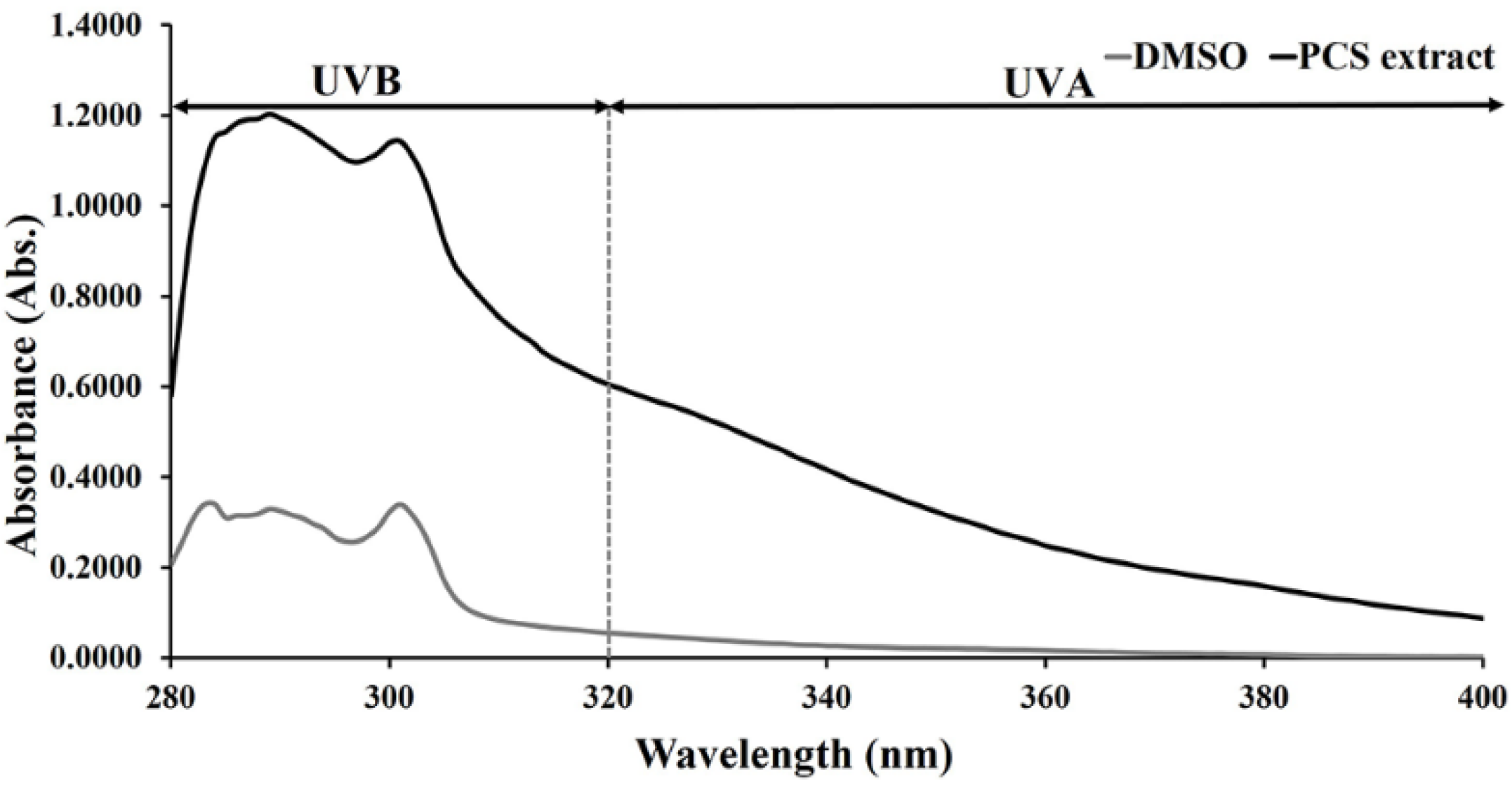
2.1. UV-Absorbing Properties of PCS Extract

Generally, the epidermal layer acts as the first line of defense against solar UV through UV absorb. The PCS extract can minimize the effects of UV on skin cells, as shown in Figure 1; 1 mg/mL of PCS extract can absorb mainly the UVB. These data indicate that PCS extract acted as physical UVB protection, preventing UVB energy from entering skin keratinocyte cells.

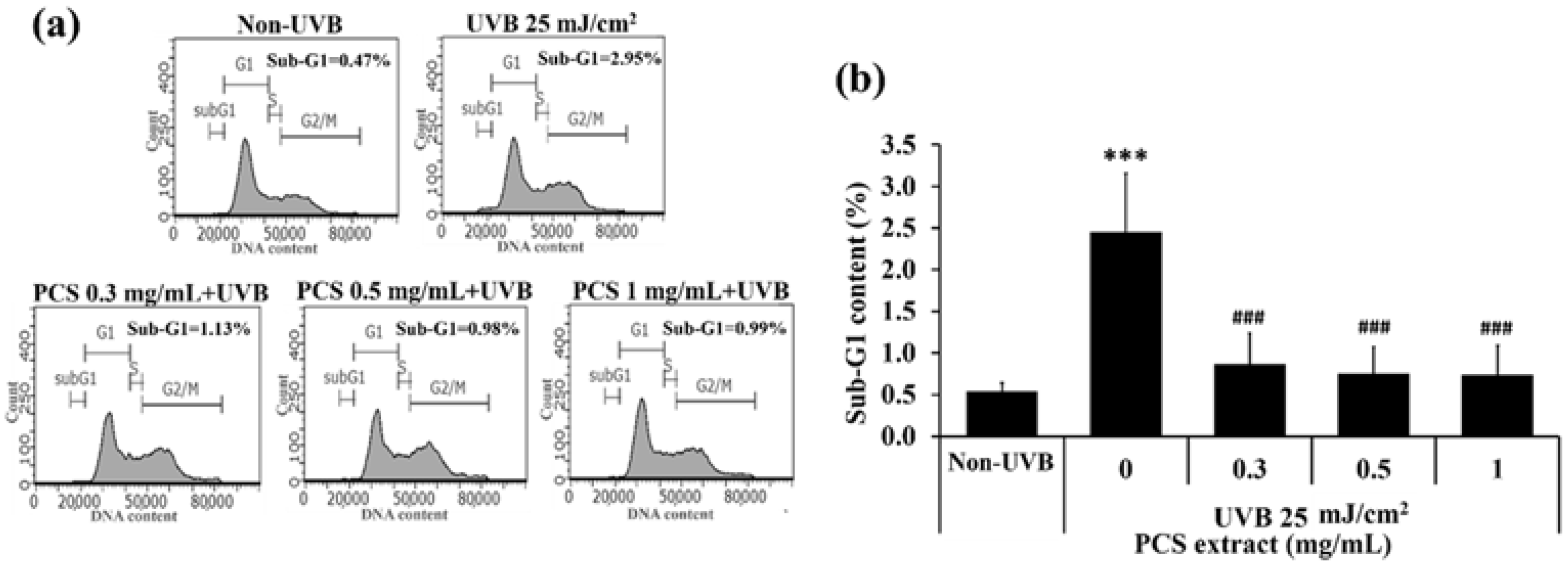
2.2. PCS Extract Attenuated UVB-Induced Cell Death in HaCaT Cells

In a previous study, we demonstrated that PCS extract at concentrations of 0.3, 0.5, and 1 mg/mL prevented cell death in UVB-treated cells [17]. Therefore, in this study, flow cytometry was conducted to examine the sub-G1 population, indicating fragmented DNA in dead cells. These results were used to confirm the anti-cell-death activity of PCS extract before subsequent experiments. The result showed that the UVB-control group significantly increased sub-G1 content at level p < 0.001 compared to the non-UVB group, whereas PCS extract reduced cell death higher than the UVB-control group (Figure 2). This result confirms that PCS extract inhibits cell death induction in UVB-treated HaCaT cells.

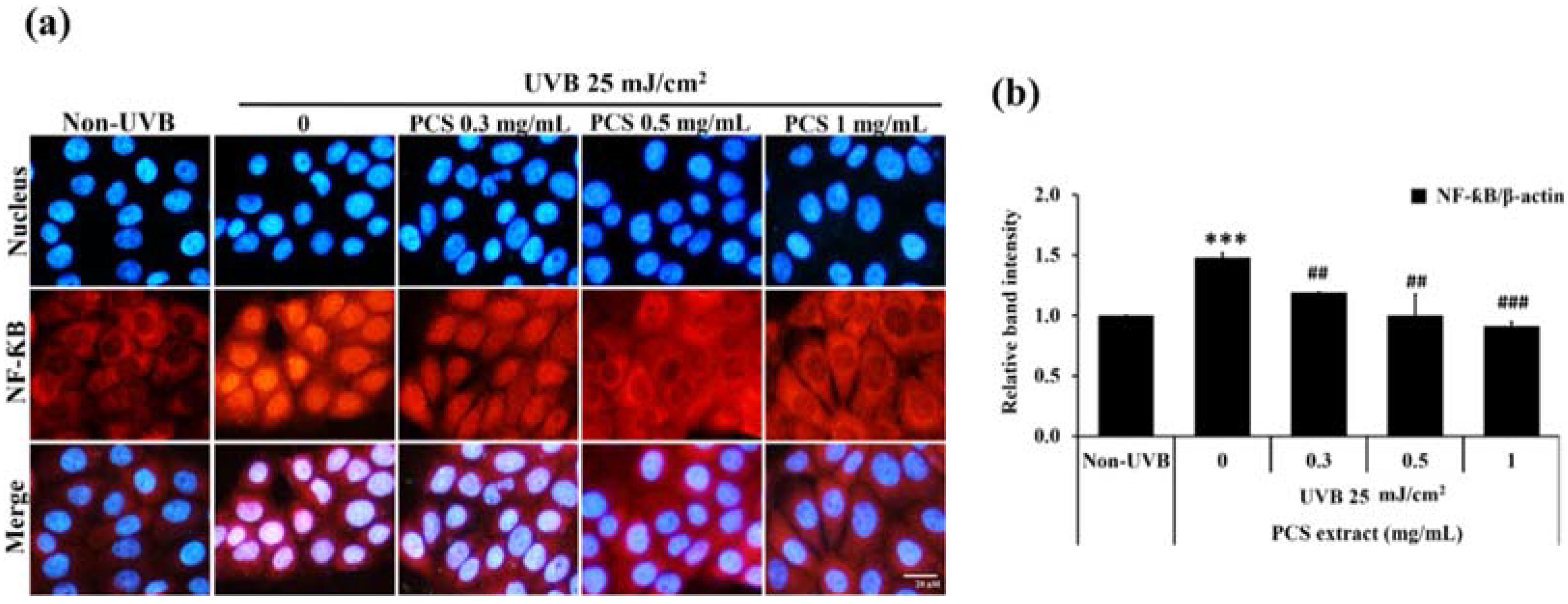
2.3. PCS Extract Decreases NF-κB Activity in UVB-Treated HaCaT Cells

The increased NF-κB function relates to cellular inflammation. Immunofluorescence staining was used to determine NF-κB activity via monitoring the translocation of NF-κB from the cytosol into the nucleus. The results demonstrated that UVB increased NF-κB nuclear translocation compared to the non-UVB group. In contrast, cells pre-treated with PCS extract showed a strong attenuation of this event compared to the UVB-control group in a dose-dependent manner, as shown in Figure 3a. Furthermore, we found that UVB significantly increased NF-κB protein expression, which was inhibited by PCS extract (Figure 3b). These results indicated that PCS extract suppressed NF-κB nuclear translocation and protein expression in UVB-induced HaCaT cells.

2.4. PCS Extract Reduced Proinflammatory Cytokine Expression in UVB-Treated HaCaT Cells

In a subsequent experiment, we investigated the expression of proinflammatory cytokines regulated by NF-κB activities, including iNOS and COX-2. In unstimulated keratinocyte cells, these proteins were at a low level. In contrast, upon the stimulation of HaCaT cells by UVB irradiation, both iNOS and COX-2 levels were significantly increased (p < 0.001) compared to the non-UVB group, whereas 0.3, 0.5, and 1 mg/mL of PCS extract significantly decreased iNOS and COX-2 levels when compared to the UVB-control group, as shown in Figure 4. These findings prove that PCS extract was an effective inhibitor in the inflammation process in UVB-exposed human keratinocyte cells.

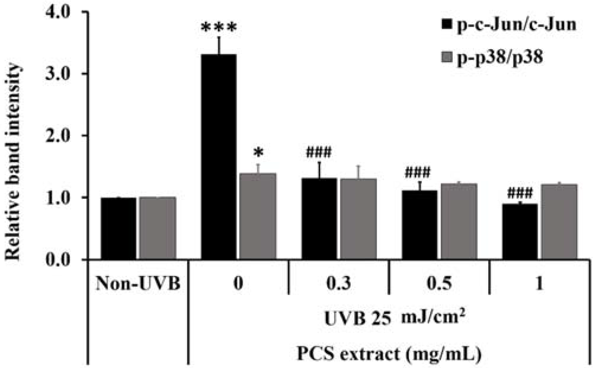
2.5. PCS Extract Decreased UVB-Induced MAPKs Phosphorylation in UVB-Treated HaCaT Cells

Further, we monitored the influence of UVB on the phosphorylation of stress-sensitive MAPKs family proteins by Western blot analysis. The results demonstrated that UVB increased p38 and c-Jun phosphorylation at the levels of p < 0.05 and p < 0.001, respectively, compared to the non-UVB group (Figure 5). In contrast, PCS extract slightly reduced phosphorylated p38 with no statistically significant difference (p < 0.001) compared to the UVB-control group. Remarkably, PCS extract strongly inhibited the phosphorylated c-Jun protein level in a dose-dependent manner compared to the UVB control group at a value of p < 0.001, as shown in Figure 5. These results show that PCS extract primarily suppressed c-Jun phosphorylation in the UVB-irradiated HaCaT cells.

3. Discussion

The outermost layer of human skin, known as the epidermis layer, is prominently composed of keratinocyte cells, which serve as a physical barrier against environmental insults such as pathogens, chemicals, and harmful radiation, especially solar UVB radiation. UVB radiation reaching the Earth accounts for only around 4–5% of total solar UV. However, solar UVB is an important inducer of photoaging, sunburn, wrinkles, inflammation, skin cell death, and increased risk of skin carcinogenesis in keratinocyte cells [1,2,3,4,13,25,26,27].
Natural plants are abundant sources of phytonutrients potent as antioxidant agents such as polyphenols, flavonoids, anthocyanins, carotenoids, and lutein. In the case of purple corn, the evidence reported that purple corn and purple maize pigment extract are rich in anthocyanin [18,19,28,29]. Similarly, a section of a kind of female corn (corn silk) is an important source of polyphenols. The ethanol and aqueous extract of purple corn silk possesses abundant polyphenol components, especially anthocyanins. In addition, these extracts showed antioxidant power through scavenging free radicals and enhancing antioxidant enzyme activities [20,21,22,23,24]. In our previous report, screening based on visible color changes of anthocyanin towards different pH values showed that adding 2 N hydrochloric acid (HCl) to PCS extract appeared as a pink–red color that turned deep green upon ammonia addition, which predicted that PCS extract might be rich in anthocyanin content. Moreover, the free-radical scavenging activity of PCS extract was investigated by using a 2,2-Diphenyl-1-(2,4,6-trinitrophenyl) hydrazyl (DPPH) assay. The effective concentration (EC50) value of PCS extract was at 0.53 mg/mL [17]. However, the phytochemicals of PCS extract need to be further confirmed.
Recently, the beneficial properties of phytonutrients in UV prevention were discovered. Previous studies found that purple corn silk extract showed potential activity against cell damage in human keratinocyte cells stimulated by UVB irradiation [17]. In this study, we were interested in the specific effects of PCS extract on human keratinocyte cells through evaluating its activity against UVB-induced skin inflammation. We found that 25 mJ/cm2 of UVB increased fragmented DNA in the UVB-treated HaCaT cells, which was attenuated by PCS extract. Hence, our results confirmed that the PCS extract prevented DNA fragmentation and cell death in the UVB-treated HaCaT cells. Generally, UVB energy induces intracellular ROS generation. Accumulated cellular ROS significantly stimulates skin inflammation. According to previous studies, PCS extract attenuates free radicals and the overproduction of ROS in HaCaT cells induced by UVB [17], and we hypothesized that PCS extract might prevent UVB-induced inflammatory response in keratinocyte cells, as it is well known that ROS directly related to NF-κB activation is a key mediator of inflammation regulating gene expression, including COX-2, iNOS, TNF-α, IL-6, IL-8, and IL-1β [8,30,31]. In keratinocyte cells, NF-κB increased the sensitivity of inflammatory response in UVB-irradiated cells and promoted inflammatory cytokine expression [7,16,32,33,34]. Interestingly, many reports showed that phytonutrients, especially a group of phenolic and proanthocyanidins, effectively attenuated NF-κB activities [19]. In this study, we found that PCS extract significantly inhibited the NF-κB activation stimulated by UVB. In addition, we observed that PCS extract decreased iNOS and COX-2 levels in the UVB-treated HaCaT cells. Likewise, previous studies suggested that UVA- and UVB-induced skin inflammation through NF-κB activation led to increased various inflammatory cytokines, which were attenuated by anthocyanin-rich extracts such as raspberry, black soybean, and strawberry extract [26,35,36]. However, we suggested that the downstream of COX-2 and iNOS involved in the inflammatory process needs to be explored to fully understand the anti-inflammatory effects of PCS extract against UVB in human keratinocyte cells.
The activation of MAPK proteins is known as a direct intracellular response signaling pathway that stimulates the expression of various target genes. Most evidence suggests that UVB strongly activates stress-sensitive JNK and p38 MAPKs to enhance skin inflammation [11]. In general, UVB causing JNK and p38 MAPK phosphorylation leads to an increase in proinflammatory cytokine expression. Papavassiliou et al. (2020) suggested that the activation of JNK mostly activated c-Jun phosphorylation at a positive regulatory domain: Ser63/73 and Thr91/93 residues. Phosphorylated c-Jun was able to form c-Jun homodimerization or heterodimerization with c-Fos [15]. This event consequently improved the activities of the transcriptional factor subunit of the activator protein 1 (AP-1) complex. The increased AP-1 activity up-regulated proinflammatory cytokines, including COX2 and iNOS [8,11,30,37]. Moreover, Spiegelman et al. (2000) suggested that activation of JNK resulted in the accumulation of β-transducin repeat-containing protein (β-TrCP), which mediated the ubiquitination of phosphorylated IκBα protein. This event indicated that JNK plays an important role in the regulation of NF-κB activation [38]. Likewise, Chen et al. (2002) considered the relationship between JNK and the NF-κB signaling pathway in inflammatory processes. The report indicated that diverse stimuli, including intra-cellular stresses and UV light, could stimulate JNK activity, potentially linked to the IκB ubiquitination, turning on NF-κB activities [39]. This evidence correlates with our study, where we found that at 25 mJ/cm2, UVB induces both c-Jun and p38 activity, as shown in Figure 5. Our results demonstrated that PCS attenuated not only NF-κB but also phosphorylated c-Jun inhibition in HaCaT cells induced by UVB. Additionally, it is well known that JNK phosphorylation activates the apoptosis pathway through regulating B-cell lymphoma-2 (BCL-2) family proteins, such as Bcl-2 suppression, and the induction of Bcl-2-associated X protein (Bax) translocation to the mitochondria, resulting in apoptosis induction [40]. The current study demonstrated that PCS suppressed sub-G1 content and c-Jun phosphorylation, while previously, studies showed that PCS extract attenuated UVB-induced apoptosis through decreased Bax and increased Bcl-2 levels in UVB-treated cells [17]. Hence, it is possible that the anti-apoptotic activity of PCS extract is through suppressed c-Jun phosphorylation in UVB-treated cells. Similarly, the red raspberry extract and cyanidin-3-glucoside (C3G) inhibited skin damage in UVB-treated HaCaT cells through attenuated p-p38 and p-c-Jun [35,41,42,43,44], while the amla fruit extract attenuated the inflammatory response to UVB by inhibiting AP-1, NF-κB, and mediator PGE2 without any impact on p38 activation [45]. Similarly, PCS extract seems to slightly reduce p38 activity without a statistically significant difference (p < 0.001) compared to the UVB-control group. Hence, a deeper understanding of the effect of PCS extract on the molecular mechanism needs to be further studied. Based on these studies, we suggested that PCS extract affects mainly through NF-κB and c-Jun activity, leading to attenuated UVB-induced skin inflammation in keratinocyte cells.

4. Materials and Methods

4.1. Reagents

Dulbecco’s modified Eagle’s medium (DMEM), fetal bovine serum (FBS), penicillin–streptomycin, and trypsin-EDTA were purchased from HiMedia (HiMedia, Laboratories, Mumbai, India). Guava cell cycle® reagent, PVDF membranes, an antibody against iNOS and β-actin, and chemiluminescence (ECL) reagent were purchased from Merck Millipore (Merck Millipore Corp., Darmstadt, Germany). An antibody against NF-κB, COX-2, p38, phosphorylated-p38, c-Jun, phosphorylated-c-Jun, and anti-mouse/-rabbit secondary antibody conjugated horseradish peroxidase (HRP) were purchased from Cell Signalling (Cell Signalling Technology, Danvers, MA, USA). Anti-mouse secondary antibody conjugated Alexafluoro-546 and fluorescent Hoechst33342 dye were purchased from Thermo Fisher Scientific (Invitrogen™, Thermo Fisher Scientific Inc., Waltham, MA, USA). Complete Mini Protease Inhibitor Cocktail was purchased from Roche Diagnostics GmbH (Roche Diagnostics GmbH, Mannheim, Germany).

4.2. Plant Extraction

The purple stigma of Zea mays L. was used to prepare the PCS extract, which was obtained from Prof. Dr. Malyn Ungsurungsie. The extraction method was as in our previous study [17]. Briefly, the purple corn style was harvested from Khonkaen province, Thailand, and kept in the laboratory at the Plant Breeding Research Centre, Khonkaen University, Thailand (Voucher No. 050914T001). The fresh specimen was dried at 50 °C in an oven to remove humidity from sample plants. Then, the specimens were ground into a powder using an electric blender. After that, 10 g of dry purple corn silk powder was immersed in 100 mL of 50% propylene glycol (PG) for 2 days, followed by the filtration of the extract solution through cheesecloth and filter paper Whatman No.4. Then, the filtrated PCS extract solution was evaporated using a vacuum evaporator and freeze-dried in a lyophilizer. Then, the PCS extract was kept in the dark at −20 °C until use.

4.3. UVB Irradiation System

A UV incubator (BIO SUN Ultraviolet VILBER LOURMAT) consisting of UVB light (2 × 30 W) at a wavelength of 312 nm was used as a UV source, and the irradiation distance was 25 mm. Cells were placed in 1 mL of phosphate-buffered saline (PBS) and exposed to a UVB lamp in a UV incubator without a plastic lid. After UVB irradiation, cells were placed in the fresh medium and incubated at 37 °C and 5% CO2 in a cell culture incubator (Forma™ Steri-Cult™, Thermo Fisher Scientific Inc., Waltham, MA, USA) before subsequent experiments.

4.4. UV-Absorbing Properties of PCS Extract

The UV-absorbing properties of PCS extract were investigated. The PCS extract at 1 mg/mL (in 100% DMSO) and 100% DMSO (a negative control) was prepared. Then, the UV-absorbing properties were measured at every 1 nm covering a range of 280–400 nm wavelengths using a spectrophotometer (Multiskan Sky, Thermo Fisher Scientific Inc., Waltham, MA, USA). The optical density (OD) of a sample is indicated as the UV-absorbing property of samples.

4.5. Cell Culture

The human keratinocyte HaCaT cell line was purchased from American Type Culture Collection (ATCC, Manassas, VA, USA). The cells were maintained in DMEM medium supplemented with 10% FBS, 100 U/mL penicillin, and 100 μg/mL streptomycin in T75 tissue culture flasks as a monolayer. Cells were grown in a humidified incubator at 37 °C and 5% CO2. At 80% confluence every 2–3 days, cells were subcultured by 0.25% trypsin-EDTA.

4.6. Flow Cytometry

Cells were seeded in 30 × 104 cells/well overnight and pre-treated with PCS extract at concentrations of 0.3, 0.5, and 1 mg/mL for 1 h. Then, the cells were exposed to 25 mJ/cm2 UVB and incubated with fresh medium at 37 °C for 24 h before being harvested using 0.25% trypsin-EDTA. Then, the supernatant was removed by centrifugation at 2000 rpm for 3 min. Cell pellets were fixed with 70% ice-cold ethanol, then washed and stained with Guava cell cycle® reagent for 30 min at room temperature in the dark. The sub-G1 content was determined using a Guava EasyCyte™ flow cytometer and GuavaSoft™ software (Merck Millipore Crop., Darmstadt, Germany).

4.7. Immunofluorescence NF-κB Staining

HaCaT cells were grown on glass coverslips in 35 mm culture dishes (30 × 104 cells/well) overnight. Cells were pre-treated with PCS extract at concentrations of 0.3, 0.5, and 1 mg/mL for 1 h followed by the UVB exposure, then incubated at 37 °C for 1 h. After that, cells were fixed with 4% paraformaldehyde for 10 min and permeabilized with 0.5% Triton X-100. Then, cells were blocked with 3% bovine serum albumin (BSA) and incubated with an antibody against NF-κB (1:250) at 4 °C overnight. After incubation, cells were incubated with secondary antibody conjugated with Alexafluoro-546 (1:500) at room temperature, and the nuclei were stained with 5 μg/mL of Hoechst33342 dye. The fluorescence signals were observed under a fluorescent microscope (IX71 Olympus, Tokyo, Japan).

4.8. Western Blot Analysis

After treatment, cells were collected by scraping and lysis with RIPA lysis buffer (50 mM Tris-HCl, 5 mM EDTA, 250 mM NaCl, 0.5% Triton X-100, pH 7.5) containing PMSF and a complete mini protease inhibitor cocktail. Then, protein concentrations were determined using Bradford assay solution kits (Bio-Rad Laboratories, Hercules, CA, USA). The protein samples were separated by gel electrophoresis (SDS-PAGE) and transferred from acrylamide gels onto PVDF membranes using a Mini Trans-Blot Cell® (Bio-Rad Laboratories, Hercules, CA, USA) before blocking the non-specific bands with 5% skim milk. Then, proteins on membranes were probed with the antibodies against NF-κB, COX-2, iNOS, p38, p-p38, c-Jun, p-c-Jun, and β-actin at dilution of 1:1000 (5% BSA) at 4 °C overnight. After that, membranes were incubated with horseradish peroxidase (HRP)-conjugated secondary antibodies for 1 h. Then, a chemiluminescence (ECL) reagent was added to enhance the immunoreactive signals. The protein expression was visualized by using a charge-coupled device (CCD) camera in a gel documentary machine (UVITEC, Alliance Q9 Advanced, Cambridge, UK).

4.9. Statistical Analysis

All data are shown as the mean ± standard deviation (SD) of three independent experiments. SPSS version 20.0 (SPSS, Inc., Chicago, IL, USA) was used to evaluate a statistical significance comparison by ANOVA using Tukey’s post hoc test. Values at p < 0.05, p < 0.01 and p < 0.001 were considered as statistically significant differences. The relative protein band intensity was quantified by Image J densitometer.

5. Conclusions

In conclusion, we found that the PCS extract presented the ability to attenuate the inflammation in human keratinocyte cells by mainly suppressing the activation of NF-κB and c-Jun, resulting in the decrease in inflammatory cytokines COX-2 and iNOS. Therefore, the PCS extract may be suitable for development as a novel natural inflammatory preventive agent due to the UVB exposure of the skin.

Author Contributions

Investigation and writing—original draft preparation, W.P.; writing—review and editing, S.I.; resources (extract preparation), M.U.; supervision, funding acquisition and validation, R.W. All authors have read and agreed to the published version of the manuscript.

Funding

This research was funded by Research and Researcher for Industries (RRi), the Thailand Research Fund (TRF) corroboration with S and J International Enterprises Public Company Limited, Bangkok, Thailand, grant number PHD57I0075.

Institutional Review Board Statement

Not applicable.

Informed Consent Statement

Not applicable.

Data Availability Statement

The datasets analyzed during the current study are available from the corresponding author on reasonable request.

Acknowledgments

We would like to thank the Research Division, Faculty of Medicine, Srinakharinwirot University, Bangkok, Thailand.

Conflicts of Interest

The authors declare no conflict of interest.

References

  1. Afaq, F.; Mukhtar, H. Effects of solar radiation on cutaneous detoxification pathways. J. Photochem. Photobiol. B 2001, 63, 61–69. [Google Scholar] [CrossRef]
  2. Ivan, A.L.; Campanini, M.Z.; Martinez, R.M.; Ferreira, V.S.; Steffen, V.S.; Vicentini, F.T.; Vilela, F.M.; Martins, F.S.; Zarpelon, A.C.; Cunha, T.M.; et al. Pyrrolidine dithiocarbamate inhibits UVB-induced skin inflammation and oxidative stress in hairless mice and exhibits antioxidant activity in vitro. J. Photochem. Photobiol. B 2014, 138, 124–133. [Google Scholar] [CrossRef] [PubMed]
  3. Park, G.; Kim, H.G.; Hong, S.P.; Kim, S.Y.; Oh, M.S. Walnuts (seeds of Juglandis sinensis L.) protect human epidermal keratinocytes against UVB-induced mitochondria-mediated apoptosis through upregulation of ROS elimination pathways. Skin Pharmacol. Physiol. 2014, 27, 132–140. [Google Scholar] [CrossRef] [PubMed]
  4. Lee, C.; Park, G.H.; Ahn, E.M.; Kim, B.A.; Park, C.I.; Jang, J.H. Protective effect of Codium fragile against UVB-induced pro-inflammatory and oxidative damages in HaCaT cells and BALB/c mice. Fitoterapia 2013, 86, 54–63. [Google Scholar] [CrossRef]
  5. Chatterjee, S. Oxidative Stress. Inflammation and Disease. In Oxidative Stress and Biomaterials; Dziubla, T., Ed.; Academic Press: Cambridge, MA, USA, 2016; pp. 35–58. [Google Scholar]
  6. Kawashima, S.; Funakoshi, T.; Sato, Y.; Saito, N.; Ohsawa, H.; Kurita, K.; Nagata, K.; Yoshida, M.; Ishigami, A. Protective effect of pre- and post-vitamin C treatments on UVB-irradiation-induced skin damage. Sci. Rep. 2018, 8, 16199. [Google Scholar] [CrossRef]
  7. Liu, X.; Shi, S.; Ye, J.; Liu, L.; Sun, M.; Wang, C. Effect of polypeptide from Chlamys farreri on UVB-induced ROS/NF-kappaB/COX-2 activation and apoptosis in HaCaT cells. J. Photochem. Photobiol. B. 2009, 96, 109–116. [Google Scholar] [CrossRef] [PubMed]
  8. Hur, S.; Lee, Y.S.; Yoo, H.; Yang, J.H.; Kim, T.Y. Homoisoflavanone inhibits UVB-induced skin inflammation through reduced cyclooxygenase-2 expression and NF-kappaB nuclear localization. J. Dermatol. Sci. 2010, 59, 163–169. [Google Scholar] [CrossRef] [PubMed]
  9. Isoherranen, K.; Punnonen, K.; Jansen, C.; Uotila, P. Ultraviolet irradiation induces cyclooxygenase-2 expression in keratinocytes. Br. J. Dermatol. 1999, 140, 1017–1022. [Google Scholar] [CrossRef]
  10. Choi, Y.J.; Moon, K.M.; Chung, K.W.; Jeong, J.W.; Park, D.; Kim, D.H.; Yu, B.P.; Chung, H.Y. The underlying mechanism of proinflammatory NF-kappaB activation by the mTORC2/Akt/IKKalpha pathway during skin aging. Oncotarget 2016, 7, 52685–52694. [Google Scholar] [CrossRef] [PubMed]
  11. Shin, S.W.; Jung, E.; Kim, S.; Kim, J.H.; Kim, E.G.; Lee, J.; Park, D. Antagonizing effects and mechanisms of afzelin against UVB-induced cell damage. PLoS ONE 2013, 8, e61971. [Google Scholar] [CrossRef]
  12. Zhi, Q.; Lie, L.; Li, F.; Zhao, J.; Yin, R.; Ming, J. The anthocyanin extracts from purple-fleshed sweet potato exhibited anti-photoaging effects on ultraviolent B-irradiated BALB/c-nu mouse skin. J. Funct. Foods 2020, 64, 103640. [Google Scholar] [CrossRef]
  13. Kuanpradit, C.; Jaisin, Y.; Jungudomjaroen, S.; Akter, M.S.; Puttikamonkul, S.; Sobhon, P.; Cummins, S.F. Attenuation of UV-B exposure-induced inflammation by abalone hypobranchial gland and gill extracts. Int. J. Mol. Med. 2017, 39, 1083–1090. [Google Scholar] [CrossRef] [PubMed] [Green Version]
  14. Jiang, F.; Guan, H.; Liu, D.; Wu, X.; Fan, M.; Han, J. Flavonoids from sea buckthorn inhibit the lipopolysaccharide-induced inflammatory response in RAW264.7 macrophages through the MAPK and NF-kappaB pathways. Food Funct. 2017, 8, 1313–1322. [Google Scholar] [CrossRef] [PubMed]
  15. Papavassiliou, A.G.; Musti, A.M. The Multifaceted output of c-Jun biological activity: Focus at the junction of CD8 T cell activation and exhaustion. Cells 2020, 9, 2470. [Google Scholar] [CrossRef]
  16. Khan, N.; Syed, D.N.; Pal, H.C.; Mukhtar, H.; Afaq, F. Pomegranate fruit extract inhibits UVB-induced inflammation and proliferation by modulating NF-kappaB and MAPK signaling pathways in mouse skin. Photochem. Photobiol. 2012, 88, 1126–1134. [Google Scholar] [CrossRef] [Green Version]
  17. Poorahong, W.; Innalak, S.; Ungsurungsie, M.; Watanapokasin, R. Protective effect of purple corn silk extract against ultraviolet-B-induced cell damage in human keratinocyte cells. J. Adv. Pharm. Technol. Res. 2021, 12, 140–146. [Google Scholar]
  18. Ramos-Escudero, F.; Muñoz, A.M.; Alvarado-Ortíz, C.; Alvarado, Á.; Yáñez, J.A. Purple corn (Zea mays L.) phenolic compounds profile and its assessment as an agent against oxidative stress in isolated mouse organs. J. Med. Food. 2012, 15, 206–215. [Google Scholar] [CrossRef] [Green Version]
  19. Pedreschi, R.; Cisneros-Zevallos, L. Antimutagenic and antioxidant properties of phenolic fractions from Andean purple corn (Zea mays L.). J. Agric. Food Chem. 2006, 54, 4557–4567. [Google Scholar] [CrossRef]
  20. Ebrahimzadeh, M.A.; Pourmorad, P.; Hafezi, S. Antioxidant activities of Iranian corn silk. Turk. J. Biol. 2008, 32, 43–49. [Google Scholar]
  21. Zilic, S.; Jankovic, M.; Basic, Z.; Vancetovic, J.; Maksimovic, V. Antioxidant activity, phenolic profile, chlorophyll and mineral matter content of corn silk (Zea mays L): Comparison with medicinal herbs. J. Cereal Sci. 2016, 69, 363–370. [Google Scholar] [CrossRef]
  22. Chaiittianan, R.; Sutthanut, K.; Rattanathongkom, A. Purple corn silk: A potential anti-obesity agent with inhibition on adipogenesis and induction on lipolysis and apoptosis in adipocytes. J. Ethnopharmacol. 2017, 6, 9–16. [Google Scholar] [CrossRef] [PubMed]
  23. Sarepoua, E.; Tangwongchai, R.; Suriharn, B.; Lertrat, K. Relationships between phytochemicals and antioxidant activity in corn silk. Int. Food Res. J. 2013, 20, 2073–2079. [Google Scholar]
  24. Senphan, T.; Yakong, N.; Aurtae, K.; Songchanthuek, S.; Choommongkol, V.; Fuangpaiboon, N.; Phing, P.L.; Yarnpakdee, S. Comparative studies on chemical composition and antioxidant activity of corn silk from two varieties of sweet corn and purple waxy corn as influenced by drying methods. Food Appl. Biosci. J. 2019, 7, 64–80. [Google Scholar]
  25. Perez-Sanchez, A.; Barrajon-Catalan, E.; Herranz-Lopez, M.; Castillo, J.; Micol, V. Lemon balm extract (Melissa officinalis L.) promotes melanogenesis and prevents UVB-induced oxidative stress and DNA damage in a skin cell model. J. Dermatol. Sci. 2016, 84, 169–177. [Google Scholar] [CrossRef]
  26. Tsoyi, K.; Park, H.B.; Kim, Y.M.; Chung, J.I.; Shin, S.C.; Lee, W.S.; Seo, H.G.; Lee, J.H.; Chang, K.C.; Kim, H.J. Anthocyanins from black soybean seed coats inhibit UVB-induced inflammatory cylooxygenase-2 gene expression and PGE2 production through regulation of the nuclear factor-kappaB and phosphatidylinositol 3-kinase/Akt pathway. J. Agric. Food Chem. 2008, 56, 8969–8974. [Google Scholar] [CrossRef]
  27. Buckman, S.Y.; Gresham, A.; Hale, P.; Hruza, G.; Anast, J.; Masferrer, J.; Pentland, A.P. COX-2 expression is induced by UVB exposure in human skin: Implications for the development of skin cancer. Carcinogenesis 1998, 19, 723–729. [Google Scholar] [CrossRef] [Green Version]
  28. Yang, Z.; Zhai, W. Identification and antioxidant activity of anthocyanins extracted from the seed and cob of purple corn (Zea mays L.). Innov. Food Sci. Emerg. Technol. 2010, 11, 169–176. [Google Scholar] [CrossRef]
  29. Zhang, Z.; Zhou, B.; Wang, H.; Wang, F.; Song, Y.; Liu, S.; Xi, S. Maize purple plant pigment protects against fluoride-induced oxidative damage of liver and kidney in rats. Int. J. Environ. Res. Public Health 2014, 11, 1020–1033. [Google Scholar] [CrossRef] [Green Version]
  30. Park, C.; Park, J.; Kim, W.J.; Kim, W.; Cheong, H.; Kim, S.J. Malonic acid isolated from Pinus densiflora inhibits UVB-induced oxidative stress and inflammation in HaCaT keratinocytes. Polymers 2021, 13, 816. [Google Scholar] [CrossRef]
  31. Li, X.Q.; Cai, L.M.; Liu, J.; Ma, Y.L.; Kong, Y.H.; Li, H.; Jiang, M. Liquiritin suppresses UVBinduced skin injury through prevention of inflammation, oxidative stress and apoptosis through the TLR4/MyD88/NFkappaB and MAPK/caspase signaling pathways. Int. J. Mol. Med. 2018, 42, 1445–1459. [Google Scholar]
  32. Hinata, K.; Gervin, A.M.; Jennifer-Zhang, Y.; Khavari, P.A. Divergent gene regulation and growth effects by NF-kappa B in epithelial and mesenchymal cells of human skin. Oncogene 2003, 22, 1955–1964. [Google Scholar] [CrossRef] [PubMed] [Green Version]
  33. Han, Y.; Jiang, Q.; Gao, H.; Fan, J.; Wang, Z.; Zhong, F.; Zheng, Y.; Gong, Z.; Wang, C. The anti-apoptotic effect of polypeptide from Chlamys farreri (PCF) in UVB-exposed HaCaT cells involves inhibition of iNOS and TGF-beta1. Cell Biochem. Biophys. 2015, 71, 1105–1115. [Google Scholar] [CrossRef] [PubMed]
  34. Liu, T.; Zhang, L.; Joo, D.; Sun, S.C. NF-kappaB signaling in inflammation. Signal. Transduct. Target. Ther. 2017, 2, 17023. [Google Scholar] [CrossRef] [PubMed] [Green Version]
  35. Wang, P.W.; Cheng, Y.C.; Hung, Y.C.; Lee, C.H.; Fang, J.Y.; Li, W.T.; Wu, Y.R.; Pan, T.L. Red raspberry extract protects the skin against UVB-induced damage with antioxidative and anti-inflammatory properties. Oxid. Med. Cell Longev. 2019, 9529676. [Google Scholar] [CrossRef] [PubMed] [Green Version]
  36. Gasparrini, M.; Forbes-Hernandez, T.Y.; Afrin, S.; Reboredo-Rodriguez, P.; Cianciosi, D.; Mezzetti, B.; Quiles, J.L.; Bompadre, S.; Battino, M.; Giampieri, F. Strawberry-based cosmetic formulations protect human dermal fibroblasts against UVA-induced damage. Nutrients 2017, 14, 605. [Google Scholar] [CrossRef] [PubMed] [Green Version]
  37. Piao, M.J.; Kang, K.A.; Zhen, A.X.; Kang, H.K.; Koh, Y.S.; Kim, B.S.; Hyun, J.W. Horse oil mitigates oxidative damage to human HaCaT keratinocytes caused by ultraviolet B irradiation. Int. J. Mol. Sci. 2019, 25, 1490. [Google Scholar] [CrossRef] [Green Version]
  38. Spiegelman, V.S.; Slaga, T.J.; Pagano, M.; Minamoto, T.; Ronai, Z.; Fuchs, S.Y. Wnt/beta-catenin signaling induces the expression and activity of betaTrCP ubiquitin ligase receptor. Mol. Cell. 2000, 5, 877–882. [Google Scholar] [CrossRef]
  39. Chen, F.; Bower, J.; Demers, L.M.; Shi, X. Upstream Signal Transduction of NF-kB Activation. Atlas Genet. Cytogenet. Oncol. Haematol. 2002, 6, 156–170. [Google Scholar] [CrossRef] [Green Version]
  40. Yue, J.; López, J.M. Understanding MAPK Signaling Pathways in Apoptosis. Int. J. Mol. Sci. 2020, 21, 2346. [Google Scholar] [CrossRef] [Green Version]
  41. Gao, W.; Wang, Y.S.; Hwang, E.; Lin, P.; Bae, J.; Seo, S.A.; Yan, Z.; Yi, T.H. Rubus idaeus L. (red raspberry) blocks UVB-induced MMP production and promotes type I procollagen synthesis via inhibition of MAPK/AP-1, NF-kappabeta and stimulation of TGF-beta/Smad, Nrf2 in normal human dermal fibroblasts. J. Photochem. Photobiol. B 2018, 185, 241–253. [Google Scholar] [CrossRef]
  42. Hu, Y.; Ma, Y.; Wu, S.; Chen, T.; He, Y.; Sun, J.; Jiao, R.; Jiang, X.; Huang, Y.; Deng, L.; et al. Protective effect of cyanidin-3-o-glucoside against ultraviolet B radiation-induced cell damage in human HaCaT keratinocytes. Front. Pharmacol. 2016, 7, 301. [Google Scholar] [CrossRef] [Green Version]
  43. Cimino, F.; Ambra, R.; Canali, R.; Saija, A.; Virgili, F. Effect of cyanidin-3-o-glucoside on UVB-induced response in human keratinocytes. J. Agric. Food Chem. 2006, 54, 4041–4047. [Google Scholar] [CrossRef]
  44. Pratheeshkumar, P.; Son, Y.O.; Wang, X.; Divya, S.P.; Joseph, B.; Hitron, J.A.; Wang, L.; Kim, D.; Yin, Y.; Roy, R.V.; et al. Cyanidin-3-glucoside inhibits UVB-induced oxidative damage and inflammation by regulating MAP kinase and NF-kappaB signaling pathways in SKH-1 hairless mice skin. Toxicol. Appl. Pharmacol. 2014, 280, 127–137. [Google Scholar] [CrossRef] [PubMed] [Green Version]
  45. Kunchana, K.; Jarisarapurin, W.; Chularojmontri, L.; Wattanapitayakul, S.K. Potential Use of Amla (Phyllanthus emblica L.) Fruit Extract to Protect Skin Keratinocytes from Inflammation and Apoptosis after UVB Irradiation. Antioxidants 2021, 10, 703. [Google Scholar] [CrossRef] [PubMed]
Figure 1. A spectrum of UV absorbance of PCS extract. The gray line shows UV absorbance of DMSO used as a negative control. The black line shows UV absorbance of PCS extract at 1 mg/mL (in DMSO). The UV absorbance was measured every 1 nm covering a range of 280–400 nm UVA and UVB wavelengths using a spectrophotometer.
Scipharm 90 00018 g001
Figure 2. Effect of PCS extract on UVB-induced cell death in HaCaT cells. Cells were pre-treated with PCS extract for 1 h and exposed to 25 mJ/cm2 UVB followed by incubation in fresh medium for 24 h. Then, DNA contents of the cells were determined using flow cytometry analysis. Cellular DNA was fixed and stained with Guava cell cycle® reagent. (a) Histogram of cell cycle distribution. (b) Sub-G1 content. Results are the average values ± SD (n = 3). *** p < 0.001; vs. the non-UVB group and ### p < 0.001 vs. the UVB control group.
Scipharm 90 00018 g002
Figure 3. Protective effect of PCS extract on UVB-induced NF-κB activity in HaCaT cells. After pre-treating cells with PCS extract for 1 h, cells were exposed to UVB, followed by incubation in fresh medium for 1 h. Then, cells were analyzed by immunofluorescence staining and Western blotting. (a) Images represent translocation of NF-κB, AlexaFloure-546 (red color) indicates NF-κB localization, and counterstain using Hoechst33342 (blue color) indicated nuclei. (b) The histogram presents the relative band intensity of NF-κB protein expression after normalization with the internal control (β-actin). Results were the average values ± SD (n = 3). *** p < 0.001 vs. non-UVB group and ## p < 0.01; ### p < 0.001 vs. the UVB control group.
Scipharm 90 00018 g003
Figure 4. Inhibitory effect of PCS extract on UVB-induced proinflammatory cytokines in HaCaT cells. Cells were pre-treated with PCS extract for 1 h before UVB irradiation. After 24 h, cells lysates were analyzed by Western blot analysis. The histogram presents the relative band intensity of iNOS and COX-2 protein expression after normalization with the internal control (β-actin). Results are the mean average ± SD (n = 3). *** p < 0.001 vs. the non-UVB group and # p < 0.05; ## p < 0.01; ### p < 0.001 vs. the UVB control group.
Scipharm 90 00018 g004
Figure 5. Protective effect of PCS extract on UVB-induced MAPKs phosphorylation in HaCaT cells. After pretreatment of the cells with PCS extract for 1 h, cells were exposed to UVB followed by incubation in fresh medium for 24 h. Then, cells were analyzed by Western blotting. The histogram presents the relative band intensity of a ratio between phosphorylated-form and total-form. Results were the average values ± SD (n = 3). * p < 0.05; *** p < 0.001 vs. the non-UVB group and ###; p < 0.001 vs. the UVB control group.
Scipharm 90 00018 g005
Publisher’s Note: MDPI stays neutral with regard to jurisdictional claims in published maps and institutional affiliations.

