
The Search for New Antibacterial Agents among 1,2,3-Triazole Functionalized Ciprofloxacin and Norfloxacin Hybrids: Synthesis, Docking Studies, and Biological Activity Evaluation


 ,
,  , , ,
, , ,
Abstract
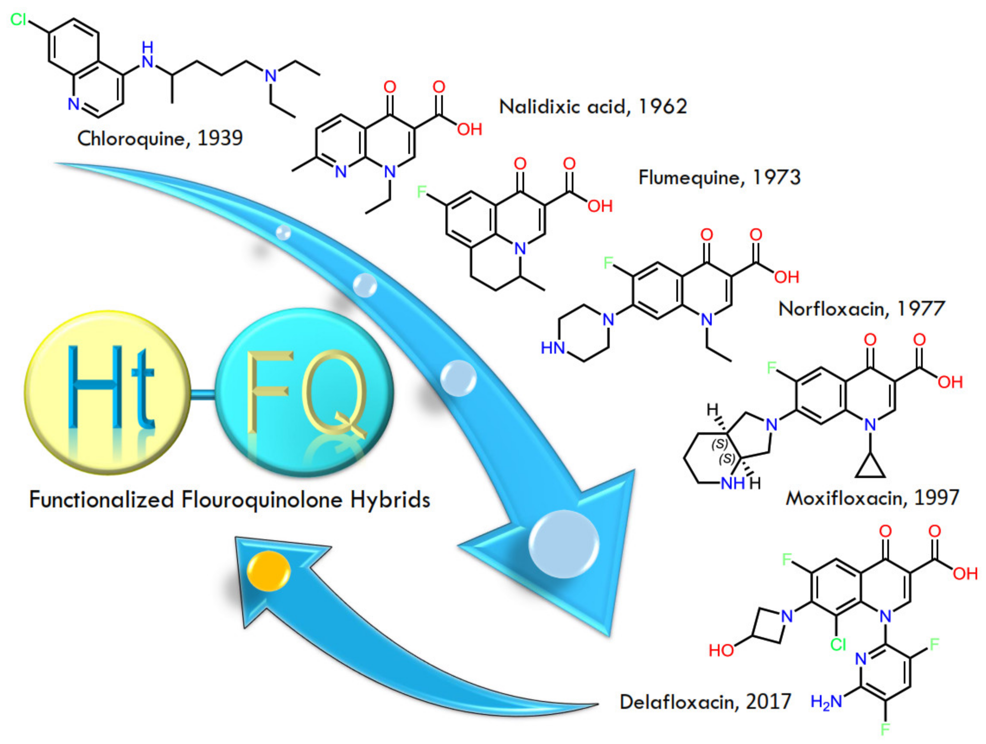
:1. Introduction
2. Materials and Methods
2.1. Chemicals and Apparatus
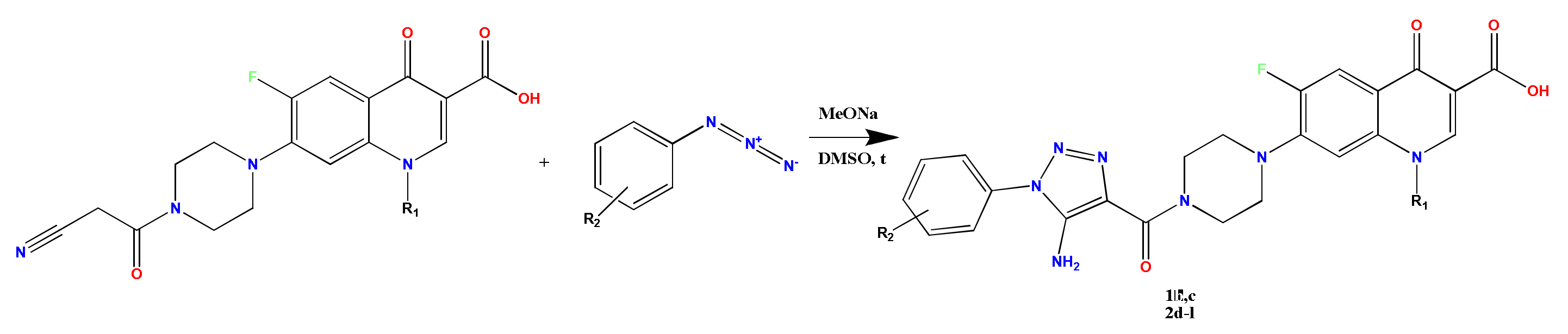
2.2. Synthesis of 7-(4-(5-Amino-1-R-1H-1,2,3-triazole-4-carbonyl)piperazin-1-yl)-1-R-6-fluoro-4-oxo-1,4-dihydroquinoline-3-carboxylic Acid 1b, c, 2d-l (General Method)
2.2.1. 7-(4-(5-Amino-1-(p-tolyl)-1H-1,2,3-triazole-4-carbonyl)piperazin-1-yl)-1-ethyl-6-fluoro-4-oxo-1,4-dihydroquinoline-3-carboxylic Acid 1b
2.2.2. 7-(4-(5-Amino-1-(4-bromophenyl)-1H-1,2,3-triazole-4-carbonyl)piperazin-1-yl)-1-ethyl-6-fluoro-4-oxo-1,4-dihydroquinoline-3-carboxylic Acid 1c
2.2.3. 7-(4-(5-Amino-1-phenyl-1H-1,2,3-triazole-4-carbonyl)piperazin-1-yl)-1-cyclopropyl-6-fluoro-4-oxo-1,4-dihydroquinoline-3-carboxylic Acid 2d
2.2.4. 7-(4-(5-Amino-1-(2-methylphenyl)-1H-1,2,3-triazole-4-carbonyl)piperazin-1-yl)-1-cyclopropyl-6-fluoro-4-oxo-1,4-dihydroquinoline-3-carboxylic Acid 2e
2.2.5. 7-(4-(5-Amino-1-(4-ethylphenyl)-1H-1,2,3-triazole-4-carbonyl)piperazin-1-yl)-1-cyclopropyl-6-fluoro-4-oxo-1,4-dihydroquinoline-3-carboxylic Acid 2f
2.2.6. 7-(4-(5-Amino-1-(4-ethylphenyl)-1H-1,2,3-triazole-4-carbonyl)piperazin-1-yl)-1-cyclopropyl-6-fluoro-4-oxo-1,4-dihydroquinoline-3-carboxylic Acid 2g
2.2.7. 7-(4-(5-Amino-1-(4-(methylthio)phenyl)-1H-1,2,3-triazole-4-carbonyl)piperazin-1-yl)-1-cyclopropyl-6-fluoro-4-oxo-1,4-dihydroquinoline-3-carboxylic Acid 2h
2.2.8. 7-(4-(5-Amino-1-(3-(methylthio)phenyl)-1H-1,2,3-triazole-4-carbonyl)piperazin-1-yl)-1-cyclopropyl-6-fluoro-4-oxo-1,4-dihydroquinoline-3-carboxylic Acid 2i
2.2.9. 7-(4-(5-Amino-1-(4-bromophenyl)-1H-1,2,3-triazole-4-carbonyl)piperazin-1-yl)-1-cyclopropyl-6-fluoro-4-oxo-1,4-dihydroquinoline-3-carboxylic Acid 2j
2.2.10. 7-(4-(5-Amino-1-(5-fluoro-2-methylphenyl)-1H-1,2,3-triazole-4-carbonyl)piperazin-1-yl)-1-cyclopropyl-6-fluoro-4-oxo-1,4-dihydroquinoline-3-carboxylic Acid 2k
2.2.11. 7-(4-(5-Amino-1-(4-(trifluoromethyl)phenyl)-1H-1,2,3-triazole-4-carbonyl)piperazin-1-yl)-1-cyclopropyl-6-fluoro-4-oxo-1,4-dihydroquinoline-3-carboxylic Acid 2l
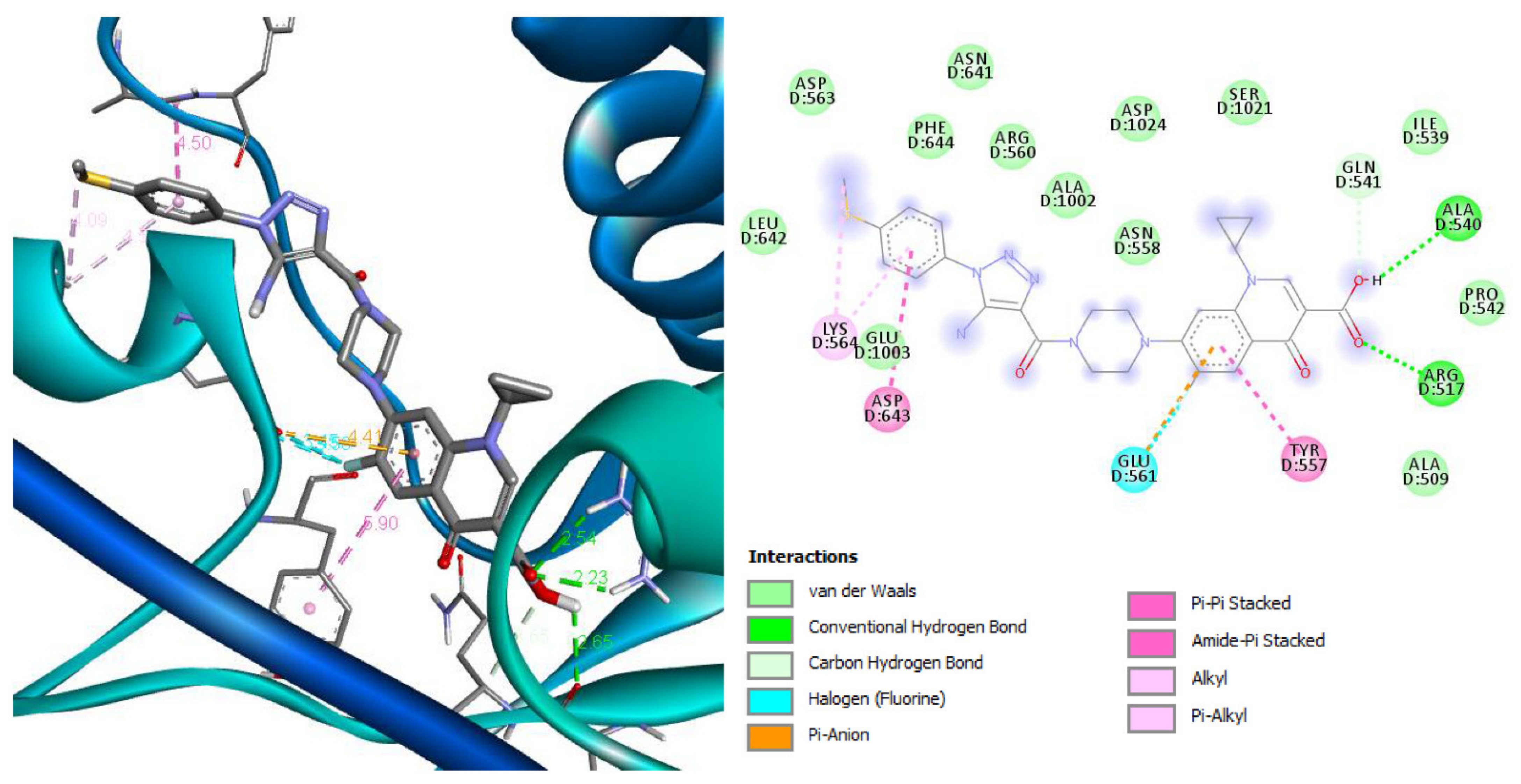
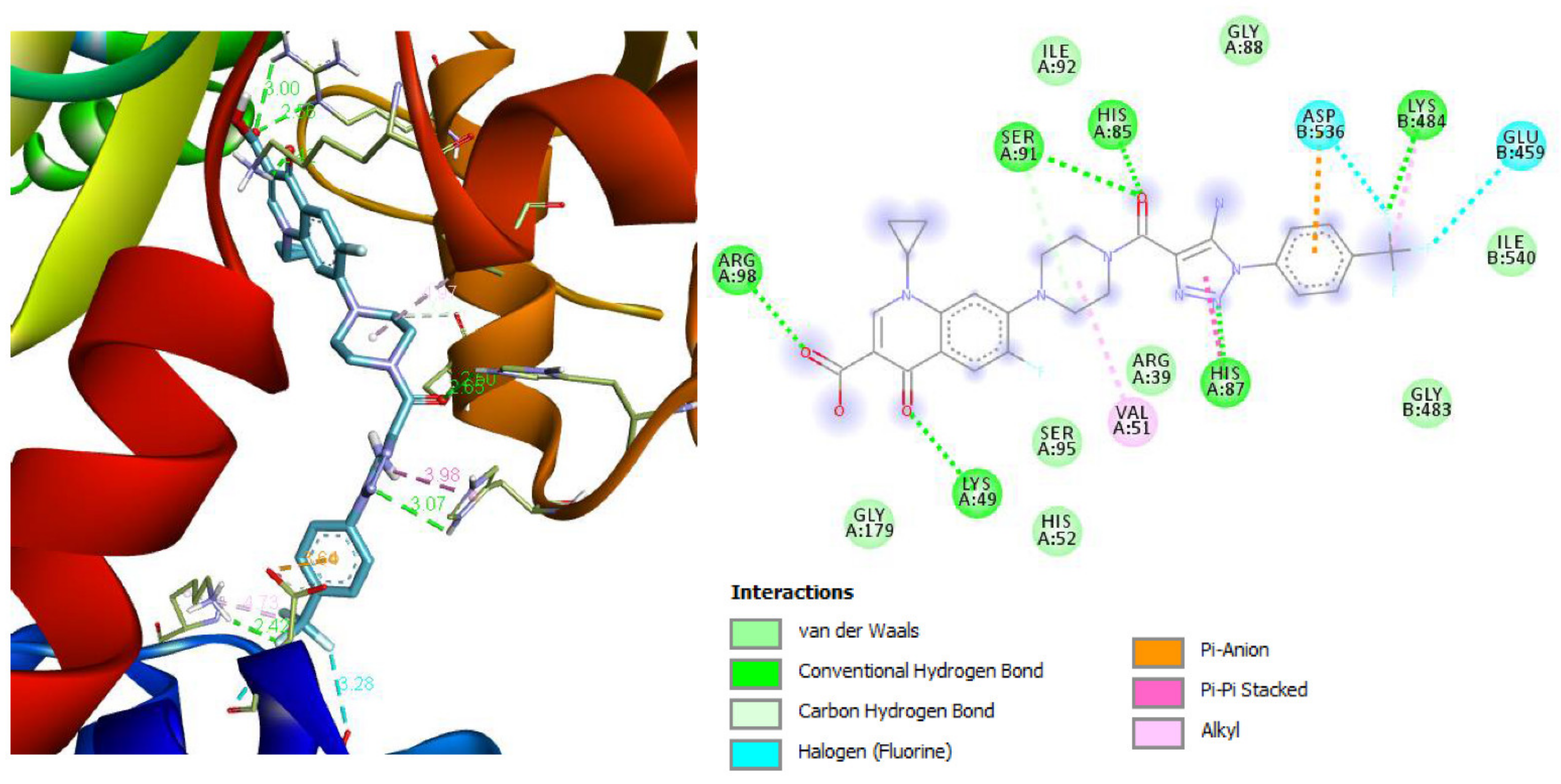
2.3. Docking Studies
2.4. Antibacterial Activity
2.4.1. Method of Double Serial Dilutions
2.4.2. Agar Diffusion Method
3. Results
3.1. Synthesis
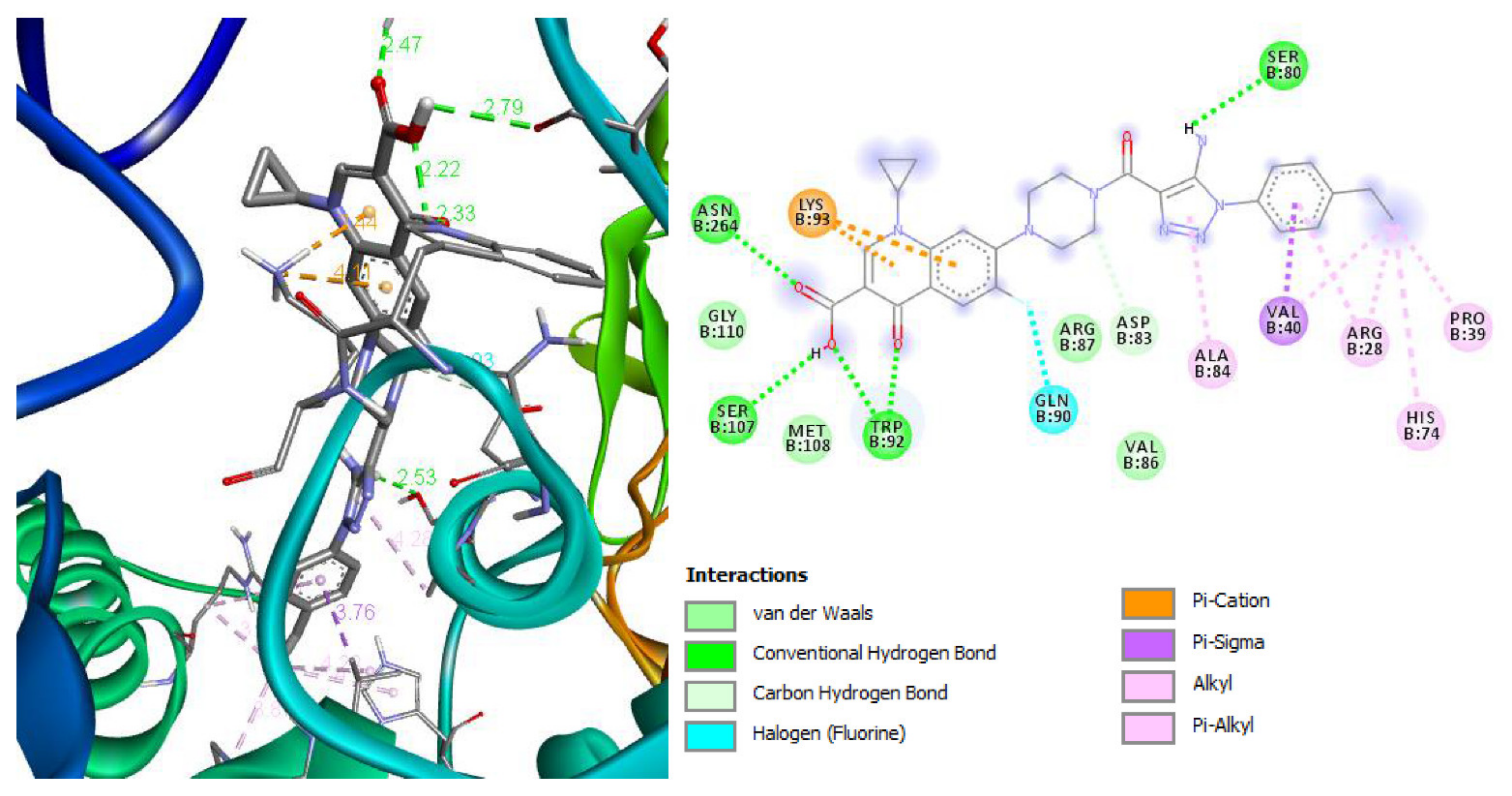
3.2. Docking Studies
3.3. Antibacterial Activity Evaluation
4. Discussion
5. Conclusions
- Quinoline heterocycle and a fluorine atom in position 6;
- Carbonyl and carboxyl fragments in the third and fourth positions of quinoline, which are involved in additional stabilization of the target molecule complex through the Mg2+ cation;
- Substitution in the seventh position of the quinoline framework by pharmacophores of heterocyclic and aromatic structures (triazole, piperazine, and phenyl fragments);
- Additional saturation of molecules with donor and acceptor substituents in the aromatic inclusions of the designed molecules also enhance the activity.
Supplementary Materials
Author Contributions
Funding
Institutional Review Board Statement
Informed Consent Statement
Conflicts of Interest
References
- Beović, B.; Doušak, M.; Ferreira-Coimbra, J.; Nadrah, K.; Rubulotta, F.; Belliato, M.; Berger-Estilita, J.; Ayoade, F.; Rello, J.; Erdem, H. Antibiotic use in patients with COVID-19: A ‘snapshot’ Infectious Diseases International Research Initiative (ID-IRI) survey. J. Antimicrob. Chemother. 2020, 75, 3386–3390. [Google Scholar] [CrossRef] [PubMed]
- Mohammed, H.H.; Abuo-Rahma, G.E.-D.A.; Abbas, S.; Abdelhafez, E.-S.M. Current Trends and Future Directions of Fluoroquinolones. Curr. Med. Chem. 2018, 26, 3132–3149. [Google Scholar] [CrossRef] [PubMed]
- Lucia, P. Quinolones: Synthesis and Antibacterial Activity. IntechOpen 2012, 255–272. [Google Scholar] [CrossRef] [Green Version]
- Suaifan, G.A.; Mohammed, A.A. Fluoroquinolones structural and medicinal developments (2013–2018): Where are we now? Bioorg. Med. Chem. 2019, 27, 3005–3060. [Google Scholar] [CrossRef]
- Zahoor, A.F.; Yousaf, M.; Siddique, R.; Ahmad, S.; Naqvi, S.A.R.; Rizvi, S.M.A. Synthetic strategies toward the synthesis of enoxacin-, levofloxacin-, and gatifloxacin-based compounds: A review. Synth. Commun. 2017, 47, 1021–1039. [Google Scholar] [CrossRef]
- Zhang, G.-F.; Liu, X.; Zhang, S.; Pan, B.; Liu, M.-L. Ciprofloxacin derivatives and their antibacterial activities. Eur. J. Med. Chem. 2018, 146, 599–612. [Google Scholar] [CrossRef]
- Lin, P.-Y.; Yeh, K.-S.; Su, C.-L.; Sheu, S.-Y.; Chen, T.; Ou, K.-L.; Lin, M.-H.; Lee, L.-W. Synthesis and Antibacterial Activities of Novel 4-Hydroxy-7-hydroxy- and 3-Carboxycoumarin Derivatives. Molecules 2012, 17, 10846–10863. [Google Scholar] [CrossRef] [Green Version]
- Pisano, M.B.; Kumar, A.; Medda, R.; Gatto, G.; Pal, R.; Fais, A.; Era, B.; Cosentino, S.; Uriarte, E.; Santana, L.; et al. Antibacterial Activity and Molecular Docking Studies of a Selected Series of Hydroxy-3-arylcoumarins. Molecules 2019, 24, 2815. [Google Scholar] [CrossRef] [Green Version]
- Cantón, R. Current microbiological aspects of community respiratory infection beyond COVID-19. Rev. Esp. Quimioter. 2021, 34, 81–92. [Google Scholar] [CrossRef]
- Karampela, I.; Dalamaga, M. Could Respiratory Fluoroquinolones, Levofloxacin and Moxifloxacin, Prove to be Beneficial as an Adjunct Treatment in COVID-19? Arch. Med. Res. 2020, 51, 741–742. [Google Scholar] [CrossRef]
- Marciniec, K.; Beberok, A.; Pęcak, P.; Boryczka, S.; Wrześniok, D. Ciprofloxacin and moxifloxacin could interact with SARS-CoV-2 protease: Preliminary in silico analysis. Pharmacol. Rep. 2020, 72, 1553–1561. [Google Scholar] [CrossRef]
- Scroggs, S.L.P.; Offerdahl, D.K.; Flather, D.P.; Morris, C.N.; Kendall, B.L.; Broeckel, R.M.; Beare, P.A.; Bloom, M.E. Fluoroquinolone Antibiotics Exhibit Low Antiviral Activity against SARS-CoV-2 and MERS-CoV. Viruses 2020, 13, 8. [Google Scholar] [CrossRef]
- Oniga, S.; Palage, M.; Araniciu, C.; Marc, G.; Oniga, O.; Vlase, L.; Prisăcari, V.; Valica, V.; Curlat, S.; Uncu, L. Design, Synthesis, Molecular Docking, And Antibacterial Activity Evaluation of Some Novel Norfloxacin Analogues. Farmacia 2018, 66, 1048–1058. [Google Scholar] [CrossRef] [Green Version]
- Marc, G.; Araniciu, C.; Oniga, S.; Vlase, L.; Pîrnău, A.; Nadăș, G.C.; Novac, C.; Matei, I.A.; Chifiriuc, M.C.; Măruțescu, L.; et al. Design, Synthesis and Biological Evaluation of New Piperazin-4-yl-(acetyl-thiazolidine-2,4-dione) Norfloxacin Analogues as Antimicrobial Agents. Molecules 2019, 24, 3959. [Google Scholar] [CrossRef] [Green Version]
- Norouzbahari, M.; Salarinejad, S.; Güran, M.; Şanlıtürk, G.; Emamgholipour, Z.; Bijanzadeh, H.R.; Toolabi, M.; Foroumadi, A. Design, synthesis, molecular docking study, and antibacterial evaluation of some new fluoroquinolone analogues bearing a quinazolinone moiety. DARU J. Pharm. Sci. 2020, 28, 661–672. [Google Scholar] [CrossRef]
- Mohammed, H.H.; Abbas, S.H.; Abdelhafez, E.S.M.; Berger, J.M.; Mitarai, S.; Arai, M.; Abuo-Rahma, G.E.D.A. Synthesis, molecular docking, antimicrobial evaluation, and DNA cleavage assay of new thiadiazole/oxadiazole ciprofloxacin derivatives. Monatshefte Chem. 2019, 150, 1809–1824. [Google Scholar] [CrossRef]
- Demirci, A.; Karayel, K.G.; Tatar, E.; Okullu, S.Ö.; Ünübol, N.; Taşli, P.N.; Kocagöz, Z.T.; Şahin, F.; Küçükgüzel, I. Synthesis and evaluation of novel 1,3,4-thiadiazole fuoroquinolone hybrids as antibacterial, antituberculosis, and anticancer agents. Turk. J. Chem. 2018, 42, 839–858. [Google Scholar] [CrossRef]
- Mokaber-Esfahani, M.; Eshghi, H.; Akbarzadeh, M.; Gholizadeh, M.; Mirzaie, Y.; Hakimi, M.; Lari, J. Synthesis and Antibacterial Evaluation of New Pyrimidyl N-Ciprofloxacin Derivatives. ChemistrySelect 2019, 4, 8930–8933. [Google Scholar] [CrossRef]
- Yadav, P.; Hada, S.; Yadav, D.K.; Kumari, N. Synthesis and antibacterial activity of 1,3-dione derivatives of 1-cyclopropyl-7-[4-(2,6-dimethyl/ dimethoxy-pyrimidin-2-yl-diazenyl)-piperzin-l-yl]-6-fluoro-4-oxo-1,4-dihydroquinolone-3 -carboxylic acid. Indian J. Chem. Sect. B Org. Med. Chem. 2018, 57, 1065–1069. [Google Scholar]
- Li, Z.; Wang, Y.; Li, M.; Zhang, H.; Guo, H.; Ya, H.; Yin, J. Synthesis and properties of dithienylethene-functionalized switchable antibacterial agents. Org. Biomol. Chem. 2018, 16, 6988–6997. [Google Scholar] [CrossRef]
- Mentese, M.; Demirbas, N.; Mermer, A.; Demirci, S.; Demirbas, A.; Ayaz, F.A. Novel Azole-Functionalited Flouroquinolone Hybrids: Design, Conventional and Microwave Irradiated Synthesis, Evaluation as Antibacterial and Antioxidant Agents. Lett. Drug Des. Discov. 2018, 15, 46–64. [Google Scholar] [CrossRef]
- Kharb, R.; Sharma, P.C.; Yar, M.S. Pharmacological significance of triazole scaffold. J. Enzym. Inhib. Med. Chem. 2010, 26, 1–21. [Google Scholar] [CrossRef] [PubMed]
- Bylov, I.E.; Bilokin, Y.V.; Kovalenko, S.M. Specific Features of Reactions of 2-Aminobenzotrifluoride and Anthranilates with Ethyl Cyanoacetate—Expeditious Routes to 3-Substituted 4-Amino- and 4-Hydroxyquinolin-2(1 H)-Ones. Heterocycl. Commun. 1999, 5, 281–284. [Google Scholar] [CrossRef]
- Silin, O.V.; Savchenko, T.I.; Kovalenko, S.M. Synthesis of 5H-Pyrazolo[4,3-c]quinolines. Heterocycles 2004, 63, 1883–1890. [Google Scholar] [CrossRef]
- Savchenko, T.I.; Silin, O.V.; Kovalenko, S.M.; Musatov, V.I.; Nikitchenko, V.M.; Ivachtchenko, A.V. Alkylation of 3-Phenyl-1H-pyrazolo[4,3-c] quinoline: Theoretical Analysis of Regioselectivity. Synth. Commun. 2007, 37, 1321–1330. [Google Scholar] [CrossRef]
- Mohapatra, R.K.; El-Ajaily, M.M.; Alassbaly, F.S.; Sarangi, A.K.; Das, D.; Maihub, A.A.; Ben-Gweirif, S.F.; Mahal, A.; Suleiman, M.; Perekhoda, L.; et al. DFT, anticancer, antioxidant and molecular docking investigations of some ternary Ni(II) complexes with 2-[(E)-[4-(dimethylamino)phenyl]methyleneamino]phenol. Chem. Pap. 2021, 75, 1005–1019. [Google Scholar] [CrossRef]
- Mohapatra, R.K.; Perekhoda, L.; Azam, M.; Suleiman, M.; Sarangi, A.K.; Semenets, A.; Pintilie, L.; Al-Resayes, S.I. Computational investigations of three main drugs and their comparison with synthesized compounds as potent inhibitors of SARS-CoV-2 main protease (Mpro): DFT, QSAR, molecular docking, and in silico toxicity analysis. J. King Saud Univ.-Sci. 2021, 33, 101315. [Google Scholar] [CrossRef]
- Bax, B.D.; Chan, P.F.; Eggleston, D.S.; Fosberry, A.; Gentry, D.R.; Gorrec, F.; Giordano, I.; Hann, M.M.; Hennessy, A.; Hibbs, M.; et al. Type IIA topoisomerase inhibition by a new class of antibacterial agents. Nature 2010, 466, 935–940. [Google Scholar] [CrossRef]
- Blower, T.R.; Williamson, B.H.; Kerns, R.J.; Berger, J.M. Crystal structure and stability of gyrase–fluoroquinolone cleaved complexes from Mycobacterium tuberculosis. Proc. Natl. Acad. Sci. USA 2016, 113, 1706–1713. [Google Scholar] [CrossRef] [Green Version]
- Laponogov, I.; Pan, X.-S.; Veselkov, D.A.; Cirz, R.T.; Wagman, A.; Moser, H.E.; Fisher, L.M.; Sanderson, M.R. Exploring the active site of the Streptococcus pneumoniae topoisomerase IV–DNA cleavage complex with novel 7,8-bridged fluoroquinolones. Open Biol. 2016, 6. [Google Scholar] [CrossRef] [Green Version]
- Волянський, Ю.Л.; Гриценко, І.С.; Широбоков, В.П. Вивчення специфічної активності протимікробних лікарських засобів; ДФЦ МОЗ: Kiev, Ukraine, 2004; p. 38. [Google Scholar]
- Визначення чутливості мікроорганізмів до антибактеріальних препаратів; Наказ МОЗ України №167 05.04.2007: Методичні вказівки; Ministry of Health of Ukraine: Kyiv, Ukraine, 2007.
- Hryhoriv, H.; Mariutsa, I.; Kovalenko, S.M.; Sidorenko, L.; Perekhoda, L.; Filimonova, N.; Geyderikh, O.; Georgiyants, V. Structural modification of ciprofloxacin and norfloxacin for searching new antibiotics to combat drug-resistant bacteria. Sci. Pharm. Sci. 2021, 5, 4–11. [Google Scholar] [CrossRef]
- Gribanov, P.S.; Topchiy, M.A.; Golenko, Y.D.; Lichtenstein, Y.I.; Eshtukov, A.V.; Terekhov, V.E.; Asachenko, A.F.; Nechaev, M.S. An unprecedentedly simple method of synthesis of aryl azides and 3-hydroxytriazenes. Green Chem. 2016, 18, 5984–5988. [Google Scholar] [CrossRef]
- Agalave, S.; Maujan, S.R.; Pore, V.S. Click Chemistry: 1,2,3-Triazoles as Pharmacophores. Chem.–Asian J. 2011, 6, 2696–2718. [Google Scholar] [CrossRef]


| Compound Number | R2 | M.p., °C | Molecular Formula, M. w. | Yield, % |
|---|---|---|---|---|
| 1b | p-Me | 250–255 | C26H26FN7O4 519.54 | 65 |
| 1c | p-Br | 300–304 | C25H23BrFN7O4 523.49 | 58 |
| 2d | H | 258–260 | C26H24FN7O4 517.52 | 66 |
| 2e | o-Me | 215–220 | C27H26FN7O4 531.54 | 37 |
| 2f | p-Et | 258–262 | C28H28FN7O4 545.58 | 68 |
| 2g | p-OMe | 260–262 | C27H26FN7O5 547.55 | 43 |
| 2h | p-SMe | 280–285 | C27H26FN7O4S 563.62 | 49 |
| 2i | m-SMe | 226–232 | C27H26FN7O4S 563.62 | 57 |
| 2j | p-Br | 275–280 | C26H23BrFN7O4 596.42 | 61 |
| 2k | 2-Me-5-F | 278–282 | C27H25F2N7O4 549.54 | 50 |
| 2l | p-CF3 | 222–228 | C27H23F4N7O4 585.52 | 49 |
| Molecule | 2XCR | 5BTL | 4KPF | ||||||
|---|---|---|---|---|---|---|---|---|---|
| Affinity DG, kcal/mol | EDoc kcal/mol | Ki µM Micromolar | Affinity DG, kcal/mol | EDoc kcal/mol | Ki µM Micromolar | Affinity DG, kcal/mol | EDoc kcal/mol | Ki µM Micromolar | |
| 1b | −9.4 | −4.96 | 233.07 µM | −9.0 | −4.90 | 255.07 µM | −9.5 | −4.30 | 700.35 µM |
| 1c | −9.4 | −5.25 | 141.96 µM | −10.8 | −5.08 | 187.55 µM | −9.1 | −4.77 | 316.56 µM |
| 2d | −9.8 | −4.16 | 891.54 µM | −8.6 | −5.24 | 144.69 µM | −9.3 | −4.45 | 547.64 µM |
| 2e | −9.6 | −4.51 | 498.52 µM | −10.6 | −6.51 | 16.79 µM | −9.6 | −5.03 | 204.77 µM |
| 2f | −10.4 | −4.96 | 230.68 µM | −8.7 | −5.06 | 195.51 µM | −9.1 | −4.73 | 341.42 µM |
| 2g | −9.8 | −5.54 | 87.25 µM | −9.6 | −4.41 | 581.77 µM | −9.2 | −4.13 | 934.16 µM |
| 2h | −9.1 | −5.66 | 70.49 µM | −8.8 | −4.90 | 256.42 µM | −9.2 | −4.65 | 388.76 µM |
| 2i | −9.3 | −4.78 | 312.12 µM | −8.7 | −5.32 | 125.18 µM | −9.0 | −4.69 | 366.76 µM |
| 2j | −9.6 | −4.60 | 423.83 µM | −9.5 | −5.16 | 165.25 µM | −8.4 | −4.01 | 1150 µM |
| 2k | −9.8 | −5.05 | 197.24 µM | −9.4 | −5.45 | 101.76 µM | −8.5 | −4.00 | 1170 µM |
| 2l | −9.9 | −5.24 | 144.73 µM | −8.8 | −5.92 | 45.87 µM | −9.7 | −3.99 | 1191 µM |
| Ciprofloxacin | −7.2 | −5.10 | 183.79 µM | −7.5 | −5.51 | 91.69 µM | −7.4 | −5.38 | 113.52 µM |
| Norfloxacin | −7.2 | −4.30 | 708.28 µM | −7.8 | −5.25 | 142.92 µM | −7.4 | −4.78 | 315.73 µM |
| Compound | Growth Retardation Zone, mm | |||||
|---|---|---|---|---|---|---|
| Reference Strains | Clinical Strains | |||||
| S. aureus АТСС 25923 | E. coli АТСС 25922 | C. albicans АТСС 885-653 | S. aureus | E. coli | C. albicans | |
| 1b | 34.3 ± 1.8 p = 0.0089 | 23.8 ± 1.6 p = 0.7521 | 30.1 ± 1.5 p = 0.0030 | 25.8 ± 1.4 p = 0.5903 | 18.7 ± 1.2 p = 0.0569 | 24.6 ± 1.5 p = 0.1984 |
| 1c | 34.4 ± 1.4 p = 0.0044 | 40.4 ± 1.9 p = 0.0008 | 17.1 ± 1.8 p = 0.2443 | 27.9 ± 1.7 p = 0.2495 | 29.3 ± 1.5 p = 0.1185 | 14.7 ± 1.2 p = 0.0298 |
| 2d | 28.8 ± 1.9 p = 0.2158 | 26.5 ± 1.8 p = 0.5553 | 25.1 ± 1.3 p = 0.0720 | 24.4 ± 1.7 p = 0.9736 | 22.3 ± 1.5 p = 0.4811 | 19.4 ± 1.4 p = 0.5773 |
| 2e | 33.1 ± 1.8 p = 0.0171 | 33.4 ± 1.4 p = 0.0075 | 16.8 ± 1.3 p = 0.1367 | 27.6 ± 1.7 p = 0.2886 | 27.4 ± 1.7 p = 0.3458 | 18.2 ± 1.4 p = 0.3242 |
| 2f | 36.1 ± 1.7 p = 0.0028 | 35.4 ± 1.7 p = 0.0038 | 15.8 ± 1.4 p = 0.0758 | 28.8 ± 1.7 p = 0.1574 | 26.3 ± 1.9 p = 0.5665 | 13.2 ± 1.3 p = 0.0123 |
| 2g | 35.7 ± 1.8 p = 0.0042 | 35.4 ± 1.4 p = 0.0021 | 22.4 ± 1.3 p = 0.4254 | 26.4 ± 1.3 0.4416 | 29.6 ± 1.5 p = 0.1001 | 19.6 ± 1.8 p = 0.6688 |
| 2h | 36.3 ± 2.6 p = 0.0108 | 29.3 ± 1.4 p = 0.1077 | 20.7 ± 1.9 p = 0.9454 | 30.3 ± 1.6 p = 0.0622 | 22.3 ± 1.7 p = 0.5044 | 18.7 ± 1.6 p = 0.4457 |
| 2i | 29.2 ± 1.5 p = 0.1121 | 32.2 ± 1.3 p = 0.0140 | 16.6 ± 2.2 p = 0.2446 | 25.3 ± 1.6 p = 0.7338 | 20.1 ± 1.4 p = 0.1547 | 11.5 ± 1.2 p = 0.0033 |
| 2j | 35.7 ± 1.67 p = 0.0034 | 28.9 ± 1.7 p = 0.1726 | 16.3 ± 1.7 p = 0.1150 | 29.2 ± 1.8 p = 0.1372 | 21.2 ± 1.6 p = 0.2786 | 13.1 ± 1.3 p = 0.0115 |
| 2k | 33.8 ± 1.4 p = 0.0064 | 42.7 ± 2.1 p = 0.00001 | 18.3 ± 1.6 p = 0.4096 | 27.3 ± 1.3 p = 0.2790 | 33.4 ± 1.3 p = 0.0079 | 16.4 ± 1.7 p = 0.1466 |
| 2l | 17.5 ± 1.8 p = 0.0572 | 32.3 ± 1.6 p = 0.0207 | 18.6 ± 1.8 p = 0.5051 | 14.1 ± 1.4 p = 0.0036 | 21.8 ± 1.6 p = 0.3987 | 13.1 ± 1.1 p = 0.0083 |
| Control | 24.2 ± 1.8 | 24.7 ± 1.6 | 20.5 ± 1.34 | 24.3 ± 1.7 | 24.4 ± 1.8 | 20.9 ± 1.6 |
Publisher’s Note: MDPI stays neutral with regard to jurisdictional claims in published maps and institutional affiliations. |
© 2021 by the authors. Licensee MDPI, Basel, Switzerland. This article is an open access article distributed under the terms and conditions of the Creative Commons Attribution (CC BY) license (https://creativecommons.org/licenses/by/4.0/).
Share and Cite
Hryhoriv, H.; Mariutsa, I.; Kovalenko, S.M.; Georgiyants, V.; Perekhoda, L.; Filimonova, N.; Geyderikh, O.; Sidorenko, L. The Search for New Antibacterial Agents among 1,2,3-Triazole Functionalized Ciprofloxacin and Norfloxacin Hybrids: Synthesis, Docking Studies, and Biological Activity Evaluation. Sci. Pharm. 2022, 90, 2. https://0-doi-org.brum.beds.ac.uk/10.3390/scipharm90010002
Hryhoriv H, Mariutsa I, Kovalenko SM, Georgiyants V, Perekhoda L, Filimonova N, Geyderikh O, Sidorenko L. The Search for New Antibacterial Agents among 1,2,3-Triazole Functionalized Ciprofloxacin and Norfloxacin Hybrids: Synthesis, Docking Studies, and Biological Activity Evaluation. Scientia Pharmaceutica. 2022; 90(1):2. https://0-doi-org.brum.beds.ac.uk/10.3390/scipharm90010002
Chicago/Turabian StyleHryhoriv, Halyna, Illia Mariutsa, Sergiy M. Kovalenko, Victoriya Georgiyants, Lina Perekhoda, Nataliia Filimonova, Olga Geyderikh, and Lyudmila Sidorenko. 2022. "The Search for New Antibacterial Agents among 1,2,3-Triazole Functionalized Ciprofloxacin and Norfloxacin Hybrids: Synthesis, Docking Studies, and Biological Activity Evaluation" Scientia Pharmaceutica 90, no. 1: 2. https://0-doi-org.brum.beds.ac.uk/10.3390/scipharm90010002