
Antineoplastic Activity of Water-Soluble Form of Novel Kinase Inhibitor 1-(4-Chlorobenzyl)-3-chloro-4-(3-trifluoromethylphenylamino)-1H-pyrrole-2,5-dione immobilized on Polymeric Poly(PEGMA-co-DMM) Carrier

 , ,
, ,  ,
,
Abstract
:
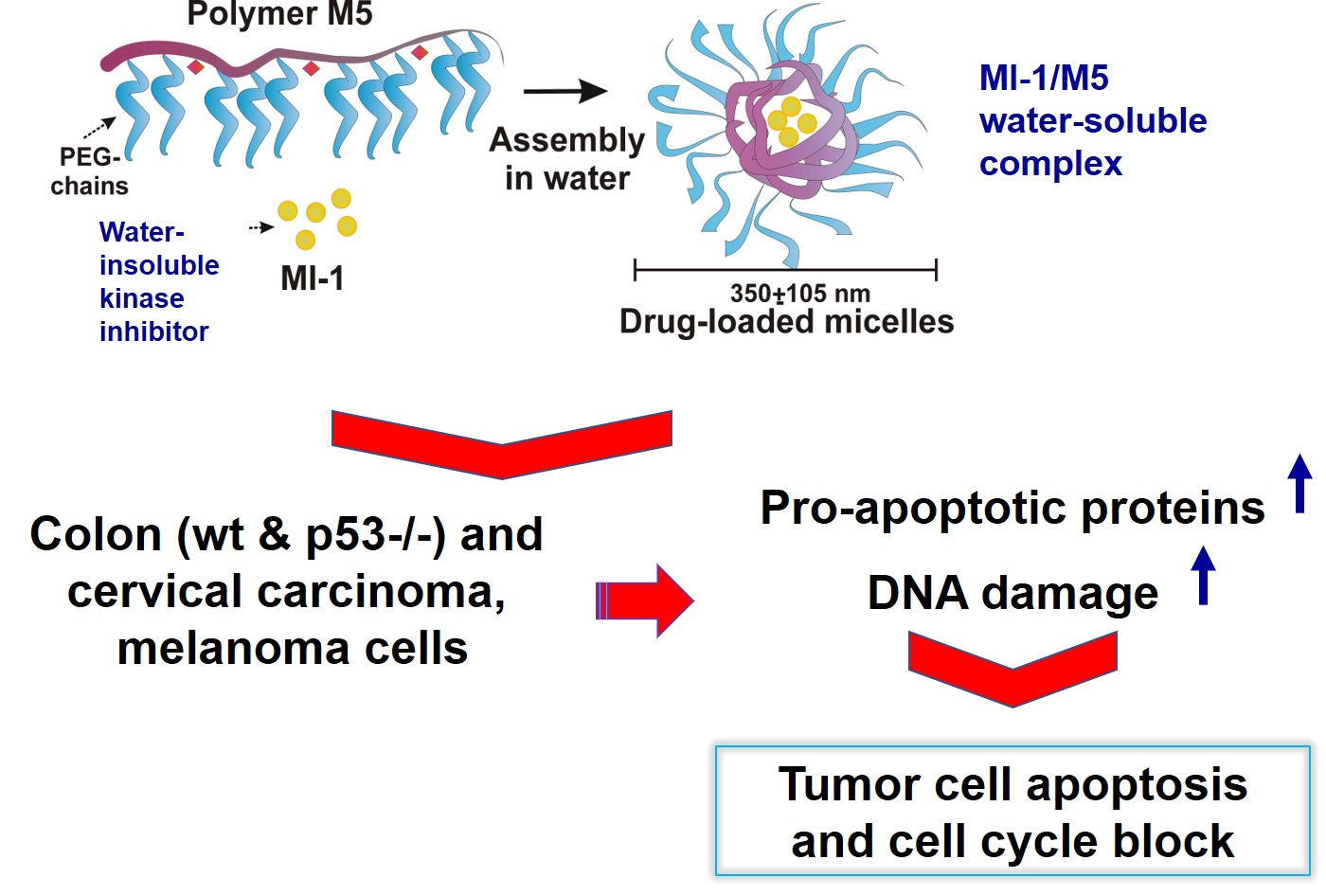
1. Introduction
2. Materials and Methods
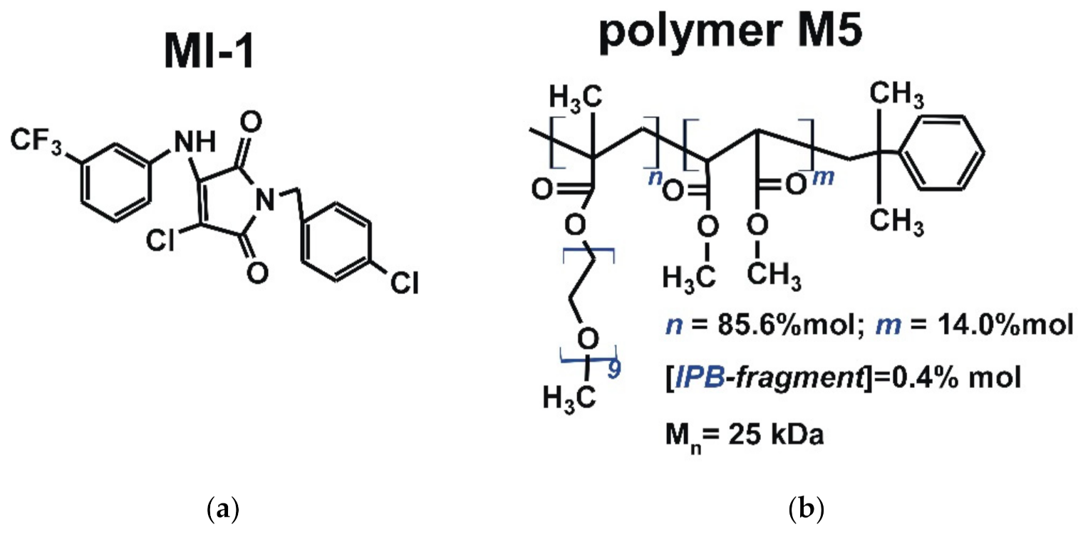
2.1. Studied Compounds
2.1.1. Synthesis and Characterization of Maleimide Derivative
2.1.2. Synthesis and Characterization of the Polymeric Carrier
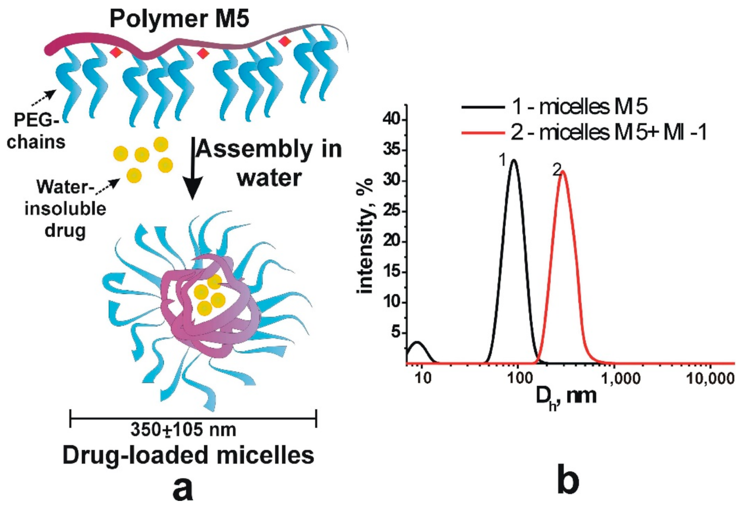
2.1.3. Synthesis and Characterization of the Complex of Maleimide Derivative with Polymer
2.1.4. Doxorubicin
2.2. Cell Culture
2.3. The MTT Assay
2.4. The Clonogenic Assay
2.5. The Western-Blot Analysis
2.6. The DNA Intercalation Assay Using Methyl Green Replacement
2.7. The Comet Assay at Alkaline Conditions
2.8. The Fluorescent Microscopy of Cells
2.9. Statistical Analysis
3. Results and Discussions
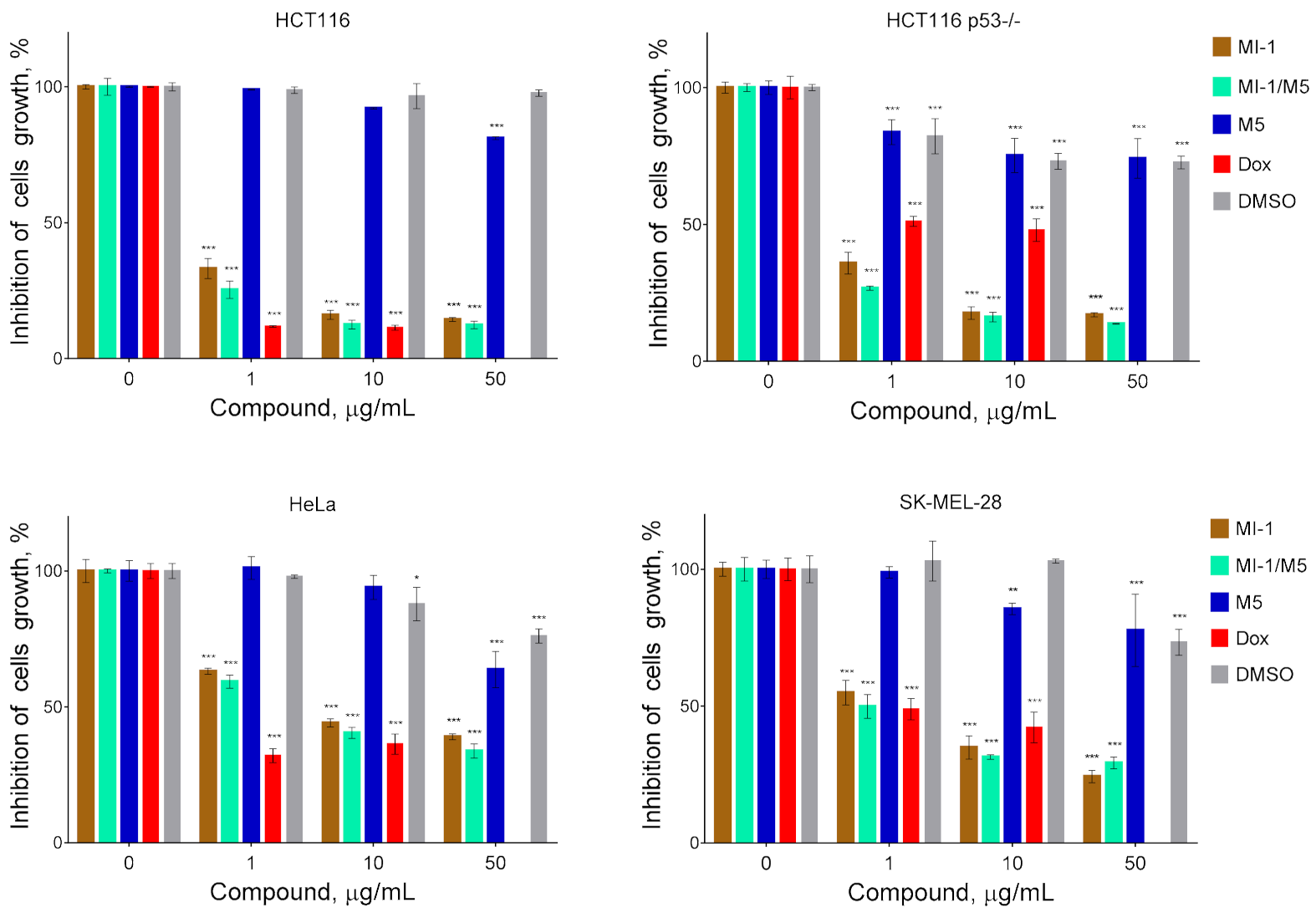
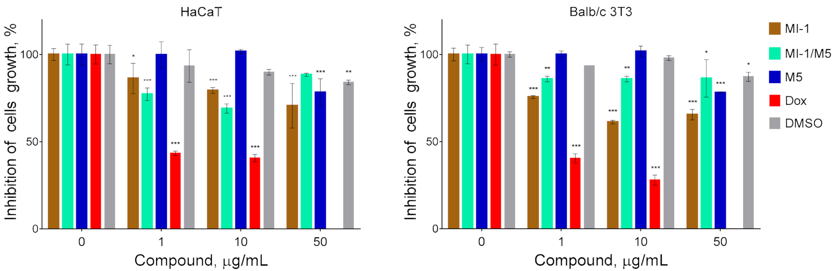
3.1. The Viability of Tumor and Pseudo-Normal Cells after Treatment
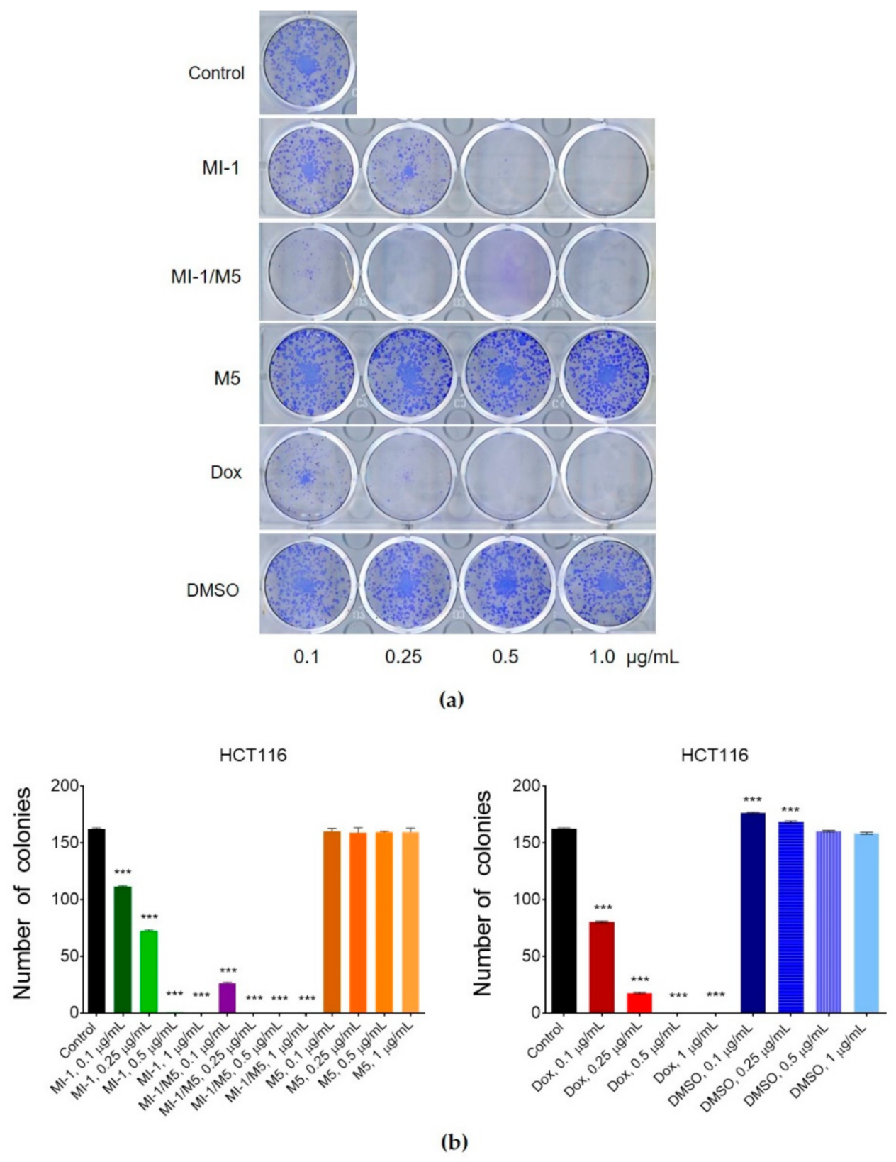
3.2. Clonogenic Response of Human Colon Carcinoma Cells to Treatment
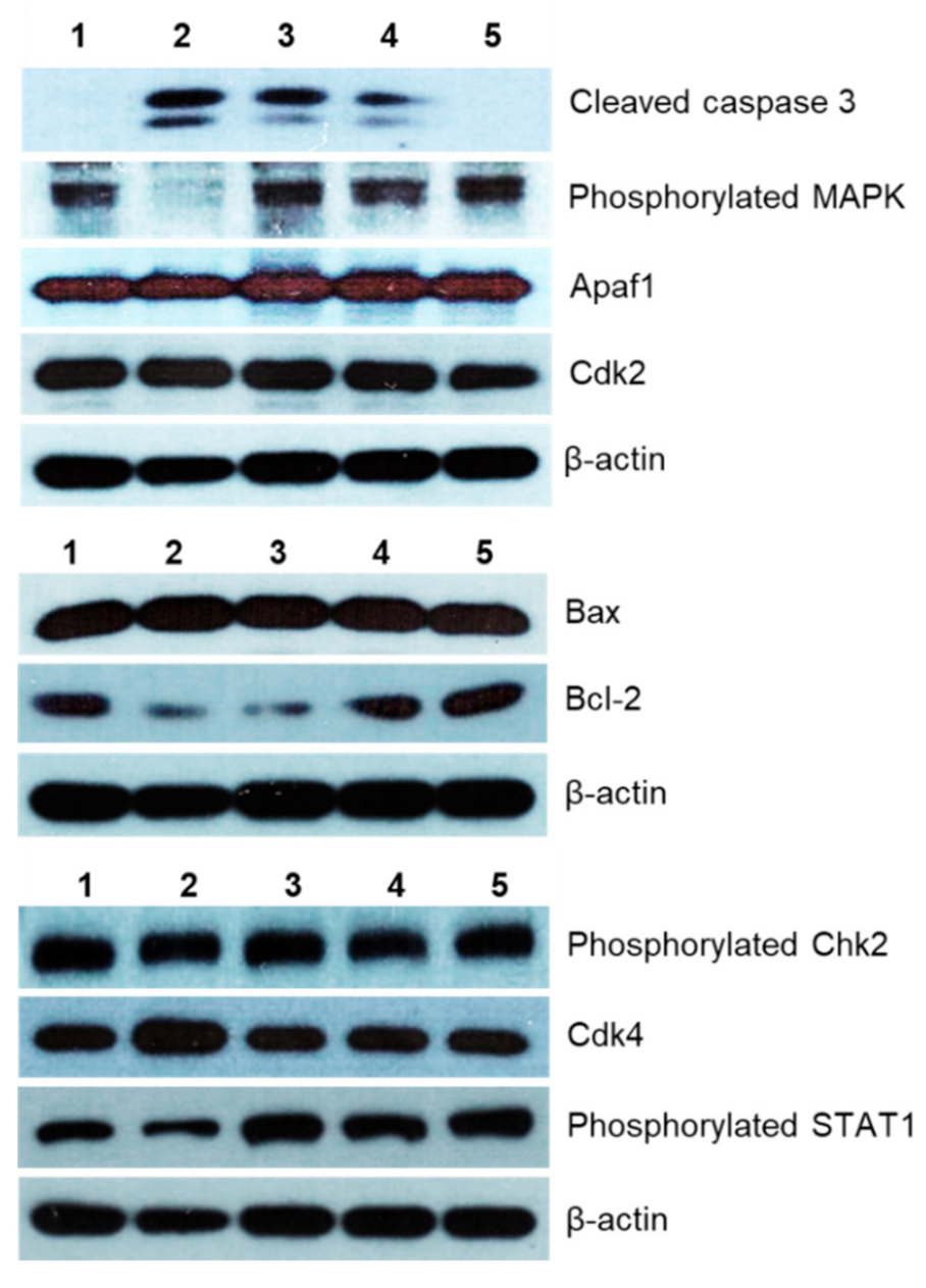
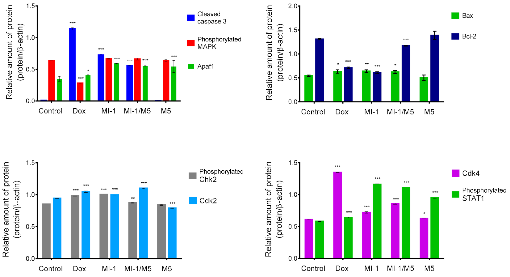
3.3. Apoptosis Induction in Treated Cells
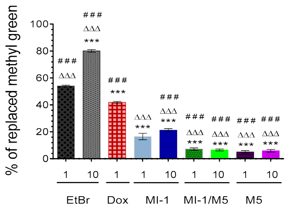
3.4. Interaction of MI-1 and Its Complex with DNA
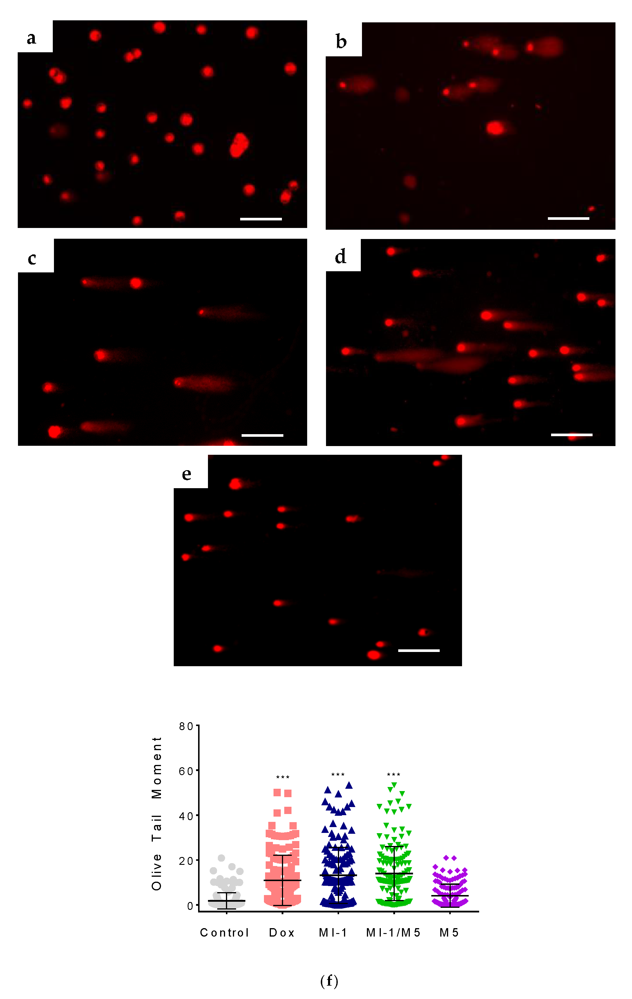
3.5. The Damage of DNA Molecule by MI-1 and Its Complex
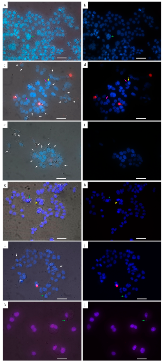
3.6. Morphological Changes in HCT116 Cells Treated with MI-1 and Its Complex
4. Conclusions
Author Contributions
Funding
Institutional Review Board Statement
Informed Consent Statement
Data Availability Statement
Acknowledgments
Conflicts of Interest
References
- Olgen, S. Overview on anticancer drug design and development. Curr. Med. Chem. 2018, 25, 1704–1719. [Google Scholar] [CrossRef] [PubMed]
- Ali, I.; Lone, M.N.; Aboul-Enein, H.Y. Imidazoles as potential anticancer agents. MedChemComm 2017, 8, 1742–1773. [Google Scholar] [CrossRef] [PubMed]
- Narvekar, M.; Xue, H.Y.; Eoh, J.Y.; Wong, H.L. Nanocarrier for poorly water-soluble anticancer drugs-barriers of translation and solutions. AAPS PharmSciTech 2014, 15, 822–833. [Google Scholar] [CrossRef] [PubMed]
- Ferrari, R.; Sponchioni, M.; Morbidelli, M.; Moscatelli, D. Polymer nanoparticles for the intravenous delivery of anticancer drugs: The checkpoints on the road from the synthesis to clinical translation. Nanoscale 2018, 10, 22701–22719. [Google Scholar] [CrossRef]
- Majumder, N.; Das, N.G.; Das, S.K. Polymeric micelles for anticancer drug delivery. Ther. Deliv. 2020, 11, 613–635. [Google Scholar] [CrossRef]
- Duchene, D.; Cavalli, R.; Gref, R. Cyclodextrin-based polymeric nanoparticles as efficient carriers for anticancer drugs. Curr. Pharm. Biotechnol. 2016, 17, 248–255. [Google Scholar] [CrossRef]
- Sun, H.; Yarovoy, I.; Capeling, M.; Cheng, C. Polymers in the co-delivery of siRNA and anticancer drugs for the treatment of drug-resistant cancers. Top. Curr. Chem. 2017, 375, 24. [Google Scholar] [CrossRef]
- Rizwanullah, M.; Alam, M.; Harshita, M.S.R.; Rizvi, M.M.; Amin, S. Polymer-lipid hybrid nanoparticles: A next-generation nanocarrier for targeted treatment of solid tumors. Curr. Pharm. Des. 2020, 26, 1206–1215. [Google Scholar] [CrossRef]
- Oliveira, A.L.C.d.L.; Schomann, T.; de Geus-Oei, L.F.; Kapiteijn, E.; Cruz, L.J.; de Araújo, R.F., Jr. Nanocarriers as a tool for the treatment of colorectal cancer. Pharmaceutics 2021, 13, 1321. [Google Scholar] [CrossRef]
- Paris, C.; Brun, O.; Pedroso, E.; Grandas, A. Exploiting protected maleimides to modify oligonucleotides, peptides and peptide nucleic acids. Molecules 2015, 20, 6389–6408. [Google Scholar] [CrossRef] [Green Version]
- Ravasco, J.M.J.M.; Faustino, H.; Trindade, A.; Gois, P.M.P. Bioconjugation with maleimides: A useful tool for chemical biology. Chemistry 2019, 25, 43–59. [Google Scholar] [CrossRef] [PubMed]
- Shi, Q.; Zhang, Y.; Huang, Z.; Zhou, N.; Zhang, Z.; Zhu, X. Precise sequence regulation through maleimide chemistry. Polym. J. 2020, 52, 21–31. [Google Scholar] [CrossRef]
- Muthyala, M.K.K.; Sahoo, S.K.; Nagasree, K.P.; Pemmadi, R.V.; Jamullamudi, R.N. Synthesis and screening of new maleimide derivatives as potential anti-tubercular agents. J. App. Pharm. Sci. 2015, 5, 44–47. [Google Scholar] [CrossRef]
- Matuszak, N.; Muccioli, G.G.; Labar, G.; Lambert, D.M. Synthesis and in vitro evaluation of N-substituted maleimide derivatives as selective monoglyceride lipase inhibitors. J. Med. Chem. 2009, 52, 7410–7420. [Google Scholar] [CrossRef]
- Pajak, B.; Orzechowska, S.; Gajkowska, B.; Orzechowski, A. Bisindolylmaleimides in anti-cancer therapy—More than PKC inhibitors. Adv. Med. Sci. 2008, 53, 21–31. [Google Scholar] [CrossRef] [PubMed] [Green Version]
- Liu, W.; Beck, J.; Schmidt, L.C.; Roolf, C.; Pews-Davtyan, A.; Rütgen, B.C.; Hammer, S.; Willenbrock, S.; Sekora, A.; Rolfs, A.; et al. Characterization of the novel indolylmaleimides’ PDA-66 and PDA-377 effect on canine lymphoma cells. Oncotarget 2016, 7, 35379–35389. [Google Scholar] [CrossRef] [Green Version]
- Van Eis, M.J.; Evenou, J.; Schuler, W.; Zenke, G.; Vangrevelinghe, E.; Wagner, J.; von Matt, P. Indolyl-naphthylmaleimides as potent and selective inhibitors of protein kinase C-a/b. Bioorg. Med. Chem Lett. 2017, 27, 781–786. [Google Scholar] [CrossRef]
- Esfandyari-Manesh, M.; Abdi, M.; Talasaz, A.H.; Ebrahimi, S.M.; Atyabi, F.; Dinarvand, R. S2P peptide-conjugated PLGA-maleimide-PEG nanoparticles containing imatinib for targeting drug delivery to atherosclerotic plaques. DARU J. Pharm. Sci. 2020, 28, 131–138. [Google Scholar] [CrossRef]
- Pichler, V.; Mayr, J.; Heffeter, P.; Dömötör, O.; Enyedy, É.A.; Hermann, G.; Groza, D.; Köllensperger, G.; Galanksi, M.; Berger, W.; et al. Maleimide-functionalised Platinum(IV) complexes as a synthetic platform for targeted drug delivery. Chem. Commun. 2013, 49, 2249–2251. [Google Scholar] [CrossRef] [Green Version]
- Gutowski, S.M.; Shoemaker, J.T.; Templeman, K.L.; Wei, Y.; Latour, R.A., Jr.; Bellamkonda, R.V.; LaPlaca, M.C.; García, A.J. Protease-degradable PEG-maleimide coating with on-demand release of IL-1Ra to improve tissue response to neural electrodes. Biomaterials 2015, 44, 55–70. [Google Scholar] [CrossRef] [Green Version]
- Hu, S.-M.; Lee, C.-Y.; Chang, Y.-M.; Xiao, J.-Q.; Kusanagi, T.; Wu, T.-Y.; Chang, N.-Y.; Christy, J.; Chiu, Y.-R.; Huang, C.-W.; et al. Vapor-phase fabrication of a maleimide-functionalized poly-p-xylylene with a three-dimensional structure. Coatings 2021, 11, 466. [Google Scholar] [CrossRef]
- Lee, W.J.; Cha, S.H. Improvement of mechanical and self-healing properties for polymethacrylate derivatives containing maleimide modified graphene oxide. Polymers 2020, 12, 603. [Google Scholar] [CrossRef] [Green Version]
- Dubinina, G.G.; Chupryna, O.O.; Platonov, M.O.; Borisko, P.O.; Ostrovska, G.V.; Tolmachov, A.O.; Shtil, A.A. In silico design of protein kinase inhibitors: Successes and failures. Anticancer Agents Med. Chem. 2007, 7, 171–188. [Google Scholar] [CrossRef]
- Kuznietsova, H.; Dziubenko, N.; Byelinska, I.; Hurmach, V.; Bychko, A.; Lynchak, O.; Milokhov, D.; Khilya, O.; Rybalchenko, V. Pyrrole derivatives as potential anti-cancer therapeutics: Synthesis, mechanisms of action, safety. J. Drug Target. 2020, 28, 547–563. [Google Scholar] [CrossRef]
- Kuznietsova, H.M.; Yena, M.S.; Kotlyar, I.P.; Ogloblya, O.V.; Rybalchenko, V.K. Anti-inflammatory effects of protein kinase inhibitor pyrrol derivate. Sci. World J. 2016, 2016, 2145753. [Google Scholar] [CrossRef] [Green Version]
- Dubinina, G.; Golovach, S.; Kozlovsky, V.; Tolmachov, A.O.; Volovenko, Y.M. Antiproliferative action of the new derivatives of l-(4-R-benzyl)-3-R1-4-(R2-phenylamino)-1H-pyrrol-2,5-dione. Zurn. Organ. Farmacevt. Khimi 2007, 5, 39–49. (In Ukrainian) [Google Scholar]
- Finiuk, N.S.; Ivasechko, I.I.; Klyuchivska, O.Y.; Kuznietsova, H.M.; Rybalchenko, V.K.; Stoika, R.S. Cytotoxic action of maleimide derivative 1-(4-Cl-benzyl)-3-chloro-4-(CF(3)-phenylamino)-1H-pyrrole-2,5-dione toward mammalian tumor cells and its capability to interact with DNA. Ukr. Biochem. J. 2020, 92, 55–62. [Google Scholar] [CrossRef]
- Byelinska, I.V.; Garmanchuk, L.V.; Khranovska, N.M.; Shelest, D.V.; Rybalchenko, T.V. Effect of maleimide derivative, protein kinases inhibitor, on the morphofunctional state of human neoplastic monoblast cell line U-937. Res. J. Pharm. Biol. Chem. Sci. 2016, 7, 1898–1905. [Google Scholar]
- Kuznietsova, H.M.; Lynchak, O.V.; Danylov, M.O.; Kotliar, I.P.; Rybalchenko, V.K. Effect of dihydropyrrol and maleimide derivatives on the state of liver and colon in normal rats and those with colorectal carcinogenesis induced by dimethylhydrazine. Ukr. Biochem. J. 2013, 85, 74–84. [Google Scholar] [CrossRef] [Green Version]
- Kuznietsova, H.M.; Luzhenetska, V.K.; Kotlyar, I.P.; Rybalchenko, V.K. Effects of 5-amyno-4-(1,3-benzothyazol-2-yn)-1-(3-methoxyphenyl)-1,2-dihydro-3H- pyrrole-3-one intake on digestive system in a rat model of colon cancer. Sci. World J. 2015, 2015, 376576. [Google Scholar] [CrossRef] [PubMed] [Green Version]
- Bielins’ka, I.V.; Lynchak, O.V.; Rybal’chenko, T.V.; Hurniak, O.M. Hematological effects of the protein kinase inhibitor maleimide derivative in dimethylhydrazin E-induced colorectal carcinogenesis of rats. Fiziolohichnyi Zhurnal 2014, 60, 40–49. (In Ukrainian) [Google Scholar] [CrossRef] [Green Version]
- Byelinska, I.V.; Lynchak, O.V.; Tsyvinska, S.M.; Rybalchenko, V.K. Morphofunctional state of blood cells after chronic exposure of the protein kinases inhibitor maleimide derivative. Fiziolohichnyi Zhurnal 2015, 61, 71–77. (In Ukrainian) [Google Scholar] [CrossRef] [Green Version]
- Finiuk, N.S.; Popovych, M.V.; Shalai, Y.R.; Mandzynets, S.M.; Hreniuh, V.P.; Ostapiuk, Y.V.; Obushak, M.D.; Mitina, N.E.; Zaichenko, O.S.; Stoika, R.S.; et al. Antineoplastic activity in vitro of 2-amino-5-benzylthiasol derivative in the complex with nanoscale polymeric carriers. Cytol. Genet. 2021, 55, 19–27. [Google Scholar] [CrossRef]
- Kuznietsova, H.M.; Hurmach, V.V.; Bychko, A.V.; Tykhoniuk, O.I.; Milokhov, D.S.; Khilya, O.V.; Volovenko, Y.M.; Rybalchenko, V.K. Synthesis and biological activity of 4-amino-3-chloro-1H-pyrrole-2,5-diones. In Silico Pharmacol. 2019, 7, 2. [Google Scholar] [CrossRef]
- Paiuk, O.L.; Mitina, N.Y.; Kinash, N.I.; Yakymovych, A.B.; Hevus, O.I.; Zaichenko, A.S. Comb-like polyethylene glycol containing oligomeric surfactants with reactive terminal groups. Ukr. Chem. J. 2018, 84, 1–9. (In Ukrainian) [Google Scholar] [CrossRef]
- Francuskiewicz, F. Polymer Fractionation; Springer: Berlin/Heidelberg, Germany, 1994; p. 217. ISBN 978-3-642-78704-1. [Google Scholar]
- Mitina, N.Y.; Riabtseva, A.O.; Garamus, V.M.; Lesyk, R.B.; Volyanyuk, K.A.; Izhyk, O.M.; Zaichenko, O.S. Morphology of the micelles formed by a comb-like PEG-containing copolymer loaded with antitumor substances with different water solubilities. Ukr. J. Phys. 2020, 65, 670. [Google Scholar] [CrossRef]
- Finiuk, N.; Klyuchivska, O.; Ivasechko, I.; Hreniukh, V.; Ostapiuk, Y.; Shalai, Y.; Panchuk, R.; Matiychuk, V.; Obushak, M.; Stoika, R.; et al. Proapoptotic effects of novel thiazole derivative on human glioma cells. Anticancer Drugs 2019, 30, 27–37. [Google Scholar] [CrossRef] [PubMed]
- NÚÑez, J.G.; Pinheiro, J.S.; Padilha, G.L.; Garcia, H.O.; Porta, V.; Apel, M.A.; Bruno, A.N. Antineoplastic potential and chemical evaluation of essential oils from leaves and flowers of Tagetes ostenii Hicken. An. Acad. Bras. Cienc. 2020, 92, e20191143. [Google Scholar] [CrossRef] [PubMed]
- Mahmood, T.; Yang, P.C. Western blot: Technique, theory, and trouble shooting. N. Am. J. Med. Sci. 2012, 4, 429–434. [Google Scholar] [CrossRef] [PubMed]
- Gallo-Oller, G.; Ordoñez, R.; Dotor, J. A new background subtraction method for Western blot densitometry band quantification through image analysis software. J. Immunol. Methods 2018, 457, 1–5. [Google Scholar] [CrossRef] [PubMed]
- Dabiri, Y.; Abu El Maaty, M.A.; Chan, H.Y.; Wölker, J.; Ott, I.; Wölfl, S.; Cheng, X. p53-dependent anti-proliferative and pro-apoptotic effects of a gold(I) N-heterocyclic carbene (NHC) complex in colorectal cancer cells. Front. Oncol. 2019, 9, 438. [Google Scholar] [CrossRef] [Green Version]
- Abu El Maaty, M.A.; Strassburger, W.; Qaiser, T.; Dabiri, Y.; Wölfl, S. Differences in p53 status significantly influence the cellular response and cell survival to 1,25-dihydroxyvitamin D3-metformin cotreatment in colorectal cancer cells. Mol. Carcinog. 2017, 56, 2486–2498. [Google Scholar] [CrossRef] [PubMed]
- Hayashi, Y.; Tsujii, M.; Kodama, T.; Akasaka, T.; Kondo, J.; Hikita, H.; Inoue, T.; Tsujii, Y.; Maekawa, A.; Yoshii, S.; et al. p53 functional deficiency in human colon cancer cells promotes fibroblast-mediated angiogenesis and tumor growth. Carcinogenesis 2016, 37, 972–984. [Google Scholar] [CrossRef]
- Boyer, J.; McLean, E.G.; Aroori, S.; Wilson, P.; McCulla, A.; Carey, P.D.; Longley, D.B.; Johnston, P.G. Characterization of p53 wild-type and null isogenic colorectal cancer cell lines resistant to 5-fluorouracil, oxaliplatin, and irinotecan. Clin. Cancer Res. 2004, 10, 2158–2167. [Google Scholar] [CrossRef] [PubMed] [Green Version]
- García, A.J. PEG-maleimide hydrogels for protein and cell delivery in regenerative medicine. Ann. Biomed. Eng. 2014, 42, 312–322. [Google Scholar] [CrossRef] [PubMed]
- Segun, P.A.; Ogbole, O.O.; Ismail, F.M.D.; Nahar, L.; Evans, A.R.; Ajaiyeoba, E.O.; Sarker, S.D. Resveratrol derivatives from Commiphora africana (A. Rich.) Endl. display cytotoxicity and selectivity against several human cancer cell lines. Phytother. Res. 2019, 33, 159–166. [Google Scholar] [CrossRef] [PubMed] [Green Version]
- Da’i, M.; Meilinasary, K.A.; Suhendi, A.; Haryanti, S. Selectivity index of Alpinia galanga extract and 1′-acetoxychavicol acetate on cancer cell lines. Indones. J. Cancer Chemoprevent. 2019, 10, 95–100. [Google Scholar]
- Beaver, C.M. The effect of culture conditions on colony morphology and proliferative capacity in human prostate cancer cell lines. Cell Biol. Toxicol. 2012, 28, 291–301. [Google Scholar] [CrossRef]
- McIlwain, D.R.; Berger, T.; Mak, T.W. Caspase functions in cell death and disease. Cold Spring Harb. Perspect. Biol. 2013, 5, a008656. [Google Scholar] [CrossRef]
- Ngoi, N.; Choong, C.; Lee, J.; Bellot, G.; Wong, A.; Goh, B.C.; Pervaiz, S. Targeting mitochondrial apoptosis to overcome treatment resistance in cancer. Cancers 2020, 12, 574. [Google Scholar] [CrossRef] [Green Version]
- Yoshida, H.; Kong, Y.Y.; Yoshida, R.; Elia, A.J.; Hakem, A.; Hakem, R.; Penninger, J.M.; Mak, T.W. Apaf1 is required for mitochondrial pathways of apoptosis and brain development. Cell 1998, 94, 739–750. [Google Scholar] [CrossRef] [Green Version]
- Lu, M.; Wang, Y.; Zhan, X. The MAPK pathway-based drug therapeutic targets in pituitary adenomas. Front. Endocrinol. 2019, 10, 330. [Google Scholar] [CrossRef] [PubMed] [Green Version]
- Zhang, Y.; Liu, Z. STAT1 in cancer: Friend or foe? Discov. Med. 2017, 24, 19–29. [Google Scholar] [PubMed]
- Topacio, B.R.; Zatulovskiy, E.; Cristea, S.; Xie, S.; Tambo, C.S.; Rubin, S.M.; Sage, J.; Kõivomägi, M.; Skotheim, J.M. Cyclin D-Cdk4,6 drives cell-cycle progression via the retinoblastoma protein’s c-terminal helix. Mol. Cell. 2019, 74, 758–770. [Google Scholar] [CrossRef]
- Baguley, B.C.; Drummond, C.J.; Chen, Y.Y.; Finlay, G.J. DNA-binding anticancer drugs: One target, two actions. Molecules 2021, 26, 552. [Google Scholar] [CrossRef]
- Rondelet, G.; Fleury, L.; Faux, C.; Masson, V.; Dubois, J.; Arimondo, P.B.; Willems, L.; Wouters, J. Inhibition studies of DNA methyltransferases by maleimide derivatives of RG108 as non-nucleoside inhibitors. Future Med. Chem. 2017, 9, 1465–1481. [Google Scholar] [CrossRef] [PubMed]
- Elmore, S. Apoptosis: A review of programmed cell death. Toxicol. Pathol. 2007, 35, 495–516. [Google Scholar] [CrossRef]


| Cell Line | IC50, µg/mL | ||||
|---|---|---|---|---|---|
| MI-1 | MI-1/M5 | M5 | Dox | DMSO | |
| Tumor cells | |||||
| HCT116 | 0.75 ± 0.06 | 0.67 ± 0.06 | ˃50 | 0.58 ± 0.04 * | ˃50 |
| HCT116 p53-/- | 0.89 ± 0.05 | 0.68 ± 0.05 ** | ˃50 | 3.60 ± 0.51 *** | ˃50 |
| HeLa | 7.22 ± 0.32 | 5.43 ± 0.30 ** | ˃50 | 0.64 ± 0.03 *** | ˃50 |
| SK-MEL-28 | 3.17 ± 0.18 | 1.00 ± 0.08 *** | ˃50 | 0.98 ± 0.07 *** | ˃50 |
| Pseudo-normal cells | |||||
| HaCaT | ˃50 | ˃50 | ˃50 | 1.04 ± 0.07 | ˃50 |
| Balb/c 3T3 | ˃50 | ˃50 | ˃50 | 0.90 ± 0.07 | ˃50 |
| Cell Line | Selectivity Index | ||||
|---|---|---|---|---|---|
| MI-1 | MI-1/M5 | M5 | Dox | DMSO | |
| HCT116 | 66.67 | 74.63 | 1.00 | 1.79 | 1.00 |
| HCT116 p53-/- | 56.18 | 73.53 | 1.00 | 0.29 | 1.00 |
| HeLa | 6.93 | 9.21 | 1.00 | 1.63 | 1.00 |
| SK-MEL-28 | 15.77 | 50.00 | 1.00 | 1.06 | 1.00 |
Publisher’s Note: MDPI stays neutral with regard to jurisdictional claims in published maps and institutional affiliations. |
© 2022 by the authors. Licensee MDPI, Basel, Switzerland. This article is an open access article distributed under the terms and conditions of the Creative Commons Attribution (CC BY) license (https://creativecommons.org/licenses/by/4.0/).
Share and Cite
Finiuk, N.; Klyuchivska, O.; Mitina, N.; Kuznietsova, H.; Volianiuk, K.; Zaichenko, A.; Rybalchenko, V.; Stoika, R. Antineoplastic Activity of Water-Soluble Form of Novel Kinase Inhibitor 1-(4-Chlorobenzyl)-3-chloro-4-(3-trifluoromethylphenylamino)-1H-pyrrole-2,5-dione immobilized on Polymeric Poly(PEGMA-co-DMM) Carrier. Sci. Pharm. 2022, 90, 7. https://0-doi-org.brum.beds.ac.uk/10.3390/scipharm90010007
Finiuk N, Klyuchivska O, Mitina N, Kuznietsova H, Volianiuk K, Zaichenko A, Rybalchenko V, Stoika R. Antineoplastic Activity of Water-Soluble Form of Novel Kinase Inhibitor 1-(4-Chlorobenzyl)-3-chloro-4-(3-trifluoromethylphenylamino)-1H-pyrrole-2,5-dione immobilized on Polymeric Poly(PEGMA-co-DMM) Carrier. Scientia Pharmaceutica. 2022; 90(1):7. https://0-doi-org.brum.beds.ac.uk/10.3390/scipharm90010007
Chicago/Turabian StyleFiniuk, Nataliya, Olga Klyuchivska, Nataliya Mitina, Halyna Kuznietsova, Kateryna Volianiuk, Alexander Zaichenko, Volodymyr Rybalchenko, and Rostyslav Stoika. 2022. "Antineoplastic Activity of Water-Soluble Form of Novel Kinase Inhibitor 1-(4-Chlorobenzyl)-3-chloro-4-(3-trifluoromethylphenylamino)-1H-pyrrole-2,5-dione immobilized on Polymeric Poly(PEGMA-co-DMM) Carrier" Scientia Pharmaceutica 90, no. 1: 7. https://0-doi-org.brum.beds.ac.uk/10.3390/scipharm90010007