Article

Cinnamomum bejolghota Extract Inhibits Colorectal Cancer Cell Metastasis and TGF-β1-Induced Epithelial-Mesenchymal Transition via Smad and Non-Smad Signaling Pathway

by
Athicha Kittiwattanokhun
1,
Sukanda Innajak
1,
Etsu Tashiro
2,
Masaya Imoto
3 and
Ramida Watanapokasin
1,*
1
Department of Biochemistry, Faculty of Medicine, Srinakharinwirot University, Bangkok 10110, Thailand
2
Laboratory of Biochemistry, Showa Pharmaceutical University, Tokyo 194-8543, Japan
3
Graduate School of Medicine, Juntendo University, Bunkyo-ku, Tokyo 113-8421, Japan
*
Author to whom correspondence should be addressed.
Submission received: 21 March 2022 / Revised: 15 April 2022 / Accepted: 26 April 2022 / Published: 9 May 2022

Abstract

:
Cinnamomum bejolghota, used in Thai traditional medicine remedies, has several biological activities including antimicrobial, antifungal, and anticancer. In colorectal cancer, epithelial-mesenchymal transition (EMT) is an initial step of cancer metastasis. Thus, this study investigated the effects of C. bejolghota bark extract (CBE) on colorectal cancer cell metastasis and transforming growth factor-β1 (TGF-β1) induced EMT in LoVo cells. The results showed that CBE could reduce cell migration, invasion, and adhesion of LoVo cells in a dose-dependent manner. In addition, our studies also showed that CBE could reverse TGF-β1-induced morphological changes as well as increase an epithelial marker, E-cadherin, while the expression of the mesenchymal marker, N-cadherin, was decreased in TGF-β1-treated LoVo cells. MMP-2 expression was effectively decreased but TIMP-1 and TIMP-2 expression was increased by the CBE treatment in LoVo cells. CBE also inhibited Smad2/3 phosphorylation and nuclear translocation as well as decreased the expression of Snail, Slug, and TCF8/ZEB1 transcription factors in LoVo cells. Moreover, CBE could inhibit TGF-β1-induced Smad-independent signaling pathway by decreased phosphorylation of ERK1/2, p38, and Akt. These findings suggest that CBE inhibited TGF-β1-induced EMT in LoVo cells via both Smad-dependent and Smad-independent pathways. Therefore, CBE may function as an alternative therapeutic treatment for colorectal cancer metastasis.

1. Introduction

Colorectal cancer (CRC) is the second cause of cancer death worldwide, accounting for nearly 70% of the mortality occur in low- and middle-income countries. There were an estimated 1.14 million new cases and 576,785 CRC deaths worldwide in 2020 [1]. CRC is a malignant tumor developed from the colorectal epithelium abnormal proliferation to an adenoma formation [2]. Surgery and chemotherapy are always the important choice of treatment for patients with CRC in an early stage. However, patients are usually diagnosed at late stage and occasionally with distant metastasis. Effective treatments of CRC metastasis are limited [3]. Therefore, the identification of effective therapeutic agents with fewer side effect plays a crucial role for CRC treatment.
Epithelial-mesenchymal transition (EMT) is one of an essential initiating processes of cancer metastasis. During EMT, tumor cells lose the properties of epithelial cell including apical-basal polarity and cell–cell contacts. Tumor cells then change the morphology from round to spindle-shaped mesenchymal characteristics and enhancing the migratory capacity [4]. It has been well known that downregulation of an epithelial marker, E-cadherin, causing disruption of cell–cell adhesion junctions, whereas the upregulation of mesenchymal marker such as vimentin and N-cadherin promoting cellular motility and invasion in cancer progression [5]. Matrix metalloproteinases (MMPs), such as MMP-2 and MMP-9, play a major role in tumor metastasis by degradation of the extracellular matrix (ECM) and stimulation of the EMT process. In addition, they are also regulated by tissue inhibitors of metalloproteinases (TIMPs) such as TIMP-1 and TIMP-2. Previous studies reported that enhancing TIMP-1 and reducing MMP-2 and -9 led to EMT inhibition [6].
Transforming growth factor β (TGF-β1), the main inducers of EMT process by stimulating epithelial cells through the binding to serine/threonine kinase receptor, known as TGF-β1 receptor type I and II (TGFβR-I, TGFβR-II) [7]. In general, TGF-β1 also has other functions that not only induce EMT but also regulate cell differentiation, proliferation, and survival in various cell types [8]. During tumor progression, EMT is induced by TGF-β1 through both canonical Smad-dependent and non-canonical Smad-independent pathways. For the Smad-dependent pathway, when TGF-β1 ligand binds to TGFβR-II, recruits TGFβRI to form an active receptor complex leading to Smad2/3-dependent signaling induction. Then TGFβRI kinase phosphorylates Smad2 and Smad3, followed by the binding to Smad4 forming complexes, which then translocate to the nucleus to regulate transcription of target genes [9]. While the non-canonical Smad pathway, TGF-β1 stimulates various downstream signaling including mitogen activated protein kinases (MAPK) such as ERK1/2, p38 MAPK, and JNK and protein kinase B (Akt) [10].
Cinnamon is one of the herbal spices obtained from inner bark that is commonly used in food flavoring, pharmaceuticals, and various industries. The Cinnamomum genus belongs to the Lauraceae family, consisting of over 250 species worldwide [11]. Cinnamomum bejolghota (Buch.-Ham.) Sweet has long been used in Thai traditional medicine, known as a Thai cinnamon or Sa-Mul-La-Wang in Thai local name. The plant is distributed in India, Bhutan, Bangladesh, Nepal, Madagascar, Myanmar, Laos, Vietnam, and Thailand [12]. The bark of Thai cinnamon has various essential oils including terpineol, linalool, pcymene, cinnamaldehyde, and cinnamic acid derivatives. On the other hand, the bark has been used as local medicine for treatment of toothache, cough, cold, and diabetes [13]. Various biological activities have been reported that the bark extract of C. bejolghota possessrd anti-diabetic, antioxidant, antibacterial, and antifungal activity [13,14]. Rao et al. reported anti-cancer activity of leaves and twig extract [15]. However, there are no reports on the effect of C. bejolghota extract (CBE) on TGF-β1-induced EMT in human colorectal cancer.
In this work, we investigated the effect of CBE on metastasis and TGF-β1-induced EMT and underlying mechanisms including the Smad-dependent and the Smad-independent pathway in human colorectal cancer LoVo cells.

2. Results

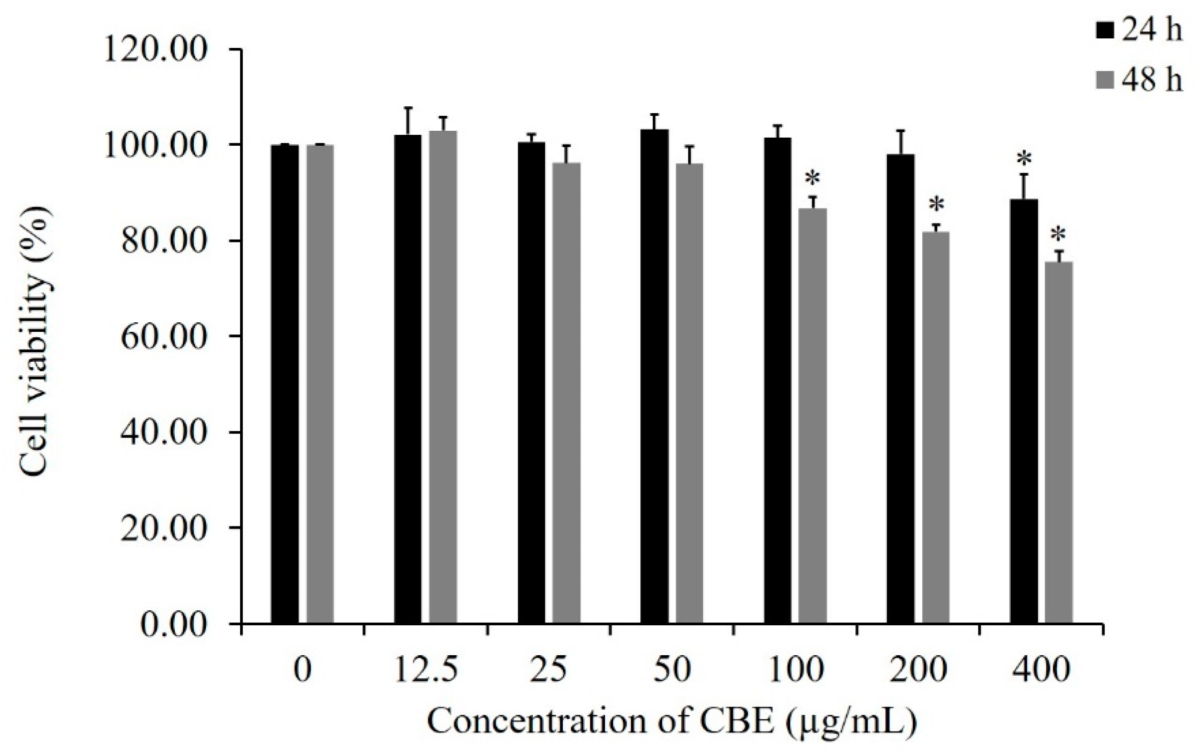
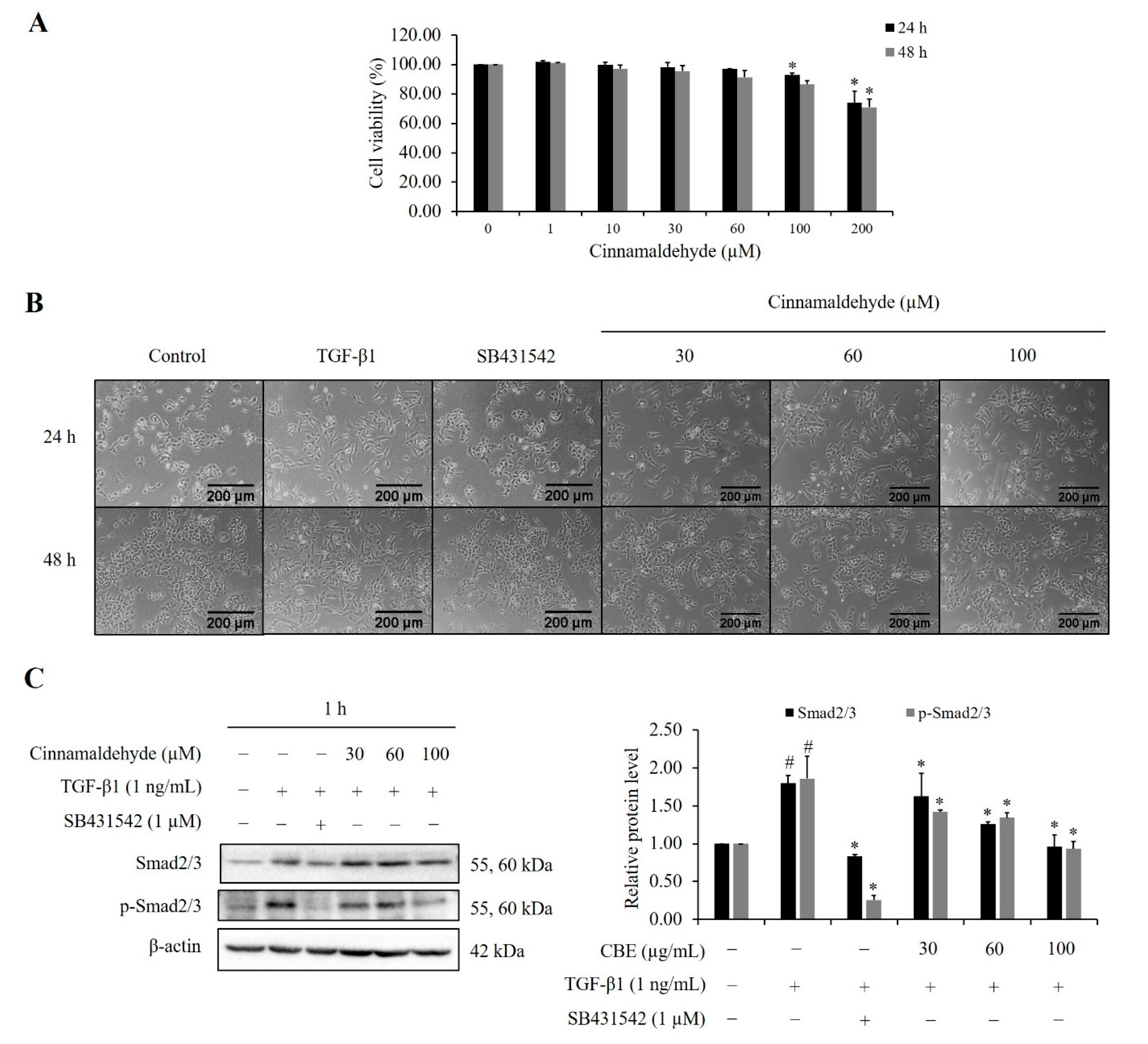
2.1. Cytotoxic Effect of CBE on the Growth of LoVo Cells

To test the cytotoxic effect of CBE on LoVo cells, cell viability was determined by MTT assay. LoVo cells were treated with varying concentrations of CBE (0, 12.5, 25, 50, 100, 200, and 400 μg/mL) for 24 and 48 h. The results showed that high concentrations of CBE reduced LoVo cell viability at 24 and 48 h (Figure 1). The sub-toxic and non-cytotoxic concentrations at 10, 30, and 100 µg/mL were chosen for further studies.

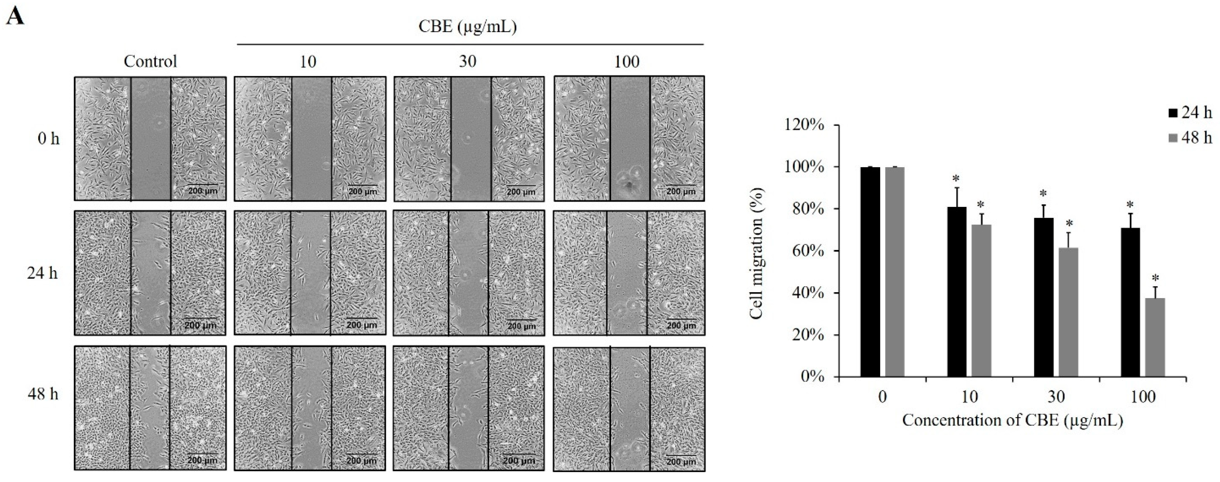
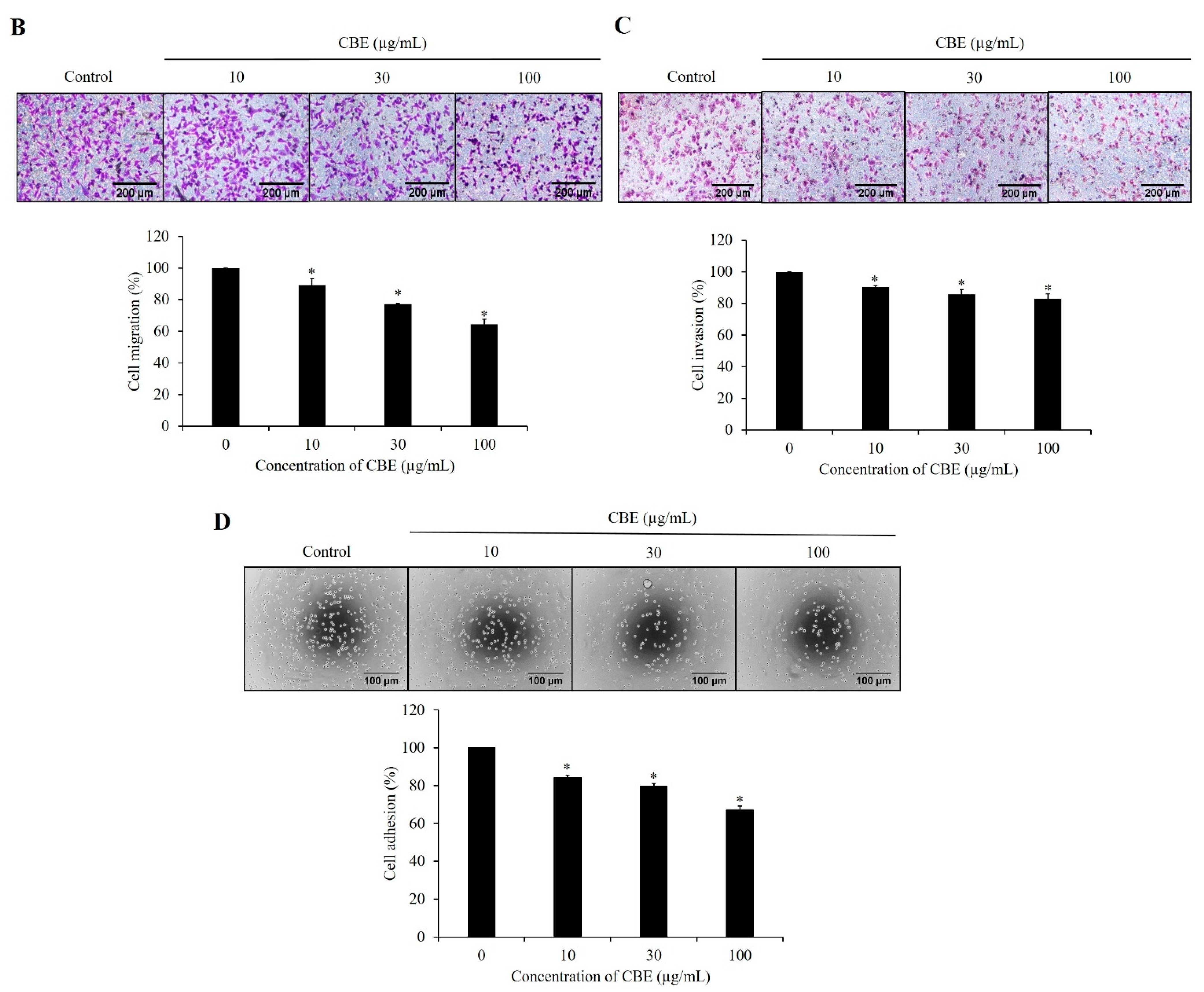
2.2. CBE Reduces Migration, Invasion, and Adhesion

Scratch assay and transwell migration assay were used to determine the effect of CBE on cell migration. The results demonstrated that CBE at 10, 30 and 100 µg/mL could inhibit cell migration in a dose-dependent manner (Figure 2A,B, p < 0.05). To determine the effect of CBE on cell invasion, Matrigel-coated transwell insert was carried out. The results showed that CBE at 10, 30 and 100 µg/mL slightly reduced the ability of LoVo cells to invade through a Matrigel-coated insert membrane (Figure 2C). Moreover, the results showed that CBE reduced adhesive ability of LoVo cells to Matrigel-coated plate in a dose-dependent manner compared to the control group (Figure 2D). These results suggested that treatment with CBE effectively inhibited migration, invasion, and adhesion of LoVo cells.

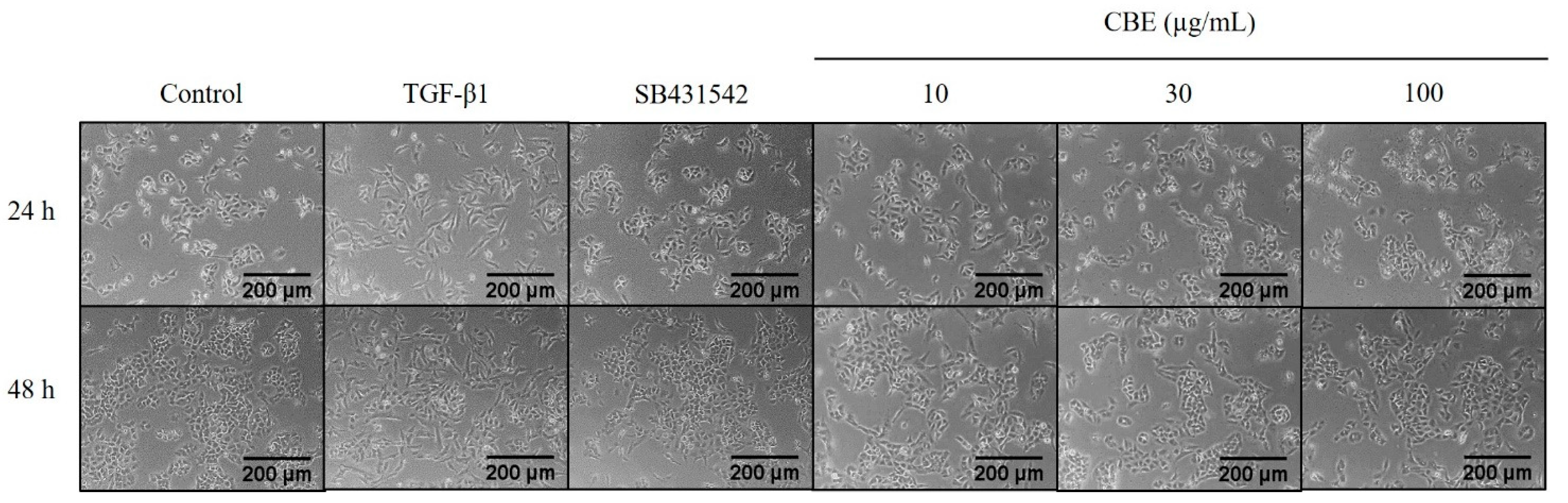
2.3. Effect of CBE on TGF-β1-Induced Morphological Changes

LoVo cells were pre-treated with CBE before being stimulated with TGF-β1. After TGF-β1 treatment, cell morphology was changed from a round shape (epithelial) to a spindle shape (mesenchymal). Cells treated with 1 µM SB431542 and 10, 30, and 100 μg/mL of CBE revealed that cells were reverted to epithelial morphology (Figure 3). Therefore, TGF-β1 treated cells showed an epithelial morphology instead of a fibroblast-like morphology, suggesting that CBE potentially reversed TGF-β1-induced EMT in LoVo cells.

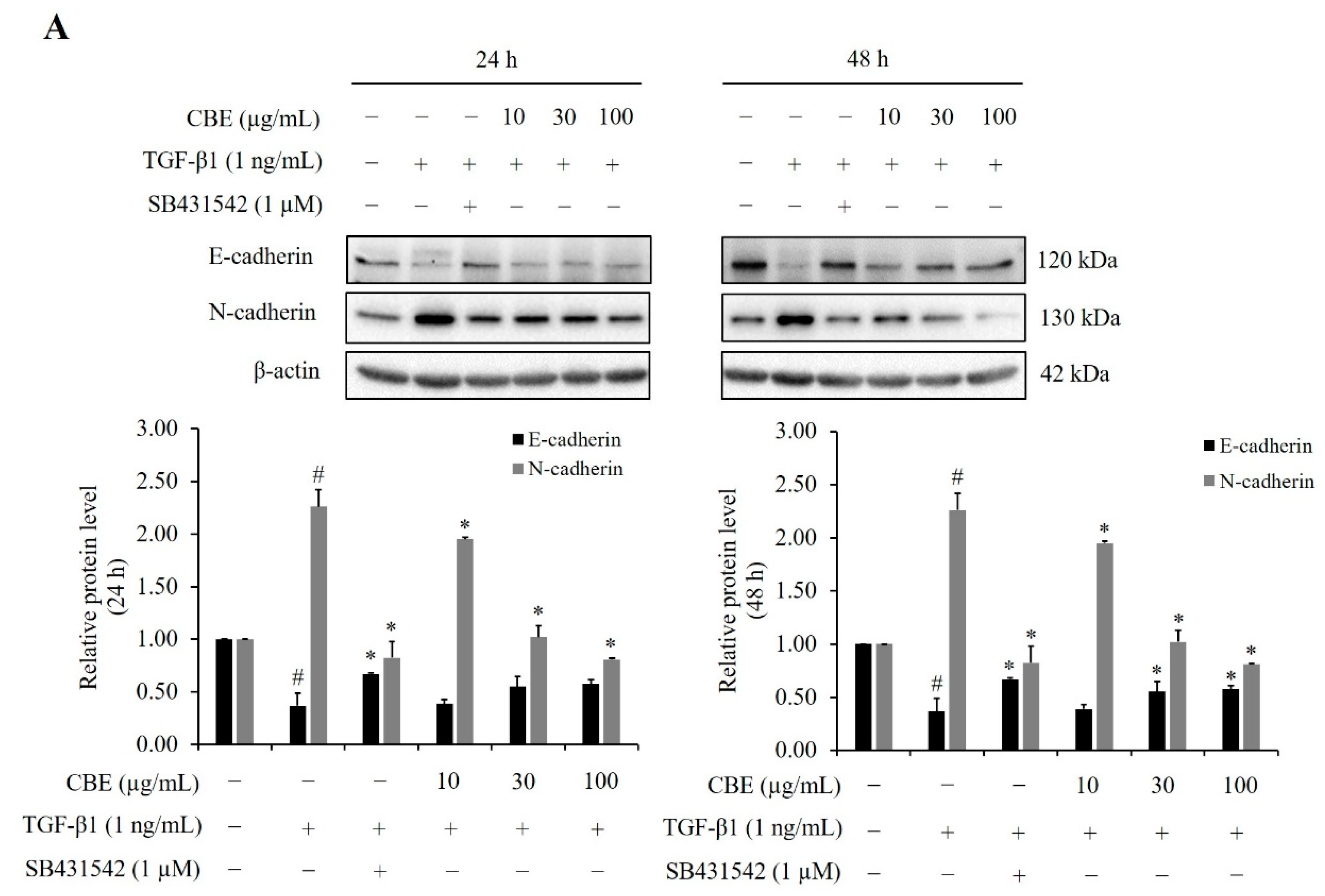
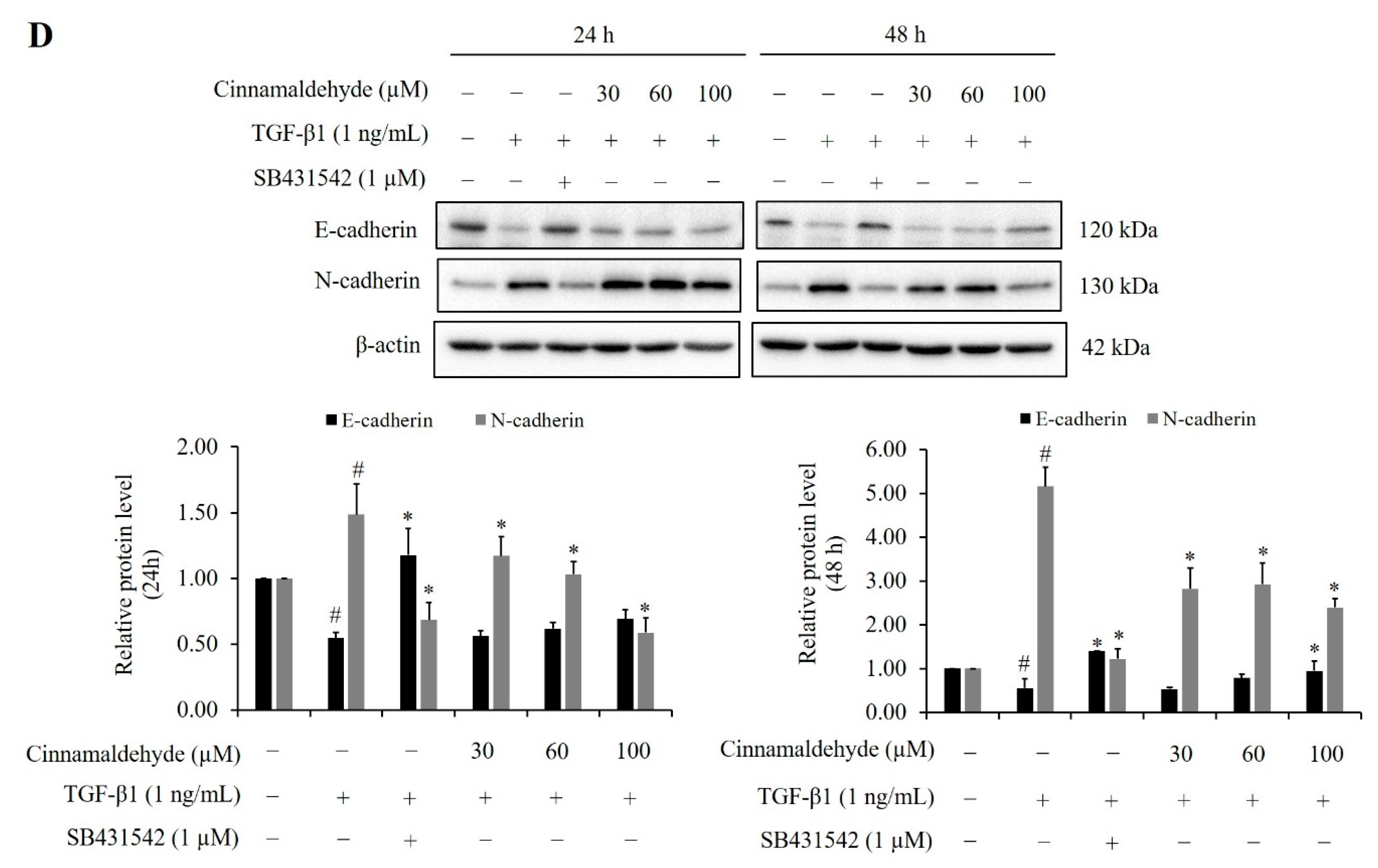
2.4. Effect of CBE on TGF-β-Induced EMT-Related Biomarker Protein Expression

It is well known that E-cadherin and N-cadherin are EMT marker proteins. Our results showed that the expression of epithelial marker E-cadherin was downregulated while the N-cadherin mesenchymal marker was upregulated after TGF-β1 treatment. Pre-treatment with CBE could increase E-cadherin expression in a concentration-dependent manner at 48 h, whereas N-cadherin expression was obviously decreased in a concentration-dependent manner at both 24 and 48 h. (Figure 4A). Similarly, qPCR results showed that CBE also downregulated N-cadherin mRNA expression after 24 h of TGF-β1 stimulation (Figure 4B). These results suggested that CBE reduced TGF-β1-induced EMT in LoVo cells.
Since MMPs exert a crucial role in the invasion of colorectal cancer cells [16], MMP-2 was apparently increased by TGF-β1, which was attenuated by CBE, whereas TIMP-1 and TIMP-2 was obviously increased in a concentration-dependent manner examined by Western blotting (Figure 4C). Our results demonstrated that downregulation of MMP-2 and upregulation of TIMPs might involve the anti-EMT activity of CBE.

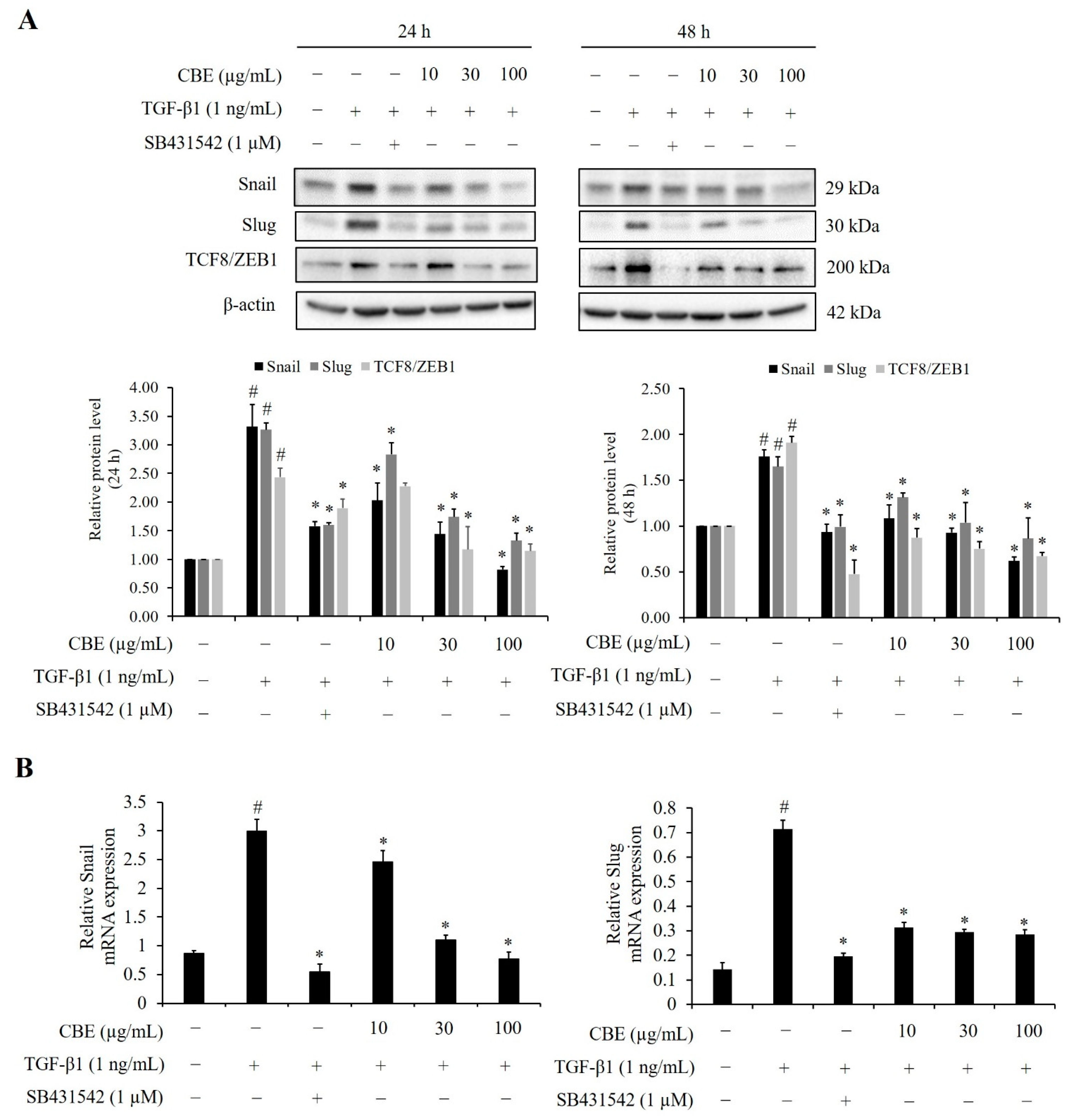
2.5. CBE Reduces TGF-β-Induced EMT-Regulated Transcription Factors

For the EMT process, several transcription factors play an important role in the control of the transcription of target genes including Snail, Slug, and TCF8/ZEB1 [17]. Upon TGF-β1 treatment, we found that the expression of Snail, Slug, and TCF8/ZEB1 in LoVo cells were upregulated, which indicated that CBE could downregulate the expression levels of these transcription factors in TGF-β1-treated LoVo cells both at 24 and 48 h (Figure 5A). Similarly, qPCR analysis revealed that CBE also decreased the Snail and Slug mRNA expression after 24 h of TGF-β1 stimulation (Figure 5B). These results suggested that CBE inhibited TGF-β1-induced EMT at the transcriptional level in LoVo cells.

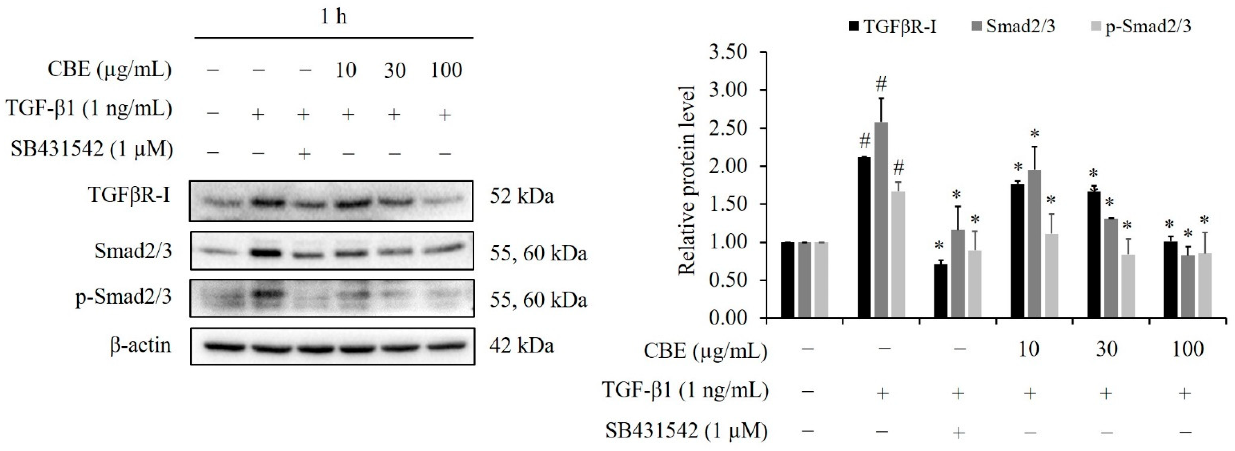
2.6. CBE Reduces TGF-β1-Induced Smad2/3 Phosphorylation

Previous studies have reported that phosphorylation of Smad2/3 by type I receptor kinase are critical steps in TGF-β1/Smad-dependent pathway in the regulating of EMT [9,18]. To investigate the effect of CBE on TGF-β1-induced phosphorylation of Smad2/3, LoVo cells were pre-treated with SB431542 and various concentrations of CBE for 30 min followed by stimulated with TGF-β1 for an additional 1 h. Western blot results showed that the total levels and phosphorylation levels of Smad2/3 were upregulated after TGF-β1 treatment for 1 h. Importantly, cells were simultaneously treated with TGF-β1 and CBE could reduce TGFβR-I expression. Furthermore, CBE also significantly downregulated total Smad2/3 and phosphorylated Smad2/3 expression (Figure 6) (p < 0.05).

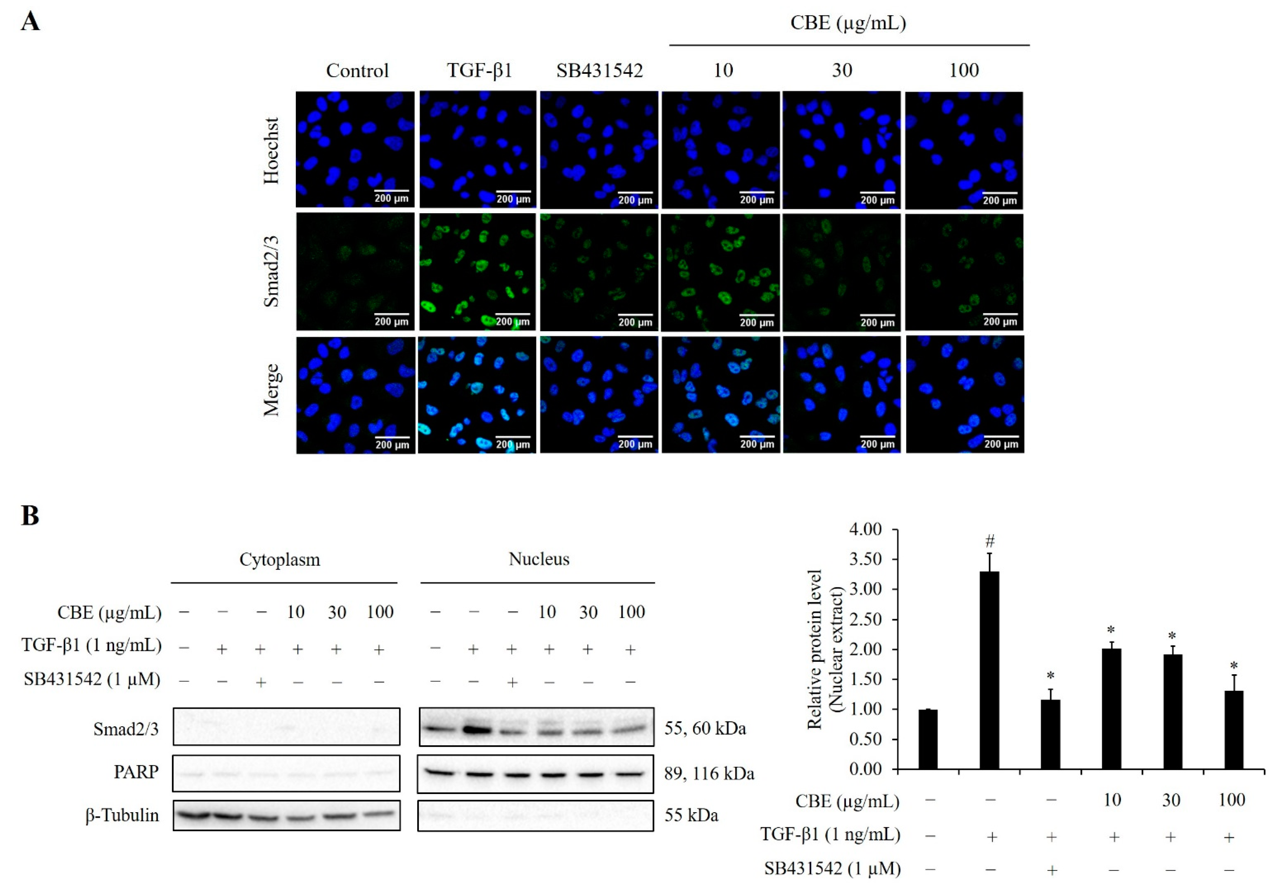
2.7. CBE Reduces TGF-β1-Induced Smad2/3 Nuclear Translocation

When stimulated with TGF-β1, Smads are phosphorylated and translocated to the nucleus and regulate target gene expression. Immunofluorescence analysis was used to assess Smad2/3 nuclear translocation. LoVo cells were treated with CBE and SB431542 for 30 min, then stimulated with TGF-β1 for 1 h. Immunofluorescence staining results showed that when treated with TGF-β1, Smad2/3 translocated into nucleus while pre-treatment with various concentrations of CBE apparently attenuated TGF-β1-induced Smad2/3 nuclear translocation in a dose-dependent manner. Likewise, SB431542 also inhibited TGF-β1-induced Smad2/3 nuclear translocation (Figure 7A). To assure Smad2/3 nuclear accumulation, cell fractionation assay was used to determine the nuclear localization. As expected, nuclear and cytoplasmic fractions showed that Smad2/3 levels in TGF-β1 treatment were upregulated, whereas SB431542 and CBE significantly reduced the levels of Smad2/3 in the nuclear fractions (Figure 7B). Therefore, these results suggested that inhibition of EMT by CBE is associated with suppression of Smad2/3 nuclear translocation.

2.8. CBE Reduces TGF-β1-Induced EMT via a Smad-Independent Pathway

The mechanism of TGF-β1-induced EMT via Smad-independent signaling pathway associated with MAPK and Akt signaling activation was reported [10]. The present study examined whether CBE could inhibit p-ERK1/2, p38, and Akt in TGF-β1-treated LoVo cells. The results showed ERK1/2, p38, and Akt were phosphorylated by TGF-β1 stimulation. Interestingly, CBE treatment could decrease phosphorylated ERK1/2, p38 and phosphorylated Akt (Ser473) similar to those of SB431542 treatment (Figure 8A,B). Therefore, our results revealed that CBE treatment reduced the activation of Smad-independent MAPK and Akt pathways in TGF-β1-induced EMT in LoVo cells.

2.9. Effect of Cinnamaldehyde on TGF-β1-Induced Smad2/3 Phosphorylation and EMT Markers Expression

Cinnamaldehyde is one of the major constituents of the Cinnamomum genus. To demonstrate the effect of cinnamaldehyde-reduced TGF-β1-induced EMT, MTT assay, morphological changes, and protein expression were determined in LoVo cells. In this study, cinnamaldehyde at various concentrations reduced cell viability in a concentration-dependent manner in both 24 h and 48 h treatments (Figure 9A). In the presence of 30, 60, and 100 µM of cinnamaldehyde could reverse cell morphology of TGF-β1-induced EMT after 24 and 48 h incubation (Figure 9B). Western blot analysis showed that cinnamaldehyde significantly reduced TGF-β1-induced phosphorylation of Smad2/3 and N-cadherin protein expression after 24 h and 48 h incubation, whereas 100 µM of cinnamaldehyde significantly increased the expression of E-cadherin after 48 h of TGF-β1 stimulation compared to cells treated with TGF-β1 in the absence of cinnamaldehyde (Figure 9C,D). These results indicated that cinnamaldehyde had an inhibitory effect on TGF-β1-induced EMT via inhibition of Smad2/3 phosphorylation.

3. Discussion

Globally, colorectal cancer is the second most common cause of cancer death. Metastasis is the important factor of patient death with CRC. Accumulating evidence suggests that EMT process is associated with CRC progression, by which epithelial cell loss of cell to cell adhesion, loss of epithelial polarity, and acquisition of migration to become mesenchymal characteristics. The most common treatment for patients with advanced colon cancer is chemotherapy, which causes many side effects such as vomiting and nausea. Currently, traditional medicine is mostly derived from natural products, which is considered an alternative treatment for the prevention of cancer. Thus, we investigated the effect of CBE on colorectal cancer cell metastasis and TGF-β1-induced EMT in LoVo cells. A cytotoxic assay showed that CBE inhibited LoVo cell proliferation in a concentration-dependent manner. It was previously reported that C. bejolghota leaf and twig extract inhibited proliferation of A549, HCT-116, and MDA-MB-23 cells [15]. At the non-cytotoxic concentration, CBE treatment reduced cell migration by scratch and transwell assay. Moreover, CBE could reduce the invasion and adhesion of LoVo cells in a concentration-dependent manner. Similarly, Koppikar et al. revealed that cinnamon extract reduced migration of human cervical tumor SiHa cells [19]. Therefore, our studies demonstrated that CBE efficiently reduced the metastatic activities of LoVo cells.
Although various species of cinnamon have been reported to have several biological properties, pharmacological effects of C. bejolghota extract have been rarely reported. This study is the first to report the effect of CBE on TGF-β1-induced EMT in CRC cells. We found that CBE reversed TGF-β1-induced morphological changes from spindle to round shape compared with the TGF-β1 and SB431542 treatment. Lin et al. showed that the extract of Cinnamomum cassia suppressed TGF-β1-induced EMT in H1299 and A549 human lung cancer cells [20]. It is well known that E-cadherin and N-cadherin are major EMT marker proteins. Many studies of herbal medicine have reported that upregulation of E-cadherin and downregulation of N-cadherin are important goals on EMT inhibition [21,22]. In this study, CBE upregulated E-cadherin and N-cadherin protein levels in TGF-β1-treated LoVo cells. Many studies have reported that TGF-β1 also induced EMT through MMP regulation, which is regulated by TIMPs [23,24]. Previous studies have reported that TIMP-1 upregulation may be responsible for the regulation of MMP-2 activity [25]. A recent study demonstrated that TGF-β1 upregulated MMP-2 transcription and suppressed TIMP-2 expression through Smad3 to promote the MMP-2 activity. Moreover, they also reported that treatment with naringenin (Smad3 inhibitor) inhibited Smad3, which subsequently suppressed MMP-2 while increasing TIMP-2 transcription in lung carcinoma cells [26]. Consistent with previous studies, we identified that TGF-β1 induced MMP-2 expression and suppressed TIMP-1 and TIMP-2 expression. Our results also showed CBE could decrease the expression of MMP-2 whereas TIMP-1 and TIMP-2 levels were increased in LoVo cells. Our findings demonstrated that CBE had an inhibitory effect on EMT protein biomarkers. Therefore, we further examined the inhibitory effect of TGF-β1-mediated EMT on colorectal cancer LoVo cells.
EMT activation by various transcription factors including Slug, Snail, and TCF8/ZEB1 protein play an important role in cancer progression and metastasis [27]. Upon TGF-β1 treatment, our results found that Snail, Slug, and TCF8/ZEB1 were increased, whereas pre-treatment with CBE could decrease the expression of these EMT transcription factors in LoVo cells. Consistent with the previous study, natural compound inhibited the TGF-β1-induced expression of Snail and Slug in colorectal cancer HT-29 and SW480 cells [28]. These studies indicated that CBE inhibited EMT via downregulation of EMT transcription factors.
Moreover, SMAD2 and KRAS mutations in LoVo cells lead to the activation of EMT and cancer metastasis [29]. Therefore, we also investigated both the Smad and the non-Smad signaling pathway. TGF-β1-stimulated EMT through Smad-dependent signaling pathway in colon cancer [30]. Notably, natural products such as resveratrol and silibinin inhibited EMT by suppressing the TGF-β1/Smad pathway in colorectal cancer cells [31,32]. Our results showed that the expression of TGFβR-I and p-Smad2/3 were upregulated by the TGF-β1 treatment, which implied that TGF-β1-stimulated EMT was related to TGFβR-I and Smad2/3 activation. CBE could suppress TGF-β1-stimulated EMT by decreasing protein levels of TGFβR-I and p-Smad2/3. Furthermore, phosphorylation of Smad proteins resulted in Smad complex translocation into the nucleus [33]. In this study, the results showed that Smad2/3 were translocated into the nucleus when treated with TGF-β1. However, pre-treatment with CBE attenuated TGF-β1-stimulated Smad2/3 nuclear translocation, which is consistent with previous study [34]. To confirm the accumulation of Smad2/3 in the nucleus, a cytoplasmic/nuclear fractionation assay was used to indicate the nuclear localization of Smad2/3. Upon TGF-β1 treatment, our results revealed that Smad2/3 levels were upregulated in the nucleus while CBE could reduce Smad2/3 levels in the nuclear fractions when compared to the cytoplasmic fractions, suggesting that inhibition of EMT by CBE was associated with the suppression of Smad2/3 nuclear translocation.
Moreover, several studies reported that TGF-β1-induced EMT was also mediated via aSmad-independent signaling pathway, including a MAPK and an Akt pathway. Wendt et al. reported the activation of ERK1/2, p38, and Akt mediated the EMT process in cancer cells [35]. We also demonstrated that treatment with CBE significantly decreased p-ERK, p-p38, and p-Akt (Ser473) expression in TGF-β1-treated cells. Overall, our study indicated that CBE could inhibit TGF-β1-induced EMT by inhibiting the activation of ERK1/2, p38, and Akt phosphorylation.
In addition, we found that CBE had antioxidant activity through DPPH scavenging in a dose-dependent manner (Figure S1). Moreover, cinnamaldehyde is one of the major constituents of the Cinnamomum genus. Likewise, Choudhury and Ahmed have reported that the bark of CBE contained cinnamaldehyde as a major composition [36]. We have investigated that a major active component of CBE may be rich in cinnamaldehyde content by using TLC. TLC results showed that the major compounds in CBE were mainly composed of cinnamaldehyde (Figure S2). Importantly, our results found that cinnamaldehyde could reduce phosphorylation of Smad2/3 and affect EMT marker expression including E-cadherin and N-cadherin. Thus, our results indicated that the inhibitory effect of CBE on TGF-β1-induced EMT may be due to the cinnamaldehyde composition in CBE.

4. Materials and Methods

4.1. Preparation of C. bejolghota Ethanolic Bark Extract

C. bejolghota bark powder was derived from Thai-Lanna Herbal Industry Company Limited, Chiangmai, Thailand. The extraction method was followed in our previous study [37]. Briefly, 100 g of C. bejolghota bark powder was mixed to maceration in 500 mL of ethanol in a conical flask. The mixture was swirled to mix entirely and was kept at room temperature (RT) for 1 day. The ethanolic extract was then filtered using filter paper Whatman No.1. Then, the filtrated CBE solution was dried using a rotary evaporator in a vacuum at 40 °C. The CBE was stored at 4 °C for further experiments. The indication of a major active component of CBE was further identified using thin-layer chromatography (TLC). Sample (CBE) and standard (cinnamaldehyde) solutions were spotted on TLC Silica gel 60 F254 plate (Merck, Darmstadt, Germany). The TLC plates were developed in toluene and ethyl acetate (93:7) as the solvent system [38]. The migrated compounds were visualized under the UV light (254 and 366 nm). The TLC plates were repeated 3 times. The antioxidant capacity of CBE was determined using 2,2-Diphenyl-1-picrylhydrazyl (DPPH) radical scavenging assay, according to the procedure described by Sadeghi et al. [39] L-ascorbic acid was utilized as a positive control.

4.2. Chemicals and Reagents

TGF-β1 was purchased from R&D Systems (Minneapolis, MN, USA). SB431542 (TGF-β1 receptor inhibitor), 3-(4, 5-dimethylthiazol-2-yl)-2,5-diphenyl-2H-tetrazolium bromide (MTT), dimethylsulfoxide (DMSO), phenylmethylsulphonyl fluoride (PMSF), penicillin G, kanamycin, and cinnamaldehyde were purchased from Sigma-Aldrich (St. Louis, MO, USA). Roswell Park Memorial Institute (RPMI) 1640 Medium was purchased from Nissui pharmaceutical (Tokyo, Japan). Fetal bovine serum (FBS) was purchased from Equitech-Bio (Kerrville, TX, USA). Matrigel was purchased from BD Biosciences (Dallas, TX, USA) The antibodies were obtained as follows: anti-N-cadherin, anti-E-cadherin from BD Biosciences (San Jose, CA, USA), anti-Smad2/3, anti-p-Smad2/3 from Santa Cruz Biotechnology (Dallas, TX, USA), anti-TIMP-1, anti-TIMP-2, anti-MMP-2, anti-TGFβR-I, anti-Snail, anti-Slug, anti-ZEB1, anti-ERK1/2, anti-p-ERK1/2, anti-p38, anti-p-p38, anti-Akt, anti-p-Akt (Ser473), and β-actin from Cell Signaling Technology (Danvers, MA, USA). TRIzol reagent was purchased from Invitrogen Life Technologies (Carlsbad, CA, USA).

4.3. Cell Culture

Human colorectal adenocarcinoma cell line, LoVo, was obtained from the American Type Culture Collection (ATCC, Manassas, VA, USA). Cells were maintained in RPMI-1640 medium supplemented with 10% FBS, 100 units/mL penicillin G, and 0.1 mg/mL kanamycin at 37 °C under a humidified 5% CO2.

4.4. Cytotoxicity Assay

MTT assay was used for determination the effect of CBE on cell viability. LoVo cell suspension were seeded at 5 × 10³ cells/well overnight. Cells were then incubated with CBE at 12.5, 25, 50, 100, 200, and 400 µg/mL for 24 h, whereas 0.5% DMSO was used as a control group. After treatment, 0.5 mg/mL MTT solution was added in each well for 4 h at 37 °C. Then DMSO was added to dissolve the formazan crystals. Absorbance was measured at 570 nm by Multiskan™ FC microplate reader (Thermo Fisher Scientific, Waltham, MA, USA) and was analyzed by the software GraphPad Prism 3.03 software (San Diego, CA, USA).

4.5. Scratch Assay

LoVo cells (6 × 10⁴ cells/well) were gently scratched and washed with phosphate-buffered saline (PBS) to remove cell debris. The cells were treated with or without CBE for 24 h and photographed by using an inverted microscope (Olympus, Tokyo, Japan). The area of migration was analyzed as described earlier [37].

4.6. Transwell Assay

Transwell Boyden chamber (8 µm pore size, Merck Millipore, Darmstadt,, Germany) was used to assess cell migration and cell invasion assay. The method for both assays were the same, but for cell invasion assay the plate was pre-coated with 30 µg Matrigel. LoVo cells suspension in 1% FBS medium (1.5 × 10⁴ cells/well) were added to the transwell chamber. After 2 h incubation, cells were treated with or without CBE, while complete medium was added to the lower chambers for 24 h. The assay was performed as described earlier [37].

4.7. Cell Adhesion Assay

LoVo cells (8 × 10⁴ cells/well) were pre-treated with or without CBE. After 24 h incubation, cells were harvested and seeded to Matrigel-coated 96-well plate and incubated for 15 min. The non-adherent cells were washed gently with PBS. Cell adhesion was determined using MTT assay. Images were obtained under a microscope.

4.8. Morphological Assessment

LoVo cells were pre-treated in 1% FBS medium with 10, 30 and 100 µg/mL CBE and 1 µM SB431542 for 30 min. After incubation, cells were incubated with 1 ng/mL TGF-β1. The morphology was monitored and photographed at 24 and 48 h using a microscope.

4.9. Western Blot Analysis

After treatment, protein samples in cell lysates were collected, lysed with RIPA buffer (50 mM Tris-HCl, pH 7.5, 5 mM EDTA, 250 mM NaCl, 0.5% Triton X-100) supplemented with PMSF and a complete mini protease inhibitor cocktail (Roche Diagnostics GmbH, Mannheim, Germany), and then separated using SDS-PAGE and electrophoretically transferred onto polyvinylidene fluoride (PVDF) membranes (Merck Millipore, Darmstadt, Germany). Membranes were blocked with blocking buffer for 1 h, then incubated overnight at 4 °C with the primary antibody as follows: anti-N-cadherin (1:1000), anti-E-cadherin (1:1000), anti-Smad2/3 (1:1000), an-ti-p-Smad2/3 (1:1000), anti-TIMP-1 (1:1000), anti-TIMP-2 (1:1000), anti-MMP-2 (1:1000), anti-TGFβR-I (1:500), anti-Snail (1:500), anti-Slug (1:500), anti-ZEB1 (1:500), anti-ERK1/2 (1:1000), anti-p-ERK1/2 (1:500), anti-p38 (1:1000), anti-p-p38 (1:1000), an-ti-Akt (1:1000), anti-p-Akt (Ser473) (1:500), and β-actin (1:10,000), and then followed by appropriate secondary antibodies at RT for 1 h. The chemiluminescence signal was detected by the Immobilon Western Chemiluminescent HRP Substrate (Merck Millipore, Darmstadt, Germany) and Chemi-Doc XRS+ System (BioRad, Hercules, CA, USA).

4.10. RNA Extraction, RT-PCR, and Quantitative Real-Time PCR (qPCR)

After 24 h incubation, total RNA was extracted with TRIzol reagent and reverse transcribed to complementary DNA (cDNA) by M-MLV reverse transcriptase (Promega, Madison, WI, USA) following the manufacturer’s instruction. qPCR was carried out using TB Green Premix Ex Taq II and detected on a Thermal Cycler Dice (Takara, Japan). The sequences of the primers used for qPCR analysis were as follows: N-cadherin 5′-GACAATGCCCCTCAAGTGTT-3′ (forward) and 5′-CCATTAAGCCGAGTGATGGT-3′ (reverse); Snail 5′-GCCTTCAACYGCAAATACTGC-3′ (forward) and 5′-CTTCTTGACA-TCTGAGTGGGTC-3′ (reverse); Slug 5′-GGGGAGAAGCCTTTTTCTTG-3′ (forward) and 5′-TCCTCATGTTGTGCAGGAG-3′ (reverse); RPL37A 5′-GCAGGAACCACAGTGCCAG ATCC-3′ (antisense) and 5′ ATTGAAATCAGCCAGCACGC-3′ (sense). The relative mRNA expression was normalized to RPL37A mRNA housekeeping gene.

4.11. Immunofluorescence Analysis

LoVo cells at 10 × 104 cells/well were cultured on glass coverslips. The cells were pre-treated with or without CBE and 1 µM SB431542 for 30 min, then stimulated with TGF-β1 for an additional 1 h. After treatment, cells were fixed with 3% paraformaldehyde and permeabilized with 0.5% Triton X-100 in phosphate-buffered saline with 3 % bovine serum albumin (BSA) in PBS. Subsequently, incubation with the primary antibody of Smad2/3 (1:1000), at RT for 1 h then incubated with the Alexa Fluor 488-conjugated goat anti-rabbit IgG, a secondary antibody (Invitrogen, Carlsbad, CA, USA) and Hoechst 33258. Cells on coverslips were mounting with 50% glycerol/PBS on glass slides and photographed using a confocal laser scanning microscope (FV1000, Olympus, Tokyo, Japan).

4.12. Cytoplasmic/Nuclear Fractionation Assay

After 1 h incubation, pellets were gently mixed with buffer A (250 mM sucrose, 10 mM HEPES-KOH, 10 mM KCl, 1.5 mM MgCl2, 1 mM EDTA, 1 mM EGTA, 1 mM DTT; pH 7.5) supplemented with a protease inhibitor cocktail tablet. Cells were then fractionated by centrifugation at 600× g, 4 °C. The pellet contains nucleus while the supernatant contains cytoplasm and mitochondria. The nuclear pellets were washed and centrifuged to eliminate cytoplasmic material. The nuclei were lysed completely in lysis buffer and centrifuged to collect the supernatant as the nuclear fraction. The supernatant containing cytoplasm and mitochondria were subsequently centrifuged for 60 min to obtain the cytoplasmic fraction. Smad2/3 level in cytoplasmic and nuclear fraction were determined using Western blot. The loading control was used to assay for contamination between the fractions, β-Tubulin (1:1000) and PARP (1:1000) (Cell Signaling Technology, Danvers, MA, USA) were used as a control for the cytoplasmic and nuclear fraction, respectively.

4.13. Statistical Analysis

Data were analyzed using one-way analysis of variance (ANOVA) to evaluate significant differences between groups (p < 0.05). Statistical analysis was presented as mean ± standard deviation (SD).

5. Conclusions

Our findings indicated that CBE had an inhibitory effect on TGF-β1-mediated EMT of human colorectal cancer cells by reversion of cell morphology. CBE also remarkably increased E-cadherin, TIMP-1 and TIMP-2, and decreased N-cadherin, MMP-2, Snail, Slug, and TCF8/ZEB1 expression by downregulation of TGF-β1 receptor through inhibition of Smad-dependent and Smad-independent signaling in LoVo cells. Hence, this study is the first to report that Cinnamomum bejolghota extract could reduce TGF-β1-induced EMT in cancer and may be valuable as a therapeutic agent in patients with CRC in the future.

Supplementary Materials

The following supporting information can be downloaded at: https://0-www-mdpi-com.brum.beds.ac.uk/article/10.3390/scipharm90020030/s1, Figure S1: DPPH-free radical scavenging (%) of L-ascorbic acid and the CBE. Figure S2: TLC chromatogram of CBE (lane A) and cinnamaldehyde (lane B). Figure S3: Original images of Western blot band intensities in Figure 4A. Figure S4: Original images of Western blot band intensities in Figure 4C. Figure S5: Original images of Western blot band intensities in Figure 5A. Figure S6: Original images of Western blot band intensities in Figure 6. Figure S7: Original images of Western blot band intensities in Figure 7. Figure S8: Original images of Western blot band intensities in Figure 8. Figure S9: Original images of Western blot band intensities in Figure 9.

Author Contributions

Supervision, R.W.; Conceptualization, R.W. and M.I.; Methodology, S.I., E.T., M.I. and R.W.; Investigation, A.K.; Formal Analysis, A.K.; Validation, S.I., E.T., M.I. and R.W.; Writing—Original Draft Preparation, A.K.; Writing—Review and Editing, A.K. and R.W.; Funding Acquisition, R.W. All authors have read and agreed to the published version of the manuscript.

Funding

This research was funded by Thailand Research Fund through the Royal Golden Jubilee PhD Program, grant number PHD/0164/2557 and Thailand Science Research and Innovation through the Strategic Wisdom and Research Institute, Srinakharinwirot University No. 012/2564.

Institutional Review Board Statement

Not applicable.

Informed Consent Statement

Not applicable.

Data Availability Statement

The datasets analyzed during the current study are available from the corresponding author on reasonable request.

Acknowledgments

We would like to thank Central laboratory, Faculty of Medicine, Srinakharinwirot University Srinakharinwirot University for supplying some instruments.

Conflicts of Interest

The authors declare no conflict of interest.

References

  1. Sung, H.; Ferlay, J.; Siegel, R.L. Global cancer statistics 2020: GLOBOCAN estimates of incidence and mortality worldwide for 36 cancers in 185 countries. CA Cancer J. Clin. 2021, 71, 209–249. [Google Scholar] [CrossRef] [PubMed]
  2. Bundgaard-Nielsen, C.; Baandrup, U.T.; Nielsen, L.P.; Sørensen, S. The presence of bacteria varies between colorectal adenocarcinomas, precursor lesions and non-malignant tissue. BMC Cancer 2019, 19, 399. [Google Scholar] [CrossRef] [PubMed]
  3. Jiang, Z.; Cao, Q.; Dai, G.; Wang, J.; Liu, C.; Lv, L.; Pan, J. Celastrol inhibits colorectal cancer through TGF-β1/Smad signalling. Onco Targets Ther. 2019, 119, 509–518. [Google Scholar] [CrossRef] [Green Version]
  4. Kalluri, R.; Weinberg, R.A. The basics of epithelial-mesenchymal transition. J. Clin. Investig. 2009, 119, 1420–1428. [Google Scholar] [CrossRef] [PubMed] [Green Version]
  5. Araki, K.; Shimura, H.; Suzuki, H.; Tsutsumi, S.; Wada, W.; Yajima, T.; Kobayahi, T.; Kubo, N.; Kuwano, H. E/N-cadherin switch mediates cancer progression via TGF-β-induced epithelial-to-mesenchymal transition in extrahepatic cholangiocarcinoma. Br. J. Cancer 2011, 105, 1885–1893. [Google Scholar] [CrossRef] [Green Version]
  6. Yan, M.D.; Yao, C.J.; Chow, J.M.; Chang, C.L.; Hwang, P.A.; Chuang, S.E.; Whang-Peng, J.; Lai, G.M. Fucoidan elevates microRNA-29b to regulate DNMT3B-MTSS1 axis and inhibit EMT in human hepatocellular carcinoma cells. Mar. Drugs 2015, 13, 6099–6116. [Google Scholar] [CrossRef] [Green Version]
  7. Xu, J.; Lamouille, S.; Derynck, R. TGF-beta-induced epithelial to mesenchymal transition. Cell Res. 2009, 19, 156–172. [Google Scholar] [CrossRef] [PubMed]
  8. Kubiczkova, L.; Sedlarikova, L.; Hajek, R.; Sevcikova, S. TGF-β—An excellent servant but a bad master. J. Transl. Med. 2012, 10, 183. [Google Scholar] [CrossRef] [PubMed] [Green Version]
  9. Hata, A.; Chen, Y.G. TGF-β signaling from Receptors to Smads. Cold Spring Harb. Perspect. Biol. 2016, 8, a022061. [Google Scholar] [CrossRef] [Green Version]
  10. Nagaraj, N.S.; Datta, P.K. Targeting the transforming growth factor-beta signaling pathway in human cancer. Expert Opin. Investig. Drugs 2010, 19, 77–91. [Google Scholar] [CrossRef] [Green Version]
  11. Rani, A.; Pande, C.; Tewari, G.; Patni, K. A review on aroma profile of Cinnamomum species in north and north east India. World J. Pharm. Res. 2017, 6, 200–221. [Google Scholar]
  12. Gogoi, B.; Kakoti, B.; Bora, S.; Borah, N.S. Pharmacognostic and preliminary phytochemical evaluation of Cinnamomum bejolghota (Buch.-Ham.) sweet bark. Indian J. Nat. Prod. Resour. 2016, 7, 59–64. [Google Scholar]
  13. Atiphasaworn, P.; Monggoot, S.; Pripdeevech, P. Chemical composition, antibacterial and antifungal activities of Cinamomum bejolghota bark oil from Thailand. J. Appl. Pharm. 2017, 7, 69–73. [Google Scholar]
  14. Gogoi, B.; Kakoti, B.; Bora, N.S.; Borah, S. Antihyperglycemic and in vivo antioxidative activity evaluation of Cinnamomum bejolghota (Buch.-Ham.) in streptozotocin induced diabetic rats: An ethnomedicinal plant in Assam. Asian Pac. J. Trop. Med. 2014, 7, S427–S434. [Google Scholar] [CrossRef] [Green Version]
  15. Rao, L.; You, Y.X.; Su, Y.; Fan, Y.; Liu, Y.; He, Q.; Chen, Y.; Meng, J.; Hu, L.; Li, Y.; et al. Lignans and neolignans with antioxidant and human cancer cell proliferation inhibitory activities from Cinnamomum bejolghota confirm its functional food property. J. Agric. Food Chem. 2020, 68, 8825–8835. [Google Scholar] [CrossRef] [PubMed]
  16. Kryczka, J.; Stasiak, M.; Dziki, L.; Mik, M.; Dziki, A.; Cierniewski, C. Matrix metalloproteinase-2 cleavage of the β1 integrin ectodomain facilitates colon cancer cell motility. J. Biol. Chem. 2012, 287, 36556–36566. [Google Scholar] [CrossRef] [PubMed] [Green Version]
  17. Ansieau, S.; Collin, G.; Hill, L. EMT or EMT-Promoting Transcription Factors, Where to Focus the Light? Front. Oncol. 2014, 4, 353. [Google Scholar] [CrossRef] [PubMed] [Green Version]
  18. Xu, L. Regulation of Smad activities. Biochim. Biophys. Acta (BBA) Gene Struct. Expr. 2006, 1759, 503–513. [Google Scholar] [CrossRef] [Green Version]
  19. Koppikar, S.J.; Choudhari, A.S.; Suryavanshi, S.A.; Kumari, S.; Chattopadhyay, S.; Kaul-Ghanekar, R. Aqueous cinnamon extract (ACE-c) from the bark of Cinnamomum cassia causes apoptosis in human cervical cancer cell line (SiHa). BMC Cancer 2010, 10, 210. [Google Scholar] [CrossRef] [Green Version]
  20. Lin, C.Y.; Hsieh, Y.H.; Yang, S.F.; Chu, S.C.; Chen, P.N.; Hsieh, Y.S. Cinnamomum cassia extracts reverses TGF-β1-induced epithelial-mesenchymal transition in human lung adenocarcinoma cells and suppresses tumor growth in vivo. Environ. Toxicol. 2017, 32, 1878–1887. [Google Scholar] [CrossRef]
  21. Kee, J.Y.; Han, Y.H.; Mun, J.G.; Park, S.H.; Jeon, H.D.; Hong, S.H. Effect of Korean Red Ginseng extract on colorectal lung metastasis through inhibiting the epithelial-mesenchymal transition via transforming growth factor-β1/Smad-signaling-mediated Snail/E-cadherin expression. J. Ginseng Res. 2019, 43, 68–76. [Google Scholar] [CrossRef] [PubMed]
  22. Hu, B.; An, H.M.; Yan, X.; Zheng, J.L.; Huang, X.W.; Li, M. Traditional Chinese medicine formulation Yanggan Jiedu Sanjie inhibits TGF-β1-induced epithelial-mesenchymal transition and metastatic potential in human hepatocarcinoma Bel-7402 cells. BMC Complementary Altern. Med. 2019, 19, 67. [Google Scholar] [CrossRef] [PubMed] [Green Version]
  23. Santibanez, J.F.; Obradović, H.; Kukolj, T.; Krstić, J. Transforming growth factor-β, matrix metalloproteinases, and urokinase-type plasminogen activator interaction in the cancer epithelial to mesenchymal transition. Dev. Dyn. 2018, 247, 382–395. [Google Scholar] [CrossRef] [PubMed] [Green Version]
  24. Hwang, K.E.; Kim, H.J.; Song, I.S.; Park, C.; Jung, J.W.; Park, D.S.; Oh, S.H.; Kim, Y.S.; Kim, H.R. Salinomycin suppresses TGF-β1-induced EMT by down-regulating MMP-2 and MMP-9 via the AMPK/SIRT1 pathway in non-small cell lung cancer. Int. J. Med. Sci. 2021, 18, 715–726. [Google Scholar] [CrossRef]
  25. Wang, N.; Zhu, M.; Tsao, S.W.; Man, K.; Zhang, Z.; Feng, Y. Up-regulation of TIMP-1 by genipin inhibits MMP-2 activities and suppresses the metastatic potential of human hepatocellular carcinoma. PLoS ONE 2012, 7, e46318. [Google Scholar] [CrossRef] [Green Version]
  26. Lian, G.Y.; Wang, Q.M.; Mak, T.S.; Huang, X.R.; Yu, X.Q.; Lan, H.Y. Inhibition of tumor invasion and metastasis by targeting TGF-β-Smad-MMP2 pathway with Asiatic acid and Naringenin. Mol. Ther.-Oncolytics 2021, 20, 277–289. [Google Scholar] [CrossRef]
  27. Brabletz, T.; Kalluri, R.; Nieto, M.A.; Weinberg, R.A. EMT in cancer. Nat. Rev. Cancer 2018, 18, 128–134. [Google Scholar] [CrossRef]
  28. Zhu, S.M.; Park, Y.R.; Seo, S.Y.; Kim, I.H.; Lee, S.T.; Kim, S.W. Parthenolide inhibits transforming growth factor β1-induced epithelial-mesenchymal transition in colorectal cancer cells. Intest. Res. 2019, 17, 527–536. [Google Scholar] [CrossRef] [Green Version]
  29. Rouillard, A.D.; Gundersen, G.W.; Fernandez, N.F.; Wang, Z.; Monteiro, C.D.; McDermott, M.G.; Ma’ayan, A. The harmonizome: A collection of processed datasets gathered to serve and mine knowledge about genes and proteins. Database 2016, 2016, baw100. [Google Scholar] [CrossRef]
  30. Hao, Y.; Baker, D.; Ten Dijke, P. TGF-β-Mediated Epithelial-Mesenchymal Transition and Cancer Metastasis. Int. J. Mol. Sci. 2019, 20, 2767. [Google Scholar] [CrossRef] [Green Version]
  31. Ji, Q.; Liu, X.; Han, Z.; Zhou, L.; Sui, H.; Yan, L.; Jiang, H.; Ren, J.; Cai, J.; Li, Q. Resveratrol suppresses epithelial-to-mesenchymal transition in colorectal cancer through TGF-β1/Smads signaling pathway mediated Snail/E-cadherin expression. BMC Cancer 2015, 15, 97. [Google Scholar] [CrossRef] [PubMed] [Green Version]
  32. Zare, Z.; Nayerpour Dizaj, T.; Lohrasbi, A.; Sheikhalishahi, Z.S.; Asadi, A.; Zakeri, M.; Hosseinabadi, F.; Abazari, O.; Abbasi, M.; Khanicheragh, P. Silibinin Inhibits TGF-β-induced MMP-2 and MMP-9 Through Smad Signaling Pathway in Colorectal Cancer HT-29 Cells. Basic Clin. Cancer Res. 2021, 12, 81–90. [Google Scholar] [CrossRef]
  33. Massagué, J.; Wotton, D. Transcriptional control by the TGF-beta/Smad signaling system. EMBO J. 2000, 19, 1745–1754. [Google Scholar] [CrossRef] [PubMed] [Green Version]
  34. Liu, X.N.; Wang, S.; Yang, Q.; Wang, Y.J.; Chen, D.X.; Zhu, X.X. ESC reverses epithelial mesenchymal transition induced by transforming growth factor-β via inhibition of Smad signal pathway in HepG2 liver cancer cells. Cancer Cell Int. 2015, 15, 114. [Google Scholar] [CrossRef] [PubMed] [Green Version]
  35. Wendt, M.K.; Allington, T.M.; Schiemann, W.P. Mechanisms of the epithelial-mesenchymal transition by TGF-beta. Future Oncol. 2009, 5, 1145–1168. [Google Scholar] [CrossRef] [Green Version]
  36. Choudhury, S.; Ahmed, R.; Barthel, A.; Leclercq, P.A. Composition of the Bark and Flower Oils of Cinnamomum bejolghota (Buch.-Ham.) Sweet from two Locations of Assam, India. J. Essent. Oil Res. 1998, 10, 245–250. [Google Scholar] [CrossRef]
  37. Kittiwattanokhun, A.; Samosorn, S.; Innajak, S.; Watanapokasin, R. Inhibitory effects on chondrosarcoma cell metastasis by Senna alata extract. Biomed. Pharmacother. 2021, 137, 111337. [Google Scholar] [CrossRef]
  38. Utchariyakiat, I.; Surassmo, S.; Jaturanpinyo, M.; Khuntayaporn, P.; Chomnawang, M.T. Efficacy of cinnamon bark oil and cinnamaldehyde on anti-multidrug resistant Pseudomonas aeruginosa and the synergistic effects in combination with other antimicrobial agents. BMC Complementary Altern. Med. 2016, 16, 158. [Google Scholar] [CrossRef] [Green Version]
  39. Sadeghi, Z.; Valizadeh, J.; Azyzian Shermeh, O.; Akaberi, M. Antioxidant activity and total phenolic content of Boerhavia elegans (choisy) grown in Baluchestan, Iran. Avicenna J. Phytomed. 2015, 5, 1–9. [Google Scholar]
Figure 1. Effect of CBE on cell viability in LoVo cells. Cells were treated with CBE for 24 and 48 h and cell viability was determined using MTT assay. Results are presented as mean values ± SD (n = 3). * p < 0.05 versus the control group. CBE, Cinnamomum bejolghota extract; SD, standard deviation.
Scipharm 90 00030 g001
Figure 2. Effect of CBE on migration, invasion, and adhesion of LoVo cells. (A) LoVo cells were treated with or without CBE at 0, 24, and 48 h and detected by scratch assay and wound area was analyzed by Image J. (B) Transwell migration assay after treatment of CBE for 24 h. (C) Transwell invasion assay after treatment of CBE for 24 h. (D) The adhesive ability of CBE-treated LoVo cells were determined by Matrigel-adhesion assay. Results are presented as mean values ± SD (n = 3). * p < 0.05 versus the control group. CBE, Cinnamomum bejolghota extract; SD, standard deviation.
Scipharm 90 00030 g002aScipharm 90 00030 g002b
Figure 3. Effect of CBE on reversion of TGF-β1-induced morphological changes in LoVo cells. Cells were pre-treated with CBE and SB431542. After incubation, cells were treated with TGF-β1 for 24 and 48 h. Cell morphology was observed under a microscope. CBE, Cinnamomum bejolghota extract.
Scipharm 90 00030 g003
Figure 4. Effect of CBE on TGF-β1-induced EMT-related biomarker proteins expression. Cells were pre-treated with CBE and SB431542 then stimulated with TGF-β1 for an additional 24 and 48 h. (A) Western blotting analysis of E-cadherin and N-cadherin EMT marker protein. (B) qPCR analysis of N-cadherin mRNA expression. (C) Western blotting analysis of MMP-2, TIMP-1, and TIMP-2. β-actin was used as an internal control. The amount of N-cadherin mRNA was normalized using RPL37A mRNA as a reference. Results are presented as mean values ± SD (n = 3). # p < 0.05 versus the control group. * p < 0.05 versus the TGF-β1 treated group. CBE, Cinnamomum bejolghota extract; SD, standard deviation.
Scipharm 90 00030 g004aScipharm 90 00030 g004b
Figure 5. CBE reduces TGF-β1-induced EMT-regulated transcription factors. Cells were pre-treated with CBE and SB431542 then stimulated with TGF-β1 for an additional 24 and 48 h. (A) Western blot analysis of transcription factors Snail, Slug, and TCF8/ZEB1 were detected. β-actin was used as an internal control. (B) qPCR analysis of Snail and Slug mRNA expression were detected after TGF-β1 stimulation for 24 h, the amount of Snail, Slug, and RPL37A mRNA were quantified and normalized using RPL37A mRNA as a reference. Results are presented as mean values ± SD (n = 3). # p < 0.05 versus the control group. * p < 0.05 versus the TGF-β1-treated group. CBE, Cinnamomum bejolghota extract; SD, standard deviation.
Scipharm 90 00030 g005
Figure 6. CBE reduces TGF-β1-induced Smad2/3 phosphorylation. LoVo cells were pre-treated with CBE and SB431542, and were then stimulated with TGF-β1 for an additional 1 h. TGFβR-I, Smad2/3, and p-Smad2/3 was determined by Western blotting. Results are presented as mean values ± SD (n = 3). # p < 0.05 versus the control group. * p < 0.05 versus the TGF-β1 treated group. CBE, Cinnamomum bejolghota extract; SD, standard deviation.
Scipharm 90 00030 g006
Figure 7. Effect of CBE on Smad2/3 nuclear translocation. Cells were pre-treated with CBE and SB431542, then stimulated with TGF-β1 for an additional 1 h. (A) Smad2/3 nuclear translocation was monitored by immunofluorescence analysis. (B) Smad2/3 localization was observed by cytoplasmic/nuclear fractionation assay. β-Tubulin and PARP were used as a control for the cytoplasmic and nuclear fraction, respectively. Data are mean values ± SD (n = 3). # p < 0.05 versus the TGF-β1-treated group. * p < 0.05 compared to the TGF-β1-treated group. CBE, Cinnamomum bejolghota extract; SD, standard deviation.
Scipharm 90 00030 g007
Figure 8. Effect of CBE on TGF-β1-induced EMT via Smad-independent signaling pathway in LoVo cells. Cells were pre-treated with 10, 30 and 100 µg/mL of CBE and 1 µM SB431542. After incubation, cells were treated with 1 ng/mL TGF-β1 for an additional 24 and 48 h. (A) Western blot analysis of p-ERK1/2, ERK1/2, p-p38, and p38 the proteins of MAPK signaling pathway. (B) Western blot analysis of p-Akt (Ser473) and Akt, proteins of Akt signaling pathway. β-actin was used as an internal control. Data are mean values ± SD of three independent experiments (n = 3). # p < 0.05 compared to the control group. * p < 0.05 compared to the TGF-β1-treated group. CBE, Cinnamomum bejolghota extract; SD, standard deviation.
Scipharm 90 00030 g008
Figure 9. Effect of cinnamaldehyde on TGF-β1- induced Smad2/3 phosphorylation and EMT markers in LoVo cells. (A) Cell viability was determined using MTT assay. (B) Cells were pre-treated with cinamaldehyde for 30 min, then cultured in TGF-β1 for an additional 24 and 48 before detection of morphological changes. (C) The phosphorylation of Smad2/3 was determined by Western blot analysis. (D) Western blotting of the E-cadherin and N-cadherin EMT marker proteins. β-actin was used as an internal control. Data are mean values ± SD of three independent experiments (n = 3). # p < 0.05 compared to the control group. * p < 0.05 compared to the TGF-β1 treated group. CBE, Cinnamomum bejolghota extract; SD, standard deviation.
Scipharm 90 00030 g009aScipharm 90 00030 g009b
Publisher’s Note: MDPI stays neutral with regard to jurisdictional claims in published maps and institutional affiliations.

