Article

Characterization of Terpene Synthase from Tea Green Leafhopper Being Involved in Formation of Geraniol in Tea (Camellia sinensis) Leaves and Potential Effect of Geraniol on Insect-Derived Endobacteria

1
Guangdong Provincial Key Laboratory of Applied Botany & Key Laboratory of South China Agricultural Plant Molecular Analysis and Genetic Improvement, South China Botanical Garden, Chinese Academy of Sciences, Xingke Road 723, Tianhe District, Guangzhou 510650, China
2
Center of Economic Botany, Core Botanical Gardens, Chinese Academy of Sciences, Xingke Road 723, Tianhe District, Guangzhou 510650, China
*
Author to whom correspondence should be addressed.
These authors have contributed equally to this work.
Submission received: 26 September 2019 / Revised: 6 November 2019 / Accepted: 27 November 2019 / Published: 30 November 2019
(This article belongs to the Special Issue Biology, Biotechnology and Bioprospecting of Microbial Biomolecules)

Abstract

:
When insects attack plants, insect-derived elicitors and mechanical damage induce the formation and emission of plant volatiles that have important ecological functions and flavor properties. These events have mainly been studied in model plants, rather than crop plants. Our study showed that tea green leafhopper (Empoasca (Matsumurasca) onukii Matsuda), a major pest infesting tea attack significantly induced the emission of geraniol from tea leaves, but did not affect the crude enzyme activity of geraniol synthase in tea leaves. An enzyme extract of E. (M.) onukii specifically produced geraniol from geraniol diphosphate. Furthermore, a terpene synthase (EoTPS) was isolated from E. (M.) onukii. This terpene synthase was able to convert geraniol diphosphate to geraniol in vitro. In addition, geraniol had in vitro ability to inhibit the growth of Acinetobacter johnsonii that is endobacterial isolated from E. (M.) onukii. This information illustrates that elicitors from piercing-sucking insects can induce the formation of volatiles from crop plants and advances our understanding of the roles of plant volatiles in the interaction among crops-insects-microorganisms.

1. Introduction

The interaction between insects and plants mediated by plant volatiles has attracted much interest [1,2], and many studies have revealed details of the early and late events related to plant volatile emissions after insect attack [3]. When plants are attacked by insects, mechanical damage is one of the first events. Although simple mechanical damage (single damage, such as one needle sting) may induce a plant response, it may be insufficient to induce the full response to insect attack because insect damage is usually continuous. Therefore, various automatic machines have been designed to simulate continuous wounding by insects [4,5]. Continuous mechanical wounding resembling insect feeding can induce the production of volatiles similar to those induced by insects [4,5]. Another important early event in the interaction between insects and plants is the entry of insect-derived elicitors into plant tissues. The most frequently reported insect-derived elicitors that induce the formation and emission of plant volatiles are fatty acid-amino acid conjugates [6,7]. Other elicitors such as β-glucosidase [8], inceptin [9], caeliferins [6], and an unidentified heat-labile constituent [10] have also been identified and shown to induce the formation and emission of plant volatiles. These elicitors have mainly been identified in chewing insects, and rarely in piercing-sucking insects. This may be because the amount of elicitors produced by small piercing-sucking insects is insufficient for analysis and difficult to obtain [2]. In addition, the first events in the interaction between insects and plants have mainly been studied in model plants, rather than horticultural crop plants.
Tea (Camellia sinensis) is an important horticultural crop in almost 30 countries, including China, Japan, India, and Kenya. Abundant volatiles compounds such as fatty acid-derivatives including (Z)-3-hexen-1-ol, (Z)-3-hexenyl acetate, (E)-2-hexenal, and (E)-2-hexenoic acid; volatile phenylpropanoids/benzenoids including benzyl nitrile, benzaldehyde, and indole; and volatile terpenes including geraniol, farnesene, ocimenes, linalool, and nerolidol were found in tea [5,11,12,13]. These volatile compounds are important quality components of teas and contribute to tea flavor [14]. Among these volatiles, terpenes are major floral and honey flavor-related volatiles [14]. During tea plant growth, many pest insects such as tea green leafhoppers, tea geometrid, tea aphids, smaller tea tortrix, and Kanzawa spider mites attack tea leaves and affect the yield and quality of the crop [13]. In response to these insect attacks, tea plants emit numerous compounds. Insect-induced volatiles may play important roles in defense [11,12]. On the other hand, insect-induced volatiles may change the flavor the tea and increase the tea quality. As a classical example, a famous oolong tea (oriental beauty), which is manufactured from tea leaves infected with tea green leafhoppers, contains characteristic volatile monoterpenes and has a unique ripe fruit and honey aroma [5,15].
In our previous study, we elucidated the effect of tea green leafhopper (Empoasca (Matsumurasca) onukii Matsuda) attack on the formation and emission of linalool in tea leaves. We found that continuous wounding was a key factor in the formation and emission of linalool from tea leaves exposed to E. (M.) onukii attack [5]. In addition, we found that E. (M.) onukii attack also significantly induced the emission of geraniol from tea leaves, but did not affect the enzyme activity of geraniol synthase (GES) in tea leaves. However, the reason is unknown. Geraniol is an acyclic monoterpene and contributes to the characteristic floral aroma and flavor in many fruits including Citrus, Vitis vinifera, and Litchi chinesis [16,17,18]. It is also an important floral aroma contributor in tea [14]. To find out whether E. (M.) onukii contains GES, E. (M.) onukii extracts were analyzed to detect insect-derived elicitors that may be related to the formation of plant volatiles. Moreover, terpene synthase genes from E. (M.) onukii were isolated, identified, and functionally characterized. In addition, the effect of geraniol on endobacterial isolated from E. (M.) onukii was evaluated. This information will advance our understanding of the roles of plant volatiles in the interaction between horticultural crops-insects-microorganisms, and provide probable evidence that piercing-sucking insect-derived elicitors can induce plant volatiles emission.

2. Materials and Methods

2.1. Plant Materials and Treatments

Tea (C. sinensis cv. Jinxuan) leaves and E. (M.) onukii were obtained from the Yingde Tea Experimental Station of the Tea Research Institute, Guangdong Academy of Agricultural Sciences (Yingde City, Guangdong, China).
To exclude effects of other factors from the tea field environment and precisely control the effect of E. (M.) onukii attack on tea leaves, we conducted insect treatment experiments in the laboratory. Thirty E. (M.) onukii were used to infest 15 shoots of intact tea leaves (one bud and three leaves) for three days. The control was untreated intact tea leaves. The experiments were carried out at 25 °C and 70% relative humidity.

2.2. Collection and Analysis of Geraniol

The methods for the collection and analysis of geraniol were described in our published paper [5]. Solid-phase microextraction (SPME, 2 cm—50/30 μm DVB/CarboxenTM/PDMS Stable FlexTM) was used to collect volatiles emitted from tea leaves in a 250 mL glass container at 25 °C for 1 h. Then, the volatiles collected on the SPME were analyzed by gas chromatography-mass spectrometry (GC-MS). The experiments were performed with three replicates.
The GC-MS system comprised a QP2010 SE instrument (Shimadzu Corporation, Kyoto, Japan) equipped with a SUPELCOWAXTM 10 column (30 m × 0.25 mm × 0.25 μm, Supelco Inc., Bellefonte, PA, USA). The GC temperature conditions were as follows: 60 °C for 3 min, increase of 4 °C/min to 150 °C, increase of 30 °C/min to 240 °C, and then hold at 240 °C for 15 min. The splitless mode was used with a splitless time of 1 min, and helium was the carrier gas (flow rate, 1.0 mL/min). The injector temperature was 230 °C. Mass spectrometry was performed in full scan mode (mass range m/z 40–200). The geraniol authentic standard was purchased from Sigma-Aldrich (Cat. Number 163333, St. Louis, MO, USA). The product was identified by comparing the mass spectra and retention time with standard substance.

2.3. Extraction and Assay Activity of Geraniol Synthase from Tea Leaves

The GES extraction and analysis methods were as described elsewhere [19], with a modification. Fresh powdered tea leaf material (1.5 g) was mixed with 5 mL buffer A (pH 7.2, 50 mM HEPES (hydroxyethyl piperazine ethanesulfonic acid) buffer containing 5 mM ascorbic acid, 10 mM MgCl2, 5 mM dithiothreitol (DTT), 5 mM sodium bisulfite, 10% glycerol, 0.1% Tween 20, and 5 mM ethylene diamine tetraacetic acid). Then, 0.05 g PVPP (polyvinyl pyrrolidone), and 0.5 mL protease inhibitor solution (0.05 g/mL, CompleteTM ULTRA Tablets, Mini, EASYpack Protease Inhibitor Cocktail, Roche, Mannheim, Germany) were added. The mixture was homogenized on ice, completed to 10 mL with buffer A. The homogenate was vortexed for 1 min followed by ultra-sonicated for 10 min at 4 °C. After centrifuge, the resultant supernatant (5 mL) was desalted over a PD-10 column (SephadexTMG-25M, GE Healthcare, Pittsburgh, PA, USA) equilibrated with buffer B (pH 7.2, 25 mM HEPES buffer containing 100 mM KCl and 10% glycerol). The protein was eluted with 5 mL buffer B, and the eluate was collected and used as the crude GES fraction from tea leaves. The experiments were performed in three replicates.
To assay the GES activity, 1 mL crude enzyme solution was mixed with geraniol diphosphate (GPP) (5 μg), and then reacted at 30 °C for 90 min. Then, volatiles were collected on the SPME (50/30 μm, DVB/CAR/PDMS, Stableflex (2 cm), Supelco Inc.) at 42 °C for 30 min. The controls were GPP or enzyme solution alone. The enzyme reaction products were analyzed by GC-MS. Column: SUPELCOWAXTM 10, 30 m × 0.25 mm, film thickness 0.25 μm, Supelco Inc. The carrier gas was helium (flow rate, 1.0 mL/min). The injection port temperature was 230 °C and operated in splitless mode. The splitless time was 1 min. The initial temperature of oven was 60 °C and kept for 3 min, then programmed to 240 °C at 10 °C/min, and held at 240 °C for 10 min. Full scan mode was applied with mass range of m/z 40–200.

2.4. Extraction and Activity Assay of Geraniol Synthase from Empoasca (Matsumurasca) onukii

Twenty E. (M.) onukii adults were separated into two parts; head and body. The insect head or body part was crushed to a powder in liquid nitrogen, and then extracted with 250 μL buffer (50 mM Tris-HCl (Ph 7.5), 5 mM DTT, 10 mM MgCl2) by ultra-sonication on ice for 10 min. The homogenate was then centrifuged at 12,000 g for 10 min (4 °C). The resultant supernatant (100 μg crude protein) was mixed with GPP (5 μg) in a reaction buffer (50 mM Tris-HCl (pH 7.5), 5 mM DTT, 10 mM MgCl2), and then reacted at 30 °C for 30 min for the GES activity assay [20]. After cooling on ice for 30 min, 25 μL of 0.1 mM ethyl n-decanoate (as an internal standard) was added. The reaction products was extracted with 250 μL of hexane:ethyl acetate (1:1, v/v). The mixture was vortexed for 30 s, followed by centrifuging at 12,000 g for 1 min (4 °C). The extraction step was repeated twice. The combined organic solvent fractions were dried by passage through a glass column packed with anhydrous sodium sulfate. The dried extracts were concentrated to 50 μL under a flow of nitrogen. 1 μL of concentrated extract was subjected to the GC-MS for analysis. Column: SUPELCOWAXTM 10, 30 m × 0.25 mm, film thickness 0.25 μm, Supelco Inc. The carrier gas was helium (flow rate, 1.0 mL/min). The injection port temperature was 230 °C and operated in splitless mode. The splitless time was 1 min. The initial temperature of the oven was 60 °C and kept for 3 min, then programmed to 240 °C at 4 °C/min, and held at 240 °C for 20 min. Full scan mode was applied with mass range of m/z 40–200. The experiments were performed in three replicates.

2.5. Empoasca (Matsumurasca) onukii Terpene Synthase Genes Cloning

Twenty E. (M.) onukii was used to isolate total RNA with 1 mL Trizol (Thermo Fishier Scientific, Waltham, MA, USA). One microgram of total RNA was used for first strand reverse transcription with random primers using PrimeScript 1st Strand cDNA Synthesis Kit according to the manufacturer’s protocol (Takara, Dalian, China). A full-length transcriptome of whole E. (M.) onukii insect was sequenced by Novogene Company (Tianjin, China; http://www.novogene.com/) with three generation PacBio sequencing technology (unpublished data). The candidate genes were identified in our transcriptome database by blast searches, using the geranylgeranyl diphosphate (GGPP) synthase described in Nasutitermes takasagoensis [21] and the terpene synthase described in Phyllotreta striolata [22]. The primers were designed with the software Primer premier 5. Nested PCR was applied to amplify the full-length open reading frames (ORFs) with PrimeSTAR Max DNA Polymerase. The primers in Tables S1 and S2 were used for ORFs cloning. The amplified PCR products were cloned into pGEM-T easy vector and sequenced. The protein sequences were aligned, and MEGA software was used to generate neighbor-joining tree [23].

2.6. Recombinant Expression of Empoasca (Matsumurasca) onukii Terpene Synthases in Escherichia Coli

The ORFs of target genes were cloned into BamHI/SalI-digested pET-32a vector using an In-fusion HD Cloning Kit (Clontech, Mountain View, CA, USA). The constructed vectors were transformed into E. coli BL21 Star (DE3) for protein expression. Cells were induced with 0.1 mM isopropyl-β-D-thiogalactopyranoside at an OD600 about 0.6. Additionally, then the cell cultures were incubated at 20 °C for another 16 h. The supernatant that were separated from the cell debris by centrifugation after sonication was incubated with Ni sepharose (GE healthcare, Pittsburgh, PA, USA) for 1 h at 4 °C and then this mixture was loaded to the column for purification according to the manufacturer’s instruction. To remove imidazole, the purified protein was loaded into the PD-10 desalting column equilibrated with 25 mM Tris-HCl for buffer exchange. The protein was eluted with equilibrated buffer and stored at −80 °C until use.

2.7. Recombinant Expression of Empoasca (Matsumurasca) onukii Terpene Synthases in Insect Cell Sf9

The putative E. (M.) onukii terpene synthases were expressed in Sf9 cell via the Bac to Bac Baculovirus Expression System (Thermo Fishier Scientific). The ORFs of target genes were cloned into BamHI/HindIII-digested pFastBac HTB vector. The constructed vectors or empty vector were transformed into the E. coli DH10Bac competent cell (Thermo Fisher Scientific) to generate recombinant bacmid. Sf9 cells was transfected with 1 μg of recombinant bacmid by lipofectimine 2000 (Thermo Fishier Scientific) for 72 h to obtain initial baculoviral stock. After amplifying for three generations, the baculoviral stock was used to infect Sf9 cells for recombinant protein expression. The Sf9 cells were collected and lysed with sonication after infecting for 72 h. The supernatant was used as crud enzyme to assay terpene synthase activity.

2.8. Recombinant Empoasca (Matsumurasca) onukii Terpene Synthase Enzyme Assay

The GES activity was evaluated using GPP as a substrate. The reaction volume was 10 mL containing 50 mM Tris HCl (pH 7.5), 10 mM MgCl2, 5 mM DTT, 5 μg GPP, 10 μg purified protein (protein expressed by E. coli), or 100 μg crude protein (protein expressed by Sf9 cells). The reactions were incubated at 30 °C for 90 min [20]. Volatile product was collected by SPME for 30 min at 42 °C. The volatiles absorbed by SPME were analyzed using GC-MS. The geraniol authentic standard was purchased from Sigma-Aldrich (Cat. Number 163333). Column: SUPELCOWAXTM 10 column, 30 m × 0.25 mm, film thickness 0.25 μm, Supelco Inc. The carrier gas was helium (flow rate, 1.0 mL/min). The injection port temperature was 230 °C and operated in splitless mode. The splitless time was 1 min. The initial temperature of oven was 60 °C and kept for 3 min, then programmed to 240 °C at 10 °C/min, and held at 240 °C for 10 min. Full scan mode was applied with a mass range of m/z 40–200. The experiments were performed in three replicates.

2.9. Extraction and Activity Assay of Geraniol Synthase from Other Insects

Two major pests of tea plants and one major pest of sweet potato were used to assay the GES activity of crude enzymes extracted from the insects. These insects were larvae of Ectropis oblique and Euproctis pseudoconspersa, and adult of Cylas formicarius. Briefly, one E. oblique/E. pseudoconspersa larvae or ten C. formicarius adults were homogenized with 250 μL buffer (50 mM Tris HCl, 10 mM MgCl2, 5 mM DTT, pH 7.5) on ice. The supernatant was transferred to a new tube and saved as crude enzyme after centrifuge at 12,000 g for 20 min. The reaction volume was 1 mL containing 50 mM Tris HCl (pH 7.5), 10 mM MgCl2, 5 mM DTT, 100 μg crude enzyme, and 5 μg GPP. The reaction was incubated at 30 °C for 90 min. The volatile product was collected by SPME for 30 min at 42 °C. The volatiles absorbed by SPME were analyzed using GC-MS. Column: SUPELCOWAXTM 10 column, 30 m × 0.25 mm, film thickness 0.25 μm, Supelco Inc. The carrier gas was helium (flow rate, 1.0 mL/min). The injection port temperature was 230 °C and operated in splitless mode. The splitless time was 1 min. The initial temperature of the oven was 60 °C and kept for 3 min, then programmed to 240 °C at 10 °C/min, and held at 240 °C for 10 min. Full scan mode was applied with a mass range of m/z 40–200. The experiments were performed in three replicates.

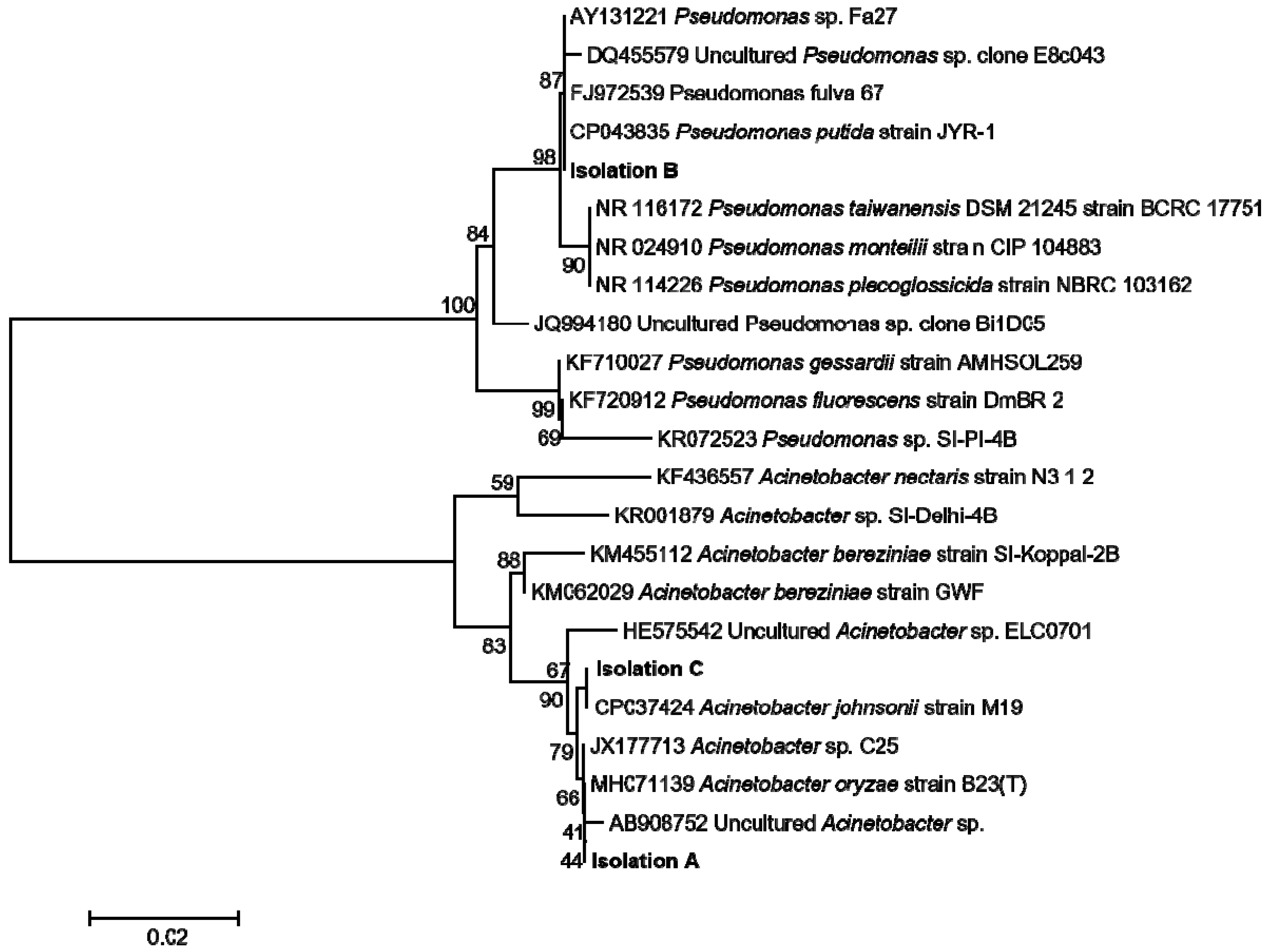
2.10. Empoasca (Matsumurasca) onukii Endobateria Isolation, Identification, and Antibacterial Activity Assay of Geraniol

The E. (M.) onukii adults were anaesthetized with CO2, and then placed on ice. The anaesthetized insects were collected in 1.5 mL eppendorf tube and washed with 70% ethanol for 1 min to kill the external bacterial. Then, the insects were washed with ddH2O twice. The washed insects were cut into pieces with dissecting forceps. The dissected insects were placed in a Luria Broth plate and incubated at 30 °C for two days. The bacteria were growing around the insect pieces. The single colonies were further isolated by plate streaking. The genomic DNA was extracted and the bacterial was identified by 16S ribosomal RNA (rRNA) sequencing. DNA was extracted with TIANamp Bacteria DNA kit (Tiangen, Beijing, China) according to the manufacturer’s instruction. The forward primer used to amplify 16S rRNA is 5′-AGTTTGATCMTGGCTCAG-3′. The reverse primer is 5′-GGTTACCTTGTTACGACTT-3′. The length of the sequenced fragment is about 1500 bp. The obtained sequences were blasted in the NCBI database to identify the similarity with other reported species and the 16S rRNA sequences were aligned using ClustalX program with the available sequences. Phylogenetic tree was generated by MEGA with the neighbor-joining method. The statistical robustness of the clusters was evaluated by bootstrap analysis after 1000 replicates.
The isolated endobacteria Acinetobacter johnsonii was used for antibacterial activity assay of geraniol. The geraniol was dissolved in Tween 20. The final concentration of geraniol in LB medium was 0, 100, 250, and 500 μg/mL, respectively. The final concentration of Tween 20 in the LB medium was 1%. One hundred microliters of the exponentially growing bacterial cells (OD600 = 0.3) was inoculated into 10 mL LB medium containing geraniol with different concentration. Then, the cultures were incubated at 30 °C for another 6 h. Then, OD600 was analyzed to evaluate the antibacterial activity.

2.11. Statistical Analyses

The statistical difference between the two groups was determined with t-test (Paired and independent samples). The p-value of less than 0.05 was considered significant. Statistical analyses were performed using SPSS Version 18.0 software (SPSS Inc. Chicago, IL, USA).

3. Results

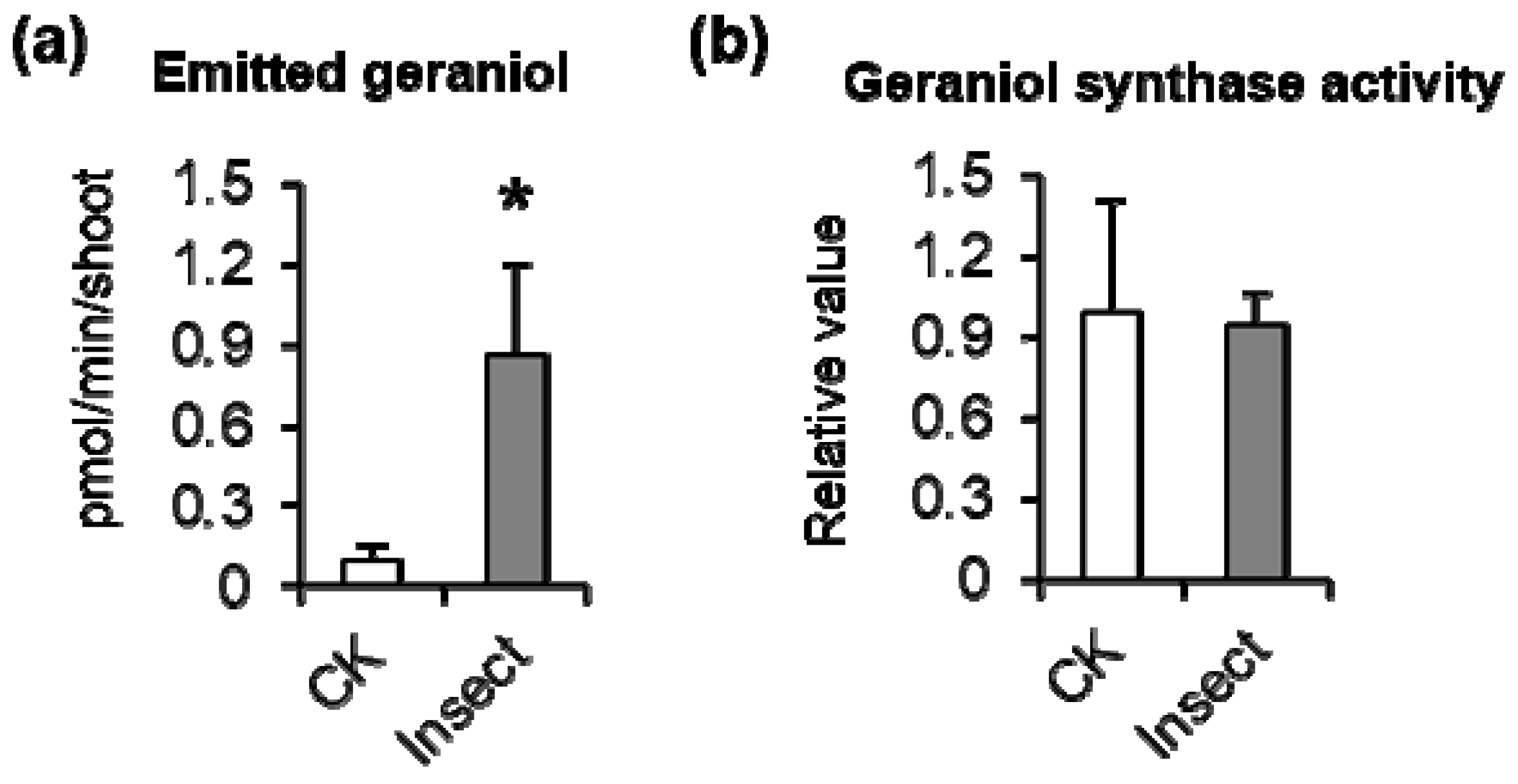
3.1. Empoasca (Matsumurasca) onukii Attack Significantly Increased Geraniol Emission from Tea Leaves

Empoasca (Matsumurasca) onukii is the corrected scientific name of tea green leafhopper in China, one of the main pests in tea production regions [24]. In tea fields, E. (M.) onukii-infested tea leaves show changes in appearance. Here, we investigated the effect of E. (M.) onukii infestation on the formation and emission of geraniol, a volatile monoterpene derived from GPP. We conducted these experiments in the laboratory to exclude the effects of other factors (e.g., other insects/climatic conditions). Compared with control tea leaves, those infested by E. (M.) onukii showed a significant increase in geraniol emission (Figure 1a). However, infestation by E. (M.) onukii did not affect the activity of GES in tea leaves (Figure 1b).

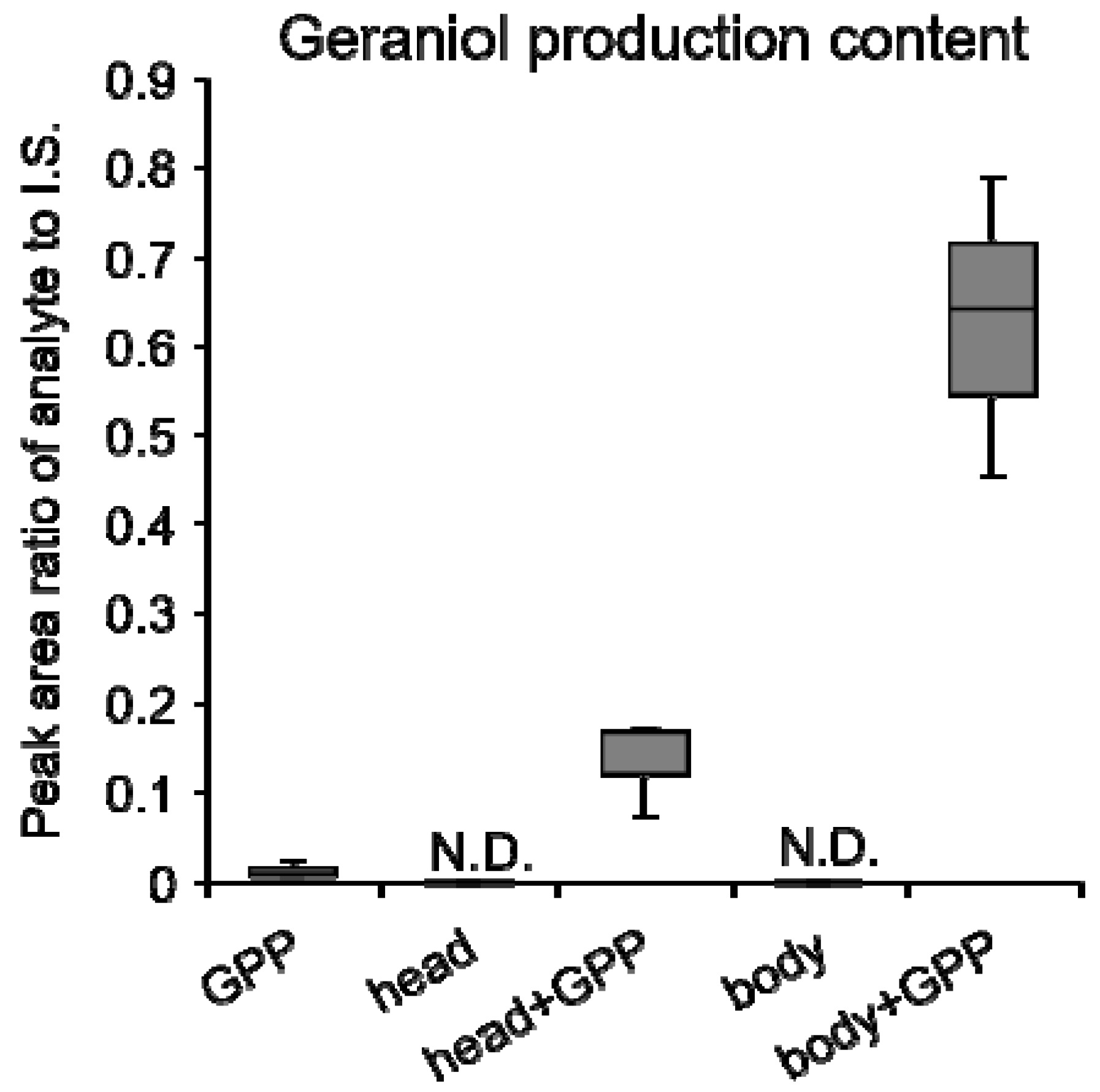
3.2. Crude Enzyme Extract of Empoasca (Matsumurasca) onukii Produced Geraniol from Greanyl Diphosphate

Since the activity of tea leaves GES was not increased after E. (M.) onukii attack, it was presumed that the geraniol accumulation may induced by GES which was from E. (M.) onukii. To investigate whether elicitors such as enzymes involved in geraniol formation were present in E. (M.) onukii, we extracted E. (M.) onukii using buffer, and added GPP as the substrate. In contrast to controls (GPP or crude extract only), the reaction solution containing GPP and the insect extract specifically produced geraniol, but not other volatile monoterpenes such as linalool. The GES activity was higher in the body part than in the head part of E. (M.) onukii (Figure 2, Table S3). These results confirmed that E. (M.) onukii contains GES, which generates geraniol from GPP.

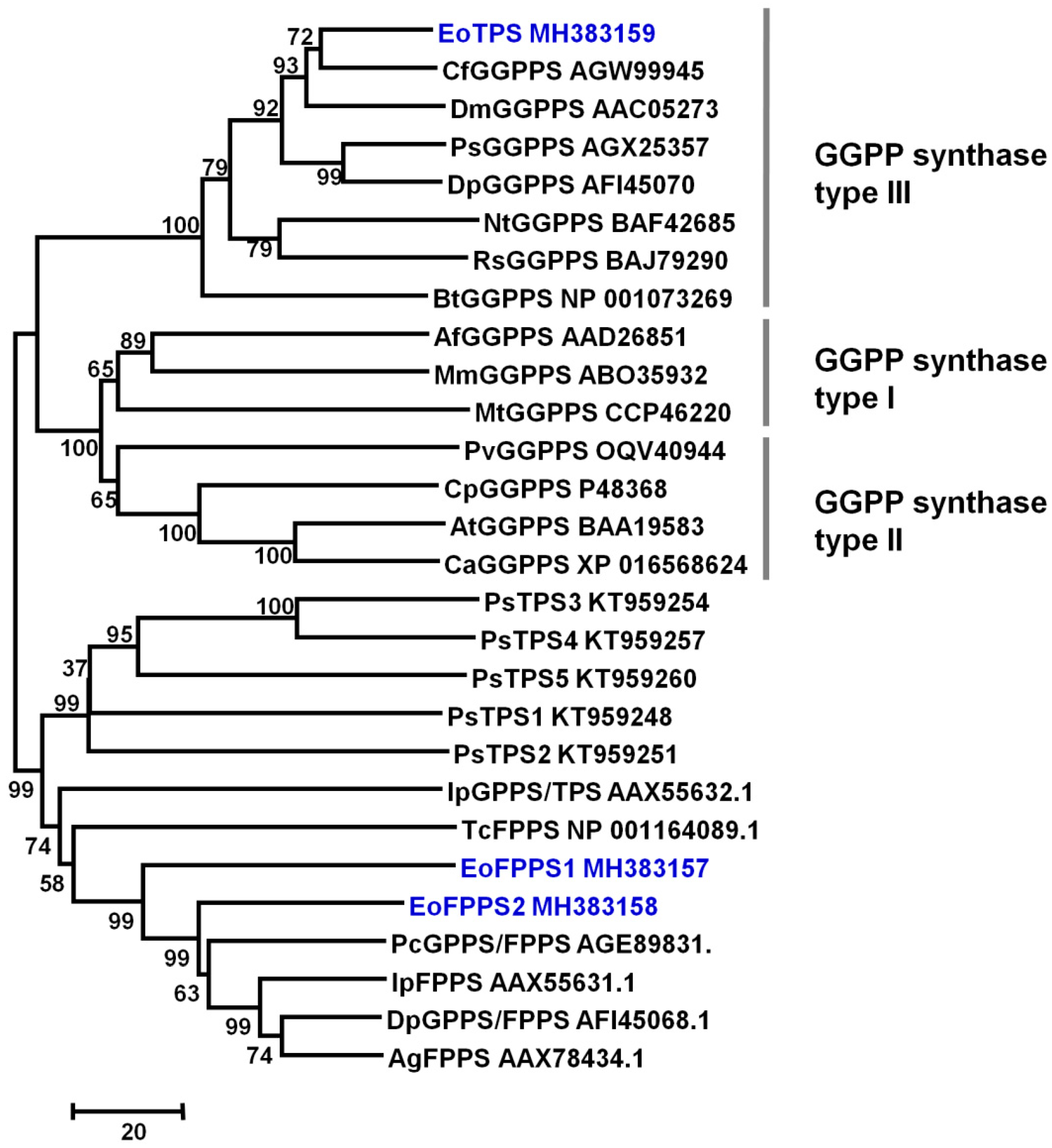
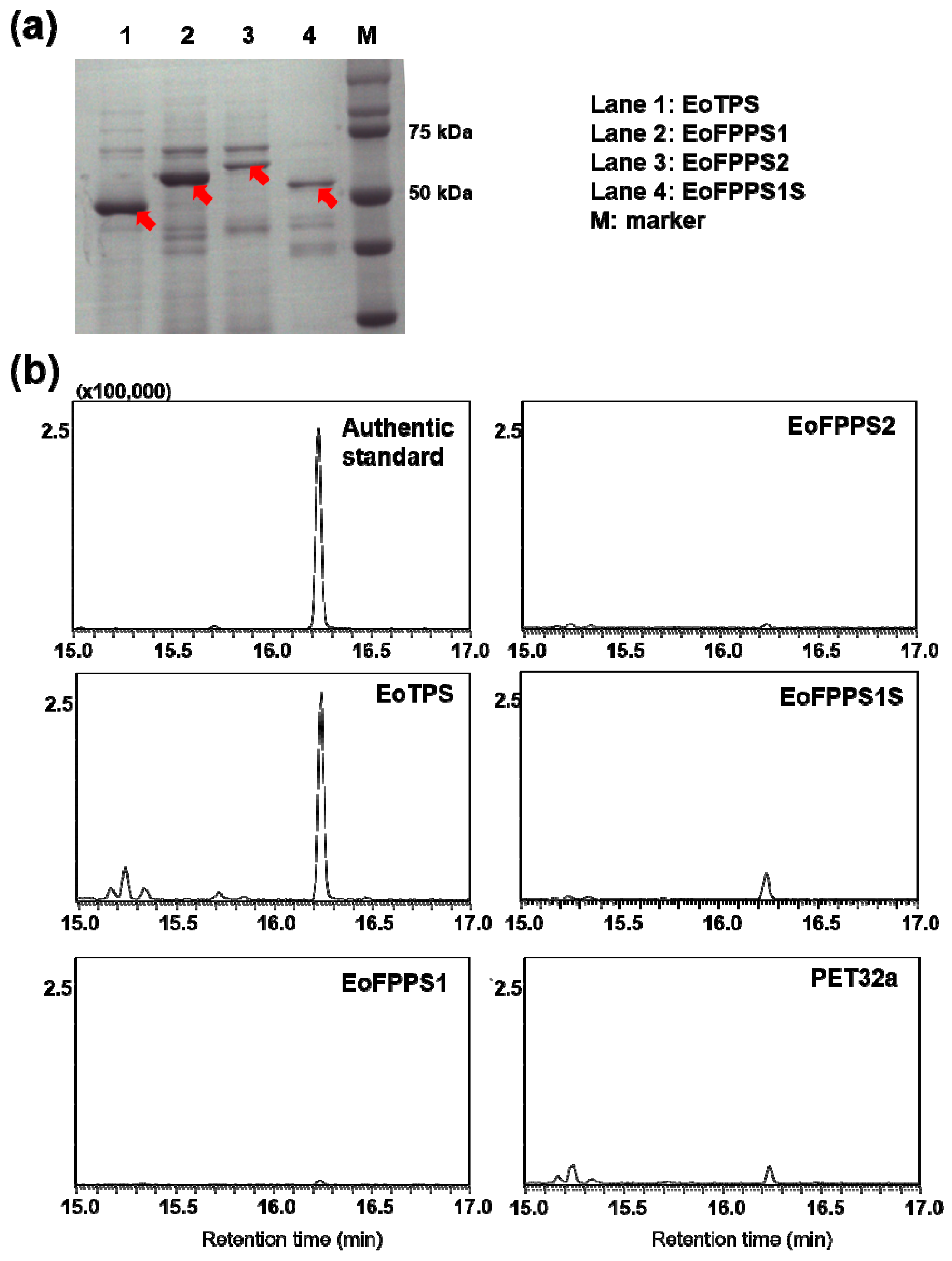
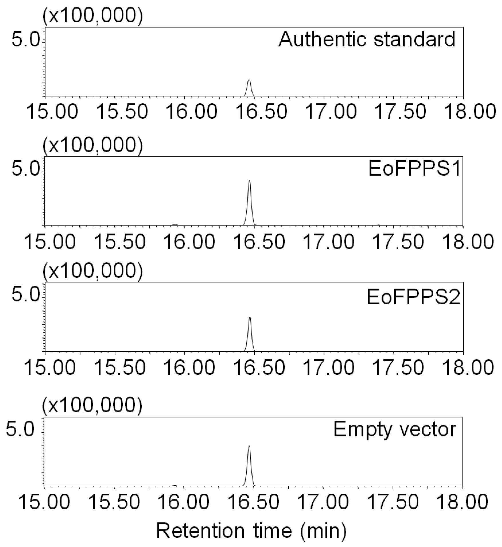
3.3. Terpene synthase from Empoasca (Matsumurasca) onukii Was Able to Convert Greanyl Diphosphate to Geraniol

Although typical terpene synthase was not found in insect genomic and transcriptomic sequences [22], the isoprenyl diphosphate synthases (IDSs) such as terpene synthases have been reported in Phyllotreta striolata and Ips pini [22,25]. A full-length transcriptome of whole E. (M.) onukii insect was sequenced with three generation PacBio sequencing technology (unpublished data). Three isoprenyl diphosphate synthases including two farnesyl diphosphate (FPPS, EoFPPS1 and EoFPPS2) homologue genes and one geranylgeranyl diphosphate synthase (GGPPS, EoTPS) homologue genes were found in this full-length transcriptome. These three genes were cloned from E. (M.) onukii successfully. The full length of EoFPPS1 (GenBank Accession Number MH383157) was 1179 bps, encoding 392 amino acids. The full length of EoFPPS2 (GenBank Accession Number MH383158) was 1287 bps, encoding 428 amino acids. The full length of EoTPS (GenBank Accession Number MH383159) was 912 bps, encoding 303 amino acids. The calculated molecular masses of EoFPPS1, EoFPPS2, and EoTPS were 45, 49, and 35 kDa, respectively. Neighbor-joining tree was generated with other isoprenyl diphosphate synthases in insect. The two farnesyl diphosphate synthase, EoFPPS1 and EoFPPS2, belong to the FPP synthase family. They clustered together with the other characterized FPPS from beetles in phylogenetic trees and were closest to the Phaedon cochleariae FPPS (Figure 3). The geranylgeranyl diphosphate synthase, EoTPS, belongs to the GGPP synthase type III family (Figure 3) and it was closest with the Choristoneura fumiferana GGPPS in the phylogenetic analysis. These genes were heterologously expressed in Escherichia coli (Figure 4a). Among these three genes, EoFPPS1 was predicted to be mitochondrion localized. To better determine the enzymatic activity of EoFPPS1, the ORF lacking pupative mitochondrial targeting peptide (the first 25 amino acids, EoFPPS1S) was also expressed in E. coli (Figure 4a). The activity assay results showed that only EoTPS converted GPP to geraniol (Figure 4b, Figure S1). It was presumed that prokaryotic expression systems may not be suitable for functional protein expression. Therefore, EoFPPS1 and EoFPPS2 were also expressed in insect Sf9 cells. The results showed that there is no difference of geraniol formation between EoFPPS1 and empty vector, or between EoFPPS2 and empty vector. Therefore, the enzyme activity assay with insect Sf9 cell expression system also proved that EoFPPS1 and EoFPPS2 could not convert GPP to geraniol (Figure 5).

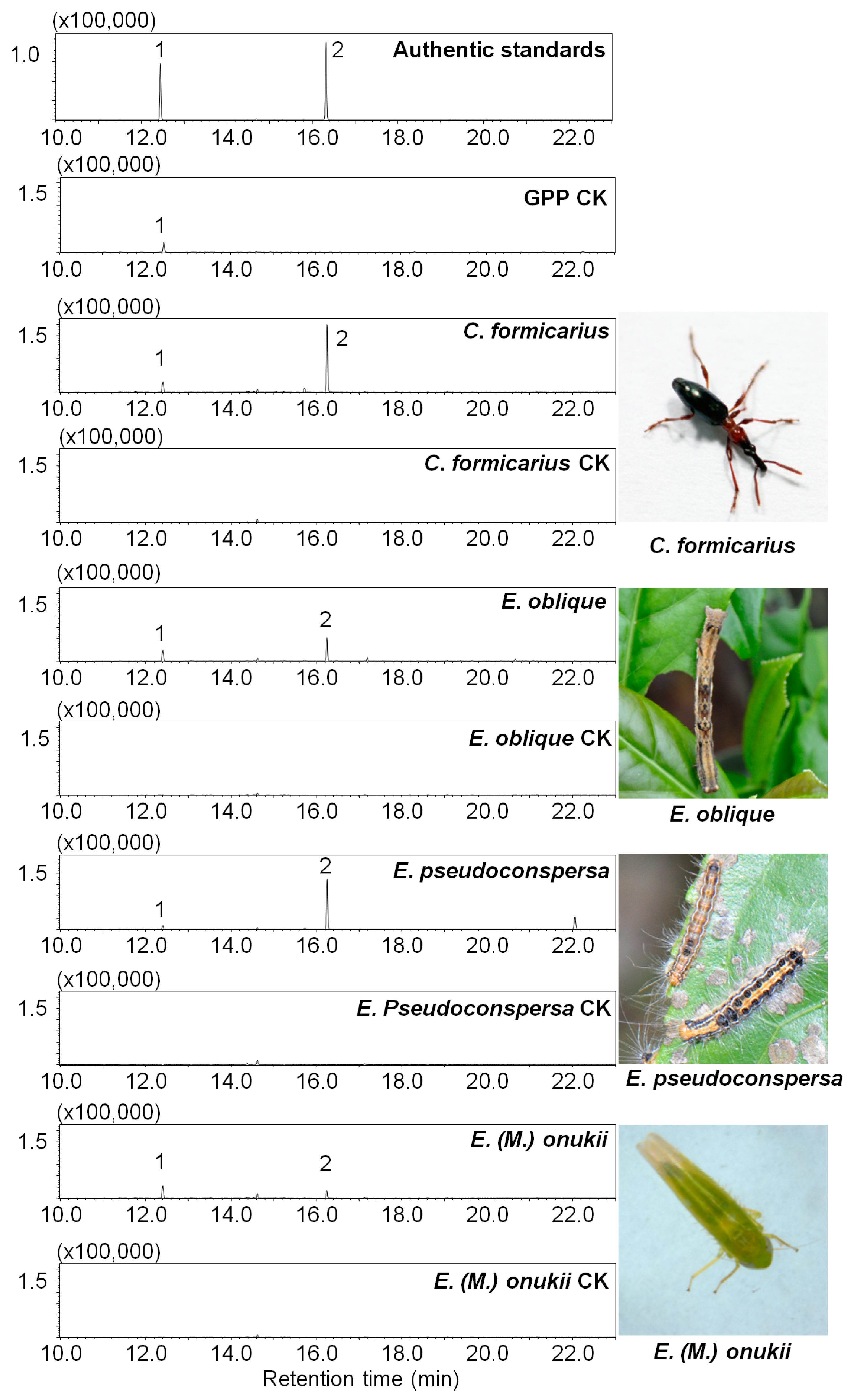
3.4. Geraniol Synthase Also Existed in Other Insects

To identify whether GES was conserved in insects, the GES activity was also determined in other insects including larvae of Ectropis oblique and Euproctis pseudoconspersa, and adult of Cylas formicarius. The activity was measured with crude enzyme extracted from different insects. The monoterpene synthase assay results showed that only geraniol was produced using GPP as a substrate (Figure 6). No significant difference was observed in linalool formation between enzyme reaction and GPP self-degradation control (Figure 6). These results suggested that GES was the monoterpene synthase that existed in these insects.

3.5. Geraniol Had in Vitro Ability to Inhibit the Growth of Endobacterial Isolated from Empoasca (Matsumurasca) onukii

To isolate the endobacteria in E. (M.) onukii, the dissected insect pieces were incubated in LB plate until the bacteria colonies appeared. Then, the grown bacterial was further purified to obtain single colony. The bacteria DNA was extracted and the 16S rRNA gene was amplified and sequenced. For preliminary identification, the 16S rRNA sequences were analyzed with BLASTn in GenBank database. Isolation A showed similarity of 99.86% with uncultured Acinetobacter sp. (AB908752). Isolation B showed similarity of 99.79% with Pseucomonas fulva (FJ972539). Isolation C showed similarity of 99.79% with Acinetobacter johnsonii (CP037424). In the next steps, these three 16S rRNA sequences were aligned with other sequences of different species in GenBank database. The neighbor-joining phylogenetic tree was constructed. The similar results were found in phylogenetic analysis (Figure 7). These endobacteria were also found in other insects [26,27,28,29]. Acinetobacter group was reported to be the major endobacteria in other insects [26]. Acinetobacter johnsonii is a common endosymbiont in insect [27,28]. Therefore, in our present study, isolation C Acinetobacter johnsonii was chosen to initially analyze the potential antibacterial activity of geraniol. The results showed that geraniol could inhibit the isolation C growth in vitro. The geraniol concentration of 500 μg/mL inhibited the isolation C growth in LB medium effectively according to the OD600 analysis (Table 1).
The isolated C colony Acinetobacter johnsonii was used for antibacterial activity assay. The experiments were performed in three replicates.

4. Discussion

To be widespread volatile, the function of geraniol in plants has been well studied. It has been shown to induce apoptosis-like cell death as a defense reaction against bacterial infection [30,31,32] and to play roles in attracting beneficial insects such as sarcophagid flies and braconid wasps [33]. In tea plants, geraniol is one of the herbivore-induced volatiles in aphid-damaged tea plants [11]. Our study confirmed that the emitted geraniol would be induced by E. (M.) onukii attack. Geraniol was reported to be the symbolic aroma which could be used to differentiate different black tea [34]. To be the important flavor-related volatile, the induced geraniol by insect may affect the tea flavor. The pathway of geraniol biosynthesis has been elucidated; it is an acyclic monoterpene alcohol that is synthesized from GPP, the universal precursor of all monoterpenes. The GES from sweet basil was the first GES to be isolated, purified, and functionally characterized. It was shown to generate geraniol from GPP, and to be localized exclusively or almost exclusively to glands [35]. Since then, GESs have been identified and characterized from the evergreen camphor tree Cinnamomum tenuipilum [36], perilla [37,38], and Madagascar periwinkle [39]. However, GES had not been isolated from tea leaves.
Terpenes are structurally diverse natural products that have diverse functions in the ecological interactions of many living organisms. Terpenes are biosynthesized by terpene synthases, which have been widely studied in plants and eukaryotic fungi. Recently, terpene synthases in insects have attracted the interest of researchers. The terpene synthase in insect was found to be an evolutionarily novel family, and the sesquiterpene aggregation pheromone synthase was identified in Phyllotreta striolata [22]. Most reported terpene synthases in insects are sesquiterpene synthases, while monoterpene synthases are rare. As one of monoterpene, geraniol has been found texist in many insects [40,41,42,43,44]. In the larvae of chrysomelid beetles, geraniol was presumed to be synthesized in fat body [43]. It could serve as a precursor of defense compound called iridoids [41,42]. Moreover, geraniol has been proven to be the active component in Nassanoff glands to attract each other in honey bees [40]. It was also reported as caste determining compound from labial glands in Melipona bees [44]. Although these important roles of geraniol had been reported, GES was not identified in the insect. In the present study, GES, a monoterpene synthase specifically producing geraniol, was extracted from E. (M.) onukii (Figure 2). Moreover, GES was found to exist in many insects in the present study (Figure 6). These observations indicate that not only sesquiterpene synthases but also monoterpene synthases occur more widely among eukaryotes than previously thought. The higher crude enzyme activity of GES was found in the body part (Figure 2), which indicated that the GES protein may be mainly located in fat body as previously reported [43] or digested system. Keeling [45] reported that the mevalonate pathway genes in midgut were upregulated after phloem feeding, which may be responsible for monoterpenoid pheromone component ipsdienol de novo synthesis. Our study further identified a TPS gene EoTPS in E. (M.) onukii, which could produce geraniol with GPP as substrate in vitro. Different from those reported terpene synthases in insect, which were all FPPS like enzymes [22,25], our study proved that the GGPPS homologue in E. (M.) onukii could function as monoterpene synthase and produce geraniol from GPP. This is the first GGPPS like terpene synthase reported in insects.
Wounding or elicitors from insects induce many events in plants. Insect elicitors such as fatty acid-amino acid conjugates [7,46], inceptin [9], caeliferins [6], and an unidentified heat-labile constituent [10] do not directly participate in the biosynthesis of plant defensive metabolites, but simulate the upstream signals of biosynthesis of plant defensive metabolites. For example, insect elicitors may resemble phytohormones that can activate the genes involved in the formation of plant volatiles, thus leading to the increased emission of plant volatiles [3]. Only β-glucosidase [8] was reported to directly react with glycosidically bound volatiles in plants to release free volatiles. The insect-derived GES detected in this study may directly contact volatile precursors such as GPP to produce free volatiles. Since the activity of GES in tea leaves was not significantly affected by the E. (M.) onukii attack, the E. (M.) onukii-derived GES may be the main contributor to geraniol emission during insect attack. The further experiments, such as RNAi of EoTPS, would be performed to confirm it. Why did E. (M.) onukii stimulate the geraniol emission itself? Additionally, is there any benefit for E. (M.) onukii to induce geraniol emission? It has been reported that the microbiome plays important roles in the interaction of plants and herbivores [47,48]. To discover the ecological function of EoTPS-induced geraniol, the endobacteria of E. (M.) onukii was isolated and identified by 16S rRNA sequencing (Figure 7). The antibacterial assay with endobacteria of isolation C Acinetobacter johnsonii showed that geraniol could inhibit the growth of this bacteria (Table 1), which suggested that the EoTPS-induced geraniol might affect the E. (M.) onukii by regulating its endobacteria. Geraniol is a wide-spread volatile in tea (C. sinensis). In addition to its great contribution to tea flavor, geraniol is also reported to be a defensive secondary metabolite in plant [49,50,51]. It has been demonstrated to be effective in repelling insects and apparently possessed insecticide properties [52,53]. Geraniol also showed well antipathogenic activity to both plant and insect pathogens [50,51]. Insect endosymbionts play an important role in insect environmental adaption. These microbiotas help insect absorb nutrition [54], metabolize the plant defensive metabolites [55], and mediate the insect reproduction [56]. Although the function of endobacteria Acinetobacter johnsonii in E. (M.) onukii is still unknown. Considering that some Acinetobacter could contribute to insecticide resistance of pest [57], it is hypothesized that geraniol might affect the E. (M.) onukii physiological activity by mediating the growth of its endobacteria. Further in situ studies need be performed to prove this hypothesis.

5. Conclusions

The most frequently reported insect-derived elicitors that induce the formation and emission of plant volatiles are identified in chewing insects, and rarely in piercing-sucking insects. In the present study, crude enzyme of E. (M.) onukii showed specific geraniol synthase activity. A E. (M.) onukii geraniol synthase EoTPS was then identified and it could convert GPP into geraniol in vitro. It was presumed that EoTPS might be delivered into tea leaves and induce the geraniol emission during insect attack. How the geraniol affects E. (M.) onukii endobacteria and the ecological function of induced geraniol for E. (M.) onukii need further study.

Supplementary Materials

The following are available online at https://0-www-mdpi-com.brum.beds.ac.uk/2218-273X/9/12/808/s1. Table S1: Primers used for gene cloning in first-round PCR. Table S2: Primers used for gene cloning in second-round PCR. Table S3: Detailed data points of Figure 2. Figure S1: Functional characterization of terpene synthases in E. (M.) onukii expressed in E. coli with three replicates.

Author Contributions

Conceptualization, Z.Y.; methodology, Z.Y., Y.Z. and X.L.; validation, Y.Z. and X.L.; formal analysis, Z.Y. and Y.Z.; investigation, Y.Z. and X.L.; writing—original draft preparation, Z.Y. and Y.Z.; writing—review and editing, Z.Y. and Y.Z.; supervision, Z.Y.; funding acquisition, Z.Y., Y.Z.

Funding

This study was supported by grants from the National Natural Science Foundation of China (31670690), the National Key Research and Development Program of China (2018YFD1000601), the Guangdong Natural Science Foundation for Distinguished Young Scholar (2016A030313652), the Youth Innovation Promotion Association of Chinese Academy of Sciences (Y821131001), and the Pearl River Science and Technology New Star Fund of Guangzhou (201806010.018).

Conflicts of Interest

The authors declare no conflict of interest.

References

  1. Pichersky, E.; Gershenzon, J. The formation and function of plant volatiles: Perfumes for pollinator attraction and defense. Curr. Opin. Plant Biol. 2002, 5, 237–243. [Google Scholar] [CrossRef]
  2. Dong, F.; Fu, X.M.; Watanabe, N.; Su, X.G.; Yang, Z.Y. Recent advances in the emission and functions of plant vegetative volatiles. Molecules 2016, 21, 124. [Google Scholar] [CrossRef] [PubMed]
  3. Maffei, M.E.; MithÖfer, A.; Boland, W. Before gene expression: Early events in plant-insect interaction. Trends Plant Sci. 2007, 12, 310–316. [Google Scholar] [CrossRef] [PubMed]
  4. Mithöfer, A.; Wanner, G.; Boland, W. Effects of feeding Spodoptera littoralis on lima bean leaves. II. Continuous mechanical wounding resembling insect feeding is sufficient to elicit herbivory-related volatile emission. Plant Physiol. 2005, 137, 1160–1168. [Google Scholar] [CrossRef] [PubMed]
  5. Mei, X.; Liu, X.Y.; Zhou, Y.; Wang, X.Q.; Zeng, L.T.; Fu, X.M.; Li, J.; Tang, J.; Dong, F.; Yang, Z.Y. Formation and emission of linalool in tea (Camellia sinensis) leaves infested by tea green leafhopper (Empoasca (Matsumurasca) onukii Matsuda. Food Chem. 2017, 237, 356–363. [Google Scholar] [CrossRef] [PubMed]
  6. Alborn, H.T.; Hansen, T.V.; Jones, T.H.; Bennett, D.C.; Tumlinson, J.H.; Schmelz, E.A.; Peter, E.A. Disulfooxy fatty acids from the American bird grasshopper Schistocerca americana, elicitors of plant volatiles. Proc. Natl. Acad. Sci. USA 2007, 104, 12976–12981. [Google Scholar] [CrossRef] [PubMed]
  7. Bonaventure, G.; VanDoorn, A.; Baldwin, I.T. Herbivore-associated elicitors: FAC signaling and metabolism. Trends Plant Sci. 2011, 16, 294–299. [Google Scholar] [CrossRef] [PubMed]
  8. Mattiacci, L.; Dicke, M.; Posthumus, M.A. β-Glucosidase: An elicitor of herbivore induced plant odor that attracts host-searching parasitic wasps. Proc. Natl. Acad. Sci. USA 1995, 92, 2039–2040. [Google Scholar] [CrossRef]
  9. Schmelz, E.A.; Carroll, M.J.; LeClere, S.; Phipps, S.M.; Meredith, J.; Chourey, P.S.; Alborn, H.T.; Teal, P.E.A. Fragments of ATP synthase mediate plant perception of insect attack. Proc. Natl. Acad. Sci. USA 2006, 103, 8894–8899. [Google Scholar] [CrossRef]
  10. Allmann, S.; Baldwin, I.T. Insects betray themselves in nature to predators by rapid isomerization of green leaf volatiles. Science 2010, 329, 1075–1078. [Google Scholar] [CrossRef]
  11. Han, B.Y.; Chen, Z.M. Composition of the volatiles from intact and mechanically pierced tea aphid-tea shoot complexes and their attraction to natural enemies of the tea aphid. J. Agric. Food Chem. 2002, 50, 2571–2575. [Google Scholar] [CrossRef] [PubMed]
  12. Ishiwari, H.; Suzuki, T.; Maeda, T. Essential compounds in herbivore-induced plant volatiles that attract the predatory mite. Neoseiulus womersleyi. J. Chem. Ecol. 2007, 33, 1670–1681. [Google Scholar] [CrossRef] [PubMed]
  13. Dong, F.; Yang, Z.Y.; Baldermann, S.; Sato, Y.; Asai, T.; Watanabe, N. Herbivore-induced volatiles from tea (Camellia sinensis) plants and their involvement in intraplant communication and changes in endogenous nonvolatile metabolites. J. Agric. Food Chem. 2011, 59, 13131–13135. [Google Scholar] [CrossRef]
  14. Yang, Z.Y.; Baldermann, S.; Watanabe, N. Recent studies of the volatile compounds in tea. Food Res. Int. 2013, 53, 585–599. [Google Scholar] [CrossRef]
  15. Cho, J.Y.; Mizutani, M.; Shimizu, B.; Kinoshita, T.; Ogura, M.; Tokoro, K.; Lin, M.; Sakata, K. Chemical profiling and gene expression profiling during the manufacturing process of Taiwan oolong tea “Oriental Beauty”. Biosci. Biotech. Bioch. 2007, 71, 1476–1486. [Google Scholar] [CrossRef] [PubMed]
  16. Ong, P.K.C.; Acree, T.E. Gas chromatography/olfactory analysis of lychee (Litchi chinesis Sonn.). J. Agric. Food Chem. 1998, 46, 2282–2286. [Google Scholar] [CrossRef]
  17. Luan, F.; Mosandl, A.; Münch, A.; Wüst, M. Metabolism of geraniol in grape berry mesocarp of Vitis vinifera L. cv. Scheurebe: Demonstration of stereoselective reduction, E/Z-isomerization, oxidation and glycosylation. Phytochemistry 2005, 66, 295–303. [Google Scholar] [CrossRef]
  18. Lan-Phi, N.T.; Shimamura, T.; Ukeda, H.; Sawamura, M. Chemical and aroma profiles of yuzu (Citrus junos) peel oils of different cultivars. Food Chem. 2009, 115, 1042–1047. [Google Scholar] [CrossRef]
  19. Abbott, E.; Hall, D.; Hamberger, B.; Bohlmann, J. Laser microdissection of conifer stem tissues: Isolation and analysis of high quality RNA, terpene synthase enzyme activity and terpenoid metabolites from resin ducts and cambial zone tissue of white spruce (Picea glauca). BMC Plant Biol. 2010, 10, 106. [Google Scholar] [CrossRef]
  20. Zhou, Y.; Zeng, L.; Liu, X.; Gui, J.; Mei, X.; Fu, X.; Dong, F.; Tang, J.; Zhang, L.; Yang, Z. Formation of (E)-nerolidol in tea (Camellia sinensis) leaves exposed to multiple stresses during tea manufacturing. Food Chem. 2017, 231, 78–86. [Google Scholar] [CrossRef]
  21. Hojo, M.; Matsumoto, T.; Miura, T. Cloning and expression of a geranylgeranyl diphosphate synthase gene: Insights into the synthesis of termite defence secretion. Insect Mol. Biol. 2007, 16, 121–131. [Google Scholar] [CrossRef] [PubMed]
  22. Beran, F.; Rahfeld, P.; Luck, K.; Nagel, R.; Vogel, H.; Wielsch, N.; Irmisch, S.; Ramasamy, S.; Gershenzon, J.; Heckel, D.G.; et al. Novel family of terpene synthases evolved from trans-isoprenyl diphosphate synthases in a flea beetle. Proc. Natl. Acad. Sci. USA 2016, 113, 2922–2927. [Google Scholar] [CrossRef] [PubMed]
  23. Kumar, S.; Stecher, G.; Li, M.; Knyaz, C.; Tamura, K. MEGA X: Moleucar evolutionary genetics analysis across computing platforms. Mol. Biol. Evol. 2018, 35, 1547–1549. [Google Scholar] [CrossRef] [PubMed]
  24. Qin, D.Z.; Zhang, L.; Xiao, Q.; Dietrich, C.; Matsumura, M. Clarification of the identity of the tea green leafhopper based on morphological comparison between Chinese and Japanese Specimens. PLoS ONE 2015, 10, e0139202. [Google Scholar] [CrossRef] [PubMed]
  25. Gilg, A.B.; Tittiger, C.; Blomquist, G.J. Unique animal prenyltransferase with monoterpene synthase activity. Naturwissenschaften 2009, 96, 731–735. [Google Scholar] [CrossRef] [PubMed]
  26. Perlatti, B.; Luiz, A.L.; Prieto, E.L.; Fernandes, J.B.; da Silva, M.F.G.; Ferreira, D.; Costa, E.N.; Júnior, A.L.B.; Forim, M.R. MALDI-TOF MS identification of microbiota associated with pest insect Diabrotica speciosa. Agric. For. Entomol. 2017, 19, 408–417. [Google Scholar] [CrossRef]
  27. Faulde, M.; Spiesberger, M. Role of the moth fly Clogmia albipunctata (Diptera: Psychodinae) as a mechanical vector of bacterial pathogens in German hospitals. J. Hosp. Infect. 2013, 83, 51–60. [Google Scholar] [CrossRef]
  28. Pidlyar, V.J.; Jangid, K.; Patole, M.S.; Shouche, Y.S. Studies on cultured and uncultured microbiota of wild Culex Quinquefasciatus mosquito midgut based on 16S ribosomal RNA gene analysis. Am. J. Trop. Med. Hyg. 2004, 70, 597–603. [Google Scholar] [CrossRef]
  29. Ceja-Navarro, J.A.; Vega, F.E.; Karaoz, U.; Hao, Z.; Jenkins, S.; Lim, H.C.; Kosina, P.; Infante, F.; Northen, T.R.; Brodie, E.L. Gut microbiota mediate caffeine detoxification in the primary insect pest of coffee. Nat. Commun. 2015, 6, 7618. [Google Scholar] [CrossRef]
  30. Jabs, T.; Dietrich, R.A.; Dangl, J.L. Initiation of runaway cell death in an Arabidopsis mutant by extracellular superoxide. Science 1996, 273, 1853–1856. [Google Scholar] [CrossRef]
  31. Izumi, S.; Nishio, Y.; Takashima, O.; Hirata, T. Monoterpenoids, potent inducer of apoptosis in cells of Marchantia polymorpha. Chem. Lett. 1997, 26, 837–838. [Google Scholar] [CrossRef]
  32. Izumi, S.; Takashima, O.; Hirata, T. Geraniol is a potent inducer of apoptosis-like cell death in the cultured shoot primordia of Matricaria chamomilla. Biochem. Biophys. Res. Commun. 1999, 259, 519–522. [Google Scholar] [CrossRef] [PubMed]
  33. James, D.G. Further field evaluation of synthetic herbivore-induced plant volatiles as attractants for beneficial insect. J. Chem. Ecol. 2005, 31, 481–495. [Google Scholar] [CrossRef] [PubMed]
  34. Aisaka, H.; Kosuge, M.; Yamanishi, T. Comparison of the flavors of Chinese “Keemun” black tea and Ceylon black tea. Agric. Biol. Chem. 1978, 42, 2157–2159. [Google Scholar] [CrossRef]
  35. Iijima, Y.; Gang, D.R.; Fridman, E.; Lewinsohn, E.; Pichersky, E. Characterization of geraniol synthase from the peltate glands of sweet basil. Plant Physiol. 2004, 134, 370–379. [Google Scholar] [CrossRef] [PubMed] [Green Version]
  36. Yang, T.; Li, J.; Wang, H.X.; Zeng, Y. A geraniol-synthase gene from Cinnamomum tenuipilum. Phytochemistry 2005, 66, 285–293. [Google Scholar] [CrossRef] [PubMed]
  37. Ito, M.; Honda, G. Geraniol synthases from perilla and their taxonomical significance. Phytochemistry 2007, 68, 446–453. [Google Scholar] [CrossRef]
  38. Masumoto, N.; Korin, M.; Ito, M. Geraniol and linalool synthases from wild species of perilla. Phytochemistry 2010, 71, 1068–1075. [Google Scholar] [CrossRef]
  39. Simkin, A.J.; Miettinen, K.; Claudel, P.; Burlat, V.; Guirimand, G.; Courdavault, V.; Papon, N.; Meyer, S.; Godet, S.; St-Pierre, B.; et al. Characterization of the plastidial geraniol synthase from Madagascar periwinkle which initiates the monoterpenoid branch of the alkaloid pathway in internal phloem associated parenchyma. Phytochemistry 2013, 85, 36–43. [Google Scholar] [CrossRef]
  40. Boch, R.; Shearer, D.A. Identification of geraniol as the active component in the Nassanoff pheromone of the honey bee. Nature 1962, 194, 704–706. [Google Scholar] [CrossRef]
  41. Lorenz, M.; Boland, W.; Dettner, K. Biosynthesis of iridodials in the defense glands of beetle larvae (Chrysomelinae). Angew. Chem. Int. Ed. Engl. 1993, 32, 912–917. [Google Scholar] [CrossRef]
  42. Laurent, P.; Braekman, J.C.; Daloze, D.; Pasteels, J. Biosynthesis of defensive compounds from beetles and ants. Eur. J. Org. Chem. 2003, 2003, 2733–2743. [Google Scholar] [CrossRef]
  43. Burse, A.; Schmidt, A.; Frick, S.; Kuhn, J.; Gershenzon, J.; Boland, W. Iridoid biosynthesis in Chrysomelina larvae: Fat body produces early terpenoid precursors. Insect Biochem. Mol. Biol. 2007, 37, 255–265. [Google Scholar] [CrossRef] [PubMed]
  44. Jarau, S.; van Veen, J.W.; Twele, R.; Reichle, C.; Herrera, E.; Gonzales, E.H.; Aguilar, I.; Francke, W.; Ayasse, M. Workers make the queens in Melipona bees: Identification of geraniol as a caster determining compound from labial glands of nurse bees. J. Chem. Ecol. 2010, 36, 565–569. [Google Scholar] [CrossRef]
  45. Keeling, C.; Blomquist, G.J.; Tittiger, C. Coordinated gene expression for pheromone biosynthesis in the pine engraver beetle, Ips pini (Coleoptera: Scolytidae). Naturwissenschaften 2004, 91, 324–328. [Google Scholar] [CrossRef]
  46. Alborn, H.T.; Turlings, T.C.J.; Jones, T.H.; Stenhagen, G.; Loughrin, J.H.; Tumlinson, J. An elicitor of plant volatiles from beet armyworm oral secretion. Science 1997, 276, 945–949. [Google Scholar] [CrossRef]
  47. Sorokan, A.V.; Rumyantsev, S.D.; Benkovskaya, G.V.; Maksimov, I.V. The ecological role of microsymbionts in the interaction of plants and herbivorous insect. Biol. Bull. Rev. 2019, 9, 275. [Google Scholar] [CrossRef]
  48. Frago, E.; Mala, M.; Weldegergis, B.T.; Yang, C.; McLean, A.; Godfray, H.C.J.; Gols, R.; Dicke, M. Symbionts protect aphids from parasitic wasps by attenuating herbivore-induced plant volatiles. Nat. Commun. 2017, 8, 1860. [Google Scholar] [CrossRef] [Green Version]
  49. Nasiou, E.; Giannakou, I.O. Effect of geraniol, a plant-based alcohol monoterpene oil, against Meloidogyne javanica. Eur. J. Plant Pathol. 2018, 152, 701–710. [Google Scholar] [CrossRef]
  50. Zhang, Z.; Yang, T.; Mi, N.; Wang, Y.; Li, G.; Wang, L.; Xie, Y. Antifungal activity of monoterpenes against wood white-rot fungi. Int. Biodeterior. Biodegrad. 2016, 106, 157–160. [Google Scholar] [CrossRef]
  51. Nardoni, S.; Ebani, V.V.; D’Ascenzi, C.; Pistelli, L.; Mancianti, F. Sensitivity of entomopathogenic fungi and bacteria to plants secondary metabolites, for an alternative control Rhipicephalus (Boophilus) microplus in cattle. Front. Pharmacol. 2018, 9, 937. [Google Scholar] [CrossRef] [PubMed]
  52. Müller, G.C.; Junnila, A.; Butler, J.; Kravchenko, V.D.; Revay, E.E.; Weiss, R.W.; Schlein, Y. Efficacy of the botanical repellents geraniol, linalool, and citronella against mosquitoes. J. Vector Ecol. 2009, 34, 2–8. [Google Scholar] [CrossRef] [PubMed]
  53. Reis, S.L.; Mantello, A.G.; Macedo, J.M.; Gelfuso, E.A.; Silva, C.P.; Fachin, A.L.; Cardoso, A.M.; Beleboni, R.O. Typical monoterpenes as insecticides and repellents against strored grain pests. Molecules 2016, 21, 258. [Google Scholar] [CrossRef] [PubMed] [Green Version]
  54. Douglas, A.E. Nutritional interactions in insect-microbial symbioses: Aphids and their symbiotic Bacteria Buchnera. Annu. Rev. Entomol. 1998, 43, 17–37. [Google Scholar] [CrossRef] [PubMed] [Green Version]
  55. Boone, C.K.; Keefover-Ring, K.; Mapes, A.C.; Adams, A.; Bohlmann, J.; Raffa, K.F. Bacteria associated with a tree-killing insect reduce concentrations of plant defense compounds. J. Chem. Ecol. 2013, 39, 1003–1006. [Google Scholar] [CrossRef] [PubMed]
  56. Johanowicz, D.L.; Hoy, M.N. The manipulation of arthropod reproduction by Wolbachia endosymbionts. Fla. Entomol. 1998, 81, 310–317. [Google Scholar] [CrossRef]
  57. Achaleke, J.; Martin, T.; Ghogomu, R.T.; Vaissayre, M.; Brévault, T. Esterase-mediated resistance to pyrethroids in field populations of Helicoverpa armigera (Lepidoptera: Noctuidae) from Central Africa. Pest Manag. Sci. 2009, 65, 1147–1154. [Google Scholar] [CrossRef]
Figure 1. Effect of Empoasca (Matsumurasca) (E. (M.)) onukii attack on emission of geraniol and geraniol synthase activity in tea leaves. (a) Effect of E. (M.) onukii attack on emission of geraniol in tea leaves in lab control experiments. (b) Effect of E. (M.) onukii attack on geraniol synthase activity. The control (CK) was intact tea leaves without treatment. The levels of enzyme activity in the control were set as 1. Student t test was used to determine the differences between control and treatment. Data are expressed as mean ± S.D. (n = 3) *, p ≤ 0.05.
Biomolecules 09 00808 g001
Figure 2. Evaluation of enzyme activity of extract of E. (M.) onukii against GPP. Twenty E. (M.) onukii adults were separated into two parts; head and body. The head or body part extract reacted with GPP. The controls were GPP or crude extract only. The geraniol production amount was represented by the GC-MS peak area ratio of m/z 69 (geraniol characteristic ion) to m/z 88 (I.S. (internal standard, ethyl n-decanoate) characteristic ion). N.D., lower than detection limit. Data are expressed as mean ± S.D. (n = 3).
Biomolecules 09 00808 g002
Figure 3. Phylogenetic analysis of terpene synthases in insects. Cf, Choristoneura fumiferana; Dm, Drosophila melanogaster; Ps, Pissodes strobe; Dp, Dendroctonus ponderosae; Nt, Nasutitermes takasagoensis; Rs, Reticulitermes speratus; Bt, Bos Taurus; Af, Archaeoglobus fulgidus; Mm, Methanococcus maripaludis; Mt, Mycobacterium tuberculosis; Pv, Pantoea vagans; Cp, Cyanophora paradoxa; At, Arabidopsis thaliana; Ca, Capsicum annuum; Ps, Phyllotreta striolata; Ip, Ips pini; Tc, Tribolium castaneum; Pc, Phaedon cochleariae; Dp, Dendroctonus ponderosae; Ag, Anthonomus grandis. Bootstrap values from 1000 replicates were used to assess the robustness of the trees.
Biomolecules 09 00808 g003
Figure 4. Sodium dodecyl sulfate-polyacrylamide gel electrophoresis (SDS-PAGE) analysis and functional characterization of terpene synthases in E. (M.) onukii expressed in E. coli. (a) SDS-PAGE of purified recombinant putative terpene synthases. EoFPPS1S was the protein lacking putative mitochondrial targeting peptide of EoFPPS1. (b) Gas chromatography-mass spectrometry (GC-MS) identification of formation of geraniol from GPP under the actions of recombinant terpene synthases. The experiments were performed in three replicates with similar results. Authentic standard: Geraniol authentic standard.
Biomolecules 09 00808 g004
Figure 5. Functional characterization of E. (M.) onukii terpene synthases expressed in insect cell Sf9. The formation of geraniol was identified by GC-MS. The formation of geraniol is not different between EoFPPS1 and empty vector or EoFPPS2 and empty vector. The experiments were performed in three replicates with similar results. Authentic standard: Geraniol authentic standard.
Biomolecules 09 00808 g005
Figure 6. Evaluation of terpene synthases in other insects. GPP reacted with crude enzymes extracted from different insects. Ectropis oblique, larvae; Euproctis pseudoconspersa, larvae; Cylas formicarius, adult; E. (M.) onukii, adult. The controls were GPP or crude enzyme only. The formation of linalool in each enzyme activity came from the GPP self-degradation. 1. Linalool; 2. geraniol. GPP CK: GPP alone in the reaction buffer; each insect CK: Crude enzyme alone in the reaction buffer. The experiments were performed in three replicates with similar results.
Biomolecules 09 00808 g006
Figure 7. Phylogenetic analysis of the isolated endobacteria. The 16S ribosomal RNA (rRNA) sequences were aligned with ClustalX and neighbor-joining tree were created with MEGA software. Bootstrap values were calculated for 1000 replicates.
Biomolecules 09 00808 g007
Table 1. Antibacterial activity assay of geraniol.
Geraniol Concentration (μg/mL)OD600
0>3
100>3
25011.980 ± 0.232
5000.018 ± 0.006

