1. Introduction
Nitrites have important effects on human health and the environment, and their quantitative assessment has been an important and challenging analytical task over the last two decades [
1,
2]. Nitrite ion (NO
2−) monitoring is especially important for potable water and foodstuff quality control, in wastewater treatment, in agriculture, and in fish farming. In particular, the recommended values for freshwater fish species are quite restrictive, requiring nitrite ions concentrations lower than 0.1 mg/L [
2].
Usually, nitrites are not present in the environment in significant concentrations (except in reducing media), as nitrate is the more stable oxidation state. Particular conditions are required for nitrites’ formation in the natural environment, such as microbial reduction in ingested nitrate in vivo; for instance, the bacteria
Nitrosomonas europaea were found to be involved in nitrite production from ammonia [
3] during stagnation of nitrate-containing and oxygen-poor drinking-water inside steel pipes. In order to gain a complete estimation of the total inorganic forms of nitrogen present in analyzed media, nitrite screening is always made in parallel to nitrates. The nitrate ion (NO
3−) is found naturally in the environment and it is present at varying concentrations in all plants. Nitrates can reach both surface water and groundwater because of agricultural activity, from wastewater disposal and from the oxidation of nitrogenous waste products in human and animal excreta, including septic tanks. Surface water nitrate concentrations can change rapidly owing to surface runoff of fertilizer, uptake by phytoplankton, and denitrification by bacteria, but groundwater concentrations generally show relatively slow changes [
2]. The most common human exposure to nitrite- and nitrate ions is through vegetables and diet proteins (nitrite is used as a preservative in many cured meats). In some circumstances, however, drinking-water can make a significant contribution to nitrate- and, occasionally, nitrite intake. In the case of bottle-fed infants, drinking-water can be the major external source of exposure to both NO
3− and NO
2− ions. By its turn, nitrite can bind to hemoglobin, the oxygen transporter molecule in the red blood cells; this interaction produces methemoglobin that binds oxygen tightly. When the methemoglobin concentration in blood is ≥10%, clinically relevant cyanosis known as ‘blue-baby syndrome’, can appear and be fatal for a child. This negative effect of nitrite on humans was first investigated by Comly in 1945 [
4]. Studies have also shown a correlation between nitrate and nitrite ingestion and increase in gastric cancer formation risks; in 2010 the IARC (International Agency for Research on Cancer) has classified “nitrate or nitrite (ingested) under conditions that result in endogenous nitrozation” as probably carcinogenic to humans [
5]. In fact, in the body NO
2− ions form carcinogenic nitrosamines [
1] when they bind with secondary amines, and it is hence essential to monitor the nitrite levels in the environment and especially in samples causing health concerns.
Traditionally nitrites are determined in solution by spectrophotometry in the presence of specific ligands [
6] or by electrochemical techniques employing bio- and chemical sensors, such as ISEs [
7,
8,
9,
10,
11]. Optical chemical sensors have been employed to a much smaller degree, despite their evident benefits, such as simplicity in preparation, no necessity of a sophisticated and high energy-consuming hardware, and no wire connections with detectors. Moreover, the analytically useful optical sensor output can be registered with common electronic devices, such as smartphones, or even without any power supply in a ‘naked-eye’ mode, when the sensor luminescence intensity change upon interaction with the analyte may be registered visually (especially useful in situations of rapid qualitative analysis and alarm cases). There is, hence, an unmet need to develop easy-to-handle and inexpensive optical sensors for fast and effective nitrite assessment. However, analyses of the last 5 years of publications on NO
2−-sensor development have shown that less than 10% of all research is dedicated to sensors with optical transduction; this research is mainly dedicated to nanomaterial-based sensors, such as metal and metal-oxide nanoparticles [
12,
13], quantum dots [
14], or liquid crystals [
15]. Some applications of polymeric sensing materials doped with nitrite-selective ligands for optode development are also reported [
16]. The accurate selection of the appropriate macromolecular ligands selectively binding to the target analyte has permitted the development of selective optodes for several inorganic anions, including nitrites [
17,
18,
19,
20]. Among these ligands (or ionophores) metalloporphyrin and metallocorrole complexes are particularly suitable for anion sensing through metal–anion coordination, a type of Lewis acid–base interaction [
21,
22]. The metallocorroles have significant potential to be integrated in optical sensors, a sensor type that has seen many technological developments and applications, including in a wide variety of smartphones [
23,
24]. This combination, when applied to environmental nitrate monitoring, could result in sensors that are less expensive, more portable, with greater ease of use and more disposable reagents.
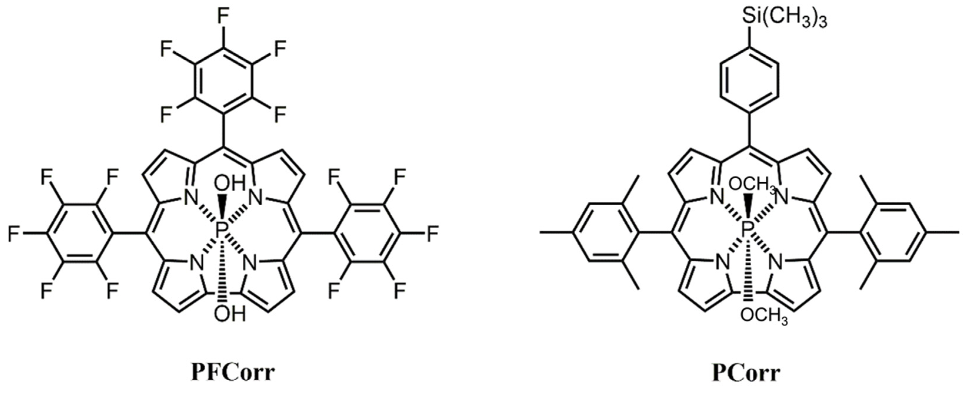
The present work investigates the properties of corrole ligands, 5,10,15-pentafluorophenyl corrole of phosphorous (V) (
PFCorr), and [10-(4-trimethylsilylphenyl)-5,15-dimesityl-corrole] phosphorous (V) (
PCorr) previously tested in [
25], for the development of selective optical sensors for rapid and low-cost detection of nitrites in natural samples with a particular attention to water quality monitoring,
Figure 1. Preliminary fluorimetric studies on ligands’ sensitivity towards different anions carried out in solution and inside the solvent polymeric membranes [
26] have been advanced in the present work. Moreover, we investigated the properties of silica nanoparticles with anchored
PFCorr units uploaded on a paper support for selective nitrite sensing. In each of these conditions the selective ligand’s fluorescence quenching was registered upon addition of an increasing amount of NO
2− ions. The sensing properties of
PFCorr on different supports were investigated in order to detect the effectiveness of nitrite assessment at recommended concentrations levels.
2. Materials and Methods
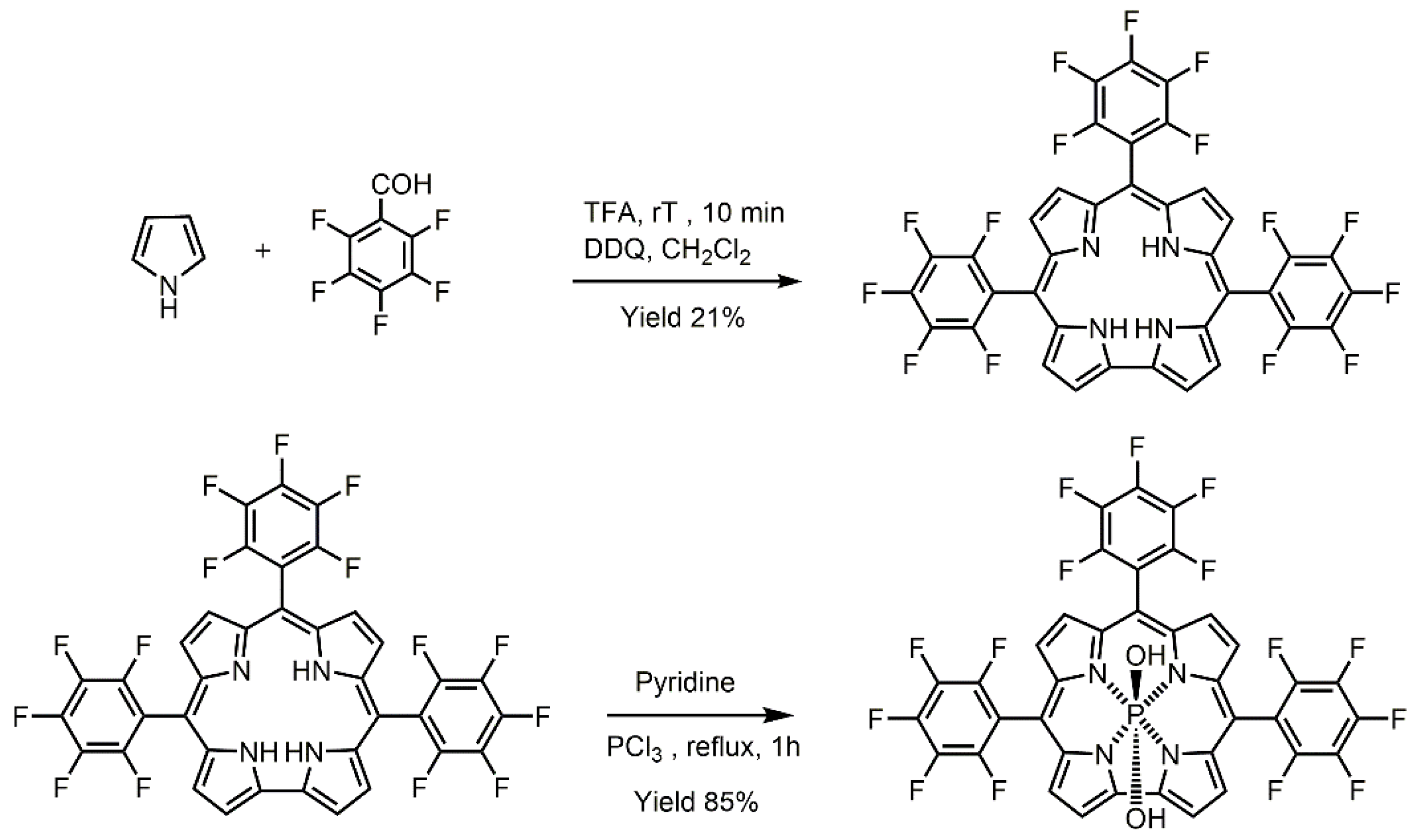
The
PCorr and
PFCorr were synthesized and fully characterized by NMR, UV-Visible and photoluminescence spectroscopy in our laboratories. For synthesis of [10-(4-trimethylsilylphenyl)-5,15-dimesityl-corrole] phosphorous (V) (
PCorr) the procedure reported in [
27] was adopted. The 5,10,15-pentafluorophenyl corrole of phosphorous (V),
PFCorr complex was obtained by a two-step procedure including free base corrole synthesis and its subsequent metalation. The procedure is schematically represented in
Figure 2.
The Poly(vinyl chloride) (PVC) high molecular weight, tris(2-ethylhexyl) phosphate (TOP), tetradecylmethyammonium chloride (TDACl), tetrabutylammonium nitrite (TBANO2), tetrahydrofuran (THF), chloroform (CHCl3), 2-(N-morpholino) ethanesulfonic acid (MES), NaNO2, NaCl, NaBr, NaNO3, CH3CO2Na, NaClO4, NaSCN, NaHCO3, and Na2SO4 salts were from Sigma-Aldrich (Milan, Italy). Ultrapure water was used for aqueous solution preparation. THF was freshly distilled prior to use. All the other chemicals were of analytical grade and used without further purification.
Polymeric membranes were prepared by incorporation of 1 wt% of PFCorr and 0.5–5 wt% of TpClPBK inside a polymeric matrix containing PVC and plasticizer in 1:2 ratio by weight. Membranes with a total weight of about 100 mg, were dissolved in 1 mL of THF. Ten microliters of each membrane cocktail was cast onto transparent glass slides, the THF solvent was allowed to evaporate overnight, and afterwards the ‘disposable’ optode sensors were tested in individual solutions of several anions on 0.01 M MES pH 5.5 background in a 1.0 × 10⁻⁸–1.0 × 10⁻² M concentration range. The calibration solutions were obtained by consecutive additions of calculated amounts of corresponding 1 M stock solution of different salts.
SHIMAZU RF-1501 fluorimeter (Shimadzu Europe, Duisburg, Germany) was employed for tests in solutions. An amount of 2.5 mL of 6 × 10
−8 M
PFCorr solution was titrated with 10
−2 M, 10
−4 M and 10
−5 M stock solutions of tetrabutylammonium nitrite (TBANO
2) in chloroform directly in a quartz cuvette of 1 cm, and light was passed upon the excitation at 413 nm (wavelength of
PFCorr Soret band in the absorption spectra). The fluorescence spectra were registered 10 min after each nitrite concentration addition. In total, eight additions of TBANO
2 were performed to reach a cumulative amount of 125 μL added to the cuvette. For the fluorimetric selectivity tests, 1 M aqueous stock solutions of the sodium salts of the following anions were used: NO
2−, NO
3−, SCN
−, ClO
4−, Cl
−, and Br
−. A single calculated addition of 1 M stock solution was made in order to obtain a final 6 × 10
−2 M concentration of each interfering ion to the 6 × 10
−8 M
PFCorr solution in DMSO. The fluorescence spectra were recorded 10 min after the addition of an interfering ion in order to exclude the dilution effect for the emission quenching. For the testing of optodes with PVC-based solvent polymeric membranes doped with
PFCorr and
PCorr, a UV-lamp (365 nm) or commercial blue-colored Light Emitting Diode (LED, 380 nm) served as a monochromic excitation light source. For experiments with the UV-lamp, 5 μL of solutions containing different (increasing) concentrations of the analyzed anion were deposited on sensing spots obtained by drop-casting
PFCorr- and
PCorr-doped PVC-based membranes on a glass support. The optode emission signal, upon illumination at 365 nm, was registered 3 min after analyte addition from the fixed distance of 10 cm by means of a common smartphone camera, and the luminescence variations were then extracted from images and transformed into analytically useful digital signals using Matlab software (v.7.9, 2009, The MathWorks, Inc., Natick, MA, USA). Due to the enhanced photosensitivity of the membranes, and in order to avoid photo degradation problems, all the experiments were carried with ‘disposable’ optical sensors, deposited on a transducer a few times prior to the testing.
PFCorr- and
PCorr-doped PVC membranes were stored in the dark before use. The measurements were repeated in triplicate. The Photoassisted Technique (PT) set up was used for measurements with a blue-colored InGaN LED (Roithner LaserTechnik, Wien, Austria, model H2A1-H385), as a monochromic external light source; and a frontally placed digital webcamera (Philips S.p.A, Milan, Italy, model Philips SPC900NC) for notebook (resolution of 352 × 288 pixels) was used as a signal detector, as described in detail in our previous work [
25]. Glass slides (0.7 cm × 3.0 cm size) with sensing membranes deposited on them were vertically immersed in polystyrene cuvettes of 1 cm path length and then laterally illuminated with an LED, and the responses of the sensing membranes were recorded from three channels representing the main visible spectrum colors: red, R (630 nm), green, G (530 nm), and blue, B (480 nm). The measurement cell was shielded from external illumination. The luminescence intensity was calculated as:
where R, G, and B correspond to the sensing membranes luminescence intensities at RGB channels, and normalized to the maximum intensity value (255) of the optical signal measured with a webcam detector. The relative optical intensity quenching was estimated as a percentage of I in the absence and in the presence of an analyte, evaluated after a background luminosity subtraction. The duration of the overall sample illumination was 50 s for the LED (during this period 10 photographic shoots were taken every 5 s, and the final optical signal was a mean value of the records) and 10 s for the UV-vis lamp.
The SiO
2 nanoparticles (SO
2NPs) were prepared according to the Stӧber method [
28], through the hydrolysis and condensation of tetraethyl orthosilicate TEOS in ethanol, in the presence of ammonia as a catalyst. Briefly, to obtain a solution [NH
3] = 0.3 M and [H
2O] = 1 M, 2.4 mL of 28% ammonium hydroxide solution and 2.16 mL of H
2O were added to 120 mL of ethanol and stirred for 10 min to ensure complete mixing. Then 7.5 mL of TEOS was added ([TEOS] = 0.28 M) and the reaction proceeded at room temperature for 24 h. Thereafter the colloidal solution was separated by high-speed centrifuge, and the silica nanoparticles were washed three times with ethanol to remove undesirable particles. The particles were dried in an oven at 100 °C for 2 h to prevent further reaction.
The SiO2 NPs were functionalized with PFCorr (SiO2NPs@PFCorr) through a two-step method called ‘post nanoparticles coating’, in which the PFCorr was added to a suspension of SiO2 nanoparticles already synthesized. After a 2 h thermal treatment at 150 °C to remove physisorbed water, SiO2 nanoparticles (80 mg) prepared previously in the first step were put in a glass flask equipped with 8 mL of toluene, followed by sonication to ensure dispersion. Five milligrams of PFCorr were then added to the flask and the mixture was stirred and refluxed at 110 °C for 3 h. The solid was recovered by ultracentrifugation and repeatedly washed with CH2Cl2, acetone, ethanol, and toluene in order to remove any excess of non-linked corrole. Particles were finally dried at 150° for 2 h. For the titration of SiO2NPs@PFCorr with NO2− ions, 4 mg of NPs were suspended in 100 mL of THF upon sonication. To this suspension the calculated amount of 1 M TBANO2 in CHCl3 was added in order to obtain the required concentration of nitrite ions. The fluorescence emission spectra were measured upon the excitation at 413 nm, 10 min after each addition, maintaining the same instrument setup.
The deposition of the functionalized nanoparticles on a paper support was made by a drop-casting method. The suspension was prepared by sonicating 2 mg of SiO2NPs@PFCorr in 1.5 mL of organic solvent (THF or toluene). Each spot on the filter paper strips WhatmanTM (Cat No 1001-150) was made by a micropipette, dripping 150 µL of the previously prepared suspension. The solvent was allowed to dry at room temperature and the operation was repeated 3 times for each spot. The fluorescence quenching of SiO2NPs@PFCorr deposited on the paper support in the presence of increasing concentrations of nitrite ions was registered upon illumination with UV-vis lamp (Vilber Lourmat Sté, Collegein, France, model VL-6.LC) at 365 nm according to the same procedure above-described for PVC-based polymeric membranes.
NMR experiments were performed in CDCl3 at 25 °C and recorded with a Bruker AV400 spectrometer (Bruker, Macerata, Italy) operating at 121.48 MHz (31P). Chemical shifts are given in ppm relative to a CDCl3 solution of PPh3 (−6.1 ppm) as an external standard for 31P spectra. For 31P NMR spectroscopy, PFCorr was dissolved in a small volume (0.8 mL) of deuterated chloroform, CDCl3. Then 200 µL of 1 M TBANO2 solution in CDCl3 was added to the PFCorr solution in order to reach an excess of nitrites in the solution ([NO2−] = 0.2 M) to estimate the axial coordination of these anionic species on the central phosphorus atom.
The SEM images were obtained with an LEO 1430 unit (Carl Zeiss Microscopy, LLC, White Plains, NY, USA) using an accelerating voltage of 10 and 15 kV. The SEM samples were prepared by depositing a suspension of SiO2NPs@PFCorr in THF, then the substrate was dried and preserved in an inert atmosphere.
3. Results and Discussion
The sensitivity of [10-(4-trimethylsilylphenyl)-5,15-dimesitylcorrole] phosphorous (V),
PCorr, to changes in nitrite ions’ concentration and ligand luminescence quenching upon analyte concentration growth was demonstrated in our previous work [
25]. In this previous research, it was supposed that the quenching fluorescence is caused by an axial coordination of rich electronic density species, such as anions, to the inner P (V) center of the macrocycle. Such coordination was previously reported in literature also for other metallocorroles [
20]; the phenomenon generally promotes quenching of corrole fluorescence and the value of this quenching is related to the concentration of anions present in the environment. In order to enhance anion-binding properties and increase ligand fluorescent properties, we introduced a high number of electronegative fluorine side-substituents in the chemical structure of the
PFCorr ligand (
Figure 1). This structural modification is expected to generate a shift of electron density around the peripheral area of the macrocycle leading to the promoted anionic sensitivity. The luminescence quantum yields, Φ
F, were estimated for both fluorophores in CHCl
3 according to the common procedure [
22], and were 0.22 and 0.26 for
PCorr and
PFCorr, respectively.
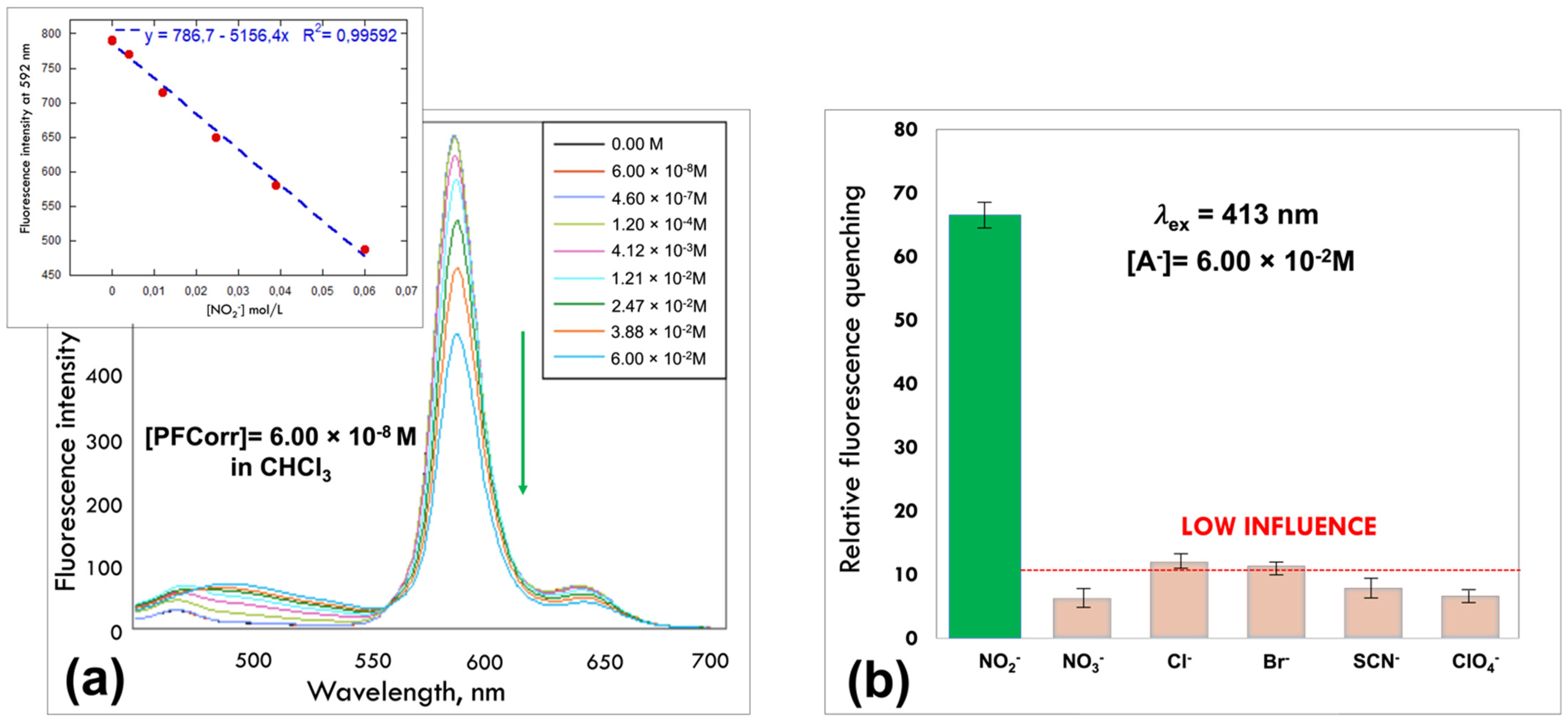
PFCorr showed a great fluorescence emission property in the region 550–680 nm upon the excitation at 413 nm, corresponding to the ligand Soret band absorbance maximum. The external core fluorination procedure, as expected, resulted in enhanced fluorescence quenching in the presence of nitrite ions: significant emission quenching of
PFCorr at 580 nm was registered in the concentration range from 6 × 10
−8 to 6 × 10
−2 M (
Figure 3a). The selectivity test showed no influence of NO
3−, Cl
−, Br
−, SCN
−, and ClO
4− interfering anions present in the same concentration (6 × 10
−2 M) in tested solutions on the selective interaction of
PFCorr ligand with nitrites (
Figure 3b).
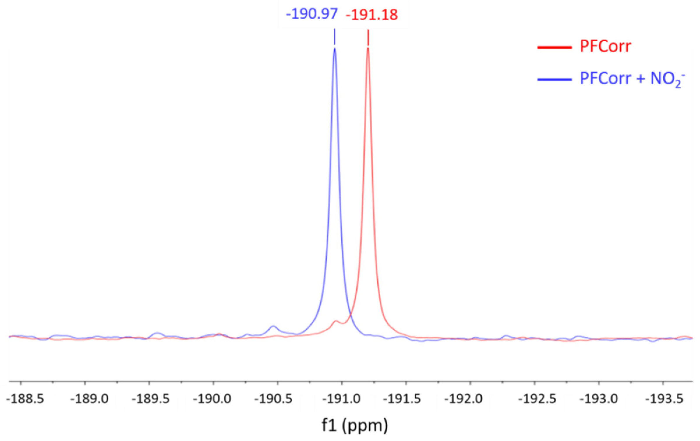
The obtained results indicate the high sensitivity of the
PFCorr ligand to nitrite ions. The coordination of NO
2− ions as an axial ligand on the central phosphorus was investigated with
31P NMR spectroscopy performed before and after the addition of NO
2− ions. As shown in
Figure 4, the chemical shift for the central phosphorus in pure
PFCorr was −191.18 ppm, which corresponds to a hexa-coordinated species with two hydroxyl groups as axial ligands [
29,
30,
31]; upon the addition of the high excess of nitrite anions, [NO
2−] = 0.2 M, the chemical shift of the phosphorus changes from −191.18 to −190.97 due to the axial coordination of the nitrite ion on the central phosphorus atom of the
PFCorr ligand.
The developments of all-solid-state optical sensors are guided by the requirements of practical utility and easy application of these devices. Consequently, the necessity of fast and accurate readouts from optical sensor outputs determines to a high degree the construction of such systems and defines the choice of the appropriate solid support used for ligand immobilization. The immobilization of binding ligands inside a polymeric matrix is one of the most widely applied methods for the bulk ion-selective optodes’ development. Moreover, over the past decade, nanomaterials [
12,
13,
14,
15] and paper-based sensors [
23,
24,
32,
33] have been developed for a wide range of analytes, including inorganic anions.
In the next step, with the aim to develop all-solid-state nitrite-selective optodes, we compared the performance of PVC/TOP membranes based on
PFCorr and previously tested in [
25]
PCorr fluorophores. While in our preliminary study the PVC-based membranes doped with
PCorr and
PFCorr were tested as cross-sensitive components in an optical sensor array [
26], the results of selectivity tests in solutions confirmed with
31P NMR nitrite-titration of
PFCorr have given evidence that suggests the possibility of employing this ligand as a fluorophore for the development of an all-solid-state nitrite-selective optode.
The extraction of the target anion inside the polymeric membrane doped with
PFCorr results in the axial coordination of the corrole–metal complex and yields a fluorescence signal. Depending on the charge of the fluorophore, an addition of different amounts of cationic lipophilic sites, TDA+ inside the membrane is required to facilitate the anion extraction. The compositions of the studied membranes are listed in
Table 1.
The amount of fluorophores in a membrane series was 0.5 and 1 wt%, while 0.5, 1, and 5 wt% of TDACl anion-exchanger were added. These amounts correspond to the 1:1, 1:2, and 1:5 molar ratios between ligand and anion-exchanger amounts for
PFCorr based membranes Mb1.1–Mb.1.3, respectively, and to 1:1 and 1:2 molar ratio between ligand and anion-exchanger amounts for
PCorr-based membranes Mb2.1–Mb.2.2, respectively (
Table 1). The fluorimetric response toward NO
2− − ions was investigated for membrane spots drop-cast on glass slides in a 0.01 M MES background solution with pH 5.5 in the concentration range 1.0 × 10
−6–1.0 ×10
−1 M.
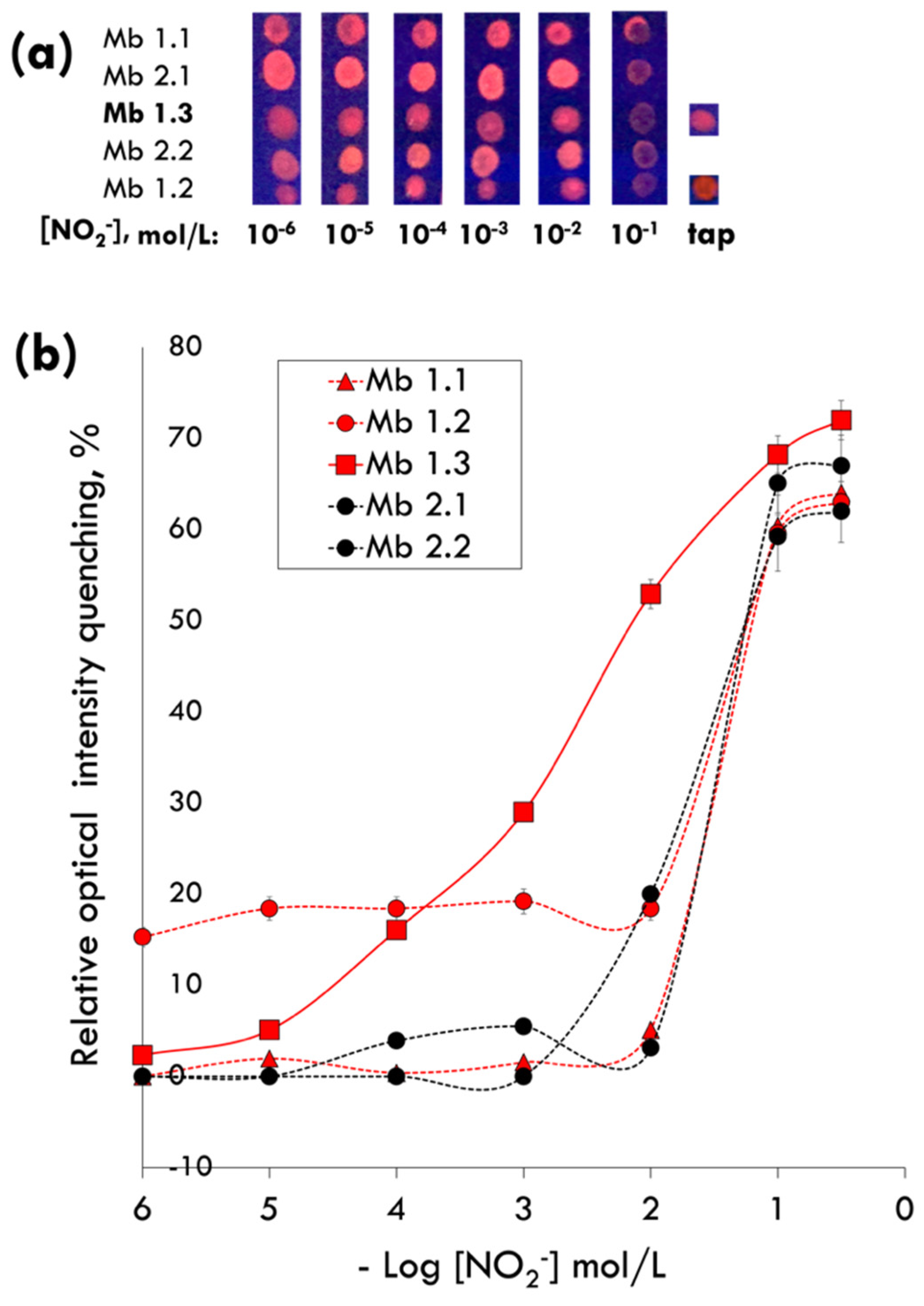
The fluorimetric response with the widest linear range was recorded for the optode with Mb 1.3, doped with 1 wt% of
PFCorr ligand and containing five-fault molar excess of anion-exchanger, TDACl (5 wt%), indicating the prevalent influence of the target anion’s transport inside the membrane phase on the optode’s sensing properties’ improvement (
Figure 5).
As it can be seen from the
Figure 5, the visibly observed luminescence intensity quenching of the sensing material indicates the suitability of the
PFCorr-based optodes to perform fast monitoring of nitrite ions in concentrations up to 10
−1 M with a low detection limit estimated as 0.27 mg/L, which is one order of magnitude lower than the maximum recommended NO
2− concentration level of 3 mg/L in drinking water [
34]. No significant luminescence quenching of membranes Mb3.1 and Mb2.1 doped respectively with
PFCorr and
PCorr was registered in tap water samples (laboratory tap water from the ‘Tor Vergata’ zone of the city of Rome, Italy, with a medium nitrite content around 0.1 mg/L according to the ACEA data [
35]) (
Figure 5).
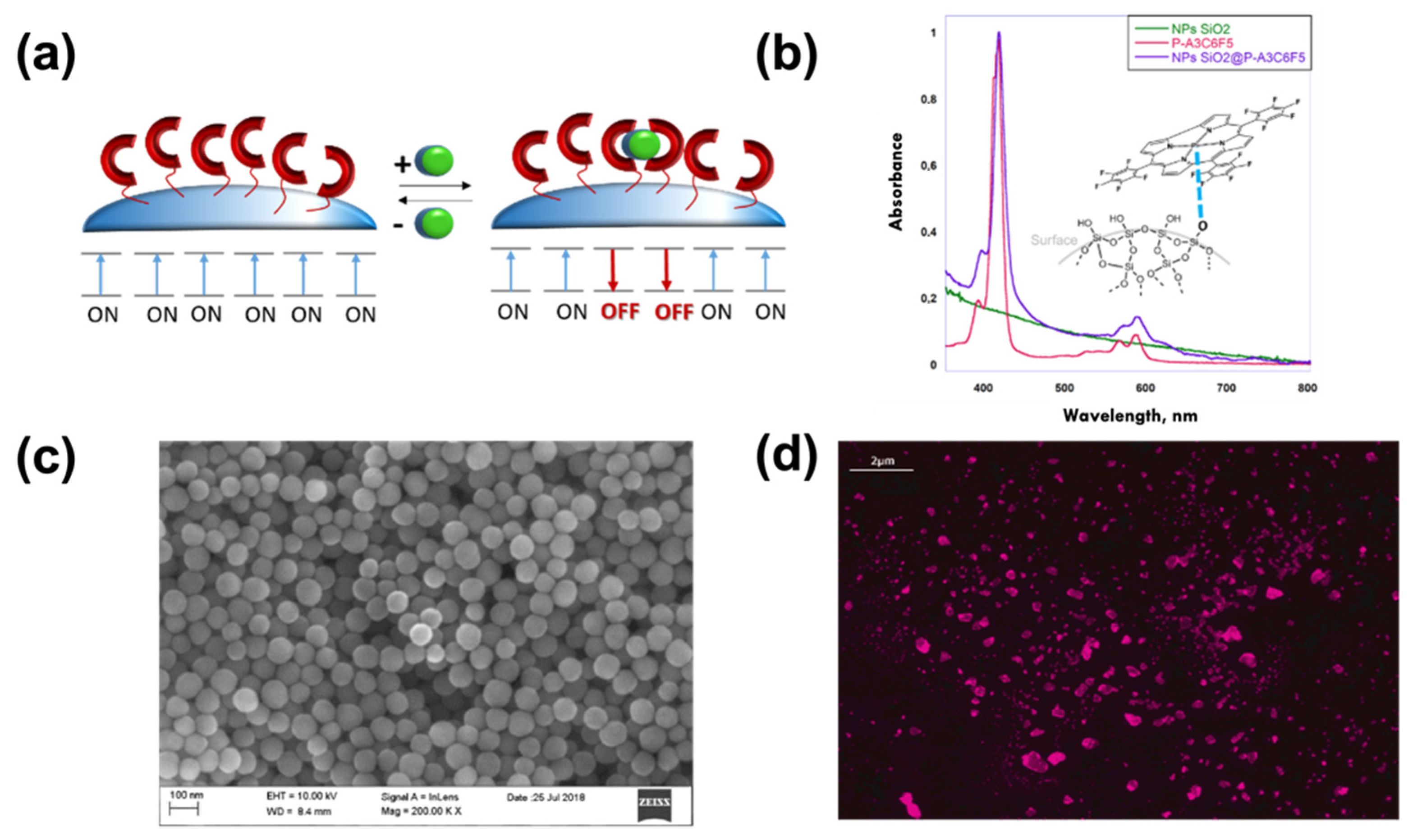
Further, in order to improve the performances of
PFCorr-based nitrite-selective optodes, we exploited the possibility to anchor ligands on the surface of silica nanoparticles, SiO
2NPs, taking advantage of the higher accessibility of sensing sites to the analyte and enhanced emission quenching through the so-called ‘dye–dye interactions’ between proximal ligands [
36]. SiO
2NPs were chosen since they are not toxic, can be obtained with a simple synthetic procedure, and are inexpensive [
28]. The
PFCorr functionalization of SiO
2NPs was made by the ‘post-coating method’ procedure, that consists of two steps, as described in detail in the
Section 2. The anchoring of
PFCorr on SiO
2NPs occurs through the axial coordination of hydroxyl groups present on the silica surface to the corrole’s central phosphorus. The wider surface area of SiO
2NPs significantly increases the number of analyte–ligand interactions, allowing the coordination of many sensing corrole units with free axial positions available for analyte binding that leads to a magnification of the sensing response (
Figure 6a). Moreover, the phenomena of ligand aggregation typical in solution (or inside a polymeric membrane) is eliminated for SiO
2NPs@
PFCorr due to the rigid fluorophore fixation on NPs; the axial nitrite coordination and the prevalence of SiO
2NPs@
PFCorr-NO
2− complexes with 1:1 stoichiometry is expected. The
PFCorr functionalization of the SiO
2NPs’ surface was confirmed by UV-vis spectra; while by SEM and fluorescent microscopy the morphologies and the responses to the light exposure of hybrid assemblies were studied (
Figure 6b–d, respectively).
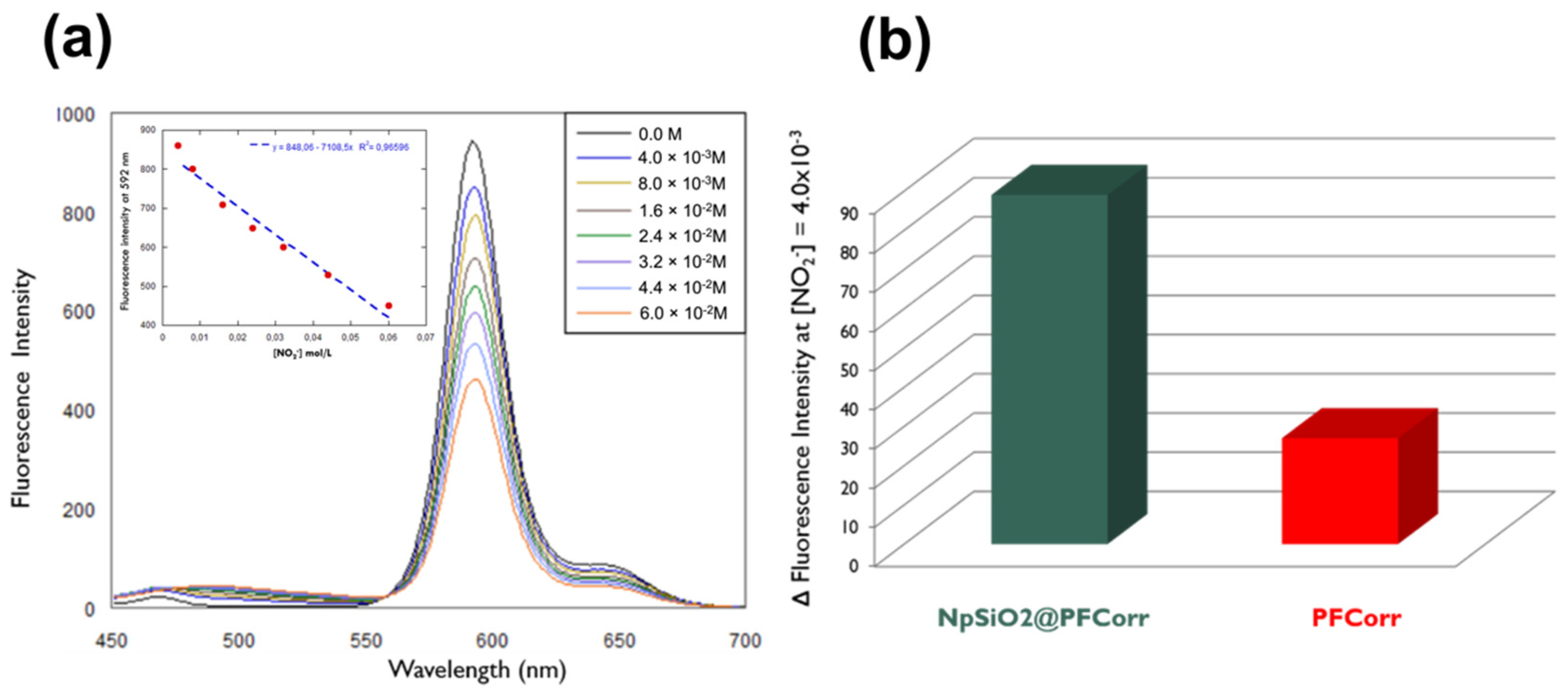
The SiO
2NPs@
PFCorr’s assembling response to nitrite anions was first checked in an aqueous suspension at increasing concentrations of analyte. The tests showed promising results in terms of the quenching effect of corrole fluorescence in the presence of nitrites. As seen in
Figure 7, the emission spectra recorded at a determinate nitrite ion concentration, [NO
2−] = 4 × 10
−3 M, showed a much greater fluorescence quenching for hybrid assemblies compared to the response of the phosphorous corrole dissolved in solution without silica nanoparticles as a solid support.
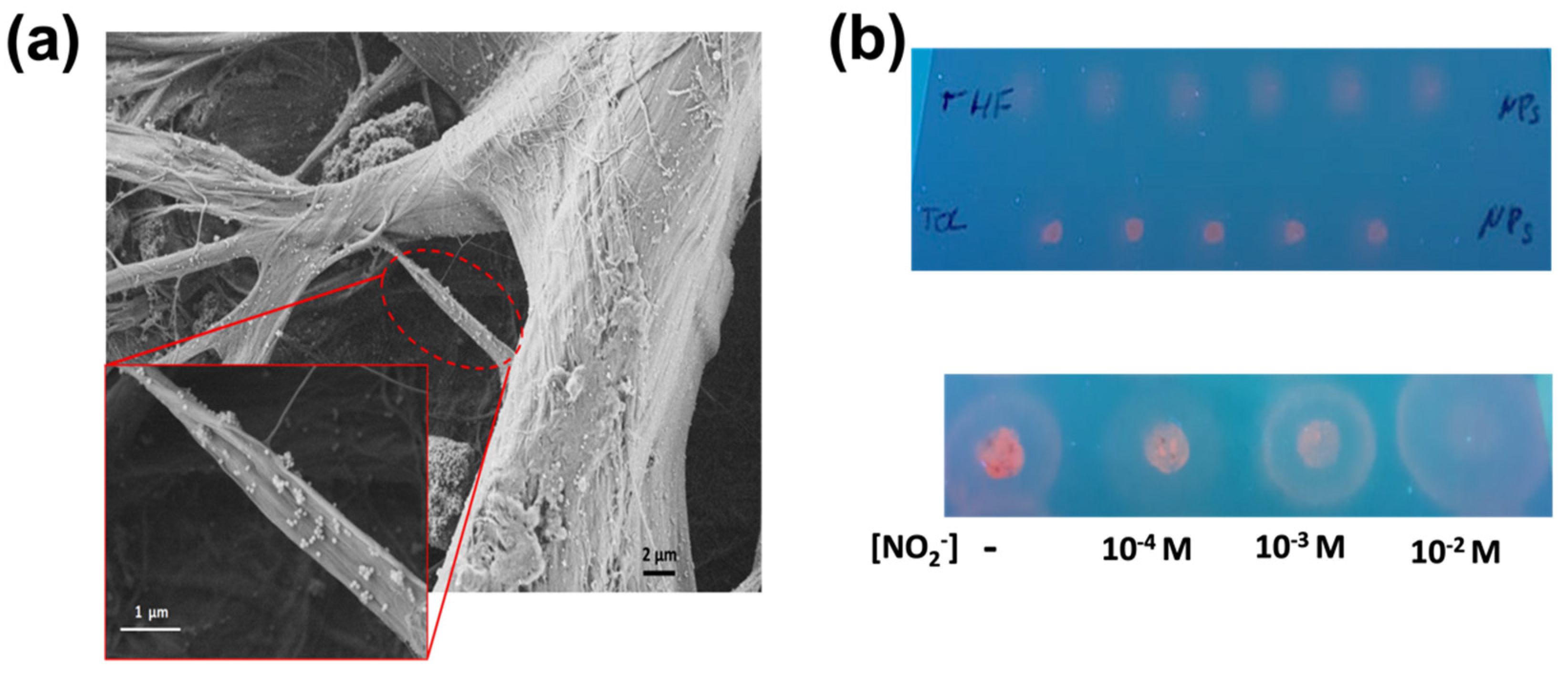
Finally, in order to apply SiO
2NPs@
PFCorr to assemble on a solid support in an optical device for fast and easy in situ measurements, they were deposited on simple filter paper Whatman
TM strips. The porosity of the paper allowed a uniform distribution of the nanoparticles on the cellulose fibers inside the material, avoiding leaching of the nanoparticles from the paper support during the liquid samples’ analysis. In
Figure 8a the SEM image demonstrates the SiO
2NPs@
PFCorr distribution on the cellulose fibers.
Figure 8b shows preliminary tests performed on paper strips using UV-lamp excitation (365 nm) for SiO
2NPs@
PFCorr deposited from THF and toluene solvents (top) and the response of on-paper SiO
2NPs@
PFCorr/THF assembly in the aqueous solutions with increasing nitrite concentrations, drop-cast on the surface of the sensing spots (bottom). The obtained results demonstrate the potential of the
PFCorr ligand for optical nitrate-ions, sensing both inside the all-solid state optodes based on polymeric membranes or in on-paper deposited SiO
2NPs@
PFCorr assemblies. In the latter case the instrument setup should be further optimized, in order to increase the potential of this innovative optical system.


 ,
, 


