Article

Detecting the Bitterness of Milk-Protein-Derived Peptides Using an Electronic Tongue

by
Arijit Nath
1,
Burak Atilla Eren
1,
John-Lewis Zinia Zaukuu
2,
András Koris
1,
Klára Pásztorné-Huszár
3,
Emőke Szerdahelyi
4 and
Zoltan Kovacs
5,*
1
Department of Food Process Engineering, Institute of Food Science and Technology, Hungarian University of Agriculture and Life Sciences, Ménesi st 44, HU-1118 Budapest, Hungary
2
Department of Food Science and Technology, College of Science, Kwame Nkrumah University of Science and Technology, Kumasi 00233, Ghana
3
Department of Refrigeration and Livestock Products Technology, Institute of Food Science and Technology, Hungarian University of Agriculture and Life Sciences, Ménesi út 43-45, HU-1118 Budapest, Hungary
4
Food Science Research Group, Institute of Food Science and Technology, Hungarian University of Agriculture and Life Sciences, Herman Ottó út 15, HU-1022 Budapest, Hungary
5
Department of Measurements and Process Control, Institute of Food Science and Technology, Hungarian University of Agriculture and Life Sciences, Somlói Street 14-16, HU-1118 Budapest, Hungary
*
Author to whom correspondence should be addressed.
Submission received: 1 May 2022 / Revised: 29 May 2022 / Accepted: 4 June 2022 / Published: 7 June 2022
(This article belongs to the Special Issue Bioinspired Chemical Sensors and Micro-Nano Devices)

Abstract

:
Bitterness is a considerable limiting factor for the application of bioactive peptides in the food industry. The objective of this study was to compare the level of bitterness of milk-protein-derived peptides using an electronic tongue (E-tongue). Liquid milk protein concentrate (LMPC) was prepared from ultra-heat-treated skimmed cow’s milk. It was initially hydrolyzed with different concentrations of trypsin, namely, 0.008 g·L−1, 0.016 g·L−1 and 0.032 g·L−1. In a later exercise, tryptic-hydrolyzed LMPC (LMPC-T) was further hydrolyzed using Lactobacillus bulgaricus and Streptococcus thermophilus. The effect of glucose in microbial hydrolysis was studied. The bitterness of peptides was evaluated with respect to quinine, a standard bittering agent. The level of bitterness of the peptides after microbial hydrolysis of LMPC-T (LMPC-T-F and LMPC-T-FG) was evaluated using a potentiometric E-tongue equipped with a sensor array that had seven chemically modified field-effect transistor sensors. The results of the measurements were evaluated using principal component analysis (PCA), and subsequently, a classification of the models was built using the linear discriminant analysis (LDA) method. The bitterness of peptides in LMPC-T-F and LMPC-T-FG was increased with the increase in the concentration of trypsin. The bitterness of peptides was reduced in LMPC-T-FG compared with LMPC-T-F. The potential application of the E-tongue using a standard model solution with quinine was shown to follow the bitterness of peptides.

1. Introduction

Milk proteins (MPs) have long been recognized as sources of high-quality protein because they can provide all types of amino acids with unique physiological and biochemical importance [1,2]. The applications of milk protein concentrate (MPC) in confectionery products, processed cheese, kefir, yogurt and muscle gain formulations are noteworthy. Depending on the dry matter, MPCs are classified as MPC-35, MPC-50, MPC-56, MPC-60, MPC-65, MPC-70, MPC-75, MPC-80, MPC-85, MPC-88 and MPC-90 [3,4]. Presently, the importance of the application of liquid milk protein concentrate (LMPC) in medium- and small-scale industries are coming to the forefront [5]. Unfortunately, some limitations of the MPs were reported [4]. MPs are listed among the “big 8” allergens due to the presence of linear and conformational epitopes in different subunits of their structures. They are responsible for protein-induced enterocolitis syndrome (FPIES), food-protein-induced enteropathy (FPE), food-protein-induced allergic proctocolitis (FPIAP) [6] and immunoglobulin E (IgE)-mediated hypersensitivity [7] in infants and adults. Therefore, the need for a reduction in the allergenic activity of MPs has motivated scientists around the globe.
In the food industry, the enzymatic hydrolysis of MPs is considered to reduce the allergenic activity of proteins. Hydrolysis of MPs with microbial-, plant- and animal- based proteases produces dietary peptides and amino acids [8]. It has been recognized as “safe” by the United States Food and Drug Administration (FDA or USFDA) and the European Food Safety Authority (EFSA). Enzymatic hydrolysis of MPs can be controlled by the selection of specific enzymes, reaction conditions and operating parameters [9]. It was demonstrated that milk-protein-derived dietary peptides confer a wide range of functionalities, including antioxidant, angiotensin-converting enzyme inhibition, antibacterial, antidiabetic, hypotensive, osteoprotective, antiobesity and immunomodulatory activities [10]. The bioavailability and nutritional values of MPs are increased due to their hydrolysis [11]. However, a plethora of literature about the beneficial effects of milk-protein-derived bioactive peptides are available, and their industrial production, commercialization and implementation in food and biopharmaceutical sectors are facing several challenges, including the (a) high processing cost related to the feedstock and equipment for both upstream and downstream processes, (b) bitter taste of bioactive peptides, (c) inadequate clinical evidence related to the bio-efficiency, (d) limited information about the bioavailability and metabolic fate of bioactive peptides and (e) complications in methodologies related to the quality assurance of bioactive peptides as biopharmaceuticals [12].
In the present investigation, the bitterness of bioactive peptides was triggered. The application of an E-tongue (objective instrument) has allowed scientists to understand human taste perception. An E-tongue is a high-sensitivity sensor-based instrument (artificial taste sensing) that is proficient at differentiating food products through pattern recognition. Similar to an electronic nose and spectroscopy, the beneficial aspects of an E-tongue are (a) rapid analytical outputs, (b) affordable operational cost, (c) easy to use, (d) small sample size and (e) harmless to the user [13]. Several models of chemical sensors have also been used to design the electronic tongue’s sensor array: electrochemical (potentiometric, voltammetric, amperometric, impedimetric, conductimetric), optical, mass and enzymatic biosensors [14]. Among these, voltammetric and potentiometric E-tongues are most commonly used for food analysis. A voltammetric E-tongue was engineered for measurements of aqueous redox-active items. However, it was shown that they have great selectivity toward biomolecules but changes in temperature influence the result. A potentiometric E-tongue is an ion-selective sensor. Their application is noteworthy due to their cost efficiency, high selectivity and flexible set-up [15]. Applications of a voltammetric E-tongue to understand the quality of yogurt [16], milk [17], honey [18], coffee [19], tea [20], fruit juice [21], meat [22] and fish [23] were reported. The potentiometric E-tongue was successfully applied to understand the quality of milk [24,25], cheddar cheese [26], tea [27], honey [28], fruit juice [29], coffee [30], meat [31] and fish [32]. Up to now, the application of an E-tongue to understand the taste of peptides produced by tryptic and microbial hydrolysis of MPs has not been reported.
In our previous investigation, allergen-free peptides with antioxidant capacity, angiotensin-converting enzyme inhibitory activity and antibacterial activity were produced via sequential tryptic and microbial hydrolysis of proteins in LMPC [33]. In this investigation, the bitterness of peptides produced via sequential tryptic and microbial hydrolysis of proteins in LMPC was evaluated using a potentiometric E-tongue. The effect of the concentration of trypsin in the proteolysis of proteins was studied. The formation of lower-molecular-weight peptides was increased after tryptic and microbial hydrolysis of LMPC. The bitterness of peptides was increased with the increase in the concentration of trypsin in the proteolysis process. The level of bitterness was reduced due to the addition of glucose in the microbial hydrolysis process.

2. Materials and Methods

2.1. Skim Milk

Skimmed milk (0.1% fat), produced via an ultra-heat treatment (UHT) of cow’s milk, was procured from local grocery shops in and around Budapest, Hungary. Throughout the experiment, milk was stored in a refrigerator at a temperature of 10 °C for a maximum of 12 h.

2.2. LMPC via a Membrane Filtration Process

An indigenous cross-flow single-channel tubular membrane module made of stainless steel (SS316) was used in the experiment to prepare the LMPC. Inside that membrane module, a tubular ceramic ultrafiltration (UF) membrane with a pore size of 5 nm (Pall Corporation, Crailsheim, Germany) was placed. A stainless steel twisted tape static turbulent promoter was used inside the ceramic membrane tube to create turbulence on the membrane surface. Detailed descriptions of the static turbulent promoter, tubular membrane and membrane module were mentioned in our previous publication [5]. During the filtration process, a trans-membrane pressure (TMP) of 3 × 10⁵ N·m−2 was monitored using pressure gauges and flow-controlling valves, which were fitted at the two opposite ends of the membrane module. The feed flow rate was controlled using hydraulic diaphragm pumps (HYDRA-CELL G03, Verder Ltd., Budapest, Hungary) and the inlet and outlet flow valves. A rotameter at the retentate end and a bypass valve were also used for controlling the retention flow rate at 200 L·h−1. Prior to filtration, membrane compaction was performed with TMP at 4 × 10⁵ N·m−2 and a retention flow rate of 200 L·h−1. Then, 1 L of UHT milk was poured into the storage tank of the membrane module and the filtration process was continued until 500 mL of permeate was achieved. The filtration process was performed at room temperature. The retentate of the filtration process was collected and considered as LMPC. The subsequent experiment was performed with LMPC.

2.3. Tryptic Digestion of Proteins in LMPC

The LMPC was pre-incubated at a temperature of 40 °C in a well-equipped bioreactor with a working volume of 500 mL. A pH sensor and a temperature sensor (Mettler-Toledo Seven Multi pH sensors; Mettler-Toledo GmbH, Schwerzenbach, Switzerland) were inserted into the bioreactor through the lid. A data cable was connected to a computer and data points were recorded using self-developed software in the R-project statistical software, ver. 3.4.3 (R Core Team, Vienna, Austria). A sterile spiral tube heat exchanger was placed in the feed tank and the isothermal condition was maintained using a temperature controller. After pre-incubation, tryptic hydrolysis of MPs was performed under a fixed agitation speed of 100 rpm and an operational temperature of 40 °C for 10 min. The effects of the tryptic hydrolysis of proteins were investigated using different concentrations of trypsin (0.008 g·L−1, 0.016 g·L−1 and 0.032 g·L−1). For this purpose, sterile 450 µL, 900 µL and 1.8 mL solutions from the stock solution of trypsin (protein concentration 0.009 g·mL−1) was inoculated with 500 mL of LMPC in a separate experiment. They are presented as LMPC-T-0.008, LMPC-T-0.016 and LMPC-T-0.032, respectively [5,34]. After 10 min of the tryptic hydrolysis of the LMPC, it was immediately transferred to another reactor with a volumetric flow rate of 120 L·h−1 using a centrifugal pump (HYDRA-CELL G03; Verder Ltd., Budapest, Hungary). In that reactor, a temperature of 70 °C was used for the inactivation of trypsin. Similar to before, a sterile spiral tube heat exchanger and a temperature controller were used in this reactor. The tryptic hydrolyzed LMPC (LMPC-T) stayed in this reactor for 30 min.

2.4. Microbial Hydrolysis

After deactivation of the trypsin at a temperature of 70 °C, the temperature of the milk was reduced to 45 °C. The effect of glucose in microbial hydrolysis of LMPC-T was studied. For this purpose, 16 mL of sterile glucose solution from a 40% glucose stock solution was added to 200 mL of LMPC-T in an aseptic condition. Lyophilized lactic acid bacteria (Thermophilic YoFlex® Mild 1.0, Chr. Hansen, Nienburg, Germany) was used for microbial hydrolysis of LMPC and LMPC-T. Microbial hydrolysis was performed in a 250 mL blue cap glass bottle. Then, 2 mL of liquid culture from stock microbial culture was inoculated to 200 mL of LMPC and LMPC-T. After inoculation, the levels of Lactobacillus bulgaricus and Streptococcus thermophilus were 1.6 × 10⁷ CFU·mL−1 and 5.4 × 10⁶ CFU·mL−1, respectively, according to Breed’s method [35]. Samples were denoted as LMPC-F, LMPC-T-F and LMPC-T-FG, respectively. All samples were incubated at a temperature of 45 °C for 6 h in an incubator (HACH, Düsseldorf, Germany) [36].

2.5. Sensory Analysis with Electronic Tongue

LMPC-F, LMPC-T-F and LMPC-T-FG were centrifuged at 5000 rpm for 15 min at a temperature of 4 °C (Z216MK; Hermle, Wehingen, Germany). Clear supernatants of the centrifuged samples were further diluted 10-fold with distilled water. The Alpha ASTREE II potentiometric E-tongue (Alpha M.O.S., Toulouse, France) was utilized in the experiment. The E-tongue was equipped with a sensor array (#1) that included seven CHEMFET (Chemically Modified Field-Effect Transistor) sensors developed for food applications [37]. The food application sensors were BB, HA, ZZ, GA CA, JE and JB. In a working electrode of the E-tongue, a lipid/polymer membrane comprises a lipid, a plasticizer and a polymer as a sensing part. The electrode potentials of the seven sensors were recorded against the reference electrode (Ag/AgCl 3 M KCl) (Metrohm, Herisau, Switzerland). The instrument also consisted of a transducer for measuring signal differences and a 16-position auto-sampler for the analysis. Each sensor in the E-tongue has a different bio-polymers layered membrane. Differences in membrane composition in the sensor affect the selectivity, which provides the sensor array cross-selectivity to tastes [38]. As the storage of the E-tongue sensors was done in a dry state, it was necessary to condition and hydrate the sensors before their use. Conditioning of the sensors was done using 0.01 N of hydrochloric acid. The error limit of each sensor was defined in the calibration step to obtain consistent and reproducible data during the measurements. Subsequently, the calibration of signals from individual sensors was accomplished. An equal volume of each diluted sample was mixed and considered for the calibration of the sensors. The calibration process was repeated until the sensors were adjusted to their target values within the error limit from the calibration [39]. It was already reported that enzymatic hydrolysis of proteins produces peptides with a bitter taste [40]. Therefore, the standard bittering agent quinin-sulphate was added to the diluted supernatant of LMPC-F and considered as the control. Two different concentrations of quinin-sulphate, i.e., 8 µM and 40 µM, were used in the experiment. The lower concentration of 8 µM (Q1) was selected to match with the human sensory threshold of the bitter taste sensation [41,42] and the higher one was selected as five times higher than the human threshold, i.e., and 40 µM (Q2). In the manuscript, they are presented as LMPC-F_Q1 and LMPC-F_Q2, respectively. The bitterness of samples was measured at room temperature in random order with the E-tongue. A total of 100 mL of sample volume was considered during each measurement. Each sample was measured nine times. DI water was used for the cleaning of sensors after each measurement. In the experiment, 120 s for sample acquisition and 10 s for sensor cleaning were considered. The average value of the last 10 s of the reading (equilibrium stage) obtained from every sensor was calculated and used for further statistical evaluation.
Two sets of experiments were performed with the E-tongue. In the first set of experiments, a comparison of the supernatant of microbial fermentation broth was carried out without quinin to understand their similar/dissimilar characteristics due to trypsin and glucose. In the second set of experiments, the bitterness of the supernatant of fermentation broth was evaluated with respect to quinin.

2.6. Statistical Analysis
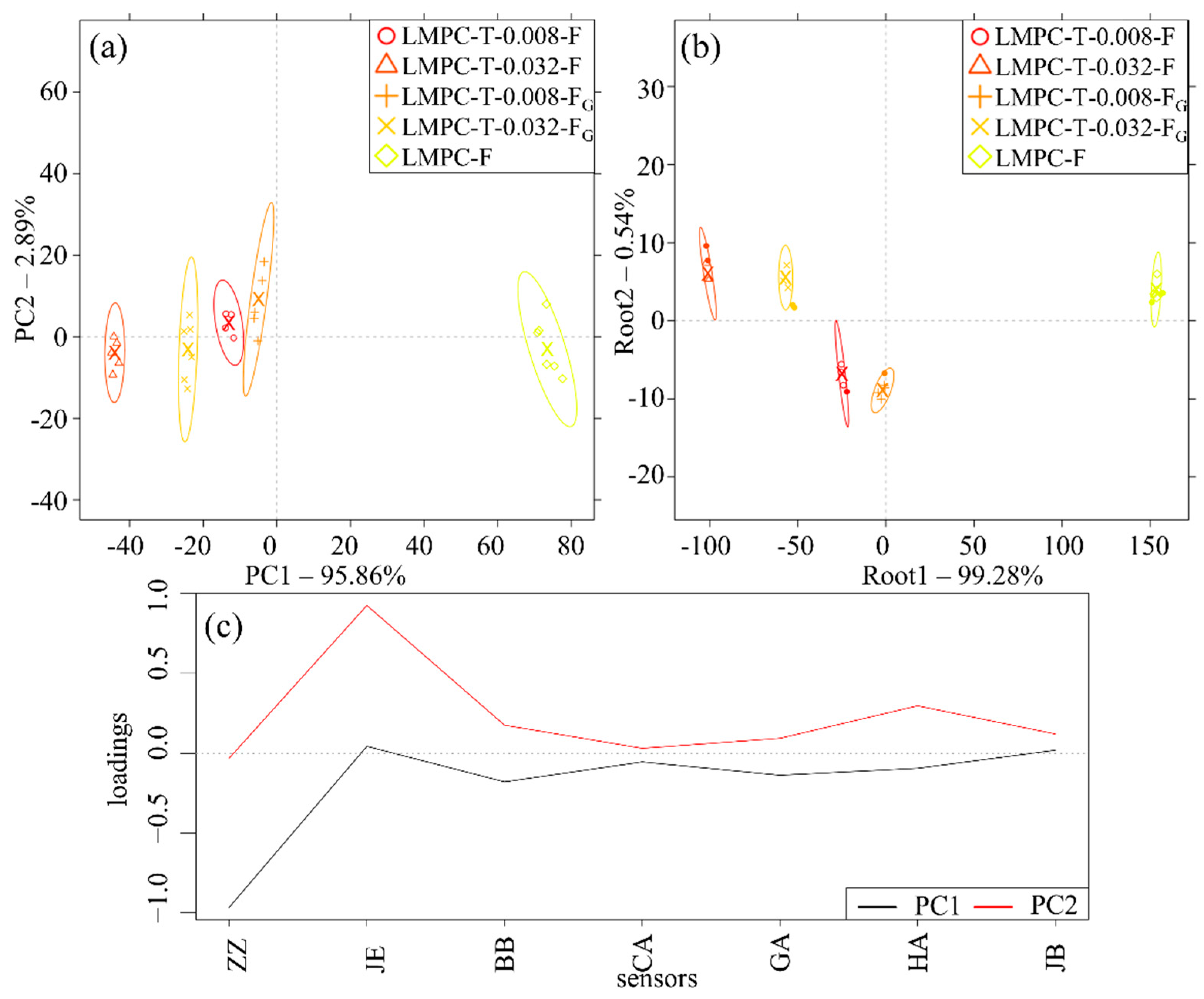
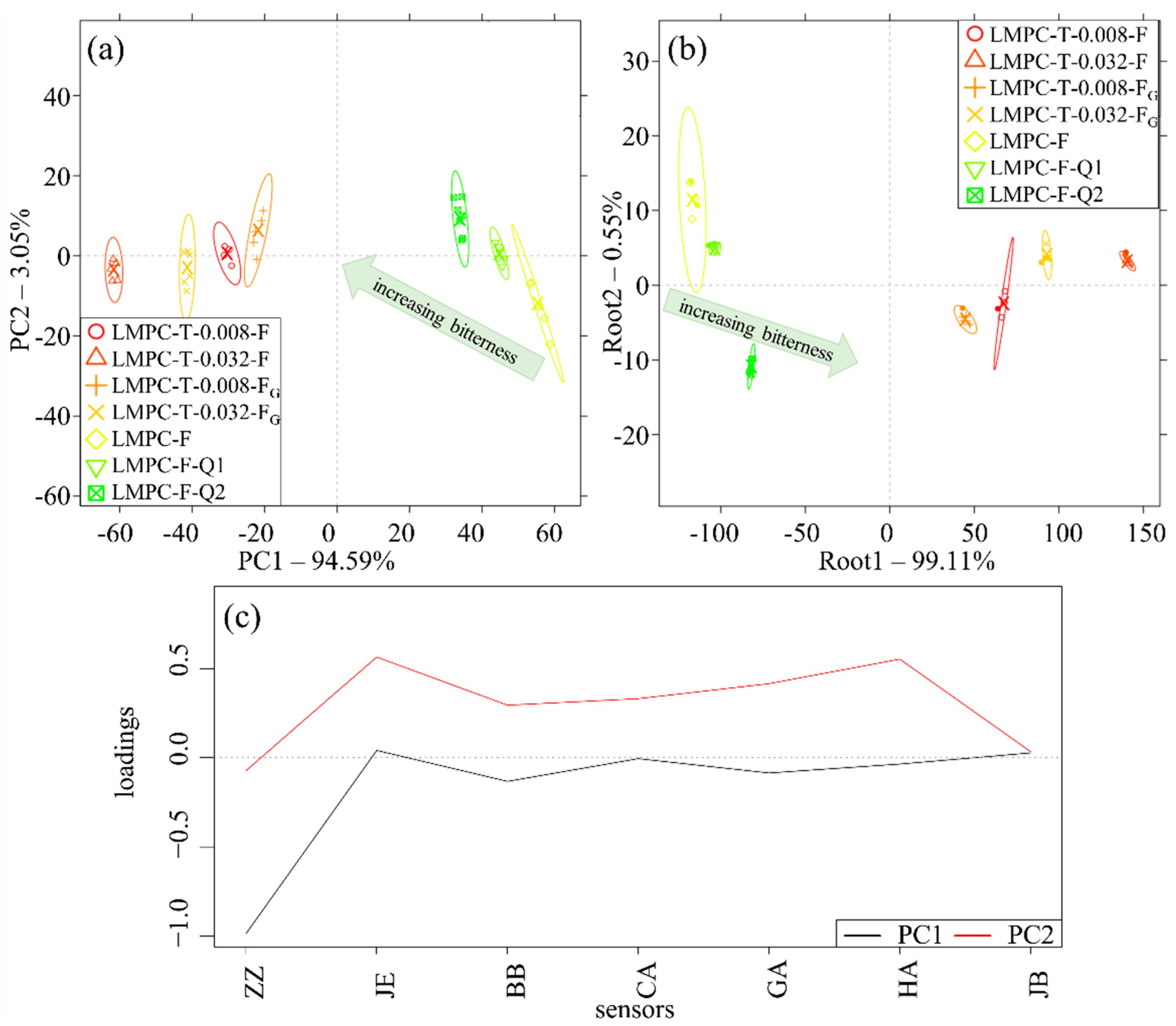
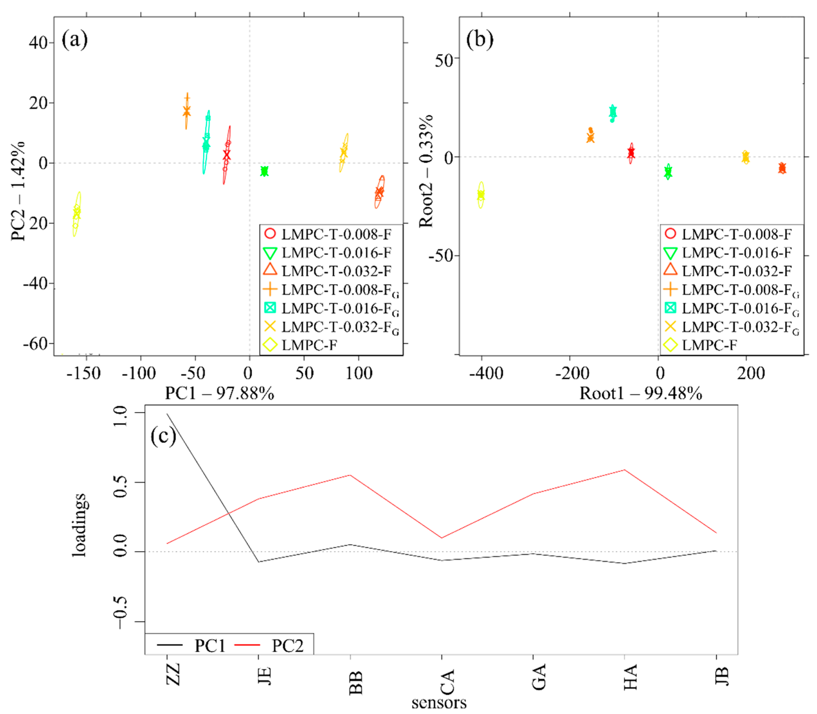
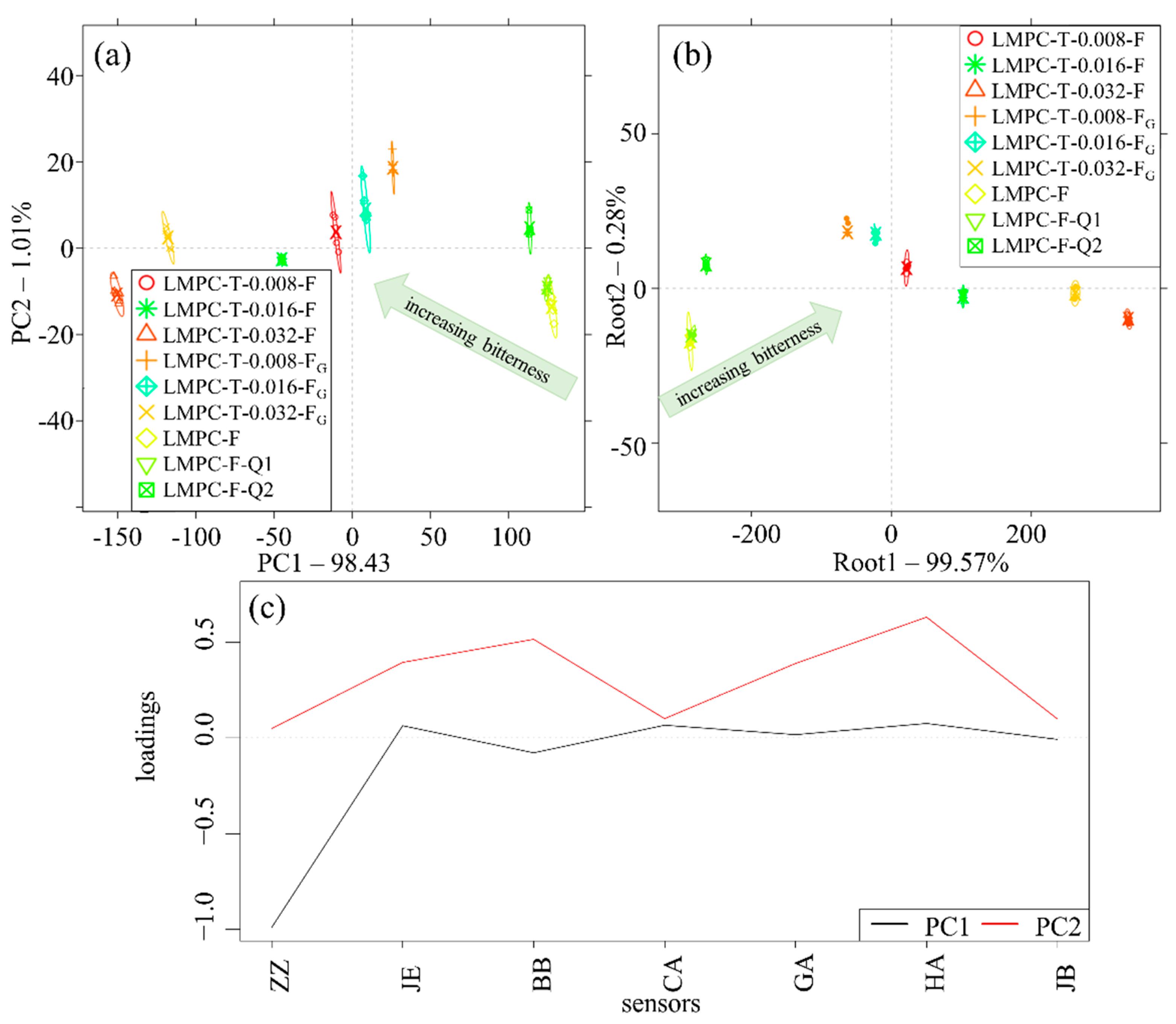
All experiments were performed in triplicates and the mean value and standard deviation were calculated. The one-way analysis of variance method followed by Tukey’s post hoc test were performed to understand the significant differences (p < 0.05) between different groups for all the analytical results. SPSS 15.0 (version 25.0) (IBM, Armonk, NY, USA) was used for the statistical analysis.
For the E-tongue results analysis, a data matrix was developed that included the results of each sample. The results of different samples and their repeats were arranged in rows of a data matrix. The seven sensors of the E-tongue were used as variables. The measurement results were evaluated using principal component analysis (PCA) [43] to understand the pattern and visualize the information. In addition to the PCA score plots, PCA-loading plots were developed to understand the contributions of different sensors of the E-tongue in the observed separation pattern. A classification of the models was built using the linear discriminant analysis (LDA) method [44] to cross-check the classification of the samples. The LDA model was tested with three-time cross-validation. One-third of the sample set was excluded from the calibration set and the generated model was validated using these retained samples. The procedure was repeated iteratively three times to ensure that all samples were included in the validation set once. The qualitative LDA model was evaluated using the ratio of correctly classified samples in the model building and cross-validation. The results of the PCA and LDA were visualized in score plots, where the separation between different groups are presented with their 95% confidence intervals. The statistical evaluation was performed using the R-project statistical software, Ver. 3.4.3 (R Core Team, Vienna, Austria) [45].

3. Results

A PCA score plot was developed based on the data of the E-tongue with LMPC-T-0.008-F, LMPC-T-0.032-F, LMPC-T-0.008-FG, LMPC-T-0.032-FG and LMPC-F (Figure 1a). The first principal component (PC1) described approximately 96% of the total variance. PC1 presented separation between the tested sample groups (LMPC-T-F and LMPC-T-FG) and LMPC-F (control). It was found that sample groups were very distinct from the control.
Based on the first discriminant factor (root 1), the LDA score plot provided a very similar arrangement of the sample groups to the results of the PCA. It represented more than 99% of the total variance (Figure 1b). The result of cross-validation was obtained and it confirmed that the E-tongue was able to recognize and predict all the samples, including control, successfully (100% correct classification).
In Figure 1c, the PCA loading plots of Figure 1a are presented. The separation patterns observed in PC1 mostly occurred by the sensor ZZ, followed by sensors BB, GA, HA and CA. Sensor JE had a significant role in the formation of PC2, followed by sensors HA, BB, GA and JB, while sensors ZZ and CA had no significant contribution in this direction.
Quinine-supplemented model solutions, namely, LMPC-F_Q1 and LMPC-F_Q2, were adopted to classify samples based on their bitterness (Figure 2). Sets of the data points of different samples were positioned with their relative similarity/dissimilarity with the quinine-supplemented model solutions. PC1 represented approximately 95% of the total variance, showing the clear separation of the LMPC-T-F and LMPC-T-FG sample groups from the LMPC-F and its quinine-substituted counterparts (Figure 2a). The tendency of separation predominantly explained their level of bitterness compared with LMPC-F using their relative position. As an example: all of the groups of LMPC-T-F samples were positioned far away from LMPC-F. According to PC1, the bitterness of samples was in the following order: LMPC-T-F > LMPC-F. It represented that all the LMPC-T-F samples were more bitter compared to LMPC-F. Furthermore, PC1 represented the comparative results of the bitterness of different samples. According to the bitterness of samples, the order herein was as follows: LMPC-T-0.032-F > LMPC-T-0.008-F and LMPC-T-0.032-FG > LMPC-T-0.008-FG. The addition of glucose in the microbial hydrolysis of proteins can modulate the bitterness of samples. The bitterness of samples was lowered in LMPC-T-FG compared to LMPC-T-F. Based on the PC1 results, LMPC-T-0.008-F > LMPC-T-0.008-FG and LMPC-T-0.032-F > LMPC-T-0.032-FG.
The results of the LDA model that was developed for the classification of the tested samples are presented in Figure 2b. It presents a very similar pattern to the one observed in the PCA (Figure 2a). Root 1 represented more than 99% of the total variance among the groups. The confidence intervals of different groups in the LDA model provided 100% correct classification in both model training and model validation.
The PCA loading plots of Figure 2a are presented in Figure 2c. According to PC1, separation mostly occurred due to sensor ZZ, followed by sensors BB and GA. Furthermore, it was noted that sensors JE, JB, CA and HA had no significant contribution to PC1. Sensors JE and HA had a significant role in the formation of PC2, followed by sensors GA, CA and BB. Sensors ZZ and JB had no significant role in PC2.
In a further experiment, LMPC-T-0.016-F and LMPC-T-0.016-FG were also considered to analyze their characteristics using the E-tongue (Figure 3). Results of the E-tongue with LMPC-T-0.016-F and LMPC-T-0.016-FG provided logical understanding, similar to Figure 1. The PCA plot shows that LMPC-F was very much dissimilar compared with LMPC-T-F and LMPC-T-FG. About 98% of the total variance of the E-tongue signals was retained in PC1 (Figure 3a). The group of LMPC-T-0.016-F was positioned between LMPC-T-0.008-F and LMPC-T-0.032-F. Similarly, LMPC-T-0.016-FG was positioned between LMPC-T-0.008-FG and LMPC-T-0.032-FG.
An LDA plot was developed for LMPC-T-0.008-F, LMPC-T-0.016-F, LMPC-T-0.032-F, LMPC-T-0.008-FG, LMPC-T-0.016-FG, LMPC-T-0.032-FG and LMPC-F (Figure 3b). The LDA plot shows very similar positions of the samples, as observed in Figure 3a. More than 99% of the total variance between groups was found based on root 1. The LDA model provided 100% correct classification in both the model training and model validation.
In Figure 3c, the PCA loading plots of Figure 3a are presented. The separation patterns observed in PC1 mostly occurred due to sensor ZZ, followed by sensors JE, BB, CA and HA. Sensors GA and JB had no significant contribution to PC1. The separation tendency observed in PC2 was mostly due to sensors BB and HA, followed by sensors JE, GA, CA, ZB and ZZ.
Similar to before, we aimed to understand the bitterness of samples with reference to quinine-supplemented model solutions. About 98% of the total variance was obtained based on PC1 (Figure 4a). The relative position of samples based on PC1 could predominantly classify the samples with their bitterness. According to PC1, the bitterness of samples was in the following order: LMPC-T-0.032-F > LMPC-T-0.016-F > LMPC-T-0.008-F and LMPC-T-0.032-FG > LMPC-T-0.016-FG > LMPC-T-0.008-FG. The addition of glucose in the microbial hydrolysis of proteins reduced the bitterness of the samples. Based on the PC1 result, LMPC-T-0.008-F > LMPC-T-0.008-FG, LMPC-T-0.016-F > LMPC-T-0.016-FG and LMPC-T-0.032-F > LMPC-T-0.032-FG.
An LDA plot was developed with LMPC-T-F, LMPC-T-FG and LMPC-F (Figure 4b). The sample groups of LMPC-T-F and LMPC-T-FG were very dissimilar to LMPC-F. About 99% of the total variance was obtained based on root 1. The positions of samples were similar to the results in PC1, as presented in Figure 4a. The LDA model provided 100% correct classification of all sample groups in both the model training and model validation.
The PCA loading plots of Figure 4a are presented in Figure 4c. According to PC1, sensor ZZ provided a significant contribution to the separation, followed by sensors JE, BB, CA and HA. Sensors GA and JB provided an insignificant contribution in this respect. According to PC2, sensors HA, GA, BB and JE provided significant contributions to the separation. Sensors JB, CA and ZZ provided an insignificant contribution to PC2.

4. Discussion

A protein or peptide becomes bitter if its average hydrophobicity Q > 1.4 kcal·residue−1 and has a molecular weight 100–6000 Da. The low molecular weight of peptides with bitterness and antioxidant capacity due to the presence of hydrophobic amino acids are produced via the proteolysis of casein and whey proteins by trypsin [5,46,47] and proteolytic systems of lactic acid bacteria [48,49,50].
According to our previous publication, peptides with lower molecular weight can be produced due to sequential tryptic and microbial hydrolysis of proteins in LMPC [33]. An SDS-PAGE image of different proteins and peptides in LMPC-F, LMPC-T-F and LMPC-T-FG with their molecular weights is shown in Figure 5.
According to this Figure, LMPC-F may have partially hydrolyzed immunoglobulin, lactoferrin and bovine serum albumin with molecular weights ~150 kDa, ~80 kDa and ~66 kDa, respectively. A protein aggregate with a molecular weight of 25–35 kDa is shown. In heat-treated milk, besides the lactosylation of proteins, the sizes of proteins in milk are altered from their original sizes. When milk is heated to more than 80 °C, the tertiary structure of whey protein unfolds [51]. At a temperature of more than 80 °C, the denaturation of α-lactalbumin is faster in the presence of β-lactoglobulin and the denaturation rate of α-lactalbumin is faster than β-lactoglobulin [52]. Subsequently, unfolded whey proteins may bind with each other or with casein molecules, especially with κ-casein in the periphery of casein micelle via covalent, hydrophobic and disulfide bonds [53,54]. It was reported that the conjugate of bovine serum albumin and β-lactoglobulin [55] and dimer of β-lactoglobulin [56] can be formed during milk processing using a UHT. However, the molecular weight of α-casein is ~25 kDa and its molecular weight is altered due to conjugation with β-lactoglobulin via a covalent bond and disulfide bond after heat treatment [54,57]. α-lactalbumin is not able to bind directly with casein because it does not contain the -SH group. α-lactalbumin may produce a hydrophobic conjugate with β-lactoglobulin [53,58]. In the SDS-PAGE image, other protein bands might be β-casein with a molecular weight of ~24 kDa, κ-casein with a molecular weight of ~19 kDa, β-lactoglobulin with a molecular weight of ~18 kDa and α-lactalbumin with a molecular weight of ~14 kDa. It is noted that with the increase in the concentration of trypsin prior to microbial hydrolysis, proteins are hydrolyzed in a dose-dependent manner. Immunoglobulin (~150 kDa), lactoferrin (~80 kDa), bovine serum albumin (~66 kDa) and κ-casein (~19 kDa) were almost hydrolyzed in all LMPC-T-F and LMPC-T-FG samples. Some casein molecules with molecular weights of ~35 kDa, ~32 kDa and ~24 kDa were retained in LMPC-T-0.008-F and LMPC-T-0.008-FG. Perhaps those were the conjugates of κ-casein with β-lactoglobulin, dimer β-lactoglobulin and β-casein, respectively. κ-casein is present in the periphery of casein globule [59]. Therefore, they have the opportunity to participate in a proteolysis reaction. The conjugate of κ-casein with β-lactoglobulin was almost hydrolyzed in LMPC-T-0.032-F and LMPC-T-0.032-FG. A protein fragment with molecular weight ~22 kDa was found in LMPC-T-0.008-F and LMPC-T-0.008-FG. It may be some peptone and γ-casein produced via the partial hydrolysis of β-casein. They were almost hydrolyzed at LMPC-T-0.016-F and LMPC-T-0.016-FG. Dimer β-lactoglobulin with a molecular weight of ~32 kDa was hydrolyzed at LMPC-T-0.016-F and LMPC-T-0.016-FG. Peptides with low molecular weight were produced with an increase in the concentration of trypsin. According to the SDS-PAGE image, the addition of glucose in the microbial hydrolysis of protein did not influence the protein profile of the hydrolysis.
It was reported that αs1-casein, αs2-casein, β-casein and κ-casein with Q values of 1.17 kcal·residue−1, 1.33 kcal·residue−1, 1.33 kcal·residue−1 and 1.29 kcal·residue−1, respectively, are potential sources of bitter peptides. Hydrophobic amino acids in –C and –N terminal positions are primarily responsible for the bitterness of peptides [60]. Trypsin is an endo-protease, which cleaves at the C-terminal side of arginine and lysine amino acid residues, except arginyl-proline and lysyl-proline. It has been reported that the peptides GPFPVI, FFVAPFPEVFGK, FALPQYLK [46], RGPPFIV [61], IHPFAQTQSLVYPFPGPIPNSLPQNIPPLTQTPVVVPPFLQPEVMGVSK, GPFPIIV and IHPFAQTQSLVYPFPGPIPN [62] are produced due to tryptic hydrolysis of casein. A proteolytic system of lactic acid bacteria consists of cell-membrane and intracellular proteases [63]. The exogenous proteins or peptides are hydrolyzed by cell-membrane proteases of lactic acid bacteria because they cannot directly uptake exogenous proteins. Hydrolysis of exogenous proteins or large molecular-weight peptides to oligopeptides takes place due to cell-membrane proteinases (PrtP), which are members of the serine family. Three types of cell-membrane proteases of lactic acid bacteria, namely, PI-type, PI/PII-type and PIII-type, hydrolyze caseins. PI-type cell-membrane protease hydrolyzes β-casein. PI/PII-type cell-membrane protease hydrolyzes β-casein and αs1-casein (lesser extent). PIII-type cell-membrane protease hydrolyzes αs1-casein, β-casein and κ-casein [64]. Oligopeptides, dipeptides and tripeptides are able to enter the biotic phase via transporter membrane proteins Opp, DtpP and DtpT [49,65] and they are hydrolyzed by numerous cytoplasmic peptidases, such as aminopeptidases (PepN, PepC, PepA, PepM, PepP and PepX), endopeptidases (PepE, PepG, PepF and PepO) and di/tri-peptidases (PepI, PepL, PepQ, PepR, PepD, PepV, and PepT) [66]. Subsequently, they are converted to nitrogenous metabolites, such as amino acids, amines, sulfur compounds and carboxylic acids by different sequential metabolic pathways [67]. The present investigation scheme is almost similar to cheese preparation and maturation, where rennet or purified chymosin and lactic acid bacteria are applied to high-fat milk in a sequential way. However, rennet or purified chymosin does not modulate the bitterness of proteins and peptides, where microbial hydrolysis of coagulated proteins and peptides may generate peptides with a bitter taste [68]. Bitter peptides produced by microbial hydrolysis of MPs during cheese maturation are listed by Lemieux and Simard, 1992 [40]. In this investigation, LMPC from skimmed UHT milk (0.1% fat) was hydrolyzed by trypsin and lactic acid bacteria for 10 min and 6 h, respectively, to avoid curdling and coagulation. Microbial hydrolysis was performed by non-bitter strains Lactobacillus delbrueckii subsp. bulgaricus and Streptococcus thermophilus. Non-bitter lactic acid strains were able to produce pyrrolidone carboxylyl peptidase, which specifically hydrolyses the peptide bond between pyrrolidone carboxylic acid residue (a bittering agent) and protein or peptide [69,70]. Therefore, in our investigation, it was found that an increase in the bitterness in hydrolyzed samples (LMPC-T-F and LMPC-T-FG) was primarily dependent on the concentration of trypsin. According to our result, bitterness was reduced due to the addition of 3% (w/v) glucose in microbial hydrolysis of LMPC-T. This can be explained by the fact that the addition of glucose influences microbial growth, as well as microbial hydrolysis of proteins, formation of aroma compounds (amines and sulfur compounds) [71], carboxylic acids and exopolysaccharides [72,73]. A high microbial cell count due to the supplementation of glucose might hydrolyze the peptides with a bitter taste and produce debittering peptides. Furthermore, it may feel that small peptides do not have the particular conformation that is required to bind with bitter taste receptors (T2Rs) [74]. The conversion of bittering peptides to debittering peptides by lactic acid bacteria was demonstrated by other investigators [75,76]. Tryptic hydrolysis of exogenous proteins prior to microbial hydrolysis produces a lower molecular weight of peptides. Therefore, their transportation to the biotic phase by cell-wall transporter proteins is facilitated. In the biotic phase, they are converted to amino acids, aroma compounds and carboxylic acids through a wide range of metabolic pathways [67,77]. According to Fernandez-Espia and Rul (1999) [78], aminopeptidase (PepS) from Streptococcus thermophilus shows a high specificity toward peptides with Arg or aromatic amino acids at the N-terminal position, which are produced via tryptic hydrolysis of protein. The investigators believed that PepS may be involved in producing amino acids from exogenous proteins, and subsequently, these amino acids may act as precursor molecules of aroma compounds. It was shown that aminopeptidase (APII) from Lactobacillus delbrueckii subsp. bulgaricus is able to hydrolyze peptides with lysine at the N-terminal position [79]. Furthermore, high microbial growth may produce higher amounts of pyrrolidone carboxylyl peptidase. It hydrolyses the peptide bond between pyrrolidone carboxylic acid residue and peptides, which is a source of bitterness [69].
According to our result, the formation of organic acids (change of pH) was related to the concentration of trypsin, as well as tryptic hydrolysis of proteins in LMPC; however, the effect of glucose was insignificant (Figure 6).
A similar observation was reported by Lorenzen and Meisel (2005) [34]. They reported that the acidity was lowered when skim MPs were hydrolyzed by trypsin and lactic acid bacteria compared with microbial hydrolysis without trypsin treatment.
In our previous publication, it was mentioned that antioxidant capacity, measured using a FRAP assay was increased in LMPC-T-F and LMPC-T-FG compared with LMPC-F [33]. The antioxidant capacity of LMPC-F was 97.05 ± 20.41 mg equivalent ascorbic acid·L−1. Changes in antioxidant capacity after sequential tryptic and microbial hydrolysis of MPs are presented in Table 1.
Hydrophobic amino acids were noted to contribute to the antioxidant capacity, and at the same time, provide the bitterness of peptides [62]. Tryptic hydrolysis of MPs produces peptides with antioxidant capacity. Some examples are as follows: VKEAMAPK from β-casein, KVLPVPQK from β-casein, AVPYPQR from β-casein, ARHPHPHLSFM from κ-casein [80], VAGTWY from β-lactoglobulin [81], ELKDLK from α-lactalbumin and ALPMHIR from β-lactoglobulin [82]. Furthermore, antioxidant peptides produced via microbial hydrolysis of MPs were identified [83]. It was mentioned that the hydrophobic residue in a peptide structure acts as a first binding site and in the presence of the second site (stimulating site), a bitter taste is detectable [84]. Smaller peptides produced via sequential proteolysis of proteins in LMPC are potential antioxidants because of the exposure of more electron-rich regions in proteins and peptides. Therefore, peptides with a higher charge-to-mass ratio and hydrophobic amino acids, such as alanine, isoleucine, leucine, methionine, phenylalanine, valine and glycine, and amino acids with imidazole moiety, such as histidine, exhibit better reducing activity toward Fe3+ and offer antioxidant capacity [85]. Sabeena Farvin and co-authors reported that the water-soluble protein fraction of yogurt containing peptides with a molecular weight of 3–10 kDa and <3 kDa had higher radical scavenging activity. The radical scavenging activity was lower for peptides with molecular weight > 30 kDa and 10–30 kDa in the water-soluble protein fraction of yogurt [86,87]. According to our results, the antioxidant capacity was somewhat reduced by the addition of glucose in the microbial hydrolysis process. This could be explained by the fact that the addition of glucose in LMPC-T induced the growth of microbes, as well as proteolysis of MPs in LMPC. Our result was similar to other investigators. Tan et al., 1993 [88] found a direct correlation between antioxidant capacity and bitterness of peptides. They reported that tryptic hydrolysis of β-casein shows a strong bitter taste, which corresponded to the strong hydrophobicity of several peptides. Further hydrolysis of tryptic digestion by aminopeptidase N from Lactococcus lactis subsp. cremoris WG2 resulted in the reduction of bitterness and hydrophobic peptides in the reaction mixture.

Justification of the Application of the E-Tongue for Evaluating Bitterness

Bitter taste perception is mediated by T2Rs in the human tongue [89], which could be activated by bitter-tasting di- and tri-peptides [90]. The realization of taste using taste sensors is accomplished by measuring the change in membrane potential caused by the electrostatic interaction and physicochemical adsorption between the lipid/polymer membrane and a taste sample [91]. Positively charged amino groups and negatively charged carboxyl groups in peptides play a great role in the interaction between peptides and the lipid–polymer membrane of the E-tongue [92]. They provide a great contribution to the bitterness sensation. It was reported that, in general, hydrophobicity and bitter intensity of peptides are directly correlated. Less bitter peptides are usually more hydrophilic and acidic than bitter peptides [93]. The stronger bitter peptides with hydrophobicity have a longer contact time with the sensor of the E-tongue [94].
The conventional method for evaluating the bitterness of a food or food ingredients is a sensory analysis using a human taste panel, which directly measures the intensity of a taste [95]; however, several disadvantages were reported. To perform a sensory analysis with a human taste panel, a large quantity of a food sample is required. In most cases, it can be difficult to prepare food or food ingredients at a large scale in the early stage of laboratory research. Furthermore, there may be a risk related to chemical or microbial contamination during production in the laboratory. An experiment with a human sensory panel can be very time-consuming because members of the panel need to be trained. As the human palate is easily saturated or fatigued, no more than 3–4 samples can be analyzed at a time [96]. Alternatively, the application of E-tongue, which mimics the human sensory response to foods offers several advantages [14]. Considering quinine as a standard bittering agent, seven different cross-selective sensors, namely, BB, HA, ZZ, GA CA, JE and JB, were able to provide information about the bitterness of MP-derived peptides in a judicious way.

5. Conclusions

Peptides with bitterness were produced due to sequential tryptic and microbial hydrolysis of MPs in LMPC. The application of the E-tongue to understand the bitterness of peptides in a distinct way was demonstrated. The E-tongue results confirmed that there was a significant difference in the taste between the control sample (LMPC-F) and the experimental samples (LMPC-T-F and LMPC-T-FG). The bitterness of peptides was increased with the increase in the concentration of trypsin used for the hydrolysis of MPs. The bitterness of peptides was reduced when microbial hydrolysis of proteins was performed with glucose compared with the absence of glucose. It might have been that the hydrolysis of bittering peptides by aminopeptidase produced by high microbial biomass in glucose supplemented LMPC-T. The reduction in bitterness in glucose-supplemented LMPC-T might have led to the secretion of a greater amount of pyrrolidone carboxylyl peptidase by the high concentration of microbes. Pyrrolidone carboxylyl peptidase specifically hydrolyses the peptide bond between pyrrolidone carboxylic acid residue and peptides. According to the result of SDS-PAGE, the formation of peptides with low molecular weight was increased with the increase in the concentration of trypsin in sequential hydrolysis of proteins in LMPC. It was found that antioxidant capacity and bitterness of peptides were directly correlated with the hydrolysis of proteins in LMPC-T-F and LMPC-T-FG. It may be the case that peptides with a low molecular weight (high charge-to-mass ratio) containing hydrophobic amino acids or nucleophiles (high electron donors) are responsible for creating the bitterness and antioxidant capacity.
Applications of the E-tongue could serve as a rapid method to understand the taste and flavor of milk and other foods. The perception of bitter taste is dependent on the individual, which may vary between communities, geographic locations and continents. In this situation, the results obtained using the E-tongue may be considered the standard to describe the organoleptic property of foods. It may be believed that the present investigation will encourage researchers to develop new peptides from food proteins, mitigate the limitation of consumption of peptides and promote their commercialization.

Author Contributions

Conceptualization, Z.K. and A.N.; methodology, A.N., E.S. and B.A.E.; software, J.-L.Z.Z.; validation, J.-L.Z.Z., Z.K., K.P.-H. and A.K.; formal analysis, A.N. and Z.K.; investigation, B.A.E., Z.K. and A.N.; resources, K.P.-H. and A.K.; data curation, J.-L.Z.Z. and Z.K.; writing, A.N. and Z.K.; writing—review and editing, A.N., Z.K. and J.-L.Z.Z.; visualization, A.K.; supervision, Z.K.; project administration, A.K. and Z.K.; funding acquisition, Z.K. All authors have read and agreed to the published version of the manuscript.

Funding

This research was supported by the European Union and co-financed by the European Social Fund (grant agreement no. EFOP-3.6.3-VEKOP-16-2017-00005), as well as by the European Union and co-financed by the European Regional Development Fund and the Hungarian Government (grant agreement no. GINOP-2.2.1-18-2020-00025).

Institutional Review Board Statement

Not required.

Informed Consent Statement

Not required.

Data Availability Statement

The data presented in this study are available on request from the first author. The data are not publicly available due to privacy reasons.

Acknowledgments

The first author acknowledges the Hungarian state postdoctoral scholarship.

Conflicts of Interest

The authors declare no conflict of interest.

References

  1. McGregor, R.A.; Poppitt, S.D. Milk Protein for Improved Metabolic Health: A Review of the Evidence. Nutr. Metab. 2013, 10, 46. [Google Scholar] [CrossRef] [PubMed] [Green Version]
  2. Davoodi, S.H.; Shahbazi, R.; Esmaeili, S.; Sohrabvandi, S.; Mortazavian, A.; Jazayeri, S.; Taslimi, A. Health-Related Aspects of Milk Proteins. Iran. J. Pharm. Res. IJPR 2016, 15, 573–591. [Google Scholar] [PubMed]
  3. Patel, A.S.; Yeung, C.K.; Brennan, C.; Zheng, H. Ingredients and Formulation. Encycl. Dairy Sci. 2022, 663–673. [Google Scholar] [CrossRef]
  4. Meena, G.S.; Singh, A.K.; Panjagari, N.R.; Arora, S. Milk Protein Concentrates: Opportunities and Challenges. J. Food Sci. Technol. 2017, 54, 3010–3024. [Google Scholar] [CrossRef]
  5. Nath, A.; Eren, B.A.; Csighy, A.; Pastorne-Huszar, K.; Kisko, G.; Abranko, L.; Toth, A.; Szerdahelyi, E.; Kovacs, Z.; Koris, A.; et al. Production of Liquid Milk Protein Concentrate with Antioxidant Capacity, Angiotensin Converting Enzyme Inhibitory Activity, Antibacterial Activity, and Hypoallergenic Property by Membrane Filtration and Enzymatic Modification of Proteins. Processes 2020, 8, 871. [Google Scholar] [CrossRef]
  6. Feuille, E.; Nowak-Węgrzyn, A. Food Protein-Induced Enterocolitis Syndrome, Allergic Proctocolitis, and Enteropathy. Curr. Allergy Asthma Rep. 2015, 15, 50. [Google Scholar] [CrossRef]
  7. Walsh, J.; Meyer, R.; Shah, N.; Quekett, J.; Fox, A.T. Differentiating Milk Allergy (IgE and Non-IgE Mediated) from Lactose Intolerance: Understanding the Underlying Mechanisms and Presentations. Br. J. Gen. Pract. 2016, 66, e609–e611. [Google Scholar] [CrossRef] [Green Version]
  8. Liang, X.; Qian, G.; Sun, J.; Yang, M.; Shi, X.; Yang, H.; Wu, J.; Wang, Z.; Zheng, Y.; Yue, X. Evaluation of Antigenicity and Nutritional Properties of Enzymatically Hydrolyzed Cow Milk. Sci. Rep. 2021, 11, 18623. [Google Scholar] [CrossRef]
  9. Abd El-Salam, M.H.; El-Shibiny, S. Preparation, Properties, and Uses of Enzymatic Milk Protein Hydrolysates. Crit. Rev. Food Sci. Nutr. 2017, 57, 1119–1132. [Google Scholar] [CrossRef]
  10. Patel, S. Functional Food Relevance of Whey Protein: A Review of Recent Findings and Scopes Ahead. J. Funct. Foods 2015, 19, 308–319. [Google Scholar] [CrossRef]
  11. Medeiros, V.; Rainha, N.; Paiva, L.; Lima, E.; Baptista, J. Bovine Milk Formula Based on Partial Hydrolysis of Caseins by Bromelain Enzyme: Better Digestibility and Angiotensin-Converting Enzyme-Inhibitory Properties. Int. J. Food Prop. 2014, 17, 806–817. [Google Scholar] [CrossRef]
  12. Li-Chan, E.C.Y. Bioactive Peptides and Protein Hydrolysates: Research Trends and Challenges for Application as Nutraceuticals and Functional Food Ingredients. Curr. Opin. Food Sci. 2015, 1, 28–37. [Google Scholar] [CrossRef] [Green Version]
  13. Aouadi, B.; Zaukuu, J.L.Z.; Vitális, F.; Bodor, Z.; Fehér, O.; Gillay, Z.; Bazar, G.; Kovacs, Z. Historical Evolution and Food Control Achievements of near Infrared Spectroscopy, Electronic Nose, and Electronic Tongue—Critical Overview. Sensors 2020, 20, 5479. [Google Scholar] [CrossRef]
  14. Zaukuu, J.-L.Z.; Bazar, G.; Gillay, Z.; Kovacs, Z. Emerging Trends of Advanced Sensor Based Instruments for Meat, Poultry and Fish Quality– a Review. Crit. Rev. Food Sci. Nutr. 2019, 60, 3443–3460. [Google Scholar] [CrossRef] [PubMed]
  15. Escuder-gilabert, L.; Peris, M. Review: Highlights in Recent Applications of Electronic Tongues in Food Analysis. Anal. Chim. Acta 2010, 665, 15–25. [Google Scholar] [CrossRef]
  16. Wei, Z.; Zhang, W.; Wang, Y.; Wang, J. Monitoring the Fermentation, Post-Ripeness and Storage Processes of Set Yogurt Using Voltammetric Electronic Tongue. J. Food Eng. 2017, 203, 41–52. [Google Scholar] [CrossRef]
  17. Wei, Z.; Wang, J. Detection of Antibiotic Residues in Bovine Milk by a Voltammetric Electronic Tongue System. Anal. Chim. Acta 2011, 694, 46–56. [Google Scholar] [CrossRef]
  18. Bougrini, M.; Tahri, K.; Saidi, T.; El, N.; El, A.; Bouchikhi, B.; Bari, N. El Classification of Honey According to Geographical and Botanical Origins and Detection of Its Adulteration Using Voltammetric Electronic Tongue. Food Anal. Methods 2016, 9, 2161–2173. [Google Scholar] [CrossRef]
  19. de Morais, T.C.B.; Rodrigues, D.R.; de Carvalho Polari Souto, U.T.; Lemos, S.G. A Simple Voltammetric Electronic Tongue for the Analysis of Coffee Adulterations. Food Chem. 2019, 273, 31–38. [Google Scholar] [CrossRef] [PubMed]
  20. Ouyang, Q.; Yang, Y.; Wu, J.; Liu, Z.; Chen, X.H.; Dong, C.; Chen, Q.; Zhang, Z.; Guo, Z. Rapid Sensing of Total Theaflavins Content in Black Tea Using a Portable Electronic Tongue System Coupled to Efficient Variables Selection Algorithms. J. Food Compos. Anal. 2019, 75, 43–48. [Google Scholar] [CrossRef]
  21. Martina, V.; Ionescu, K.; Pigani, L.; Terzi, F.; Ulrici, A.; Zanardi, C.; Seeber, R. Development of an Electronic Tongue Based on a PEDOT-Modified Voltammetric Sensor. Anal. Bioanal. Chem. 2007, 387, 2101–2110. [Google Scholar] [CrossRef] [PubMed]
  22. Apetrei, I.M.; Apetrei, C. Application of Voltammetric E-Tongue for the Detection of Ammonia and Putrescine in Beef Products. Sens. Actuators B Chem. 2016, 234, 371–379. [Google Scholar] [CrossRef]
  23. Ruiz-rico, M.; Fuentes, A.; Masot, R.; Alcañiz, M.; Fernández-segovia, I.; Manuel, J. Use of the Voltammetric Tongue in Fresh Cod (Gadus Morhua) Quality Assessment. Innov. Food Sci. Emerg. Technol. 2013, 18, 256–263. [Google Scholar] [CrossRef]
  24. Dias, L.A.; Peres, A.M.; Veloso, A.C.A.; Reis, F.S.; Vilas-Boas, M.; Machado, A.A.S.C. An Electronic Tongue Taste Evaluation: Identification of Goat Milk Adulteration with Bovine Milk. Sens. Actuators B Chem. 2009, 136, 209–217. [Google Scholar] [CrossRef]
  25. Li, D.; Peng, J.; Kwok, L.; Zhang, W.; Sun, T. Metabolomic Analysis of Streptococcus Thermophilus S10-Fermented Milk. LWT 2022, 161, 113368. [Google Scholar] [CrossRef]
  26. Lipkowitz, J.B.; Ross, C.F.; Diako, C.; Smith, D.M. Discriminating Aging and Protein-to-Fat Ratio in Cheddar Cheese Using Sensory Analysis and a Potentiometric Electronic Tongue. J. Dairy Sci. 2018, 101, 1990–2004. [Google Scholar] [CrossRef]
  27. Xu, M.; Wang, J.; Zhu, L. The Qualitative and Quantitative Assessment of Tea Quality Based on E-Nose, E-Tongue and E-Eye Combined with Chemometrics. Food Chem. 2019, 289, 482–489. [Google Scholar] [CrossRef] [PubMed]
  28. Dias, L.G.; Veloso, A.C.A.; Sousa, M.E.B.C.; Estevinho, L.; Machado, A.A.S.C.; Peres, A.M. A Novel Approach for Honey Pollen Profile Assessment Using an Electronic Tongue and Chemometric Tools. Anal. Chim. Acta 2015, 900, 36–45. [Google Scholar] [CrossRef] [Green Version]
  29. Hong, X.; Wang, J. Use of Electronic Nose and Tongue to Track Freshness of Cherry Tomatoes Squeezed for Juice Consumption: Comparison of Different Sensor Fusion Approaches. Food Bioprocess Technol. 2015, 8, 158–170. [Google Scholar] [CrossRef]
  30. Dong, W.; Hu, R.; Long, Y.; Li, H.; Zhang, Y.; Zhu, K.; Chu, Z. Comparative Evaluation of the Volatile Profiles and Taste Properties of Roasted Coffee Beans as Affected by Drying Method and Detected by Electronic Nose, Electronic Tongue, and HS-SPME-GC-MS. Food Chem. 2019, 272, 723–731. [Google Scholar] [CrossRef] [PubMed]
  31. Gil, L.; Barat, J.M.; Baigts, D.; Martínez-máñez, R.; Soto, J.; Garcia-breijo, E.; Aristoy, M.; Toldrá, F.; Llobet, E. Monitoring of Physical—Chemical and Microbiological Changes in Fresh Pork Meat under Cold Storage by Means of a Potentiometric Electronic Tongue. Food Chem. 2011, 126, 1261–1268. [Google Scholar] [CrossRef]
  32. Han, F.; Huang, X.; Teye, E.; Haiyang, G. Quantitative Analysis of Fish Microbiological Quality Using Electronic Tongue Coupled with Nonlinear Pattern Recognition Algorithms. J. Food Saf. 2015, 35, 336–344. [Google Scholar] [CrossRef]
  33. Nath, A.; Csighy, A.; Eren, B.A.; Tjandra Nugraha, D.; Pásztorné-Huszár, K.; Tóth, A.; Takács, K.; Szerdahelyi, E.; Kiskó, G.; Kovács, Z.; et al. Bioactive Peptides from Liquid Milk Protein Concentrate by Sequential Tryptic and Microbial Hydrolysis. Processes 2021, 9, 1688. [Google Scholar] [CrossRef]
  34. Lorenzen, P.; MEISEL, H. Influence of Trypsin Action in Yoghurt Milk on the Release of Caseinophosphopeptide-rich Fractions and Physical Properties of the Fermented Products. Int. J. Dairy Technol. 2005, 58, 119–124. [Google Scholar] [CrossRef]
  35. Beshkova, D.; Simova, E.; Frengova, G.; Simov, Z. Production of Flavour Compounds by Yogurt Starter Cultures. J. Ind. Microbiol. Biotechnol. 1998, 20, 180–186. [Google Scholar] [CrossRef]
  36. Li, S.; Tang, S.; He, Q.; Hu, J.; Zheng, J. Changes in Proteolysis in Fermented Milk Produced by Streptococcus Thermophilus in Co-Culture with Lactobacillus Plantarum or Bifidobacterium Animalis Subsp. Lactis during Refrigerated Storage. Molecules 2019, 24, 3699. [Google Scholar] [CrossRef] [Green Version]
  37. Alpha MOS. Technical Note T-SAS-04 Astree Electrochemical Sensor Technology. 2009. Available online: www.alpha-mos.com (accessed on 1 June 2022).
  38. Kovács, Z.; Dalmadi, I.; Lukács, L.; László, S.; Szántai-Kohegyi, K.; Kókai, Z.; Fekete, A. Geographical Origin Identification of Pure Sri Lanka Tea Infusions with Electronic Nose, Electronic Tongue and Sensory Profile Analysis. J. Chemom. 2010, 24, 121–130. [Google Scholar] [CrossRef]
  39. Rachid, O.; Simons, F.E.R.; Rawas-Qalaji, M.; Simons, K.J. An Electronic Tongue: Evaluation of the Masking Efficacy of Sweetening and/or Flavoring Agents on the Bitter Taste of Epinephrine. AAPS PharmSciTech 2010, 11, 550–557. [Google Scholar] [CrossRef] [PubMed] [Green Version]
  40. Lemieux, L.; Simard, R.E. Bitter Flavour in Dairy Products. II. A Review of Bitter Peptides from Caseins: Their Formation, Isolation and Identification, Structure Masking and Inhibition. Lait 1992, 72, 335–385. [Google Scholar] [CrossRef]
  41. Breslin, P.A.S. Human Gustation and Flavour. Flavour Fragr. J. 2001, 16, 439–456. [Google Scholar] [CrossRef]
  42. Keast, R.S.J.; Roper, J. A Complex Relationship among Chemical Concentration, Detection Threshold, and Suprathreshold Intensity of Bitter Compounds. Chem. Senses 2007, 32, 245–253. [Google Scholar] [CrossRef] [Green Version]
  43. Brereton, R.G. Exploratory Data Analysis. In Chemometrics for Pattern Recognition; John Wiley & Sons, Ltd.: Chichester, UK, 2009; pp. 47–106. ISBN 9780470746462. [Google Scholar]
  44. Berrueta, L.A.; Alonso-Salces, R.M.; Héberger, K. Supervised Pattern Recognition in Food Analysis. J. Chromatogr. A 2007, 1158, 196–214. [Google Scholar] [CrossRef] [PubMed]
  45. R Core Team. R: A Language and Environment for Statistical Computing 2017; R Foundation for Statistical Computing: Vienna, Austria, 2017. [Google Scholar]
  46. Matoba, T.; Hayashi, R.; Hata, T. Isolation of Bitter Peptides from Tryptic Hydrolysate of Casein and Their Chemical Structure. Agric. Biol. Chem. 1970, 34, 1235–1243. [Google Scholar] [CrossRef] [Green Version]
  47. PÉLISSIER, J.P.; Manchon, P. Comparative study of the bitter taste of enzymic hydrolysates from cow, ewe and goat caseins. J. Food Sci. 1976, 41, 231–233. [Google Scholar] [CrossRef]
  48. Abraham, A.G.; De Antoni, G.L.; Añon, M.C. Proteolytic Activity of Lactobacillus Bulgaricus Grown in Milk. J. Dairy Sci. 1993, 76, 1498–1505. [Google Scholar] [CrossRef]
  49. Liu, E.; Zheng, H.; Shi, T.; Ye, L.; Konno, T.; Oda, M.; Shen, H.; Ji, Z.-S. Relationship between Lactobacillus Bulgaricus and Streptococcus Thermophilus under Whey Conditions: Focus on Amino Acid Formation. Int. Dairy J. 2016, 56, 141–150. [Google Scholar] [CrossRef]
  50. Tzvetkova, I.; Dalgalarrondo, M.; Danova, S.; Iliev, I.; Ivanova, I.; Chobert, J.M.; Haertlé, T. Hydrolysis of Major Dairy Proteins by Lactic Acid Bacteria from Bulgarian Yogurts. J. Food Biochem. 2007, 31, 680–702. [Google Scholar] [CrossRef]
  51. Relkin, P.; Mulvihill, D.M. Thermal Unfolding of Β-lactoglobulin, A-lactalbumin, and Bovine Serum Albumin. A Thermodynamic Approach. Crit. Rev. Food Sci. Nutr. 1996, 36, 565–601. [Google Scholar] [CrossRef]
  52. Anema, S.G.; McKenna, A.B. Reaction Kinetics of Thermal Denaturation of Whey Proteins in Heated Reconstituted Whole Milk. J. Agric. Food Chem. 1996, 44, 422–428. [Google Scholar] [CrossRef]
  53. Anema, S.G. Heat-Induced Changes in Caseins and Casein Micelles, Including Interactions with Denatured Whey Proteins. Int. Dairy J. 2021, 122, 105136. [Google Scholar] [CrossRef]
  54. Dalgleish, D.G. Denaturation and Aggregation of Serum Proteins and Caseins in Heated Milk. J. Agric. Food Chem. 1990, 38, 1995–1999. [Google Scholar] [CrossRef]
  55. Gezimati, J.; Singh, H.; Creamer, L.K. Heat-Induced Interactions and Gelation of Mixtures of Bovine β-Lactoglobulin and Serum Albumin. J. Agric. Food Chem. 1996, 44, 804–810. [Google Scholar] [CrossRef]
  56. Manderson, G.A.; Hardman, M.J.; Creamer, L.K. Effect of Heat Treatment on the Conformation and Aggregation of β-Lactoglobulin A, B, and C. J. Agric. Food Chem. 1998, 46, 5052–5061. [Google Scholar] [CrossRef]
  57. Jang, H.D.; Swaisgood, H.E. Disulfide Bond Formation Between Thermally Denatured β-Lactoglobulin and κ-Casein in Casein Micelles. J. Dairy Sci. 1990, 73, 900–904. [Google Scholar] [CrossRef]
  58. Hong, Y.-H.; Creamer, L.K. Changed Protein Structures of Bovine β-Lactoglobulin B and α-Lactalbumin as a Consequence of Heat Treatment. Int. Dairy J. 2002, 12, 345–359. [Google Scholar] [CrossRef]
  59. Cheema, M.; Mohan, M.S.; Campagna, S.R.; Jurat-Fuentes, J.L.; Harte, F.M. The Association of Low-Molecular-Weight Hydrophobic Compounds with Native Casein Micelles in Bovine Milk. J. Dairy Sci. 2015, 98, 5155–5163. [Google Scholar] [CrossRef] [PubMed] [Green Version]
  60. Gordon, W.G.; Groves, M.L. Primary Sequence of Beta, Gamma, and Minor Caseins. J. Dairy Sci. 1975, 58, 574–582. [Google Scholar] [CrossRef]
  61. Minamiura, N.; Matsumura, Y.; Fukumoto, J.; Yamamoto, T. Bitter Peptides in Cow Milk Casein Digests with Bacterial Proteinase Part I. Isolation and Determination of Amino Acid Sequence of a Bitter Peptide. Agric. Biol. Chem. 1972, 36, 588–595. [Google Scholar] [CrossRef]
  62. Bumberger, E.; Belitz, H.D. Bitter Taste of Enzymic Hydrolysates of Casein. I. Isolation, Structural and Sensorial Analysis of Peptides from Tryptic Hydrolysates of Beta-Casein. Z. Lebensm. Unters. Forsch. 1993, 197, 14–19. [Google Scholar] [CrossRef] [PubMed]
  63. Kieliszek, M.; Pobiega, K.; Piwowarek, K.; Kot, A.M. Characteristics of the Proteolytic Enzymes Produced by Lactic Acid Bacteria. Molecules 2021, 26, 1858. [Google Scholar] [CrossRef] [PubMed]
  64. Park, Y.W.; Nam, M.S. Bioactive Peptides in Milk and Dairy Products: A Review. Korean J. Food Sci. Anim. Resour. 2015, 35, 831–840. [Google Scholar] [CrossRef] [PubMed] [Green Version]
  65. Courtin, P.; Monnet, V.; Rul, F. Cell-Wall Proteinases PrtS and PrtB Have a Different Role in Streptococcus Thermophilus/Lactobacillus Bulgaricus Mixed Cultures in Milk. Microbiology 2002, 148, 3413–3421. [Google Scholar] [CrossRef] [PubMed] [Green Version]
  66. Tagliazucchi, D.; Martini, S.; Solieri, L. Bioprospecting for Bioactive Peptide Production by Lactic Acid Bacteria Isolated from Fermented Dairy Food. Fermentation 2019, 5, 96. [Google Scholar] [CrossRef] [Green Version]
  67. Zhang, C.; Zhang, Y.; Li, H.; Liu, X. The Potential of Proteins{,} Hydrolysates and Peptides as Growth Factors for Lactobacillus and Bifidobacterium: Current Research and Future Perspectives. Food Funct. 2020, 11, 1946–1957. [Google Scholar] [CrossRef]
  68. Shinoda, I.; Tada, M.; Okai, H.; Fukui, S. Bitter Taste of H-Pro-Phe-Pro-Gly-Pro-Ile-Pro-OH Corresponding to the Partial Sequence (Positions 61 ~ 67) of Bovine β -Casein, and Related Peptides. Agric. Biol. Chem. 1986, 50, 1247–1254. [Google Scholar] [CrossRef]
  69. Sullivan, J.J.; Jago, G.R. A Model for Bitter Peptide Formation and Degradation in Cultured Dairy Products. Aust. J. Dairy Technol. 1970, 25, 111. [Google Scholar]
  70. Sullivan, J.J.; Jago, G.R. Pyrroli-Donecarboxylyl Peptidase Activity in Streptococcus Cremoris ML1. Aust. J. Dairy Technol. 1970, 25, 141. [Google Scholar]
  71. Chen, C.; Zhao, S.; Hao, G.; Yu, H.; Tian, H.; Zhao, G. Role of Lactic Acid Bacteria on the Yogurt Flavour: A Review. Int. J. Food Prop. 2017, 20, S316–S330. [Google Scholar] [CrossRef] [Green Version]
  72. Zhang, T.; Zhang, C.; Li, S.; Zhang, Y.; Yang, Z. Growth and Exopolysaccharide Production by Streptococcus Thermophilus ST1 in Skim Milk. Brazilian J. Microbiol. 2011, 42, 1470–1478. [Google Scholar] [CrossRef] [Green Version]
  73. Malaka, R.; Maruddin, F.; Dwyana, Z.; Vargas, M.V. Assessment of Exopolysaccharide Production by Lactobacillus Delbrueckii Subsp. Bulgaricus Ropy Strain in Different Substrate Media. Food Sci. Nutr. 2020, 8, 1657–1664. [Google Scholar] [CrossRef]
  74. Kim, M.-R.; Choi, S.-Y.; Lee, C.-H. Molecular Characterization and Bitter Taste Formation of Tryptic Hydrolysis of 11S Glycinin. J. Microbiol. Biotechnol. 1999, 9, 509–513. [Google Scholar]
  75. Bouchier, P.J.; O’Cuinn, G.; Harrington, D.; FitzGerald, R.J. Debittering and Hydrolysis of a Tryptic Hydrolysate of β-Casein with Purified General and Proline Specific Aminopeptidases from Lactococcus Lactis Ssp. Cremoris AM2. J. Food Sci. 2001, 66, 816–820. [Google Scholar] [CrossRef]
  76. Shimamura, T.; Nishimura, T.; Iwasaki, A.; Odake, S.; Akuzawa, R. Degradation of a Bitter Peptide Derived from Casein by Lactic Acid Bacterial Peptidase. Food Sci. Technol. Res. 2009, 15, 191–194. [Google Scholar] [CrossRef] [Green Version]
  77. Savijoki, K.; Ingmer, H.; Varmanen, P. Proteolytic Systems of Lactic Acid Bacteria. Appl. Microbiol. Biotechnol. 2006, 71, 394–406. [Google Scholar] [CrossRef] [PubMed]
  78. Fernandez-Espla, M.D.; Rul, F. PepS from Streptococcus Thermophilus. A New Member of the Aminopeptidase T Family of Thermophilic Bacteria. Eur. J. Biochem. 1999, 263, 502–510. [Google Scholar] [CrossRef] [Green Version]
  79. López-Fandiño, R.; Ardö, Y. Effect of Heat Treatment on the Proteolytic/Peptidolytic Enzyme System of a Lactobacillus Delbrueckii Subsp. Bulgaricus Strain. J. Dairy Res. 1991, 58, 469–475. [Google Scholar] [CrossRef]
  80. Rival, S.G.; Boeriu, C.G.; Wichers, H.J. Caseins and Casein Hydrolysates. 2. Antioxidative Properties and Relevance to Lipoxygenase Inhibition. J. Agric. Food Chem. 2001, 49, 295–302. [Google Scholar] [CrossRef]
  81. Power, O.; Fernández, A.; Norris, R.; Riera, F.A.; FitzGerald, R.J. Selective Enrichment of Bioactive Properties during Ultrafiltration of a Tryptic Digest of β-Lactoglobulin. J. Funct. Foods 2014, 9, 38–47. [Google Scholar] [CrossRef]
  82. Ballatore, M.B.; Bettiol, M. del R.; Vanden Braber, N.L.; Aminahuel, C.A.; Rossi, Y.E.; Petroselli, G.; Erra-Balsells, R.; Cavaglieri, L.R.; Montenegro, M.A. Antioxidant and Cytoprotective Effect of Peptides Produced by Hydrolysis of Whey Protein Concentrate with Trypsin. Food Chem. 2020, 319, 126472. [Google Scholar] [CrossRef]
  83. Tonolo, F.; Fiorese, F.; Moretto, L.; Folda, A.; Scalcon, V.; Grinzato, A.; Ferro, S.; Arrigoni, G.; Bindoli, A.; Feller, E.; et al. Identification of New Peptides from Fermented Milk Showing Antioxidant Properties: Mechanism of Action. Antioxidants 2020, 9, 117. [Google Scholar] [CrossRef] [Green Version]
  84. Matoba, T.; Hata, T. Relationship between Bitterness of Pepticles and Their Chemical Structures. Agric. Biol. Chem. 1972, 36, 1423–1431. [Google Scholar] [CrossRef]
  85. Apak, R.; Özyürek, M.; Güçlü, K.; Çapanoğlu, E. Antioxidant Activity/Capacity Measurement. 1. Classification, Physicochemical Principles, Mechanisms, and Electron Transfer (ET)-Based Assays. J. Agric. Food Chem. 2016, 64, 997–1027. [Google Scholar] [CrossRef] [PubMed]
  86. Sabeena Farvin, K.H.; Baron, C.P.; Nielsen, N.S.; Jacobsen, C. Antioxidant Activity of Yoghurt Peptides: Part 1-in Vitro Assays and Evaluation in ω-3 Enriched Milk. Food Chem. 2010, 123, 1081–1089. [Google Scholar] [CrossRef]
  87. Sabeena Farvin, K.H.; Baron, C.P.; Nielsen, N.S.; Otte, J.; Jacobsen, C. Antioxidant Activity of Yoghurt Peptides: Part 2—Characterisation of Peptide Fractions. Food Chem. 2010, 123, 1090–1097. [Google Scholar] [CrossRef]
  88. Tan, P.S.; van Kessel, T.A.; van de Veerdonk, F.L.; Zuurendonk, P.F.; Bruins, A.P.; Konings, W.N. Degradation and Debittering of a Tryptic Digest from Beta-Casein by Aminopeptidase N from Lactococcus Lactis Subsp. Cremoris Wg2. Appl. Environ. Microbiol. 1993, 59, 1430–1436. [Google Scholar] [CrossRef] [Green Version]
  89. Jaggupilli, A.; Howard, R.; Upadhyaya, J.D.; Bhullar, R.P.; Chelikani, P. Bitter Taste Receptors: Novel Insights into the Biochemistry and Pharmacology. Int. J. Biochem. Cell Biol. 2016, 77, 184–196. [Google Scholar] [CrossRef]
  90. Upadhyaya, J.; Pydi, S.P.; Singh, N.; Aluko, R.E.; Chelikani, P. Bitter Taste Receptor T2R1 Is Activated by Dipeptides and Tripeptides. Biochem. Biophys. Res. Commun. 2010, 398, 331–335. [Google Scholar] [CrossRef]
  91. Kobayashi, Y.; Habara, M.; Ikezazki, H.; Chen, R.; Naito, Y.; Toko, K. Advanced Taste Sensors Based on Artificial Lipids with Global Selectivity to Basic Taste Qualities and High Correlation to Sensory Scores. Sensors 2010, 10, 3411–3443. [Google Scholar] [CrossRef] [PubMed] [Green Version]
  92. Miyanaga, Y.; Tanigake, A.; Nakamura, T.; Kobayashi, Y.; Ikezaki, H.; Taniguchi, A.; Matsuyama, K.; Uchida, T. Prediction of the Bitterness of Single, Binary- and Multiple-Component Amino Acid Solutions Using a Taste Sensor. Int. J. Pharm. 2002, 248, 207–218. [Google Scholar] [CrossRef]
  93. Ley, J.P. Masking Bitter Taste by Molecules. Chemosens. Percept. 2008, 1, 58–77. [Google Scholar] [CrossRef]
  94. Liu, B.Y.; Zhu, K.X.; Peng, W.; Guo, X.N.; Zhou, H.M. Effect of Sequential Hydrolysis with Endo- and Exo-Peptidase on Bitterness Properties of Wheat Gluten Hydrolysates. RSC Adv. 2016, 6, 27659–27668. [Google Scholar] [CrossRef]
  95. Bleibaum, R.N.; Stone, H.; Tan, T.; Labreche, S.; Saint-Martin, E.; Isz, S. Comparison of Sensory and Consumer Results with Electronic Nose and Tongue Sensors for Apple Juices. Food Qual. Prefer. 2002, 13, 409–422. [Google Scholar] [CrossRef]
  96. Newman, J.; Egan, T.; Harbourne, N.; O’Riordan, D.; Jacquier, J.C.; O’Sullivan, M. Correlation of Sensory Bitterness in Dairy Protein Hydrolysates: Comparison of Prediction Models Built Using Sensory, Chromatographic and Electronic Tongue Data. Talanta 2014, 126, 46–53. [Google Scholar] [CrossRef] [PubMed]
Figure 1. PCA score plot (a), LDA score plot (b) and PCA loading plots (c), which were developed based on the results of the E-tongue with aqueous solutions of LMPC-T-0.008-F, LMPC-T-0.032-F, LMPC-T-0.008-FG, LMPC-T-0.032-FG and LMPC-F (ellipses show the 95% confidence interval, filled dots on the LDA figure represent the observations used in the cross-validation). LMPC: liquid milk protein concentrate, T: trypsin, F: fermentation with lactic acid bacteria in absence of glucose, FG: fermentation with lactic acid bacteria in presence of glucose.
Chemosensors 10 00215 g001
Figure 2. PCA score plot (a), LDA score plot (b) and PCA loading plots (c), which were developed based on the results of the E-tongue with aqueous solutions of LMPC-T-0.008-F, LMPC-T-0.032-F, LMPC-T-0.008-FG, LMPC-T-0.032-FG, LMPC-F, LMPC-F_Q1 and LMPC-F_Q2 (ellipses show the 95% confidence intervals, filled dots on the LDA figure represent the observations used in the cross-validation). LMPC: liquid milk protein concentrate, T: trypsin, F: fermentation with lactic acid bacteria in the absence of glucose, FG: fermentation with lactic acid bacteria in the presence of glucose.
Chemosensors 10 00215 g002
Figure 3. PCA score plot (a), LDA score plot (b) and PCA loading plots (c), which were developed based on the results of the E-tongue with aqueous solutions of LMPC-T-0.008-F, LMPC-T-0.016-F, LMPC-T-0.032-F, LMPC-T-0.008-FG, LMPC-T-0.016-FG, LMPC-T-0.032-FG and LMPC (ellipses show the 95% confidence intervals, filled dots on the LDA figure represent the observations used in the cross-validation). LMPC: liquid milk protein concentrate, T: trypsin, F: fermentation with lactic acid bacteria in the absence of glucose, FG: fermentation with lactic acid bacteria in the presence of glucose.
Chemosensors 10 00215 g003
Figure 4. PCA score plot (a), LDA score plot (b) and PCA loading plots (c), which were developed based on the results of the E-tongue with aqueous solutions of LMPC-T-0.008-F, LMPC-T-0.016-F, LMPC-T-0.032-F, LMPC-T-0.008-FG, LMPC-T-0.016-FG, LMPC-T-0.032-FG, LMPC, LMPC_Q1 and LMPC_Q2 (ellipses show the 95% confidence intervals, filled dots on the LDA figure represent the observations used in the cross-validation). LMPC: liquid milk protein concentrate, T: trypsin, F: fermentation with lactic acid bacteria in the absence of glucose, FG: fermentation with lactic acid bacteria in the presence of glucose.
Chemosensors 10 00215 g004
Figure 5. SDS-PAGE image of proteins and peptides in LMPC-F, LMPC-T-F and LMPC-T-FG [33]. LMPC: liquid milk protein concentrate, T: trypsin, F: fermentation with lactic acid bacteria in the absence of glucose, FG: fermentation with lactic acid bacteria in the presence of glucose.
Chemosensors 10 00215 g005
Figure 6. Changes in pH in LMPC-F, LMPC-T-F and LMPC-T-FG with time. LMPC: liquid milk protein concentrate, T: trypsin, F: fermentation with lactic acid bacteria in the absence of glucose, FG: fermentation with lactic acid bacteria in the presence of glucose.
Chemosensors 10 00215 g006
Table 1. Comparison of the antioxidant capacity of LMPC-T-F and LMPC-T-FG. Antioxidant capacity is expressed in mg equivalent ascorbic acid·L−1. The results are presented as a mean value with standard deviation (±values). In superscript, dissimilar letters represent a significant difference between results, as evaluated using Tukey’s post hoc method [33]. LMPC: liquid milk protein concentrate, T: trypsin, F: fermentation with lactic acid bacteria in the absence of glucose, FG: fermentation with lactic acid bacteria in the presence of glucose.
SamplesAntioxidant Capacity
(mg eqv. Ascorbic Acid·L−1)
SamplesAntioxidant Capacity
(mg eqv. Ascorbic Acid·L−1)
LMPC-T-0.008-F122.16 ± 20.25 ALMPC-T-0.008-FG99.08 ± 18.61 a
LMPC-T-0.016-F131.75 ± 27.42 ALMPC-T-0.016-FG108.22 ± 28.76 a
LMPC-T-0.032-F147.34 ± 26.68 aLMPC-T-0.032-FG128.31 ± 32.04 a
Publisher’s Note: MDPI stays neutral with regard to jurisdictional claims in published maps and institutional affiliations.

