
Self-Assembled Inkjet Printer for Droplet Digital Loop-Mediated Isothermal Amplification


Beijing Key Laboratory of Microanalytical Methods and Instrumentation, Key Laboratory of Bioorganic Phosphorus Chemistry & Chemical Biology, Ministry of Education, Department of Chemistry, Tsinghua University, Beijing 100084, China
*
Author to whom correspondence should be addressed.
Chemosensors 2022, 10(7), 247; https://0-doi-org.brum.beds.ac.uk/10.3390/chemosensors10070247
Submission received: 3 June 2022
/
Revised: 22 June 2022
/
Accepted: 24 June 2022
/
Published: 28 June 2022
(This article belongs to the Special Issue Feature Papers on Luminescent Sensing)
Abstract
:Developing rapid and inexpensive diagnostic tools for molecular detection has been pushed forward by the advancements of technical aspects. However, attention has rarely been paid to the molecular detection methodology using inkjet printing technique. Herein, we developed an approach that employed a self-assembled inkjet printer as the enabling technology to realize droplet digital loop-mediated isothermal amplification in a low-cost and practical format. An inkjet printer is a self-assembled tool for the generation of discrete droplets in controllable volumes from a picoliter to a nanoliter. A microfluidic chip serves as a droplets reservoir to perform droplet digital LAMP assays. The inkjet printer approach successfully quantified the HPV16 from CaSki cells. This self-assembled and practical inkjet printer device may therefore become a promising tool for rapid molecular detection and can be extended to on-site analysis.
1. Introduction
Molecular detection plays a critical role in diseases prevention, prediction, diagnosis, treatment, and prognosis. Advances in science and technology have made the molecular detection methodology become universally available, but they are usually limited to clinical or laboratory use due to a complex workflow and bulky instruments. Therefore, developing rapid and practical molecular detection methods, especially combining detection specificity and sensitivity with portability, user-friendliness and cost-effectiveness is still highly desired [1]. Generally, the content of the target nucleic acid cannot reach the level required for detection. Therefore, nucleic acid amplification is a primary and fundamental step for target detection during which process a large number of target copies are produced [2]. Polymerase chain reaction (PCR) remains to be commonly accepted for nucleic acid amplification with a turnaround time approaching 2 h [3,4,5,6]. However, PCR thermocycling requires precise temperature control, limiting its application in special scenarios. An isothermal amplification method, such as loop-mediated isothermal amplification (LAMP), is an attractive alternative to a conventional PCR due to its remarkable rapidity and thermal simplicity [7,8,9,10]. The LAMP technique eliminates the need for temperature control instrumentation while reducing the amplification time, thus making it more suitable for resource-limited settings.
Digitalization is today changing the field of molecular analysis [11,12]. Digitalization relies on compartmentalizing the reaction sample aliquot into large numbers of small-volume partitions, with each partition containing either zero or one copy of the target DNA to be amplified. After amplification, the partitions containing the target DNA molecule present a fluorescent signal. Finally, the target DNA concentration in the original sample is calculated using Poisson distribution [13,14]. A variety of microfluidic-based LAMP methods have been developed for molecular detection, which allows for the design of different precise micropatterns for compartmentalizing a sample into small-volume partitions, generating large numbers of droplets for digital analysis [15,16,17,18,19,20]. However, many of them still require complex processing devices and precise microfeatures, leading to its implementation in a cumbersome manner.
The development of diagnostic tools for molecular detection is urgently needed. Although few studies have been previously devoted to the usage of inkjet printer setups, such as acoustic [21], electrohydrodynamic (EHD) [22], or laser-assisted [23], issues of complex assembly and resource requirements still exist. Herein, we reported an inkjet printer as the enabling technology to realize droplet digital LAMP in a low-cost and practical format. An inkjet printer is an easily self-assembled tool for the generation of monodisperse droplets in a controllable volume from a picoliter to a nanoliter [24]. A microfluidic chip serves as a droplets collection chamber to perform LAMP assays. By virtue of this approach, droplets can be accurately printed by an inkjet printer and collected in a microfluidic chip. Each individual droplet supported the LAMP amplification of the molecules. We employed an inkjet printer device for the quantitative analysis of human papilloma virus (HPV) 16 from CaSki cells, and after the Poisson distribution calculation, a good correlation (R2 value was 0.989) was obtained between the observed number and the expected number. These findings demonstrated the self-assembled inkjet printer for the droplet digital LAMP was feasible and reliable. This self-assembled inkjet printer proposed herein may serve as a potential tool for a droplet digital LAMP analysis in resource-limited settings.
2. Materials and Methods
2.1. Inkjet Printer Apparatus
The inkjet printer (Fuji Electrics, Tokyo, Japan) with piezoelectric ceramic was employed to print droplets. Using the bending mode of piezoelectric ceramics that closely adhered to the chamber of inkjet chip, the injection of droplets is controlled by laboratory-programmed software. A VW-9000 high-speed microscope (Keyence, Osaka, Japan) was employed for the observation of droplets ejected by the inkjet microchip. A 75 mm capillary tube (Funakoshi, Tokyo, Japan) was provided for holding and supplying inkjet solution. The electromotive X-Y stage MMU-30X (Chuo Precision Industrial, Tokyo, Japan) was controlled by laboratory-programmed software. The driving conditions (driving voltage and pulse width) of the piezoelectric ceramic on the inkjet chip are also controlled by laboratory-programmed software to produce monodisperse droplets of different sizes. The printed droplets are entrained by immiscible mineral oil and introduced into a fused quartz capillary (530 μm i.d., GL Science, Tokyo, Japan) for final collection in the microfluidic chip. The mineral oil (Sigma-Aldrich, Beijing, China) was supplemented with 3 wt % of Span 80 surfactant (Sigma-Aldrich, Beijing, China).
2.2. DNA Extraction
HPV16 DNA was extracted from cervical cancer cell lines, CaSki cells (Cancer Institute and Hospital, Chinese Academy of Medical Science, Beijing, China) by using TIANamp Micro DNA kit (Tiangen, Beijing, China) according to the manufacturer’s recommended protocol. The extracted HPV16 DNA was diluted to a final concentration of 10 ng/μL and stored at −20 °C for further use.
2.3. Microfluidic Chip Fabrication
Polydimethylsiloxane (PDMS, Sylgard 184, Dow Corning, Midland, MI, USA) was used to fabricate the microfluidic chip. The microfluidic chip in this study consisted of two layers. The top layer is a collection chamber for storing droplets and performing LAMP reaction, and the bottom layer is glass substrate spin-coated with PDMS. In droplet digital LAMP, the droplets printed by inkjet printer were water-in-oil (W/O) emulsions such that both samples and reagents were encapsulated in oil. Therefore, a thin PDMS layer was spin-coated on the glass substrate to maintain hydrophobicity [25]. The collection chamber in the top layer had a total capacity of 1.57 mL; the bottom layer is a flat surface to seal the collection chamber. For the top layer, PDMS and a curing agent were mixed together at a weight ratio of 5:1 and poured onto the cell culture dish with a thickness of about 0.5 cm. After the PDMS was cured and cut into appropriate size, a biopsy puncher with 2 cm diameter was used for punching a hole which was used as the droplets’ reservoir. In terms of the bottom layer, 10:1 ratio mixed PDMS was spin-coated on the thin glass substrate to prevent the droplets from directly contacting the glass. When the PDMS was incompletely cured, we manually assembled the two layers together, followed by thermal bonding in the oven at 80 °C for 3 h. The whole microfluidic chip does not need complex microstructure, thus eliminating the need of photomasks.
2.4. LAMP Primers
The DNA sequence of HPV16 was obtained from the National Center for Biotechnology Information (NCBI) website. The design of HPV16 primers was referred to the previous related studies [26]. The HPV16 primers were designed by software Primer Explorer (http://primerexplorer.jp/e/ (accessed on 6 February 2019) and synthesized by Sangon Biotechnology (Shanghai, China). LAMP reaction requires four specific primers: a forward outer primer (F3), a backward outer primer (B3), a forward inner primer (FIP), and a back inner primer (FIP). These primer sequences are: FIP(5′-3′): TTCTGCTTGTCCAGCTGGACGCAATTAAATGACAGCTCAGAG; BIP(5′-3′): CCGGACAGAGCCCATTACAAT GTGTGTGCTTTGTACGCA; F3(5′-3′): AGACAACTGATCTCTACTGTT; B3(5′-3′): CTTC CAAAGTACGAATGTCTAC.
2.5. Pretreatments and Assembly of Inkjet Chip
The pretreatment of inkjet chip was performed according to the instructions provided by the manufacturer, which was described previously [27]. Prior to use, the microchannels of the inkjet chip were washed thoroughly with alcohol to eliminate internal air bubbles, then the microchannels were cleaned with ultrapure water, followed by drying with air. Afterward, the prepared sample solution was transported through the microchannel into the inkjet chip using a capillary. The inkjet chip with piezoelectric ceramic was integrated into the whole inkjet printer system, which was connected with the voltage and position control circuit by computer software. A driving voltage of 90 V and pulse width of 10 μs was applied on the piezoelectric actuator of the inkjet chip to print droplets. There may hide some common and potential drawbacks, such as satellite droplets [28] and nozzle clogging [29,30] during the inkjet printing process; proper pretreatments of the inkjet chip could effectively avoid the non-desirable effects.
2.6. Droplet Digital LAMP
The LAMP reaction mixture was prepared using the LoopAmp DNA amplification kit (Eiken Chemical, Tochigi, Japan) and the calcein fluorescence indicator kit (Eiken Chemical, Tochigi, Japan) according to the manufacturer’s protocols. The LAMP reaction was carried out in 25 μL mixture using the following reagents (final concentrations): 12.5 μL 2 × reaction mix (RM) containing 40 mM Tris-HCl (pH 8.8), 20 mM KCl, 16 mM MgSO4, 20 mM (NH4)2SO4, 0.2% Tween 20, 1.6 M betaine, 2.8 mM each of deoxynucleoside triphosphate (dNTP). The amount of primer needed for one reaction was 40 pmol for FIP and BIP, 20 pmol for LF and LB (if needed), and 5 pmol for F3 and B3, 2 μL DNA, 1 μL Bst DNA polymerase, and 1 μL calcein (fluorescent detection reagent) was added to LAMP mixture before the LAMP reaction. Finally, distilled water was used to make up a deficiency. The entire sample preparation process was performed on ice in order to prevent the inactivation of Bst DNA polymerase. Subsequently, the premixed LAMP reaction mixture was introduced into the inkjet chip and printed discrete droplets using the inkjet printer. The microfluidic chip was used as droplets reservoir. The mineral oil was prefilled in the microfluidic chip in order to prevent droplet evaporation and cross-contamination. The microfluidic chip was sealed with an optical adhesive film, which is covered on the top layer to prevent the evaporation of mineral oil. Lastly, the microfluidic chip was incubated at 65 °C for 60 min and terminated at 80 °C for 5 min to perform LAMP amplification. Fluorescence images after LAMP reaction were recorded using a microscope (Leica DMI 4000 B, Wetzlar, Germany) equipped with a CCD camera (Leica DFC 300 FX, Wetzlar, Germany).
2.7. LAMP Feasibility
LAMP feasibility was tested in the centrifuge tube. A total of 25 μL LAMP reaction mixture was prepared as above described, except that extracted HPV16 DNA was used as the positive sample and nuclease-free H2O as the negative control. Labeled two tubes, respectively. Afterward, added all LAMP reaction components to the labeled tube, capped the tube, and vortexed the tube to mix the components. Centrifuged the tube briefly to spin down the contents and eliminate air bubbles. After LAMP reaction, it was observed with the aid of 365 nm UV irradiation. Fluorescence spectra were measured with a F-7000 spectrometer (Hitachi, Tokyo, Japan).
3. Results
3.1. Droplets Generation Using Inkjet Printer
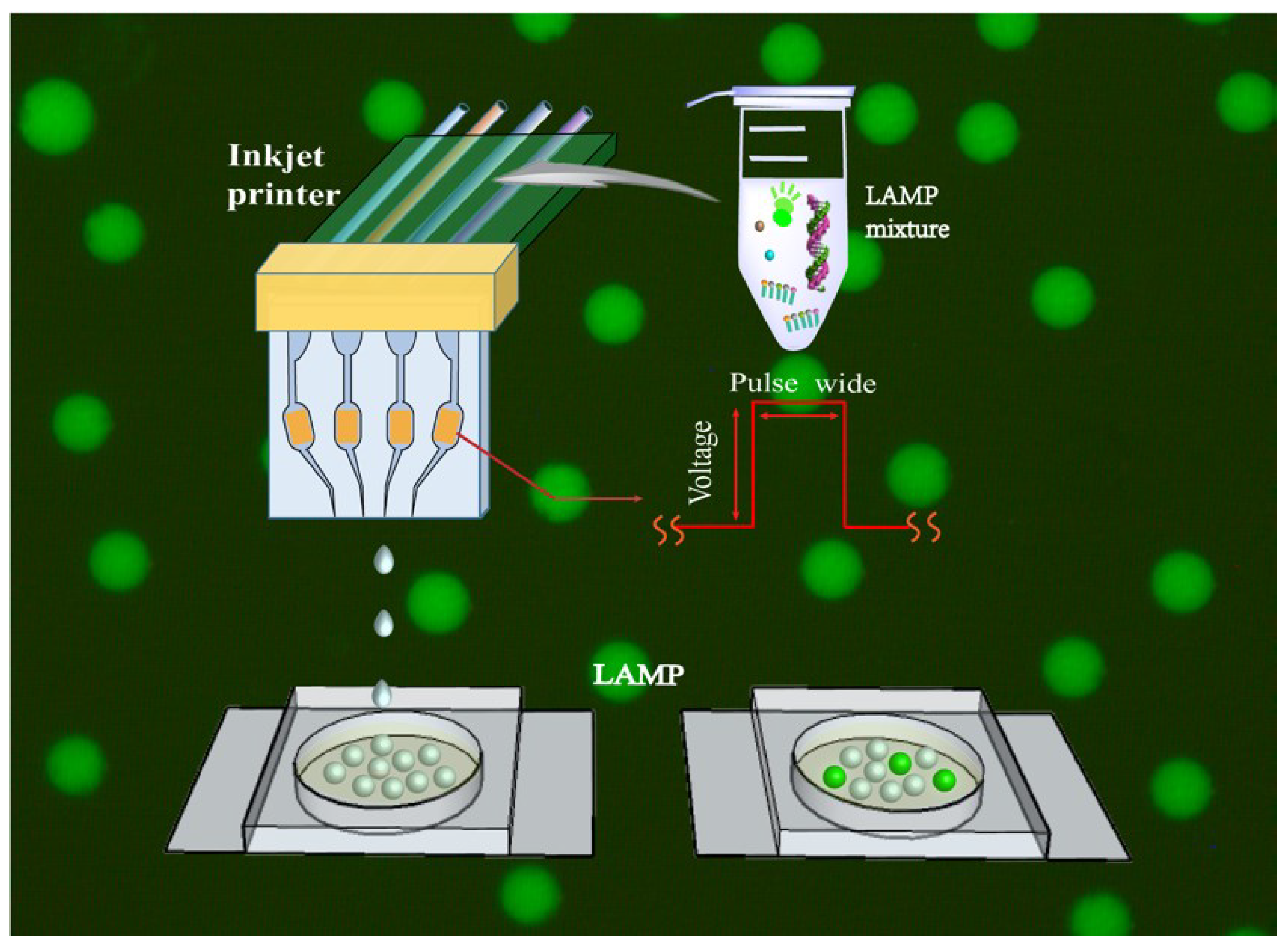
The operating workflow of the inkjet printer for the droplet digital LAMP assay is schematically illustrated in Figure 1. The extracted HPV16 DNA and LAMP reaction mixture was loaded into the inkjet printer to print the droplets and finally collected in the microfluidic chip for the LAMP reaction. In each individual droplet, the LAMP reaction was initiated, along with the accumulation of the reaction products. After the LAMP reaction, the fluorescence that amplifies the target DNA accumulated within the droplets was imaged and analyzed. Calcein was used as the fluorescent metal indicator for the LAMP product detection, as its fluorescence was sensitive to changes in the concentration of metal ions in the solution. The fluorescence signal after the LAMP reaction was subject to digital analysis, and the accurate quantification of the target was obtained through Poisson statistical analysis by the ratio of the positive fluorescent LAMP droplets.
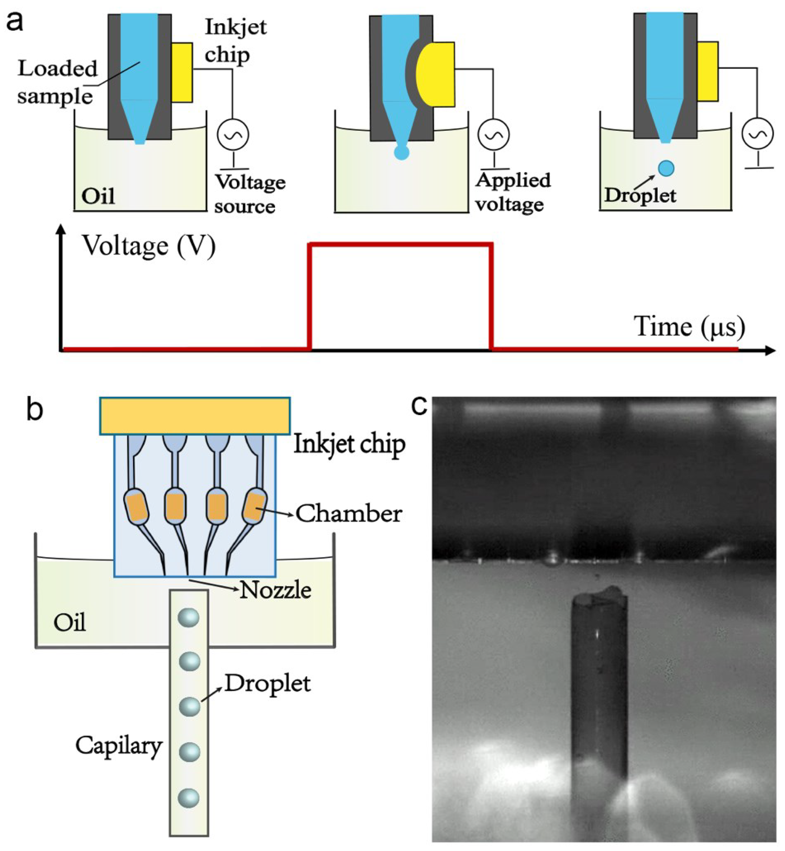
The inkjet printer is a reusable device that can be easily assembled for droplet generation. Briefly, the inkjet chip needs to be immersed in mineral oil. Adjust the position and distance between the nozzle and capillary using a high-speed microscope. By manually adjusting the X-Y stage, the nozzle on the inkjet chip would be moved to the top of the capillary, and then control the piezoelectric ceramic on the inkjet chip through a waveform generator, as shown in Figure 2a. Because the piezoelectric ceramic is tightly attached to the chamber of the inkjet chip, by adjusting the driving voltage and pulse width applied on the piezoelectric ceramic, discrete droplets can be printed. The resulting droplets fell directly and were introduced into the capillary, before finally being collected in the microfluidic chip, as shown in Figure 2b. The printed droplet size mainly depends on the driving voltage and pulse width; thus, the droplet size can be fine-tuned by changing these two parameters, which is different from the traditional microfluidic method.
To improve the dynamic range and accuracy of the digital LAMP analysis, 10,000 droplets were printed using the inkjet printer and collected in a microfluidic chip for subsequent analysis. The printing process of the inkjet printer was observed and recorded by a high-speed microscope, as shown in Figure 2c. Because the evaporation of oil and droplets may occur during the LAMP reaction process, the microfluidic chip was prefilled with some mineral oil; thus, the resulting printed droplets can be submerged in mineral oil.
3.2. The Feasibility of LAMP
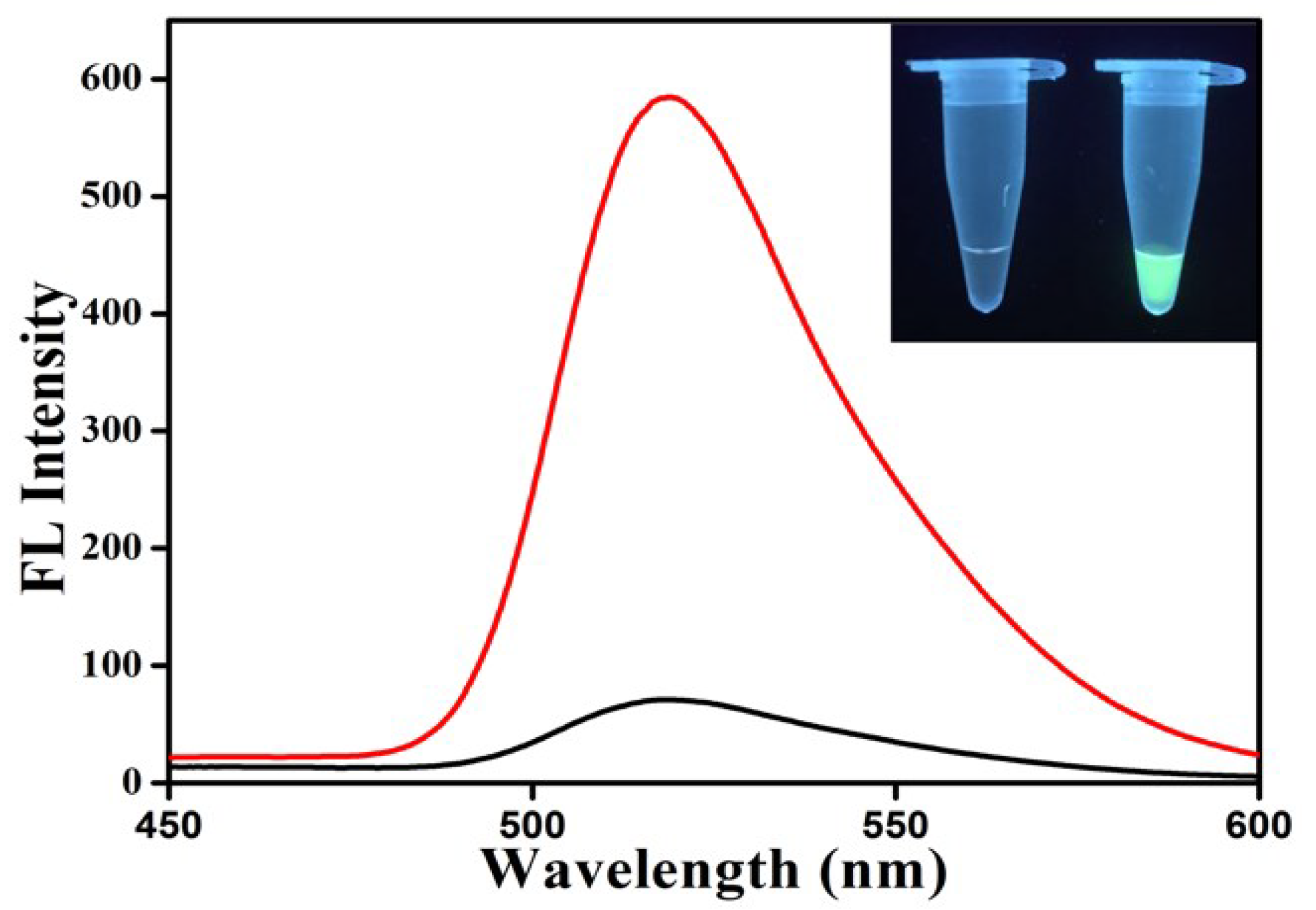
To examine the LAMP feasibility, extracted HPV16 DNA was utilized as the positive sample and nuclease-free H2O as the negative control. The LAMP feasibility assay was carried out in a centrifuge tube to simplify the testing process and facilitate the results identification. The red line and black line represent the fluorescence spectra of the positive sample and the negative control after the LAMP reaction, respectively. For a positive reaction, the color change could be characterized with the aid of an ultraviolet light at 365 nm. As depicted in Figure 3, it can be clearly observed that the fluorescence intensity appeared to be increasing when the extracted DNA was used as the template but not with the negative control (nuclease-free H2O), indicating that the LAMP reaction was successful and feasible for HPV16 detection.
3.3. ddLAMP by Inkjet Printer
Quantification analysis of target nucleic acid is of great importance for device application. In order to examine the capability of the proposed inkjet printer approach for the droplet digital LAMP, serial 10-fold dilutions of HPV16 DNA samples ranging from 10−6 fg/μL to 10−2 fg/μL were prepared for the droplet digital LAMP assay. Five HPV16 DNA samples, including the negative control, were used as input templates for the LAMP reaction. Diluted HPV16 DNA samples (including negative control) were premixed with LAMP reagents and loaded into the inkjet printer through the microchannel. For each sample with a different concentration, the experiment was conducted three times. By applying the driving voltage, discrete microdroplets were printed and subsequently collected in the microfluidic chip.
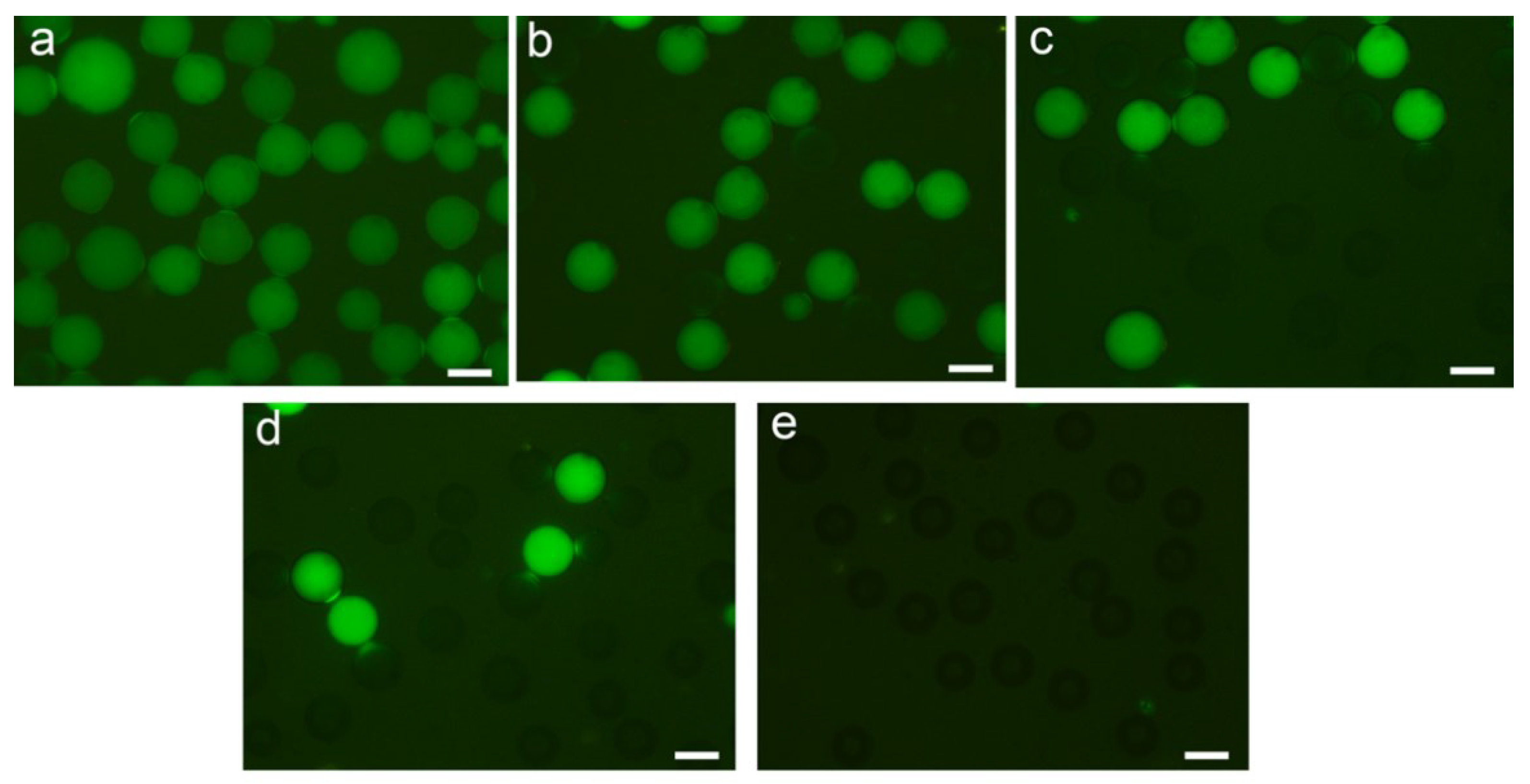
Following the isothermal LAMP reaction, representative fluorescent images were obtained by microscope, as shown in Figure 4. A droplet within the target HPV16 generates fluorescence after amplification, that is, a positive droplet, while droplets without target HPV16 have no fluorescent signal. It can be seen that the fraction of the positive droplets increased proportionally with the concentration of DNA. The increasing fraction of the positive droplets occurs due to a rise in the number of target molecules in the original sample, and consequently, the probability of a droplet containing at least one target molecule also increases, which in turn leads to an increase in the number of fluorescence droplets after the LAMP endpoint reaction.
In principle, the minimum concentration for digital HPV16 analysis is a single molecule. The presence of more than one copy of HPV16 molecule in a droplet may result in a non-linear relationship between the positive signals and the copy number of HPV16 molecules. If the sample is at a lower concentration, the copy number within the droplets can be measured by directly counting the positive fluorescent droplets. This can be calculated by Poisson statistical analysis. If the sample is at a very low concentration, the results of the LAMP reaction may appear to be all negative, this phenomenon was confirmed in our experiment, as shown in Figure 4e. When 99% of the droplets exhibited positive signals, 1% of the droplets therefore exhibited negative signals. It was further analyzed by Poisson statistics, and the expected average DNA copy number per droplet (λ) was calculated. Therefore, the number of DNA templates was about 4.6 copies/droplet. The droplet diameter printed by the inkjet printer is about 60 μm, and the droplet volume follows the equation: V = 4/3 × π × r3. Thus, the maximum theoretical dynamic range of the inkjet printer method for the droplet digital LAMP was from 1 to 4 × 10⁴ copies/μL.
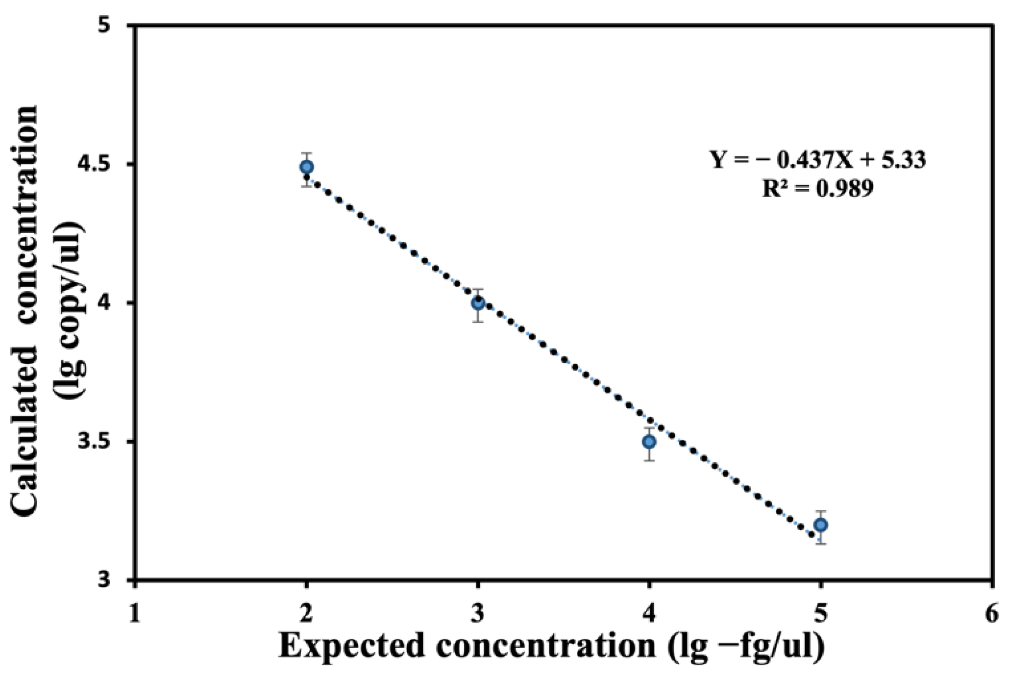
In the present study, the proposed inkjet printer method was employed to measure the concentration of target HPV16. After the Poisson statistical analysis, it was found that the calculated concentration of the HPV16 showed a good linear correlation with the expected concentration (R2 value was 0.989), as shown in Figure 5. A good correlation of the calculated and expected concentration of the HPV16 demonstrated the capability of the inkjet printer for the droplet digital LAMP.
4. Conclusions
Herein, we reported a self-assembled inkjet printer that can reliably perform droplet digital LAMP assays. The performance of the inkjet printer for the droplet digital LAMP was characterized and employed for HPV16 detection, and the experimental calculated concentration has a good correlation with the expected concentration, indicating this method enables digital nucleic acid quantitative analysis. Soft dip-pen nanolithography (DPN) is an atomic force microscopy (AFM) setup optimized to precisely control the deposition of biomolecules [31]. AFM is a powerful tool to address biomolecular morphology [32] and the individual biorecognition [33] based on adhesion force. Nevertheless, a self-assembled inkjet printer can be a cost-effective solution for fast biomolecular detection. In comparison with previous devices, the self-assembled inkjet printer simplifies the operation procedure and reduces instrument complexity which greatly increases the portability of these assays. We envision such a self-assembled inkjet printer as a promising and practical tool that can be extended to on-site analysis, especially in a resource-limited environment.
Author Contributions
Conceptualization, Z.F.; methodology, Z.F.; software, Z.F.; validation, Z.F.; formal analysis Z.F.; investigation, Z.F.; resources, Z.F.; data curation, Z.F. and Y.S.; writing—original draft preparation, Z.F.; writing—review and editing, Y.S.; visualization, Y.S.; supervision, J.-M.L.; project administration, J.-M.L.; funding acquisition, J.-M.L. All authors have read and agreed to the published version of the manuscript.
Funding
This research was funded by the National Natural Science Foundation of China (No. 21727814, 21621003).
Institutional Review Board Statement
Not applicable.
Informed Consent Statement
Not applicable.
Data Availability Statement
Not applicable.
Acknowledgments
The authors wish to thank Tsinghua University for the space and equipment needed for this research.
Conflicts of Interest
The authors declare no conflict of interest. The funders had no role in the design of the study; in the collection, analyses, or interpretation of data; in the writing of the manuscript; or in the decision to publish the results.
References
- Francesko, A.; Cardoso, V.F.; Lanceros-Méndez, S. Lab-On-a-Chip Technology and Microfluidics; Elsevier: Amsterdam, The Nederlands, 2019. [Google Scholar]
- Shang, Y. Molecular mechanisms of oestrogen and SERMs in endometrial carcinogenesis. Nat. Rev. Cancer 2006, 6, 360–368. [Google Scholar] [CrossRef] [PubMed]
- Higuchi, R.; Fockler, C.; Dollinger, G. Kinetic PCR analysis: Real-time monitoring of DNA amplification reactions. Biotechnology 1993, 11, 1026–1030. [Google Scholar] [CrossRef] [PubMed]
- Else, E.A.; Swoyer, R.; Zhang, Y.; Taddeo, F.J.; Bryan, J.T.; Lawson, J.; Van Hyfte, I.; Roberts, C.C. Comparison of real-time multiplex human papillomavirus (HPV) PCR assays with INNO-LiPA HPV genotyping extra assay. J. Clin. Microbiol. 2011, 49, 1907–1912. [Google Scholar] [CrossRef] [PubMed] [Green Version]
- Gravitt, P.E.; Burk, R.D.; Lorincz, A. A comparison between real-time polymerase chain reaction and hybrid capture 2 for human papillomavirus DNA quantitation. Cancer Epidemiol. Prev. Biomark. 2003, 12, 477–484. [Google Scholar]
- Young, L.S.; Bevan, I.S.; Johnson, M.A.; Bolmfiled, P.I.; Woodman, C.B.J. The polymerase chain reaction: A new epidemiological tool for investigating cervical human papillomavirus infection. BMJ 1989, 298, 14–18. [Google Scholar] [CrossRef] [Green Version]
- Notomi, T.; Okayama, H.; Masubuchi, H.; Yonekawa, T.; Watanabe, K.; Amino, N.; Hase, T. Loop-mediated isothermal amplification of DNA. Nucleic Acids Res. 2000, 28, e63. [Google Scholar] [CrossRef] [Green Version]
- Notomi, T.; Mori, Y.; Tomita, N. Loop-mediated isothermal amplification (LAMP): Principle, features, and future prospects. J. Microbol. 2015, 53, 1–5. [Google Scholar] [CrossRef]
- Tomita, N.; Mori, Y.; Kanda, H.; Notomi, T. Loop-mediated isothermal amplification (LAMP) of gene sequences and simple visual detection of products. Nat. Protoc. 2008, 3, 877–882. [Google Scholar] [CrossRef]
- Asiello, P.J.; Baeumner, A.J. Miniaturized isothermal nucleic acid amplification, a review. Lab Chip 2011, 11, 1420–1430. [Google Scholar] [CrossRef]
- Oliveira, B.; Veigas, B.; Fernandes, A.R.; Guas, H.; Baptista, P.V. Fast prototyping microfluidics: Integrating droplet digital lamp for absolute quantification of cancer biomarkers. Sensors 2020, 20, 1624. [Google Scholar] [CrossRef] [Green Version]
- Sanders, R.; Huggett, J.F.; Bushell, C.A.; Cowen, S.; Scott, D.J.; Foy, C.A. Evaluation of digital PCR for absolute DNA quantification. Anal. Chem. 2011, 83, 6474–6484. [Google Scholar] [CrossRef] [PubMed]
- Peng, H.; Zhu, M.; Gao, Z.; Liao, C.; Jia, C.; Wang, H.; Zhou, H.; Zhao, J. A centrifugal microfluidic emulsifier integrated with oil storage structures for robust digital LAMP. Biomed. Microdevices 2020, 22, 18. [Google Scholar] [CrossRef] [PubMed]
- Wang, P.; Jing, F.; Li, G.; Wu, Z.; Cheng, Z.; Zhang, J.; Zhang, H.; Jia, C.; Jin, Q.; Mao, H.; et al. Absolute quantification of lung cancer related microRNA by droplet digital PCR. Biosens. Bioelectron. 2015, 74, 836–842. [Google Scholar] [CrossRef] [PubMed]
- Rane, T.D.; Chen, L.; Zec, H.C.; Wang, T.H. Microfluidic continuous flow digital loop-mediated isothermal amplification (LAMP). Lab Chip 2015, 15, 776–782. [Google Scholar] [CrossRef] [PubMed] [Green Version]
- Schulz, M.; Ruediger, J.; Landmann, E.; Bakheit, M.; Frischmann, S.; Rassler, D.; Homann, A.R.; Stetten, F.; Zengerle, R.; Paust, N. High dynamic range digital assay enabled by dual-volume centrifugal step emulsification. Anal. Chem. 2021, 93, 2854–2860. [Google Scholar] [CrossRef] [PubMed]
- Ma, Y.D.; Chang, W.H.; Luo, K.; Wang, C.H.; Liu, S.Y.; Yen, W.H.; Lee, G.B. Digital quantification of DNA via isothermal amplification on a self-driven microfluidic chip featuring hydrophilic film-coated polydimethylsiloxane. Biosens. Bioelectron. 2018, 99, 547–554. [Google Scholar] [CrossRef]
- Yu, Z.; Lyu, W.; Yu, M.; Wang, Q.; Qu, H.; Ismagilov, R.F.; Han, X.; Lai, D.; Shen, F. Self-partitioning SlipChip for slip-induced droplet formation and human papillomavirus viral load quantification with digital LAMP. Biosens. Bioelectron. 2020, 155, 112107. [Google Scholar] [CrossRef] [Green Version]
- Xia, Y.; Yan, S.; Zhang, X.; Ma, P.; Du, W.; Feng, X.; Liu, B.F. Monte Carlo modeling-based digital loop-mediated isothermal amplification on a spiral chip for absolute quantification of nucleic acids. Anal. Chem. 2017, 89, 3716–3723. [Google Scholar] [CrossRef]
- Yuan, H.; Chao, Y.; Shum, H.C. Droplet and microchamber-based digital loop-mediated isothermal amplification (dLAMP). Small 2020, 16, e1904469. [Google Scholar] [CrossRef]
- Kuznetsova, I.; Smirnov, A.; Anisimkin, V.; Gubin, S.; Kolesov, V. Inkjet printing of plate acoustic wave devices. Sensors 2020, 20, 3349. [Google Scholar] [CrossRef]
- Ahn, J.-H.; Hong, H.-J.; Lee, C.-Y. Temperature-sensing inks using electrohydrodynamic inkjet printing technology. Materials 2021, 14, 5623. [Google Scholar] [CrossRef] [PubMed]
- Koch, L.; Deiwick, A.; Chichkov, B. Capillary-like formations of endothelial cells in defined patterns generated by laser bioprinting. Micromachines 2021, 12, 1538. [Google Scholar] [CrossRef] [PubMed]
- Hayashida, K.; Nambala, P.; Reet, N.V.; Büscher, P.; Kawai, N.; Mutengo, M.M.; Musaya, J.; Namangala, B.; Sugimoto, C.; Yamagishi, J. Development of a bio-inkjet printed LAMP test kit for detecting human African trypanosomiasis. PLoS Negl. Trop. Dis. 2020, 14, e0008753. [Google Scholar] [CrossRef] [PubMed]
- Trantidou, T.; Elani, Y.; Parsons, E.; Ces, O. Hydrophilic surface modification of PDMS for droplet microfluidics using a simple, quick, and robust method via PVA deposition. Microsyst. Nanoeng. 2017, 3, 16091. [Google Scholar] [CrossRef] [PubMed]
- Fan, Z.; Feng, X.; Zhang, W.; Li, N.; Zhang, X.; Lin, J.-M. Visual detection of high-risk HPV16 and HPV18 based on loop-mediated isothermal amplification. Talanta 2020, 217, 121015. [Google Scholar] [CrossRef]
- Fan, Z.; Zhou, Z.; Zhang, W.; Lin, J.-M. Inkjet printing based ultra-small MnO2 nanosheets synthesis for glutathione sensing. Talanta 2021, 225, 121989. [Google Scholar] [CrossRef]
- Oktavianty, O.; Haruyama, S.; Ishii, Y. Enhancing droplet quality of edible ink in single and multi-drop methods by optimization the waveform design of DoD inkjet printer. Processes 2022, 10, 91. [Google Scholar] [CrossRef]
- Lau, G.-K.; Milan, S. Ink-Jet printing of micro-electro-mechanical systems (MEMS). Micromachines 2017, 8, 194. [Google Scholar] [CrossRef] [Green Version]
- Gutiérrez, E.; De, J.; Barreto, J.D.J.; Garcia-Hernandez, S.; González-Solorzano, M.G. Decrease of nozzle clogging through fluid flow control. Metals 2020, 10, 1420. [Google Scholar] [CrossRef]
- Rani, E.; Mohshim, S.A.; Ahmad, M.Z.; Goodacre, R.; Ahmad, S.A.A.; Lu, S.W. Polymer pen lithography-fabricated DNA arrays for highly sensitive and selective detection of unamplified ganoderma boninense DNA. Polymers 2019, 11, 561. [Google Scholar] [CrossRef] [Green Version]
- Marcuello, C.; Frempong, G.A.; Balsera, M.; Medina, M.; Lostao, A. Atomic force microscopy to elicit conformational transitions of ferredoxin-dependent flavin thioredoxin reductases. Antioxidants 2021, 10, 1437. [Google Scholar] [CrossRef] [PubMed]
- Marcuello, C.; Miguel, R.D.; Lostao, A. Molecular recognition of proteins through quantitative force maps at single molecule level. Biomolecules 2022, 12, 594. [Google Scholar] [CrossRef] [PubMed]
Figure 1.
Schematic illustration of the self-assembled inkjet printer for droplet digital loop-mediated isothermal amplification. (Not real scale).
Figure 1.
Schematic illustration of the self-assembled inkjet printer for droplet digital loop-mediated isothermal amplification. (Not real scale).

Figure 2.
Droplet generation using inkjet printer. (a) Schematic illustration of a droplet generation cycle by piezoelectric inkjet printer. (b) Schematic illustration of droplets generation by inkjet printer. (c) Microscopic image of droplet injection by inkjet printer.
Figure 2.
Droplet generation using inkjet printer. (a) Schematic illustration of a droplet generation cycle by piezoelectric inkjet printer. (b) Schematic illustration of droplets generation by inkjet printer. (c) Microscopic image of droplet injection by inkjet printer.

Figure 3.
The fluorescence spectra of extracted HPV16 DNA and nuclease-free H2O after LAMP reaction. Red line: fluorescence spectra of extracted HPV16 DNA after LAMP reaction. Black line: fluorescence spectra of nuclease-free H2O after LAMP reaction.
Figure 3.
The fluorescence spectra of extracted HPV16 DNA and nuclease-free H2O after LAMP reaction. Red line: fluorescence spectra of extracted HPV16 DNA after LAMP reaction. Black line: fluorescence spectra of nuclease-free H2O after LAMP reaction.

Figure 4.
Droplet digital LAMP results by the proposed inkjet printer with different concentrations of HPV16 DNA. The scale bar is 50 μm. (Representative image sections cropped). (a) Representative fluorescent image of LAMP reaction results with 10−2 fg/μL HPV16 DNA. (b) Representative fluorescent image of LAMP reaction results with 10−3 fg/μL HPV16 DNA. (c) Representative fluorescent image of LAMP reaction results with 10−4 fg/μL HPV16 DNA. (d) Representative fluorescent image of LAMP reaction results with 10−5 fg/μL HPV16 DNA. (e) A representative nontemplate control (NTC) image after LAMP reaction.

Figure 5.
The correlation between the calculated concentration and the expected concentration of HPV16.
Figure 5.
The correlation between the calculated concentration and the expected concentration of HPV16.

Publisher’s Note: MDPI stays neutral with regard to jurisdictional claims in published maps and institutional affiliations. |
© 2022 by the authors. Licensee MDPI, Basel, Switzerland. This article is an open access article distributed under the terms and conditions of the Creative Commons Attribution (CC BY) license (https://creativecommons.org/licenses/by/4.0/).
Share and Cite
MDPI and ACS Style
Fan, Z.; Sun, Y.; Lin, J.-M. Self-Assembled Inkjet Printer for Droplet Digital Loop-Mediated Isothermal Amplification. Chemosensors 2022, 10, 247. https://0-doi-org.brum.beds.ac.uk/10.3390/chemosensors10070247
AMA Style
Fan Z, Sun Y, Lin J-M. Self-Assembled Inkjet Printer for Droplet Digital Loop-Mediated Isothermal Amplification. Chemosensors. 2022; 10(7):247. https://0-doi-org.brum.beds.ac.uk/10.3390/chemosensors10070247
Chicago/Turabian StyleFan, Zhaoxuan, Yucheng Sun, and Jin-Ming Lin. 2022. "Self-Assembled Inkjet Printer for Droplet Digital Loop-Mediated Isothermal Amplification" Chemosensors 10, no. 7: 247. https://0-doi-org.brum.beds.ac.uk/10.3390/chemosensors10070247
Note that from the first issue of 2016, this journal uses article numbers instead of page numbers. See further details here.