Article

Handheld Enzymatic Luminescent Biosensor for Rapid Detection of Heavy Metals in Water Samples

by
Kirill A. Lukyanenko
1,2,†,
Ivan A. Denisov
1,2,*,†,
Vladimir V. Sorokin
2,†,
Anton S. Yakimov
3,
Elena N. Esimbekova
2,4 and
Peter I. Belobrov
2
1
Laboratory for Digital Controlled Drugs and Theranostics, Federal Research Center “Krasnoyarsk Science Center SB RAS”, Krasnoyarsk 660036, Russia
2
Laboratory of Bioluminescent Biotechnologies, Siberian Federal University, Krasnoyarsk 660041, Russia
3
Research Institute of Molecular Medicine and Pathobiochemistry, Krasnoyarsk State Medical University named after Prof. V.F. Voino-Yasenetsky, Krasnoyarsk 660022, Russia
4
Laboratory of Photobiology, Institute of Biophysics SB RAS, Krasnoyarsk 660036, Russia
*
Author to whom correspondence should be addressed.
These authors contributed equally to this work.
Submission received: 15 February 2019 / Revised: 18 March 2019 / Accepted: 19 March 2019 / Published: 26 March 2019
(This article belongs to the Special Issue Chemical Sensors for Heavy Metals/Toxin Detection)

Abstract

:
Enzymatic luminescent systems are a promising tool for rapid detection of heavy metals ions for water quality assessment. Nevertheless, their widespread use is limited by the lack of test procedure automation and available sensitive handheld luminometers. Herein we describe integration of disposable microfluidic chips for bioluminescent enzyme-inhibition based assay with a handheld luminometer, which detection system is based on a thermally stabilized silicon photomultiplier (SiPM). Microfluidic chips were made of poly(methyl methacrylate) by micro-milling method and sealed using a solvent bonding technique. The composition of the bioluminescent system in microfluidic chip was optimized to achieve higher luminescence intensity and storage time. Results indicate that developed device provided comparable sensitivity with bench-scale PMT-based commercial luminometers. Limit of detection for copper (II) sulfate reached 2.5 mg/L for developed biosensor. Hereby we proved the concept of handheld enzymatic optical biosensors with disposable chips for bioassay. The proposed biosensor can be used as an early warning field-deployable system for rapid detection of heavy metals salts and other toxic chemicals, which affect bioluminescent signal of enzymatic reaction.

1. Introduction

Significant problem affecting human health is the global pollution of water sources by various types of organic and inorganic toxic substances [1]. Analytical methods and tools that allow rapid and inexpensive monitoring of the environment are required. Traditional analytical methods such as mass spectrometry and chromatography are highly expensive and therefore their application on an ongoing basis or in the field is limited. In this context, biosensors appear as suitable alternatives or complementary analytical tools [2].
A biosensor can be defined as an independently integrated receptor transducer device, which is capable of providing selective quantitative or semi-quantitative analytical information using a biological recognition element [3]. Molecular biosensors use enzymes, immunosystems, tissues, organelles or whole cells as recognition elements to detect chemical compounds usually by electrical, thermal or optical signals [4]. Biosensors are more favorable, reliable, accurate, cost effective, and easy to use compared to other conventional lab-based detection techniques due to their portability, reusability, real-time response, high specificity and selectivity [5]. It is worth noting that the combination of all these qualities in one device is still one of the challenges in the development of field deployable biosensors [6]. Despite these difficulties, for more than 40 years of their history, biosensors have found wide commercial application in a variety of fields, such as medical tests, food quality analyses, biothreat, environmental protection, etc. [7].
When using biological objects as recognition elements, it is always worth considering the fact that in nature there are no two absolutely identical organisms or cells. Such biodiversity introduces a certain error in the measurement. The use of individual molecules, such as proteins, avoids this problem and comes as close as possible to the accuracy and unambiguity of chemical measurements [8]. A necessary condition for this is the presence of such a detector that does not affect the measuring element of the biosensor when measured.
Among various types of biosensors, the optical ones tend to be the most promising, especially in the field of environmental pollution control. This is due to their high sensitivity, no need for extensive sample preparation, multitarget sensing, the possibility of compact design, and in most cases label-free detection, except for quantum dots and fluorescence-based methods [9,10]. Bioluminescence-based biorecognition elements have great potential for producing costs effective and compact optical biosensors because of their low cost [11,12]. A bacterial coupled enzyme system NAD(P)H:FMN-oxidoreductase and luciferase (Red+Luc) can be used as such kind of biorecognition element for development of rapid assays of water quality [13,14]. Luciferase catalyzes the oxidation of long-chain aliphatic aldehydes involving reduced flavin mononucleotide; one of the products of this reaction is a quantum of light in the blue-green spectrum. To provide luciferase with reduced flavin mononucleotide, the luciferase reaction is coupled with the reaction catalyzed by NAD(P)H:FMN-oxidoreductase [15,16,17].
The interaction of toxicants with the coupled enzyme system Red+Luc leads to the change of such bioluminescence parameters as the luminescence intensity, delay in the output of the reaction to the maximum luminescence emission and alteration of chemical reaction constants. This approach does not provide information on the chemical composition of the harmful compounds in the sample. However, it makes it possible to assess the effect of these unknown compounds on the biological system and provides a warning signal that chemical analysis is required to identify the substance.
One of the trends in the development of optical biosensors in the field of environmental pollution control and early warning systems is integration of microelectronics and microfluidics into optical biosensors for miniaturization of optical biorecognition elements [18]. This leads to the development of disposable chips [19,20], which are more suitable for use with portable biosensors and protect samples from cross-contamination. Recently we have introduced disposable luciferase-based microfluidic chips to perform enzymatic assay [21,22,23]. These chips can be used for rapid assay of water pollution. However, there was a lack of portable luminometers suitable for the application with microfluidic chips.
The development of portable luminometers imposes restrictions on their size, including the size of the optical detection system. Silicon photomultipliers (SiPM) seems to be promising photodetectors for portable biosensors, because of the several advantages [24,25]: low operating voltage (smaller than 50 V), insensitivity to magnetic fields, compact dimensions, and immunity to damage from light overexposure, high gain (106). They are cheaper than compact photomultiplier tubes and relatively new to the market. SiPM potential is being currently studied for biomedical application [26], compact imaging systems [27,28], etc. by a lot of research groups in the world [29,30]. Sensitivity of the SiPM detector depends on the ambient temperature, which can be overcome by using a photodetector cooling system [31].
Here we introduce the integration of luciferase-based microfluidic chips with a portable luminometer in order to develop handheld enzymatic luminescent biosensor for rapid detection of heavy metals in water samples. To develop a biosensor, it was necessary (i) to design a portable luminometer; (ii) to optimize the composition of bioluminescent system in microfluidic chip.

2. Materials and Methods

2.1. Reagents

The following reagents were used: flavin mononucleotide (FMN) (CHEBI: 17621, SERVA Electrophoresis GmbH, Heidelberg, Germany), reduced nicotinamide adenine dinucleotide (NADH) (CHEBI: 16908, GERBU Biotechnik GmbH, Heidelberg, Germany), ethanol (CHEBI:16236, Merck, Darmstadt, Germany), tetradecanal (CHEBI:84067, Merck, Darmstadt, Germany), starch from potato (CHEBI:28017, Sigma-Aldrich, St. Louis, MO, USA), gelatin from porcine skin (CHEBI:5291, Sigma-Aldrich, St. Louis, MO, USA), potassium phosphate buffer with pH 7.0 (CHEBI:63036, Fluka Chemie AG, Buchs, Switzerland), PMMA (CHEBI:61369, SafPlast Innovative, Kazan, Russia), 1,2-dichloroethane (Soyuzhimprom, Novosibirsk, Russia) and acetone (Vekton, St. Petersburg, Russia). Lyophilized preparations of purified enzymes were produced at the Laboratory of Nanobiotechnology and Bioluminescence of the Institute of Biophysics SB RAS (Krasnoyarsk, Russia). One vial of preparation contained 0.5 mg of luciferase EC 1.14.14.3 (Photobacterium leiognathi) from recombinant strain of Escherichia coli and 0.18 activity units of NAD(P)H:FMN-oxidoreductase EC 1.5.1.29 (Vibrio fischeri).

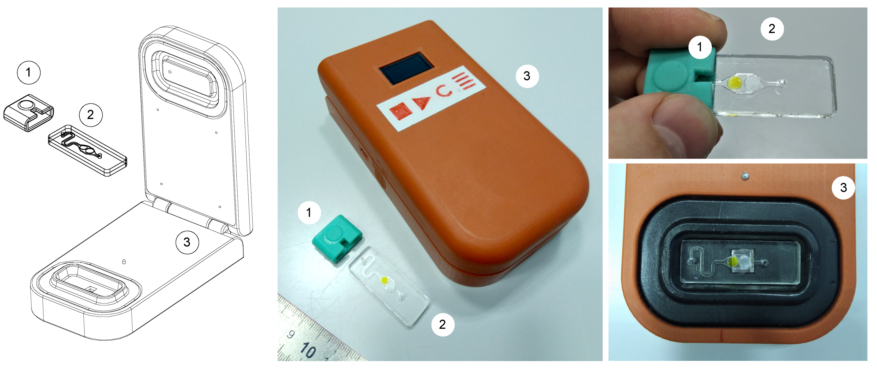
2.2. Biosensor Fabrication

The housing of the portable luminometer was designed using a CAD software KOMPAS-3D (Ascon, St. Petersburg, Russia). Then the housing was manufactured by a micro-milling method with the CNC milling machine Modela MDX-40A (Roland, Hamamatsu, Japan) and model plastic Necuron 1300 (NECUMER GmbH, Bohmte, Germany). The sample compartment was manufactured by pressing heated polyvinyl chloride (PVC) into the mould, which was made by the same technique as the housing of the device. Disposable microfluidic chips were manufactured in accordance with the technique described in detail earlier [21,22]. In brief, the body of the chip consisted of two PMMA (polymethylmethacrylate) substrates. One of the substrates contained a channelized surface made by micro-milling [32]. The enzymes and substrates of bioluminescent system Red+Luc were immobilized in the reaction chamber of the chip in the form of dried droplets of starch gel. The chips were sealed at room temperature using solvent bonding technique.
Vinylpolysiloxane A-silicone (Zhermack, Badia Polesine, Italy) was used as a material for sampler fabrication. A mould for casting was made by micro-milling.

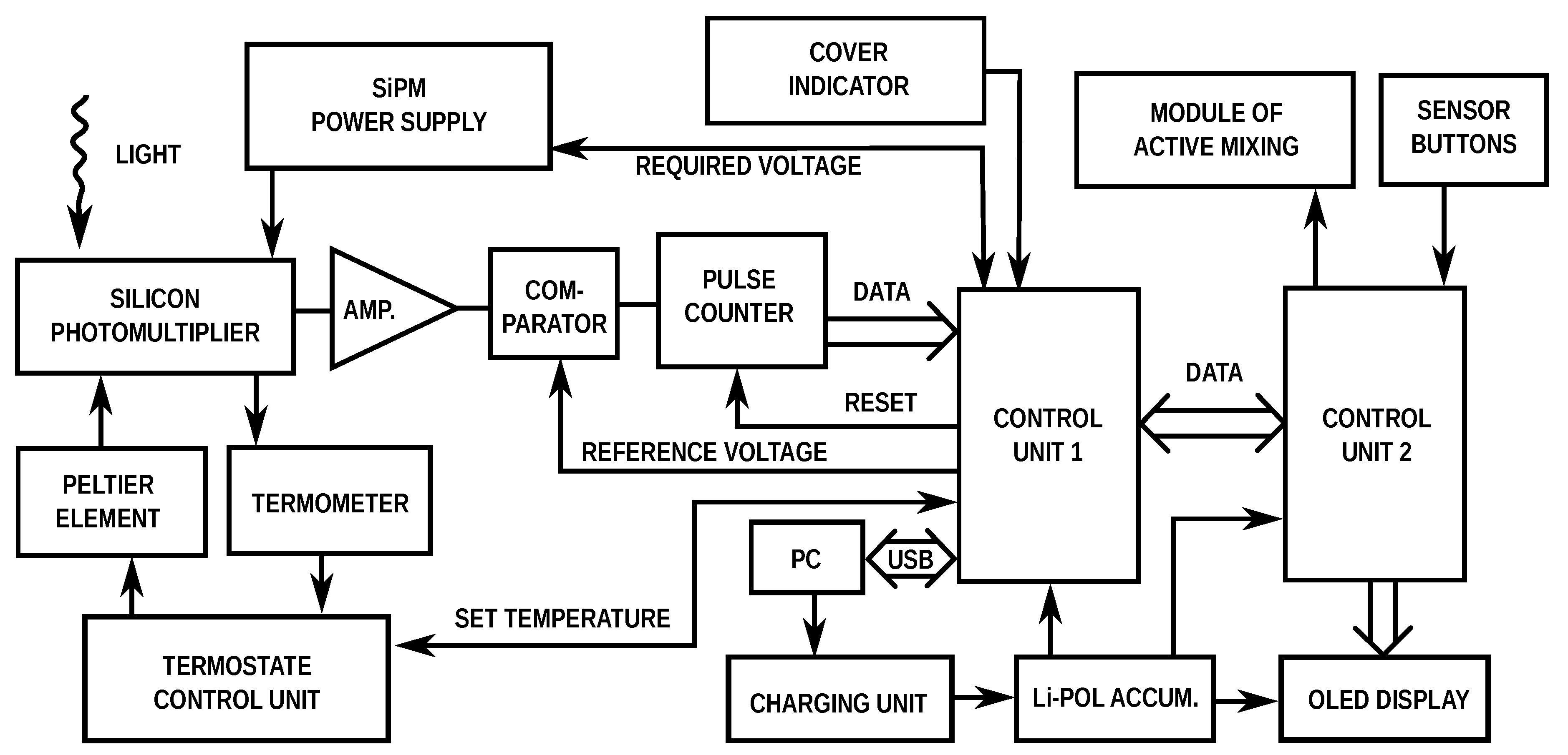
2.3. Signal Detection

The SiPM consisted of a PM6650-EB (KETEK GmbH, München, Germany) containing an array of 14,272 avalanche photodiodes with individual cell dimensions of 50 × 50 μ m2 providing an active area of 6 × 6 mm2. The signal from the SiPM was amplified by operational amplifier AD8062 (Analog Devices, Norwood, MA, USA), and then counted by a dual 4-bit binary counter MM74HC393M (Fairchild Semiconductor, Sunnyvale, CA, USA) and microcontroller LPC2103 (NXP Semiconductors, Eindhoven, Netherlands) as peaks counts per second expressed as relative luminescence units (RLU). This information was shown on the biosensor display by microcontroller STM32F103C8B6 (STMicroelectronics, Ginebra, Switzerland) and the data were also transmitted to a personal computer (PC) through a USB interface. The programs for microcontrollers were developed with Oberon programming language [33] using Astrobe (CFB Software, Burnside, Australia) and O7 (Alexander Shiryaev, Yegoryevsk, Russia) compilers. The program for PC was developed with open-source integrated development environment BlackBox Component Builder (Oberon microsystems AG, Zürich, Switzerland).
The intensity of bioluminescent signal from microfluidic chip was measured by the developed device and GloMax 20/20 luminometer (Promega, Fitchburg, WI, USA) in the mode for kinetics measurement.

2.4. Testing Procedure

The testing procedure was as follows. At the first stage the chip was filled with liquid sample using a sampler adapter for microfluidic chips. At the 30th second after the sample introduction mixing with sampler adapter started. Ten pushes on the sampler membrane resulted in uniform distribution of the bioluminescent system reagents in the reaction chamber of the chip. Then the chip was placed in the luminometer, where the luminescence intensity detection was performed. At the final stage the waste chip was removed from the luminometer. Then the procedure was repeated with a new chip.
The activity of the immobilized coupled enzyme system Red+Luc was measured in the following way. At the beginning, we registered the control luminescence intensity of the enzyme system ( I c ). For I c registration, the chip was filled with distilled water sample. The chip was placed in the luminometer. Then the luminescence intensity of the analyzed sample was measured ( I e x p ). For I e x p registration, the chip was filled with analyzed liquid sample.
For each measurement, at least 12 repetitions were carried out. Statistical analysis was performed using t-distribution at a 95% confidence level.

3. Results and Discussion

3.1. Design of Portable Luminometer

The biosensor was composed of three main parts: a compact and portable luminometer, a disposable microfluidic chip and a sampler adapter (Figure 1). The microfluidic chip had the dimensions 14.4 × 40.5 mm, which was less than a traditional glass microscope slide, and contained components of the coupled enzyme system Red+Luc co-immobilized into starch gel [23].
The compact luminometer measured 80 × 140 × 41 mm3 and weighed less than 300 g. It consisted of a sample compartment, a silicon photomultiplier with a thermal stabilization system, a battery and a microcontroller electronics (Figure 2). All the elements were combined in a hard plastic housing that shielded the internal components from light and dust. The principal design was inspired by the work of Wojciechowski [34]. Because of the nature of the readout method, shielding from ambient light was extremely important. The sample compartment was made from opaque PVC. The size of the sample compartment measured 41 × 15 × 5 mm3, thus making it possible to use not only special microfluidic chips but also other planar samples (test strips, slices, etc.).
The designed software proved to be versatile and user-friendly, plotting in real-time during measurement and choosing the signal integration time. The information about the current measurement was duplicated on the biosensor display. All the measurements could be started and stopped using the buttons built into the housing of the biosensor.
The portable luminometer had the possibility of autonomous operation due to the built-in rechargeable battery. It was equipped with a USB-mini standard interface for battery recharging and communication with a laptop, personal computer or tablet.
The development of biosensors within the point-of-care concept assumes that potential users should not possess skills to work with laboratory equipment, such as pipettes. Taking this into consideration, a special sampler adapter for microfluidic chips was developed (Figure 1). This adapter may be used by an unprepared user. The principle of operation of the sampler adapter was as follows. When the chip was placed in the sampler, the chip output channel was located under the clamping ring. The negative pressure in the channel of the chip was created after pressing and releasing the upper part of the sampler. Due to this, the sample was sucked into the microfluidic chip. The sample volume was approximately 45 μ L.
Previously, we demonstrated that it was necessary to use an active mixing method for the disposable microfluidic chips, since the passive one did not ensure even distribution of the reagents in the reaction chamber [22]. This uniform distribution was important for the bioluminescent system, since it provided the maximum intensity of luminescence. Therefore, the sampler was used as active mixer. After sampling, periodic clicking on the top of the sampler resulted in active mixing in the chip. It was found that the optimal number of clicks was 10 times. This number provided the most uniform mixing of reagents in reaction chamber, which resulted in higher luminescence intensity level and reproducibility.

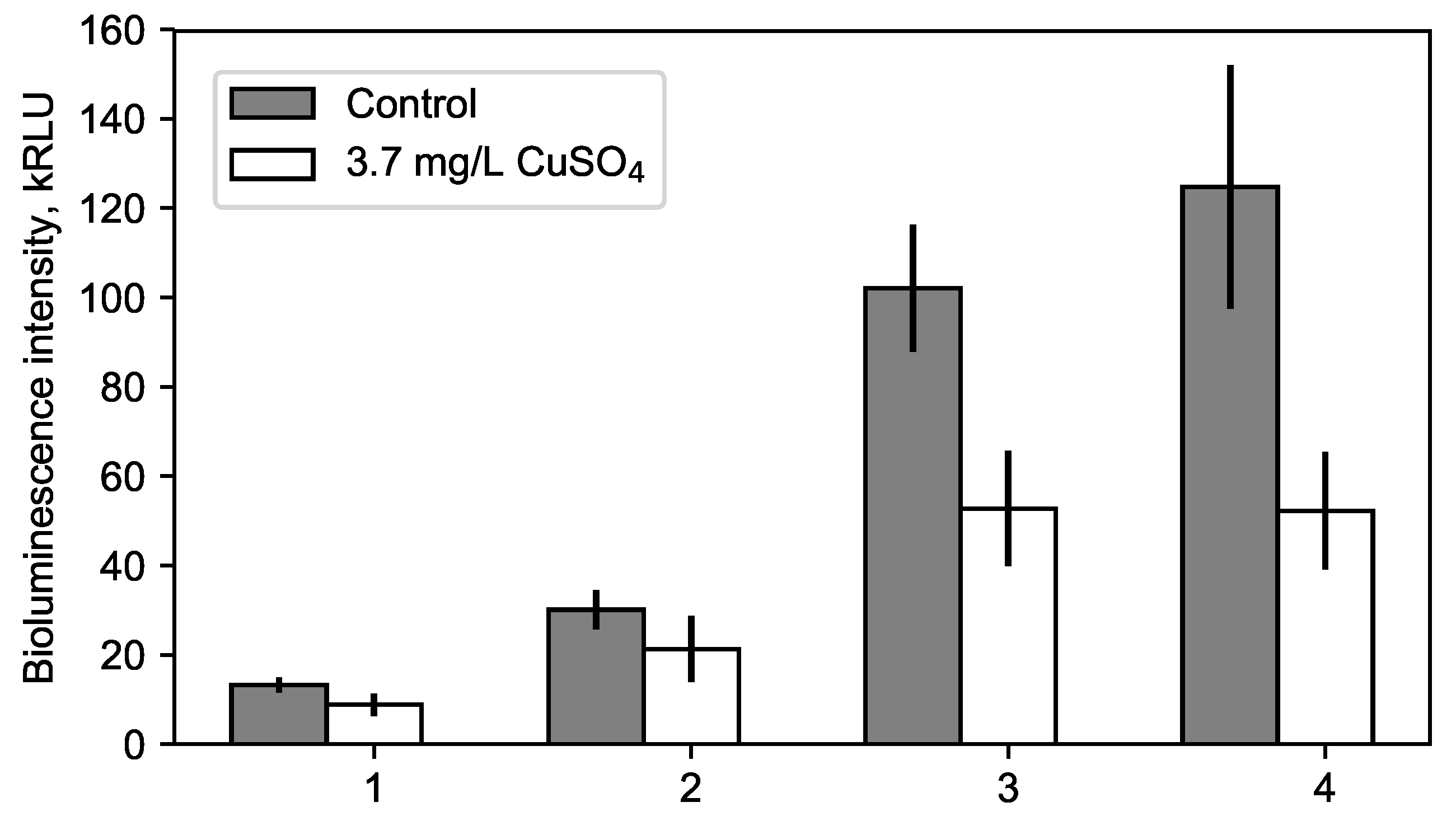
3.2. Optimization of Microfluidic Chip Composition and Storage Conditions

To adapt microfluidic chips for work in the portable luminometer and to improve the storage time of the reagents we optimized concentrations of enzymes and their substrates. It was necessary to achieve luminescence intensity which was high enough to be determined by the SiPM detector.
Four different compositions were used to make microfluidic chips. Microfluidic chips under standard composition (No.1, see Table 1) contained 0.14 μ g of luciferase and 52.3 × 10⁻⁶ activity units of NAD(P)H:FMN-oxidoreductase, 0.00044% of aldehyde, 0.11 mM of NADH. Composition No.2 contained a doubled concentration of aldehyde, composition No.3 in addition to this contained 35% increased concentration of enzymes and composition No.4 in addition to this contained doubled concentration of NADH. Chips of all compositions contained 4 mM of FMN. The chips were tested with the standard laboratory GloMax 20/20 luminometer. The results are shown in Figure 3.
It was demonstrated that the rise of aldehyde concentration led to the increase in the level of luminescence, while the sensitivity to toxin remained the same. The increase in the number of enzymes by 35% led to a sharp increase in the intensity of luminescence by the factor of 5 and a slight loss of sensitivity to the toxic substance. The increase in the concentration of NADH did not affect the intensity of luminescence, but at the same time, increased the sensitivity of the system to the toxic substance. Thus, an excess concentration of aldehyde and NADH in the enzyme system is needed to increase luminescence intensity and sensitivity to toxic compounds. Compositions No.3 and 4 provided sufficient luminescence intensity to be used with portable luminometer.
Changes in the composition of the bioluminescent system did not have a significant effect on the storage time of the chips. With an increased amount of the enzymes, loss of sensitivity to toxin was observed with a storage time of more than one month. A decrease in the luminescence intensity and sensitivity was observed with increasing storage time. Significant reduction of luminescence intensity and sensitivity was observed after 4 months storage at +4 °C. The system completely lost luminescence intensity and sensitivity to toxic substances after 3 months storage at 25 °C. When stored at −18 °C and below the bioluminescent system retained its activity and sensitivity to toxins for 6 months without significant reduction, which was a good result for an immobilized enzyme system [35]. Conventional cell-based biosensors retain their activity only for 1 week [36]. Despite the availability of technologies that allow the bacterial activity to last for several months [37,38,39], their use requires the availability of certain equipment and specially trained personnel.

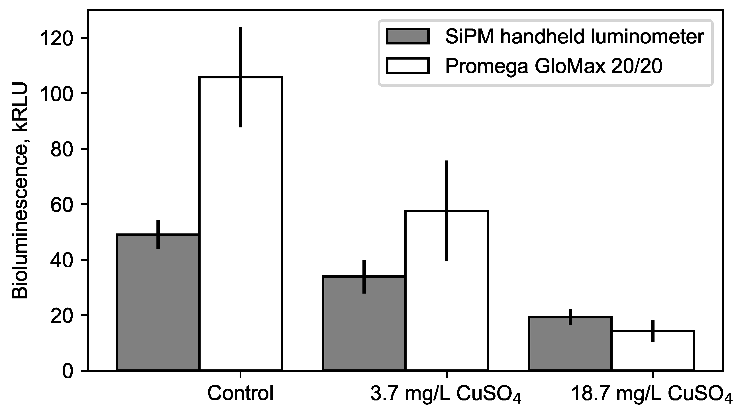
3.3. Sensitivity of the Portable Luminometer

The sensitivity of the SiPM depended on its temperature. To ensure the optimal sensitivity, the system of thermal stabilization was developed. This system was based on a Peltier element, which was controlled by a microcontroller. This system provided thermostabilization of the photodetector at the level of 21 °C. In addition to this, a focusing prism was placed on the detector to improve the light harvest.
To compare the analytical performance of the developed portable SiPM-based luminometer and a bench-scale commercial luminometer GloMax 20/20 we fulfilled a detection of copper (II) sulfate in water by both instruments (Figure 4).
A control sample (distilled water) and a copper(II) sulphate were measured. After sampling and activation of the bioluminescence reaction, each chip was first placed in GloMax 20/20, where the intensity of the luminescence reached the maximum value. After that, the chip was immediately removed from GloMax 20/20 and placed in a sample compartment of SiPM-base handheld luminometer, where the measurement continued. Such actions were permissible, because upon reaching the maximum intensity of the bioluminescence the signal remained at that level for 10–20 s.
The results of measurements showed that both luminometers provided comparable level of sensitivity. As can be seen from the Figure 4 there is no significant difference between suggested luminometer and GloMax for the 18.7 mg/L. With less concentration and more light intensity this difference can be distinguished. The 2 times lower resolution is related to the size of the surface area of the photodetectors (36 mm2 for SiPM, and approx. 490 mm2 for PMT) and different optical system for light condensation. The optical system of the suggested device can be improved by using of anti-reflection coating for glass prism and better back mirror quality. Registered limit of detection for copper(II) sulphate using proposed portable luminometer was 2.5 mg/L.

4. Conclusions

This paper has described the first proofing of concept of handheld biosensor that was designed to detect harmful environmental pollutants in liquid samples. The handheld biosensor consisted of a portable SiPM-based luminometer, disposable microfluidic chips with immobilized bioluminescent enzyme system and a sampler adapter for chips.
Portability and possibility of autonomous operation allow the proposed biosensor to be applied as an early warning system for environmental protection in the field. For example, the biosensor can be used by environmental control services during field work, when it is not possible to take expensive laboratory equipment and carry out on-site analyzes. In this case, the user will have the opportunity to make a preliminary bioassay by means of the proposed portable biosensor. The compact luminometer can be applied in other areas such as medical diagnostics (point-of-cate testing) or sanitary, provided there are appropriate microfluidic chips available.

Author Contributions

Investigation, K.A.L., A.S.Y., I.A.D., V.V.S.; formal analysis, K.A.L.; visualization, K.A.L.; software design, I.A.D.; hardware design, I.A.D. and V.V.S.; original draft preparation, K.A.L. and I.A.D.; review and editing, A.S.Y., E.N.E., P.I.B.; methodology, P.I.B.; supervision, E.N.E. and P.I.B. All authors have read and approved the final manuscript.

Funding

This research was funded by Russian Foundation for Basic Research, Government of Krasnoyarsk Territory, Krasnoyarsk Regional Fund of Science, to the research project #18-44-242003: “Designing an enzyme reagent for bioluminescent analysis: mechanisms for increasing sensitivity and accuracy”.

Acknowledgments

The authors acknowledges V.A. Kratasyuk for the facilities support, scientific basis of enzymatic bioassays and the initial impulse for biosensors development; V.I. Lonshakova-Mukina and A.E. Bezrukikh for help with gel immobilization of enzymes; V.M. Golovin for consultations about SiPM physics and utilization; A.S. Bukatin, I.V. Kukhtevich and K.I. Belousov for the discussion of microfluidic chip design.

Conflicts of Interest

The authors of the research K.A.L., I.A.D., V.V.S. are the authors of handheld luminometer studied and described in the article. The E.N.E. is one of the authors of immobilized reagent based on bacterial coupled enzyme system NAD(P)H:FMN-oxidoreductase and luciferase. The funders had no role in the design of the study; in the collection, analyses, or interpretation of data; in the writing of the manuscript, or in the decision to publish the results.

Abbreviations

The following abbreviations are used in this manuscript:
Red+LucNAD(P)H:FMN-oxidoreductase and Luciferase;
PMTPhotomultiplier Tube;
SiPMSilicon Photomultiplier;
FMNFlavin Mononucleotide;
NADHNicotinamide Adenine Dinucleotide;
CADComputer-aided Design.

References

  1. Schwarzenbach, R.P.; Egli, T.; Hofstetter, T.B.; von Gunten, U.; Wehrli, B. Global water pollution and human health. Annu. Rev. Environ. Resour. 2010, 35, 109–136. [Google Scholar] [CrossRef]
  2. Rodriguez-Mozaz, S.; de Alda, M.J.L.; Marco, M.P.; Barceló, D. Biosensors for environmental monitoring: A global perspective. Talanta 2005, 65, 291–297. [Google Scholar] [CrossRef]
  3. Thévenot, D.R.; Toth, K.; Durst, R.A.; Wilson, G.S. Electrochemical biosensors: Recommended definitions and classification. Biosens. Bioelectron. 2001, 16, 121–131. [Google Scholar] [CrossRef]
  4. Nagel, B.; Dellweg, H.; Gierasch, L. Glossary for chemists of terms used in biotechnology (IUPAC Recommendations 1992). Pure Appl. Chem. 1992, 64, 143–168. [Google Scholar] [CrossRef] [Green Version]
  5. Luka, G.; Ahmadi, A.; Najjaran, H.; Alocilja, E.; DeRosa, M.; Wolthers, K.; Malki, A.; Aziz, H.; Althani, A.; Hoorfar, M. Microfluidics integrated biosensors: A leading technology towards lab-on-a-chip and sensing applications. Sensors 2015, 15, 30011–30031. [Google Scholar] [CrossRef] [PubMed]
  6. Michelini, E.; Cevenini, L.; Calabretta, M.M.; Spinozzi, S.; Camborata, C.; Roda, A. Field-deployable whole-cell bioluminescent biosensors: So near and yet so far. Anal. Bioanal. Chem. 2013, 405, 6155–6163. [Google Scholar] [CrossRef]
  7. Bahadır, E.B.; Sezgintürk, M.K. Applications of commercial biosensors in clinical, food, environmental, and biothreat/biowarfare analyses. Anal. Biochem. 2015, 478, 107–120. [Google Scholar] [CrossRef]
  8. Taylor, J. Quality Assurance of Chemical Measurements; Routledge: New York, NY, USA, 2018. [Google Scholar]
  9. Narsaiah, K.; Jha, S.N.; Bhardwaj, R.; Sharma, R.; Kumar, R. Optical biosensors for food quality and safety assurance—A review. J. Food Sci. Technol. 2012, 49, 383–406. [Google Scholar] [CrossRef] [PubMed]
  10. Ligler, F.S. Perspective on optical biosensors and integrated sensor systems. Anal. Chem. 2008, 81, 519–526. [Google Scholar] [CrossRef]
  11. Roda, A.; Guardigli, M.; Michelini, E.; Mirasoli, M. Bioluminescence in analytical chemistry and in vivo imaging. TrAC Trends Anal. Chem. 2009, 28, 307–322. [Google Scholar] [CrossRef]
  12. Roda, A.; Mirasoli, M.; Michelini, E.; Di Fusco, M.; Zangheri, M.; Cevenini, L.; Roda, B.; Simoni, P. Progress in chemical luminescence-based biosensors: A critical review. Biosens. Bioelectron. 2016, 76, 164–179. [Google Scholar] [CrossRef] [PubMed]
  13. Esimbekova, E.; Kondik, A.; Kratasyuk, V. Bioluminescent enzymatic rapid assay of water integral toxicity. Environ. Monit. Assess. 2013, 185, 5909–5916. [Google Scholar] [CrossRef]
  14. Esimbekova, E.; Kratasyuk, V.; Shimomura, O. Application of Enzyme Bioluminescence in Ecology. In Bioluminescence: Fundamentals and Applications in Biotechnology—Volume 1; Springer: Berlin/Heidelberg, Germany, 2014; Volume 144, pp. 67–109. [Google Scholar]
  15. Hastings, J.W.; Gibson, Q.H. Intermediates in the bioluminescent oxidation of reduced flavin mononucleotide. J. Biol. Chem. 1963, 238, 2537–2554. [Google Scholar] [PubMed]
  16. Hastings, J.W.; Riley, W.H.; Massa, J. The purification, properties, and chemiluminescent quantum yield of bacterial luciferase. J. Biol. Chem. 1965, 240, 1473–1481. [Google Scholar]
  17. Petushkov, V.; Kratasyuk, G.; Rodionova, N.; Fish, A.; Belobrov, P. Two-enzyme NADH:FMN-oxidoreductase-luciferase system from luminescent bacteria. Biochem. Acad. Sci. USSR 1984, 49, 93–604. [Google Scholar]
  18. Long, F.; Zhu, A.; Shi, H. Recent advances in optical biosensors for environmental monitoring and early warning. Sensors 2013, 13, 13928–13948. [Google Scholar] [CrossRef]
  19. Fiorini, G.S.; Chiu, D.T. Disposable microfluidic devices: Fabrication, function, and application. BioTechniques 2005, 38, 429–446. [Google Scholar] [CrossRef]
  20. Kuo, J.S.; Chiu, D.T. Disposable microfluidic substrates: Transitioning from the research laboratory into the clinic. Lab Chip 2011, 11, 2656–2665. [Google Scholar] [CrossRef]
  21. Lukyanenko, K.; Denisov, I.; Yakimov, A.; Esimbekova, E.; Belousov, K.; Bukatin, A.; Kukhtevich, I.; Sorokin, V.; Evstrapov, A.; Belobrov, P. Analytical Enzymatic Reactions in Microfluidic Chips. Appl. Biochem. Microbiol. 2017, 53, 775–780. [Google Scholar] [CrossRef]
  22. Lukyanenko, K.A.; Belousov, K.I.; Denisov, I.A.; Yakimov, A.S.; Esimbekova, E.N.; Bukatin, A.S.; Evstrapov, A.A.; Belobrov, P.I. Active mixing of immobilised enzymatic system in microfluidic chip. Micro Nano Lett. 2017, 12, 377–381. [Google Scholar] [CrossRef]
  23. Denisov, I.; Lukyanenko, K.; Yakimov, A.; Kukhtevich, I.; Esimbekova, E.; Belobrov, P. Disposable luciferase-based microfluidic chip for rapid assay of water pollution. Luminescence 2018, 33, 1054–1061. [Google Scholar] [CrossRef] [PubMed]
  24. Dolgoshein, B.; Balagura, V.; Buzhan, P.; Danilov, M.; Filatov, L.; Garutti, E.; Groll, M.; Ilyin, A.; Kantserov, V.; Kaplin, V.; et al. Status report on silicon photomultiplier development and its applications. Nucl. Instrum. Methods Phys. Res. Sect. A Accel. Spectrom. Detect. Assoc. Equip. 2006, 563, 368–376. [Google Scholar] [CrossRef] [Green Version]
  25. Kovaltchouk, V.; Lolos, G.; Papandreou, Z.; Wolbaum, K. Comparison of a silicon photomultiplier to a traditional vacuum photomultiplier. Nucl. Instrum. Methods Phys. Res. Sect. A Accel. Spectrom. Detect. Assoc. Equip. 2005, 538, 408–415. [Google Scholar] [CrossRef]
  26. Pasquardini, L.; Pancheri, L.; Potrich, C.; Ferri, A.; Piemonte, C.; Lunelli, L.; Napione, L.; Comunanza, V.; Alvaro, M.; Vanzetti, L.; et al. SPAD aptasensor for the detection of circulating protein biomarkers. Biosens. Bioelectron. 2015, 68, 500–507. [Google Scholar] [CrossRef] [PubMed]
  27. Xiao-Hui, Z.; Yu-Jin, Q.; Cui-Lan, Z. Design and development of compact readout electronics with silicon photomultiplier array for a compact imaging detector. Chin. Phys. C 2012, 36, 973. [Google Scholar]
  28. Wu, J.; Liu, X.; Wang, L.; Dong, L.; Pu, Q. An economical fluorescence detector for lab-on-a-chip devices with a light emitting photodiode and a low-cost avalanche photodiode. Analyst 2012, 137, 519–525. [Google Scholar] [CrossRef]
  29. Dinu, N.; Amara, Z.; Bazin, C.; Chaumat, V.; Cheikali, C.; Guilhem, G.; Puill, V.; Sylvia, C.; Vagnucci, J. Electro-optical characterization of SiPM: A comparative study. Nucl. Instrum. Methods Phys. Res. Sect. A Accel. Spectrom. Detect. Assoc. Equip. 2009, 610, 423–426. [Google Scholar] [CrossRef]
  30. Eckert, P.; Schultz-Coulon, H.C.; Shen, W.; Stamen, R.; Tadday, A. Characterisation studies of silicon photomultipliers. Nucl. Instrum. Methods Phys. Res. Sect. A Accel. Spectrom. Detect. Assoc. Equip. 2010, 620, 217–226. [Google Scholar] [CrossRef] [Green Version]
  31. Li, H.; Lopes, N.; Moser, S.; Sayler, G.; Ripp, S. Silicon photomultiplier (SPM) detection of low-level bioluminescence for the development of deployable whole-cell biosensors: Possibilities and limitations. Biosens. Bioelectron. 2012, 33, 299–303. [Google Scholar] [CrossRef] [Green Version]
  32. Guckenberger, D.J.; de Groot, T.E.; Wan, A.M.; Beebe, D.J.; Young, E.W. Micromilling: A method for ultra-rapid prototyping of plastic microfluidic devices. Lab Chip 2015, 15, 2364–2378. [Google Scholar] [CrossRef] [Green Version]
  33. Schagaev, I.; Kaegi-Trachsel, T. Programming Language for Safety Critical Systems. In Software Design for Resilient Computer Systems; Springer: Cham, Germany, 2016; pp. 159–182. [Google Scholar]
  34. Wojciechowski, J.R.; Shriver-Lake, L.C.; Yamaguchi, M.Y.; Fureder, E.; Pieler, R.; Schamesberger, M.; Winder, C.; Prall, H.J.; Sonnleitner, M.; Ligler, F.S. Organic photodiodes for biosensor miniaturization. Anal. Chem. 2009, 81, 3455–3461. [Google Scholar] [CrossRef] [PubMed]
  35. Sassolas, A.; Blum, L.J.; Leca-Bouvier, B.D. Immobilization strategies to develop enzymatic biosensors. Biotechnol. Adv. 2012, 30, 489–511. [Google Scholar] [CrossRef] [PubMed]
  36. Cevenini, L.; Calabretta, M.M.; Tarantino, G.; Michelini, E.; Roda, A. Smartphone-interfaced 3D printed toxicity biosensor integrating bioluminescent “sentinel cells”. Sens. Actuators B Chem. 2016, 225, 249–257. [Google Scholar] [CrossRef]
  37. Michelini, E.; Roda, A. Staying alive: New perspectives on cell immobilization for biosensing purposes. Anal. Bioanal. Chem. 2012, 402, 1785–1797. [Google Scholar] [CrossRef] [PubMed]
  38. Date, A.; Pasini, P.; Daunert, S. Construction of spores for portable bacterial whole-cell biosensing systems. Anal. Chem. 2007, 79, 9391–9397. [Google Scholar] [CrossRef] [PubMed]
  39. Yagur-Kroll, S.; Schreuder, E.; Ingham, C.J.; Heideman, R.; Rosen, R.; Belkin, S. A miniature porous aluminum oxide-based flow-cell for online water quality monitoring using bacterial sensor cells. Biosens. Bioelectron. 2015, 64, 625–632. [Google Scholar] [CrossRef] [PubMed]
Figure 1. The biosensor consists of a SiPM-based handheld luminometer (3), a disposable microfluidic chip (2) and a sampler adapter for chips (1). The portable luminometer has the capability of autonomous operation, for this purpose it is equipped with a battery, a display and control buttons. The disposable microfluidic chips contain enzymes and substrates of the coupled enzyme bioluminescent system NAD(P)H:FMN-oxidoreductase+luciferase co-immobilized into starch gel.
Chemosensors 07 00016 g001
Figure 2. The principal block diagram of electrical circuits used in proposed device.
Chemosensors 07 00016 g002
Figure 3. The enzyme system composition in a microfluidic chip was optimized to achieve higher luminescence intensity. Four different compositions were tested (Table 1). The standard composition (1) contained 0.14 μ g of luciferase and 52.3 × 10⁻⁶ activity units of NAD(P)H:FMN-oxidoreductase, 0.00044% of aldehyde, 0.11 mM of NADH and 4 mM of FMN. Composition (2) contained a doubled concentration of aldehyde, composition (3) in addition contained 35% increased concentration of enzymes and composition (4) in addition contained doubled concentration of NADH.
Chemosensors 07 00016 g003
Figure 4. Sensitivity comparison of the biosensor and Promega GloMax 20/20 luminometer by measuring copper(II) sulphate at different concentrations.
Chemosensors 07 00016 g004
Table 1. Concentrations of enzymes and their substrates in microfluidic chips.
[Aldehyde], %Luciferase, μ gOxidoreductase, μ U[NADH], mM
No.10.000440.1452.30.11
No.20.000880.1452.30.11
No.30.000880.1968.50.11
No.40.0008818.9068.00.22

