A Review: Electrochemical Biosensors for Oral Cancer
by
 ,
,
Yen-Tzu Lin
1,†, 
Sorour Darvishi
2,†,
Anant Preet
1, 
Tzu-Yen Huang
3,
Sheng-Hsuan Lin
4,
Hubert H. Girault
2,
Ligang Wang
2 and
Tzu-En Lin
1,*
1
Institute of Biomedical Engineering, College of Electrical and Computer Engineering, National Chiao Tung University, Hsinchu 30010, Taiwan
2
École Polytechnique Fédérale de Lausanne (EPFL), Valais Wallis, CH-1950 Sion, Switzerland
3
Department of Otolaryngology-Head and Neck Surgery, Kaohsiung Medical University Hospital, Kaohsiung Medical University, Kaohsiung 80756, Taiwan
4
Institute of Data Science and Engineering, National Chiao Tung University, Hsinchu 30010, Taiwan
*
Author to whom correspondence should be addressed.
†
The first and second author contributed equally to the work.
Chemosensors 2020, 8(3), 54; https://0-doi-org.brum.beds.ac.uk/10.3390/chemosensors8030054
Submission received: 1 June 2020
/
Revised: 1 July 2020
/
Accepted: 8 July 2020
/
Published: 13 July 2020
Abstract
:Oral cancer poses a serious threat worldwide owing to its soaring case-fatality rate and its metastatic characteristics of spreading to the other parts of the body. Despite the recent breakthroughs in biomedical sciences, the detection of oral cancer at an early stage is still challenging. Conventional diagnosis in clinics and optical techniques to detect oral cancer in the initial stages are quite complicated as well as not completely accurate. To enhance the survival rate of oral cancer patients, it is important to investigate the novel methodologies that can provide faster, simpler, non-invasive, and yet ultraprecise detection of the onset of oral cancer. In this review, we demonstrate the promising aspects of an electrochemical biosensor as an ideal tool for oral cancer detection. We discuss the cutting-edge methodologies utilizing various electrochemical biosensors targeting the different kinds of biomarkers. In particular, we emphasize on electrochemical biosensors working at the molecular levels, which can be classified into mainly three types: DNA biosensors, RNA biosensors and protein biosensors according to the types of the analytes. Furthermore, we focus on the significant electrochemical methods including cyclic voltammetry (CV), differential pulse voltammetry (DPV) and electrochemical impedance spectroscopy (EIS) to analyze the oral cancer biomarkers (such as IL-6, IL-8, CYFRA 21-1, CD 59 and CIP2A) present in body fluids including saliva and serum, using non-invasive manner. Hence, this review provides essential insights into the development of pioneering electrochemical biosensors for the detection of oral cancer at an early stage.

1. Introduction
Cancers occurring in the oral cavity are among the most common malignancies in developed countries [1,2]. Each year, about 300,000 new cases of oral cancer are reported and, unfortunately, results in over 140,000 deaths worldwide [3]. In the United States, about 53,000 Americans are diagnosed with oral or oropharyngeal cancer annually, causing over 9750 deaths, killing approximately 1 person every hour [4,5]. Premalignant oral lesions or early-stage cancers are usually asymptomatic, thus usually being ignored at the initial stages, leading to a high mortality rate, especially for male tobacco and alcohol users [6]. It is also reported that oral cancer is associated with betel quid chewing in various communities [7,8,9]. Besides the high fatalities, oral cancer is also a critical factor in reducing productivity in developing countries from early deaths [3,10]. Conventional methods for the diagnosis of oral cancer and premalignant lesions (e.g., leukoplakia and erythroplakia) mainly rely on visual inspection and biopsy. Oral cancer includes several types of carcinoma, most often refers to as squamous cell carcinoma (SCC) [11]. To investigate the biomarkers responsible for these malignancies, and to diagnose oral cancer in the early stages, biosensor advancement is vital [12].
At present, the initial diagnosis of oral cancer is made by performing an invasive method such as tissue biopsy of the affected region, followed by further assessment using non-invasive medical imaging techniques including computed tomography (CT), magnetic resonance imaging (MRI) or positron emission tomography (PET) scans which are quite expensive and complicated [1,13]. In comparison to conventional inspection methods, electrochemical biosensors provided with relatively high sensitivity, enhanced specificity, and non-invasive detection methods for biomolecules [14,15]. Furthermore, these biosensors won’t be much affected by challenges like optical interference or sample turbidity [16]. In this review, we discuss the cutting-edge methodologies utilizing various electrochemical biosensors for detecting oral cancer. In general, they can be classified into mainly three types: DNA biosensors, RNA biosensors and protein biosensors based on their sensing targets. Furthermore, we focus on the significant electrochemical methods including cyclic voltammetry (CV), differential pulse voltammetry (DPV) and electrochemical impedance spectroscopy (EIS) to analyze the oral cancer biomarkers (Figure 1). Biomarkers can be detected in the body fluids such as serum and saliva, as shown in Table 1. In the following sections, we summarize the fundamental methodologies that are essential for electrochemical biosensors development for monitoring the behavior of numerous biomarkers in oral cancer.
2. Electrochemical Methodologies for Biosensing
2.1. DNA Biosensor
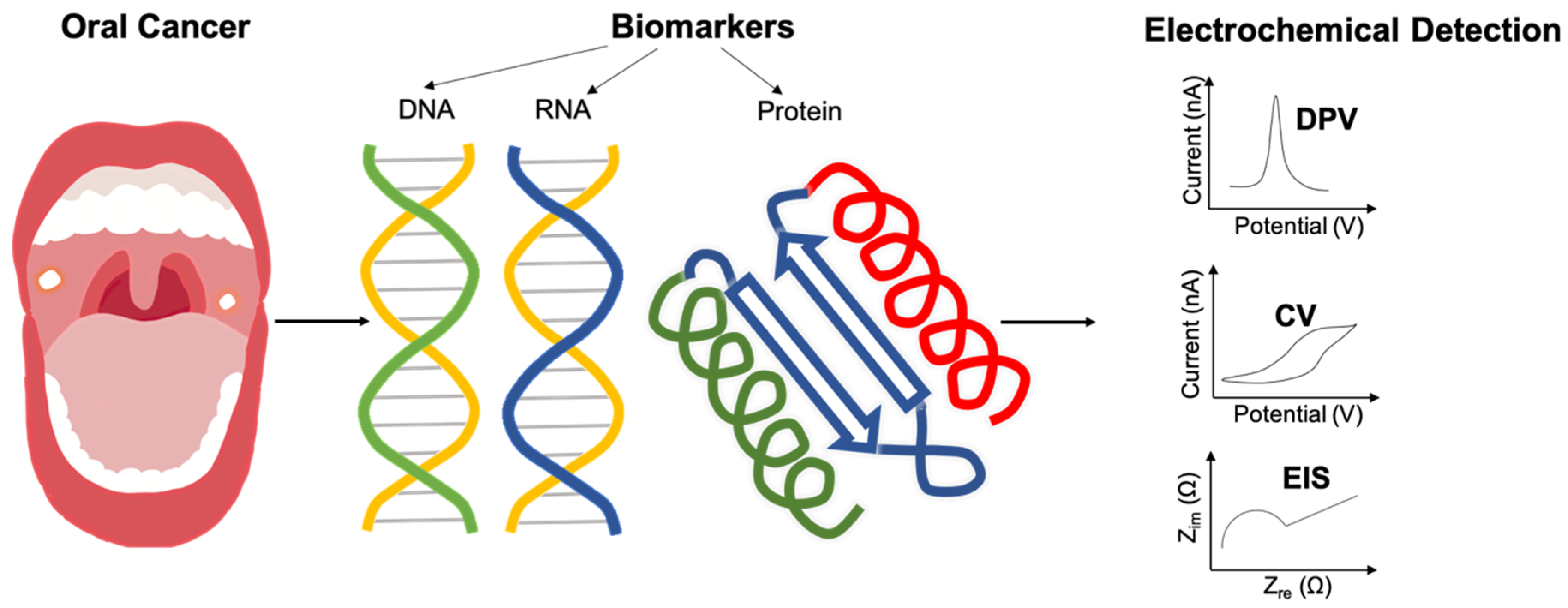
In most cases, target DNA collected from human body fluid needed to be centrifuged and purified. DNA biosensors detects the target DNA by modifying the working electrode with complementary sequences [17,18]. Usually, the gold electrode, screen-printed carbon electrode and glassy carbon electrode are utilized as the working electrode for the electrochemical set-up, while indium tin oxide (ITO) can also be used [19]. Ma et al. developed a ratiometric electrochemical DNA sensor for direct analysis of target DNA in artificial saliva specimens, by integrating the exonuclease III-assisted amplification with dual-signal ratiometric output mode [20]. Thus, it enables the detection of a lower concentration of biomarkers. Firstly, oral cancer overexpressed 1 (ORAOV1) DNA is hybridized with a ferrocene-labeled hairpin probe (Fc-Probe), then separated by exonuclease III releasing the target DNA, which is followed by the next amplification cycle. The amount of the residual Fc probe is quantified by using an electrochemical method. The residual Fc probe is hybridized with the methylene blue-labeled hairpin probe (MB-labeled probe), then a modified MB-labeled probe is fixed on the gold electrode and used as a working electrode. Once the Fc probe combines with the MB-labeled probe, the electric current would decrease. Thus, the more the ORAOV1 DNA presents in saliva, the more Fc probe would be consumed, and the higher current would be detected (as shown in Figure 2a). Chen et al. also developed a DNA biosensor to detect ORAOV1 [21]. They modified signal probes that contain target recognition sequences, nicking the endonuclease recognition sequence and G-rich sequence on a bare gold electrode, forming a cycle after a hybridized signal probe cleaved by the nicking enzyme. Finally, the addition of hemin to bind with a G-rich sequence that is located on signal probes and catalyzed the H2O2-mediated oxidation of 3,3′,5,5′ tetramethylbenzidine (TMB) to acquire current signal. However, the performance of DNA biosensors is not always very satisfactory along with the metal electrode because of DNA denaturation. To overcome this challenge, Wei et al. introduced a biocompatible DNA dendrimer system, a short nanoscale of DNA modified on the working electrode surface. They adopted a 16-Array electrochemical chip with three electrodes to detect multiple biomarkers simultaneously as shown in Figure 2b [22]. Eventually, the signal level got higher, and the amount of denatured species get lower.
2.2. RNA Biosensor
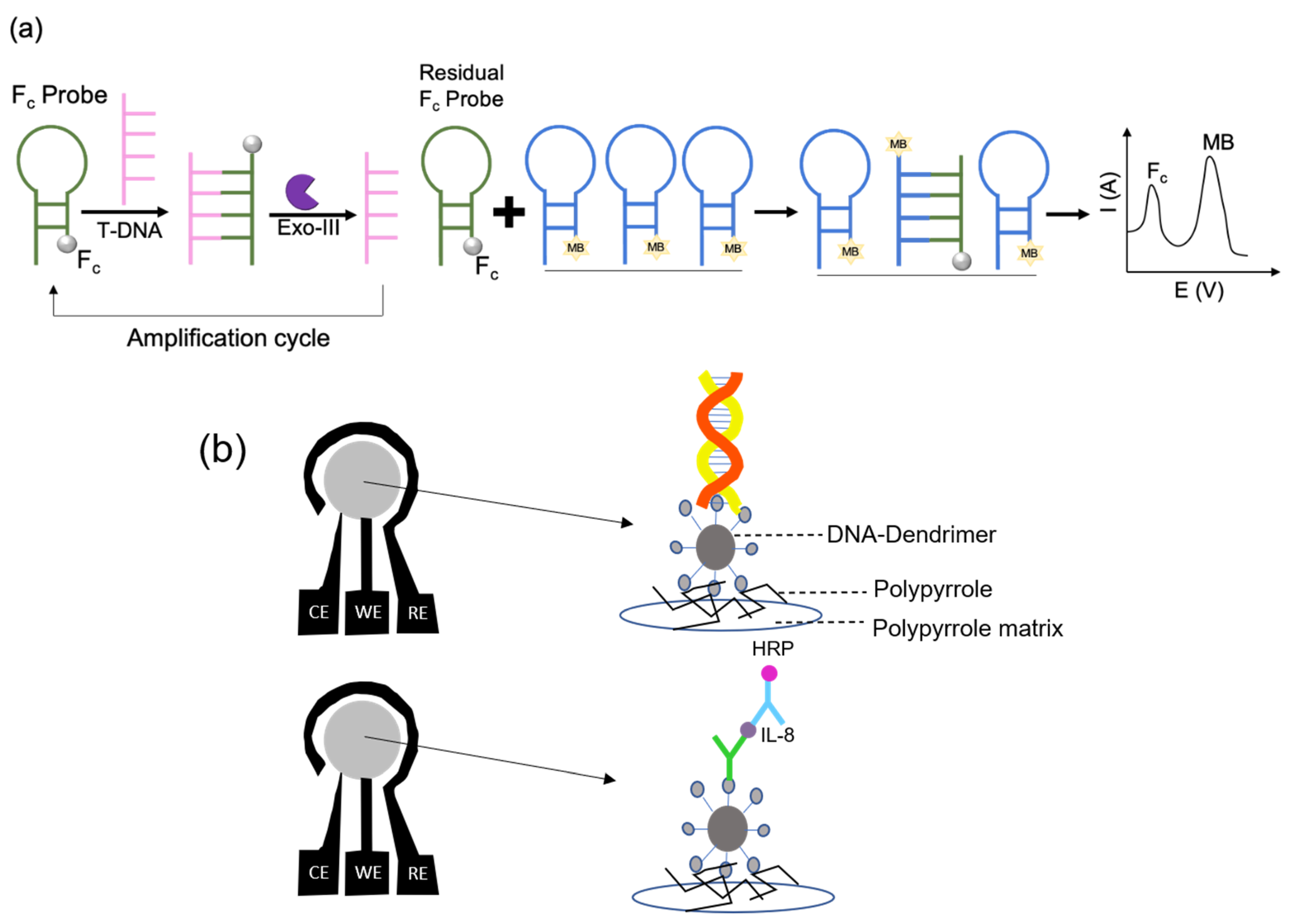
RNA biosensors are often combined with magnetic beads utilizing the junction strategy to ensure the biosensors achieve better sensitivity and a higher distinction ability. Wang et al. reported the modification of capture probes on magnetic beads. As depicted in Figure 3a, a magnetic bead is capable of binding numerous capture probes along with the target and the signal probe, getting absorbed on the surface of the electrically magnetic-controllable working electrode on the addition of a voltage on the electric coil [10,11]. Hence, the electrochemical current signal increased because horseradish peroxidase (HRP) catalyzed the H2O2-mediated oxidation of TMB after immersing the working electrode in TMB-H2O2 solution. As illustrated in Figure 3b, the magnetic beads were captured on the dual screen-printed carbon electrode (SPdCE) as working electrodes so that target mRNA and target interleukin-8 (IL-8) could be detected simultaneously in the same samples. Furthermore, hydroquinone (HQ) can be used as a redox mediator in the H2O2 solution to monitor the current generation and analysis of each target’s concentration in artificial saliva.
2.3. Protein Biosensor
Electrochemical biosensors for measuring protein cancer biomarkers offer a sensitive, quick, and cheap diagnosis framework for point of care (POC) and clinical analysis [23]. In these biosensors, the surface of the electrodes usually is modified with receptors such as antibodies or aptamers. Consequently, the explicit protein analytes are measured with the assistance of quantifiable and electroactive species generated from themselves or a signal transducer that has been added into the solution. Immunosensors are highly selective since the interactions between an antibody and an antigen act like a lock and key binding mechanism [24]. Protein biomarkers for cancer detection are broadly applicable for measuring elements that are considered as indicators of abnormal biological processes, disease processes, or responses to therapeutic intermediation [19,25]. Theses biomarkers usually can be extracted from serum, tissue, or saliva, and their expression levels can be expressive of disease states. Several tumor markers can be easily found in saliva which makes the salivary sample in a high level of interest for the non-invasive detection of oral cancer [26,27]. Recent studies have shown both cell-free mRNAs and proteins in saliva display diagnostic values for oral cancer, as previously mentioned [28]. One of the pioneers for the multiplexed electrochemical sensors for the determination of salivary biomarkers for oral cancer detection was reported by Wei et al. as mentioned in Section 2.1 the detection of a certain protein [22]. Their results showed that the multiplex detection of both interleukin-8 (IL-8) mRNA and protein allows an accurate diagnosis for oral cancer detection. In another study, human saliva samples used for the detection of protein IL-8 and IL-8 mRNA by the development of electrochemical magnetobiosensors [29]. In this research, highly sensitive and selective methodologies were reported. Moreover, the detection limits are 0.21 nM for IL-8 mRNA and 72.4 pg mL−1 for IL-8 protein in undiluted saliva samples. The development of reliable immunosensor for POC application for early detection of certain proteins is of interest, especially for an ultra-low concentrated sample without interference from the variety of other non-analyte proteins in serum and saliva [29,30,31,32,33,34]. The gold standard method for clinical detection of biomarkers is enzyme-linked immunosorbent assay (ELISA) [31]. ELISA has the limits of detection (LOD) of 1–3 pg mL−1 for many protein analytes [19,30]. However, ELISA is limited by costly test kits and equipment, long time of measuring, and difficulties in multiplexing. Consequently, ELISA is not the first choice for the application of POC diagnostics.
Compared with traditional biosensors, aptamer-based biosensors characterized label-free, high sensitivity for electrochemical detection [35,36]. Qureshi et al. developed a capacitive aptasensor to monitor human epidermal growth factor receptor 2 (HER2) protein which was an overexpressed biomarker in many cancers including ovarian, lung, gastric, and oral cancers [37]. Anti-HER2 aptamers (ssDNA) are immobilized on a gold microelectrode surface. Then, dilute human serum was spiked with various concentrations of HER2. Ultimately, the relationship between capacitance and HER2 concentration was measured. The results showed that a dynamic range was between 0.2 and 2 ng mL−1, without adding any redox-chemicals [35]. In another studies, aptamers were utilized for the detection of IL-6 protein. For instance, Kumar et al. reported a nano-aptamer sensor to detect IL-6 in biofluids like sweat using EIS. In this study, aptamers against IL-6 were immobilized on gold nanoparticles deposited on the gold working electrode for capturing the analyte [38]. The detection limit was estimated as 0.02 pg mL−1. Furthermore, the aptasensors had 90% of the original signal of impedance response after 2 weeks which indicates the high stability of the sensor. Another example is that Tertis et al. developed an IL-6 aptasesnsor based on a glassy carbon electrode modified by p-aminobenzoic acid, p-aminothiophenol and gold nanoparticles [39]. The aptasensor showed the detection limit of 1.6 pg mL−1 and high selectivity in the blood sample.
Another type of protein sensor is based on immunoassays. In a typical sandwich immunoassay, a recognized element, such as primary antibody, is immobilized on an electrode surface to capture the specific analyte. In the next step, secondary antibody conjugated with an enzyme, for instance, horseradish peroxidase (HRP), is added into the solution for binding the antigen (the analyte). HRP can convert its substrate to electrochemically active species, so that the chemical signal can be converted to electrochemical signal through this step [29,40,41,42,43]. For instance, Heineman’ group pioneered in enzyme-linked electrochemical detection of the analyte using sandwich immunoassays [32]. In this research, two main procedures are being utilized for multiplex electrochemical protein identification. First, the primary antibody was used for binding the analytes, and then the secondary antibodies were attached to nanoparticles. Second, electrodes were utilized by immobilizing with various antibodies and for measuring the electrochemical signals. In addition to their research, the improvement of delicate and precise electrochemical biosensing frameworks relies on bioreceptor, redox mediators and transducer networks. In recent years, nanomaterials are popular, and they are widely applied in the biosensor field owing to their high surface-to-volume ratio. New carbon or metal nanomaterials have been innovated, such as graphene and its derivatives, with special properties including high electron transfer rate, high biocompatibility, and thus many studies combine nanomaterials with immunosensors [29,33,44,45,46]. Nanostructure based immunosensors increase electrode surface area, which can provide 10-fold or more antibody coverage compared to flat surfaces, leading to enhanced sensitivity of up to 1000-fold and lower limit of detection [34,47,48,49].
In addition to oral cancer biosensors, electrochemical biosensors systems for the detection of cancer biomarkers using similar principles have been developed for several types of cancer including lung cancer [47,50], melanoma [51,52,53] breast cancer [36,54,55] and prostate cancer [56]. Electrochemical signal amplification in these biosensors could be achieved by estimating electroactive chemical responses released or consumed from an enzyme, for example HRP [20,54,57]. Other electrochemical detection methods relied on special nanoparticles labeled on antibodies. This complex could be further dissolved to gain detectable electroactive species [25,32,34,58,59,60,61]. Protein sensors for targeting various important biomarkers were listed in the following sections.
2.3.1. Proteins Biosensors Targeting IL-6 and IL-8
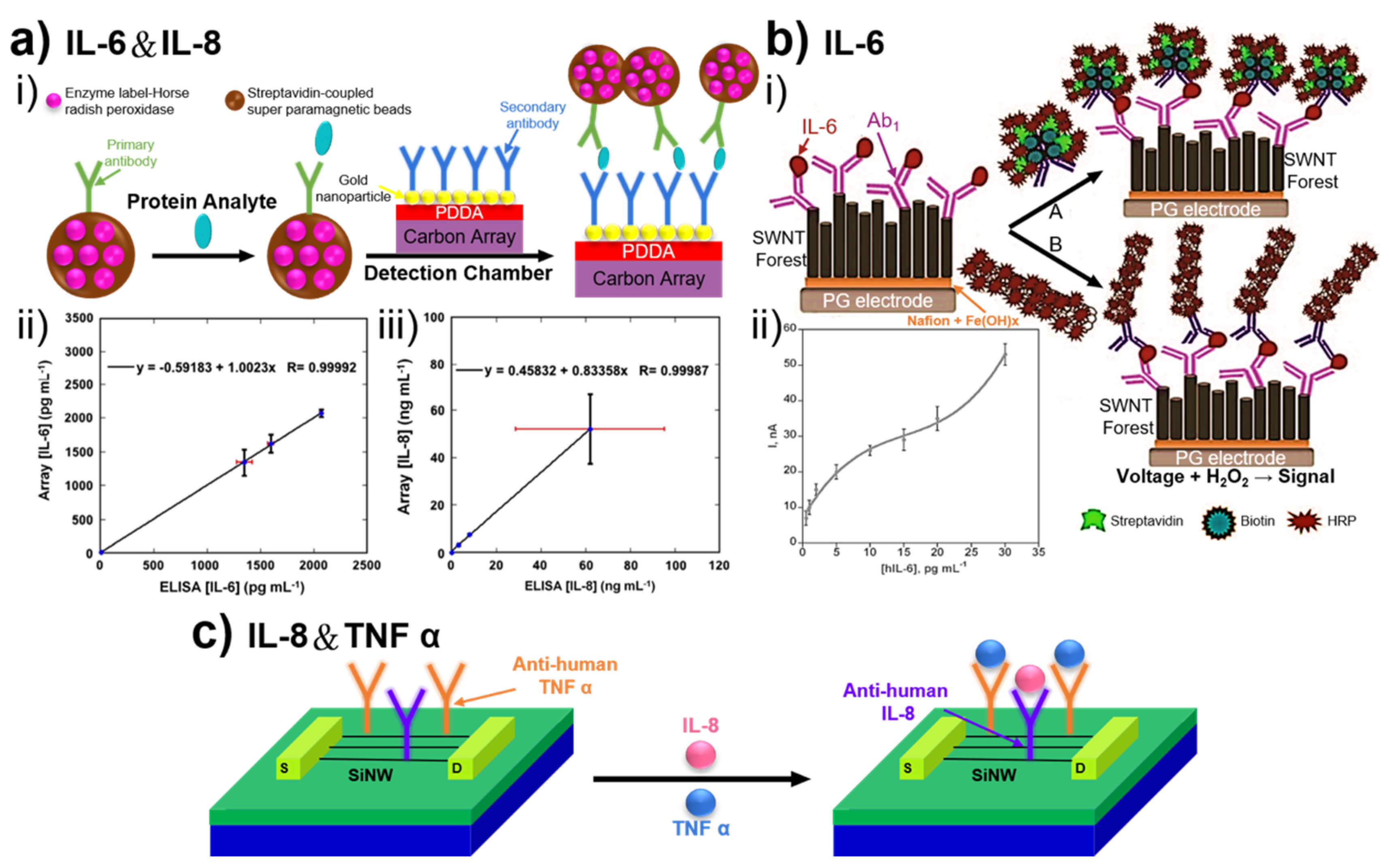
IL-6 and IL-8 are cellular proteins that are particularly relevant to oral squamous cell carcinoma (OSCC) [62,63,64,65]. The uncontrolled expression of these cytokines could be a sign of the development of tumor growth and metastasis [65]. Usually, the normal expression levels of these cytokines in keratinocytes are low. IL-6 is an interleukin that behaves as both a pro-inflammatory cytokine and an anti-inflammatory myokine, that is involved in acute-phase reaction, growth regulation and differentiation of cells [60,66]. Patients with oral cancer have 20 to 1000 pg mL−1 of IL-6 in the serum, while, in healthy individuals, it is <6 pg mL−1 [17]. IL-8 is a 8-kDa cytokine related to the angiogenic and mitotic processes, and inflammatory response. The serum levels in healthy individuals are lower than 13 pg mL−1 compared with 20–1000 pg mL−1 or more in patients [67]. Figure 4 depicts electrochemical immunosensors based on the detection of biomarkers IL-6 and IL-8 for targeting oral cancer.
Otieno and coworkers developed a sensor array coated with gold nanoparticle–antibody conjugates in a poly(dimethylsiloxane) microchannel [31]. In their investigation, protein analytes were captured on Ab2–MP–HRP bioconjugates from serum or other biological samples. Figure 4a(i) illustrates the principle for fabricating the sensor. The accuracy of the sensor was verified (Figure 4a(ii,iii)). The comparison of the obtained results showed good correlations with standard ELISAs. In another study, Malhotra and coworkers reported an ultrasensitive electrochemical immunosensor for targeting IL-6 in head and neck squamous cell carcinoma (HNSCC) cells [25]. The mean concentration of IL-6 in the serum of patients with HNSCC is higher than 20 pg mL−1 compared with 6 pg mL−1 in healthy individuals. In this research, the high sensitivity was achieved by nanostructured single-walled nanotube (SWNT) forest platforms. Two strategies were chosen for the multilabel detection in the amperometric immunosensor, as shown in Figure 4b(i): using 14–16 HRPs labeled Ab2; or adding active HRPs modified on carboxylated carbon nanotubes.
In another study, Zhang et al. reported a field-effect transistor (FET) sensor for the detection of two biomarkers of OCSS in saliva samples, IL-8 and TNF-α, by using a silicon nanowire (SiNW) (Figure 4c) [32]. The detection of biomarkers in real saliva sample is very tricky since it contains complex components. They overcame this problem by functionalizing with two different antibodies immobilized on the surface of the electrode for capturing IL-8 and tumor necrosis factor α (TNF-α) and avoiding the cross-reactivity of biomarkers carefully (Figure 4c).
In recent years, two-dimensional (2D) graphene-based materials are widely used in biosensors because of their unique properties including high mechanical stability, biocompatibility and high rate of electron transfer [33,68,69]. The advantages of graphene-based biosensors can be roughly concluded into two points. First, graphene oxide (GO) contains certain functional groups that can be easily covalently bonded to biomolecules. Second, the structural defects of GO, reduced graphene oxide (rGO) and graphene-based quantum dots (GQDs) can be used to immobilize the biomolecules onto their surfaces. Graphene-based sensors are successfully used for the incorporation of nanoparticles and/or hydrogel for electrochemical detection of various analytes including glucose [70,71], DNA, cholesterol oxidase, and NADH, etc. [72]. Verma and coworkers fabricated immunosensor based on the gold nanoparticles-reduced graphene oxide (AuNPs-rGO) composites for non-invasive IL-8 detection in the saliva sample [49]. The reported immunosensor has shown fast detection (9 min) of IL-8 and offers high sensitivity with an experimental linear dynamic range of 500 fg mL−1 to 4 ng mL−1 and a detection limit of 72.73 ± 0.18 pg mL−1. The high sensitivity and rapid detection were due to the high rate of electron transfer through the thin film of AuNPs-rGO nanocomposites.
2.3.2. Protein Biosensors Targeting Other Biomarkers
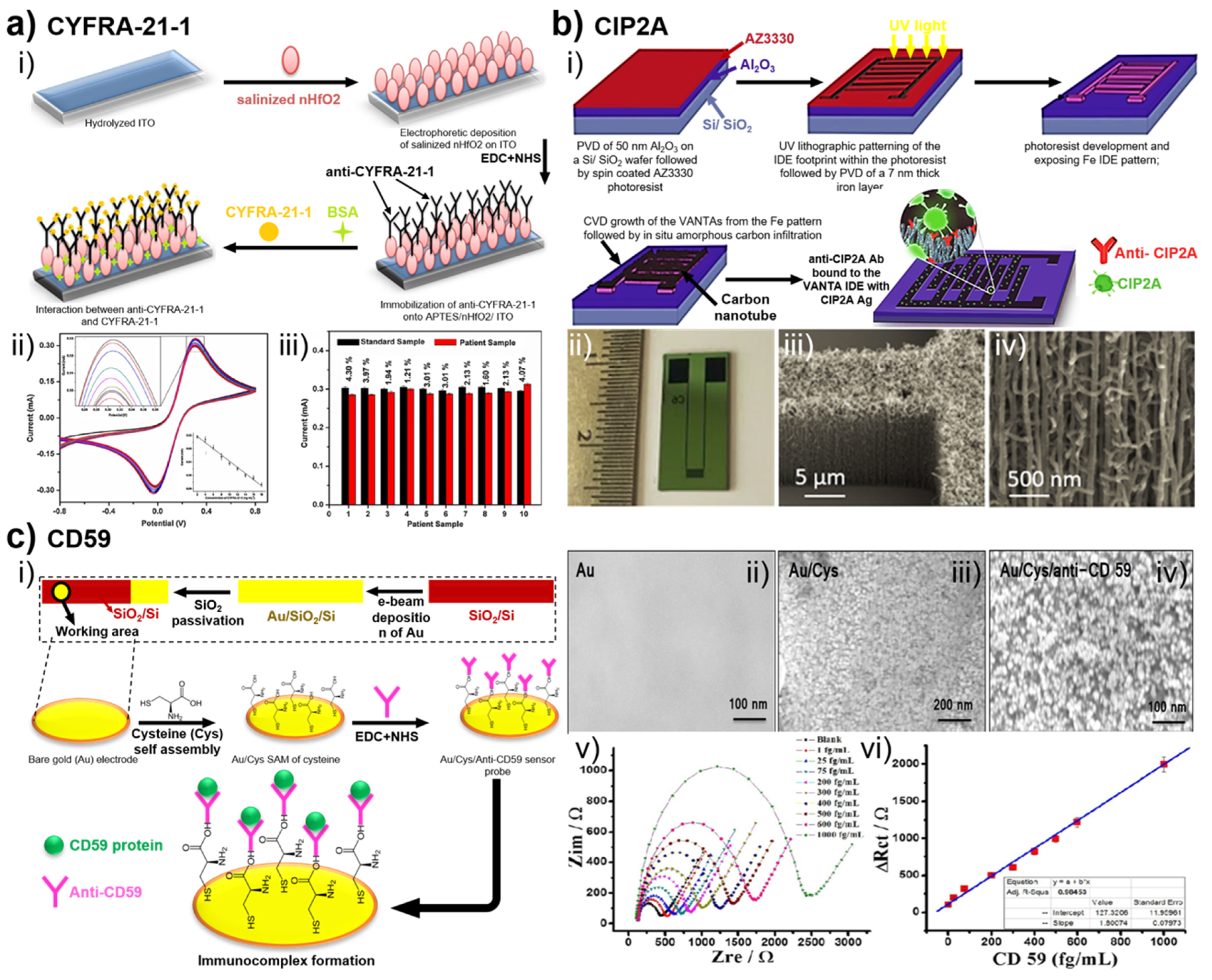
Besides IL-6 and IL-8, some other biomarkers have also been employed for electrochemical detection of oral cancer. Kumar and coworkers reported a non-invasive, label-free immunosensor based on nanostructured hafnium oxide (hafnia) deposited onto indium tin oxide (ITO) coated glass for detection of CYFRA-21-1 in human saliva [34]. CYFRA-21-1 is a remarkable tumor marker for squamous cell carcinoma (SCC). However, the clinical value of CYFRA-21-1 in OSCC has not been validated [73]. Figure 5a(i) shows the steps of the fabrication of BSA/anti-CYFRA-21-1/APTES/nHfO2/ITO immunosensor. The BSA/anti-CYFRA-21-1/APTES/nHfO2/ITO electrode responded according to the concentration of CYFRA-21-1 (2–18 ng mL−1), shown in Figure 5a(ii). In this biosensor, it was found that the CV peak current gradually decreased linearly with increased concentration of CYFRA-21-1. The decreased current was due to the formation of electrically insulating antigen–antibody complex which prevents the electron transfer through redox mediator. The current responses recorded for the real samples (Figure 5a(iii)) were matched with the current signals obtained for standard samples of the same concentration. This result showed an excellent correlation between the current from the real sample.
The first application of vertically aligned carbon nanotube array (VANTA) interdigitated electrodes (IDEs) for electrochemical detection of CIP2A was reported by Ding and coworkers [54]. IDEs worked as electrochemical transducers, consisting of several electrodes which were electrically connected together [74,75]. CIP2A is a cancer biomarker for a variety of human cancers including lung, breast and gastric cancers. However, CIP2A is extensively expressed in OSCC cell lines, and malignant human oral epithelial tissues [76]. The scheme in Figure 5b(i) represents the steps of the fabrication of immunosensor. The dimensions of VANTA IDEs are shown in Figure 5b(ii). Moreover, the highly porous VANTA structures were observed with field-effect scanning electron microscopy (FESEM) (Figure 5b(iii,iv)) in two magnifications. The label-free immunosensor for detecting CIP2A showed a wide linear sensing range (1−100 pg mL−1) with a good detection limit of 0.24 pg mL−1 in saliva. Furthermore, the electrochemical immunosensor showed higher sensitivity than the corresponding CIP2A ELISA test.
Moreover, Choudhary and coworkers developed a label-free impedimetric immunosensor to diagnose oral cancer by detecting CD59 [48]. The immunosensor probe was fabricated by immobilizing CD59 antibodies (anti-CD59) on a self-assembled molecular layer of L-cysteine (Cys) on a gold electrode. The stepwise of fabrication of the immunosensor is shown in Figure 5c(i). The microphotographs for Au, Au/Cys and Au/Cys/anti- CD59 surfaces are shown in Figure 5c(ii–iv), respectively. CD59 is one of the most fundamental and clinically relevant early-stage markers, that can be used to diagnose oral cancer [44]. Figure 5c(v) shows the EIS response of immunosensor in different concentrations of CD59. The Rct value increased with an increase of CD59 concentrations due to the formation of the insulation layer between CD59 and immunosensor. The immunosensor showed a linear range of detection between 1.0 and 1000 fg mL−1 (Figure 5c(vi)).
3. Conclusions
In this review, DNA biosensor, RNA biosensor and protein biosensor for several salivary biomarkers (such as IL-6, IL-8, TNF-α, CYFRA 21-1, CD 59 and CIP2A) have been discussed as promising candidates to provide crucial information for developing non-invasive oral cancer diagnosis. Therefore, the major challenge faced in developing such a diagnostic system accounts for the relatively lower amounts of target analytes present in saliva, which is taken as the diagnostic fluid. However, as the novel and highly sensitive electrochemical biosensor techniques are emerging, the accuracy of analyzing the vital biomarkers in the saliva has been enhanced as well. In addition, these electrochemical sensors meet the requisites, including easy-to-use, non-invasiveness and low cost. The ultrasensitivity and the specificity of biosensors can be improved by combining with MBs, signal probes or nanomaterials on the working electrode so that the electrochemical signal can be more evident. SPdCE and 16-array chips offer the system to detect multiple relevant oral cancer biomarkers simultaneously. However, as oral cancer exhibits some biomarkers that are also corresponding to other types of cancers, their distinctive detection is still an issue. Efforts have been made to differentiate the normal tissues from the different cancerous tissues at various stages by utilizing electrochemical sensing. In the future, the development of electrochemical biosensors on-chip will be one of the main non-invasive detection methodologies of oral cancer at earlier stages in a relatively more precise manner.
Author Contributions
Conceptualization, Y.-T.L., S.D. and T.-E.L.; validation, L.W.; formal analysis, T.-E.L.; investigation, T.-Y.H. and S.-H.L.; resources, H.H.G.; data curation, Y.-T.L. and S.D.; writing—original draft preparation, T.-E.L., Y.-T.L., S.D., A.P. and L.W.; writing—review and editing, T.-E.L., Y.-T.L., S.D., and A.P.; project administration, T.-E.L.; funding acquisition, T.-E.L., S.-H.L., and H.H.G.; All authors have read and agreed to the published version of the manuscript.
Funding
This research was funded by Young Scholar Fellowship Program by Ministry of Science and Technology (MOST) in Taiwan, grant number MOST 108-2636-E-009-012 and 109-2636-B-009-001.
Acknowledgments
Thanks to S.-J. Ji and C.-Y. Chien of the Ministry of Science and Technology (National Taiwan University) for the consulting and data collection.
Conflicts of Interest
The authors declare no conflict of interest.
References
- Montero, P.H.; Patel, S.G. Cancer of the oral cavity. Surg. Oncol. Clin. N. Am. 2015, 24, 491–508. [Google Scholar] [CrossRef] [PubMed] [Green Version]
- Warnakulasuriya, S. Global epidemiology of oral and oropharyngeal cancer. Oral Oncol. 2009, 45, 309–316. [Google Scholar] [CrossRef]
- Chaturvedi, P.; Singh, A.; Chien, C.Y.; Warnakulasuriya, S. Tobacco related oral cancer. BMJ 2019, 365, l2142. [Google Scholar] [CrossRef]
- Siegel, R.L.; Miller, K.D.; Goding Sauer, A.; Fedewa, S.A.; Butterly, L.F.; Anderson, J.C.; Cercek, A.; Smith, R.A.; Jemal, A. Colorectal cancer statistics. CA Cancer J. Clin. 2020, 70, 145–164. [Google Scholar] [CrossRef] [Green Version]
- Jemal, A.; Siegel, R.; Ward, E.; Hao, Y.; Xu, J.; Murray, T.; Thun, M.J. Cancer statistics. CA Cancer J. Clin. 2008, 58, 71–96. [Google Scholar] [CrossRef] [PubMed]
- Messadi, D.V. Diagnostic aids for detection of oral precancerous conditions. Int. J. Oral Sci. 2013, 5, 59–65. [Google Scholar] [CrossRef] [PubMed]
- Yen, A.M.F.; Chen, S.C.; Chen, T.H.H. Dose-response relationships of oral habits associated with the risk of oral pre-malignant lesions among men who chew betel quid. Oral Oncol. 2007, 43, 634–638. [Google Scholar] [CrossRef]
- Kumar, M.; Nanavati, R.; Modi, T.; Dobariya, C. Oral cancer: Etiology and risk factors: A review. J. Cancer Res. Ther. 2016, 12, 458–463. [Google Scholar] [CrossRef]
- Chuang, S.L.; Su, W.W.Y.; Chen, S.L.S.; Yen, A.M.F.; Wang, C.P.; Fann, J.C.Y.; Chiu, S.Y.H.; Lee, Y.C.; Chiu, H.M.; Chang, D.C.; et al. Population-based screening program for reducing oral cancer mortality in 2,334,299 taiwanese cigarette smokers and/or betel quid chewers. Cancer 2017, 123, 1597–1609. [Google Scholar] [CrossRef] [Green Version]
- Pearce, A.; Sharp, L.; Hanly, P.; Barchuk, A.; Bray, F.; de Camargo Cancela, M.; Gupta, P.; Meheus, F.; Qiao, Y.L.; Sitas, F.; et al. Productivity losses due to premature mortality from cancer in Brazil, Russia, India, China, and South Africa (BRICS): A population-based comparison. Cancer Epidemiol. 2018, 53, 27–34. [Google Scholar] [CrossRef]
- Gupta, B.; Johnson, N.W.; Kumar, N. Global epidemiology of head and neck cancers: A continuing challenge. Oncology 2016, 91, 13–23. [Google Scholar] [CrossRef] [PubMed]
- Srinivas, P.R.; Kramer, B.S.; Srivastava, S. Trends in biomarker research for cancer detection. Lancet Oncol. 2001, 2, 698–704. [Google Scholar] [CrossRef]
- Pollaers, K.; Hinton-Bayre, A.; Friedland, P.L.; Farah, C.S. AJCC 8th edition oral cavity squamous cell carcinoma staging—Is it an improvement on the AJCC 7th edition? Oral Oncol. 2018, 82, 23–28. [Google Scholar] [CrossRef] [PubMed]
- Mishra, S.; Saadat, D.; Kwon, O.; Lee, Y.; Choi, W.S.; Kim, J.H.; Yeo, W.H. Recent advances in salivary cancer diagnostics enabled by biosensors and bioelectronics. Biosens. Bioelectron. 2016, 81, 181–197. [Google Scholar] [CrossRef]
- Chen, X.J.; Zhang, X.Q.; Liu, Q.; Zhang, J.; Zhou, G. Nanotechnology: A promising method for oral cancer detection and diagnosis. J. Nanobiotechnol. 2018, 16, 52. [Google Scholar] [CrossRef]
- Hasanzadeh, M.; Shadjou, N.; dela Guardia, M. Non-invasive diagnosis of oral cancer: The role of electro-analytical methods and nanomaterials. TrAC Trends Anal. Chem. 2017, 91, 125–137. [Google Scholar] [CrossRef]
- Riedel, F.; Zaiss, I.; Herzog, D.; Götte, K.; Naim, R.; Hörmann, K. Serum levels of interleukin-6 in patients with primary head and neck squamous cell carcinoma. Anticancer Res. 2005, 25, 2761–2766. [Google Scholar]
- Ilkhani, H.; Sarparast, M.; Noori, A.; Bathaie, S.Z.; Mousavi, M.F. Electrochemical aptamer/antibody based sandwich immunosensor for the detection of EGFR, a cancer biomarker, using gold nanoparticles as a signaling probe. Biosens. Bioelectron. 2015, 74, 491–497. [Google Scholar] [CrossRef]
- Tan, Y.; Wei, X.; Zhao, M.; Qiu, B.; Guo, L.; Lin, Z.; Yang, H.H. Ultraselective homogeneous electrochemical biosensor for DNA species related to oral cancer based on nicking endonuclease assisted target recycling amplification. Anal. Chem. 2015, 87, 9204–9208. [Google Scholar] [CrossRef]
- Ma, R.N.; Wang, L.L.; Wang, H.F.S.; Jia, L.P.; Zhang, W.; Shang, L.; Xue, Q.W.; Jia, W.L.; Liu, Q.Y.; Wang, H.F.S. Highly sensitive ratiometric electrochemical DNA biosensor based on homogeneous exonuclease III-assisted target recycling amplification and one-step triggered dual-signal output. Sens. Actuators B Chem. 2018, 269, 173–179. [Google Scholar] [CrossRef]
- Chen, J.; Zhang, J.; Guo, Y.; Li, J.; Fu, F.; Yang, H.H.; Chen, G. An ultrasensitive electrochemical biosensor for detection of DNA species related to oral cancer based on nuclease-assisted target recycling and amplification of DNAzyme. Chem. Commun. 2011, 47, 8004–8006. [Google Scholar] [CrossRef] [PubMed]
- Wei, F.; Patel, P.; Liao, W.; Chaudhry, K.; Zhang, L.; Arellano-Garcia, M.; Hu, S.; Elashoff, D.; Zhou, H.; Shukla, S.; et al. Electrochemical sensor for multiplex biomarkers detection. Clin. Cancer Res. 2009, 15, 4446–4452. [Google Scholar] [CrossRef] [PubMed] [Green Version]
- Aluoch, A.O.; Sadik, O.A.; Bedi, G. Development of an oral biosensor for salivary amylase using a monodispersed silver for signal amplification. Anal. Biochem. 2005, 340, 136–144. [Google Scholar] [CrossRef] [PubMed]
- Wei, F.; Liao, W.; Xu, Z.; Yang, Y.; Wong, D.T.; Ho, C.M. Bio/Abiotic interface constructed from nanoscale DNA dendrimer and conducting polymer for ultrasensitive biomolecular diagnosis. Small 2009, 5, 1784–1790. [Google Scholar] [CrossRef] [PubMed] [Green Version]
- Malhotra, R.; Patel, V.; Vaqué, J.P.; Gutkind, J.S.; Rusling, J.F. Ultrasensitive electrochemical immunosensor for oral cancer biomarker IL-6 using carbon nanotube forest electrodes and multilabel amplification. Anal. Chem. 2010, 82, 3118–3123. [Google Scholar] [CrossRef] [Green Version]
- Saxena, S.; Sankhla, B.; Sundaragiri, K.; Bhargava, A. A review of salivary biomarker: A tool for early oral cancer diagnosis. Adv. Biomed. Res. 2017, 6, 90. [Google Scholar] [CrossRef]
- Malon, R.S.P.; Sadir, S.; Balakrishnan, M.; Córcoles, E.P. Saliva-based biosensors: Noninvasive monitoring tool for clinical diagnostics. Biomed Res. Int. 2014, 2014, 962903. [Google Scholar] [CrossRef]
- Markopoulos, A.K.; Michailidou, E.Z.; Tzimagiorgis, G. Salivary markers for oral cancer detection. Open Dent. J. 2010, 4, 172–178. [Google Scholar] [CrossRef] [Green Version]
- Torrente-Rodríguez, R.M.; Campuzano, S.; Ruiz-Valdepeñas Montiel, V.; Gamella, M.; Pingarrón, J.M. Electrochemical bioplatforms for the simultaneous determination of interleukin (IL)-8 MRNA and IL-8 protein oral cancer biomarkers in raw saliva. Biosens. Bioelectron. 2016, 77, 543–548. [Google Scholar] [CrossRef]
- Wang, Z.W.; Zhang, J.; Guo, Y.; Wu, X.Y.; Yang, W.J.; Xu, L.J.; Chen, J.H.; Fu, F.F. A novel electrically magnetic-controllable electrochemical biosensor for the ultra sensitive and specific detection of attomolar level oral cancer-related microRNA. Biosens. Bioelectron. 2013, 45, 108–113. [Google Scholar] [CrossRef]
- Otieno, B.A.; Krause, C.E.; Latus, A.; Chikkaveeraiah, B.V.; Faria, R.C.; Rusling, J.F. On-line protein capture on magnetic beads for ultrasensitive microfluidic immunoassays of cancer biomarkers. Biosens. Bioelectron. 2014, 53, 268–274. [Google Scholar] [CrossRef] [PubMed] [Green Version]
- Zhang, Y.; Chen, R.; Xu, L.; Ning, Y.; Xie, S.; Zhang, G.J. Silicon nanowire biosensor for highly sensitive and multiplexed detection of oral squamous cell carcinoma biomarkers in saliva. Anal. Sci. 2015, 31, 73–78. [Google Scholar] [CrossRef] [Green Version]
- Kumar, S.; Sharma, J.G.; Maji, S.; Malhotra, B.D. Nanostructured zirconia decorated reduced graphene oxide based efficient biosensing platform for non-invasive oral cancer detection. Biosens. Bioelectron. 2016, 78, 497–504. [Google Scholar] [CrossRef] [PubMed]
- Kumar, S.; Kumar, S.; Tiwari, S.; Augustine, S.; Srivastava, S.; Yadav, B.K.; Malhotra, B.D. Highly sensitive protein functionalized nanostructured hafnium oxide based biosensing platform for non-invasive oral cancer detection. Sens. Actuators B Chem. 2016, 235, 1–10. [Google Scholar] [CrossRef]
- Qureshi, A.; Gurbuz, Y.; Niazi, J.H. Label-free capacitance based aptasensor platform for the detection of HER2/ErbB2 cancer biomarker in serum. Sens. Actuators B Chem. 2015, 220, 1145–1151. [Google Scholar] [CrossRef]
- Lin, T.E.; Chen, W.H.; Shiang, Y.C.; Huang, C.C.; Chang, H.T. Colorimetric detection of platelet-derived growth factors through competitive interactions between proteins and functional gold nanoparticles. Biosens. Bioelectron. 2011, 29, 204–209. [Google Scholar] [CrossRef] [PubMed]
- Slamon, D.J.; Godolphin, W.; Jones, L.A.; Holt, J.A.; Wong, S.G.; Keith, D.E.; Levin, W.J.; Stuart, S.G.; Udove, J.; Ullrich, A.; et al. Studies of the HER-2/Neu proto-oncogene in human breast and ovarian cancer. Science 1989, 244, 707–712. [Google Scholar] [CrossRef]
- Kumar, L.S.S.; Wang, X.; Hagen, J.; Naik, R.; Papautsky, I.; Heikenfeld, J. Label free nano-aptasensor for interleukin-6 in protein-dilute bio fluids such as sweat. Anal. Methods 2016, 8, 3440–3444. [Google Scholar] [CrossRef]
- Tertis, M.; Leva, P.I.; Bogdan, D.; Suciu, M.; Graur, F.; Cristea, C. Impedimetric aptasensor for the label-free and selective detection of interleukin-6 for colorectal cancer screening. Biosens. Bioelectron. 2019, 137, 123–132. [Google Scholar] [CrossRef]
- Thomas, J.H.; Kim, S.K.; Hesketh, P.J.; Halsall, H.B.; Heineman, W.R. Microbead-based electrochemical immunoassay with interdigitated array electrodes. Anal. Biochem. 2004, 328, 113–122. [Google Scholar] [CrossRef]
- Li, H.; Wei, Q.; He, J.; Li, T.; Zhao, Y.; Cai, Y.; Du, B.; Qian, Z.; Yang, M. Electrochemical immunosensors for cancer biomarker with signal amplification based on ferrocene functionalized iron oxide nanoparticles. Biosens. Bioelectron. 2011, 26, 3590–3595. [Google Scholar] [CrossRef] [PubMed]
- Thomas, J.A.; Schnell, F.; Kaveh-Baghbaderani, Y.; Berensmeier, S.; Schwaminger, S.P. Immunomagnetic separation of microorganisms with iron oxide nanoparticles. Chemosensors 2020, 8, 17. [Google Scholar] [CrossRef] [Green Version]
- Evtugyn, G.; Hianik, T. Electrochemical immuno- and aptasensors for mycotoxin determination. Chemosensors 2019, 7, 10. [Google Scholar] [CrossRef] [Green Version]
- Vasilescu, A.; Fanjul-Bolado, P.; Titoiu, A.M.; Porumb, R.; Epure, P. Progress in electrochemical (bio)sensors for monitoring wine production. Chemosensors 2019, 7, 66. [Google Scholar] [CrossRef] [Green Version]
- Shellaiah, M.; Sun, K.W. Review on nanomaterial-based melamine detection. Chemosensors 2019, 7, 9. [Google Scholar] [CrossRef] [Green Version]
- Zakaria, A.B.M.; Leszczynska, D. Electrochemically prepared unzipped single walled carbon nanotubes-MnO2 nanostructure composites for hydrogen peroxide and glucose sensing. Chemosensors 2019, 7, 1. [Google Scholar] [CrossRef] [Green Version]
- Tiwari, S.; Gupta, P.K.; Bagbi, Y.; Sarkar, T.; Solanki, P.R. L-Cysteine capped lanthanum hydroxide nanostructures for non-invasive detection of oral cancer biomarker. Biosens. Bioelectron. 2017, 89, 1042–1052. [Google Scholar] [CrossRef]
- Choudhary, M.; Yadav, P.; Singh, A.; Kaur, S.; Ramirez-Vick, J.; Chandra, P.; Arora, K.; Singh, S.P. CD 59 targeted ultrasensitive electrochemical immunosensor for fast and noninvasive diagnosis of oral cancer. Electroanalysis 2016, 28, 2565–2574. [Google Scholar] [CrossRef]
- Verma, S.; Singh, A.; Shukla, A.; Kaswan, J.; Arora, K.; Ramirez-Vick, J.; Singh, P.; Singh, S.P. Anti-IL8/AuNPs-RGO/ITO as an immunosensing platform for noninvasive electrochemical detection of oral Cancer. ACS Appl. Mater. Interfaces 2017, 9, 27462–27474. [Google Scholar] [CrossRef] [PubMed]
- Khanmohammadi, A.; Aghaie, A.; Vahedi, E.; Qazvini, A.; Ghanei, M.; Afkhami, A.; Hajian, A.; Bagheri, H. Electrochemical biosensors for the detection of lung cancer biomarkers: A review. Talanta 2020, 206, 120251. [Google Scholar] [CrossRef]
- Darvishi, S.; Pick, H.; Lin, T.E.; Zhu, Y.; Li, X.; Ho, P.C.; Girault, H.H.; Lesch, A. Tape-stripping electrochemical detection of melanoma. Anal. Chem. 2019, 91, 12900–12908. [Google Scholar] [CrossRef] [PubMed]
- Lin, T.-E.; Lu, Y.-J.; Sun, C.-L.; Pick, H.; Chen, J.-P.; Lesch, A.; Girault, H.H. Soft electrochemical probes for mapping the distribution of biomarkers and injected nanomaterials in animal and human tissues. Angew. Chem. Int. Ed. 2017, 56, 16498–16502. [Google Scholar] [CrossRef] [PubMed]
- Lin, T.E.; Bondarenko, A.; Lesch, A.; Pick, H.; Cortés-Salazar, F.; Girault, H.H. Monitoring tyrosinase expression in non-metastatic and metastatic melanoma tissues by scanning electrochemical microscopy. Angew. Chem. Int. Ed. 2016, 55, 3813–3816. [Google Scholar] [CrossRef] [PubMed] [Green Version]
- Pachauri, N.; Dave, K.; Dinda, A.; Solanki, P.R. Cubic CeO2 implanted reduced graphene oxide-based highly sensitive biosensor for non-invasive oral cancer biomarker detection. J. Mater. Chem. B 2018, 6, 3000–3012. [Google Scholar] [CrossRef]
- Hasanzadeh, M.; Shadjou, N.; dela Guardia, M. Early stage screening of breast cancer using electrochemical biomarker detection. TrAC Trends Anal. Chem. 2017, 91, 67–76. [Google Scholar] [CrossRef]
- Singh, S.; Gill, A.A.S.; Nlooto, M.; Karpoormath, R. Prostate cancer biomarkers detection using nanoparticles based electrochemical biosensors. Biosens. Bioelectron. 2019, 137, 213–221. [Google Scholar] [CrossRef]
- Ding, S.; Das, S.R.; Brownlee, B.J.; Parate, K.; Davis, T.M.; Stromberg, L.R.; Chan, E.K.L.; Katz, J.; Iverson, B.D.; Claussen, J.C. CIP2A immunosensor comprised of vertically-aligned carbon nanotube interdigitated electrodes towards point-of-care oral cancer screening. Biosens. Bioelectron. 2018, 117, 68–74. [Google Scholar] [CrossRef] [Green Version]
- Aydın, M.; Aydın, E.B.; Sezgintürk, M.K. A highly selective electrochemical immunosensor based on conductive carbon black and star PGMA polymer composite material for IL-8 biomarker detection in human serum and saliva. Biosens. Bioelectron. 2018, 117, 720–728. [Google Scholar] [CrossRef]
- Verma, S.; Singh, S.P. Non-invasive oral cancer detection from saliva using Zinc oxide-reduced graphene oxide nanocomposite based bioelectrode. MRS Commun. 2019, 9, 1227–1234. [Google Scholar] [CrossRef]
- Deckert, F.; Legay, F. Development and validation of an IL-6 immuno-receptor assay based on surface plasmon resonance. J. Pharm. Biomed. Anal. 2000, 23, 403–411. [Google Scholar] [CrossRef]
- Malhotra, R.; Urs, A.B.; Chakravarti, A.; Kumar, S.; Gupta, V.K.; Mahajan, B. Correlation of cyfra 21-1 Levels in saliva and serum with CK19 MRNA expression in oral squamous cell carcinoma. Tumor Biol. 2016, 37, 9263–9271. [Google Scholar] [CrossRef] [PubMed]
- Alevizos, I.; Mahadevappa, M.; Zhang, X.; Ohyama, H.; Kohno, Y.; Posner, M.; Gallagher, G.T.; Varvares, M.; Cohen, D.; Kim, D.; et al. Oral cancer in vivo gene expression profiling assisted by laser capture microdissection and microarray analysis. Oncogene 2001, 20, 6196–6204. [Google Scholar] [CrossRef] [PubMed] [Green Version]
- Babiuch, K.; Kuśnierz-Cabala, B.; Kęsek, B.; Okoń, K.; Darczuk, D.; Chomyszyn-Gajewska, M. Evaluation of proinflammatory, NF-KappaB dependent cytokines: IL-1α, IL-6, IL-8, and TNF-α in tissue specimens and saliva of patients with oral squamous cell carcinoma and oral potentially malignant disorders. J. Clin. Med. 2020, 9, 867. [Google Scholar] [CrossRef] [PubMed] [Green Version]
- Sahibzada, H.A.; Khurshid, Z.; Khan, R.S.; Naseem, M.; Siddique, K.M.; Mali, M.; Zafar, M.S. Salivary IL-8, IL-6 and TNF-α as potential diagnostic biomarkers for oral cancer. Diagnostics 2017, 7, 21. [Google Scholar] [CrossRef] [Green Version]
- Chen, Z.; Malhotra, P.S.; Thomas, G.R.; Ondrey, F.G.; Duffey, D.C.; Smith, C.W.; Enamorado, I.; Yeh, N.T.; Kroog, G.S.; Rudy, S.; et al. Expression of proinflammatory and proangiogenic cytokines in patients with head and neck cancer. Clin. Cancer Res. 1999, 5, 1369–1379. [Google Scholar] [PubMed]
- Tanaka, T.; Narazaki, M.; Kishimoto, T. Il-6 in inflammation, immunity, and disease. Cold Spring Harb. Perspect. Biol. 2014, 6, a016295. [Google Scholar] [CrossRef]
- Gokhale, A.S.; Haddad, R.I.; Cavacini, L.A.; Wirth, L.; Weeks, L.; Hallar, M.; Faucher, J.; Posner, M.R. Serum concentrations of interleukin-8, vascular endothelial growth factor, and epidermal growth factor receptor in patients with squamous cell cancer of the head and neck. Oral Oncol. 2005, 41, 70–76. [Google Scholar] [CrossRef]
- Chen, X.; Jia, X.; Han, J.; Ma, J.; Ma, Z. Electrochemical immunosensor for simultaneous detection of multiplex cancer biomarkers based on graphene nanocomposites. Biosens. Bioelectron. 2013, 50, 356–361. [Google Scholar] [CrossRef]
- Wei, Q.; Mao, K.; Wu, D.; Dai, Y.; Yang, J.; Du, B.; Yang, M.; Li, H. A novel label-free electrochemical immunosensor based on graphene and thionine nanocomposite. Sens. Actuators B Chem. 2010, 149, 314–318. [Google Scholar] [CrossRef]
- Darvishi, S.; Souissi, M.; Kharaziha, M.; Karimzadeh, F.; Sahara, R.; Ahadian, S. Gelatin methacryloyl hydrogel for glucose biosensing using ni nanoparticles-reduced graphene oxide: An experimental and modeling study. Electrochim. Acta 2018, 261, 275–283. [Google Scholar] [CrossRef]
- Darvishi, S.; Souissi, M.; Karimzadeh, F.; Kharaziha, M.; Sahara, R.; Ahadian, S. Ni nanoparticle-decorated reduced graphene oxide for non-enzymatic glucose sensing: An experimental and modeling study. Electrochim. Acta 2017, 240, 388–398. [Google Scholar] [CrossRef]
- Kuila, T.; Bose, S.; Khanra, P.; Mishra, A.K.; Kim, N.H.; Lee, J.H. Recent advances in graphene-based biosensors. Biosens. Bioelectron. 2011, 26, 4637–4648. [Google Scholar] [CrossRef] [PubMed]
- Zhong, L.P.; Zhu, H.G.; Zhang, C.P.; Chen, W.T.; Zhang, Z.Y. Detection of serum cyfra 21-1 in patients with primary oral squamous cell carcinoma. Int. J. Oral Maxillofac. Surg. 2007, 36, 230–234. [Google Scholar] [CrossRef] [PubMed]
- Fowler, J.D.; Allen, M.J.; Tung, V.C.; Yang, Y.; Kaner, R.B.; Weiller, B.H. Practical chemical sensors from chemically derived graphene. ACS Nano 2009, 3, 301–306. [Google Scholar] [CrossRef] [PubMed] [Green Version]
- Li, X.; Batchelor-McAuley, C.; Shao, L.; Sokolov, S.V.; Young, N.P.; Compton, R.G. Quantifying single-carbon nanotube-electrode contact via the nanoimpact method. J. Phys. Chem. Lett. 2017, 8, 507–511. [Google Scholar] [CrossRef]
- Böckelman, C.; Hagström, J.; Mäkinen, L.K.; Keski-Säntti, H.; Häyry, V.; Lundin, J.; Atula, T.; Ristimäki, A.; Haglund, C. High CIP2A immunoreactivity is an independent prognostic indicator in early-stage tongue cancer. Br. J. Cancer 2011, 104, 1890–1895. [Google Scholar] [CrossRef] [Green Version]
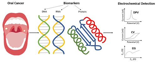
Figure 1.
Schematic illustration of an oral cancer-detecting process by using electrochemical biosensors. Abbreviations: Differential pulse voltammetry (DPV); Cyclic voltammetry (CV); Electrochemical impedance spectroscopy (EIS).
Figure 1.
Schematic illustration of an oral cancer-detecting process by using electrochemical biosensors. Abbreviations: Differential pulse voltammetry (DPV); Cyclic voltammetry (CV); Electrochemical impedance spectroscopy (EIS).

Figure 2.
(a) Schematic illustration of the Exo-III involved in the DNA target amplification cycle and the signal readout of electrochemical DNA biosensor. (b) The three-electrode configuration on the electrochemical chip and illustration of biomolecules immobilize on DNA dendrimer. Abbreviations: Ferrocene-labeled hairpin probe (Fc); Exonuclease III (Exo-III); Methylene blue (MB); Counter electrode (CE); Working electrode (WE); Reference electrode (RE); Horseradish peroxidase (HRP); Interleukin 8 (IL-8).

Figure 3.
(a) The principle of the magnetic-controllable electrochemical RNA biosensor. (b) The modified MBs on SPdCE for the simultaneous determination of IL-8 protein and IL-8 mRNA. Abbreviations: Magnetic bead (MB); Horseradish peroxidase (HRP); 3,3′,5,5′ Tetramethylbenzidine (TMB); Interleukin 8 (IL-8); Streptavidin-modified magnetic bead (Strep-MB); Carboxylic acid-modified magnetic bead (HOOC-MB); Hydroquinone (HQ).

Figure 4.
Electrochemical immunosensors based on the detection of biomarkers IL-6 and IL-8 for targeting oral cancer. (a) (i) Conceptual strategy for detection of protein biomarker by micro-fluidic immunoarray. Correlation plots of immunoarray assay results for conditioned media of different cell lines vs. standard ELISA assays for: (ii) IL-6; and (iii) IL-8. (b) (i) Multilabel amperometric immunosensor; and (ii) influence of IL-6 concentration on steady-state current for immunosensor using the bioconjugate. (c) Schematic diagram illustrating SiNW arrays functionalized with two antibodies and two biomarkers. (a) Adapted with permission from ref. [31]. Copyright 2014 Elsevier. (b) Adapted with permission from ref. [25]. Copyright 2010 American Chemical Society. Abbreviations: Poly(diallyldimethylammonium chloride) (PDDA); Interleukin 8 (IL-8); Interleukin 6 (IL-6); Anitbody1 (Ab1); Anitbody2 (Ab2); Single-walled nanotube (SWNT); tumor necrosis factor α (TNF-α).

Figure 5.
(a) (i) Schematic representation of the fabrication of BSA/anti-CYFRA-21-1/APTES/nHfO2/ITO immunosensor for oral cancer detection. (ii) The electrochemical response of modified immunoelectrode in different concentrations of CYFRA-21-1. Insets show the calibration curves of peak current and CYFRA-21-1 concentration. (iii)The comparison between the standard sample and patient samples. (b) (i) Schematic representation of the fabrication steps of VANTA IDEs; (ii) optical image of a VANTA IDE (the values of numbers on the ruler are in centimeters); and (iii, iv) FESEM images of VANTAs from the IDE devices in two magnifications. (c) (i) Schematic representation of the fabrication and the detection principle: (top) the stepwise fabrication of immunosensor; and (below) steps of the fabrication of immunocomplex formation. FESEM images of: bare (ii) Au; (iii) Au/Cys; and (iv) Au/Cys/Anti-CD59 surfaces. (v) EIS responses for CD59 detection in 5 mM zobell’s solution and (vi) calibration plot from 1 to 1000 fg mL−1 CD59 concentrations. (a) Adapted with permission from ref. [34]. Copyright 2016 Elsevier. (b) Adapted with permission from ref. [57]. Copyright 2018 Elsevier. (c) Adapted with permission from ref. [48]. Copyright 2014 Elsevier. Abbreviations: 1-Ethyl-3-(3-dimethylaminopropyl)carbodiimide (EDC); N-Hydroxysuccinimide (NHS).


Table 1.
Biomarkers that have been used for oral cancer electrochemical biosensors and the experimental conditions.
Table 1.
Biomarkers that have been used for oral cancer electrochemical biosensors and the experimental conditions.
| Biomarker | Sample | Electrochemical Method | Detection Limits | The Levels of The Biomarker in Normal Case and Cancer Patient | References |
|---|---|---|---|---|---|
| Amylase | Samples spiked in potassium ferrocyanide | Cyclic voltammetry | 1.57 pg mL−1 | - | [23] |
| IL-8 protein, IL-1β protein and IL-8 mRNA | Samples spiked in buffer | cyclic square-waveform | Protein:100–200 fg mL−1 mRNA IL-8:10 aM | IL-8 protein, OSCC patient: 720 pg mL−1; Normal: 250 pg mL−1 IL-8 mRNA, patient: 16 fM; Normal:2 fM | [24] |
| Interleukin-6 (IL-6) | HNSCC cell lines | Amperometry | 2.5 × 10⁻¹⁴ M | IL-6 protein, HNSCC patient: more than 20 pg mL−1; Normal: less than 6 pg mL−1 | [25] |
| microRNA | Artificial saliva | Cyclic voltammetry Chronoamperometry | 2.2 × 10⁻¹⁹ M | - | [30] |
| IL-6 protein, IL-8 protein | Serum | Amperometry | IL-6:5 fg mL−1, IL-8:7 fg mL−1 | IL-6 protein, HNSCC patient: more than 20 pgmL−1; Normal: less than 6 pg ml−1 IL-8 protein, patient: 720 pg mL−1; Normal: 250 pg mL−1 | [31] |
| Oral Cancer Overexpressed 1 | Human saliva | Differential pulse voltammetry | 0.35 pM | - | [19] |
| IL-8 protein, TNF-α | Artificial saliva | I-V Curve | 100 fg mL-1 | IL-8 protein, patient: 720 pg mL−1; Normal: 250 pg mL−1 | [32] |
| IL-8 mRNA IL-8 protein | Human saliva | Amperometry | IL-8 mRNA: 0.21 nM IL-8: 72.4 pg mL−1 | IL-8 mRNA, patient: 16 fM; Normal:2 fM IL-8 protein, patient: 720 pg mL−1; Normal: 250 pg mL−1 | [29] |
| CYFRA-21-1 | Samples spiked in PBS buffer | Cyclic voltammetry Electrochemical impedance spectroscopy | 0.21 ng mL−1 (calculated) | CYFRA-21-1 protein, normal: 3.8 ng mL−1; patient:17.46 ± 1.46 ng mL−1 | [34] |
| CYFRA-21-1 | Samples spiked in PBS buffer | Cyclic voltammetry Differential pulse voltammetry | 0.122 ng mL−1 (calculated) | CYFRA-21-1 protein, normal: 3.8 ng mL−1; Patient:17.46 ± 1.46 ng mL−1 | [33] |
| CYFRA-21-1 | Artificial Saliva | Differential pulse voltammetry | 0.001 ng mL−1 | CYFRA-21-1 protein, normal: 3.8 ng mL−1; Patient:17.46 ± 1.46 ng mL−1 | [47] |
| CD59 | Human saliva | Cyclic voltammetry Electrochemical impedance spectroscopy | Treated Saliva: 0.84 ± 0.04 fg mL−1 Raw saliva: 1.46 ± 0.05 fg mL−1 | - | [48] |
| IL-8 protein | Human saliva | Cyclic voltammetry Differential pulse voltammetry | 72.73 ± 0.18 pg mL−1 (calculated) | IL-8 protein, patient: 720 pg mL−1; normal: 250 pg mL−1 | [49] |
| Oral Cancer Overexpressed 1 | Artificial saliva | Alternating current voltammetric Electrochemical impedance spectroscopy | 12.8 fM | - | [20] |
| CIP2A | Human saliva | Cyclic voltammetry Electrochemical impedance spectroscopy | 0.24 pg mL−1 | - | [57] |
| CYFRA-21-1 | Human saliva | Differential pulse voltammetry Electrochemical impedance spectroscopy | 0.625 pg mL−1 | CYFRA-21-1 protein, normal: 3.8 ng mL−1; Patients: 17.46 ± 1.46 ng mL−1 | [54] |
| IL-8 protein | Human serum and saliva | Cyclic voltammetry Electrochemical impedance spectroscopy Single Frequency Impedance | 3.3 fg mL−1 | IL-8 protein, patient: 720 pg mL−1; Normal: 250 pg mL−1 | [58] |
| IL-8 protein | Human saliva | Cyclic voltammetry Differential pulse voltammetry | 51.53 ± 0.43 pg mL−1 (calculated) | IL-8 protein, patient: 720 pg mL−1; Normal: 250 pg mL−1 | [59] |
© 2020 by the authors. Licensee MDPI, Basel, Switzerland. This article is an open access article distributed under the terms and conditions of the Creative Commons Attribution (CC BY) license (http://creativecommons.org/licenses/by/4.0/).
Share and Cite
MDPI and ACS Style
Lin, Y.-T.; Darvishi, S.; Preet, A.; Huang, T.-Y.; Lin, S.-H.; Girault, H.H.; Wang, L.; Lin, T.-E. A Review: Electrochemical Biosensors for Oral Cancer. Chemosensors 2020, 8, 54. https://0-doi-org.brum.beds.ac.uk/10.3390/chemosensors8030054
AMA Style
Lin Y-T, Darvishi S, Preet A, Huang T-Y, Lin S-H, Girault HH, Wang L, Lin T-E. A Review: Electrochemical Biosensors for Oral Cancer. Chemosensors. 2020; 8(3):54. https://0-doi-org.brum.beds.ac.uk/10.3390/chemosensors8030054
Chicago/Turabian StyleLin, Yen-Tzu, Sorour Darvishi, Anant Preet, Tzu-Yen Huang, Sheng-Hsuan Lin, Hubert H. Girault, Ligang Wang, and Tzu-En Lin. 2020. "A Review: Electrochemical Biosensors for Oral Cancer" Chemosensors 8, no. 3: 54. https://0-doi-org.brum.beds.ac.uk/10.3390/chemosensors8030054
Note that from the first issue of 2016, this journal uses article numbers instead of page numbers. See further details here.