Low Energy Beta Emitter Measurement: A Review
by
 ,
,
Hara Kang
1,2, 
Sujung Min
1,2,
Bumkyung Seo
2,
Changhyun Roh
2,3,* ,
,
Sangbum Hong
2,* and
Jae Hak Cheong
1,* 1
Department of Nuclear Engineering, Kyung-Hee University, Yongin-si 17104, Gyeonggi-do, Korea
2
Decommissioning Technology Research Division, Korea Atomic Energy Research Institute, Daejeon 34057, Korea
3
Quantum Energy Chemical Engineering, University of Science and Technology (UST), 217 Gajeong-ro, Daejeon 34113, Korea
*
Authors to whom correspondence should be addressed.
Chemosensors 2020, 8(4), 106; https://0-doi-org.brum.beds.ac.uk/10.3390/chemosensors8040106
Submission received: 22 September 2020
/
Revised: 23 October 2020
/
Accepted: 25 October 2020
/
Published: 28 October 2020
(This article belongs to the Special Issue Radiation-Based Sensors and Nanosensors)
Abstract
:The detection and monitoring systems of low energy beta particles are of important concern in nuclear facilities and decommissioning sites. Generally, low-energy beta-rays have been measured in systems such as liquid scintillation counters and gas proportional counters but time is required for pretreatment and sampling, and ultimately it is difficult to obtain a representation of the observables. The risk of external exposure for low energy beta-ray emitting radioisotopes has not been significantly considered due to the low transmittance of the isotopes, whereas radiation protection against internal exposure is necessary because it can cause radiation hazard to into the body through ingestion and inhalation. In this review, research to produce various types of detectors and to measure low-energy beta-rays by using or manufacturing plastic scintillators such as commercial plastic and optic fiber is discussed. Furthermore, the state-of-the-art beta particle detectors using plastic scintillators and other types of beta-ray counters were elucidated with regard to characteristics of low energy beta-ray emitting radioisotopes. Recent rapid advances in organic matter and nanotechnology have brought attention to scintillators combining plastics and nanomaterials for all types of radiation detection. Herein, we provide an in-depth review on low energy beta emitter measurement.
1. Introduction
Globally, the characterization of residual radioactivity is of important concern to treat radioactive waste generated during the operation and decommissioning in nuclear facilities. The characterization is essential to estimate the radiological hazard and support decision making at decommissioning sites [1,2]. A final status survey has been carried out to meet the regulation release via lower radioisotope concentrations to the derived concentration guideline level (DCGL) [3]. Mainly, low energy beta-ray emitting radioisotopes such as 3H, 14C, and 63Ni are usually detected by a liquid scintillation counter (LSC). However, this approach requires a long time for pretreatment and waste generated after the analysis is harmful to the environment [4,5,6,7,8]. Additionally, the specific and large size equipment of the LSC makes it unsuitable for measuring beta-ray emitting isotopes on site due to its complex systems [9]. Furthermore, the LSC is suitable to measure beta-rays due to their low atomic number and density because the elemental composition of plastics is H and C. Additionally, plastic scintillators are easy to manufacture in desired size and can be inexpensively fabricated in a large size [10]. Above all, characteristics of the different additives in the plastic scintillator grant special properties such as high Z material loading for gamma-ray measurement or boron loading for thermal neutron detection [11]. Due to these characteristics, plastic detectors have been used to measure β-rays [12]. The plastic scintillator converts radiation into scintillation [13], and then a spectrum analysis of incident radiation is performed by converting scintillation into current through a photosensor and amplifying it. Since the physical/chemical properties vary depending on the type of scintillator, it is important to select an appropriate scintillator upon target radiation [14]. Thus, the characteristics of low energy beta-ray emitting isotopes and trends regarding in-situ detectors to measure low-energy beta-ray emitting isotopes were investigated.
This article provides an overview of the characteristics of low energy beta-rays and the characteristics of radioisotopes occurring at the decommissioning site. In addition, we elucidated commercial plastics and detectors to carry out an analysis of the technical requirements and detector structure for an in-situ beta-ray measurement by analyzing technology trends for a low energy beta-ray measurement. Thus, recent commercial detectors for measurement of low energy beta-ray emitting isotopes were investigated. Furthermore, the technical status of the measurement of radiation levels for monitoring or measuring low energy β-emitting radionuclides at home and abroad was investigated. Ultimately, this paper could provide basic data for the development of technologies for measurement of low energy β-emitting isotopes.
2. Low Energy Beta-Emitter Characteristics
Radioisotopes generated at decommissioning sites such as 60Co and 137Cs are easy to measure without requiring chemical separation from other isotopes. In the meantime, beta-ray emitters that cannot penetrate a thick medium should be completely separated from other isotopes for measurement [15,16]. Low energy β emitters mainly originate from neutron activation due to their low atomic mass. Radiation protection against internal exposure via intake, inhalation, and ingestion is essential [17,18], while risk arising from external exposure is very slight because of its low energy and short range [19,20].
There are numerous radioisotopes at the decommissioning site. Tritium and carbon-14 are the main isotopes that emit low energy β-ray, with respective maximum energy of 18.6 keV and 156 keV [21]. Owing to their low energy, they are not treated as radioisotopes that require protection from external exposure. Nevertheless, these radionuclides interact in some environmental mechanisms and turn into various forms influencing the human body. Additionally, low energy of the beta-emitters leads to some difficulties in detection and consequently conventional detectors such as the ionization chamber or the Geiger–Muller counter are incapable of measuring low energy beta emitters. Table 1 shows representative beta-emission isotopes, and also presents the characteristics and human effects of low-energy beta-emitters at the decommissioning site.
2.1. 3H
Although tritium has the same chemical behavior as that of hydrogen, this isotope emits radiation via beta decay, unlike hydrogen or deuterium [22]. Tritium has two neutrons and one proton and releases beta-rays that are converted into a stable isotope (Equation (1)).
In general, the main contribution to the accumulation of 3H in concrete is the neutron radiation reaction of 6Li(n, α)3H [23]. Additionally, neutron capture of 2H(n, γ)3H and 3He(n, p)3H in the reactor evaporator is mainly detected in nuclear facilities. Tritium travels only 6 mm in air, and it cannot penetrate the dead layer of skin [18,24]. It also penetrates only about several micrometers in graphite [25] and less than 1 pm in water [26]. Nevertheless, as a result of having the same chemical properties as hydrogen, tritium tends to replace stable hydrogen in the human body in a gaseous state or the form of tritiated water. Tritium gases are rarely dissolved in the human body, and objects can be exposed to and adsorb the vapor of tritium. Only 0.004% or less of tritium gas is absorbed once inhaled, but 98–99% of it is absorbed by the human body when it is breathed for 4–5 min in air saturated by evaporating tritiated water [27].
2.2. 14C
14C is mainly produced by the neutron capture reaction of 14N(n, p)14C, 13C(n, α)14C, and 17O(n, α)14C at the reactor core during the operation of a nuclear reactor [21]. Among others, the reaction 14N(n, p)14C at the concrete shield [28] around the reactor core is the main contribution of the 14C production due to the high neutron cross-section [23,29]. In addition, 14C, which is highly volatile, exists mainly in the form of carbonate and is highly mobile in groundwater and in the form of CO2 [30]. Stable isotopes 14N and 17O are very common in building materials, and they are mainly detected in radioactive metals from reactors and can be detected in all materials examined for neutrons.
2.3. 36Cl
36Cl is a radioisotope with a 301,000 year half-life that decays to the stable state of 36Ar emitting maximum energy of 709 keV (98.1%) or electron capture (1.9%). 36Cl is created via neutron activation of rocks on the ground. Additionally, neutron activation of the stable state radioisotope in nuclear fuel, graphite, coolant, steel, and ion-exchangers produces 36Cl via 35Cl(n, γ)36Cl [31,32].
2.4. 63Ni
63Ni is the most abundant radioisotope in nuclear facilities at decommissioning, from graphite, pipes, and concrete to ion-exchangers [33]. 63Ni is created by the reaction of stable Ni and Cu of 62Ni(n, r)63Ni, 63Cu(n, p)63Ni, and decays to 63Cu after emission of β-ray energy of 66.95 keV. Ni has high resistance to water and air and is used for metal protection coating alloys for corrosion resistance metal. Therefore, 62Ni produces 63Ni via neutron activation in structural iron and steel of nuclear reactors and internal components and 63Ni is released through corrosion of the surface and circulating coolant of metals such as stainless steel or Inconel.
2.5. 90Sr
90Sr is mainly produced by nuclear fission and emits 90Y after high-energy beta-decay. It is found in radioactive waste such as ion exchange resin, filter sludge, and at the bottom of the evaporator. In addition, 90Sr is highly soluble and thus easily transported through precipitation and groundwater. 90Sr constitutes a long-term biological hazard as it accumulates in bone tissues and can lead to cancer via ingestion [31].
2.6. 94Nb
94Nb is generated by nuclear fission or neutron activation of a stable state of 235U and 239Pu at the nuclear reactor. STable 94Nb exists in large quantities in reactor vessel material and fuel cladding components with large amounts of Inconel [34]. 94Nb is mainly generated by neutron capture and decay to 94Mo with 472 keV of β-ray.
2.7. 99Tc
99Tc is not a naturally occurring radioisotope but it is generated by nuclear fission of uranium and plutonium. 99Tc is mainly found at the bottom of the evaporator or radioactive waste such as in the filter and sludge [35]. The main form of 99Tc is [TcO4]−, which is highly mobile in groundwater and has a long half-life, which can pose long-term radiological risks.
2.8. 129I
129I is mainly produced by uranium nuclear fission in the reactor [36] and is not a naturally occurring nuclide. 129I is found in radioactive waste such as ion exchange resin, enriched waste fluid of coolant, filter sludge, and cartridge filters. When inhaled and ingested, most is dissolved in body fluids and deposited in the thyroid gland.
2.9. 241Pu
In the case of 241Pu, it is produced in the reactor through neutron absorption by the β decay of 241Am of trans-uranium elements [37]. In general, plutonium waste is classified as high-level waste, but various wastes generated during reactor operations are also present in low-level waste.
3. Beta Ray
Gamma-ray emitters such as 60Co and 152Eu can be easily detected when analyzing the γ spectrum, but in the case of β emitters, it is difficult to distinguish radioisotopes due to poor energy resolution in the spectrum [30]. This is particularly noticeable in the low-energy β area, and there is also a problem that energy measurement is difficult because the noise generated in low-energy areas overlaps with the radioisotopes [32]. Figure 1a,b show the track of γ and β-ray through a medium. As shown in Figure 1a, gamma-rays lose energy for a short period of time due to the photoelectric effect, Compton scattering, and electron pair production [38,39].
The interaction within the medium of the γ-ray can be classified according to the energies of the incident photons, which are shown in Table 2.
Meanwhile, β particle continuously loses energy in the medium (shown in Figure 1b) [40,41]. As a result, the effective penetration range of the electron is short because the total range through which particles have moved and the electrons travel in a straight line do not match [42,43]. Therefore, low-energy β isotopes such as 3H and 14C are analyzed by methods such as the separation of radioisotopes by a liquid scintillation counter (LSC) [44,45] or beta-ray induced X-ray spectrometry (BIXS) [46] due to their short range, making it difficult to transfer energy to detectors. The number of neutrons in a nucleus is excessive and it tends to emit electrons (β particles) [42]. This is called β-decay, the neutron converts into a proton and β particle (electron), and nuclear conversion occurs with electron emission [30]. On the other hand, when the number of protons in a nucleus is excessive, the proton is converted into a neutron and electron. Nucleus conversion then occurs, releasing positrons (β+ particles), which is called β+ decay. When the particles are emitted from radioactive decay, the sum of the energy should be constant, and thus an anti-neutrino is released with β− particle emission, and a neutrino is released with a β+ particle (Table 2) [47]. Due to this mechanism, the emitted energy from beta decay distributed between the energy of β particles and the energy of the neutrinos produces a continuous energy spectrum. β decay leads the nucleus to enter an excited state, and excess energy released by emitting one or more photons. Additionally, excess energy emitted via internal conversion transfers the surplus energy that subtracted binding energy to the orbital electron, to produce an internal conversion electron.
4. Interaction with Matter
Electrons besieged by the Coulomb electric field that interacts with all of the particles passing through it [48,49]. For most of the interactions, only a minuscule fraction of the kinetic energy of an incident particle can be transferred [29]. These interactions are similar to an electron losing its energy by friction. This process is commonly referred to as CSDA, or continuous slowing-down approximation [32]. In principle, there are three interaction processes.
- Hard collisions:Inelastic scattering with atomic electrons generates excitation or ionization of electrons, and delta-rays (secondary electrons) are originated. The probability of this interaction is proportional to the atomic number, Z.
- Interactions by Coulomb force with an external nucleus field:Inelastic scattering with nuclei results in photons means Bremsstrahlung. The probability of this interaction process is proportional to Z2.
- Soft collisions:Elastic scattering, in which electrons lose a small amount of energy, is necessary to satisfy the conservation of momentum with a collision. The probability of this interaction is proportional to Z2.
Beta particles have the same mass and charge as the electrons, which differ from their origin. Beta particles are emitted from the nucleus during radioactive decay, while electrons are produced or exist outside of the nucleus of an atom. Additionally, beta particles are generated by pair production that contains negatively charged particles (negatrons) and positively charged particles (positrons) at the same time. Each particle is released in a different direction at an angle of 180° in pair production by the conversion of gamma radiation in the vicinity of a nucleus.
There are two mechanisms by which beta particles interact with matter, ionization and electron orbital excitation [50], dissipating their kinetic energy. Plus, there is a third mechanism via which beta particles interact with matter, their radioactive energy is dissipated via Bremsstrahlung production, which distinguishes the beta particle compared to other radiation. Thus, a β particle has stopping power, described by Equation (2), which is given by the sum of the collisional and radioactive contributions [51].
The expected value of the energy loss rate per unit path length, x, is called the stopping power (dT/dx)Y,T,Z by the type of charged particle, Y, in the medium of atomic number, Z, and the kinetic energy of the charged particle, T. Dividing the stopping power by the density () of the absorbing medium gives the mass stopping power (dT/dx) [48]. Stopping power can be divided into collision stopping power and radiative stopping power. Collision stopping power is the energy loss rate resulting from the sum of soft collisions and hard collisions, commonly referred to as collision interactions. Unless otherwise specified, radiation stopping power is assumed to originate solely from photons (bremsstrahlung). The energy consumed by radiative collision is generated from the track of a charged particle, and it leads to ionization and excitation nearby the track of the particle. The mass collision stopping power may be expressed as follows in Equation (3):
where c is the collision interaction, s is the soft collision, and h is the hard collision.
5. Scintillation Process
Scintillation is a phenomenon that a substance is illuminated by external energy such as radiation, electric field. When radiation is incident to the scintillator, it interacts with particles in the medium to transfer energy to the periphery and excite scintillation molecules. Excited molecules are then immediately stabilized and emit photons with as much light as the difference in the energy level when returning to the ground state [39]. These scintillation characteristics depend on the type of scintillation material, incident radiation, and the nature of secondary charged particles. Beta particles are absorbed in matter and their energy dissipates by colliding with molecules in the scintillator, which transfer the energy via heat, ionization, and excitation. For efficient energy transfer between beta particles and the scintillator, a scintillation cocktail, which converts kinetic energy of beta particles into light energy, is added to the scintillator. As the molecules excited by the scintillator transfer their energy to other molecules or solutions, electrons are excited. After an excited electron emits photon light in the ultraviolet region, it goes to the ground state. One of the key performance measures of the scintillator is the light yield, which is defined below [52].
The intensity of photon light from the scintillator is proportional to the initial energy of the beta particle through linear conversion of the photosensor. Photosensitive devices such as a photomultiplier tube (PMT) or silicon photomultiplier tube (SiPM) amplify the light and convert detected photon light into an electrical form to detect emitted photons efficiently. The photomultiplier tube collects the total photon light produced within the scintillator, and the inside face of the PMT is uniformly coated with a photosensitive material that converts photon light energy into electrical energy. The electrons are drawn to the electrodes in the photomultiplier tube by the positive potential of the electrodes to produce more electrons. These secondary electrons are attracted to the following electrodes and repeat the next diodes that make up the PMT (Figure 2). The electrons amplified at each electrode stage produce an electrical pulse proportional to the photons.
PMT is very sensitive and generates small pulses even when there is almost no light and produces noise that appears in the background area of the sample measurement. Additionally, noise is caused by external factors such as heat, cosmic rays, and fall out, and it is difficult to distinguish low energy β-ray from this noise [53]. Photons incident to the photo-sensor generate photoelectric effects and are converted into photoelectrons, which are amplified by electrical signals to a sufficient level for spectral analysis through preamplifiers and main amplifiers. As the height of the converted current signal pulse is proportional to the energy of the incident radiation, analysis of the incident radiation from the pulse-height is possible, and the amount of incident radiation can be derived.
6. Characteristics of the Scintillator
The scintillator acts by converting radiation into scintillation and converts its light into current and amplifies it through the light sensor to perform an energy analysis of incident radiation. Scintillators suitable for radiation detectors afford the following characteristics [27,30,31,32,40,42,48,51,53].
- High scintillation efficiency of radiation energy:The luminescence efficiency of the scintillator is given by the ratio of energy lost by radiation within the scintillator to energy converted to scintillation, and the luminescence intensity of the scintillator varies depending on the type of scintillator and the quality of radiation. Higher scintillation efficiency increases the luminescence sensitivity of the scintillator due to its high energy absorption and conversion efficiency to photons. The luminescence efficiency of an organic scintillator is a function of the luminescence intensity of the anthracene to the electrons. A high light output means that when radiation with the same energy is incident, the number of photons produced is high, which means that the amount of data is high, which has a significant effect on the resolution. The high light output has high luminescence efficiency because it has excellent linearity proportional to the intensity of light emitted by the scintillator and the energy of incident radiation.
- High transparency:Higher transparency increases the amount of light that reaches the PMT, which increases the efficiency of light collection. It can be obtained by minimizing the self-absorption, internal attenuation of the emitted photons, and high transfer efficiency to the photosensor.
- Short decay time of scintillation:The time taken for a luminescence phenomenon to dissipate, called the scintillation attenuation time, classifies luminescence characteristics according to the time remaining in the material as follows:Fluorescence: Light stops as soon as the energy is cut off.Phosphorescence: Residual light remains even after cutting off the incident energy.A short attenuation time of scintillation can count high dose rates of radiation because of the decrease in the dead time and because the signal pulses are rapidly produced. In addition, the precision is proportional to the attenuation time of the scintillator, which improves the possibility of simultaneous measurement applications due to the short rise time of the signal pulse. Generally, there is an attenuation time of several seconds for an inorganic scintillator and several nanoseconds for organic scintillators.
- The wavelength distribution of the scintillation is suitable for the spectral sensitivity characteristics of the photomultiplier tube.
When radiation that enters a scintillator is converted into light, it has much smaller energy and longer wavelengths than original radiation. If the wavelength distribution of the scintillation and the spectral sensitivity characteristics of the PMT are suitable, the photons can be efficiently converted into photoelectrons, thus achieving high photoelectron emission efficiency, defined as quantum efficiency. Therefore, the output wavelengths of light emitted from the scintillator should be consistent with the absorption wavelength and refractive index (e.g., the refractive index of glass, 1.5) of the photosensor. Therefore, the scintillator should be fabricated or selected with consideration of its compatibility with the response functions, such as the optimal wavelength area of the photosensor. In addition, there are indicators of comparable performance and influencing factors for determining optimal scintillation, such as radiation integrity, low cost, large-scale production possibility, thermal/mechanical integrity at the environment, and the applicability of pulse shape discrimination techniques. In addition, scintillators for beta-ray measurement have to be thin to detect the incident radiation only for beta-rays and avoid interference from gamma-rays [54].
6.1. Types of Scitillators
The types of scintillators can be classified as follows, depending on their condition and chemical composition.
- Depending on the condition: solids, liquids, and gases.
- Depending on chemical composition: inorganic and organic scintillator.
Depending on the type of scintillator and the scintillation mechanism, the radiation under the measurement varies, and the characteristics of scintillators according to chemical composition are shown in Table 3.
6.1.1. Inorganic Scintillator
An inorganic scintillator contains about 0.1% of impurities such as Eu, Ce, and Tl to enhance the luminescence and form an energy level at which scintillation can occur [55]. Therefore, it is not an exciton, a weakly coupled electron-electric pair, but an extrinsic crystal resulting from luminescence in impurities. Since a high-density material is added, it is used for measuring γ-rays, which have a long range in the medium. The following two types of inorganic scintillator.
- NaI(Tl):It has a high density (3.67 g/cm3) and contains high atomic number composition (I, Z = 53), and thus offers efficient detection of γ-rays and excellent linearity. Its use has been expanded to large-capacity detectors such as monitoring nuclear power plants, medical care, and security searches due to its relatively low price. However, it is used to measure γ and medium-hard X-rays rather than measuring α, β, and soft X-rays, which have weak permeability, because they have low mechanical thermal impact and have to be sealed with aluminum to block contact with the air [56].
- BGO (Bismuth Germanate, Bi4Ge3O12):The high atomic number (Z(Bi) = 83) and high density (7.3 g/cm3) result in excellent detection efficiency and are used for γ-ray and X-ray measurements. In addition, the attenuation time of luminescence is very short, and thus it is used as a detector such as in X-ray CT and PET and has excellent mechanical strength and chemical properties. However, it has a low intensity of luminescence, resulting in lower energy resolution than NaI(Tl) [57,58].LiI(Eu) is also used for thermal neutron measurements and ZnS(Ag) is used for α-ray measurements.
6.1.2. Organic Scintillator
The organic scintillator is an aromatic hydrocarbon compound and has a benzene-ring structure [59]. Organic scintillators are classified into organic crystals, plastics, liquid scintillators, etc., depending on their densities [60]. Unlike inorganic scintillators, the luminescence is of molecular origin, forming an ion pair (exciton) by electron excitation between energy levels within the composition molecules [55]. The exciton then moves just below the conduction band within the scintillator crystal and falls down to the valence band via capture by the cation. Finally, photons are emitted by the formation of electron-hole pair recombination.
6.2. Liquid Scintillator
It is widely used for measurements such as 3H, 14C, etc., due to the high detection efficiency in low energy β-rays. Since the samples containing 3H and 14C are measured directly in the liquid scintillator, there is no absorption or attenuation of the β-ray at the incident window. Due to its low atomic number and density, the luminescence efficiency of the liquid scintillator is not high.
6.3. Gas Scintillator
Luminescence sensitivity is weaker than a solid scintillator because of its small density, but the attenuation time is the shortest. The luminescence efficiency is very small for γ-rays, electrons, and neutrons but relatively large for α-rays or fission fragments.
7. Inorganic Nanomaterials
Conventional plastic scintillators are fabricated with an organic solvent containing a scintillation cocktail and then dried it. Therefore, the atomic number and density of the components are low, resulting in low light conversion efficiency in the scintillator. Additionally, they have poor detection efficiency and energy resolution. Adding materials with high atomic numbers and nanomaterials with various properties can increase the light conversion efficiency and density of the scintillator. Plastic scintillators containing nanomaterials of high atomic number and organic/inorganic hybrid scintillators with the addition of a high atomic number inorganic nanomaterial to the conventional plastic are manufactured to compensate for the shortcomings of the organic scintillator (low energy resolution and low measurement efficiency) while having the advantage of the inorganic scintillator. As conventional inorganic nanomaterials such as CdTe and ZnO have poor light conversion efficiency due to poor light absorption because of structural defects or low density, the luminescence rate is increased by adding activators such as Ce3+. Below is a summary of the types and characteristics of inorganic nanomaterials.
7.1. Perovskite (Calcium Titanium Oxide Mineral)
Perovskite, with a chemical formula of ABO3 [61], consists of a material with a high atomic number and high density, and it leads to a high light absorption rate, high charge diffusion coefficient, and excellent charge movement, resulting in high light conversion efficiency in the scintillator [56,62]. However, it has shortcomings including vulnerability to moisture and oxygen because of its ionic compound, and also is environmentally harmful, addictive, and prone to oxidation due to Pb, its main component. Therefore, to overcome these disadvantages, a perovskite that is not addictive and has high atmospheric safety using Sn instead of Pb was recently developed [63].
7.2. CdTe Structure of LaF3:Ce/CdTe
The CdTe structure of LaF3: Ce/CdTe is a Ce3+ doped fluorescent with Ce3+ as an activator with a very fast response speed and emits scintillation in the UV range. The CdTe luminescence of LaF3: Ce/CdTe is about five times stronger than pure CdTe due to improved energy transfer and an absorbent rate of light and stability of structure defects [64].
7.3. CeF3/ZnO
ZnO is a semiconductor with a wide bandgap of 60 MeV and has a disadvantage of a low light emission rate due to its high exciton binding energy, and it has fast scintillation speed but relatively low density. To improve this, CeF3/ZnO with Ce3+ doping is developed, and energy transfer from CeF3 in the scintillator to ZnO occurs. Compared with simple ZnO nanoparticles, the luminescence is 30 times higher and the X-ray luminescence is four times higher [65].
8. Beta-Ray Detector Technology
8.1. Commercial Plastic Scintillator
Depending on the type of scintillator, the physical and chemical properties such as light output and density vary, and thus it is important to choose the appropriate scintillator according to the radiation being measured. Additionally, the longer wavelength of light emitted from the scintillator matches the sensitivity of the PMT, there is less loss of light, and the wavelength of the absorption and emission of the radiation varies from one scintillator to another. Therefore, in this section, the characteristics of commercial scintillators mainly used for the measurement of β particles are compared.
8.1.1. Eljen Technology
As shown in Figure 3, EJ-260 is a suitable scintillator for sensitivity of the light sensor consistent with the long wavelength of emission. The green fluorescence emission of EJ-260 is sufficiently short in wavelength, and the scintillation efficiency is sufficient to use with PMT, which is sensitive to blue wavelengths. The characteristics of EJ-260 are shown in Table 4.
8.1.2. Saint-Gobain
BC-400 and BC-404 are suitable for measuring γ, α, and β-rays below 5 MeV. In the case of BC-400, it is a commonly used scintillator, whereas BC-404 is used for fast counting. BC-408 meanwhile is efficient for X, α, and β-ray measurements below 100 KeV and has high light output. It can be made to a large extent, suitable for measuring pollution in a wide site. Furthermore, BC-428 is a scintillator emitting green fluorescence, similar in efficiency to BC-400, but in the case of light output, it has 56% efficiency in non-alkali photocathode PMT. The emission spectrum and the characteristics of each plastic scintillator are shown in Figure 4 and Table 5, respectably.
CaF2:Eu is used to detect hundreds of keV of γ-rays due to the low atomic number that constitutes the substance and is suitable for detecting β particles due to low back-scattering. In addition, CaF2:Eu is non-hygroscopic and thus radiation in the solution can directly contact the crystals. Additionally, with resistance to heat and mechanical impact, it can be made in various forms. Figure 5 is a scintillation emission spectrum of CaF2:Eu with a maximum emission value near 435 nm. CaF2:Eu penetrates visible light well but absorbs light at 400 nm, which partially overlaps with the scintillation emission area, causing self-absorption of the scintillator. Thus, for applications requiring optimal energy resolution, less than 1 inch of length is appropriate due to the self-absorption of CaF2:Eu.
8.2. Commercial Detector
Commercial detectors for β measurement with high user convenience have been manufactured in various countries, including the United States and the Netherlands.
8.2.1. United States
In 2013, the Savannah River National Laboratory (SRNL) developed a system for rapid analysis of tritium and other β or α particles at the site. Figure 6 shows a portable rapid tritium analysis system (PORTAS) detector produced by SRNL that combines a sample holder, a small PMT, and a multi-channel analyzer into a single package. These packages are surrounded by aluminum cases and entirely shielded from outside light. Radioactive isotopes are measured by putting radionuclides in a liquid scintillating cocktail container. The container is fixed by a screw cap at the end of the case, and then by attaching the cap, light is blocked. Ortec fabricated a multi-purpose α/β counter using a gas flow proportional detector or a double phosphor scintillator (Figure 7). The α/β systems of gas flow proportional counter type produced by Ortec are shown in Table 6.
Ortec used a gas flow-type proportional counter tube to fabricate an automatic/manual detection system and a single/multiple detection systems and P-10 gas for filling gas (Figure 8). The automatic detector system is mainly used for relatively short count times to count multiple samples. Table 6 shows Ortec’s automated detection systems. In Table 7, the manual single detector is compared to a long count time to measure the small number of samples.
MPC-9604 is an α/β multi-detector system (MDS) for low-level background radiation measurements and is used for rapid sample throughput and high sensitivity (Figure 9). Each MPC-9604 contains four separated 2.25-inch diameter pancake type gas-flow proportional counters and aluminum windows. By connecting up to 12 MPC-9604 devices to a single PC, a total of 48 independent channels could be used. The MDS system is shielded with a large gas flow proportional counter guard detector to block the cosmic rays and a 4-inch lead shield to remove background noise. It also incorporates amplifiers of spectroscopy grade to process signals and linear low voltage power supply devices to remove electrical interference. These signals are transmitted through the shielded cable. A RFI (radio frequency interface) guard and metal enclosure are applied to eliminate noise.
Ortec has developed a dual phosphor-type counter, ASC-950-DP automatic sample changer and MPC-900-DP manual single sample changer (Figure 10). It is mainly used for health physics where rapid counts are required such as for smear or air filter measurements. A 1.5 kg lightweight probe was produced to monitor low/middle/high energy β particles in a wide-area plane, such as floors and walls (Figure 11a). The probe consists of a rectangular large-area window of 600 cm2, making it sensitive to the detection of low-level energy-emitting isotopes in a wide area. It was shown to have 19% efficiency and sensitivity for 60Co, 26% for 36Cl, and 24% for 90Sr/90Y. BP19AD/BP19DD beta probes applied large BC-400 plastic scintillator probes inside the light-alloy die-cast housing (Figure 11b). The reaction with the low energy β-ray is good results from a low background. HP-380B and HP-380AB are portable survey meters with a high α/β sensitivity including low background counts (Figure 12a). The detector can be used as a smart probe, including a memory device that stores all calibration and function parameters. Using ZnS(Ag) for α-ray detection and a plastic scintillator for β-ray detection, a dual phosphor material for distinguishing α-ray and β-ray was conjugated. In addition, a fine mesh made of stainless-steel was used to protect the detector’s external area from holes inside of the lightweight aluminum housing. In addition, a dual phosphor scintillation probe consisting of DP8A and DP8B is used to monitor α/β surface contamination such as floors and walls. It has a high sensitivity to distribute radioactive contamination using a large-area probe of 600 cm2 (Figure 12b). In Table 8, the characteristics of the probes from ThermoFisher are compared.
AMS-4 is a continuous detection system for early warning of worker exposure by airborne emission such as β-ray emission particles, radioactive iodine, and noble gas in the air (Figure 13). Both fixed and portable types are available due to its light and rigid composition with a size of 32.5 cm H × 27.9 cm W × 22.2 cm D and weight of 3.4 kg. Table 9 shows the specifications of each function of the AMS-4. It provides DAC (Derived Air Concentration)-based alerts for radionuclides specified in 10 CFR Part 20. An Ar/CO2 gas proportional detector can monitor the discharge of effluent through an in-line sampling head and provides a real-time γ-ray background subtraction function using the remote sampling function.
HP-210 and HP-360 are designed to detect effluent from radiation workplaces and to prevent the spread of contamination inside and outside the laboratory, designed for daily contamination surveys of all surfaces that could be exposed to radiation, such as individuals, tables, floors, and equipment (Figure 14). High sensitivity using thin mica windows protected by etched (stainless-steel screens) to β-ray emission of surface contamination (40 keV) can meet laboratory environmental and safety requirements. For HP-210T, high-density tungsten shielding enables relatively low β-ray monitoring in a γ background, and aluminum-housed HP-210AL allows low energy β-ray monitoring in the low energy background area. Table 10 shows the specifications of the Geiger–Mueller detectors from ThermoFisher (Waltham, MA, USA).
8.2.2. Netherlands
Scionix produced an α/β detector using SIPM (J-60035-4P) as a light sensor. Figure 15 shows a plastic scintillator mixed with ZnS(Ag) with an α/β detector produced by Scionix with a polyester housing and a double aluminum miler (mylar, 0.9 mg/cm2) incident window to block out light. Other characteristics of this commercial detector are shown in Table 11.
8.2.3. Japan
In 2013, Japan produced a measurement system to analyze 90Sr underwater (Figure 16) based on a combination of a plastic detector and a gas flow type Geiger–Muller counter tube surrounded by a lead shield. The electrons collected by each detector are processed through a simultaneous circuit. For 90Sr measurement in groundwater in a depth of 4 m, a 2 Bq/L detection limit was obtained within 10 days [81,82,83]. Likewise, the pico-beta analyzer requires a long average analysis time of two to four days per bottle of the liquid sample and requires pretreatment such as fuming nitric acid or the strontium resin method [82]. In addition, due to the thick lead shielding and the nature of the gas flow-type proportional counter, a Q gas supply system is essential, making it difficult to move outside.
8.3. Flow Cell Detector
Tritium inflows to the human body via inhalation or ingestion, and the formed organically bound tritium (OBT) leads to the detriment of DNA [84]. Thus, the maximum tritium concentration of drinking water is legislated in every country (EPA of U.S: 740 Bq/L, EURATOM of EU: 100 Bq/L) [85,86]. Furthermore, detectable technologies with rapidity and precision to prevent tritium pollution are required. Conventional low energy beta-emitting isotopes are measured by a liquid scintillation counter (LSC) due to the short range of the beta particle. However, a LSC is not appropriate for an in-situ measurement, and pretreatment for sampling and long analysis time are required. In the case of a gas detector, a complicated system where the ionizing chamber and gas tank are combined with the detector make automatic work infeasible. While the solid detector can measure beta-rays directly, the performance is degraded by erosion from water. To compensate for these defects, studies on fabricating a flow-cell type detector to monitor and prevent low beta-ray effluents have been carried out at home and abroad.
8.3.1. Korea
In UNIST, a study on real-time tritium monitoring at underwater was performed. The tritiated water is electrolyzed using a proton exchange membrane (PEM) cell, and is measured using a gas proportional counter. Figure 17 presents the composition of the electrolysis system with the PEM cell.
After putting tritium water into a water container, it is supplied to the PEM cell by connecting a power supply to the water pump. In Figure 18, HT gas, H2 gas, and a small quantity of water are produced and only generate gases that are captured by the probe, which comprises of a plastic scintillator and two PMT (R 878, Hamamatsu) inside an acryl case.
The above system is used to measure the radioactive variation of tritium per unit of mass according to the current applied to the PEM cell. It also calculates the ratio of the current change rate to the change in radioactivity per unit mass in the number of electrolyzed tritium and predicts the trend of radioactive change due to the change in current. As a result, the current was optimized at 7 A, with a detection efficiency of 31.3% ± 1.3% and a minimum detectable activity (MDA) calculated at 10.3 ± 0.8 kBq/m3 for five minutes. Additionally, the detection efficiency of a LSC is 34.4% ± 0.2%, and it showed a detection efficiency of 0.91 ± 0.04 for gas tritium produced by electrolysis.
8.3.2. Japan
In 2017, the National Institute for Fusion Science (NIFS) developed a monitoring system based on flow cell detectors for real-time measurement of underwater tritium concentrations. The flow cell detector was fabricated using a granulated CaF2 solid scintillator. Figure 19a shows three types of flow cell detectors used in the experiment: a single diameter of 3 mm cells, a series of 3 mm cells in diameter, a single diameter of 5 mm cells. The flow cell is made of Teflon PFA tubes (Figure 19a). Figure 19b shows the configuration of the flow cell detectors for tritium, including flow cells, a pair of PMTs, high voltage power, coincidence factor modules, flow cell pumps, and sample bottles. The sample bottle is a model simulating an effluent tank at the radiation facility and is filled with tritiated water. The flow cell pump sends the tritiated water to the flow cell, which is placed between the two PMTs for the coincidence system. Samples of various concentrations ae then produced by diluting the commercial tritiated water with distilled water and measured for 600 and 10,000 s using each flow cell. As a result of the study on the relationship between the count speed and concentration by passing samples through flow cells, the 5 mm diameter three cell series accurately measured a low tritium concentration of 10 Bq/mL while maintaining linearity between the count speed and the tritium concentration.
8.3.3. UK
In 2019, Lancaster University from the UK conducted a study on the fabrication of scintillation detectors to measure tritium in groundwater. First, CaF2:Eu, a non-hygroscopic inorganic scintillator, was selected and manufactured as a homogeneous scintillator with CaF2:Eu powder using two methods: a chemical method and a granulation method. Due to the quantum mechanical nature of the nanosize particles, reducing the radius of the scintillation particles results in structural changes, such as expansion of the absorption and emission bands and increase of the forbidden band, which leads to an increase of the scintillator luminescence. A study was conducted to fabricate scintillation particles with a small radius via two methods. Chemical approaches, such as a reverse micelle method, electrodeposition, and precipitation methods, were used, and the sizes of particles for each process are shown in Table 12.
The other is granulation, a pure mechanical method, which is carried out to produce fine particles. There are two methods of granulation, ball milling and use of a mortar and pestle to pulverize a monocrystalline scintillator to produce particles of a desired size. Figure 20 shows a comparison of the scanning electron microscope (SEM) image of CaF2:Eu pulverized with the two methods. The size distribution of particles produced by the mortar and pestle method for the two CaF2:Eu crystals showed that the distribution of particle size, 2–11 μm and 2–50 μm, with the centers of each particle of 7 μm and 10 μm, respectively.
As producing CaF2:Eu powder through various methods, chemical approaches were found to be unsuitable due to the use of harmful chemicals. Whereas the mortar and pestle method was found to be suitable due to its simplicity. Furthermore, the fine-sized particles of CaF2:Eu were successfully manufactured. Additionally, the radius of scintillator particles with the highest value of energy accumulated by 3H, 210Pb, and 14C calculated by the Geant 4 computational simulation were 3.5 μm, 30 μm, and 150 μm, respectively. The scintillator consists of CaF2:Eu particles with a radius of 3.5 μm deposited on a PDMS substrate and compared with a single crystal CaF2:Eu inorganic scintillator. As shown in Table 13, the sum counts of the scintillator with a particle radius of 3.5 μm is 15% higher, increasing the number of photons produced within the scintillator, leading to greater efficiency to measure radioisotopes.
Figure 21 represents a prototype flow cell that was created to detect a short-time concentration spike of radioisotopes spilled to the river (Figure 21).
The flow cell is made by machining aluminum blocks and a lid made with transparent perspex. The center of the lid was embedded with SiPM and the inlet and outlet of the flow cell were made on the side. Two sets of flow cells were fabricated for the experiment, one consists of a three-layer perspex disk, with a diameter of 4 cm and a gap of 8 mm between each layer, and the other is 12 layers of polycarbonate disks, with a diameter of 4 cm, a thickness of 1 mm, and a spacing of 1 mm. SiPM (Sense C-series 60035) was selected for the light sensor used in the detector with a smaller operating voltage (29.7 V) compared to PMT. The results by using tritiated water of 1000 Bq/mL concentration are shown in Table 14. The flow cell consisting of 12 layers has 95% higher efficiency than a flow cell consisting of three layers.
8.3.4. EU
In 2019, Southwestern European Instruments (SEI) established a real-time low-radiation tritium monitoring system to measure low-level tritium within a river near nuclear power plants. To this end, two types of flow cell prototype detectors were fabricated to obtain an optimized flow cell detector. Geant 4 simulation results showed that most (99.7%) β particles reaching the fiber optic surface were emitted at a distance of less than 5 μm, and only radioactive decay from very thin layers of water near the fiber-optic surface was detected. Therefore, cladding was excluded to maximize the exposed detection area due to the low energy of tritium (average 6 keV). Additionally, lead bricks were used to remove cosmic and natural radioactivity to reach tritium levels below 100 Bq/L. In addition, the maximum tritium concentration of drinking water was presented by EURATOM. To this end, a cosmic veto detector consisting of two layers of plastic scintillator (Epic crystal) was used, with the entire detection being positioned within a Pb shield of several centimeters of thickness. Figure 22 shows a prototype detector of the IFIC version, consisting of 64 optical fiber BCF-12 (Saint. Gobain crystals) 25 cm in length and 1 mm in diameter.
The fiber optic was connected to two PTFE containers and measured by two SiPMs configured in the coincidence mode to eliminate noise from the light detector. TRITIUM-1 IFICs were used to assess the stability of optical fibers over time and showed a stable response for nine months. Figure 23 shows an Aveiro version of a prototype detector with a larger detection surface consisting of a larger number of fibers (400) than other prototypes. The optical fibers were positioned between the two PMTs (Hamamatsu R2154-02) for the coincidence mode to increase the sensitivity of tritium detection by eliminating PMT noise. Figure 24 shows a Tritium-2 prototype module, which is the final detector to be installed in the Arrocampo dam at the Alamaz nuclear power plant in Spain. It contains 500 optical fibers with a length of 25 cm and a diameter of 1 mm, arranged in 4 × 4 SiPM (Hamamatsu) for a coincidence system. It is also positioned in a parallel container made of Teflon walls, and the reflection of light is optimized.
8.4. Beta-Ray Detector Understudying
Studies other than the flow cell on the measurement for low energy beta ray emitting radioisotopes have been carried out. This section described the detector fabrication to measure beta ray emitting radioisotopes in radioactivity contamination and radioactive waste at decommissioning sites.
8.4.1. Myong-Ji University
Myong-Ji University from Korea studied 90Sr, a major artificial radioisotope generated via nuclear fission among the radioisotopes outpoured to the ocean from the Fukushima Daiichi nuclear power generator, for real-time monitoring. Therefore, physical and structural detection efficiency and a water/dustproof detector for 90Sr measurement underwater was designed and produced. Additionally, the photon transfer efficiency of PMT with detector thickness is calculated and optimized using LightTools software. Detection characteristics of 90Sr/90Y in an underwater environment were evaluated by developing a PMT and SiPM photosensor based scintillation detector. Figure 25 represents a prototype of the scintillation detector applied to a large-area probe. In light of the maximum energy of the beta ray and physical characteristics of the scintillator, a 1 mm thick CaF2:Eu inorganic scintillator and a 6 mm × 6 mm of the small size SiPM photosensor were exploited and a Teflon reflector was used as the reflector. The voltage gain of SiPM is as high as that of PMT, but the detection area is relatively small and that decreases the detection efficiency. To compensate for this shortcoming, a study on fabricating a large-scale probe was carried out.
Based on results of the SiPM linked CaF2:Eu scintillation detector, an in-situ detector was produced as shown in Figure 26. PMT B51B03 (ADIT Inc., Sweetwater, IN, USA) exhibited a circular CaF2:Eu scintillator with the same area of photoreception of PMT with a diameter of 50.8 nm and thickness of 1 mm.
In additiion, minimum detectable concentration was 330 Bq/L for 10 min, but it did not retain the domestic emission standard of 20 Bq/L, suggesting the possibility of real-time monitoring for 90Sr.
8.4.2. KAERI
In 2017, KAERI conducted a study to in-situ measure radiological contamination from beta rays such as 90Sr and 238U. Plastic scintillators were fabricated adding nanomaterials such as Gd2O3, CdS, and CdTe. Detection efficiency with scintillator thickness was evaluated through MCNP (Monte Carlo N-Particle) simulation to measure the high energy beta ray of 90Sr (maximum energy of 2.3 MeV), showing good detection efficiency above 3 mm thickness. In the case of a low energy beta ray of 204Tl (maximum energy of 763 keV), the detection efficiency was higher with 1 mm thickness. From the results of a MCNP computational simulation for 545.9 keV low energy beta ray emission by 90Sr (standard activity 18.0 kBq), plastic scintillator thickness above 6 mm was found to be appropriate. Considering that beta-ray contamination at the decommissioning site was 0.0629 Bq/g, much lower than the radioactivity of 18.0 kBq at the experimental source, the minimum thickness to fully absorb beta-rays was calculated to be 4 mm, even considering the shielding of other radiation emitted from the site. The fabricated plastic scintillator is a mixture of organic scintillator into epoxy, using 2,5-Diphenyloxazole (PPO) as the first material and 1,4-bis(5-phenyloxazol-2-yl) benzene (POPOP) as the second. PPOs absorb UV generated by beta rays and emit 320 nm of visible light (visible light–violet, VL–V), while POPOP acts as a wave shift that absorbs VL–V and emits 420 nm of visible light (visible light–blue, VL–B) of a long wavelength. As a result, plastic produced by mixing PPO 0.2 wt % and POPOP 0.01 wt % had the highest ratio of scintillation at 380–446 nm (VL–V to VL–B) and the highest emission intensity at a 420 nm wavelength.
For PS-0201 and commercial plastic, BC-400 (Saint. Gobain) in terms of emission intensity and transmission ratio with the light, the emission intensity at a wavelength of 420 nm showed that PS-0201 was 20 times weaker than BC-400 (Figure 27). Comparing the transmittance of scintillators, the transmittance of PS-0201 at 420 nm was 83.0%, which was 3.7% higher than the value (79.3%) of BC-400. Additionally, a comparison of radiation absorbance for 90Sr (18.0 kBq, 545.9 keV β-ray) showed 96% incident β-ray absorbance in PS-0201, while commercial scintillators had a transmittance of less than 2% and 98% absorbance, respectively.
A study on improving the detection efficiency of the fabricated plastic scintillator was then carried out by mixing each nanomaterial of Gd2O3, CdS, and CdTe with the premixture, and the transmittance and relative efficiency of each scintillator were compared (Figure 28). Comparing the transmittance at 420 nm wavelengths (Figure 28a), CdTe and Gd2O3 had a transmittance of about 80%, while CdS had a low transmittance of about 20%. When calculating the relative efficiency of each plastic scintillator, it was found that the plastic scintillator with 0.1 wt % of CdTe added was the highest (Figure 28b).
8.4.3. UNIST
In 2019, UNIST assembled a plastic detector to measure radiological contamination by long-lived beta-ray emitting radioisotope at the decommissioning site (Figure 29). Not only beta-rays but also various radioisotopes are jumbled at the nuclear facility and decommissioning sites, a scanning detector that could immediately distinguish beta-rays and gamma-rays was developed. The radiation sensitivity due to scintillator thickness was then compared through an experiment and a computational simulation using MCNP.
Plastic scintillators with different thicknesses of 1 mm and 10 mm and a diameter of 50 mm were used to assemble the detector bundle and PMT was attached using an optical cement, as shown in Figure 30. For two types of contamination, homogenous and surface contamination, 90Sr and 60Co sources were measured for 600 s at a point 100 mm from the detector. The measured contaminated areas were defined as an area of 40 cm × 40 cm and a depth of 50 cm for homogeneous contamination, and an area of 40 cm × 40 cm and depth of 5 cm for surface contamination. The detection efficiency of each source by two different thicknesses of the plastic scintillator is shown in Table 15.
The experiment showed that two scintillators for β-ray emitting isotopes had similar detection efficiency, but for γ-ray emitters, the detection efficiency was three times higher in 10 mm thick scintillators. Simulating a detector in soil by MCNP with a plastic scintillator 12 mm in diameter and 20 mm in thickness to measure a 90Sr source resulted in an effective distance of 19 mm, an effective volume of 37.3 cm3, and the detection efficiency of 4.2%. Based on the results above, the MDA satisfying release criteria of 90Sr were calculated in 335 s for the concentration of 1.0 Bq/g, and 33,100 s for 0.1 Bq/g. In addition, the surface was scanned to detect radioactive hotspots at site surface contamination, and the MDC result of 34 dpm/100 cm2 over two-minute measurement time was lower than the MDC of the Geiger–Muller and the gas proportional modulus (550 dpm/100 cm2 and 170 dpm/100 cm2, respectively). However, for the above results it was assumed that soil contamination by 90Sr is homogeneous at the contaminated site, and this may result in a decrease of detection efficiency as soil characteristics may differ from actual measurements.
8.4.4. CEA
In 2017, French Alternative Energies and Atomic Energy Commission (CEA) from France developed a digital auto radiography (DA) technique to characterize radioactive waste through localization and quantification of residual radioactive contamination. Auto radiography was originally developed for biological research but found non-destructive and sensitive in order of α, β, γ-rays, which used as equipment provides radiological images of samples. The screens used in the digital auto radiography are shown in Figure 30.
Radiation-sensitive detection was assembled with two screens, a phosphor crystal screen and a backing screen. TR (tritium) screens did not have protective layers and were sensitive to β-rays of tritium (average energy of about 6 keV), whereas the MS (multisensitive) screen was coated with a protective layer. Radiation energy was exposed to a layer of 633 nm to produce Eu3+ in an unstable state, which emitted 390 nm photons, returned to its initial Eu2+ state, and was collected by a photomultiplier tube. Trapped electrons in Br traps of BaFBr:Eu2+ crystal were excited by a 633 nm laser, then released and re-excited electrons of the Eu2+ valence band, transitioned in a meta-stable Eu3+ band with the release of 390 nm energy. After scanning the DA screen, it can be exposed to strong white light, removing radiological information within minutes and reusing it. The time to initialize radiological information depends on the energy of the isotope, the exposure time, and the intensity of the white light source. Methods and order of radiation measurement using digital auto radiography are as follows.
- (1)
- Obtain radiological information based on the ratio of radiation, exposure time, etc., using MS/TR screens where a MS screen and TR screen are stacked.
- (2)
- Scan the screen with a 663 nm laser scanner and then collect the information in digital light units for each compartment.
- (3)
- Use OptiQuant software to quantify the digital light unit and map the two-dimensional radiation traces.
These screens can be placed and used multiple times at different locations to obtain a two-dimensional radiological mapping. In addition, a study on identifying particle types and energy of radioisotopes at the decommissioning site was carried out. The radiation intensity was compared by placing a source at the top of the stacked screens, and collecting the digital light unit (DLU) signals detected from each screen to calculate the ratio of the DLU. By comparing the response of two subsequently stacked screens to determine the signal reduction ratio, radioisotopes were then deduced by determining the last screen on which the signal can be detected according to the maximum energy of the radioisotopes. Figure 31 shows the ratio of signals of β-ray emitting isotopes to radiation exposure time between successive screens.
Radiological information was collected on one screen for 3H, a second screen for 14C, and 20 screens for 90Sr/90Y. DA technology provides high spatial resolution to monitor radioactive images directly so that contamination of radioactive waste can be observed in a two-dimensional map. This allowed us to identify the hot spots and homogeneity of certain wastes, thereby improving efficiency and ensuring sample representativeness. Thus, the DA radiography technique can observe surfaces or wastes at the contaminated site as a complementary measure if the conventional detection technique (camera, probe, etc.) does not detect radioactive contamination, or if it is difficult to determine the presence of an isotope. However, these technologies can only identify contamination of one radioisotope, not a mixed radioactive environment.
8.4.5. Canada
In 2018, McMaster University from Hamilton, Canada demonstrated Thick Gas Electron Multiplier (THGEM) technology to produce a proportional counter for measuring low-energy β-rays (Figure 32). For tritium measurements through the conventional gas proportional counter, the amount of tritium measured is severely overestimated because other isotopes commonly exist in the sampled gases. For GEM detectors developed in 2005, spatial ionization cluster size information could be used to distinguish tritium from other sources. Therefore, a recent study was conducted to produce a thick gas electron multiplier (THGEM), which is similar in structure to GEM but is five to 20 times larger in size. Figure 32a represents a THGEM based β-ray detector to produce a 42 mm × 42 mm thick detector.
In addition, the THGEM detector consists of aluminum vacuum chambers (Figure 32b), copper collimators, collection anodes, and voltage dividers. With the distance between the source and the detector set at 10 mm, the experiment was conducted with THGEM detectors in low-pressure TE-propane and P-10 gases.
The principle of measurement of the proportional counter is as follows. When radiation particles pass through cylinders, gases are ionized by accelerated ion pairs via an electric field in the chamber. If a low voltage is applied to a proportional counter, only α particles can be measured, but if the applied voltage increases above the threshold voltage, separation detection of α particles and β particles is possible, distinguishing particles by distinguishing the pulse height due to the difference in specific-ionization. Compared to complex GEMs, THGEM detectors are easy to fabricate and have robust features because of their simple design and application of commercial PCB design software. They can also compensate for the shortcomings of the existing gas proportional counter with low detection efficiency due to the limited surface area. They are also appropriate for monitoring contamination of beta ray emitting isotopes in nuclear facilities as costs per unit area are economical.
9. Scintillators
The Sahani group from the Defence Laboratory (DRDO) in India showed a ZnO:Ga nanorod scintillator for α-ray detection (Figure 33). A scintillator of a one-dimensional thin layer was produced, due to the properties of α particles, which have high mass and charge and short-range in matter. Gallium served as a dopant in zinc oxide matrix by the hydrothermal method that forms a seed on a florine-doped tin oxide (FTP) glass substrate, followed by low-temperature solution growth. The ZnO seed layer is deposited by spin-coating a gel that contains zinc acetate and is preheated to form a nanoseed. This process is repeated until obtaining the desired seed layer. The seeded substrate is then kept in a solution containing a gallium and zinc precursor for the Ga doped seed layer growth and heated at 450 °C. Finally, a semitransparent ZnO:Ga/glass scintillator is produced (Figure 33a), and the diameter of the nanorods was about 150 ± 10 nm. The optical band gap was 3.22 eV, slightly less than the value (3.37 eV) of pristine ZnO nanorods due to Ga defect states that lower the absorption edge. Additionally, the peak of the photoluminescence (PL) spectrum is 393 nm. The α radiation detector constituting the developed scintillator coupled with a photomultiplier using silicon grease is fabricated. They were wrapped using black tape to block external light. Figure 33b shows a schematic of the fabricated detector.
Figure 34a presents pulse height spectra for three different sources and counts of the experiment; the distance between the sources to the detector was 5 mm and the counting time was 300 s. It resulted in a nearly linear net count rate versus the activity spectra (Figure 34b).
Herein, we present recent commercial plastic scintillators and detectors regarding fabricated flow cells and other detectors to measure low energy -rays. Table 16 shows the commercial scintillators produced by Sain. Gobain, Eljen technology, and Epic crystal.
In commercial detectors, United States, Netherland, and Japan fabricated user-friendly beta ray measurement systems. SRNL from US fabricated an in-situ liquid scintillation detector that has high detection efficiency to supplement shortcomings of the in-situ solid scintillator, long measurement time, and complicated design. Additionally, Ortec assembled a gas flow type and dual phosphor type detector to measure alpha and beta rays. Table 17 presents the dual phosphor type and gas-flow type proportional counter.
The dual phosphor type counter has better portability than others because it operates without external devices. However, the detection efficiency for 90Sr/90Y sources was 45%, which was lower than that of gas-flow type counters (WPC-1150-GFW-3: 63%, MPC-1000 (GFL/GFW): 55%, MPC-9604: 55%). As the diameter of the detector was increased, the background count rate decreased for extreme low-level α/β ray measurement in the proportional counter. On the other hand, detectors with high background count rates (dual phosphor type and WPC-1150-GFW-3) are suitable for health physics and environmental monitoring, which require fast count rates. In the US, systems that typically use 2 inches of detector have a β background factor of 1–2 cm for low level radioactive monitoring systems.
MPC-9604, a multiple detection system for low-level background radioactivity measurements, is suitable for analysis that requires high sensitivity and throughput. MPC-9604 has a low background count rate of 0.4–0.7, diminished external radioactivity, electrical interference, and background noise by applying a virgin lead shield, cosmic guard detector, spectroscopy grade amplifier, RFI guard, and shielding cable. ThermoFisher Scientific Co. Ltd. (Waltham, MA, USA) produced a lightweight probe weighing 0.5–1.55 kg to monitor low/medium/high energy β-rays (Table 18). It has high sensitivity to low-energy emitting radioisotopes due to its large-area window. In BP19DD probes, a plastic scintillator (BC-400) in a light-alloy housing is exploited for higher measurement efficiency in low/medium/high energy than for other counters. For HP-210, a thin mica window protected by etched stainless-steel screens improved the sensitivity to β rays.
Scionix from the Netherlands produced a SiPM-based detector that could detect α/β-rays by fabricating nanomaterials with incorporation of a plastic compound. Table 19 presents a comparison of the flow cell detectors from different countries. In Japan, a measurement system composed of plastic detectors and Geiger–Muller counters for an underwater 90Sr analysis presented a low detection limit of 2 Bq/L. UNIST, Japan, United Kingdom, EU, and others have conducted studies on the fabrication of flow cells to monitor tritium on-site in real-time. Table 19 represents a LSC system with flow cells produced in each country. By connecting two PMTs with the flow cell for the coincidence mode, the noise from PMT was eliminated to increase the sensitivity of tritium detection. The Aveiro detector from EU was placed inside a lead shield and cosmic-rays were removed using a veto detector and a 4 × 4 SiPM (Hamamatsu) was arranged for a coincidence system. Additionally, parallel containers with inner Teflon walls were used to optimize the reflection of light, and all of these were placed inside the lead shield. Lancaster University from the UK carried out a study to maximize energy deposition using a CaF2:Eu inorganic scintillator fabricated by granulation. They found that tritium, 14C, and 210Pb of each isotope had different radius particles that maximize the deposition efficiency: 3.5 μm, 30 μm, and 150 μm, respectively. They then compared the count of the tritium source with a CaF2:Eu single crystal scintillator, and found that scintillators fabricated with particle size maximizing energy deposition efficiency had a 15% higher count. Additionally, a scintillator was fabricated by spraying 3.5 μm radius CaF2:Eu particles on a PMSD substrate and flow cells were produced to determine the detection efficiency with the number of scintillator layers. The experiments showed that flow cells with 12 layers had 95% higher efficiency than those with three layers, and they confirmed improved detection efficiency by increasing the surface area consisting of the flow cell with multiple layers. UNIST and EU used commercial plastic scintillators while Japan and United Kingdom used granulated CaF2 to constitute the flow cell. UNIST, EU, and the UK fabricated flow cells with multiple plastic plates or optical fibers to broaden the surface area where water and scintillators contact, and Japan formed three layers of detector. The results showed that UNIST and EU had lower minimum detectable activity (MDA) of 0.01 Bq/mL and 0.1 Bq/mL, respectively, compared to Japan (10 Bq/mL).
Beyond research on flow cells, various studies have been carried out to measure low energy β-ray emitting isotopes, and the characteristics of the detectors and counters from each country are compared in Table 20. Myong-Ji University assembled a CaF2:Eu detector to monitor 90Sr in the marine environment. To this end, they produced two prototypes of plastic detectors using SiPM and PMT as a photosensor, respectively. While the detector with SiPM should have a probe with large-area to compensate for low light detection efficiency, the PMT coupled detector had a small diameter of 50.8 mm and possibility of monitoring 90Sr was confirmed. KAERI fabricated plastic scintillators by mixing PPO and POPOP and compared them with BC-400. The fabricated plastic scintillator showed 3.7% better light transmittance than BC-400, while its luminous intensity was 20 times weaker. Additionally, the relative efficiency of CdTe, Gd2O3, and CdS nanomaterials was compared, and CdTe had the highest efficiency among them. UNIST assembled a bundle with different thicknesses of two plastic scintillators and measured a standard source buried in soil. Additionally, the detection efficiency of detectors in homogeneous soil was obtained through MCNP simulations and MDA was derived. Furthermore, CEA developed a reusable auto radiography screen for in situ measurements of low energy β-rays in decommissioning sites. Detection of radioisotopes and radiological mapping of isotopes can be achieved by stacking several screens, but only one isotope can be detected.
Monitoring systems in the US typically use 2 inches of detector and a β background factor of 1–2 cm. MPC-9604 from Ortec is a multi-detection system for measuring low-level background radioactivity, with application of a virgin lead shield, cosmic guard detector, spectroscopy grade amplifier, RFI guard, shielding cable, etc., resulting in a low background count rate of 0.4–0.7 by reducing external radioactivity, electrical interference, and background noise. Additionally, Aveiro detectors produced in the EU used veto detectors to remove spacecraft and arranged a 4 × 4 SiPM (Hamamatsu) for a coincidence system. Additionally, the interior has a Teflon wall to optimize the light reflection in the parallel form of the container, all of which are placed inside a lead shield
10. Conclusions
Characterization of the residual radioactivity of radioactive waste generated for the operation and decommissioning in nuclear facilities is an important concern in nuclear power plants. The initial characterization gives information to conduct radiation protection by understanding the requirements for the safety of workers. The final characterization is critical for the release of radioactive waste generated from the decommissioning of nuclear facilities under the regulations of each country. Therefore, precise characterization is vital for managing radioactive waste generated at the decommissioning site. For γ-ray emitting isotopes such as 60Co and 137Cs, it is easy to measure and analyze radioactivity, and chemical separation from other radioisotopes is hardly required. On the other hand, for β-ray emission it is difficult to distinguish isotopes through a spectrum analysis because β-rays cannot penetrate a thick medium and the energy resolution of the spectrum is poor. These characteristics are conspicuous in low energy β-ray emitting isotopes such as 3H, 14C, and 63Ni, and it is difficult to distinguish them from the noise that occurs in low energy areas. Low energy beta-ray emitters have low penetration, and thus the risk of external exposure is negligible while inflow in the human body via intake or inhalation is critical and thus people should be protected against radiation exposure. Therefore, commercial plastic scintillators and optical fibers have been exploited to assemble flow cell or various types of detectors and to conduct research using these detectors for radiation measurement.
Detection of low energy β-rays has been mainly carried out through the liquid scintillation counter, as low energy is difficult to transfer to the detector due to its short-range in the medium. However, this method takes a lot of time for pretreatment and sampling and also is not ecofriendly. Furthermore, large equipment and complex systems make it unsuitable for use directly at the site. In the case of commercial detectors, various companies have produced portable detectors and low-level radioactive monitoring systems for user convenience. The SRNL group developed portable liquid scintillator detectors to compensate for the shortcomings of long measurement times and complex design. Ortec’s MPC-9604 was shown to have a low background count rate of 0.4–0.7 by reducing external radioactivity, electrical interference, and background noise by applying a virgin lead shield, cosmic guard detector, spectroscopy grade amplifier, RFI guard, shielding cable, etc. ThermoFisher Scientific produced a large-area window to assemble a probe with high sensitivity to low-energy radioisotopes.
Subsequently, studies on low energy β ray measurement using plastic scintillators were carried out. From flow cells, various studies have been carried out to measure low-energy β-emitting isotopes. Studies have been conducted in many countries, including South Korea, Japan, the UK, and the EU, to produce flow cells for monitoring radioisotopes released underwater, including tritium in particular. By connecting the two PMTs with the flow cell and forming a coincidence system, noise was eliminated from the PMT to increase the sensitivity of radioisotope detection. In the case of the Aveiro detector of the EU, the detector is placed inside a lead shield and the cosmic-rays are removed using a veto detector. Additionally, the inside of the container has Teflon walls, which optimize the reflection of light. In Lancaster, UK, a CaF2:Eu inorganic scintillator was fabricated by granulation to maximize energy deposition efficiency. It has been shown that the detection efficiency varies with the radius of scintillation particles. A flow cell was then composed with a scintillator made by spraying 3.5 um radius CaF2:Eu particles upon PMSD substrates. In the case of UNIST, EU, and the UK, flow cells with 13 thin plastic plates and 500 optical fibers were assembled to make the surface area where water and scintillators contact while Japan formed a three cell detector.
From flow cells, various types of detectors are made with plastic, which has the advantage of reducing the time, cost, etc., required for pretreatment to analyze samples and wasteless, convenient composition that can be applied to the site. Myong-Ji University intended to monitor 90Sr underwater by producing CaF2:Eu detectors. To this end, two prototypes of plastic detectors were produced using SiPM and PMT. Although a large-scale detector was required to compensate for the low light detection efficiency when SiPM was used, a small detector with a diameter of 50.8 mm was produced to confirm the possibility of field monitoring of 90Sr with PMT. KAERI compared relative efficiency by adding CdTe, Gd2O3, and CdS nanomaterials to plastic scintillators, and found that CdTe was the most efficient. CEA in France developed a reusable auto radiography screen for in-situ detection of low-energy β-rays. In the case of detectors with plastic scintillators, the system is simple and mechanically robust, making it more suitable for use in field measurement compared to other types of detectors. It is also easy to process and can be manufactured in desired sizes and shapes and on a large scale as it is cheaper than other scintillators such as NaI and BGO. It was also found to be suitable for detecting low-energy β-emitting isotopes due to their low atomic number and density. However, the development of beta-ray measuring detectors combined with nanomaterials is still a significant issue. The following are major challenges for beta-ray measurement: first, low light conversion efficiency due to short atypical and self-absorbing; second, difficulty in distinguishing noise from low energy, and various approaches combining nanomaterials will lead us to seize upstairs. Low energy β emitters originate mostly from neutron activation due to their low atomic mass. This includes 3H, 14C, 63Ni, 94Nb, 99Tc, 129I, and 241Pu. Among these, 3H, 14C, and 36Cl can be detected in nuclear reactors, concrete barriers, rebars, and aluminum alloys. Tritium easily changes into HTO by reacting with the surrounding water. Additionally, when tritiated water evaporates, about 98% of it in saturated air is absorbed into the body by inhalation. For 14C, it has high mobility in groundwater and is volatile in a gaseous state (CO2). Thus, the risk for external exposure of low energy β-rays is less because the penetration distance within tissue is very short [74], but radiation protection against inflow into the body is essential. However, the measurement of low energy β-rays has poor energy resolution, and noise from low-energy areas makes it difficult to distinguish radioisotopes and energy. Furthermore, low energy makes it difficult to transfer energy to the detector, and thus an isotope analysis is performed using methods such as a liquid scintillation counter or scaling factor. Unlike γ-ray emitting isotopes, beta-rays are difficult to measure because of their low penetration and short range, and furthermore pretreatment is essential because it should be measured after being separated from other radioisotopes. However, this method requires a lot of manpower, cost, and time to produce samples, and produces waste after sample measurement. Therefore, this paper investigated the status of the technology on the beta-ray measurement, which has characteristics of being waste-free and reduced time and cost required for pretreatment and sampling, and is also applicable for in-situ measurement. Owing to a simple system and mechanical durability plastic scintillators are applicable to in-situ measurement. Next, the commercial detectors and status of prototype detectors that have been fabricated to monitor low energy β-ray emitting isotopes were investigated. For commercial detectors, portable detectors and low-level monitoring systems were produced for user convenience in United States, the Netherlands, and elsewhere. In the case of scintillator fabrication, it has been shown that the efficiency varies with the radius of scintillation particles. Additionally, for the measurement of low energy β-ray emitting isotopes, flow cells were produced to monitor those isotopes released underwater. Studies have been carried out to increase the detection efficiency of the flow cell and to lower MDA by using an organic/inorganic scintillator. The count rate of the flow cell in the form of multiple layers was found to be higher than that of the flow cell in a single scintillator layer. This may be used as basic data according to the development of technology on the measurement of low energy β-ray.
In this paper, we provided an outlook on the progress in the development of radiological approaches for the determination of beta rays in low energy beta particle measurements in the environments. Thus, low energy beta particle measurements are of significant concern in nuclear facilities including decommissioning sites at nuclear power plants. The main achievements are briefly summarized in this study. The characterization and the localization of surface and matrix contamination are of primary convern at the decommissioning site in a nuclear facility. In this regard, digital authoradiography measurements capable of giving a real time image of the contaminated obervables and providing an accurate mapping of contamination at the nuclear facility will address a major challenge to investigate residual contamination on site and to proceed toward proper remediation of a contaminated facility and decommissioning site. Furthermore, nanotechnology-based advances in low energy beta particle detection and monitoring have brought attention to the production of scintillators including nanomaterials.
Author Contributions
Conceptualization, original draft preparation, review and editing: H.K., S.M., and B.S.; supervision, C.R., S.H., and J.H.C. All authors have read and agreed to the published version of the manuscript.
Funding
This research received no external funding.
Acknowledgments
This work was supported by a grant from the National Research Foundation of Korea (NRF), funded by the Korean government, Ministry of Science ICT (No. 2017M2A8A5015143).
Conflicts of Interest
The authors declare no conflict of interest.
References
- Wrona, D.J.; Stewart-Smith, D.; Bless, A. Trojan Nuclear Plant Decommissioning Plan and License Termination (PGE-1078), 9th ed.; VPN-004-2001; NRC: Washington, DC, USA, 2001. [Google Scholar]
- Dlouhy, Z.; Crégut, A.; Genova, M.; Cross, M.T.; Reisenweaver, D.W.; Laraia, M.; Cross, M.T.; Sivintsev, Y.; Smith, R.I. Radiological Characterization of Shut Down Nuclear Reactors for Decommissioning Purposes, Technical Reports Series No. 389; IAEA: Vienna, Austria, 1998. [Google Scholar]
- The Boeing Company, Santa Susana Field Laboratory. Final Status Survey Report: Characterization and Final Status Survey Radioactive Materials Handling Facility Perimeter; Cabrera Project No.05-1018.00; Cabrera Services: Las Vegas, NV, USA, 2006. [Google Scholar]
- Tatsuhiko, U.; Takao, K.; Masahiro, T.; Satoshi, T.; Takeshi, I.; Katsuyoshi, T. Detection Efficiency of Plastic Scintillator for Gaseous Tritium Sampling and Measurement system. Fusion Eng. Des. 2010, 85, 1474–1478. [Google Scholar]
- Warwick, P. Sample Preparation and Presentation. Anal. Proc. 1990, 27, 109–116. [Google Scholar] [CrossRef]
- Bea, J.W.; Kim, H.R. In situ beta-gamma separation algorithm for cost-effective assessment of radioactive waste resources. Int. J. Energy Res. 2018, 42, 4761–4769. [Google Scholar]
- Shah, K.S.; Squillante, M.R.; Entine, G. HgI2 Low Energy Beta Particle Detector. IEEE Trans. Nucl. Sci. 1990, 37, 152–154. [Google Scholar] [CrossRef]
- Carlsson, S. Liquid Scintillation Counting; Department of Radiation Physlcs Linköping University: Uddevalla, Sweden, 1993. [Google Scholar]
- Genoud, G.; Lehmuskoski, J.; Bell, S.; Palonen, V.; Oinonen, V.; Soivi, M.L.K.; Reinikainen, M. Laser Spectroscopy for Monitoring of Radiocarbon in Atmospheric Sample. Anal. Chem. 2019, 91, 12315–12320. [Google Scholar] [CrossRef] [PubMed] [Green Version]
- Lee, C.H.; Son, J.; Kim, T.H.; Kim, Y.K. Characteristics of Plastic Scintillators Fabricated by a Polymerization Reaction. Nucl. Eng. Technol. 2017, 49, 592–597. [Google Scholar] [CrossRef] [Green Version]
- Hamel, M.; Soumaré, M.; Burešová, H.; Bertrand, G.H.V. Tuning the Decay Time of Plastic Scintillator. Dyes Pigm. 2019, 165, 112–116. [Google Scholar] [CrossRef]
- Vo, H.H.; Kanamaru, S.; Marquet, C.; Nakamura, H.; Nomachi, M.; Piquemal, F.; Ricol, J.S.; Sugaya, Y.; Yasuda, K. Energy Resolution of Plastic Scintillation Detector for Beta Rays. IEEE Trans. Nucl. Sci. 2008, 55, 3717–3724. [Google Scholar] [CrossRef]
- Yanagida, T.; Fujimoto, Y.; Kurosawa, S.; Kamada, K.; Takahashi, H.; Fukazawa, Y.; Nikl, M.; Chani, V. Temperature Dependence of Scintillation Properties of Bright Oxide Scintillators for Well-Logging. Jpn. J. Appl. Phys. 2013, 52, 076401. [Google Scholar] [CrossRef]
- Kumar, A.; Waker, A.J. An Experimental Study of the Relative Response of Plastic Scintillators to Photons and Beta Particles. Radiat. Meas. 2012, 47, 930–935. [Google Scholar] [CrossRef]
- Povinec, P. Multiwire Proportional Counters for Low-Level 14C and 3H Measurements. Nucl. Instrum. Methods 1987, 156, 441–446. [Google Scholar] [CrossRef]
- Leskinen, A.; Paatero, S.S.; Räty, A.; Tyrkkö, T.M.; Markku, T.I. Determination of 14C, 55Fe, 63Ni and Gamma Emitters in Activated RPV Steel Samples: A Comparison between calculations and experimental Analysis. J. Radioanal. Nucl. Chem. 2020, 323, 399–413. [Google Scholar] [CrossRef] [Green Version]
- Radiation Safety for Consumer Products. IAEA Safety Standards; Specific Safetey Guide No. SSG-36; IAEA: Vienna, Austria, 2016. [Google Scholar]
- Wang, K.Z.; Sun, L.; Xiong, K.H.; Chen, W.B.; Wang, Y.Y.; Bian, H.H.; Cui, F.M.; Liu, Y.L. Monitoring of Tritium Internal Exposure Doses of Heavy-Water Reactor Workers in Third Qinshan Nuclear Power Plant. J. Int. Dose-Response Soc. 2019, 17, 1–8. [Google Scholar] [CrossRef] [Green Version]
- Shah, K.S.; Squillante, M.R.; Entine, G. A Low-Energy Beta-Particle Imaging System for Measuring Tritium Distribution. IEEE Trans. Nucl. Sci. 1990, 37, 152–154. [Google Scholar] [CrossRef]
- Decisions on the Adoption of OECD Legal Instruments Radiation Protection Standards for Gaseous Tritium Light Devices; OECD: Paris, France, 1973.
- Hou, X. Tritium and 14C in the Environment and Nuclear Facilities: Sources and Analytical Methods. J. Nucl. Fuel Cycle Waste Technol. 2018, 16, 11–39. [Google Scholar] [CrossRef]
- Kohan, L.M.; Sanguanmith, S.; Meesungnoen, J.; Causey, P.; Stuartb, C.R.; Gerin, J.P.J. Self-Radiolysis of Tritiated Water. 1. A Comparison of the Effects of 60Co γ-Rays and Tritium β-Particles on Water and Aqueous Solutions at Room Temperature. RSC Adv. 2013, 3, 19282–19299. [Google Scholar] [CrossRef]
- Kang, M.J.; Hong, S.B.; Chung, U.S.; Park, J.H.; Chung, K.H.; Choi, G.S.; Lee, C.W. A Correlation of the 60Co and Beta-emitting Radionuclides in the Activated Concrete of KRR-2. J. Nucl. Sci. Technol. 2008, 45, 658–661. [Google Scholar] [CrossRef] [Green Version]
- Sign, T.E. Tritium, Health Physics Society Specialists in Radiation Safety, Herndon. 2020. Available online: https://hps.org/documents/tritium_fact_sheet.pdf (accessed on 3 July 2020).
- Ohuchi, H.; Kondo, Y.; Asakura, Y.; Kawano, T. Tritium measurement in high gamma-ray radiation fields by using an imaging plate. Fusion Sci. Technol. 2011, 60, 944–947. [Google Scholar] [CrossRef]
- Wunderly, S.W. Solid Scintillation Counting: A New Technique for Measuring Radiolabeled Compound. Int. J. Radiat. Appl. Instrum. 1988, 40, 569–573. [Google Scholar] [CrossRef]
- Yoon, S.K.; Seo, H.; Lee, C.; Ahn, S.K.; Kim, H.D. A State of the Art on Technology of Fast Neutron Measurement Based on Organic Scintillators; Korea Atomic Energy Research Institute: DaeJeon, Korea, 2018. [Google Scholar]
- Endo, A.; Harada, Y.; Kawasaki, K.; Kikuchi, M. Measurement of Depth Distributions of 3H and 14C Induced in Concrete Shielding of an Electron Accelerator facility. Appl. Radiat. Isot. 2004, 60, 955–958. [Google Scholar] [CrossRef]
- Miramonti, L. A Plastic Scintillator Detector for Beta Particles. Radiat. Meas. 2002, 35, 347–354. [Google Scholar] [CrossRef]
- Adams, J.P.; Carboneau, M.L.; Allred, W.E. National Low-Level Waste Management Program R, Idaho National Engineering and Environmental Laboratory; DOE/LLW-127; Lockheed Martin Idaho Technologies Company: Idaho Falls, ID, USA, 1995; Volume 11. [Google Scholar]
- Courti, A.; Bouisset, P.; Chavallier, P. Beta Spectrometry for Environmental Radioactivity Measurement. Radioprotection 2002, 37, 911–916. [Google Scholar] [CrossRef]
- Borg, J. Dosimetry of Low-Energy Beta Radiation; Riso National Laboratory: Roskilde, Denmark, 1996. [Google Scholar]
- Konig, W.; Schupfner, R.; Schottelkopf, H. A Fast and Very Sensitive LSC Procedure to Determine Fe-55 in Steel and Concrete. J. Radioanal. Nucl. Chem. 1955, 193, 119–125. [Google Scholar] [CrossRef]
- Adams, J.P.; Carboneau, M.L. Program Radionuclide Report Series; Lockheed Martin Idaho Technologies Company: Idaho Falls, ID, USA, 1999. [Google Scholar]
- Dawson, P.M.; Watson, D.D.; Hylko, J.M. Lessons Learned Following the Successful Decommissioning of a Reaction Vessel Containing Lime Sludge and Technetium-99. In Proceedings of the WM ’02 Conference, Tucson, AZ, USA, 24–28 February 2002. [Google Scholar]
- Warwick, P.; Reading, D.; Croudace, I. Measurement of 36Cl and 129I in Decommissioning Wastes. Analyst 1999, 124, 627–632. [Google Scholar]
- Peterson, J.; MacDonell, M.; Haroun, L.; Monette, F. Radiological and Chemical Fact Sheets to Support Health Risk Analyses for Contaminated Areas; Argonne National Laboratory Environmental Science Division: Argonne, IL, USA, 2007. Available online: https://www.remm.nlm.gov/ANL_ContaminantFactSheets_All_070418.pdf (accessed on 20 September 2020).
- Ragheb, M. Gamma Ray Interaction with Matter. Available online: http://mragheb.com/NPRE%20402%20ME%20405%20Nuclear%20Power%20Engineering/Gamma%20Rays%20Interactions%20with%20Matter.pdf (accessed on 20 September 2020).
- Interactions of Gammas. Available online: https://www.physlab.org/wp-content/uploads/2016/04/Gamma-1.pdf (accessed on 20 September 2020).
- Bae, J.W.; Kim, H.R. Plastic Scintillator Beta Ray Scanner for In-Situ Discrimination of Beta Ray and Gamma Ray Radioactivity in Soil. Nucl. Eng. Technol. 2020, 52, 1259–1265. [Google Scholar] [CrossRef]
- Huffert, A.M.; Meck, R.A.; Miller, K.M. Background as a Residual Radioactivity Criterion for Decommissioning; NUREG-1501; NRC: Washington, DC, USA, 1994. [Google Scholar]
- Alton, T.L. In-Situ Monitoring of Groundwater Radionuclides with Emphasis on Tritium Detection. Ph.D. Thesis, Lancaster University, Lancaster, UK, 2019. [Google Scholar]
- Fichet, P.; Bultel, A.; Markelj, S.; Moreno, C. Review of the Different Techniques to Analyse Tritium; TRANSAT-D2. 1; CEA: Paris, France, 2017. [Google Scholar]
- Furuta, E.; Iwasaki, N.; Kato, Y.; Tomozoe, Y. A New Tritiated Water Measurement Method with Plastic Scintillator Pellets. Isot. Environ. Health Stud. 2016, 52, 560–566. [Google Scholar] [CrossRef]
- Yoshihara, Y.; Furuta, E.; Ohyama, R.; Yokota, S.; Kato, Y.; Yoshimura, T.; Ogiwara, K. Measurement of Tritium with Plastic Scintillator Surface Improvement with Plasma Treatment. Fusion Sci. Technol. 2017, 67, 654–657. [Google Scholar] [CrossRef]
- Matsuyama, M.; Murai, T.; Watanabe, K. Quantitative Measurement of Surface Tritium by Beta-Ray-Induced X-Ray Spectrometry (BIXS). Fusion Sci. Technol. 2017, 41, 505–509. [Google Scholar] [CrossRef]
- Voronina, E.V.; Maslennikova, A.E.; Dulov, E.N. Recording Beta Spectrum with a Scintillation Counter Passing of β-Radiation Through Matter; Kazan Federal University, the Methodical Commission of the Institute of Physics: Kazan, Russia, 2013. [Google Scholar]
- Attix, F.H. Introduction to Radiological Physics and Radiation Dosimetry, 1st ed.; Wiley-VCH: Weinheim, Germany, 1986; pp. 160–168. [Google Scholar]
- Yanagiada, A.T. Inorganic Scintillating Materials and Scintillation Detectors. Proc. Jpn. Acad. Ser. B Phys. Biol. Sci. 2018, 94, 75–97. [Google Scholar] [CrossRef] [Green Version]
- Majkowski, I. Radioactivity Measurements at Regulatory Release Levels; No. 6186; OECD: Paris, France; NEA: Paris, France, 2006. [Google Scholar]
- L’Annunziata, M.F. Radioactivity, 1st ed.; Elsevier Science: Amsterdam, The Netherlands, 2016; pp. 167–201. [Google Scholar]
- Hajagos, T.J.; Liu, C.; Cherepy, N.J.; Pei, O. High-Z Sensitized Plastic Scintillators: A Review. Adv. Mater. 2018, 30, 1706956. [Google Scholar] [CrossRef]
- Kessler, M.J. Liquid Scintillation Analysis; PerkinElmer: Waltham, MA, USA, 2015. [Google Scholar]
- Maekawa, T.; Sumita, A.; Makino, S. Thin Beta-ray Detectors using Plastic Scintillator Combined with Wavelength-shifting Fibers for Surface Contamination Monitoring. J. Nucl. Sci. Technol. 2012, 35, 886–894. [Google Scholar] [CrossRef]
- Cerrito, L. Radiation and Detectors, 1st ed.; Springer: London, UK, 2017; pp. 157–159. [Google Scholar]
- Zhou, Y.; Zhao, Y. Chemical Stability and Instability of Inorganic Halide Perovskites. Energy Environ. Sci. 2019, 8, 37–58. [Google Scholar] [CrossRef]
- Kim, H.R. Development of the Beta-Ray Distribution Measurement Technology of the Site; Korea Atomic Energy Research Institute: Dae-Jeon, Korea, 2017. [Google Scholar]
- Kang, B.S. Scintillation Detector; Konyang University: Nonsan, South Korea, 2018. [Google Scholar]
- Pourtangestani, K. Optimization of Plastic Scintillator Thicknesses for Online Beta/Gamma Detection; University of Ontario Institute of Technology: Oshawa, ON, Canada, 2010. [Google Scholar]
- Leroy, C.; Rancoita, P.G. Principles of Radiation Interaction in Matter and Detection, 4th ed.; World Scientific: Hackensack, NJ, USA, 2009; pp. 41–48. [Google Scholar]
- Yin, W.J.; Yang, J.H.; Kang, J.; Yan, Y.; Wei, S.H. Halide perovskite materials for solar cells: A theoretical review. J. Mater. Chem. A 2015, 3, 8926–8942. [Google Scholar] [CrossRef]
- Chen, Q.; Wu, J.; Ou, X.; Huang, B.; Almutlaq, J.; Zhumekenov, A.A.; Guan, X.; Han, S.; Liang, L.; Yi, Z.; et al. All-Inorganic Perovskite Nanocrystal Scintillators. Nature 2018, 561, 88–93. [Google Scholar] [CrossRef] [PubMed]
- Ke, W.; Kanatzidis, M.G. Prospects for Low-Toxicity Lead-Free Perovskite Solar Cells. Nat. Commun. 2019, 10, 965. [Google Scholar] [CrossRef] [PubMed]
- Hossu, M.; Liu, Z.; Yao, M.; Ma, L.; Chen, W. X-ray Luminescence of CdTe Quantum Dots in LaF3:Ce/CdTe Nanocomposites. Am. Inst. Phys. 2012, 100, 013109. [Google Scholar] [CrossRef]
- Sahi, S.; Chen, W. Luminescence Enhancement in CeF3/ZnO Nanocomposites for Radiation Detection. Radiat. Meas. 2013, 59, 139–143. [Google Scholar] [CrossRef]
- Green Emitting Plastic Scintillator EJ-260, EJ-262 (ElJen Technology). Available online: https://eljentechnology.com/images/products/data_sheets/EJ-260_EJ-262.pdf (accessed on 20 September 2020).
- BC400 BC404 BC408 BC412 BC416 Data Sheet (Saint. Gobain). Available online: https://www.crystals.saint-gobain.com/sites/imdf.crystals.com/files/documents/bc400-404-408-412-416-data-sheet.pdf (accessed on 20 September 2020).
- Calcium Fluoride_CaF2Eu Scintillation Material (Saint. Gobain). Available online: https://www.crystals.saint-gobain.com/sites/imdf.crystals.com/files/documents/calcium-fluoride-data-sheet_69768.pdf (accessed on 20 September 2020).
- DiPrete, D.P.; Whiteside, T.; Pak, D.J.; DiPrete, C.C. System and Method for Assaying Radiation. U.S. Patent 8,581,195, 21 November 2011. [Google Scholar]
- Alpha, Beta Counting Manual Systems Brochure (Ortec). Available online: https://www.ortec-online.com/-/media/ametekortec/brochures/protean%20ab-manual.pdf (accessed on 20 September 2020).
- Alpha, Beta Automatic Counting Systems Brochure (Ortec). Available online: https://www.ortec-online.com/-/media/ametekortec/brochures/protean%20ab-automatic.pdf (accessed on 20 September 2020).
- MDS Alpha, Beta Counting Multi-Detector System (Ortec). Available online: https://www.ortec-online.com/products/radiochemistry-health-physics-research-industrial/alpha-beta-counters/alpha-beta-counting-s (accessed on 20 September 2020).
- BP17A Beta Probes (ThermoFisher Scientific). Available online: https://www.thermofisher.com/order/catalog/product/BP17B?SID=srch-srp-BP17B (accessed on 20 September 2020).
- BP19AD/BP19BDD Beta Probes (ThermoFisher Scientific). Available online: https://www.thermofisher.com/order/catalog/product/BP19RD?SID=srch-srp-BP19RD (accessed on 20 September 2020).
- HP-380AB Alpha-Beta Scintillation Hand Probes (ThermoFisher Scientific). Available online: https://www.thermofisher.com/order/catalog/product/HP380B?SID=srch-srp-HP380B (accessed on 20 September 2020).
- DP8A/DP8B Alpha-Beta Probes (ThermoFisher Scientific). Available online: https://www.thermofisher.com/order/catalog/product/DP8A?SID=srch-srp-DP8A (accessed on 20 September 2020).
- AMS-4 Continuous Air Monitor (ThermoFisher Scientific). Available online: https://www.thermofisher.com/document-connect/document-connect.html?url=https%3A%2F%2Fassets.thermofisher.com%2FTFS-Assets%2FLSG% (accessed on 20 September 2020).
- HP-210 Hand Probe (ThermoFisher Scientific). Available online: https://www.thermofisher.com/document-connect/document-connect.html?url=https%3A%2F%2Fassets.thermofisher.com%2FTFS-Assets%2FLSG%2FSpecifica (accessed on 20 September 2020).
- HP-360 Hand Probe Alpha, Beta, Gamma (ThermoFisher Scientific). Available online: https://www.thermofisher.com/document-connect/document-connect.html?url=https%3A%2F%2Fassets.thermofisher.com%2FTFS-Asset (accessed on 20 September 2020).
- 51BM0.25/SIP-EJ444-E3-X2 Data Sheet (Scionix). Available online: https://scionix.nl/wp-content/uploads/2019/02/51B0.25_SIP-E3-EJ444-X2.pdf (accessed on 20 September 2020).
- Applying Strontium Resin Method for the Analysis of Strontium-90; Tokyo Electric Power Company: Tokyo, Japan, 2014.
- Counting Loss of Beta-Ray Measuring in Ray Measuring in Fukushima Daiichi Nuclear Power Station; Tokyo Electric Power Company: Tokyo, Japan, 2014.
- Start of Introducing ICP-MS Method for Analyzing Water Samples for Strontium; Tokyo Electric Power Company: Tokyo, Japan, 2014.
- Kim, S.B.; Baglan, N.; Davis, P.A. Current understanding of organically bound tritium (OBT) in the environment. J. Environ. Radioact. 2013, 126, 83–91. [Google Scholar] [CrossRef]
- Radionuclides in Drinking Water: A Small Entity Compliance Guide; EPA: Washington, DC, USA, 2002.
- Laying Down Requirements for the Protection of the Health of the General Public with Regard to Radioactive Substances in Water Intended for Human Consumption. In Proceedings of the Euratom Treaty for Opinion of the European Economic and Social Committee, Brussels, Belgium, 22 October 2013.
- Kang, K.; Bae, J.W.; Kim, H.R. Detection of Tritium Generated by Proton Exchange Membrane Electrolysis by Optimization of Electrolysis Conditions. J. Radioanal. Nucl. Chem. 2019, 322, 1417–1421. [Google Scholar] [CrossRef]
- Kawano, T.; Uda, T.; Yamamoto, T.; Ohashi, H. Tritium Water Monitoring System Based on CaF2 Flow-Cell Detector. Fusion Sci. Technol. 2017, 60, 952–955. [Google Scholar] [CrossRef] [Green Version]
- Kawano, T.; Ohashi, H.; Hamada, Y.; Jamsranjav, E. Comparative Testing of Various Flow-Cell Detectors Fabricated using CaF2 Solid Scintillator. Fusion Sci. Technol. 2017, 67, 404–407. [Google Scholar] [CrossRef] [Green Version]
- Azevedo, C.D.R.; Baeza, A.; Bras, M.; Camara, T.; Cerna, C.; Chauveau, E.; Gil, J.M.; Corbacho, J.A.; Delgado, V.; Diaz, J.; et al. TRITIUM—A Real-Time Monitor System for Water Quality Surveillance; Conference Paper; IEEE: New York, NY, USA, 2018. [Google Scholar]
- Azevedo, C.D.R.; Baeza, A.; Bras, M.; Camara, T.; Cerna, C.; Chauveau, E.; Gil, J.M.; Corbacho, J.A.; Delgado, V.; Diaz, J.; et al. TRITIUM-A Quasi Real-Time Low Activity Tritium Monitoring for Water. EPJ Web Conf. 2020, 225, 03008. [Google Scholar] [CrossRef]
- Park, H.M. Applicability of in situ detectors for radiological assessment of strontium-90 in rivers and lakes. J. Radioanal. Nucl. Chem. 2018, 317, 1051–1057. [Google Scholar] [CrossRef]
- Nam, J.S.; Choi, Y.S.; Hong, S.B.; Seo, B.K.; Moon, J.K.; Choi, J.W. Study on the Characteristics of a Scintillator for Beta-ray Detection using Epoxy Resin. EPJ Web Conf. 2017, 153, 07005. [Google Scholar] [CrossRef] [Green Version]
- Nam, J.S.; Kim, Y.U.; Hong, S.B.; Seo, B.K.; Kim, K.H. Performance Evaluation of a Plastic Scintillator for Making a In-Situ Beta Detector. SAE Mulli 2017, 67, 1080–1085. [Google Scholar] [CrossRef]
- Nam, J.S.; Hong, S.B.; Seo, B.K.; Choi, Y.S.; Moon, J.K. Plastic Scintillator Method of Manufacturing Thereof the Same. Korea Patent No. 10-2018-0079705, 2018. Available online: http://kpat.kipris.or.kr/kpat/biblioa.do?method=biblioFrame (accessed on 20 September 2020).
- Waste Characterization and In Situ Measurement; CEA: Lyon, France, 2017.
- Haudebourg, R.; Fichet, P. A Non-Destructive and On-Site Digital Autography-Based Tool to Identify Contaminating Radionuclide in Nuclear Wastes and Facilities to be Dismantled. J. Radioanal. Nucl. Chem. 2016, 309, 551–561. [Google Scholar] [CrossRef]
- Fichet, P.; Desnoyers, I.; Mougel, C. New Developments of Autoradiography Technique to Improve Alpha and Beta Measurements for Decommissioing Facilities; CEA: Lyon, France, 2016. [Google Scholar]
- Haudebourg, R. Preliminary Identification of Alpha and Beta Contaminations through Digital Autoradiography, International Symposium on Preparation for Decommissioning; CEA: Lyon, France, 2016. [Google Scholar]
- Matthew, R. Development of a Thick Gas Electron Multiplier Based Beta-Ray Detector. Ph.D. Thesis, McMaster University, Hamilton, ON, Canada, 2018. [Google Scholar]
- Sahani, R.M.; Kumari, C.; Pandya, A.; Dixit, A. Efficient Alpha Radiation Detector using Low Temperature Hydrothermally Grown ZnO:Ga Nanorod Scintillator. Nat. Res. 2019, 9, 11354. [Google Scholar] [CrossRef]
Figure 1.
Radiation interaction in scintillator (a) gamma-ray interaction and (b) beta-ray interaction.
Figure 1.
Radiation interaction in scintillator (a) gamma-ray interaction and (b) beta-ray interaction.

Figure 2.
Basic composition of the scintillation detector.

Figure 3.
EJ-260: (a) EJ-260 scintillator, (b) EJ-260 scintillator emission spectrum with wavelength [66].
Figure 3.
EJ-260: (a) EJ-260 scintillator, (b) EJ-260 scintillator emission spectrum with wavelength [66].

Figure 4.
Emission spectrum with wavelength of plastic scintillation (a) BC-400, (b) BC-404, (c) BC-408, and (d) BC-428 [67].
Figure 4.
Emission spectrum with wavelength of plastic scintillation (a) BC-400, (b) BC-404, (c) BC-408, and (d) BC-428 [67].

Figure 5.
Emission spectrum with wavelength of CaF2:Eu [68].
Figure 5.
Emission spectrum with wavelength of CaF2:Eu [68].

Figure 6.
Portable rapid tritium analysis system (PoRTAS) detector: (a) PoRTAS detector and cap and (b) PoRTAS system plane [69].
Figure 6.
Portable rapid tritium analysis system (PoRTAS) detector: (a) PoRTAS detector and cap and (b) PoRTAS system plane [69].


Figure 8.
Gas-flow type proportional counter: (a) MPC-900-GFL and (b) MPC-1000-GFL [71].
Figure 8.
Gas-flow type proportional counter: (a) MPC-900-GFL and (b) MPC-1000-GFL [71].

Figure 9.
Gas-flow type proportional counter: (a) MPC-9604 and (b) multi-detector system (MDS) composition linked with a P-10 gas tank [72].
Figure 9.
Gas-flow type proportional counter: (a) MPC-9604 and (b) multi-detector system (MDS) composition linked with a P-10 gas tank [72].

Figure 10.
Dual phosphor type counter: (a) ASC-950-DP automatic sample changer and (b) MPC-900-DP manual single sample changer [70,71].


Figure 12.
(a) HP-380AB alpha-beta scintillation hand probes [75] and (b) DP8A/DP8B alpha-beta probes [76].

Figure 13.
AMS-4 beta air monitor [77].
Figure 13.
AMS-4 beta air monitor [77].


Figure 15.
Silicon photomultiplier tube (SiPM) based alpha-beta detector, Scionix: (a) α/β detector and (b) detector plane [80].
Figure 15.
Silicon photomultiplier tube (SiPM) based alpha-beta detector, Scionix: (a) α/β detector and (b) detector plane [80].

Figure 16.
Pico-beta analyzer, TEPCO [82].
Figure 16.
Pico-beta analyzer, TEPCO [82].

Figure 17.
Electrolysis system using the proton exchange membrane (PEM) cell [87].
Figure 17.
Electrolysis system using the proton exchange membrane (PEM) cell [87].

Figure 18.
The flow cell detector probe [87].
Figure 18.
The flow cell detector probe [87].

Figure 19.
Flow cell system: (a) the prototype flow cell, (b) composition of the detection system [88,89].

Figure 20.
SEM images of the CaF2 particle with the granulation method: (a) the ball milling method and (b) the mortar and pestle method [42].
Figure 20.
SEM images of the CaF2 particle with the granulation method: (a) the ball milling method and (b) the mortar and pestle method [42].

Figure 21.
The flow cell fabricated [42].
Figure 21.
The flow cell fabricated [42].

Figure 22.
Tritium-1, IFIC proto-type detector: (a) bundle of the optical fibers, (b) PTFT container [90].
Figure 22.
Tritium-1, IFIC proto-type detector: (a) bundle of the optical fibers, (b) PTFT container [90].

Figure 23.
TRITIUM-1, AVEIRO detector prototype [90].
Figure 23.
TRITIUM-1, AVEIRO detector prototype [90].

Figure 24.
TRITIUM-2 detector: (a) TRITIUM-2 probe prototype and (b) detection composition with the TRITIUM-2 module [91].
Figure 24.
TRITIUM-2 detector: (a) TRITIUM-2 probe prototype and (b) detection composition with the TRITIUM-2 module [91].

Figure 25.
SiPM-CaF2:Eu prototype detector [92].
Figure 25.
SiPM-CaF2:Eu prototype detector [92].

Figure 26.
In-situ prototype detector [92].
Figure 26.
In-situ prototype detector [92].

Figure 27.
BC-400 and fabricated plastic scintillators: (a) emission intensity with wavelength and (b) transmittance with wavelength [93,94].

Figure 28.
Compared plastic scintillators with nanomaterial: (a) transmittance with wavelength and (b) relative efficiency of scintillators with nanomaterial [95].
Figure 28.
Compared plastic scintillators with nanomaterial: (a) transmittance with wavelength and (b) relative efficiency of scintillators with nanomaterial [95].

Figure 29.
A plastic detector composed with two scintillators: (a) conceptual diagram and (b) plastic detector bundle [40].
Figure 29.
A plastic detector composed with two scintillators: (a) conceptual diagram and (b) plastic detector bundle [40].


Figure 31.
Signal discrimination ratio with radiation exposure time of the radioisotope (a) 14C, (b) 36Cl, (c) 90Sr/90Y discrimination ratio [97].
Figure 31.
Signal discrimination ratio with radiation exposure time of the radioisotope (a) 14C, (b) 36Cl, (c) 90Sr/90Y discrimination ratio [97].

Figure 32.
(a) THGEM detector composition and (b) aluminum chamber [100].
Figure 32.
(a) THGEM detector composition and (b) aluminum chamber [100].

Figure 33.
(a) Semitransparent nature of grown nanorods (ZnO:Ga/glass) and (b) pulse height spectra for different α sources [101].
Figure 33.
(a) Semitransparent nature of grown nanorods (ZnO:Ga/glass) and (b) pulse height spectra for different α sources [101].

Figure 34.
(a) Pulse height spectrum for different α sources and (b) net count rate versus activity for α sources [101].
Figure 34.
(a) Pulse height spectrum for different α sources and (b) net count rate versus activity for α sources [101].


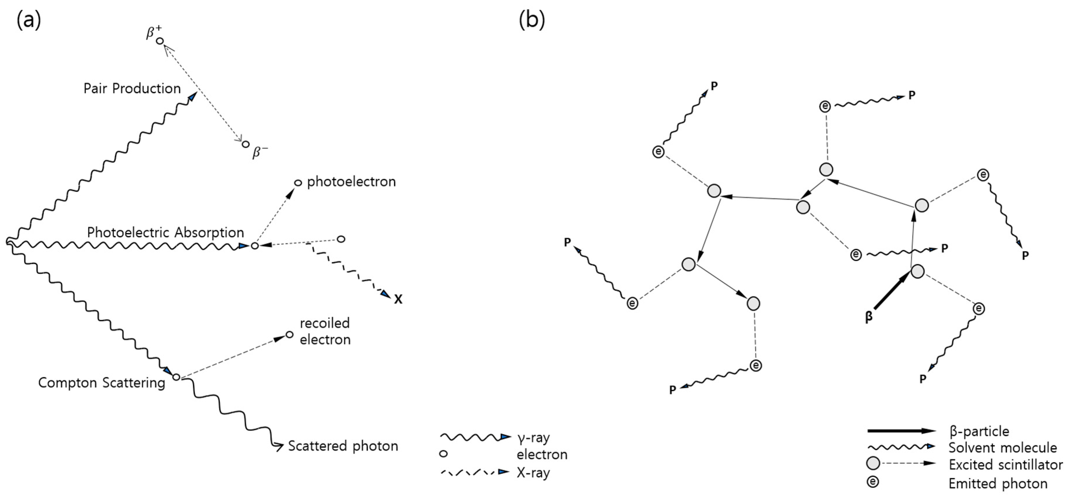
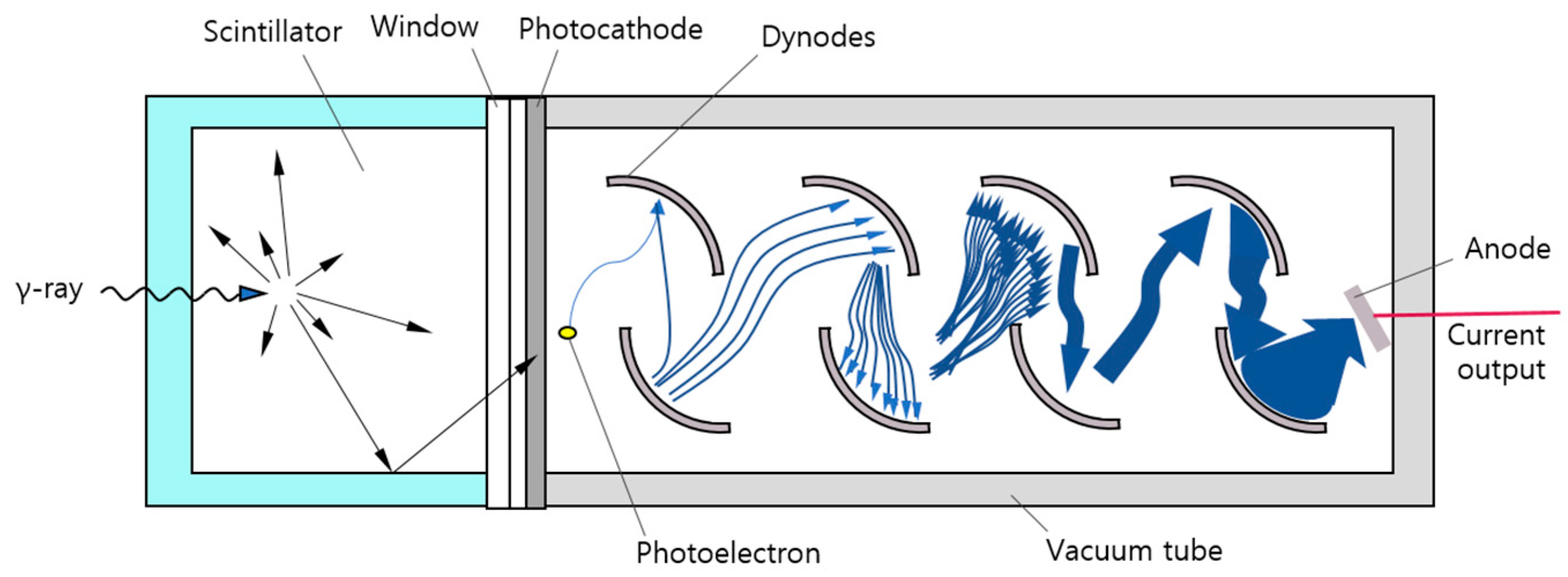
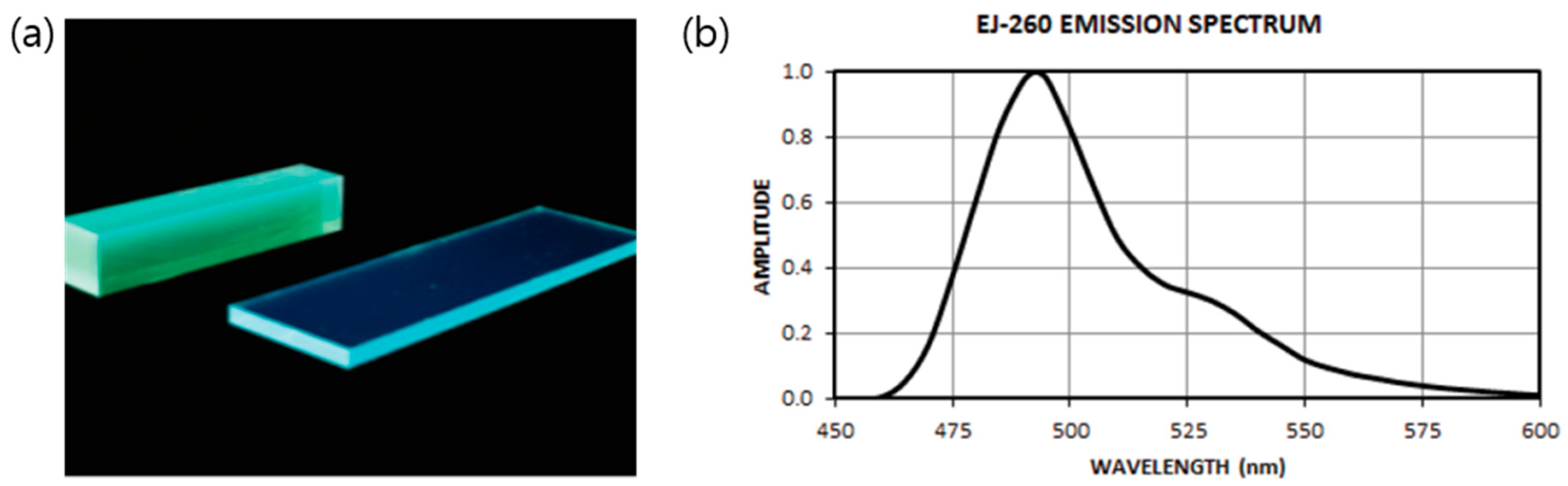
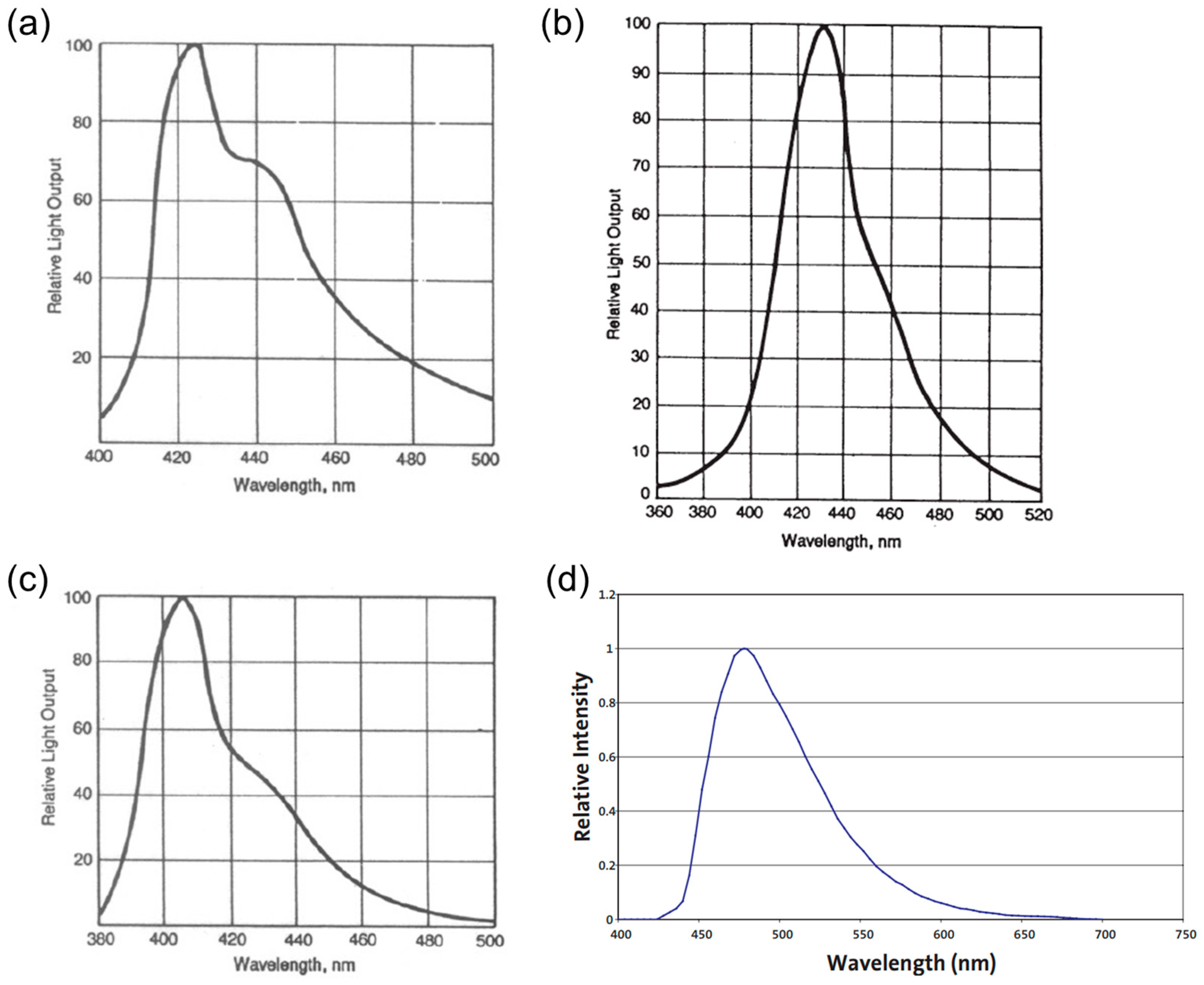
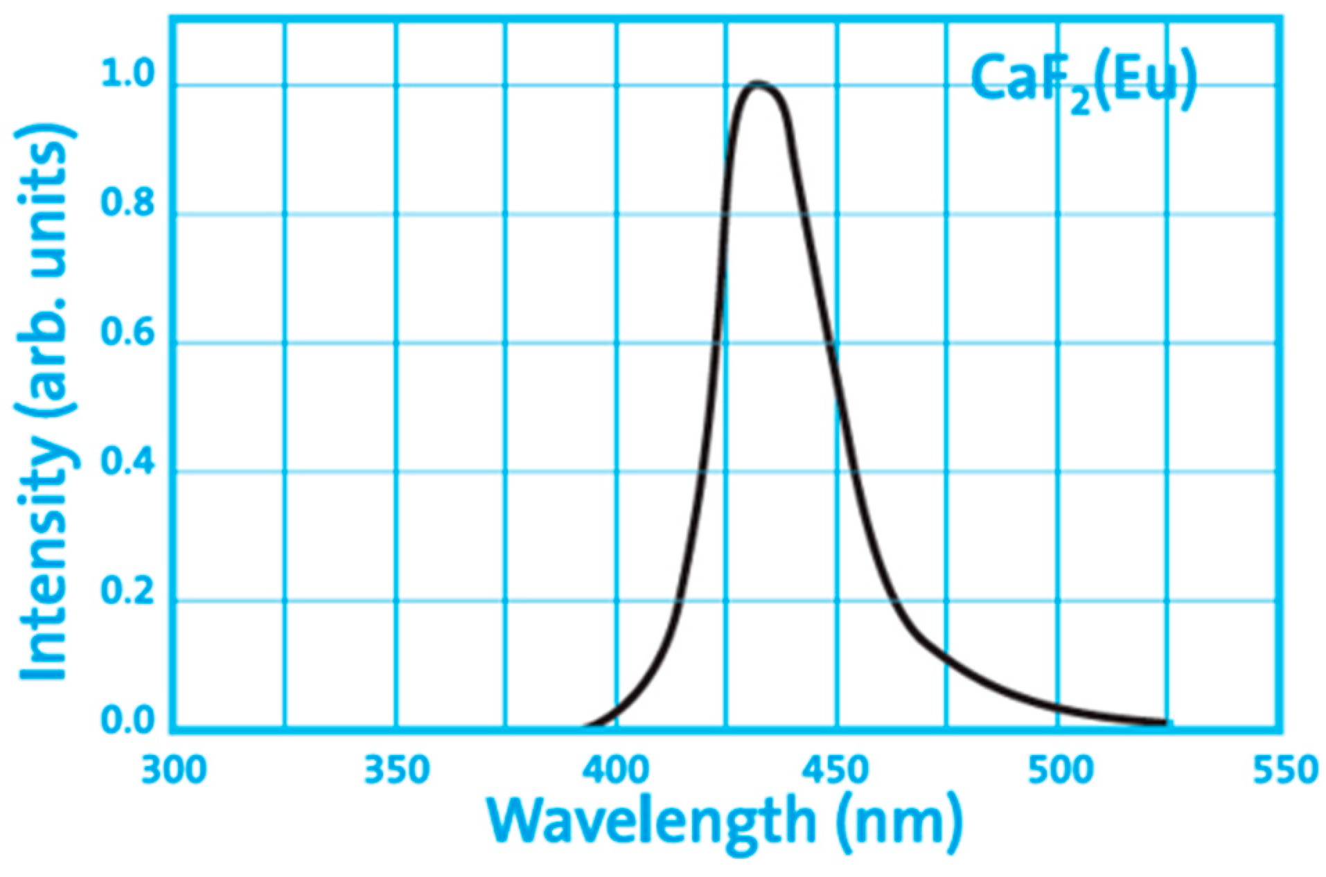
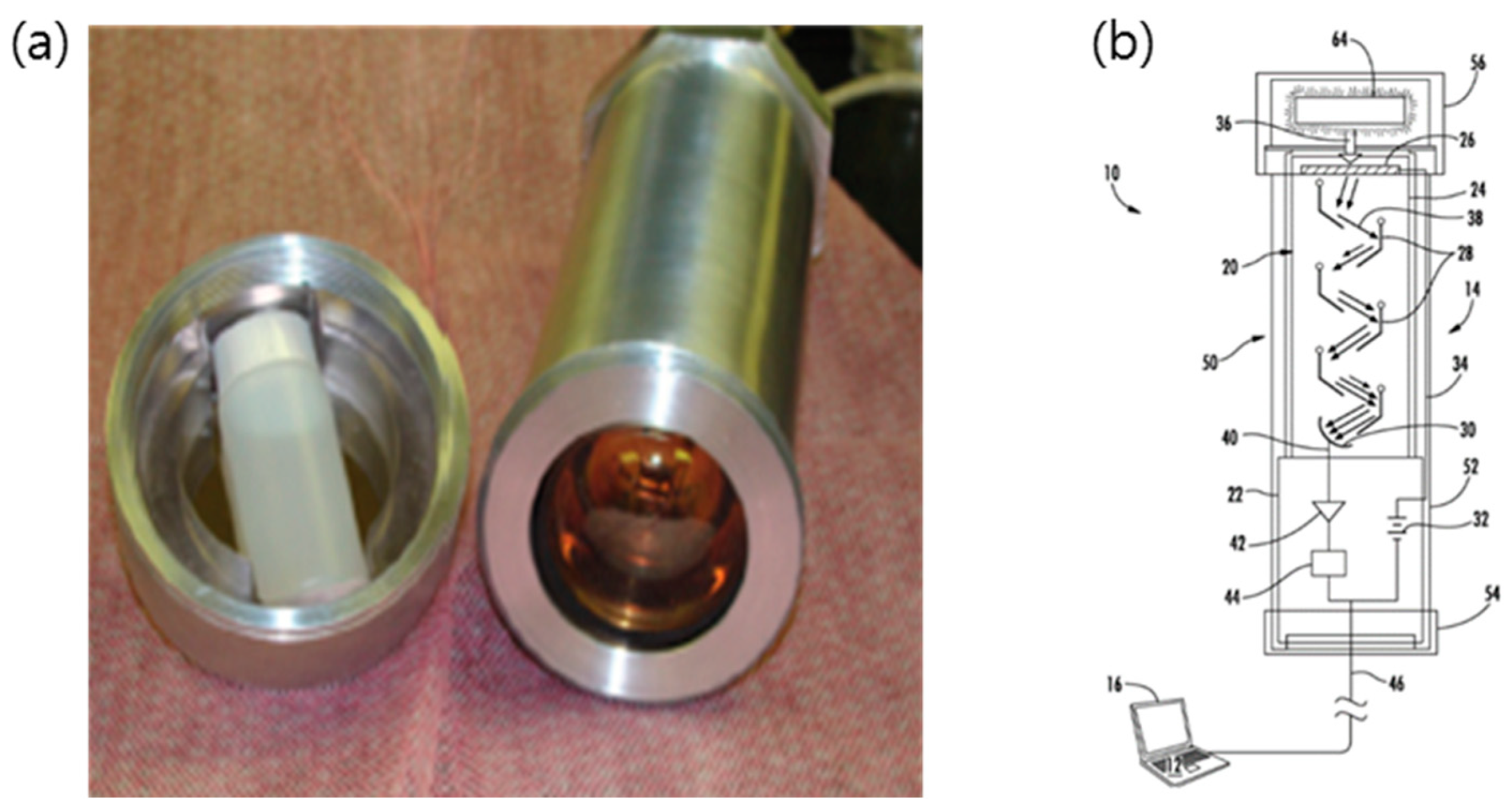
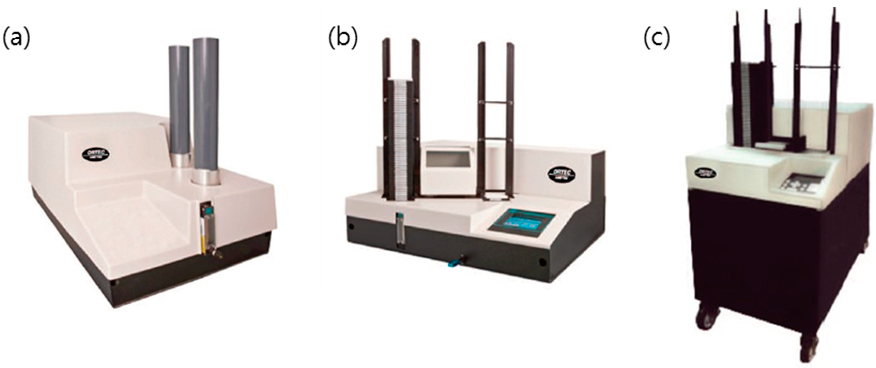
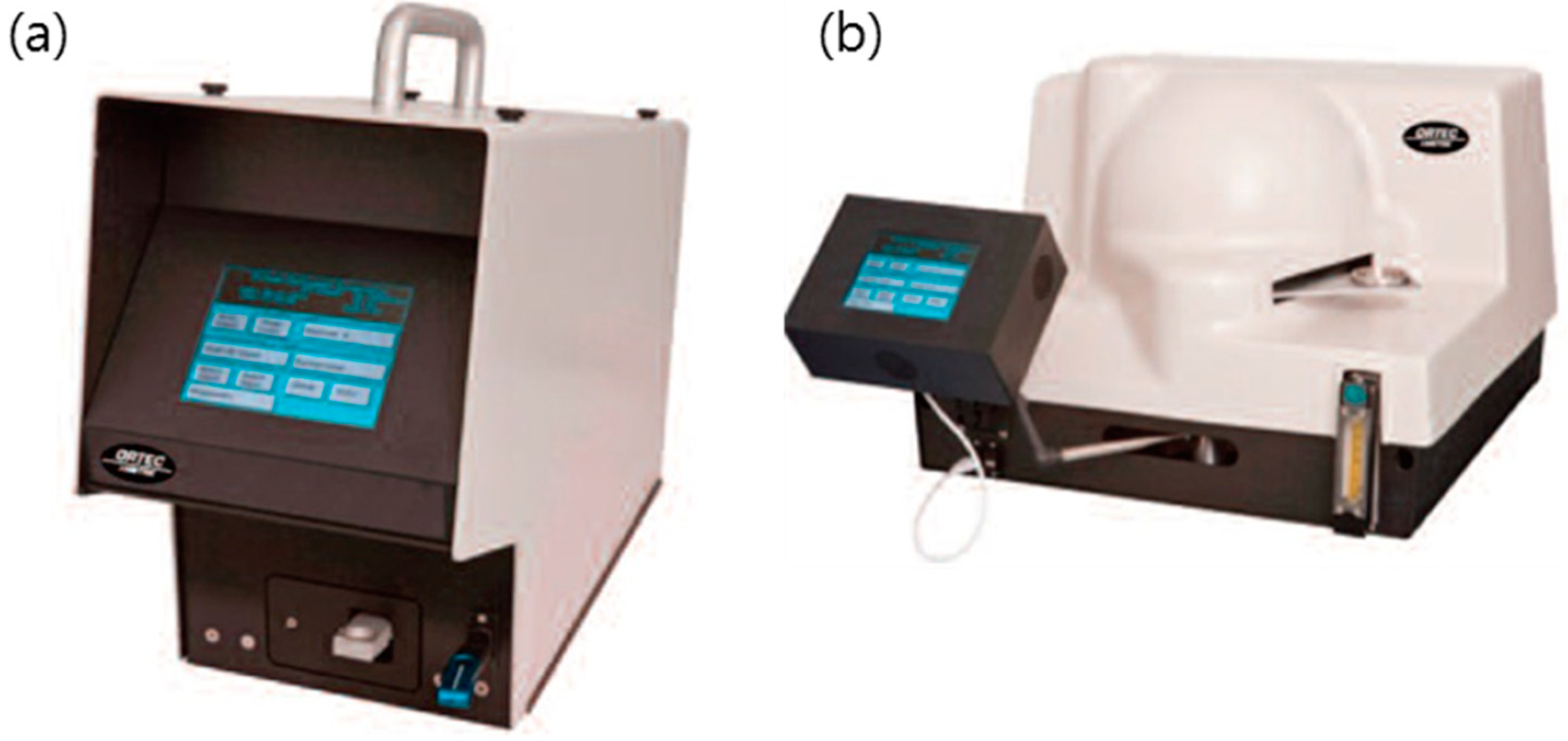
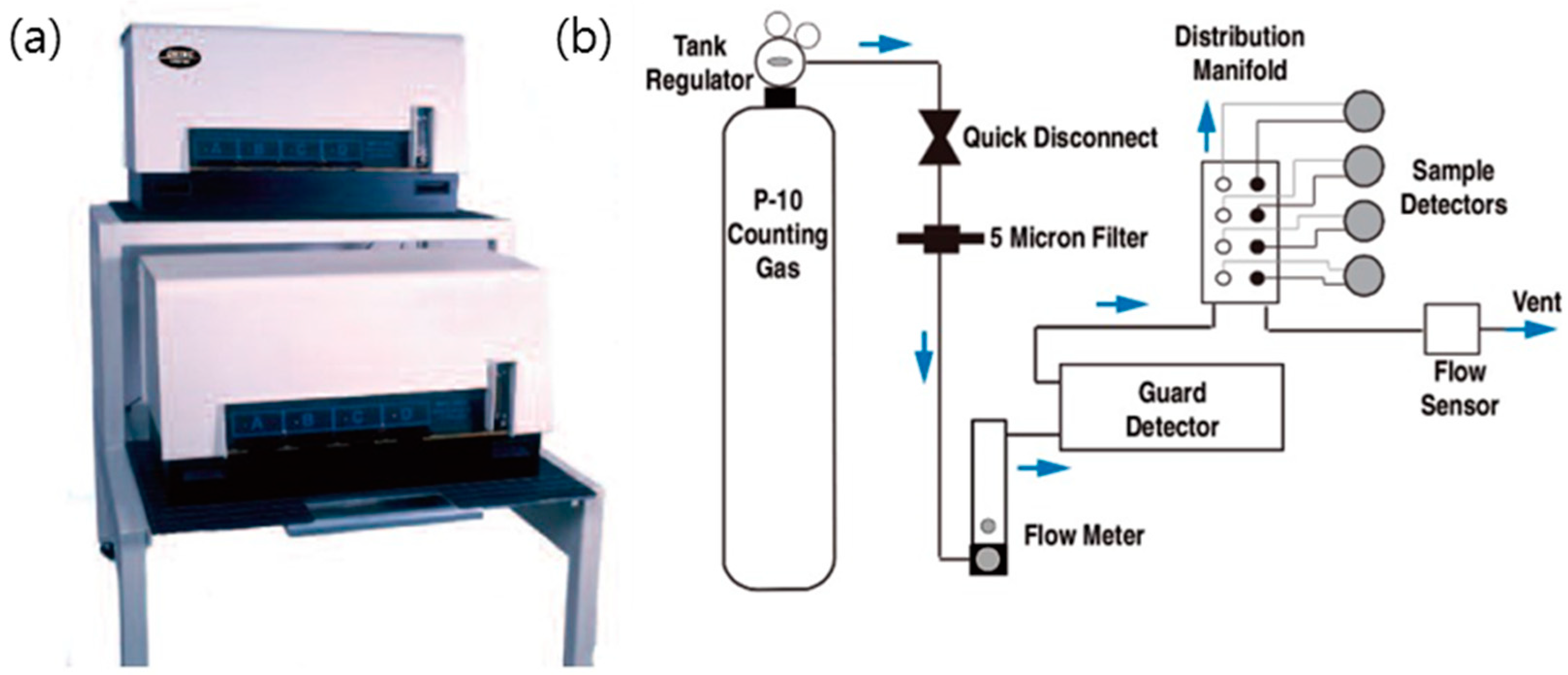
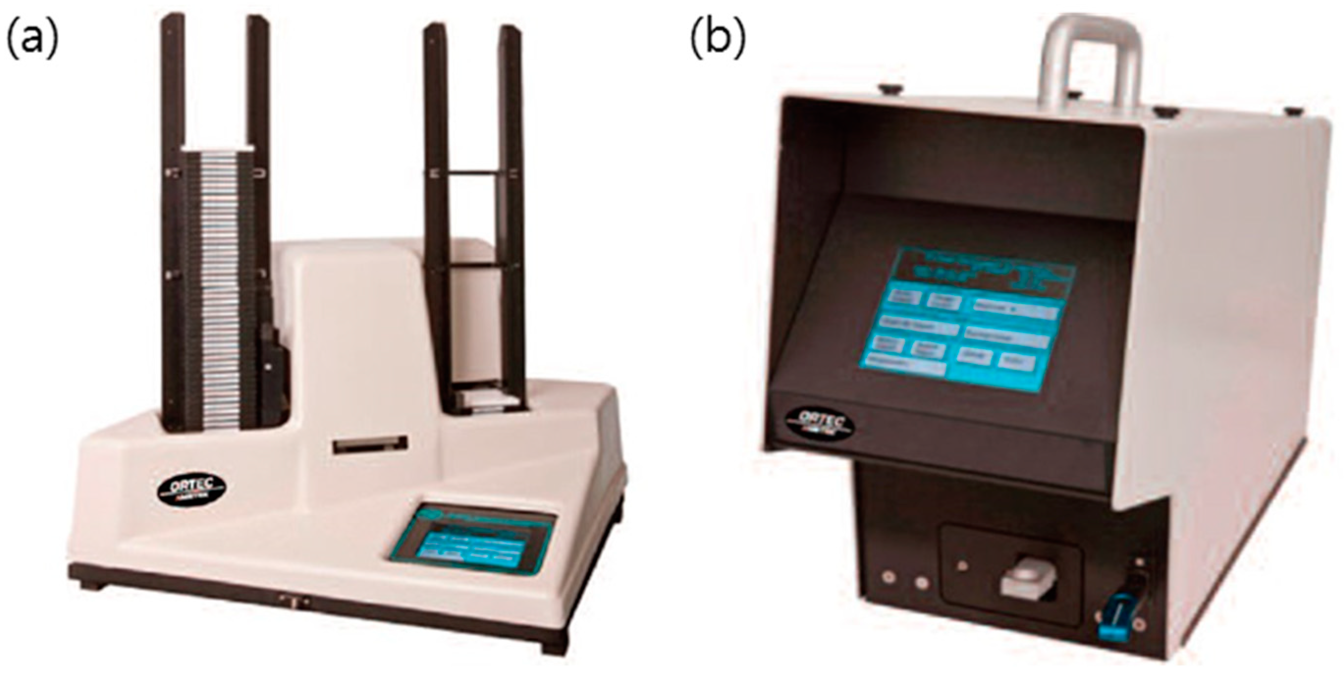
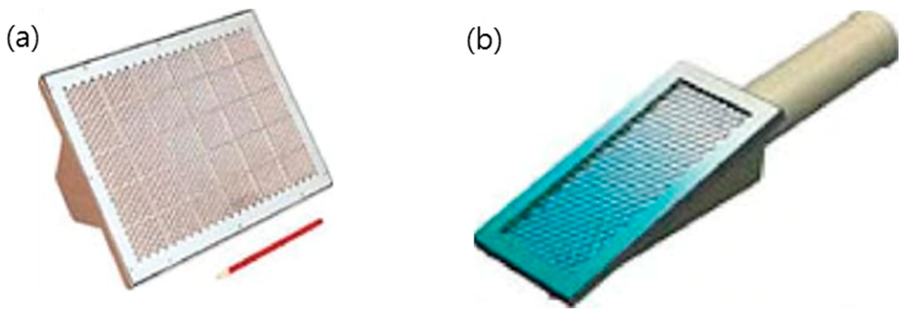
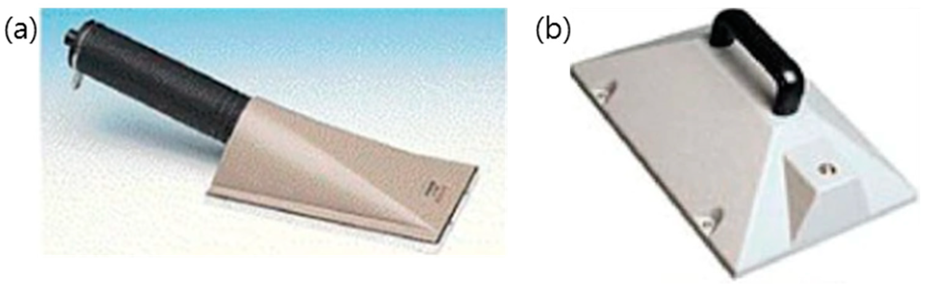
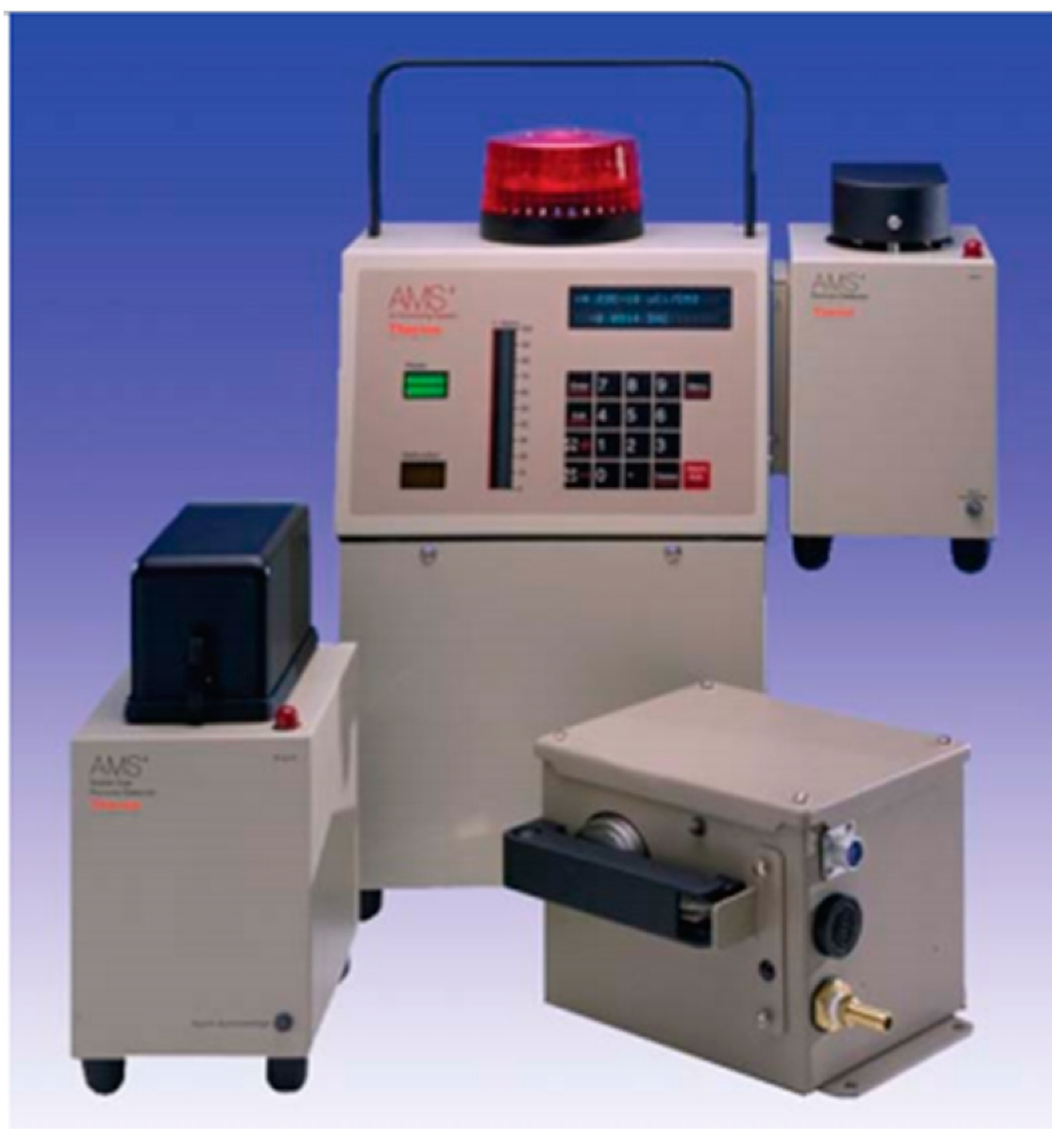
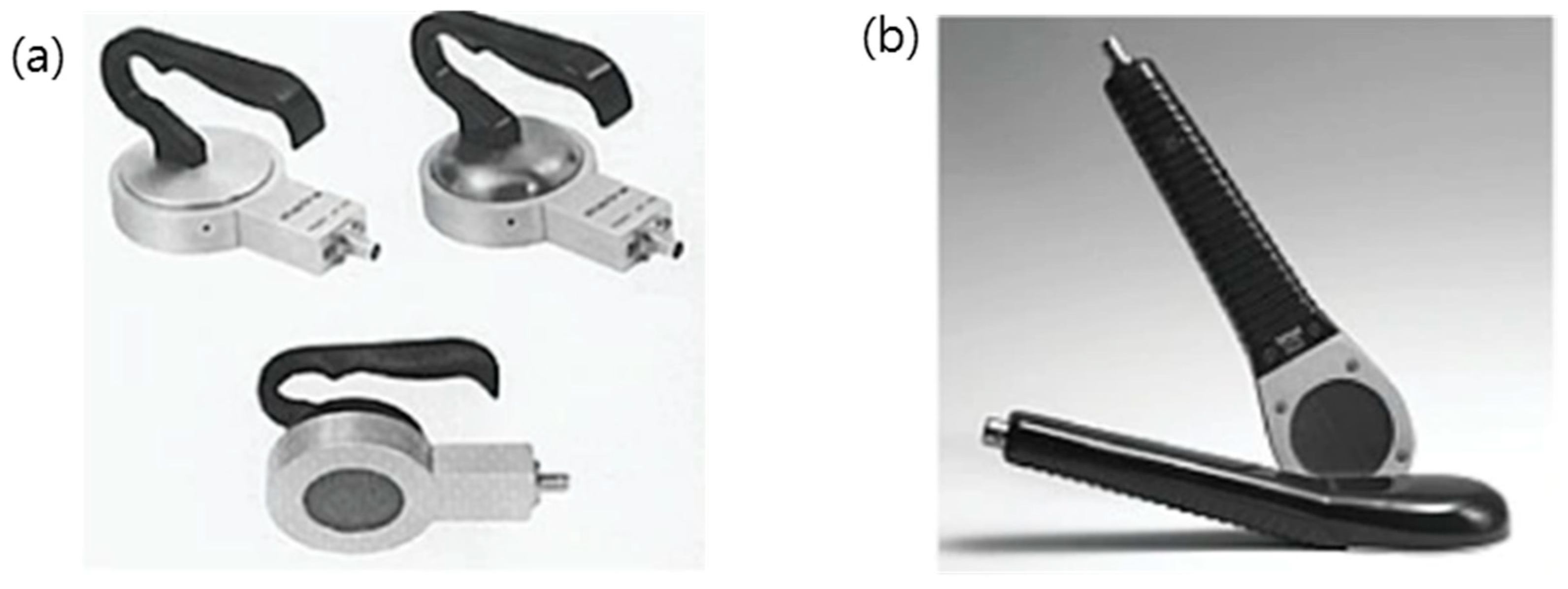
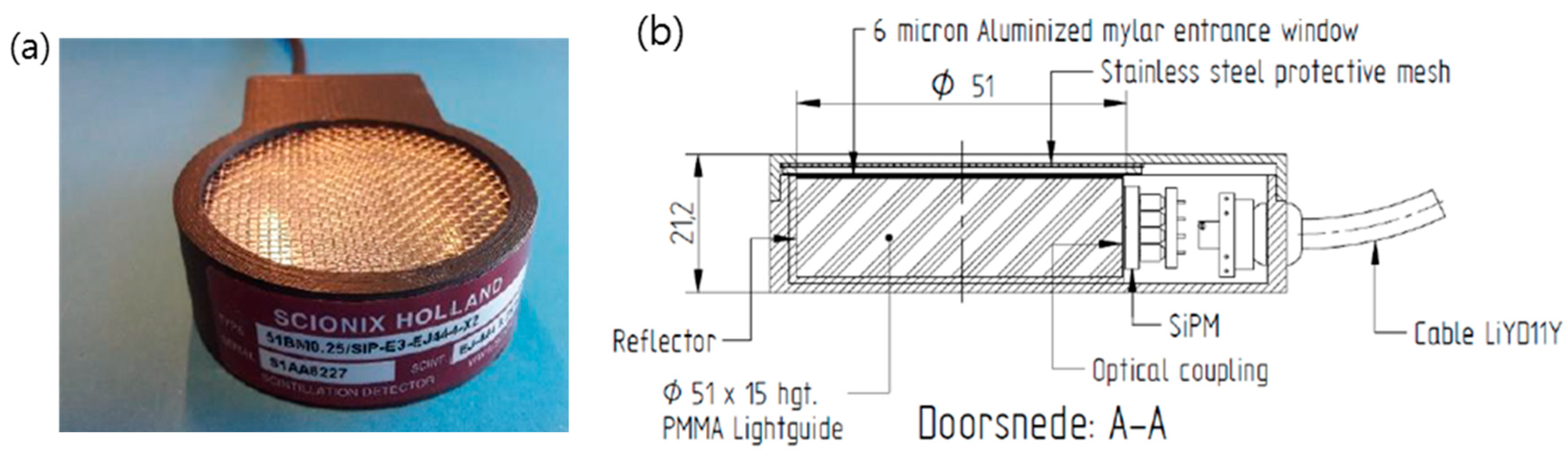
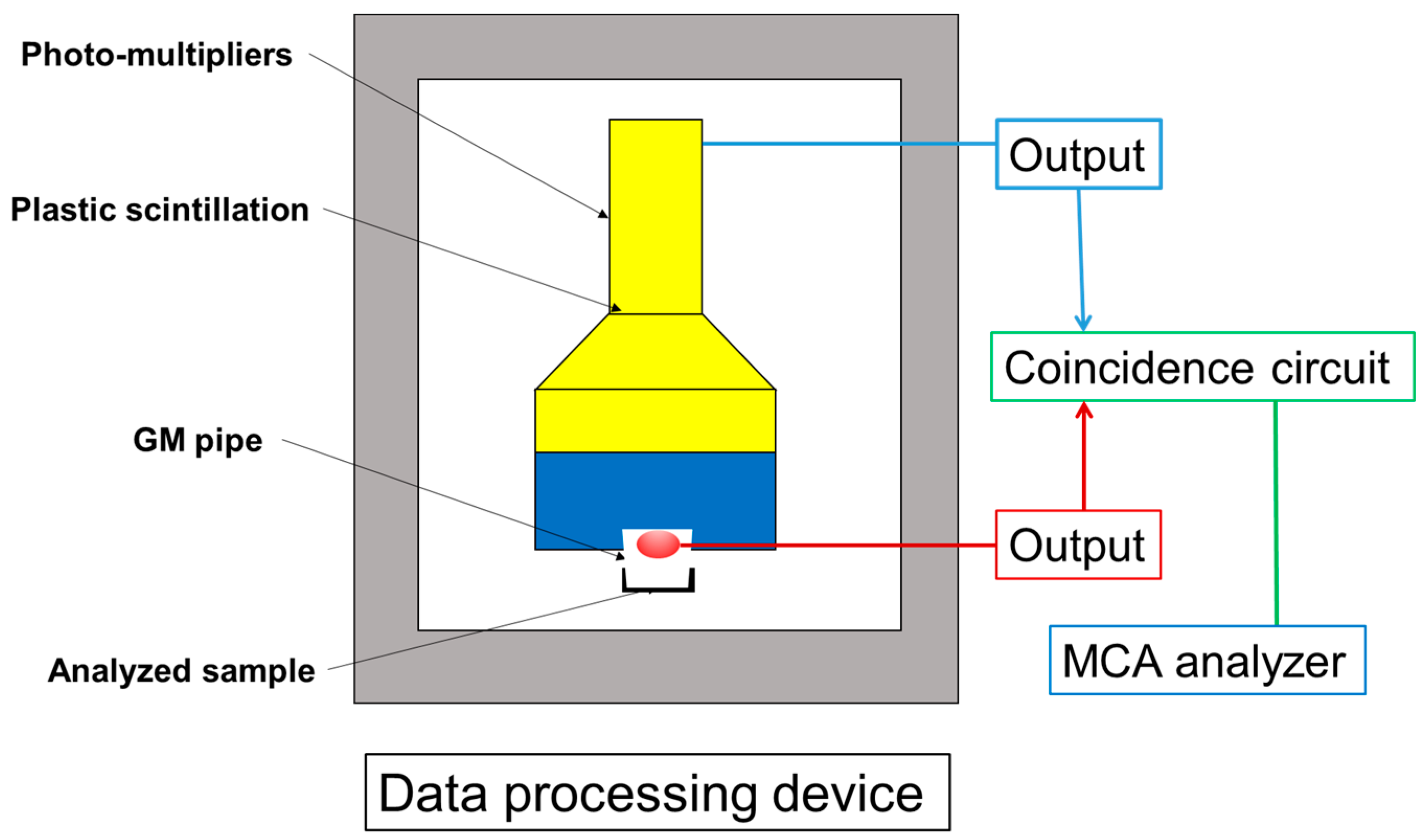
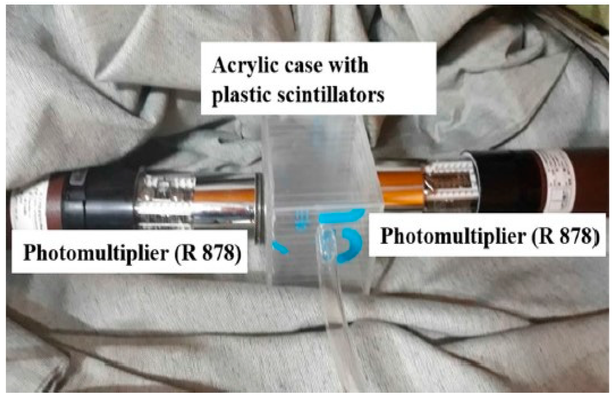
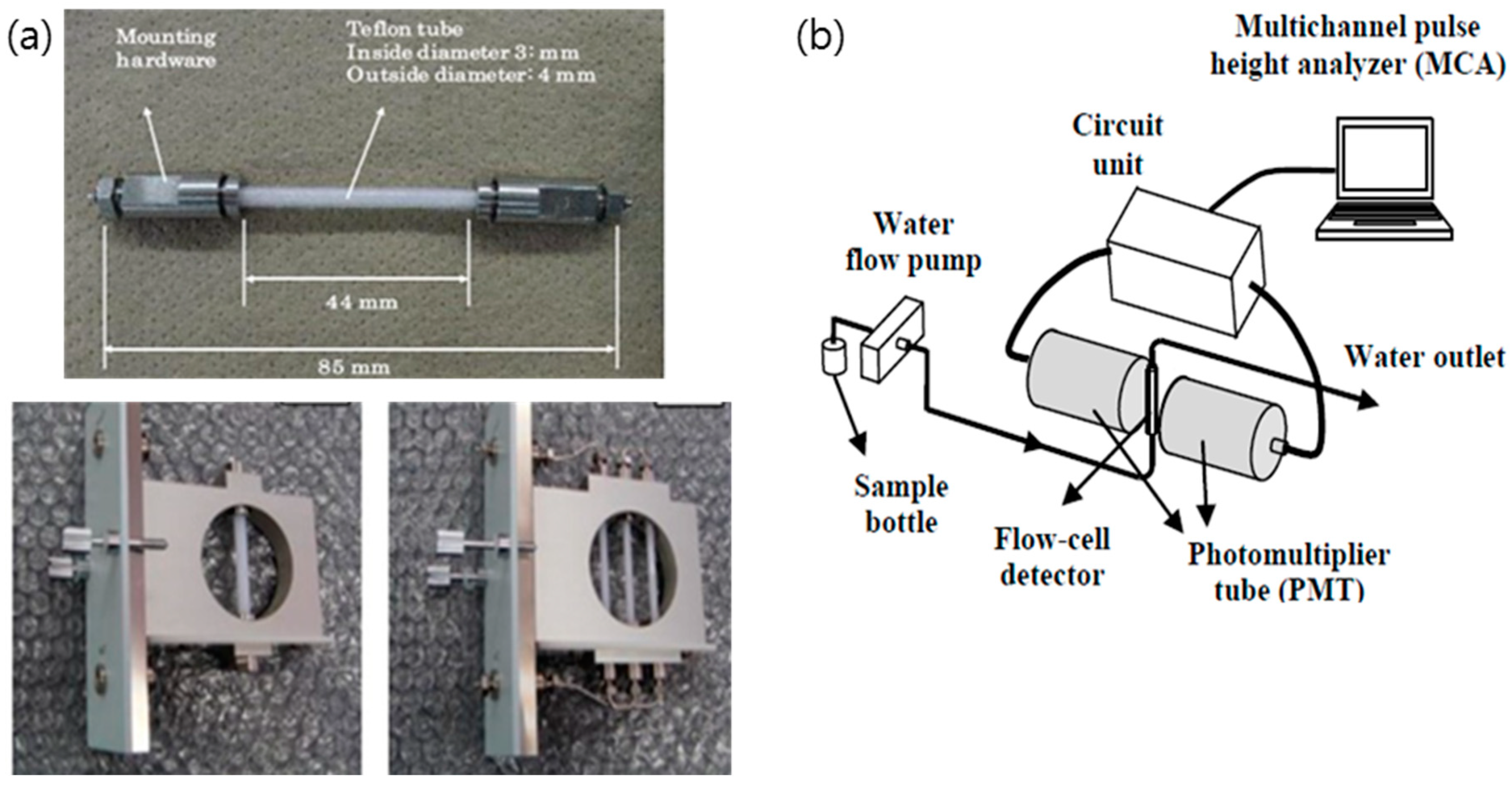
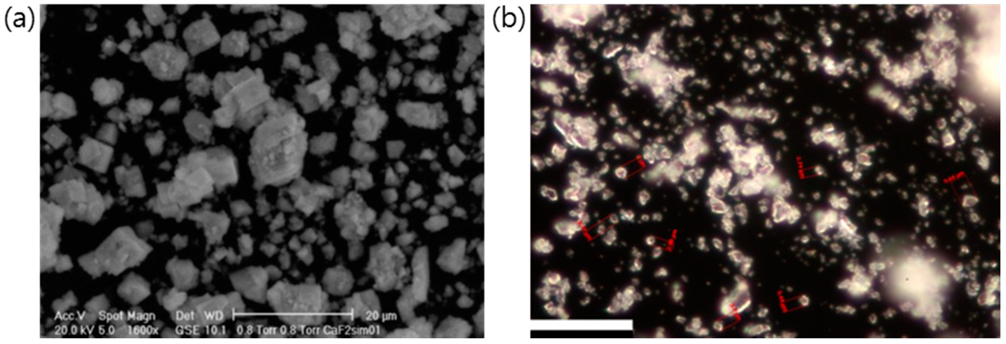
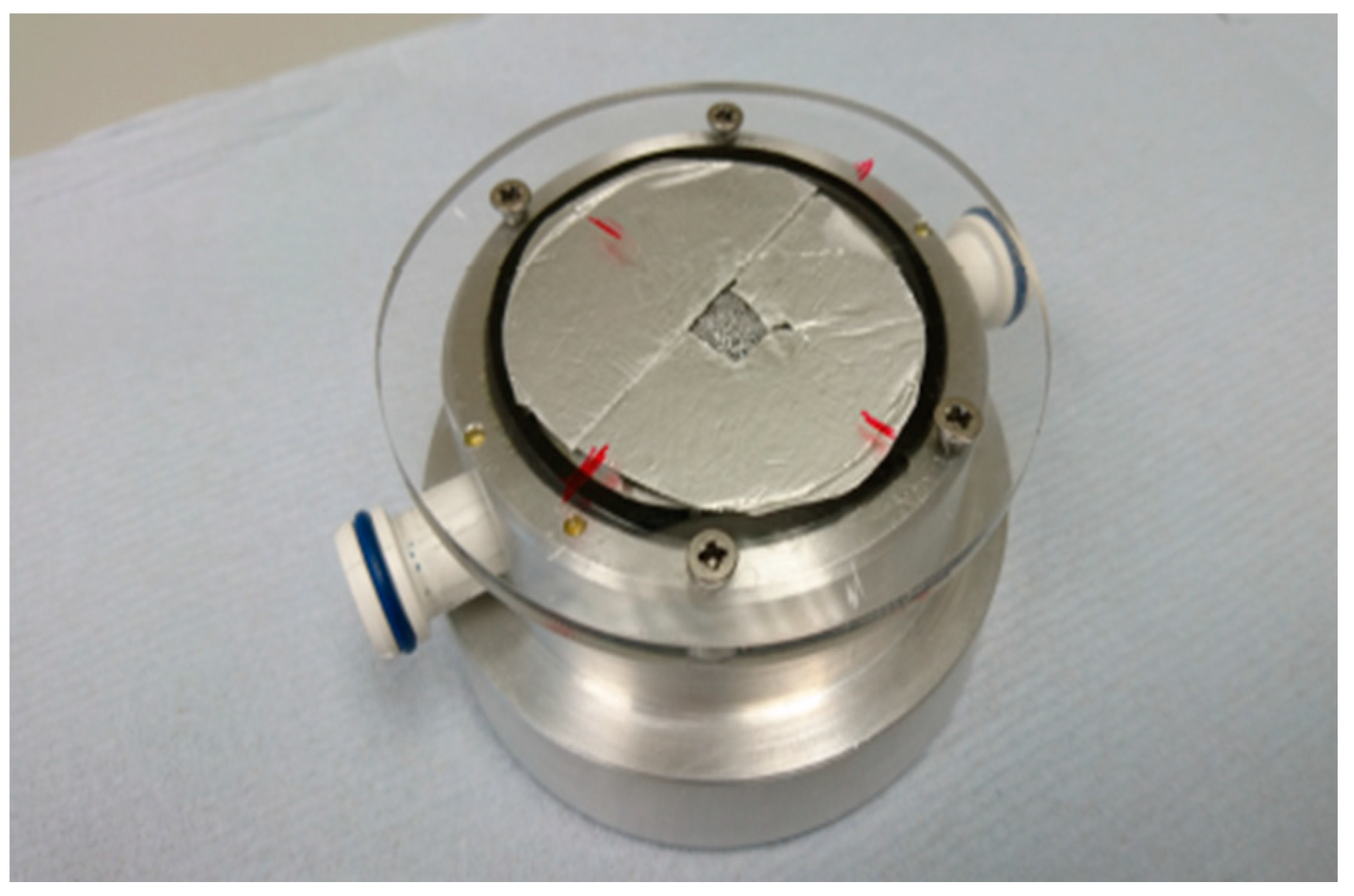
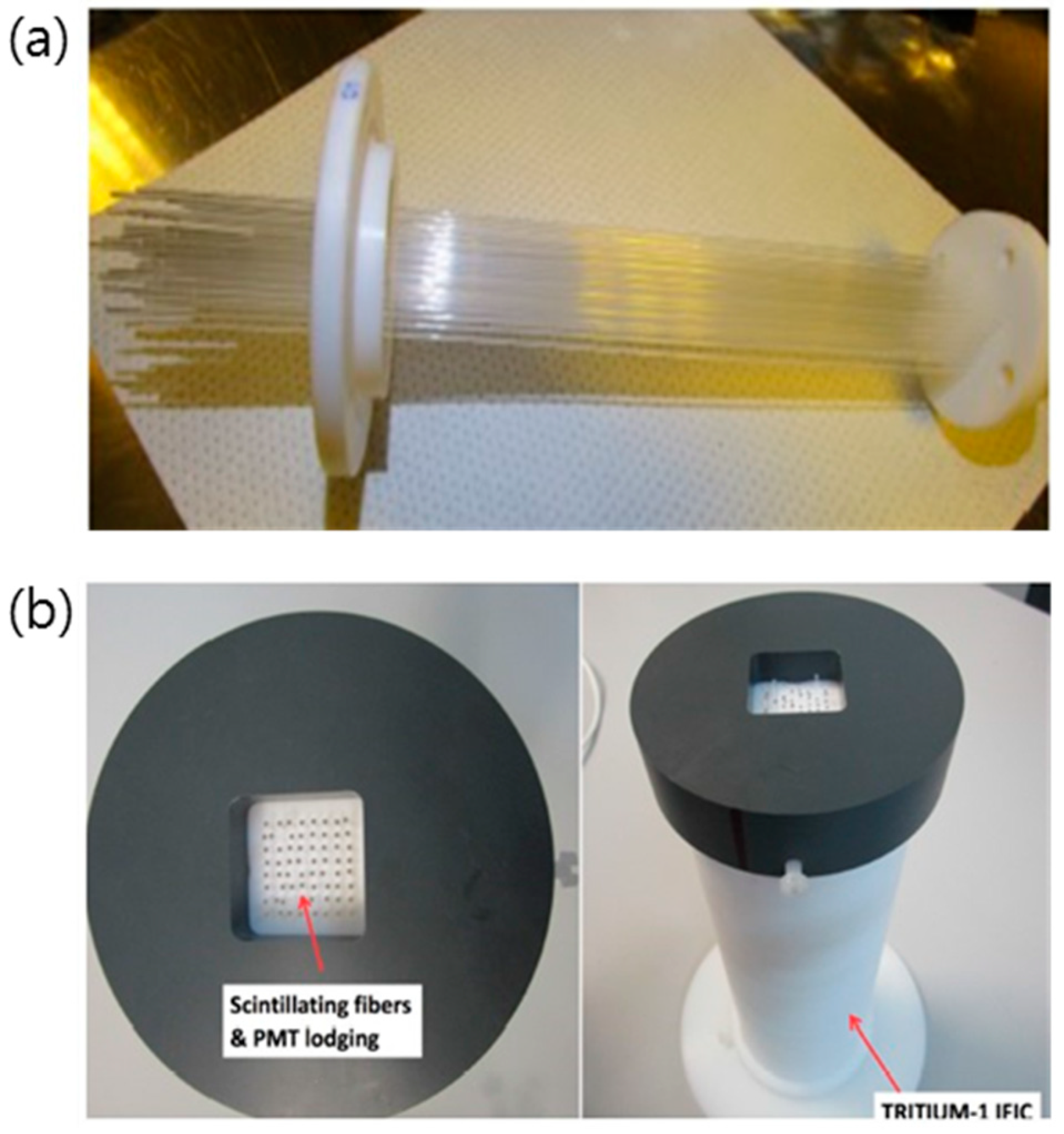
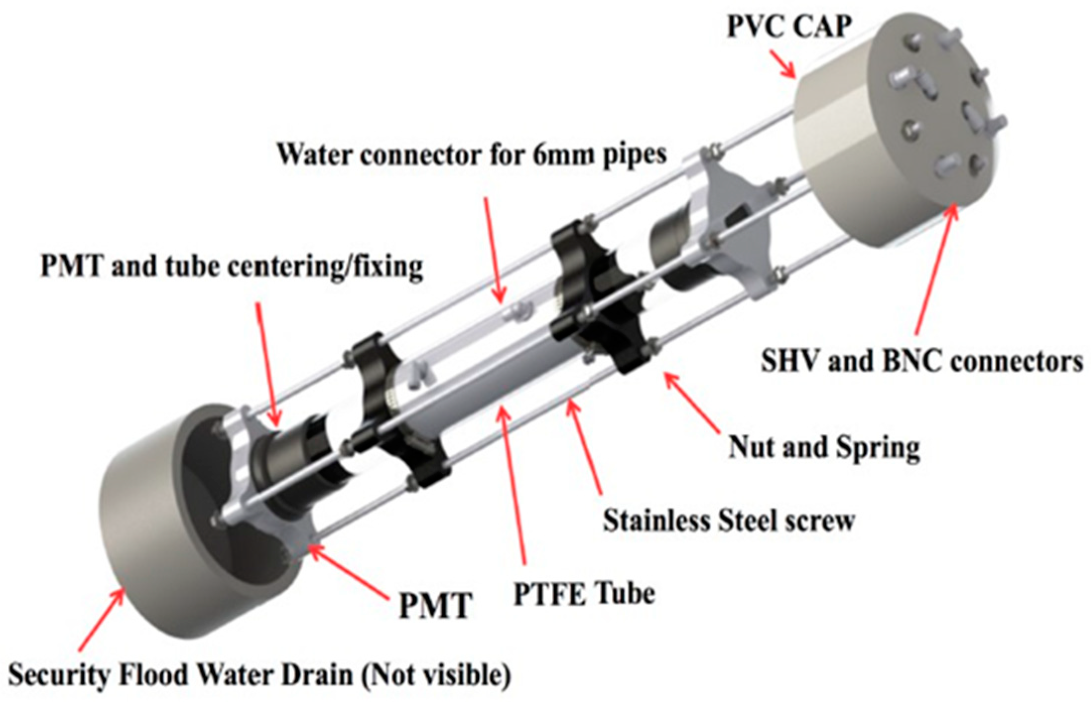
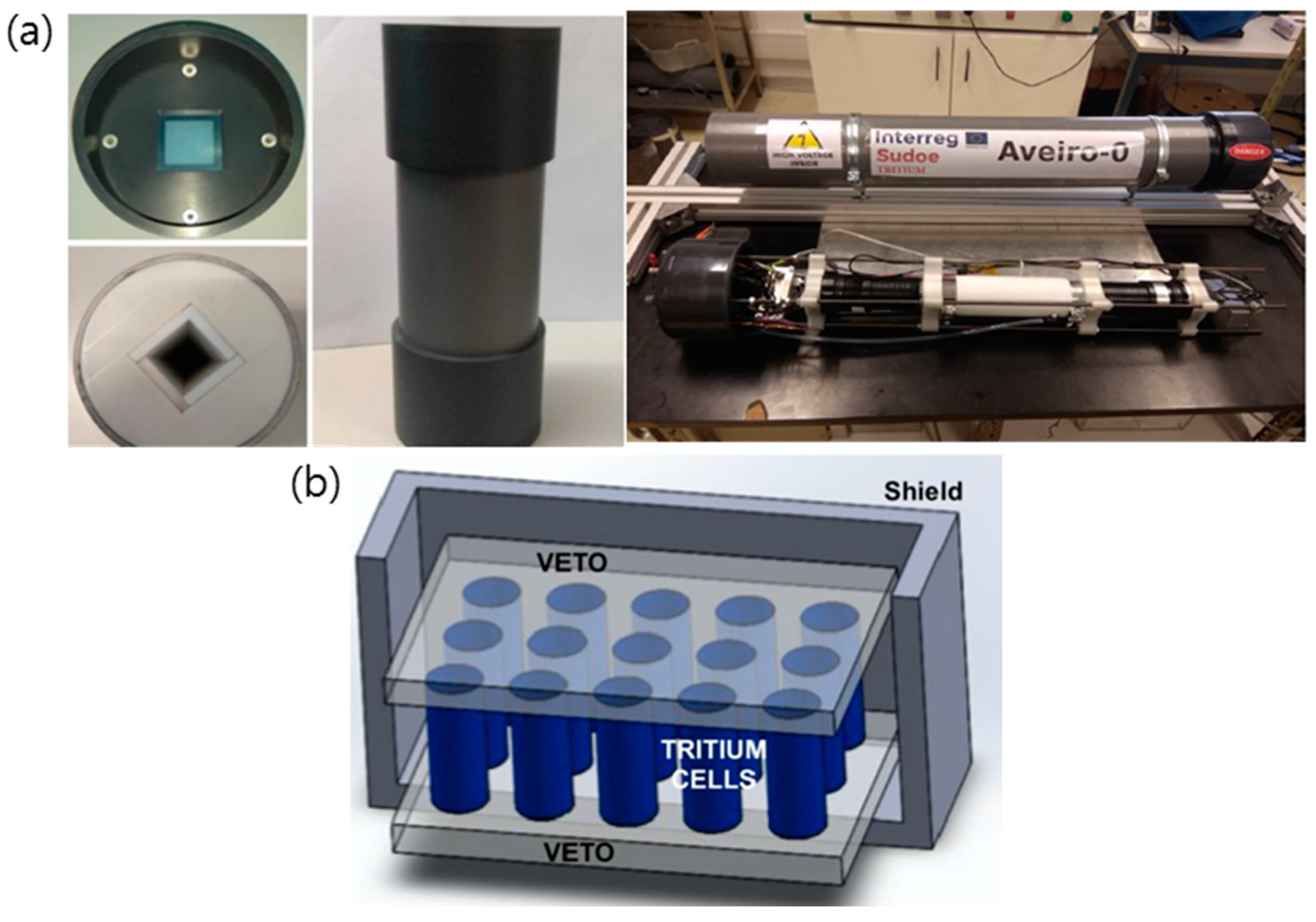
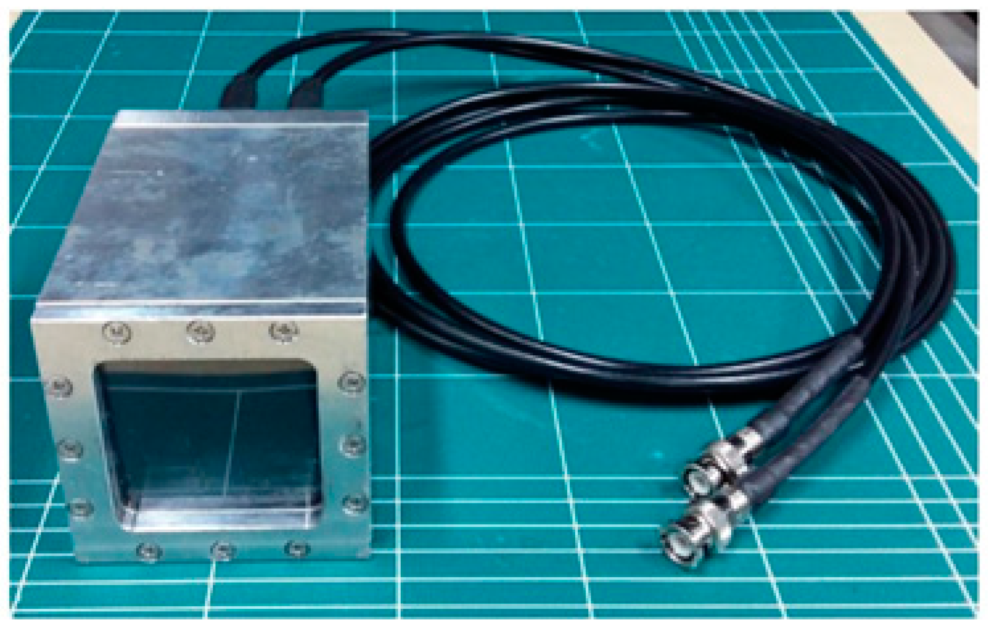
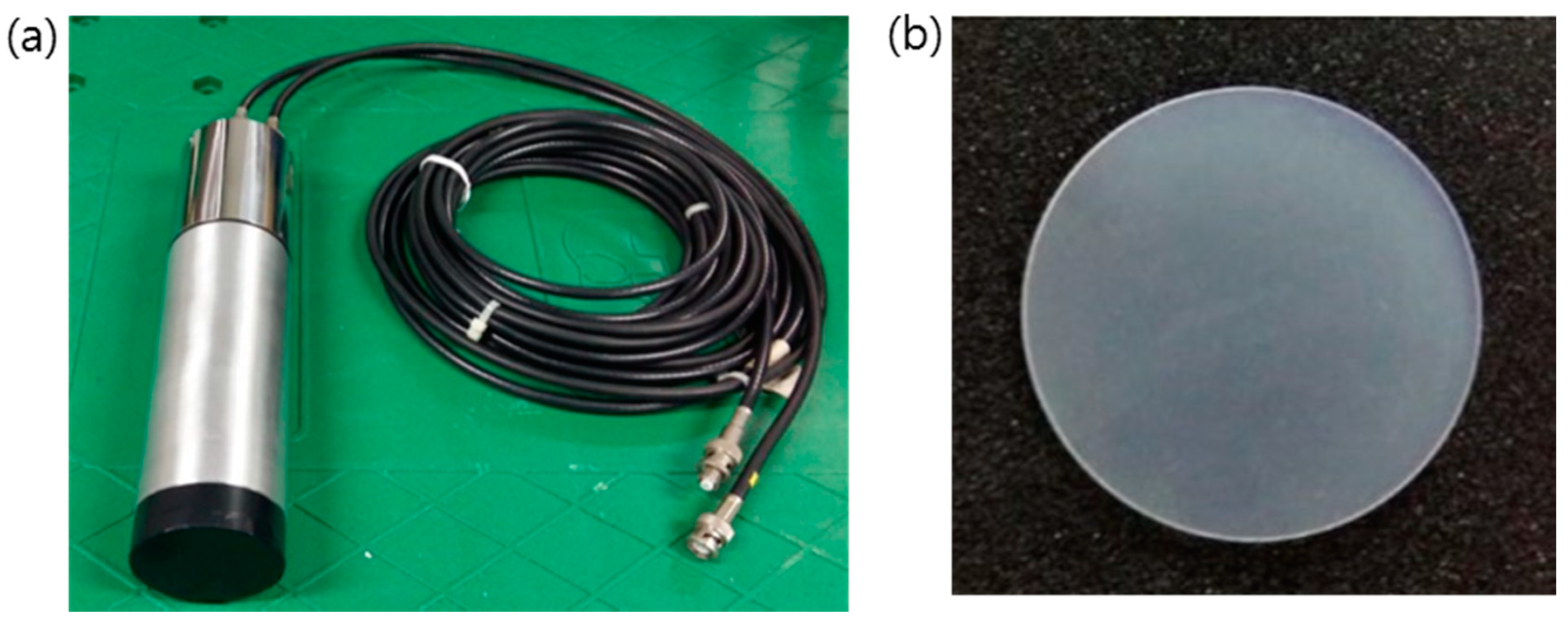
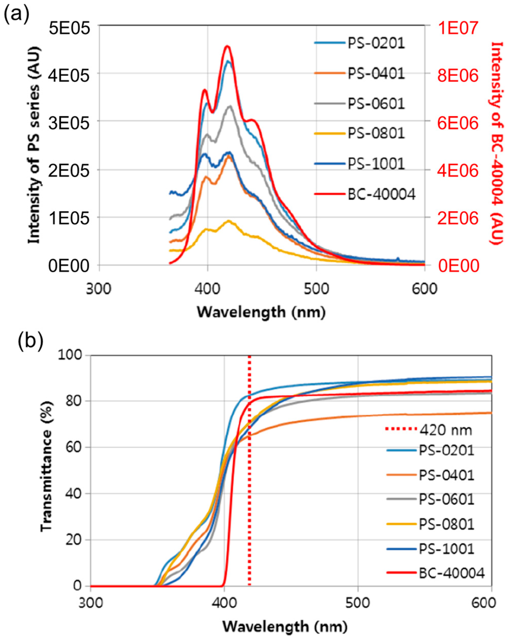
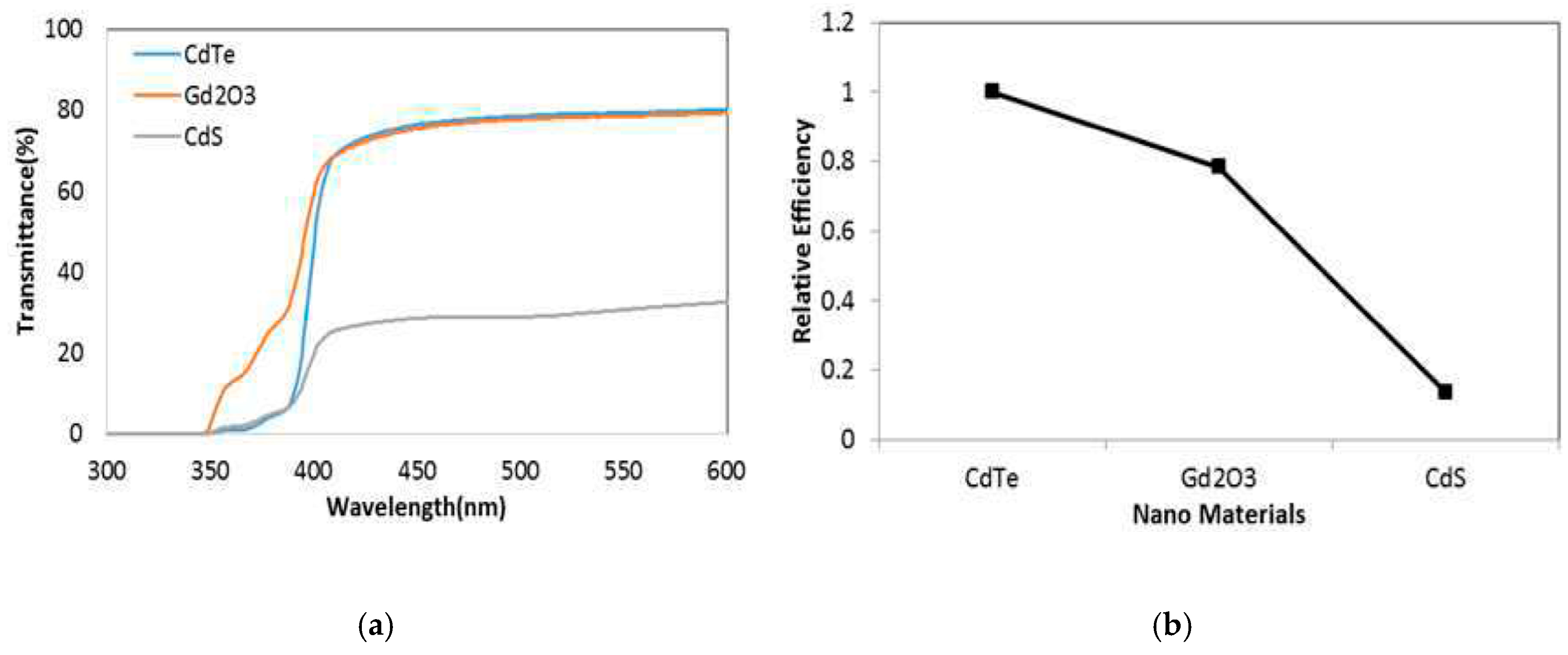
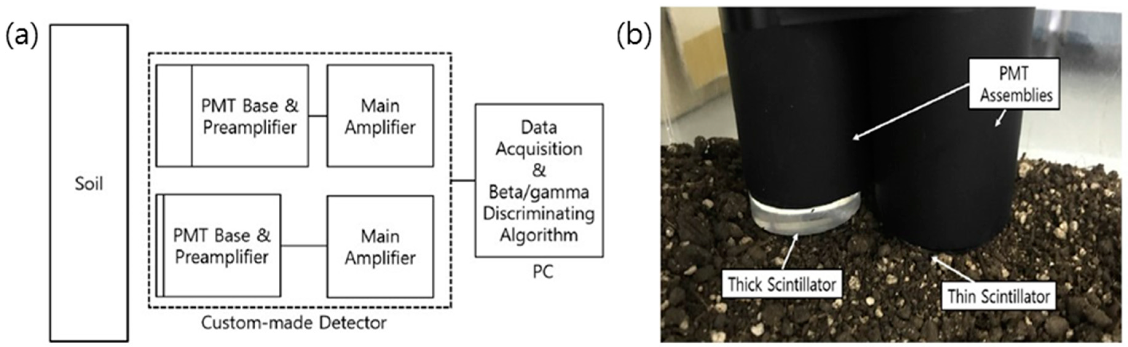
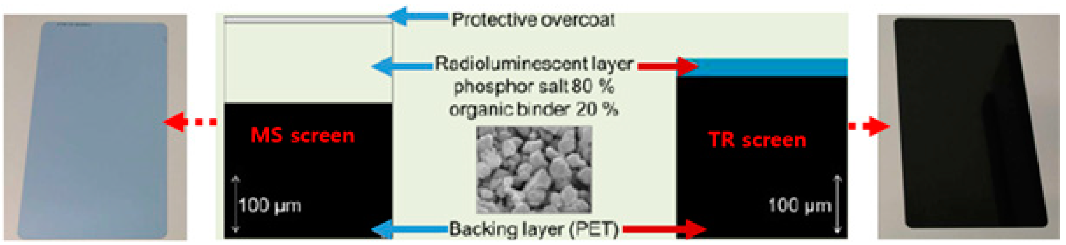
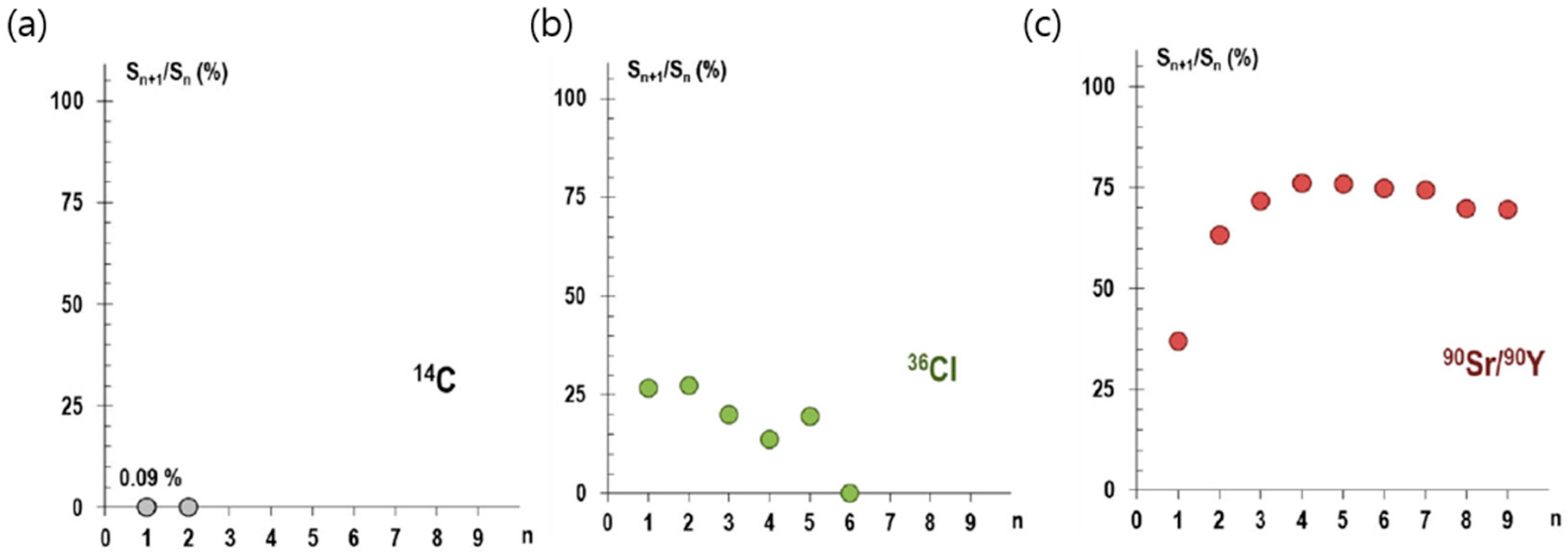
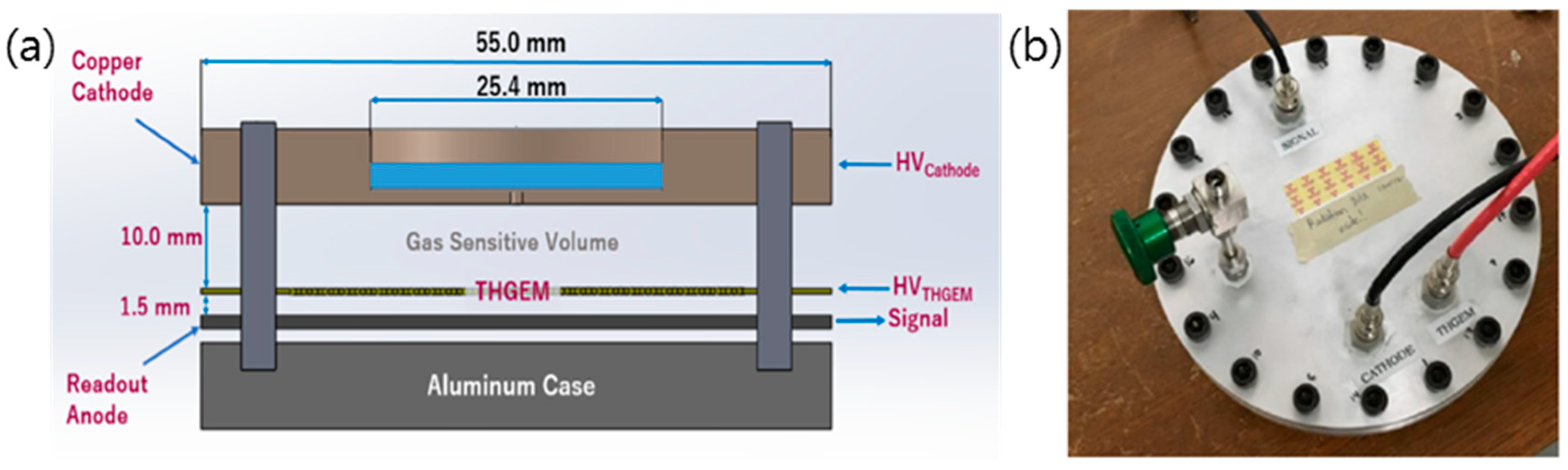
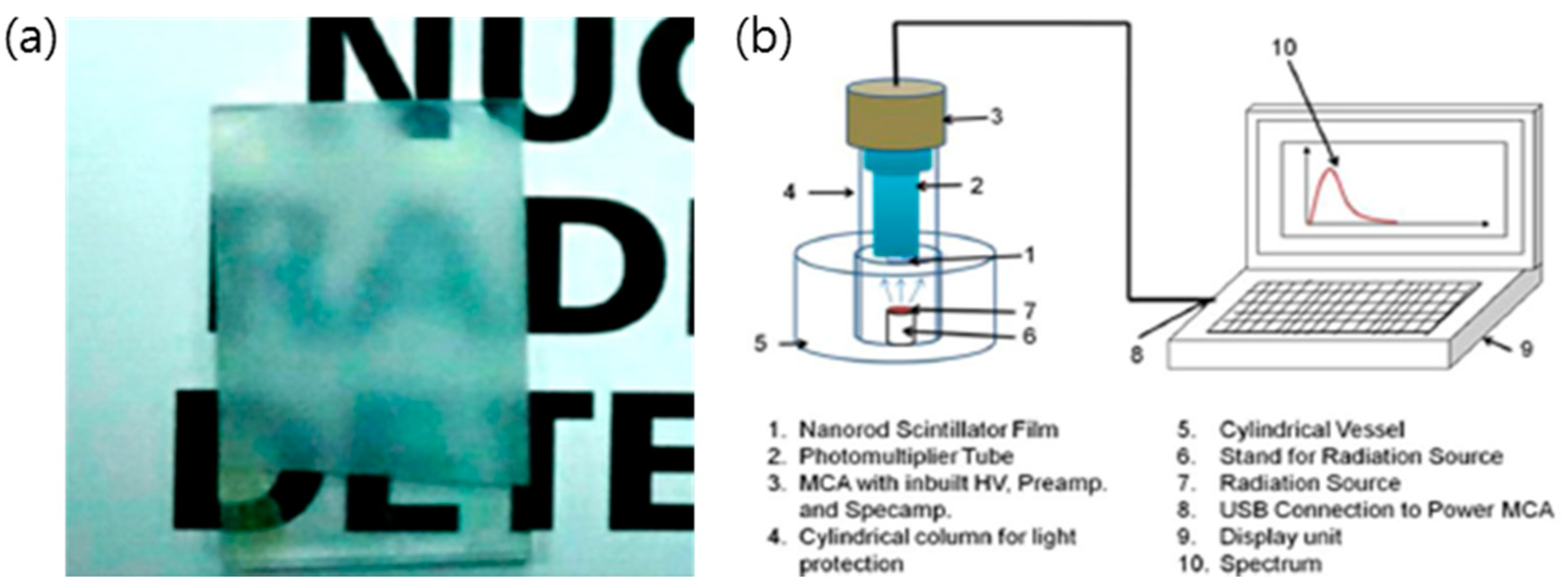
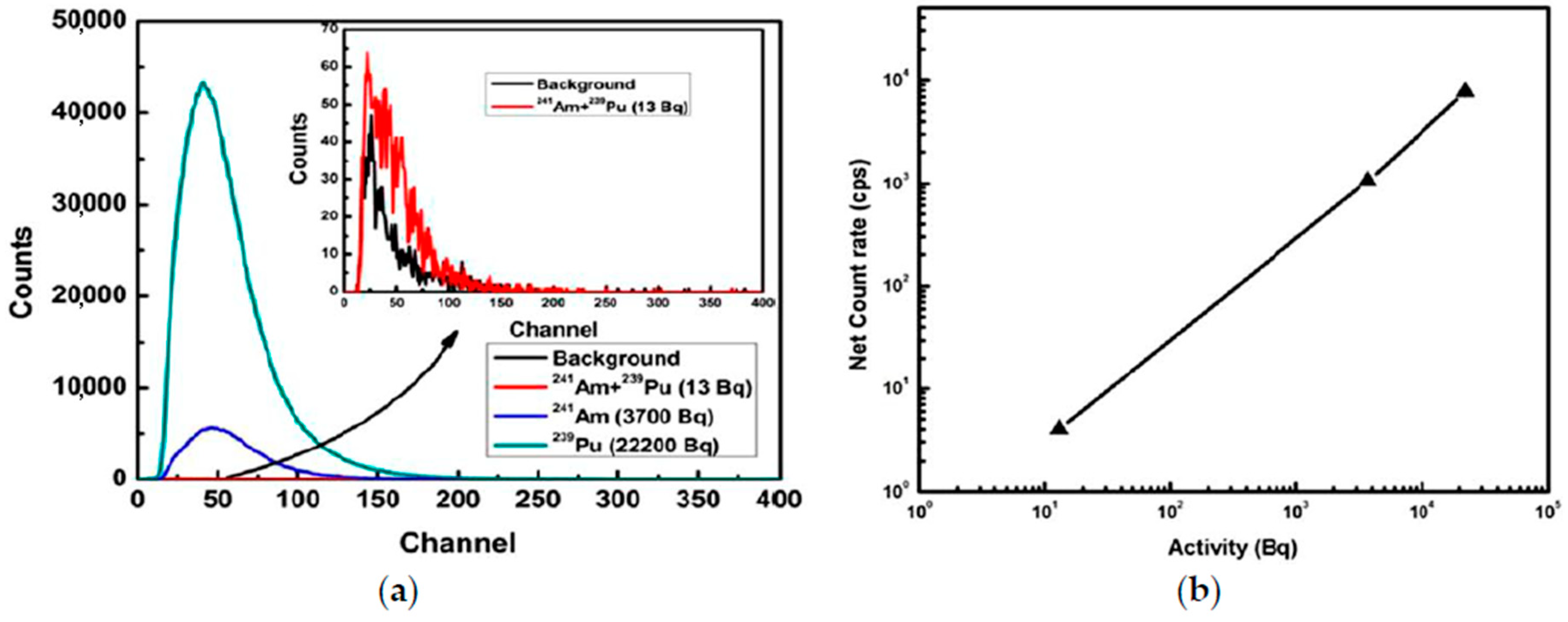
Table 1.
Characteristics of beta-ray emitters.
| Nuclide | Half-Life | Principal Radiation |
|---|---|---|
| 3H | 12.3 y | 0.0186 MeV |
| 14C | 5.73 × 10³ y | 0.156 MeV |
| 36Cl | 3.01 × 10⁵ y | 0.709 MeV |
| 63Ni | 100 y | 0.659 MeV |
| 90Sr/90Y | 29.1 y | 0.546 MeV + 2.28 MeV |
| 94Nb | 2 × 10⁴ y | 0.47 MeV |
| 99Tc | 2.13 × 10⁵ y | 0.293 MeV |
| 129I | 1.6 × 10⁷ y | 0.15 MeV |
| 241Pu | 14.1 y | 0.21 MeV |
Table 2.
Gamma interaction with the energy region.
| Energy Region | Interaction Phenomena |
|---|---|
| E < 30 keV | Photoelectron Effect and Auger Electrons |
| 30 keV < E < 2 MeV | Compton Scattering |
| E > 2 MeV | Pair Production |
Table 3.
Comparison of an organic scintillator and inorganic scintillator.
| Organic Scintillator | Inorganic Scintillator | |
|---|---|---|
| Strength | - short decay time (few ns) - counts signal rapidly - relatively inexpensive price | - high detection efficiency and emission intensity - good energy linearity - stable crystal structure (mechanical, chemically stable) |
| Weakness | - bad energy linearity - low atomic number and density - low possibility of photoelectron effect - bad energy linearity | - long decay time (hundreds of ns) - expensive price |
Table 4.
Characteristics of the EJ-260 scintillator.
| Basic Properties | Characteristics |
|---|---|
| Light Output (% Anthracene) | 60 |
| Scintillation Efficiency (photons/1 MeV e−) | 9200 |
| Wavelength of Maximum Emission (nm) | 490 |
| Light Attenuation Length (cm) | 350 |
| Rise Time (ns) | 1.5 |
| Decay Time (ns) | 9.2 |
| Polymer Base | Polyvinyltoluene |
| Refractive Index | 1.58 |
| Light Output | 95% of that at 20 °C |
Table 5.
Characteristics of plastic scintillator.
| Properties | BC-400 | BC-404 | BC-408 | BC-428 | CaF2:Eu |
|---|---|---|---|---|---|
| Light Output (% Anthracene) | 65 | 68 | 64 | 36 | - |
| Wavelength of Maximum Emission (nm) | 423 | 408 | 425 | 480 | 435 |
| Light Attenuation Length (cm) | 160 | 140 | 210 | 150 | - |
| Rise Time (ns) | 0.9 | 0.7 | 0.9 | 1.6 | - |
| Decay Time (ns) | 2.4 | 1.8 | 2.1 | 12.5 | 940 |
| Polymer Base | PVT | PVT | PVT | PVT | - |
| Density (g/cc) | 1.023 | 1.023 | 1.023 | 1.032 | 3.18 |
| Refractive Index | 1.58 | 1.58 | 1.58 | 1.58 | 1.47 |
Table 6.
Compared properties by the Ortec automatic count system.
| Properties | IPC-650 (IPC-650-HP) | WPC-1050 | WPC-1150-GFW-3 |
|---|---|---|---|
| 90Sr/90Y Efficiency | 55% | 55% | 63% |
| Window | Windowless | Aluminized, 80 μg/cm2 | Aluminized, 80 μg/cm2 |
| β BKG Performance | 0.7 cpm | 0.7 cpm (0.9 cpm Warranted) | 1.5 cpm |
| Guard Detector | X | Gas-Flow Type Proportional Counter | Gas-Flow Type Proportional Counter |
| Detector Diameter | 5.7 cm (2.25 inch) | 5.7 cm (2.25 inch) | 8.9 cm (3.5. in) dia Gas Flow Detector |
| Shielding | 10.2 cm (4 inch) | 10 cm (2.4 inch) | 10.2 cm (4 inch) |
| Detector Type | Hemispherical Style | Pancake Style | Pancake Style |
| Notes | - Use in Radiochemical and Health Physics (IPC-650-HP) - Use in Count for Ultra-Low-Level α/β | - Use In Radiochemical and Health Physics | - Use in Health Physics and Environmental Monitoring - Use in Air Sampler of Large Capacity Air Filter |
Table 7.
Compared properties by the Ortec Manual Single Counter.
| Properties | MPC-900(GFL/GFW) | MPC-1000(GFL/GFW) |
|---|---|---|
| 90Sr/90Y Efficiency | 55% | 55% |
| Window | Aluminized, 80 μg/cm2 (GFW: Windowless) | Aluminized, 80 μg/cm2 (GFW: Windowless) |
| β BKG Performance | 35 cpm (55 cpm Warranted) | 0.7 cpm (0.9 cpm Warranted) |
| Guard Detector | X | Cosmic Guard Detector (Large Area Gas Flow Proportional Counter) |
| Planchet Diameter | 5.1 cm (2 inch) | 5.1 cm (2 inch) |
| Shielding | - | 10 cm (2.4 inch) Thick |
| Detector Type | Hemispherical Style | Pancake Style |
| Notes | - Use in the place where not far and not require high sensitivity for β-ray measurement | - Ultra low-level background radioactive counter |
Table 8.
Compared characteristics of the probes from ThermoFisher.
| Model | BP17A | BP19AD | BP19DD | HP380B | HP380AB | DP8A/DP8B |
|---|---|---|---|---|---|---|
| Probe Type | Beta | Beta | Beta | Beta Scintillation | Alpha-Beta Scintillation | Alpha-Beta |
| Window Area [cm2] | 600 | 100 | 100 | 100 | 100 | 600 |
| Weight [kg] | 1.5 | 0.5 | 0.5 | 0.59 | 0.59 | 1.55 |
| BKG Measurement | s−1 < 3 cpm < 1800 | s−1 < 10 cpm < 600 | s−1 < 8 cpm < 480 | s−1 < 10 cpm < 600 | s−1 < 10 cpm < 600 | s−1 < 30 cpm < 1800 |
| Efficiency (%) | ||||||
| 14C | - | 14 | 21 | 28 | - | - |
| 36Cl | 26 | 49 | 48 | - | - | 31 |
| 60Co | 19 | 32 | 34 | - | - | 12 |
| 90Sr/90Y | 24 | 51 | 51 | 52 | 44 | 36 |
| 99Tc | - | - | - | - | 18 | - |
| 241Am | - | - | - | - | 36 | 28 |
Table 9.
AMS-4 specifications.
| Function | Radial Sampling Head | Inline Sampling Head | Noble Gas Sampling Head |
|---|---|---|---|
| Probe Type | Proportional (Ar/CO2) | ||
| Size (cm3) | 24.9 H × 14.2 W × 18.5 D | 24.1 H × 23.6 W × 16.3 D | 29.5 H × 14.2 W × 18.5 D |
| 4π Efficiency (%) | |||
| 60Co | 8.5 | 5.75 | - |
| 90Sr/90Y | 17 | 12 | - |
| 85Kr | - | - | 6.4 |
| 133Xe | - | - | 4.4 |
Table 10.
Specifications of HP-210/HP-360 series pancake Geiger–Mueller detectors.
| Model | HP-210 | HP-360 | ||
|---|---|---|---|---|
| Size (cm3) | 16.5 L × 8.9 W × 9.7 H | 24.8 L × 6.8 W × 7 H | ||
| Weight (kg) | 0.7–1.9 | 0.23 | ||
| Dead Time (μs) | 50 | |||
| Mica Window Size (cm. diameter)/Thickness (mg/cm2) | 4.45/1.4–2.0 | |||
| β Efficiency (%) | with Screen | 14C | 6 | |
| 60Co | 16 | |||
| 90Sr/90Y | 32 | |||
| 99Tc | 15 | |||
| 137Cs | 22 | |||
| without Screen | 14C | 6 | - | |
| 60Co | 16 | - | ||
| 90Sr/90Y | 32 | - | ||
| 99Tc | 15 | - | ||
| 137Cs | 22 | - | ||
Table 11.
Characteristics of Scionix alpha-beta detector.
| Protection | 72% Transmission Stainless Steel grid |
|---|---|
| Power Supply | +5–16 V (3 mA) |
| Electrical Connections | ERA0S302CLL Flying Leads (LEMO) |
| Performance | |
| Pulse Rise Time (0–100%) | 150 ns (betas) 530 ns (alphas) |
| Pulse Fall Time (1/e) | 1.2 μs (betas) 530 ns (alphas) |
| Gain | Approximately 75 mV/MeV(betas) Approximately 500 mV/5.48 MeV (alphas) |
Table 12.
Particle diameter with each chemical method.
| Method | Particle Diameter (nm) |
|---|---|
| Reverse Micelle Method | 20 nm |
| Electrodeposition | 500 nm |
| Precipitation | 10 ± 2 nm (depend on pH) |
| Chemical Coprecipitation | 30–35 nm, 15–20 nm |
Table 13.
Scintillator efficiency to counting time.
| Counting Time | 600 s | 1200 s |
|---|---|---|
| Single Crystal Scintillator | 151,393 ± 406 | 293,481 ± 568 |
| Fabricated Scintillator | 173,421 ± 228 | 339,450 ± 305 |
Table 14.
Sum count comparison with the scintillation detectors of different layers.
| Scintillator | Sum Count (6 h) |
|---|---|
| 3 Layers Flow Cell | 271 ± 31 |
| 12 Layers of Flow Cell | 11,660 ± 150 |
Table 15.
Radiation detection efficiency with source and scintillator thickness.
| Scintillator Thickness | 1 mm | 10 mm |
|---|---|---|
| 90Sr | 1.5% | 1.5% |
| 60Co | 0.067% | 0.21% |
Table 16.
Commercial scintillators.
| Item | Saint. Gobain | Eljen Technology | Epic Crystal | |||
|---|---|---|---|---|---|---|
| Scintillator | BC-400 | BC-404 | BC-408 | BC-428 | EJ-260 | Plastic Scintillator |
| Light Output, % Anthracene | 65 | 68 | 64 | 36 | 60 | 50–60 |
| Emission Wavelength (nm) | 423 | 408 | 425 | 480 | 490 | 415 |
| Base Material | Polyvinyltoluen | Polyvinyltoluen | Polysterene | |||
| Density (g/cm3) | 1.032 | 1.023 | 1.05 | |||
Table 17.
Dual phosphor type and gas-flow type proportional counter.
| Item | Dual Phosphor Type Counter | WPC-1150-GFW-3 | MPC-1000-GFL | MPC-9604 |
|---|---|---|---|---|
| 90Sr/90Y Efficiency | 45% | 63% | 55% | 55% |
| Window | Aluminized, 80 μg/cm2 | Aluminized, 80 μg/cm2 | Aluminized, 80 μg/cm2 | Aluminized, 100 μg/cm2 |
| β BKG Performance | 40 cpm (65 cpm Warranted) | 1.5 cpm | 0.7 cpm (0.9 cpm Warranted) | 0.4–0.7 cpm (0.9 cpm Warranted) |
| Guard Detector | - | Gas-Flow type Proportional Counter | Cosmic Guard Detector (Large-Area Gas Flow Proportional Counter) | Cosmic Guard Detectors |
| Detector Diameter | 5.1 cm (2 inc) | 8.9 cm (3.5 inch) | 5.1 cm (2 inch) | 81.3 cm × 41 cm × 41 cm |
| Shielding | - | 10.2 cm (4 inch) | 10 cm (2.4 inch) thick | 4 inch 4π Virgin Lead Shield |
| Detector Type | ZnS + Plastic Dual Phosphore | Pancake Style | Pancake Style | Gas Flow Type |
| Note | • Health physic field requiring rapid count such as smear, air filter count | • Health physic and environment monitoring • Large capacity air sampler | • Extremely low-level background radioacivity counter | • Spectroscopy grade amplifier • RFI guard • Linear low voltage supplier |
Table 18.
ThermoFisher counter.
| Model | BP19DD | AMS-4 | HP-210 | |
|---|---|---|---|---|
| counter type | Plastic probe | Proportional | Pancake GM | |
| Size (cm3) | 100 cm2 | 24.9 H × 14.2 W × 18.5 D | 16.5 L × 8.9 W × 9.7 H | |
| Weight (kg) | 0.5 | 3.4 | 0.7–1.9 | |
| Efficiency (%) | 14C | 21 | - | 6 |
| 36Cl | 48 | - | - | |
| 60Co | 34 | 8.5 | 16 | |
| 90Sr/90Y | 51 | 17 | 32 | |
| 99Tc | - | - | 15 | |
| 137Cs | - | - | 22 | |
Table 19.
Flow cell detector.
| LSC | UNIST | Lancaster | NIFS | EU | |
|---|---|---|---|---|---|
| Scintillator | Liquid Scintillator | Plastic Scintillator | CaF2:Eu + PMSD Substrate | Granulated CaF2 | Plastic Scintillator (Epic crystal) |
| Photosensor | - | PMT (R878, Hamamatsu) (×2) | SiPM (Sense C-series 60035) | PMT (×2) | PMT (R2154-02, Hamamatsu) (×2) |
| Flow Cell Composition | - | Thin Plate (×13) | Thin Plate (Φ 4 cm, 1 mm Thick, 1 mm Gap) (×12) | (Φ 5 mm) Cell (×3) | (Φ 1 mm, Length 25 cm) × 500 fiber |
| Flow Cell Tube/Case Material | - | Acryl Case | Aluminum Case | Teflon Tube | PTFE Case |
| Anti-Coincidence | - | × | × | O | |
| Minimum Detectable Activity | 0.077 Bq/L | 0.01 Bq/mL | - | 10 Bq/mL | Below 0.1 Bq/mL |
| Detection Efficiency | 34.4% ± 0.2% | 31.3% ± 1.3%, | - | - | - |
| Note | - | • Gas measurement after tritium electrolysis using PEM | • Total count comparison with with scintillator particle radius | - | • Cosmic veto detector |
Table 20.
Scintillation detector and proportional counter.
| Myong-Ji University | KAERI | UNIST | EU | McMaster University | |
|---|---|---|---|---|---|
| Detector Type | Plastic Detector | - | Plastic Detector | Digital Auto Radiography | Gas Proportional Detector |
| Scintillator | CaF2:Eu | Plastic (epoxy, Hardener + PPO, POPOP) | Epic crystal | BaFBr:Eu | - |
| Scintillator Emission Wavelength (nm) | 435 | 420 | 415 | 390 | - |
| Scintillator Composition | Φ 50.8 mm, 1 mm Thick Disc | Φ 50 mm, 4 mm Thick Disc | Φ 50 mm, 1 mm/10 mm Thick Disc | Phosphor Screen | 42 mm × 42 mm, 0.4 mm Thick |
| Photosensor | PMT (ADIT Inc, B51B03) | - | PMT | - | - |
| Reflector | Teflon Reflector | - | - | - | - |
| MDA | 330 Bq/L | - | 34 dpm/100 cm2 | - | - |
| Note | - | • 20 times lower emission intensity compared with BC-400 • 3.7% higher transmittance compared with BC-400 | • Detector bundle composited with the two different thicknesses of plastic scintillator | • Single radioisotope detection | • Uncomplicated design and fabrication • Robust system • Inexpensive fabrication expense |
Publisher’s Note: MDPI stays neutral with regard to jurisdictional claims in published maps and institutional affiliations. |
© 2020 by the authors. Licensee MDPI, Basel, Switzerland. This article is an open access article distributed under the terms and conditions of the Creative Commons Attribution (CC BY) license (http://creativecommons.org/licenses/by/4.0/).
Share and Cite
MDPI and ACS Style
Kang, H.; Min, S.; Seo, B.; Roh, C.; Hong, S.; Cheong, J.H. Low Energy Beta Emitter Measurement: A Review. Chemosensors 2020, 8, 106. https://0-doi-org.brum.beds.ac.uk/10.3390/chemosensors8040106
AMA Style
Kang H, Min S, Seo B, Roh C, Hong S, Cheong JH. Low Energy Beta Emitter Measurement: A Review. Chemosensors. 2020; 8(4):106. https://0-doi-org.brum.beds.ac.uk/10.3390/chemosensors8040106
Chicago/Turabian StyleKang, Hara, Sujung Min, Bumkyung Seo, Changhyun Roh, Sangbum Hong, and Jae Hak Cheong. 2020. "Low Energy Beta Emitter Measurement: A Review" Chemosensors 8, no. 4: 106. https://0-doi-org.brum.beds.ac.uk/10.3390/chemosensors8040106
Note that from the first issue of 2016, this journal uses article numbers instead of page numbers. See further details here.