Biomarkers Determination Based on Surface-Enhanced Raman Scattering


1
Teaching Affairs Office, Jilin Normal University, Siping 136000, China
2
Department of Chemistry, Institute for Molecular Science and Fusion Technology, Kangwon National University, Chunchon 24341, Korea
3
College of Chemistry, Jilin Normal University, Siping 136000, China
*
Authors to whom correspondence should be addressed.
Chemosensors 2020, 8(4), 118; https://0-doi-org.brum.beds.ac.uk/10.3390/chemosensors8040118
Submission received: 26 October 2020
/
Revised: 19 November 2020
/
Accepted: 20 November 2020
/
Published: 22 November 2020
(This article belongs to the Section Optical Chemical Sensors)
Abstract
:An overview of noteworthy new methods of biomarker determination based on surface-enhanced Raman scattering (SERS) is presented. Biomarkers can be used to identify the occurrence and development of diseases, which furthers the understanding of biological processes in the body. Accurate detection of a disease-specific biomarker is helpful for the identification, early diagnosis and prevention of a disease and for monitoring during treatment. The search for and discovery of valuable biomarkers have become important research hotspots. Different diseases have different biomarkers, some of which are involved in metabolic processes. Therefore, the fingerprint characteristics and band intensities in SERS spectra have been used to identify metabolites and analyze markers. As a promising technique, SERS has been widely used for the quantitative and qualitative determination of different types of biomarkers for different diseases. SERS techniques provide new technologies for the diagnosis of disease-related markers and determining the basis for clinical treatment. Herein, several SERS-based methods with excellent sensitivity and selectivity for the determination of biomarkers for tumors, viruses, Alzheimer’s disease, cardiac muscle tissue injury, and cell activity are highlighted.
1. Introduction
Various diseases are caused by lifestyle habits, environmental pollution, occupational hazards, natural and biological factors, chronic stimulation and trauma, iatrogenic factors and other external factors, and certain diseases are fatal [1,2]. Disease is the greatest threat to human life and health. For example, cardiovascular and cerebrovascular diseases are characterized by rapid onset and high mortality [3]. The high incidence and low survival rates of cancer are major problems that the medical community must overcome [1,2]. Therefore, early diagnosis and treatment are the most effective ways to increase patient survival [4]. In research on the human body and the exploration of disease mechanisms, many studies tend to be conducted at the level of single cells, proteins, and genes [5]. Therefore, the demand for related disease diagnosis and analysis technology is increasing. Various measurement methods and standards for the early diagnosis of different diseases should be developed. Recently, biomarkers that indicate certain or possible changes in the structure or function of systems, organs, tissues, and cells have been widely used in disease diagnosis [6,7,8,9,10,11].
Biomarkers generally refer to a certain characteristic biochemical index in a general physiological, pathological or therapeutic process that can be objectively measured and evaluated [9,10]. By measuring biomarkers, the current physiological status of the body can be determined. Therefore, biomarkers have been employed to determine the developmental stage of a disease and the effectiveness of drug treatment. The examination of disease-specific biomarkers can aid in disease identification, prevention, and monitoring during treatment. The search for and discovery of valuable biomarkers have become important focuses of current research. Early detection of tumors and other diseases based on biomarkers is of great importance [6,7,8,9,10,11]. For example, in liver cancer, clinical diagnosis is mainly based on histopathology and imaging diagnosis, but the detection of small tumors is quite difficult. Therefore, biomarkers have been established as important indicators for monitoring the occurrence and development of diseases. For example, a liver cancer marker, α-fetoprotein (AFP), can be used as an auxiliary index, but it is difficult to detect the more accurate functional metric AFP-L3% due to its low content and similar structure to those of AFP-L1 and AFP-L2. It is difficult to detect AFP-L3 with traditional detection technology. Thus, it is very important to develop a new method that is sensitive to AFP-L3 [12]. Recently, unique advantages have been shown for surface-enhanced Raman scattering (SERS) technology. Fingerprint information can be accurately measured by SERS technology; therefore, AFP-L3% can be measured and used as a strong basis for diagnosis [13].
Raman spectroscopy is a very useful chemical and biological analysis tool. It is based on the Raman effect and has been widely used in many research fields [14,15]. The Raman effect is caused by the inelastic scattering of light by molecules. The intensity of Rayleigh scattering is only approximately 10−3 of the scattering of the incident light, and the intensity of Raman scattering is only 10−3–10−6 of the Rayleigh intensity. Therefore, the normal Raman effect is inherently weak. For bulk samples, a high-energy-density laser light source and a large number of sampled molecules facilitate easy acquisition of Raman spectra. However, the sensitivity of Raman spectroscopy cannot meet the requirements for studying ultrathin films, especially surface molecules. This is mainly due to the small number of molecules: Raman scattering by molecules is a secondary photon process, and the Raman scattering cross section of each molecule is approximately 10−30/cm2. Therefore, a large number of molecules must be sampled to obtain a sufficient rate of conversion of laser photons into Raman photons. To compensate for these shortcomings, Raman derivative techniques are gradually being developed. Surface-enhanced Raman scattering (SERS) was first observed by the British scientist Fleischmann on an electrochemically rough metal electrode surface, which significantly enhanced the Raman signal of a pyridine molecule [16]. Van Duyne [17] and Creighton [18] et al. studied the same system in detail, excluding an increase in the number of molecules and the influence of resonance, and pointed out that the 5–6 orders of magnitude enhancement of the Raman signal from pyridine molecules is caused by the rough electrode surface. This discovery aroused widespread interest in the scientific community, and this phenomenon was named SERS. SERS eliminates the above issues and has provided an excellent tool for studying surface molecules because it can detect monolayer or even submonolayer materials and provide rich information about the structure of matter at the molecular level. The SERS effect has a large enhancement factor. Coin metals, such as Ag, Au and Cu, are the most common SERS enhancers, among which Ag exhibits the best enhancement effect. Other metals, such as Li, Na, K, and transition metals (i.e., Fe, Co, Ni, etc.) can also produce SERS effects [19]. Recently, semiconductors have been shown to exhibit excellent SERS enhancement and broadened the applicability of SERS-active substrates [20].
SERS is an important future research direction for molecular spectroscopy [21,22,23]. SERS technology has the advantages of fast analysis, low sample concentration requirements, no sample pretreatment, no sample damage, and high sensitivity. Due to its characteristics of being fast, nondestructive and insensitive to water systems, SERS technology has unique advantages in biological molecule detection and monitoring of the human body [24,25,26,27]. SERS has high detection sensitivity that can reach the single-molecule level [28,29]. In addition, with recent advances in laser technology and nanoscience, the stability, reproducibility and sensitivity of detection have been greatly improved and can meet the strict requirements for early disease diagnosis. Currently, researchers have used SERS technology to explore biological systems, from simple biological macromolecules, such as amino acids, proteins, and nucleic acids, to complex systems, such as cells and tissues [30,31,32]. SERS has significant advantages in biomolecule analysis and monitoring, and many studies on protein and DNA monitoring have been reported. With the continuous attention of researchers, the study of protein markers has expanded from simple adsorption behavior to single-molecule testing, immune responses, and protein structure and function studies. At present, two monitoring methods, labeled and label-free, have been developed for biomacromolecules [33]. The label-free method mainly reflects biomolecule information and is mainly used for the quantitative and qualitative analysis of biomacromolecules with cofactors or high Raman scattering cross sections. For the labeled detection method, SERS probe molecules are used to indirectly reflect changes in the contents of biomacromolecules and interactions between biomacromolecules and ligands.
Generally, nuclear magnetic resonance, mass spectrometry, electrochemistry, and cryoelectron microscopy are used for biomedical and bioanalytical studies and provide rich and powerful information. However, these methods are limited in spatial resolution and in in vivo single-cell studies. A SERS spectrum contains native fingerprint vibrational information from a sample, which has several advantages: (1) SERS-active substrates can be designed with different sizes, shapes, and coatings for different purposes. (2) SERS is suitable for long-term monitoring. (3) SERS can realize single-molecule detection. (4) SERS provides fingerprint information for biological systems and produces bands with a very narrow bandwidth. As SERS technology continues to be developed, biological applications of SERS need to consider three aspects [34,35,36,37,38,39]. (1) The coordinated promotion of the SERS method to improve its applicability, sensitivity, accuracy, specificity and biocompatibility: According to previous studies, SERS technology offers excellent enhancement and high sensitivity, accuracy, and specificity when applied in the field of life science research, but it is difficult to achieve these characteristics at the same time. It is necessary to consider not only all the characteristics of the SERS substrate but also the characteristics of the system to be analyzed. (2) In situ extraction, specific recognition and SERS signal acquisition of target molecules in complex systems: Using SERS technology to detect tumor cells is very complex, along with the study of related tumor markers and early intracellular processes. For early diagnosis, the content/proportion of target molecules is very low. These factors require that a SERS analysis platform offers not only SERS detection but also separation and specific recognition. (3) Elimination of environmental interferences and extraction of effective spectral information: In addition to the small amount of target molecular information, there are many other components of molecular Raman information, especially environmental background interferences. The development of two-dimensional correlation spectroscopy or mathematical methods can effectively eliminate interferences from the environmental background in the SERS spectrum, and rapidly developing artificial intelligence technology can be used to extract effective target molecular Raman information from complex spectral signals.
Glycoproteins and metabolites are associated with the occurrence and development of many diseases, such as infections, tumors, cardiovascular disease, liver disease, kidney disease, diabetes and some genetic diseases [40,41]. In addition, glycoproteins and glycolipids on the cell surface can enter the surrounding environment or the blood circulation and can be used as abnormal markers to provide information for clinical diagnosis; when an individual is suffering from disease, the glycoproteins in the body fluid often have specific strong or weak changes, which can help in diagnosis or to determine a prognosis. Many diseases are often related to variations in glycoproteins (such as Golgi glycoproteins, platelet membrane activating glycoproteins, or serum acidic glycoproteins), making the detection of glycoproteins very important. For tumor marker determination, the detection methods’ specificity and sensitivity have been improved, and the diagnosis of liver cancer has become increasingly accurate. In addition to enzyme-linked immunosorbent assay (ELISA) and radioimmunoassay [42,43], the methods used to detect biomarkers in serum include agar immunodiffusion, immunoconvection electrophoresis, reverse indirect hemagglutination, radioimmunoassay rocket electrophoresis, etc. [44,45,46,47]. These methods are not very sensitive or are complex and have been eliminated from use. More importantly, for the detection of tumor markers, researchers have been developing highly sensitive, selective and accurate early diagnosis methods.
With improvements at the medical level, clinical diagnosis requires detection methods with lower detection limits and higher sensitivity. In addition, with the popularity of certain concepts and the development of materials science, SERS technology has made tremendous advances in analysis of biological systems. In particular, recently developed nanostructures and nanomaterials are hotspots in research on the specific recognition of biomolecules, greatly reducing the detection limit. Thus, methods of detection for many disease-related biomarkers based on SERS have been developed. Herein, we outline several SERS-based biomarker determination methods exhibiting excellent accuracy, sensitivity, and selectivity. The detection of multiple tumor markers, the detection of biomarkers for various diseases (Alzheimer’s disease and cardiac muscle tissue injury), the detection of viruses based on viral nonstructural proteins, the determination of cell activity, and the monitoring of cell apoptosis are discussed.
2. Detection of Multiple Tumor Markers
2.1. Carcinoembryonic Antigen (CEA) Biomarker for the Diagnosis of Multiple Cancers
In the past, CEA has been used as a specific marker for the early diagnosis of colon cancer and rectal cancer. Many clinical practices have shown increased CEA values not only in gastrointestinal cancer but also in breast cancer, lung cancer and other malignant tumors [48]. Therefore, CEA is a broad-spectrum tumor marker. Although it cannot be used as a specific indicator for the diagnosis of certain malignant tumors, it still has significant clinical value in the differential diagnosis, disease monitoring and curative effect evaluation of malignant tumors.
Chon et al. [49,50] and Rong et al. [51] designed a series of new materials with SERS activity for the detection of CEA. For example, hollow gold nanospheres have high SERS activity on their surface. More importantly, magnetic beads are used as support substrates for immune complexes. Magnetic beads can be used in immunoassays of cancer markers with high reproducibility and sensitivity. CEA can be rapidly detected by this method, and a picogram-scale detection limit can be reached, making this technique more sensitive than ELISA. Ganesan et al. used an ultrashort pulsed laser to fabricate a 3D biocompatible aluminum-based quantum structure for CEA determination [52]. In addition, molecularly imprinted techniques and sandwich immunoassay techniques have been designed for the quantitative evaluation of CEA, and the detection limit is as low as the nanogram scale [53,54].
2.2. Alpha Fetoprotein (AFP) Biomarker for Early Diagnosis of Hepatocellular Carcinoma (HCC)
AFP is closely related to the occurrence of liver cancer and many kinds of tumors. It can have a high concentration in multiple tumor types and can be used as a positive detection index for many kinds of tumors. At present, AFP is mainly used as a serum marker for primary liver cancer and for the diagnosis and treatment monitoring of primary liver cancer. Considering the close relationship between AFP and liver cancer, many researchers have studied the application of AFP in many aspects, focusing on the early diagnosis of liver cancer, the observation of drug efficacy, the completion of surgery, the inspection of distant metastasis, the prognosis of patients and gene therapy [12]. SERS has been recognized as a good candidate for quantitative and qualitative analysis. Various types of SERS-active substrates and nanoprobes have been designed and used for the quantitative analysis of AFP. For example, Zhou et al. designed nano-Si immune probes and SiC@Ag SERS-active immune probes for the quantitative analysis of AFP. Due to the strong plasmonic effect of SiC@Ag, the probes exhibited excellent SERS activity with an enhancement factor of 105 and a detection limit of 0.46 fg/mL for AFP. [55]
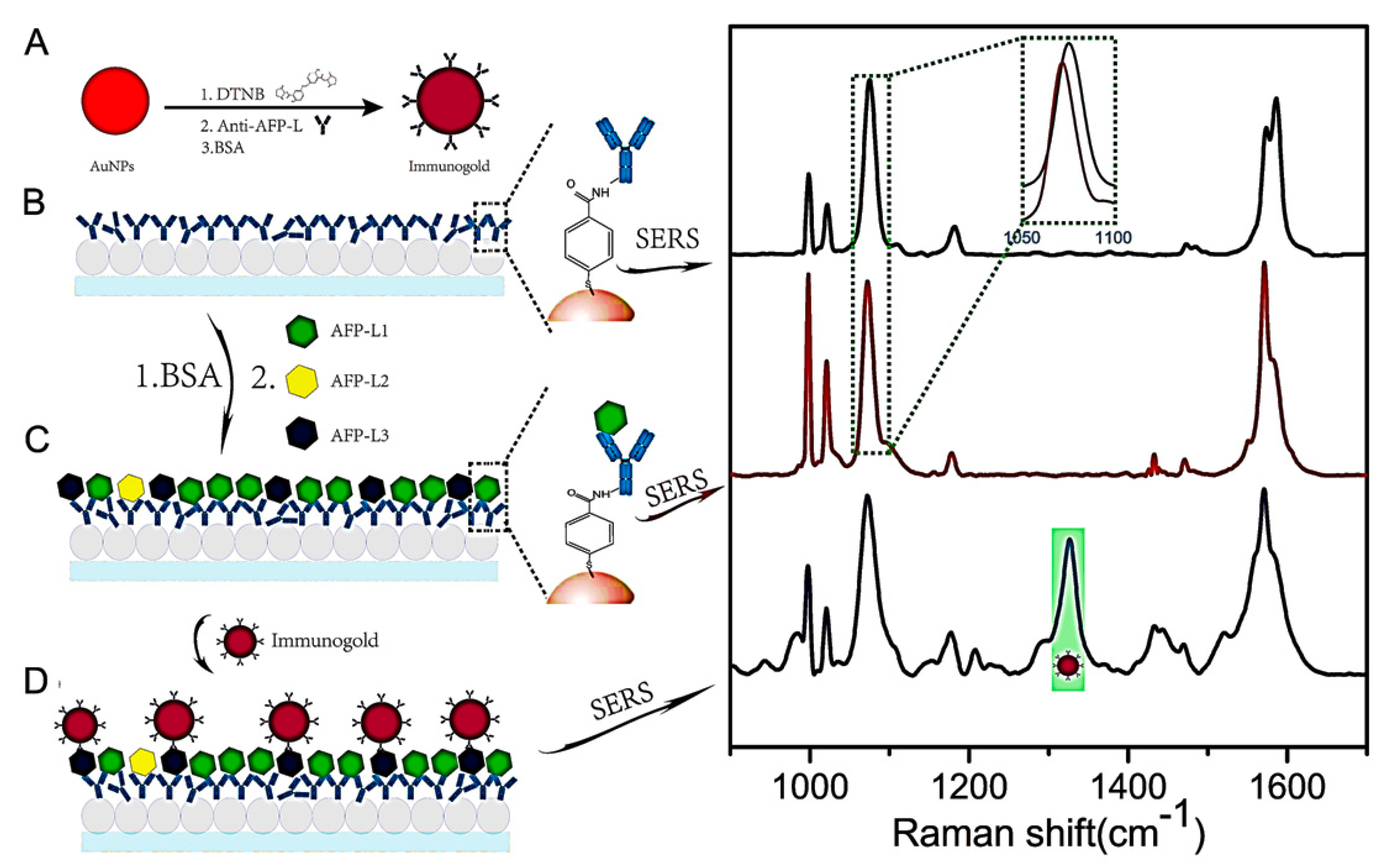
It has been confirmed that AFP produced during liver disease progression is different from normal AFP in the glycosyl chain. AFP is divided into three categories based on the action of AFP and adzuki bean agglutinin: AFP-L1, AFP-L2 and AFP-L3. AFP-L1 mainly exists in the cells of the embryonic stage belonging to normal human AFP; AFP-L2 comes from the AFP secreted by pregnant women; AFP-L3 is the protein specifically produced by hepatoma cells. If the total AFP concentration is detected, this concentration includes both the AFP-L3 concentration and the AFP-L1 and AFP-L2 concentrations, the latter of which are not specific to HCC. Thus, protein chips also produce very serious false-positive results. In the diagnosis, when AFP-L3 accounts for more than 10% of the total AFP, the patient can be diagnosed with early liver cancer, and AFP-L3% has been proposed as a new heteroplastic diagnostic index. Therefore, developing a protein chip capable of reading AFP-L3% with a heterogeneous resolution is very important and has many application prospects. A detection method for AFP-L3 based on SERS technology in the early stage was developed and effectively overcame the shortcomings of traditional AFP detection in the diagnosis of tumor diseases. The detection of AFP-L3 provides a new basis for the early diagnosis of liver cancer and can improve the detection accuracy for hepatitis, cirrhosis and liver cancer diagnosis [56]. Ma et al. [13] designed two SERS probes for multiple homologue detection with high sensitivity and specificity (Figure 1) and reported the quantitative analysis of AFP and AFP-L3 with high accuracy.
2.3. Diagnosis of Early Gastric Cancer (EGC) and Advanced Gastric Cancer (AGC)
Due to an unclean diet, increased work pressure, and Helicobacter pylori infection, gastric cancer has increasingly begun to develop in younger people. Gastric cancer can occur in any part of the stomach, and more than half of the cases occur in the antrum [57]. Most gastric cancers are adenocarcinomas. There are no obvious symptoms in the early stage, or there are nonspecific symptoms, such as upper abdominal discomfort and belching, that are often similar to those of chronic gastric diseases, such as gastritis and gastric ulcer and are easily overlooked. As the early diagnosis rate for gastric cancer is still low, it is very important to develop new diagnostic methods for early diagnosis. Recently, Hakim et al. [58], Amal et al. [59], and Peng et al. [60] found that human breath contains more than 3000 volatile organic compounds (VOCs). Some respiratory products can be used as markers in the diagnosis of early gastric cancer from occurrence to development. Certain VOCs are generated by cancer cells or tissues that undergo changes in metabolism or oxidative stress. Therefore, they can serve as cancer VOC biomarkers without influencing clinical symptoms.
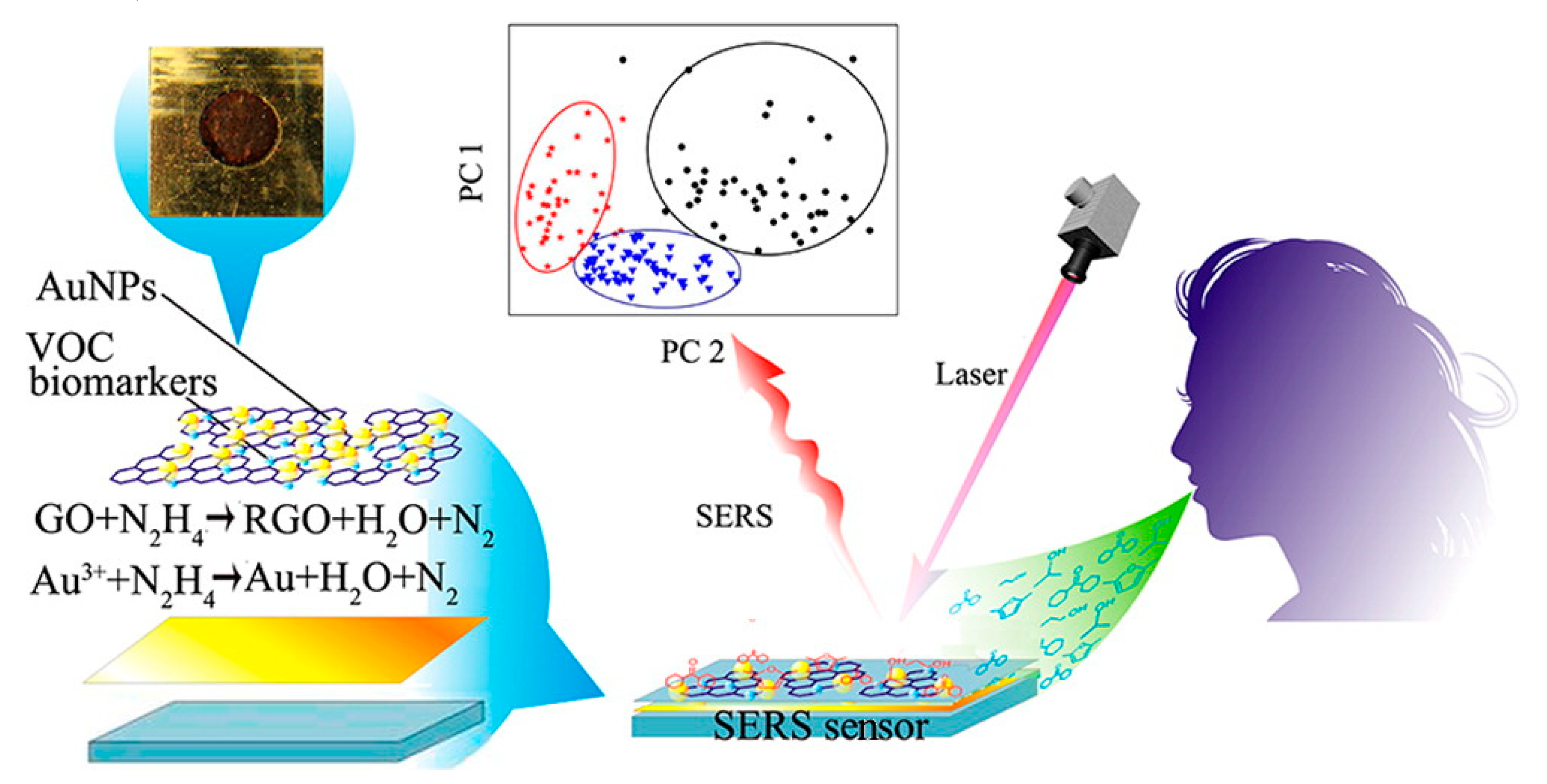
Chen et al. [61] designed Au nanoparticle-decorated reduced graphene oxide (RGO) composites, where RGO enhances the adsorption of biomarkers and Au nanoparticles act as SERS-active substrates for the effective determination of the adsorbed biomarkers (Figure 2). Most importantly, 14 VOC biomarkers in breath could be identified, and 14 Raman bands as fingerprints of biomarker patterns were selected for the diagnosis of patients at different stages. Breath analysis based on SERS offers a new method for the fast, low-cost and reliable diagnosis of diseases, showing clinical applicability.
2.4. Diagnosis of Pancreatic Cancer (PC)
Pancreatic cancer (PC) is a highly malignant tumor of the digestive tract that is difficult to diagnose and treat. Approximately 90% of cases are ductal adenocarcinomas originating from the ductal epithelium. The mortality and morbidity of PC have increased significantly in recent years. Carbohydrate antigen (CA) 19-9, matrix metallopeptidase 7 (MMP7) and mucin 4 (MUC4) have high sensitivity and good specificity for PC, with a positive rate of 85–95%. Thus, they are employed as biomarkers for the diagnosis of PC [62].
SERS provides a powerful method for the classification, early diagnosis and metastasis monitoring of PC. Banaei et al. [63] designed multiple detection systems, consisting of CA 19-9, MMP7 and MUC4, based on SERS. Microarray structures were used to accurately control antibody capture based on immunoassays of various serum markers of PC. Levels of various biomarkers in the sera of normal adults and individuals with pancreatitis and PC were measured. The results show that this method has great potential for the early diagnosis of PC. Beyene et al. developed a gold nanoflower (AuNF)-based SERS method for the detection of MUC4 and demonstrated approaches to improve the reliability of the method. The proposed method is very sensitive, and the detection limit is at the picogram level [64]. Pang et al. designed a magnetic SERS-active substrate for the one-step detection of microRNAs in exosomes and residual plasma from blood, which are biomarkers for the early detection of pancreatic cancer. The proposed method is fast and sensitive and can be used to test ultramicro-scale samples, which is helpful for the early detection of pancreatic cancer [65].
2.5. Specific Detection of Prostate Antigen (PSA) in the Diagnosis of Prostate Diseases and Prostate Cancer
Prostate-specific antigen (PSA) is a protein produced by the prostate. An increase in PSA in the blood may be a sign of cancer, but it may also be caused by aging, hypertrophy or inflammation of the prostate [66]. The main problems with the PSA test are false-positive results and overdiagnosis. Once a diagnosis is confirmed to be positive, the patient usually undergoes more tests, such as biopsy, which has other risks, such as bleeding, infection and urinary incontinence. Tumors in most prostate cancer patients grow slowly, and patients are more likely to die from other diseases before the prostate tumor develops symptoms. Prostate cancer is the most commonly diagnosed nonskin cancer. Most prostate cancer patients have a good prognosis, with a 10-year survival rate of 95%.
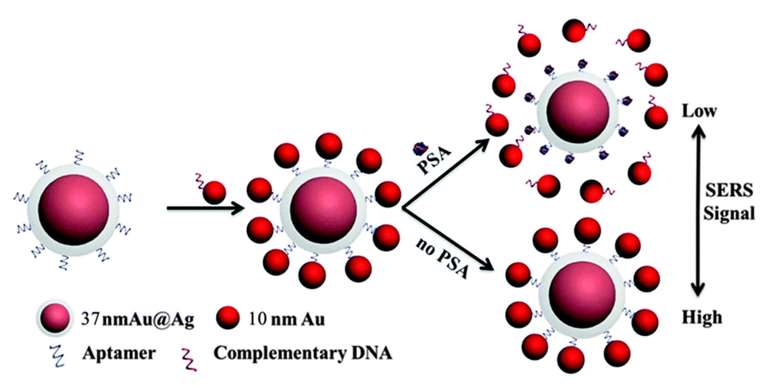
For the detection of prostate tumor markers, researchers have prepared various types of SERS-active substrates and SERS probes (Figure 3) [67,68,69,70,71]. SERS technology can be used to establish new methods that address the problems with traditional methods. Cheng and Gao et al. [72,73] designed a new diagnostic tool that measures the concentrations of total PSA (t-PSA), free PSA (f-PSA) and t-PSA/f-PSA in serum. The possibility of prostate cancer should be considered when concentrations of t-PSA and f-PSA in serum increase and those of t-PSA/f-PSA decrease. This tool will help improve the specificity and accuracy of prostate cancer diagnosis. In addition, the levels of t-PSA and f-PSA were also slightly increased in patients with prostatitis, prostatic hypertrophy, nephritis, prostatic polyps and urogenital diseases. Monolayer graphene, which has an electronic structure and interface properties that are highly sensitive to PSA, is employed as a Raman substrate. During specific binding to PSA and its aptamer, the G band of graphene undergoes a frequency shift, the extent of which is dependent on the PSA concentration [74]. Ag-decorated electrospun fibers and magnetic beads were employed for the recyclable determination of PSA with high sensitivity. The determination method produced uniform and reproducible SERS signals and exhibited excellent sensitivity (picogram scale) [75,76].
3. Structure Determination of Alzheimer’s Disease Markers Based on SERS Technology
In recent years, as life expectancy has continued to increase, the incidence of senile diseases has increased. Alzheimer’s disease has attracted increasing attention because it can cause memory loss, behavioral changes, and poor quality of life. The structural changes of a biomarker can be used as a reference for the identification of Alzheimer’s disease [77]. Two abnormally folded proteins, amyloid-β and tau, accumulate in the brain and cause Alzheimer’s disease. A label-free SERS-based method was employed to monitor variations in the conformation of the amyloid-β and tau proteins [78]. Lipid bilayer-encapsulated Ag nanoparticle-, nanofluidic- and electrochemical-based SERS and laser nanotextured SERS-active substrates were employed to study the conformational changes of amyloid-β proteins [78,79,80,81,82]. Ma et al. designed an Ag film to discriminate single-site phosphorylated S396 in a Tau410 protein, which is an important biomarker in Alzheimer’s disease [83]. Park et al. employed a sequential nanotransfer printing technique to fabricate a graphitic nanolayer-coated three-dimensional SERS substrate that successfully detected the two abnormally folded proteins and analyzed the secondary structural changes quantitatively [84]. The fingerprint information from SERS spectra helps to understand conformational changes that cause conformational diseases. In addition, by using SERS to study the interaction between amyloid-forming proteins and a biomimetic membrane, it was found that this interaction is of great importance for understanding the pathophysiology of Alzheimer’s disease.
4. Diagnosis of Cardiac Muscle Tissue Injury Based on the Cardiac Muscle Troponin T (cTnT) Biomarker
Acute myocardial infarction is myocardial necrosis caused by acute and persistent ischemia and hypoxia of the coronary arteries [85]. Recently, Wei et al. [86] found that cardiac muscle troponin T (cTnT) is a good marker for monitoring myocardial injury and necrosis, which has important clinical significance for the diagnosis and risk stratification of acute myocardial infarction (the diagnostic sensitivity is 100%, the specificity is 91%, and the duration is long). An elevated troponin value indicates myocardial injury, which can be seen in acute myocardial infarction.
When the SERS and ELISA techniques were combined, the quantitative determination of cardiovascular diseases exhibited excellent sensitivity. Yu et al. [87] employed 3,3′,5,5′-tetramethylbenzidine (TMB) as a probe molecule for indirect detection of cTnT. Herein, TMB was reduced to TMB2+ in the presence of cTnT. As cTnT increased, TMB2+ also increased synchronously. The proposed method exhibited an excellent linear relationship with a wide linear range. Thus, the established SERS-based method has potential applications in the bioanalysis and early diagnosis of diseases.
5. The Determination of Viral Nonstructural Proteins
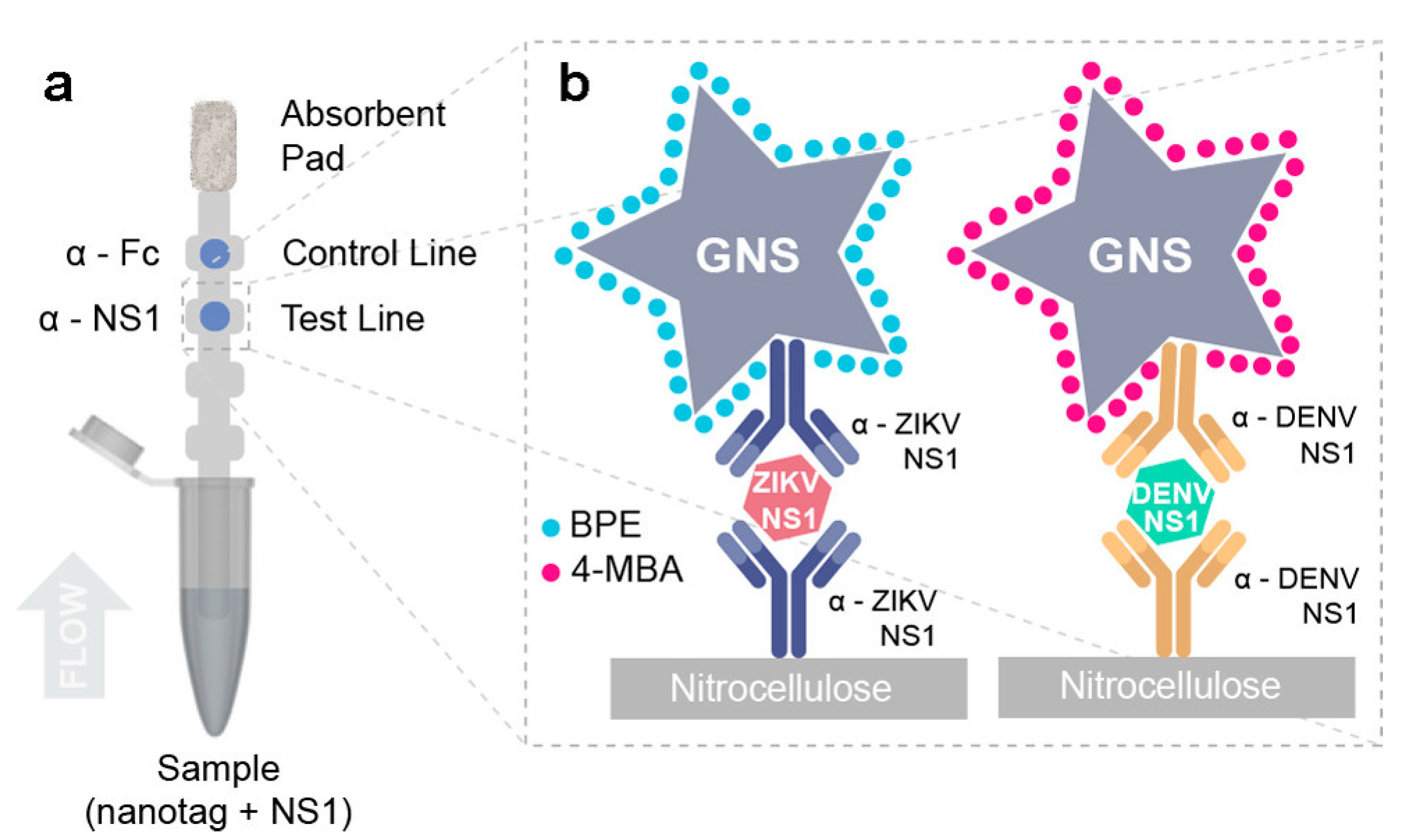
Viral structural proteins are necessary for the survival of mature virus particles, such as the viral capsid protein. Nonstructural proteins refer to enzymes that are expressed during viral infection and replication and that play a role in proliferation [88]. However, they do not bind to the mature virus and are not a part of the virus structure. Sánchez-Purrà et al. [89] found that viral nonstructural proteins can be employed as biomarkers for virus determination. Dengue virus (DENV) and Zika virus (ZIKV) have the same transmission vector, and infected patients may have concurrent Zika and dengue infections. In addition, both diseases exhibit similar nonspecific symptoms, but the disease outcomes are greatly different and affect patient treatment. SERS has been shown to be a good candidate method for the determination of dengue (DENV-3) and ZIKV (Figure 4). Their viral nonstructural proteins were detected based on a SERS technique. The authors designed 1,2-bis(4-pyridyl)ethylene (BPE)- and 4-mercaptobenzoic acid (MBA)-decorated gold nanostars as SERS probes that were specifically conjugated to antibodies to distinguish ZIKV and DENV biomarkers. The proposed SERS-based multiplexed assay exhibited excellent sensitivity compared to the colorimetric readout.
6. Detection of Cell Activity Based on Surface-Enhanced Resonance Raman Spectroscopy (SERRS)
The traditional quantitative test of cell activity (most commonly used in the MTT test) involves adding exogenous agents, such as MTT (thiazolyl blue tetrazolium bromide, [3-(4,5-dimethyl-2-thiazolyl)-2,5-diphenyl-2H-tetrazolium bromide]), to a cell suspension, and amber dehydrogenase in living cells can reduce MTT to water-insoluble formazan. Dead cells do not have this reducing function because they cannot produce dehydrogenase through metabolism. The traditional MTT method is quantitative because of colorimetry. It has many disadvantages, such as abundant interferences from other substances and low detection sensitivity (approximately 1.0 μg/mL), and is prone to false-positive results [90].
Based on the traditional MTT cell activity test, Mao et al. [91,92] developed a more sensitive cell activity identification method (a SERRS-MTT method) by exploiting the high sensitivity and high selectivity of SERRS (Figure 5). Based on the characteristics of SERRS, which provides fingerprint spectral information, the SERRS-MTT detection method is expected to avoid interferences from background substances (such as residual MTT reagent, serum, exogenous drugs) in cell suspensions. In addition, the SERRS-MTT method not only simplifies the testing procedure but is expected to have higher accuracy in evaluating the effectiveness of anticancer drug effects compared with the traditional MTT method.
A confocal Raman microscope imaging system can comprehensively analyze the chemical and physical properties of biological samples and visualize chemical components by generating depth profiles and 3D images. The characteristic Raman bands of biological macromolecules can be used to track the changes in various components before and after apoptosis through a Raman imaging system. The introduction of a SERS probe not only improves the sensitivity of Raman imaging but also realizes the dynamic observation of intracellular organelle interactions, providing a more convenient and intuitive detection method for the study of organelle interactions. On the other hand, a new type of formazan Raman probe developed based on the traditional MTT method to detect cell viability was combined with Raman imaging technology to determine in situ single-cell viability based on resonance Raman scattering (Figure 5). This method overcomes the limitations of the traditional MTT method in terms of complicated sample pretreatment steps and detection sensitivity and provides a new method for the in situ real-time evaluation of cell apoptosis.
7. Monitoring of Apoptosis Based on Cytochrome c (Cyt c)
Traditional methods for detecting the release of Cyt c from apoptotic cell mitochondria include immunoblotting, mass spectrometry, aptamer-labeled fluorescence spectroscopy, etc. [93,94,95]. Although these methods have high sensitivity, the sample pretreatment steps (protein purification, ligand preparation, etc.) are complicated, and these existing methods cannot distinguish the redox state of Cyt c. SERS was employed to establish a non-label-based method for fast and direct detection of Cyt c that can achieve fast and ultrasensitive detection of Cyt c released from apoptotic mitochondria [96]. Raman spectroscopy can be used to directly quantify the Cyt c released from mitochondria extracted from apoptotic cells, with a sensitivity of 2 μM, and the sensitivity increased 103 times after the introduction of SERS. This method can provide a way to study the regulation of apoptosis after organelle interaction.
8. Summary and Outlook
In recent years, the development of biomarker detection based on SERS technology has been rapid and comprehensive. With the rapid development of nanotechnology, SERS technology has received increased attention. From tumor identification, organelle functionality, and virus detection to identification of cell activity, SERS technology is playing an increasingly important diagnostic role. However, advances in SERS technology still face many challenges. For example, the poor repeatability of SERS substrate preparation results in poor repeatability of experimental data and affects the application and promotion of SERS. The following issues need to be resolved. (1) The development of the preparation and application of new universal SERS-active nanostructures with high sensitivity, specificity, and biocompatibility and low interference for biomarker determination is urgent. The optimal SERS-active nanostructures do not cause irreproducible changes in the spectrum. In addition to the improvement and development of the original SERS-enhanced substrate preparation method, it is also necessary to develop more kinds of SERS-active substrates and establish new SERS-enhanced substrate preparation technologies. (2) New SERS testing technology suitable for medical testing needs to be developed. Combining SERS technology with biomedical technologies, especially nondestructive detection and analysis technologies, will lead to new SERS detection technologies and the comprehensive analysis of biomarker information to fully utilize the advantages of SERS technology in disease-related biomarker detection with high sensitivity and specificity. (3) It is important to develop simple and effective protein bioconjugation methods. The adsorption behavior and structure of biomarkers in contact with substrates have not been clearly explored, resulting in insufficient interpretation of protein signals. Therefore, SERS technology should be used as a method to study the structure and function of biomarkers. (4) It is also important to extract information from target molecules in complex systems. Most methods focus on only characteristic SERS signals and ignore the processing and analysis of signal resolution and the quantitative structure–activity relationship of the full spectrum. Therefore, exploring new algorithms and building logic trees for complex systems will help solve and analyze practical problems. With new and noteworthy developments and diverse applications of SERS in various scientific fields, the impact of SERS will continue to advance and grow exponentially.
Author Contributions
Writing—original draft preparation, C.S., S.G. and S.J.; writing—review and editing, L.C. and Y.M.J. All authors have read and agreed to the published version of the manuscript.
Funding
This work was also supported by the National Research Foundation of Korea (NRF) grants funded by the Korea government (No. NRF-2018R1A2A3074587 and No. NRF-2020K2A9A2A06036299).
Conflicts of Interest
The authors declare no conflict of interest.
References
- GBD 2015 Mortality and Causes of Death Collaborators. Global, regional, and national life expectancy, all-cause mortality, and cause-specific mortality for 249 causes of death, 1980–2015: A systematic analysis for the Global Burden of Disease Study 2015. Lancet 2016, 388, 1459–1544. [Google Scholar] [CrossRef] [Green Version]
- Mendis, S.; Puska, P.; Norrving, B. Global Atlas on Cardiovascular Disease Prevention and Control; World Health Organization: Geneva, Switzerland; World Heart Federation and the World Stroke Organization: Geneva, Switzerland, 2011; pp. 3–18. [Google Scholar]
- Perez, M.M.; Ferrer, M.D.; Lazo-Rodriguez, M.; Canals, A.Z.; Banon-Maneus, E.; Campistol, J.M.; Miller, S.; Garg, R.; Gold, A.; Salcedo, C.; et al. A novel assay to measure calcification propensity: From laboratory to humans. Sci. Rep. 2020, 10, 17578. [Google Scholar] [CrossRef]
- Paraskevaidi, M.; Morais, C.L.M.; Lima, K.M.G.; Snowden, J.S.; Saxon, J.A.; Richardson, A.M.T.; Jones, M.; Mann, D.M.A.; Allsop, D.; Martin-Hirsch, P.L.; et al. Differential diagnosis of Alzheimer’s disease using spectrochemical analysis of blood. Proc. Natl. Acad. Sci. USA 2017, 114, E7929–E7938. [Google Scholar] [CrossRef] [PubMed] [Green Version]
- Vera, M.; Biswas, J.; Senecal, A.; Singer, R.H.; Park, H.Y. Single-cell and single-molecule analysis of gene expression regulation. Annu. Rev. Genet. 2016, 50, 267–291. [Google Scholar] [CrossRef] [PubMed] [Green Version]
- Piccoli, S.P.; Garofolo, F. Biomarker assay validation. Bioanalysis 2018, 10, 889–891. [Google Scholar] [CrossRef] [Green Version]
- Rosado, M.; Silva, R.; Bexiga, M.G.; Jones, J.G.; Manadas, B.; Anio, S.I. Advances in biomarker detection: Alternative approaches for blood-based biomarker detection. Adv. Clin. Chem. 2019, 92, 141–199. [Google Scholar]
- Jing, J.; Gao, Y. Urine biomarkers in the early stages of diseases: Current status and perspective. Discov. Med. 2018, 25, 57–65. [Google Scholar]
- Drabovich, A.P.; Martínez-Morillo, E.; Diamandis, E.P. Toward an integrated pipeline for protein biomarker development. Biochim. Biophys. Acta 2015, 1854, 677–686. [Google Scholar] [CrossRef]
- Rossetti, C.; Abdel Qader, A.; Grønhaug Halvorsen, T.; Sellergren, B.; Reubsaet, L. Antibody-free biomarker determination: Exploring molecularly imprinted polymers for pro-gastrin releasing peptide. Anal. Chem. 2014, 86, 12291–12298. [Google Scholar] [CrossRef] [Green Version]
- Hashkavayi, A.B.; Raoof, J.B.; Park, K.S. Sensitive electrochemical detection of tryptophan using a hemin/G-quadruplex aptasensor. Chemosensors 2020, 8, 100. [Google Scholar] [CrossRef]
- Wu, M.; Liu, H.; Liu, Z.; Liu, C.; Zhang, A.; Li, H. Analysis of serum Alpha-Fetoprotein (AFP) and AFP-L3 levels by protein microarray. J. Int. Med. Res. 2018, 46, 4297–4305. [Google Scholar] [CrossRef] [PubMed] [Green Version]
- Ma, H.; Sun, X.; Chen, L.; Cheng, W.; Han, X.X.; Zhao, B.; He, C. Multiplex immunochips for high-accuracy detection of AFP-L3% based on surface-enhanced Raman scattering: Implications for early liver cancer diagnosis. Anal. Chem. 2017, 89, 8877–8883. [Google Scholar] [CrossRef] [PubMed]
- Vo-Dinh, T.; Yan, F.; Wabuyele, M.B. Surface-enhanced Raman scattering for biomedical diagnostics and molecular imaging. Surf. Enhanc. Raman Scatt. 2006, 103, 409–426. [Google Scholar]
- Kneipp, J.; Wittig, B.; Bohr, H.; Kneipp, K. Surface-enhanced Raman scattering: A new optical probe in molecular biophysics and biomedicine. Theor. Chem. Acc. 2010, 125, 319–327. [Google Scholar] [CrossRef]
- Fleischmann, M.; Hendra, P.J.; McQuillan, A.J. Raman spectra of pyridine adsorbed at a silver electrode. Chem. Phys. Lett. 1974, 26, 163–166. [Google Scholar] [CrossRef]
- Jeanmaire, D.L.; Van Duyne, R.P. Surface Raman electrochemistry: Part I. Heterocyclic, aromatic and aliphatic amines adsorbed on the anodized silver electrode. J. Electroanal. Chem. 1977, 84, 1–20. [Google Scholar] [CrossRef]
- Albrecht, M.G.; Creighton, J.A. Anomalously intense raman spectra of pyridine at a silver electrode. J. Am. Chem. Soc. 1977, 99, 5215–5219. [Google Scholar] [CrossRef]
- Tian, Z.Q.; Yang, Z.L.; Ren, B.; Li, J.F.; Zhang, Y.; Lin, X.F.; Hu, J.W.; Wu, D.Y. Surface-enhanced Raman scattering from transition metals with special surface morphology and nanoparticle shape. Faraday Discuss. 2006, 132, 159–170. [Google Scholar] [CrossRef] [Green Version]
- Ji, W.; Li, L.; Song, W.; Wang, X.; Zhao, B.; Ozaki, Y. Enhanced Raman scattering by ZnO superstructures: Synergistic effect of charge transfer and Mie resonances. Angew. Chem. Int. Ed. 2019, 58, 14552–14556. [Google Scholar]
- Aroca, R. Surface-Enhanced Vibrational Spectroscopy; John Wiley & Sons Ltd.: Chichester, UK, 2006. [Google Scholar]
- Kneipp, K.; Moskovits, M.; Kneipp, H. Surface-Enhanced Raman Scattering-Physics and Applications; Springer: Berlin, Germany, 2006. [Google Scholar]
- Šašić, S. Pharmaceutical Applications of Raman Spectroscopy; John Wiley & Sons Ltd.: Hoboken, NJ, USA, 2008. [Google Scholar]
- Han, X.X.; Zhao, B.; Ozaki, Y. Surface-enhanced Raman scattering for protein detection. Anal. Bioanal. Chem. 2009, 394, 1719–1727. [Google Scholar] [CrossRef]
- Han, X.X.; Zhao, B.; Ozaki, Y. Label-free detection in biological applications of surface-enhanced Raman scattering. TrAC Trend. Anal. Chem. 2012, 38, 67–78. [Google Scholar] [CrossRef]
- Murgida, D.H.; Hildebrandt, P. Proton-coupled electron transfer of cytochrome c. J. Am. Chem. Soc. 2001, 123, 4062–4068. [Google Scholar] [CrossRef]
- Siebert, F.; Hildebrandt, P. Vibrational Spectroscopy in Life Science; Wiley-VCH: Weinheim, Germany, 2008. [Google Scholar]
- Nie, S.M.; Emory, S.R. Probing single molecules and single nanoparticles by surface-enhanced Raman scattering. Science 1997, 275, 1102–1106. [Google Scholar] [CrossRef] [PubMed]
- Kneipp, K.; Wang, Y.; Kneipp, H.; Perelman, L.T.; Itzkan, I.; Dasari, R.R.; Feld, M.S. Single molecule detection using Surface-Enhanced Raman Scattering (SERS). Phys. Rev. Lett. 1997, 78, 1667–1670. [Google Scholar] [CrossRef] [Green Version]
- Boken, J.; Khurana, P.; Thatai, S.; Kumar, D.; Prasad, S. Plasmonic nanoparticles and their analytical applications: A review. Appl. Spectrosc. Rev. 2017, 52, 774–820. [Google Scholar] [CrossRef]
- Smith, E.; Dent, G. Modern Raman Spectroscopy—A Practical Approach; John Wiley & Sons Ltd.: Chichester, UK, 2005. [Google Scholar]
- Han, X.X.; Ji, W.; Zhao, B.; Ozaki, Y. Semiconductor-enhanced Raman scattering: Active nanomaterials and applications. Nanoscale 2017, 9, 4847–4861. [Google Scholar] [CrossRef]
- Chen, L.; Cai, L.; Ruan, W.; Zhao, B. Surface-enhanced Raman Spectroscopy (SERS): Protein application. In Encyclopedia of Analytical Chemistry; John Wiley & Sons Ltd.: Chichester, UK, 2014. [Google Scholar]
- Stiles, P.L.; Dieringer, J.A.; Shah, N.C.; Van Duyne, R.P. Surface-enhanced Raman Spectroscopy. Annu. Rev. Anal. Chem. 2008, 1, 601–626. [Google Scholar] [CrossRef] [Green Version]
- Crawford, A.C.; Skuratovsky, A.; Porter, M.D. Sampling error: Impact on the quantitative analysis of nanoparticle-based surface-enhanced Raman scattering immunoassays. Anal. Chem. 2016, 88, 6515–6522. [Google Scholar] [CrossRef]
- Shin, H.; Jeong, H.; Park, J.; Hong, S.; Choi, Y. Correlation between cancerous exosomes and protein markers based on Surface-Enhanced Raman Spectroscopy (SERS) and Principal Component Analysis (PCA). ACS Sens. 2018, 3, 2637–2643. [Google Scholar] [CrossRef]
- Qiu, L.; Wang, W.; Zhang, A.; Zhang, N.; Lemma, T.; Ge, H.; Toppari, J.J.; Hytönen, V.P.; Wang, J. Core–shell nanorod columnar array combined with gold nanoplate–nanosphere assemblies enable powerful in situ SERS detection of bacteria. ACS Appl. Mater. Interfaces 2016, 8, 24394–24403. [Google Scholar] [CrossRef]
- Sharma, B.; Bugga, P.; Madison, L.R.; Henry, A.; Blaber, M.G.; Greeneltch, N.G.; Chiang, N.; Mrksich, M.; Schatz, G.C.; Van Duyne, P.R. Bisboronic acids for selective, physiologically relevant direct glucose sensing with surface-enhanced Raman spectroscopy. J. Am. Chem. Soc. 2016, 138, 13952–13959. [Google Scholar] [CrossRef] [PubMed]
- Hu, W.; Ye, S.; Zhang, Y.; Li, T.; Zhang, G.; Luo, Y.; Mukamel, S.; Jiang, J. Machine learning protocol for surface-enhanced Raman spectroscopy. J. Phys. Chem. Lett. 2019, 10, 6026–6031. [Google Scholar] [CrossRef] [PubMed]
- Silsirivanit, A. Glycosylation markers in cancer. Adv. Clin. Chem. 2019, 89, 189–213. [Google Scholar] [PubMed]
- Rosen, R.T.; Hiserodt, R.D.; Fukuda, E.K.; Ruiz, R.J.; Zhou, Z.; Lech, J.; Rosen, S.L.; Hartman, T.G. The determination of metabolites of garlic preparations in breath and human plasma. Biofactors 2000, 13, 241–249. [Google Scholar] [CrossRef]
- Lee, J.; Kim, H.; Heo, Y.; Yoo, Y.K.; Han, S.I.; Kim, C.; Hur, D.; Kim, H.; Kang, J.Y.; Lee, J.H. Enhanced paper-based ELISA for simultaneous EVs/Exosome isolation and detection using streptavidin agarose-based immobilization. Analyst 2020, 145, 157–164. [Google Scholar] [CrossRef]
- Dwenger, A. Radioimmunoassay: An overview. J. Clin. Chem. Clin. Biochem. 1984, 22, 883–894. [Google Scholar]
- Lim, S.I.; Jeong, W.; Tark, D.S.; Yang, D.K.; Kweon, C.H. Agar gel immunodiffusion analysis using baculovirus-expressed recombinant bovine leukemia virus envelope glycoprotein (gp51/gp30(T-)). J. Vet. Sci. 2009, 10, 331–336. [Google Scholar] [CrossRef]
- Maier, T.V.; Schmitt-Kopplin, P. Capillary electrophoresis in metabolomics. Methods Mol. Biol. 2016, 1483, 437–470. [Google Scholar]
- Macaulay, M.E. The IgM and IgG response to Bordetella pertussis vaccination and infection. J. Med. Microbiol. 1981, 14, 1–7. [Google Scholar] [CrossRef]
- Durrington, P.N.; Whicher, J.T.; Warren, C.; Bolton, C.H.; Hartog, M. A comparison of methods for the immunoassay of serum apolipoprotein B in man. Clin. Chim. Acta 1976, 71, 95–108. [Google Scholar] [CrossRef]
- Duffy, M.J. Carcinoembryonic antigen as a marker for colorectal cancer: Is it clinically useful? Clin. Chem. 2001, 47, 624–630. [Google Scholar] [CrossRef] [PubMed] [Green Version]
- Chon, H.; Lee, S.; Son, S.W.; Oh, C.H.; Choo, J. Highly sensitive immunoassay of lung cancer marker carcinoembryonic antigen using surface-enhanced Raman scattering of hollow gold nanospheres. Anal. Chem. 2009, 81, 3029–3034. [Google Scholar] [CrossRef] [PubMed]
- Chon, H.; Lee, S.; Yoon, S.-Y.; Chang, S.-I.; Lim, D.W.; Choo, J. Simultaneous immunoassay for the detection of two lung cancer markers using functionalized SERS nanoprobes. Chem. Chommun. 2011, 47, 12515–12517. [Google Scholar] [CrossRef] [PubMed]
- Rong, Z.; Wang, C.; Wang, J.; Wang, D.; Xiao, R.; Wang, S. Magnetic immunoassay for cancer biomarker detection based on surface-enhanced resonance Raman scattering from coupled plasmonic nanostructures. Biosens. Bioelectron. 2016, 84, 15–21. [Google Scholar] [CrossRef]
- Ganesan, S.; Venkatakrishnan, K.; Tan, B. Wrinkled metal-based quantum sensor for in vitro cancer diagnosis. Biosens. Bioelectron. 2020, 151, 111967. [Google Scholar] [CrossRef]
- Carneiro, M.C.C.G.; Sousa-Castillo, A.; Correa-Duarte, M.A.; Sales, M.G.F. Dual biorecognition by combining molecularly imprinted polymer and antibody in SERS detection. Application to carcinoembryonic antigen. Biosens. Bioelectron. 2019, 146, 111761. [Google Scholar] [CrossRef]
- Song, C.; Yang, Y.; Yang, B.; Min, L.; Wang, L. Combination assay of lung cancer associated serum markers using surface-enhanced Raman spectroscopy. J. Mater. Chem. B 2016, 4, 1811–1817. [Google Scholar] [CrossRef]
- Zhou, L.; Zhou, J.; Feng, Z.; Wang, F.; Xie, S.; Bu, S. Immunoassay of tumor markers in human serum based on SI nanoparticles and SiC@Ag SERS-active substrate. Analyst 2016, 141, 2534–2541. [Google Scholar] [CrossRef]
- Li, D.; Mallory, T.; Satomura, S. AFP-L3: A new generation of tumor marker for hepatocellular carcinoma. Clin. Chim. Acta 2001, 313, 15–19. [Google Scholar] [CrossRef]
- Kang, G.; Hwang, W.C.; Do, I.G.; Wang, K.; Kang, S.Y.; Lee, J.; Park, S.H.; Park, J.O.; Kang, W.K.; Jang, J.; et al. Exome sequencing identifies early gastric carcinoma as an early stage of advanced gastric cancer. PLoS ONE 2013, 8, e82770. [Google Scholar] [CrossRef] [Green Version]
- Hakim, M.; Broza, Y.Y.; Barash, O.; Peled, N.; Phillips, M.; Amann, A.; Haick, H. volatile organic compounds of lung cancer and possible biochemical pathways. Chem. Rev. 2012, 112, 5949–5966. [Google Scholar] [CrossRef] [PubMed]
- Amal, H.; Ding, L.; Liu, B.-B.; Tisch, U.; Xu, Z.-Q.; Shi, D.-Y.; Zhao, Y.; Chen, J.; Sun, R.-X.; Liu, H.; et al. The scent fingerprint of hepatocarcinoma: In-vitro metastasis prediction with volatile organic compounds (VOCs). Int. J. Nanomed. 2012, 7, 4135–4146. [Google Scholar]
- Peng, G.; Hakim, M.; Broza, Y.Y.; Billan, S.; Abdah-Bortnyak, R.; Kuten, A.; Tisch, U.; Haick, H. Detection of lung, breast, colorectal, and prostate cancers from exhaled breath using a single array of nanosensors. Br. J. Cancer 2010, 103, 542–551. [Google Scholar] [CrossRef] [PubMed]
- Chen, Y.; Zhang, Y.; Pan, F.; Liu, J.; Wang, K.; Zhang, C.; Cheng, S.; Lu, L.; Zhang, W.; Zhang, Z.; et al. Breath analysis based on surface-enhanced Raman scattering sensors distinguishes early and advanced gastric cancer patients from healthy persons. ACS Nano 2016, 10, 8169–8179. [Google Scholar] [CrossRef]
- Chu, L.C.; Goggins, M.G.; Fishman, E.K. Diagnosis and detection of pancreatic cancer. Cancer J. 2017, 23, 333–342. [Google Scholar] [CrossRef]
- Banaei, N.; Foley, A.; Houghton, J.M.; Sun, Y.; Kim, B. Multiplex detection of pancreatic cancer biomarkers using a SERS based immunoassay. Nanotechnology 2017, 28, 455101. [Google Scholar] [CrossRef] [Green Version]
- Beyene, A.B.; Hwang, B.J.; Tegegne, W.A.; Wang, J.S.; Tsai, H.C.; Su, W.N. Reliable and sensitive detection of pancreatic cancer marker by gold nanoflower-based SERS mapping immunoassay. Microchem. J. 2020, 158, 105099. [Google Scholar] [CrossRef]
- Pang, Y.; Wang, C.; Lu, L.; Wang, C.; Sun, Z.; Xiao, R. Dual-SERS biosensor for one-step detection of microRNAs in exosome and residual plasma of blood samples for diagnosing pancreatic cancer. Biosens. Bioelectron. 2019, 130, 204–213. [Google Scholar] [CrossRef]
- Ward, A.M.; Catto, J.W.; Hamdy, F.C. Prostate specific antigen: Biology, biochemistry and available commercial assays. Ann. Clin. Biochem. 2001, 38, 633–651. [Google Scholar] [CrossRef] [Green Version]
- Ma, W.; Yin, H.; Xu, L.; Wu, X.; Kuang, H.; Wang, L.; Xu, C. Ultrasensitive aptamer-based SERS detection of PSAs by heterogeneous satellite nanoassemblies. Chem. Commun. 2014, 50, 9737–9740. [Google Scholar] [CrossRef]
- Ballentine Carter, H. Prostate-Specific Antigen (PSA) screening for prostate cancer. JAMA 2018, 319, 1866–1868. [Google Scholar] [CrossRef] [PubMed]
- Yoon, K.J.; Seo, H.K.; Hwang, H.; Pyo, D.; Eom, I.-Y.; Hahn, J.H.; Jung, Y.M. Bioanalytical application of SERS immunoassay for detection of prostate-specific antigen. Bull. Korean Chem. Soc. 2010, 31, 1215–1218. [Google Scholar] [CrossRef] [Green Version]
- Schlücker, S. SERS microscopy: Nanoparticle probes and biomedical applications. ChemPhysChem 2009, 10, 1344–1354. [Google Scholar] [CrossRef] [PubMed]
- Qu, B.; Chu, X.; Shen, G.; Yu, R. A novel electrochemical immunosensor based on colabeled silica nanoparticles for determination of total prostate specific antigen in human serum. Talanta 2008, 76, 785–790. [Google Scholar] [CrossRef]
- Cheng, Z.; Choi, N.; Wang, R.; Lee, S.; Moon, K.C.; Yoon, S.Y.; Chen, L.; Choo, J. Simultaneous detection of dual prostate specific antigens using surface-enhanced Raman scattering-based immunoassay for accurate diagnosis of prostate cancer. ACS Nano 2017, 11, 4926–4933. [Google Scholar] [CrossRef]
- Gao, R.; Cheng, Z.; Wang, X.; Yu, L.; Guo, Z.; Zhao, G.; Choo, J. Simultaneous immunoassays of dual prostate cancer markers using a SERS-based microdroplet channel. Biosens. Bioelectron. 2018, 119, 126–133. [Google Scholar] [CrossRef]
- Liu, S.; Huo, Y.; Bai, J.; Ning, B.; Peng, Y.; Li, S.; Han, D.; Kang, W.; Gao, Z. Rapid and sensitive detection of prostate-specific antigen via label-free frequency shift Raman of sensing graphene. Biosens. Bioelectron. 2020, 158, 112184. [Google Scholar] [CrossRef]
- Du, Y.; Liu, H.; Chen, Y.; Tian, Y.; Zhang, X.; Gu, C.; Jiang, T.; Zhou, J. Recyclable label-free SERS-based immunoassay of PSA in human serum mediated by enhanced photocatalysis arising from ag nanoparticles and external magnetic field. Appl. Surf. Sci. 2020, 528, 146953. [Google Scholar] [CrossRef]
- Yun, B.J.; Koh, W.G. Highly sensitive SERS-based immunoassay platform prepared on silver nanoparticle-decorated electrospun polymeric fibers. J. Ind. Eng. Chem. 2020, 82, 341–348. [Google Scholar] [CrossRef]
- Tan, C.-C.; Yu, J.-T.; Tan, L. Biomarkers for preclinical Alzheimer’s Disease. J. Alzheimers Dis. 2014, 42, 1051–1069. [Google Scholar] [CrossRef]
- Choi, I.; Huh, Y.S.; Erickson, D. Ultra-sensitive, label-free probing of the conformational characteristics of amyloid beta aggregates with a SERS active nanofluidic device. Microfluid. Nanofluid. 2012, 12, 663–669. [Google Scholar] [CrossRef]
- Karaballi, R.A.; Merchant, S.; Power, S.; Brosseau, C.L. Electrochemical Surface-Enhanced Raman Spectroscopy (EC-SERS) study of the interaction between protein aggregates and biomimetic membranes. Phys. Chem. Chem. Phys. 2018, 20, 4513–4526. [Google Scholar] [CrossRef] [PubMed]
- Bhowmik, D.; Mote, K.R.; MacLaughlin, C.M.; Biswas, N.; Chandra, B.; Basu, J.K.; Walker, G.C.; Madhu, P.K.; Maiti, S. Cell-membrane-mimicking lipid-coated nanoparticles confer Raman enhancement to membrane proteins and reveal membrane-attached amyloid-β conformation. ACS Nano 2015, 9, 9070–9077. [Google Scholar] [CrossRef] [PubMed]
- Buividas, R.; Dzingelevičius, N.; Kubiliūtė, R.; Stoddart, P.R.; Truong, V.K.; Ivanova, E.P.; Juodkazis, S. Statistically quantified measurement of an Alzheimer’s marker by surface-enhanced Raman scattering. J. Biophotonics 2015, 8, 567–574. [Google Scholar] [CrossRef] [PubMed]
- Yang, J.K.; Hwang, I.J.; Cha, M.G.; Kim, H.I.; Yim, D.; Jeong, D.H.; Lee, Y.S.; Kim, J.H. Reaction kinetics-mediated control over silver nanogap shells as surface-enhanced Raman scattering nanoprobes for detection of Alzheimer’s disease biomarkers. Small 2019, 15, 1900613. [Google Scholar] [CrossRef]
- Ma, H.; Liu, S.; Liu, Y.; Zhu, J.; Han, X.X.; Ozaki, Y.; Zhao, B. In-situ fingerprinting phosphorylated proteins via surface-enhanced Raman spectroscopy: Single-site discrimination of tau biomarkers in Alzheimer’s disease. Biosens. Bioelectron. 2021, 171, 112748. [Google Scholar] [CrossRef]
- Park, H.J.; Cho, S.; Kim, M.; Jung, Y.S. Carboxylic acid-functionalized, graphitic layer-coated three-dimensional SERS substrate for label-free analysis of Alzheimer’s disease biomarkers. Nano Lett. 2020, 20, 2576–2584. [Google Scholar] [CrossRef]
- Mueller, M.; Vafaie, M.; Biener, M.; Giannitsis, E.; Katus, H.A. Cardiac troponin T: From diagnosis of myocardial infarction to cardiovascular risk prediction. Circ. J. 2013, 77, 1653–1661. [Google Scholar] [CrossRef] [Green Version]
- Wei, B.; Jin, J.-P. Troponin T isoforms and posttranscriptional modifications: Evolution, regulation and function. Arch. Biochem. Biophys. 2011, 505, 144–154. [Google Scholar] [CrossRef] [Green Version]
- Yu, Z.; Chen, L.; Wang, Y.; Wang, X.; Song, W.; Ruan, W.; Zhao, B.; Cong, Q. A SERS-active enzymatic product used for the quantification of disease-related molecules. J. Raman Spectrosc. 2014, 45, 75–81. [Google Scholar] [CrossRef]
- Xu, S.; Ci, Y.; Wang, L.; Yang, Y.; Zhang, L.; Xu, C.; Qin, C.; Shi, L. Zika Virus NS3 is a canonical RNA helicase stimulated by NS5 RNA polymerase. Nucleic Acids Res. 2019, 47, 8693–8707. [Google Scholar] [CrossRef] [PubMed]
- Sánchez-Purrà, M.; Carré-Camps, M.; Puig, H.; Bosch, I.; Gehrke, L.; Hamad-Schifferli, K. Surface-enhanced Raman spectroscopy-based sandwich immunoassays for multiplexed detection of Zika and dengue viral biomarkers. ACS Infect. Dis. 2017, 3, 767–776. [Google Scholar] [CrossRef] [PubMed]
- Nga, N.T.H.; Ngoc, T.T.B.; Trinh, N.T.M.; Thuoc, T.L.; Thao, D.T.P. Optimization and application of MTT assay in determining density of suspension cells. Anal. Biochem. 2020, 610, 113937. [Google Scholar] [CrossRef] [PubMed]
- Mao, Z.; Liu, Z.; Chen, L.; Yang, J.; Zhao, B.; Jung, Y.M.; Wang, X.; Zhao, C. Predictive value of the surface-enhanced resonance Raman scattering-based MTT assay: A rapid and ultrasensitive method for cell viability in situ. Anal. Chem. 2013, 85, 7361–7368. [Google Scholar] [CrossRef]
- Mao, Z.; Liu, Z.; Yang, J.; Han, X.; Zhao, B.; Zhao, C. In situ semi-quantitative assessment of single-cell viability by resonance Raman spectroscopy. Chem. Commun. 2018, 54, 7135–7138. [Google Scholar] [CrossRef]
- Song, B.; Li, J.; Li, J. Pomegranate peel extract polyphenols induced apoptosis in human hepatoma cells by mitochondrial pathway. Food Chem. Toxicol. 2016, 93, 158–166. [Google Scholar] [CrossRef]
- Tyurina, Y.Y.; Tyurin, V.A.; Kapralova, V.I.; Wasserloos, K.; Mosher, M.; Epperly, M.W.; Greenberger, J.S.; Pitt, B.R.; Kagan, V.E. Oxidative lipidomics of γ-radiation-induced lung injury: Mass spectrometric characterization of cardiolipin and phosphatidylserine peroxidation. Radiat. Res. 2011, 175, 610–621. [Google Scholar] [CrossRef] [Green Version]
- Zhang, H.; Zhang, B.; Di, C.; Ali, M.C.; Chen, J.; Li, Z.; Si, J.; Zhang, H.; Qiu, H. Label-free fluorescence imaging of cytochrome c in living systems and anti-cancer drug screening with nitrogen doped carbon quantum dots. Nanoscale 2018, 10, 5342–5349. [Google Scholar] [CrossRef]
- Zhu, J.; Jiang, M.; Ma, H.; Zhang, H.; Cheng, W.; Li, J.; Cai, L.; Han, X.X.; Zhao, B. Redox-state-mediated regulation of cytochrome c release in apoptosis revealed by surface-enhanced Raman scattering on nickel substrates. Angew. Chem. Int. Ed. 2019, 58, 16499–16503. [Google Scholar] [CrossRef]
Figure 1.
Schematic of a SERS-based immunoassay: (A) preparation of immunogold; (B) antibody capture chip fabrication; (C,D) reproduced with permission from [13]. (Copyright 2017, ACS.)
Figure 1.
Schematic of a SERS-based immunoassay: (A) preparation of immunogold; (B) antibody capture chip fabrication; (C,D) reproduced with permission from [13]. (Copyright 2017, ACS.)

Figure 2.
Schematic diagrams of a SERS sensor and overview of the processes involved in the use of breath for the determination of early gastric cancer (EGC) and advanced gastric cancer (AGC). AuNPs, gold nanoparticles; VOCs, volatile organic compounds; RGO, reduced graphene oxide; GO, graphene oxide; PC, principal component analysis. (Reproduced with permission from [61]. Copyright 2016, ACS.)

Figure 3.
Schematic of a method for prostate-specific antigen (PSA) detection based on aptamer-directed core–satellite assemblies. (Reproduced with permission from [67]. Copyright 2014, RSC.)
Figure 3.
Schematic of a method for prostate-specific antigen (PSA) detection based on aptamer-directed core–satellite assemblies. (Reproduced with permission from [67]. Copyright 2014, RSC.)

Figure 4.
Sandwich immunoassay: (a) schematic of a dipstick sandwich immunoassay and (b) sandwiches formed by each antibody pair, NS1 and GNS−Ab conjugate, for both ZIKV and DENV NS1 at the test line. Fc, the Fe region of the protein; NS1, nonstructural protein 1; ZIKA, Zika virus; DENV, dengue virus; GNS, gold nanostar. (Reproduced with permission from [89]. Copyright 2017, ACS.)

Figure 5.
Schematic of the detection of cell activity based on the SERRS-MTT assay. (Reproduced with permission from [91]. Copyright 2013, ACS.)
Figure 5.
Schematic of the detection of cell activity based on the SERRS-MTT assay. (Reproduced with permission from [91]. Copyright 2013, ACS.)

Publisher’s Note: MDPI stays neutral with regard to jurisdictional claims in published maps and institutional affiliations. |
© 2020 by the authors. Licensee MDPI, Basel, Switzerland. This article is an open access article distributed under the terms and conditions of the Creative Commons Attribution (CC BY) license (http://creativecommons.org/licenses/by/4.0/).
Share and Cite
MDPI and ACS Style
Song, C.; Guo, S.; Jin, S.; Chen, L.; Jung, Y.M. Biomarkers Determination Based on Surface-Enhanced Raman Scattering. Chemosensors 2020, 8, 118. https://0-doi-org.brum.beds.ac.uk/10.3390/chemosensors8040118
AMA Style
Song C, Guo S, Jin S, Chen L, Jung YM. Biomarkers Determination Based on Surface-Enhanced Raman Scattering. Chemosensors. 2020; 8(4):118. https://0-doi-org.brum.beds.ac.uk/10.3390/chemosensors8040118
Chicago/Turabian StyleSong, Chao, Shuang Guo, Sila Jin, Lei Chen, and Young Mee Jung. 2020. "Biomarkers Determination Based on Surface-Enhanced Raman Scattering" Chemosensors 8, no. 4: 118. https://0-doi-org.brum.beds.ac.uk/10.3390/chemosensors8040118
Note that from the first issue of 2016, this journal uses article numbers instead of page numbers. See further details here.