Nanomaterials Based Electrochemical Sensors for Serotonin Detection: A Review


Department of Chemistry, Physics and Environment, Faculty of Sciences and Environment, “Dunărea de Jos” University of Galaţi, 47 Domneasca Street, 800008 Galaţi, Romania
*
Author to whom correspondence should be addressed.
Chemosensors 2021, 9(1), 14; https://0-doi-org.brum.beds.ac.uk/10.3390/chemosensors9010014
Submission received: 22 December 2020
/
Revised: 9 January 2021
/
Accepted: 12 January 2021
/
Published: 14 January 2021
(This article belongs to the Special Issue Voltammperometric Sensors)
Abstract
:The present review deals with the recent progress made in the field of the electrochemical detection of serotonin by means of electrochemical sensors based on various nanomaterials incorporated in the sensitive element. Due to the unique chemical and physical properties of these nanomaterials, it was possible to develop sensitive electrochemical sensors with excellent analytical performances, useful in the practice. The main electrochemical sensors used in serotonin detection are based on carbon electrodes modified with carbon nanotubes and various materials, such as benzofuran, polyalizarin red-S, poly(L-arginine), Nafion/Ni(OH)2, or graphene oxide, incorporating silver-silver selenite nanoparticles, as well as screen-printed electrodes modified with zinc oxide or aluminium oxide. Also, the review describes the nanocomposite sensors based on conductive polymers, tin oxide-tin sulphide, silver/polypyrole/copper oxide or a hybrid structure of cerium oxide-gold oxide nanofibers together with ruthenium oxide nanowires. The presentation focused on describing the sensitive materials, characterizing the sensors, the detection techniques, electroanalytical properties, validation and use of sensors in lab practice.
1. Introduction
Serotonin, also known as 5-hydroxytriptamin, is a key messenger mediating a series of central and peripheral functions in the human body. As a neurotransmitter in the central nervous system, it is necessary in various brain functions and associated to anxiety and behaviour. Besides, the serotonin at the level of the central nervous system contributes to the neuronal control of intestine movements and the secretion of intestinal fluids [1]. However, the actions of serotonin extend beyond interneuronal communication at the level of the central nervous system and the enteric nervous system towards the peripheral tissues. Serotonin mediates numerous non-neuronal processes, such as the function of the bladder, respiratory system, haemostasis, vascular tone, immunity and intestinal inflammation [2,3,4].
Serotonin is also a regulator of both parts of the energy balance, i.e., energy accumulation and energy consumption. Serotonin at the level of the central nervous system is involved in appetite regulation and subsequently nutrient intake [5]. The inhibitory effect of serotonin on appetite led to authorizing the antagonist of serotonin receptors in obesity treatment [6]. Furthermore, the parameters pertaining to digestion, insulin production and liver regeneration are dependent on the signal transmission mediated by serotonin at the peripheral level [7,8,9,10]. Other studies prove that reducing serotonin synthesis at the peripheral level and signal transmission in adipose tissue may prevent obesity, insulin resistance and non-alcoholic fatty liver disease due to intensifying energy consumption at the level of the brown and beige adipose tissues [11,12,13].
The detection of serotonin by means of electrochemical sensors is of great interest because these have attractive features such as simple sample preparation, relative low cost, high sensitivity and selectivity, and the relatively easy operation.
The electrochemical sensors have been successfully employed in the detection and quantification of serotonin in different body fluids, such as whole blood, serum, cerebrospinal fluid and urine, in a very small amount of sample and with minimal pre-treatment of the samples. The level of serotonin in body fluids is important because it is a biomarker in several diseases, such as depressive disorders, carcinoid tumours, and diabetes.
The normal levels of serotonin in body fluids are in the range of nanomolar in serum samples, in urine, and or in cerebrospinal fluid, and these levels have been achieved by using sensors based on nanomaterials and their nanocomposites.
Serotonin quantification in body fluids is very useful in medical diagnostics. Therefore, the development and the correlations between the classical methods and those based on electrochemical sensors are desired. The analyses carried with the electrochemical sensors could be useful in the screening and monitoring analysis of serotonin after validation, being the core of the point-of-care concept.
2. Serotonin Synthesis and Metabolism. Transmission of the Serotonin-Mediated Signal
2.1. Synthesis In Vivo of Serotonin
In mammals, serotonin is synthesized from tryptophan. Serotonin synthesis is closely connected to the available level of tryptophan, kynurenine synthesis and the tryptophan-hydroxylase enzyme (Tph). Tryptophan generates the precursor of serotonin, i.e., 5-hydroxytryptophan (5-HTP), which is then rapidly converted into serotonin by means of the enzyme aromatic amino acid decarboxylase (AAAD). Tryptophan-hydroxylase exerts its action through two isoforms: tryptophan-hydroxylase 1 (Tph1), mainly present at the periphery of nervous system, and tryptophan-hydroxylase 2 (Tph2), mainly present in the brainstem at the level of medial raphe nucleus and the enteric nervous system [14,15,16].
In addition to serotonin synthesis, most of the tryptophan is converted into kynurenine. The conversion of tryptophan to kynurenine requires the enzyme indolamin-2,3-dioxygenase (IDO), which is found everywhere in the body except the liver, or tryptophan-2,3-dioxygenase (TDO), which is found in the liver tissue [17,18,19].
The schematic presentation of serotonin synthesis starting from L-tryptophan and the degradation of serotonin to 5-hydroxyindolacetic acid is presented in Figure 1.
Activating indolamin-2,3-dioxygenase by pro-inflammatory cytokines such as gamma interferon (IFN-γ) and the alpha tumour necrosis factor (TNF-α) reduces serotonin levels and increases kynurenine levels [18,19,21]. The activation of indolamin-2,3-dioxygenase is connected to raising the amount of kynurenine and decreasing the amount of serotonin, which is a phenomenon associated to the initiation of the depressive syndrome [22]. Thus, in addition to tryptophan, the pathway of kynurenine is important in controlling serotonin synthesis and availability.
2.2. Release and Transport upon Serotonin Binding
Stimulating enterochromaffin cells leads to serotonin release in the interstitial space of the cells in the area of the gastrointestinal tract. Enterochromaffin cells act as a mediator of the signal responding to post-prandial releases in the intestinal lumen. The released serotonin activates receptors in order to induce intestinal bowel movements. However, serotonin should be removed from the intestinal space in order to cease its action when it is no longer necessary [23].
The high levels of serotonin generated by the enterchromaffin cells require a well-regulated control system in order to remove serotonin from the interstitial space of the intestine to stop the serotonin-mediated processes and prevent serotonin toxicity. Removing serotonin from the interstitial space takes place either by locking it in the enterocytes or by its transport in the circulatory system. The enterocytes of the intestinal mucous assimilate serotonin through a serotonin transporter (SERT), and then they degrade it by means of monoamine oxidase (MAO). The amount of serotonin left enters the circulation through the capillary area in the submucous of the intestinal wall. Upon reaching the circulatory flow, most serotonin is sequestered by SERT inside platelets, where it is stored as dense granules [24]. Like many other cell types, platelets have the ability to degrade serotonin under the form of granules by means of MAO [25]. Besides, circulating platelets may release serotonin as a response to stimuli, in which case it may result in vessel constriction and stimulation of platelet aggregation and thus coagulation [26].
Taking into account numerous factors, including sample contamination by platelets, physiopathological alterations inducing platelet frailty or anticoagulant methods for plasma isolation, it is difficult to assess the level of free serotonin in the bloodstream. Given these difficulties of assessment, it is common practice to use as a detection procedure the detection in urine of the serotonin metabolites, as they are much stabler, such as the 5-hydroxyindolacetic acid (HIAA) [27].
2.3. Metabolism of Serotonin
Most of the serotonin amount is degraded by MAO enzyme. MAO has two isoforms: MAO-A and MAO-B, the MAO-A enzyme displaying a much higher affinity for serotonin [28]. The MAO-dependent catabolic product of serotonin is the 5-hydroxyindol aldehyde, which is further metabolized to 5-hydroxyindolaceticacid by action of enzyme aldehyde-dehydrogenase [29]. Serotonin may also be metabolized to N-acetyl-serotonin by means of enzyme arylalkylamin-N-acetyltransferase and then to melatonin under the action of the enzyme hydroxyindole O-methyl transferase [30]. As shown above, serotonin may also be metabolized by means of IDO to follow the pathway of kynurenine.
2.4. Signal Mediation by Serotonin upon Receptors
Serotonin may bind to one of the specific receptors (hydroxytryptamine receptors-HTR), classified in seven families (HTR1-7) [31]. Except for HTR3, which is a ligand-gated ionic channel, the other six families contain receptors coupled with G protein (guanine nucleotide-binding proteins). Thus, serotonin is able to initiate two intracellular mechanisms: membrane depolarization or alterations of the G protein mediated by secondary messengers such as cyclic adenosine monophosphate (cAMP), inositol triphosphate (IP3), and diacylglycerol (DAG). In short, the HTR1 and HTR5 families initiate the transmission of the signal coupled with the Gi/G0 protein, which subsequently reduce the level of cAMP. On the contrary, the HTR4, HTR6 and HTR7 are coupled with the Gs protein and increase the cellular cAMP level. Finally, the HTR2 family is coupled with Gq/G11 proteins and rise IP3 and DAG levels [32]. Gi, G0, Gq and G11 are proteins from the G protein family. When the serotonin binds the receptors in the human body, a series of biochemical phenomena take place, which produce effects on various human organs. For example, the binding of serotonin to 5-HT3 serotonergic receptors induces nausea and vomiting. The binding of serotonin to 5-HT1B receptors dilates cerebral blood vessels with the onset of migraine.
By activating the 5-HT2C and 5-HT1C serotonergic receptors, bradycardia occurs by decreased heart rate below 60. The decreasing of heart rate occurs by activating the amplified vasopressin baroreceptor reflex and consequently increasing vagal tone, along with decreasing the tone of the sympathetic system.
2.5. Roles of Serotonin on Human Body
Serotonin is a compound that behaves like a hormone in the blood and that acts as a neurotransmitter in the brain. In other words, serotonin is one of the communication units that neurons capture and emit to influence each other, creating brain activation dynamics and chain effects. Thus, serotonin favours the crossing of information between neurons and that, beyond the brain, serves very different purposes. In fact, the highest concentrations of serotonin are not in the brain but in the gastrointestinal tract. In the intestines, the serotonin has a very important role in the regulation of digestion. Too high levels of serotonin are linked to the appearance of diarrhea, while an excessive deficit of this compound can cause constipation. In addition, serotonin also influences the appearance or absence of appetite. General maladjustment in the production of serotonin can have drastic effects on several factors that affect the mood and behaviour. Serotonin has been associated with the symptoms of depression, since people with a disorder of this type tend to have low concentrations of serotonin in the blood. Among the basic maintenance functions of the integrity of the human body that could be related to serotonin is thermal regulation. A correlation between serotonin levels and sexual libido has been proven. High levels of serotonin are associated with a lack of sexual desire, while low levels would promote the appearance of behaviours aimed at satisfying this need. Serotonin also serves to stabilize the emotional state of the human being in stressful situations. Specifically, it serves to inhibit aggressiveness and violent behaviours that can derive from it. Throughout the day, serotonin levels increase and decrease, describing the curves of the circadian rhythm. In this way, the production of serotonin influences regulating the ability to sleep, favouring or hindering the conciliation of sleep. Several studies have revealed that serotonin levels could affect bone density; high circulating levels of serotonin in the gut could be related to lower bone density and osteoporosis [33,34,35].
3. Analytical Methods of Serotonin Detection
Taking into account the importance of serotonin in the human body, the accurate measurement of serotonin concentration in biological samples is of utmost interest to research.
Abnormal serotonin levels have been associated with hypertensive neuropsychiatric disorders, neurodegenerative diseases, vascular complications from metabolic disorders, and diabetes. In addition, high or low levels have been observed in various diseases, such as carcinoid tumours, depressive disorders and diabetes. In these cases, serotonin values were found to be around 300 nM in the blood and 3 nM in the cerebrospinal fluid in depressed patients. Values of approximately 300 µM were detected in urine samples from patients with carcinoid tumours. Normal serotonin values are in the range of 270 nM–1490 nM in serum, 300 nM–1650 nM in urine and below 0.0568 nM in cerebrospinal fluid [35].
To date, various analytical techniques for serotonin detection have been applied, among which spectrophotometry [36], high-performance liquid chromatography (HPLC) [37,38,39,40,41,42], chemiluminiscence [40,41], fluorescence [43,44,45], electrophoresis [46,47,48,49,50], as well as electrochemical techniques can be mentioned.
Traditional analysis methods provide sensitive and selective serotonin detection in complex samples, but they generally require a long time, are expensive, need chemical reagents and solvents, need complex processing and may only be performed by qualified personnel [51,52,53].
The techniques based on electrochemical detection have a series of attractive features that make them useful in analytical practice, such as short analysis time, low cost, use of low-concentration aqueous solutions, simple sample preparation, etc. Besides, they are highly sensitive, being able to detect serotonin directly in biological samples, thus being useful in on-line, in-line, on-site and real-time screening tests, etc. [52]. The main disadvantage of these methods is low selectivity, which may, however, be considerably improved by using innovative sensitive materials or biological receptors. [53].
However, the complementarity between the methods and the possibility of establishing correlations between the results obtained by various techniques are features to take into account. The electrochemical sensors could be validated by the standard method and used for screening analysis as rapid tests.
Electrochemical sensors may be classified into two types, such as enzymatic sensors/biosensors and non-enzymatic sensors [54]. The former are based on the efficiency of the biocatalitic activity of enzymes, while the latter depend on the electrocatalytic properties of sensitive materials, which favours electro-oxidation reactions.
The first non-enzymatic sensors were manufactured using conventional electrodes such as glassy carbon, platinum and gold electrodes. Even if these materials are inert, the high potential necessary to electrooxidize serotonin at the level of the working electrodes often leads to fouling the detection interface owing to the phenomenon of absorbing the oxidation product on the electrode surface. This undesirable process modifies the surface of the working electrode and results in reduced sensitivity, selectivity and reproducibility [55].
In order to significantly decrease the electrooxidation potential of serotonin, many nanomaterials have been created, characterized and used, such as noble metals, polymeric/metallic composites, transitional metals/metallic oxides, hydroxides, carbon nanocomposites, in building modified electrodes [56]. In this manner, one may obtain sensors with superior analytical features, e.g., wide linearity range and low detection limit. Nonenzymatic sensors may be analysed from several points of view, such as according to the sensitive material, design or detection mechanism.
For the electrochemical detection of serotonin, the voltammetric techniques are usually employed. Among these techniques, cyclic voltammetry (CV), square wave voltammetry (SWV) and differential pulse voltammetry (DPV) are the most used [57]. Cyclic voltammetry is a technique very useful for initial electrochemical studies of unknown systems for obtaining information regarding the complex reactions at the electrode surface [58]. In the case of SWV, the potential between the working electrode and a reference electrode is changed over time, following a pulse pattern. SWV is highlighted by a very good sensitivity, the absence of background currents and a high working speed [59]. DPV is a fast and effective electroanalytical technique, which is widely used since it reduces the effects of background currents, and that is why the results are characterized by high sensitivity and low detection limits [60].
This review presents the recent evolutions and applying various electrochemical sensors based on various nanomaterials to detect serotonin. The content mainly covers articles published in the last decade, highlighting the sensitive nanomaterials used, analytical characteristics, detection methods and mechanisms of electro-oxidation of serotonin on non-enzymatic electrodes.
3.1. Nonenzymatic Detection of Serotonin
Developing novel sensitive materials for applications in the field of electrochemical sensors is a very promising research direction. In the past few decades, the progress made in the field of nanotechnology and nanomaterials has led to creating sensors with high sensitivity, selectivity, stability and reproducibility [61]. Another important feature that was greatly improved due to nanotechnologies was sensor miniaturization and integrating the entire measurement-performing system (e.g., voltammperometric measurements) into a single device [62]. Thus, it ensures system portability and the use of a very small volume of sample to analyse.
Numerous sensitive materials, such as carbon, nano-carbon, glassy carbon, carbon nanotubes, graphene oxides, gold nanoparticles, platinum nanoparticles, benzofuran derivatives, Nafion/Ni(OH)2, and polyalizarin-S, were used to measure serotonin in various experimental conditions [63,64,65,66,67,68,69,70,71,72,73,74,75,76,77,78,79,80,81,82,83,84,85,86,87,88,89,90,91,92,93,94,95,96,97,98,99,100,101,102,103,104].
Irrespective of the material used to build the sensor, the mechanism of the serotonin oxidoreduction includes a process of transferring 2 electrons and 2 protons, resulting in the formation of the quinone derivative, as shown in Figure 2.
The use of different sensing materials mainly affects the redox process by the lowering of the peak potential and increment of the peak current. Furthermore, the existence of nanostructures in the sensitive layer also improves the selectivity, for example, by the specific steric selective interaction between the serotonin and the active centre from the sensor surface.
3.1.1. Sensors Based on Carbonaceous Nanomaterials and Their Composites
The high electrical conductivity of carbonaceous nanomaterials has made these indispensable in various technical fields [105]. Due to the miniature size of these nanomaterials, the electroconductive properties of these nanostructures may only be exploited if they are homogeneously incorporated in adequate matrices [106].
A recent article describes the creation and use of a new nanocomposite consisting of multi-walled carbon nanotubes (CNT) functionalized with benzofuran derivatives (DC) and ionic liquid (IL), in order to modify a glassy carbon electrode (GC). Two well-defined redox peaks were observed for the benzofuran derivative immobilized on the glassy carbon electrode by the direct transfer of electrons between the benzofuran derivative and the GC electrode. It was observed that the novel sensor manifests excellent electrochemical activity regarding norepinephrine (NE) and serotonin (5-hydroxytryptamine, 5-HT) oxidation.
The detection mechanism of this sensor is shown in Figure 3.
An electrocatalytic behaviour for NE oxidation on the IL-DC-CNT/GC surface could through the electrochemical–chemical (EC) catalytic mechanism. In this scheme, NE is oxidized in the catalytic chemical reaction (C’) by the oxidized form of 7-(1,3-dithiolan-2-yl)-9,10-dihydroxy-6H-benzofuro[3,2-c]chromen-6-one (DCox), this product being obtained through an electrochemical (E) reaction. As a result, NE is oxidized at a potential of 180 mV on the IL-DC-CNT/GC surface, while the same NE is oxidized at a potential of 400 mV on the unmodified electrode. The detection process takes place is the same way for serotonin.
Besides, no obvious interference was discovered in NE and 5-HT detection in the presence of compounds that may generate interferences, such as the ascorbic acid and the uric acid, which coexist with NE and 5-HT in biological samples. Differential pulse voltammetry showed two linearity ranges between the sensor signal and the concentration of the NE, 0.1–30 µM and 30–1000 µM, and one linearity range, 5–900 µM, for 5-HT. The detection limits for NE and 5-HT were 49 nM, and 2 µM, respectively. The potential difference of 220 mV between the peaks corresponding to NE and 5-HT is big enough to allow for the simultaneous measurement of the mixture of NE and 5-HT, without significant interference. The very good sensitivity and selectivity of the voltammetric responses and the very low detection limit, together with the ease of manufacture, make the modified electrode a very useful tool in the accurate determination of the content of NE and 5-HT in human serum [102].
In another article dealing with an electrode of glassy carbon (GC) modified with multi-walled carbon nanotubes (MWCNTs) and Nafion/Ni(OH)2. Nafion/Ni(OH)2-MWCNTs/glassy carbon electrode (GCE) sensor was used for the electrochemical oxidation of dopamine and serotonin, using cyclic voltammetry, differential pulse voltammetry, and chronoamperometry.
The nanometric characteristics of Ni(OH)2 were studied by means of scanning electron microscopy (SEM) and transmission electron microscopy (TEM). Figure 4a shows a typical image of Ni(OH)2 synthesized by the precipitation-coordination method onto MWNTs. It may be seen that Ni(OH)2 is shaped like a plate and the size is 50–80 nanometres, as well as a slight agglomeration. Figure 4b shows a TEM image of the nanometric characteristics of Ni(OH)2. The TEM results show that nanoparticles have the same size as in the SEM image, proving the efficiency of the deposition technique.
The modified electrode worked as an effective electron mediator in the electrochemical detection of dopamine and serotonin in the presence of ascorbic acid. The voltammetric techniques were able to separate the anodic peaks of dopamine and serotonin, and the interference with the ascorbic acid was negligible in dopamine and serotonin measurements.
The data obtained from the differential pulse voltammetry showed that the anodic peak currents were directly proportional to concentration within the range 0.05–25 µmol·L−1 with the detection limit of 0.015 µmol·L−1 for dopamine, and the concentration range 0.008–10 µmol·L−1 with the detection limit of 0.003 µmol·L−1 for serotonin. The electrode is a useful sensor in serotonin electroanalysis because the manufacturing process is simple, it has a wide linearity range, a low detection limit, high stability and good reproducibility [103].
The next study focuses on a new stable and sensitive electrochemical sensor, developed by a double modification of a glassy carbon electrode (GCE) with poly-alizarin red S (AzrS) and multi-walled carbon nanotubes (MWCNTs). The chemically modified sensor (poly-AzrS/MWCNTs/GCE) was used to study the electrocatalytic oxidation of serotonin in a 0.1 M phosphate buffer solution of pH 6 by means of electrochemical techniques, such as cyclic voltammetry, differential pulse voltammetry, and electrochemical impedance spectroscopy.
The scheme of sensor development and detection mechanism are shown in Figure 5.
The electrochemical sensor is efficient in mediating electron transport between the electrochemical reaction and the electrode in the detection of serotonin. Also, the sensor shows well-separated oxidation peaks for serotonin and adrenalin in the case of the simultaneous detection of these analytes in the same solution. This study focused on the effect of experimental variables on the electrochemical behaviour (the potential scan rate, pH of the solution, simultaneous detection, accumulation time, and concentration). The detection and quantification limits of serotonin were 1.8 × 10⁻⁷ mol·L−1, and 17.52 × 10⁻⁷ mol·L−1, respectively. The electrochemical sensor was used in the direct analysis of serotonin in human serum samples, exhibiting higher sensitivity, stability and reproducibility as compared to the unmodified glassy carbon electrode [104].
Another study makes a comparison between the performance of the unmodified glassy carbon electrode and the modified glassy carbon electrode with poly(L-arginine)(P-Arg), reduced graphene oxide (rGO) and gold nanoparticles (Au NPs) in order to obtain an electrode used for the simultaneous detection of dopamine, serotonin and L-tryptophan in the presence of ascorbic acid. The modified GCE was developed by layer-by-layer deposition and electrochemical deposition. The morphology of the modified electrode surface was studied by means of scanning electronic microscopy, and the electrochemical characterizations were carried out by cyclic voltammetry and electrochemical impedance spectroscopy. The modified electrode showed an excellent electrochemical activity towards dopamine (DA), serotonin (5-HT) and L-tryptophan (L-Trp) at pH 7. The sensor responses obtained by differential pulse voltammetry were as follows: for dopamine, the peak current was linearly dependent with the concentration in two ranges, 1–50 nM and 1–50 µM, the applied potential was 202 mV (vs. Ag/AgCl), and the detection limit was 1 nM; for serotonin, 10–500 nM and 1–10 µM, 381 mV, 30 nM, and, for L-tryptophan, 10–70 nM and 10–100 µM, 719 mV, 0.1 µM. The GCE/P-Arg/rGO/Au NP electrode could successfully separate the voltammetric signals of the targeted analytes, obtaining well-defined peaks by means of differential pulse voltammetry. The sensor has a high electrochemical activity, efficiency, excellent stability and reproducibility towards the electrooxidation of dopamine, serotonin and L-tryptophan. Therefore, the sensor may be used in the analysis of dopamine, serotonin and L-tryptophan in biological samples and used in medical diagnosis [107].
Very good results were also obtained in serotonin detection when using sensors based on an electrode modified by a hybrid nanocomposite poly(3,4-ethylenedioxythiophene)—reduced graphene oxide–silver nanoparticles (PEDOT/rGO/Ag NPs). The structure and properties of the nanocomposite material were described and characterized by various methods, such as X-ray photoelectron spectroscopy (XPS), X-ray diffraction (XRD), SEM, TEM, Fourier-transform infrared (FT-IR), UV-Vis and Raman spectroscopy. The results of the electrochemical impedance spectroscopy (EIS) revealed a very low resistance of the transfer rate (198 Ω) for the modified electrode. The sensor shows good performance in serotonin detection, using various electrochemical techniques such as cyclic voltammetry, differential pulse voltammetry, and chronoamperometry. Better sensing properties of the PEDOT/rGO/Ag NPs/GCE sensor were obtained when the differential pulse voltammetry was used as the detection technique, with a low detection limit of 0.1 nM, high operational stability, reproducibility, short response time and high sensitivity. The electrochemical response of PEDOT/rGO/Ag NPs/GCE towards serotonin was studied by DPV. Figure 6a shows DPV curves of a sensor in serotonin solutions of various concentrations obtained by the successive addition of serotonin amounts in phosphate buffer solution (PBS) of pH = 7.4. Figure 6b shows the linear dependence between the oxidation peak current of the sensor and serotonin concentration.
The oxidation of serotonin at the sensitive element of PEDOT/rGO/Ag NPs/GCE occurred at 0.280 V and it had a good linear response within the range 1 nM and 0.5 mM. The detection limit obtained was 0.1 nM. The analysis of the real samples and the interference studies proved that the modified electrode may be successfully used in the medical and pharmaceutical fields [108].
Another team of researchers developed a sensitive and selective electrochemical sensor for serotonin detection based on silver selenide self-assembled on reduced graphene (rGO-Ag2Se). The mechanism of serotonin detection with the sensor based on rGO-Ag2Se is presented in the Figure 7.
The facilitation of the electron transport of rGO-Ag2Se increases the anodic peak current due to the serotonin oxidation at the electrode-sensitive surface.
The size of nanoparticles was assessed to be about 60–70 nm by high-resolution electron transmission microscopy (HR-TEM). In the absorption spectrum of rGO-Ag2Se in the UV range, a maximum was observed at 258 nm. X-ray diffraction (XRD) confirmed the amorphous character of rGO and the crystalline structure of Ag2Se nanoparticles. The surface of the glassy carbon electrode (GCE) was modified with rGO-Ag2Se and studied by means of cyclic voltammetry and electrochemical impedance spectroscopy. A clear anodic peak related to serotonin oxidation was observed both by cyclic voltammetry and by differential pulse voltammetry. Cyclic voltammetry proved that rGO facilitates electron transport and hence the increase in the redox peak current. By means of electrochemical impedance spectroscopy, it was found that the resistance of the transfer velocity of rGO-Ag2Se is 81.3 Ω and also the existence of electron transfer facilitation at the level of rGO-Ag2Se/GCE. In optimal conditions, rGO-Ag2Se/GCE has good sensitivity for serotonin detection, and the sensor’s response is directly proportional to serotonin concentration within the range 0.1 to 15 µM with a detection limit of 29.6 nM. The sensor manufactured in this study has good stability (18 days), high selectivity and satisfactory results on real samples, being a useful tool in diagnosing illnesses associated to abnormal serotonin release [109].
Another class of sensor used in serotonin detection is those based on screen-printed technology. An interesting paper described a new electrochemical sensor specifically developed for the simultaneous detection of norepinephrine and serotonin. The electrochemical behaviour of norepinephrine and serotonin was studied by means of cyclic voltammetry and square wave voltammetry with a screen-printed electrode (SPE) modified with an MWNTs-ZnO-chitosan composite. The potentials of the peaks related to norepinephrine and serotonin were around 90 and 280 mV, respectively, with a very good separation of the peaks, which may facilitate the simultaneous analysis of both analytes in complex samples. The results showed that the electrochemical response to norepinephrine and serotonin was much improved due to the important catalytic activity of the sensitive material. The currents of the peaks related to noradrenaline and serotonin linearly depend on their concentrations in the ranges 0.5–30 µM and 0.05–1 µM, respectively, with detection limits of 0.2 and 0.01 µM. The modified electrode, MWNTs-ZnO-chitosan/SPE, should be stored at 4 °C; in this condition, it remains stable for 3 months. The sensor based on screen-printed technology is low cost and highly replicable. The sensor may be successfully used in analyzing norepinephrine and serotonin in clinical samples [110].
Another paper proposed a novel and simple method to detect serotonin in mouse brain tissue, using a sensor based on MWCNTs/Al2O3/chitosan deposited on a screen-printed electrode. This electrode showed excellent electrocatalytic activity to serotonin oxidation on real samples of brain tissue. Square wave voltammetry was used to optimize the experimental conditions for sensing. The sensor displayed excellent voltammetric responses of high sensitivity towards serotonin in optimal conditions. The current of the peak related to serotonin is proportional to a serotonin concentration between 0.01 and 1.0 µM, with a detection limit of 0.005 µM. As compared to the high-performance liquid chromatography (HPLC) method, the proposed method also has good results in detecting serotonin at the brain level and may be used in depression diagnosis. The experimental results showed that the modified electrode may be used in the fast detection of serotonin in depressions, with the potential to become a future monitoring tool in such neurological disorders [111].
In a study also aimed at detecting serotonin, a new sensitive electrochemical method is described, using a sensor based on edge plane pyrolytic graphite (EPPGS) modified with polymelamine. Melamine was used to modify the sensitive surface through electropolymerization on the EPPGS, in acidic conditions. The purpose was the forming of a conducting polymer layer on the EPPGS, in order to increase the sensitivity and selectivity. To characterize the surface of the conducting polymer-modified sensor, the researchers used field emission scanning electronic microscopy (FE-SEM) and electrochemical impedance spectroscopy. Electrochemical measurements were performed using square wave voltammetry and cyclic voltammetry. Square wave voltammograms were recorded in 20 µM serotonin solution (support electrolyte PBS of pH 7.2) both with the unmodified EPPGS and for the polymelamine modified EPPGS using the SWV optimized parameters. The results obtained are presented in Figure 8.
When applying a potential from 0 to 600 mV, a well-defined peak was observed at the modified sensor (328 mV) and the unmodified sensor (340 mV) corresponding to serotonin oxidation. A remarkable improvement of the current peak with a less positive serotonin oxidation potential in the case of the modified EPPGS, as shown in Figure 8, clearly evinces that the polymelamine modified sensor has excellent electrocatalytic properties, which improve the kinetics of the electrochemical process of serotonin oxidation. Therefore, the polymelamine-modified sensor showed excellent electroanalytical activity towards the electrochemical serotonin oxidation, with a more intense peak and a shift of peak potential to a less positive potential as compared to the unmodified sensor.
The linearity range for serotonin detection was situated between 1–100 and 0.1–100 µM with a detection limit of 492 nM for the unmodified sensor and 30 nM for the conducting polymer-modified sensor. For validation of the modified sensor, serotonin detection was performed in human serum and plasma with good results. Additionally, the sensor was able to successfully separate the voltammetric signal of serotonin from the signals of the metabolites normally present in biological fluids. The efficiency of the sensor in this study was therefore clearly proved for serotonin detection. The wide linearity range, low detection limit, long-term stability and reproducibility provide successful approaches to extend the method proposed for the analysis of serotonin in clinical samples [112].
The following work describes the sensing performances of a high quality nanohybrid material containing graphene (GR) encapsulated in an alloy of AuAg, with homogeneous structure. The nanohybrid material has a homogeneous structure and the synthesis is possible with good reproducibility, without the need for an external stabilizer, reducing agents or polymer-based ligands. Due to the advantages of a unique molecular architecture, this nanohybrid material was used as an efficient electrocatalyst in electroanalysis. The AuAg-GR-based sensor is useful in identifying and detecting the serotonin neurotransmitter in a wide concentration range, between 2.7 nM and 4.82 µM, and it has a very low detection limit of 1.6 nM. The interferences are insignificant, and the reproducibility of measurements is excellent. In addition, the AuAg-GR-based sensor was successfully used in the accurate detection of serotonin in human samples [113].
The sensors based on carbonaceous materials and their nanocomposites presented above are useful in the detection of serotonin. It could be remarked that the carbonaceous nanomaterials facilitate the electron transfer and have high electrocatalytic activities, electron transfer rates, and high electrical conductivity. The role of other components in the sensitive layer is, in general, to increase the sensitivity and the selectivity. In all cases, the detection mechanism of serotonin by voltamperometric sensors includes the transfer of two electrons and two protons, which take place at a lower potential do to the catalytic properties of the sensitive layer. The kinetics of the redox process is even more influenced by the sensitive compounds, increasing the detection rate and the sensitivity. The detection technique most appropriate for the quantification of serotonin seems to be DPV, but even the difficulty to achieve the optimal parameters for the measurements is less highlighted.
3.1.2. Sensors Based on Conducting Polymers and Their Composites
Glassy carbon electrodes (GCEs) have been widely used for electrochemical determinations due to their remarkable physical and chemical characteristics. GCEs have a low oxidation rate and are chemically inert. For these reasons, these electrodes have been modified with various polymers for the electrochemical determination of neurotransmitters such as serotonin and dopamine.
The development of conducting composite materials has the purpose the maintaining of the useful properties of each component and the improving of the sensitive characteristics due to synergistic effects between the components.
Filik et al. developed the electrochemical sensor based on the GCE modified with poly-(safranine O) to detect serotonin in human serum. GCE was modified by electrooxidizing polymerization of safranin O by applying a potential between -0.8 and 1.2 V vs. Ag/AgCl at a scan rate of 50 mV/s (15 cycles). The simultaneous detection of serotonin, ascorbic acid and dopamine was successfully achieved due to the relatively low background current and different electrochemical potentials for oxidation of dopamine and ascorbic acid [114].
Gong et al. used 5,5-ditetradecyl-2- (2-tri-methylammonioethyl) -1,3-dioxane bromide (DTDB) self-assembled bi-layer lipid membrane (BLM) attached to the surface of the GCE to electrochemically detection of serotonin. DTDB/GCE was manufactured by adding a solution of DTDB and chloroform to the electrode surface, followed by immersing the electrode in a phosphate buffer solution, pH 6.0. The modified electrode showed a significant improvement in the electrochemical response of serotonin with a decrease in over potential by about 30 mV, which led to the detection of serotonin in the presence of interfering compounds such as ascorbic acid. GCE/DTDB demonstrated an effective elimination of the interference phenomenon from ascorbic acid, at a concentration of ascorbic acid 100 times higher compared to serotonin [115].
Simultaneous detection of norepinephrine and serotonin cannot be performed on a GCE due to overlapping oxidation potentials and electrode contamination. To overcome these shortcomings, a 3-amino-5-mercapto-1,2,4-triazole (AMTa) film was used to modify the surface of a GCE. The development of this modified electrode was performed by the electropolymerization of AMTa by application of a potential from -0.2 to 1.7 V with a scan rate of 50 mV/s. Thus, with this modified electrode, the simultaneous detection of norepinephrine and serotonin with a potential difference of 150 mV between norepinephrine and serotonin was obtained [116].
Wang et al. developed an electrochemical sensor for the detection of serotonin using a GCE coated with a film of C-undecyl-calix resorcinarene, this compound acting as a molecular receptor. A potential of +1.75 V was applied for 300 s, followed by a cyclic voltammetry scan at a potential between 0.30 and 1.25 V, at a scan rate of 50 mV/s [117].
Selvarajan et al. developed a new nanocomposite containing silver, polypyrrole and copper oxide (Ag/PPy/Cu2O) using ultrasonication and oxidative polymerization. For this purpose, Ag NPs-decorated Cu2O was covered with a layer of polypyrrole, and the resulting composite was drop casted on the surface of the GCE. The nanocomposite shown showed high electrocatalytic activity, acceptable repeatability, stability, rapid response, and good selectivity to compounds interfering in serotonin detection [118].
A recent work described a new electrochemical sensor based on the nanocomposite material SnO2-SnS2 for the selective and simultaneous detection of the biomarkers involved in depression (serotonin and tryptophan) in the presence of ascorbic acid. The composite material SnO2-SnS2 obtained through one hydrothermal method was characterized using XRD, FE-SEM, FITR, UV-Vis, electrochemical impedance spectroscopy, cyclic voltammetry, and square wave voltammetry. The glassy carbon electrode modified with SnO2-SnS2 exhibited two well-defined oxidation peaks at 0.43 and 0.83 V, corresponding to serotonin and tryptophan, respectively, in a phosphate buffer solution at pH 7. In optimal conditions linear dependences were obtained between the peak currents and the analyte concentrations in the range 0.1–700 µM for serotonin, and 0.1–800 µM for tryptophan, with low detection limits of 45 and 59 nM, respectively. The excellent performance characteristics of this sensor were attributed to the synergic effect between SnO2 and SnS2 and the fact that the electrochemically active surface of SnO2-SnS2 is much larger than that of regular electrodes. The applicability of the sensor used in this study was proved for detecting serotonin and tryptophan in human serum and spirulina samples. As a result, the sensor based on SnO2-SnS2 could be useful in the analysis of serotonin and tryptophan in clinical and diagnosis research [119].
Another study describes for the first time the synthesis of a new hybrid structure based on CeO2-Au nanofibers (CeO2-Au NFs) and RuO2 nanowires (RuO2NWs) through a combination of processes consisting of electrospinning and thermal annealing. The hybrid CeO2-Au nanofibers were manufactured through one technique, which include electrospinning and annealing. The amorphous precursors were efficiently converted at a relatively low temperature into a RuO2NWs-CeO2-Au NFs monocrystalline structure. The new RuO2NWs-CeO2-Au NFs hybrid structure, combined with graphite oxide (GO) and functionalized multi-walled carbon nanotubes (f-MWCNTs), were further employed to modify a screen-printed carbon electrode (SPCE) to develop a useful and sensitive electrochemical method for the simultaneous detection of serotonin, dopamine, and ascorbic acid. The scheme of the manufacturing process and the detection principle of the sensor is shown in Figure 9.
In optimal working conditions, linear models for calibration were obtained in the ranges 0.01–150, 0.01–120 and 0.5–100 µM with detection limits of 2.4, 2.8 and 160 nM for serotonin, dopamine, and uric acid, respectively. The electrochemical sensor developed in this study facilitated the oxidation processes of the analytes, shifting the oxidation potentials towards lower values, thus avoiding the overlap of the oxidation peak potentials, and ensuring a better resolution. This sensor was successfully used in detecting serotonin, dopamine and uric acid in biological fluids and pharmaceutical samples [120].
A study from 2020 reports the electrochemical detection of serotonin with a polypyrrole (PPy) modified carbon screen-printed electrode (SPCE) doped with green ferric oxide (III) nanoparticles. The green ferric oxide nanoparticles were synthetized from the leaf extract (Fe3O4NPsL) and flowers extracts (Fe3O4NPsF), respectively, of the plant Callistemon viminalis. The modified carbon screen-printed electrodes, SPCE-PPy-Fe3O4NPsL and SPCE-PPy-Fe3O4NPsF, demonstrated good electrocatalytic activity in detecting serotonin compared to other electrodes reported in literature. The dynamic range of serotonin detection had detection limit values between 0.007 and 0.1 µM for the SPCE-PPy-Fe3O4NPsL electrode, and 0.021 and 0.020 for the SPCE-PPy-Fe3O4NPsF electrode, respectively. Limit of detection (LOD) values proved that the SPCE-PPy-Fe3O4NpsF sensor is more efficient, while the SPCE-PPy-Fe3O4NPsL sensor showed an optimal current response to serotonin and had the best results in terms of stability and simultaneous detection in the presence of 100 nM ascorbic acid and 0.1 nM serotonin. Furthermore, the SPCE-PPy-Fe3O4NPsL and SPCE-PPy-Fe3O4NPsF sensors were successfully used for the analysis of serotonin in fruits (bananas) [65]. In conclusion, the modified screen-printed electrode showed faster electron transport, as well as a better electrochemical response in the detection of serotonin compared to other electrodes. Serotonin detection at the modified SPCE-PPy-Fe3O4NPs electrode was a diffusion-controlled process. The study also demonstrated that the SPCE-PPy-Fe3O4NPsL and SPCE-PPy-Fe3O4NPsF sensors could be used to detect serotonin in real samples.
In the case of sensors and their nanocomposites, the excellent sensing properties towards serotonin could be remarked even in complex samples and in the presence of competing and noncompeting analytes. It could be related to the synergic effects of nanostructures which include conducting polymers and doping agents or other electroactive nanomaterials. Additionally, the conducting polymers are biocompatible and could be used for reducing the effects of biofouling and interferences. The DPV detection technique could assure the sensitive and selective detection of serotonin in the complex samples, in the presence of other electroactive compounds such as dopamine, ascorbic acid, uric acid, etc. The design of sensors, which seems to be more appropriate in the practice, is the screen-printed one. This design includes all the electrodes on the same device, is easy to connect to the potentiostat/galvanostat and requires a small amount of sample.
It can be stated that research on the simple, sensitive and selective detection of serotonin by means of electrochemical sensors were successfully accomplished. The electrochemical sensors were based on various sensitive nanomaterials and nanocomposites with an electrocatalytic effect for serotonin oxidation. The sensitive properties were improved by using optimal working conditions (e.g., pH, temperature, and support electrolyte) and the use of sensitive voltammetric techniques (e.g., differential pulse voltammetry).
As is presented in this review, there is a wide variety of electrochemical sensors used to detect serotonin. In order to summarize the studies presented above, the analytical performances of the main electrochemical sensors developed mainly in the last 10 years used for serotonin detection, are included in Table 1.
As can be seen in the Table 1, a great variety of sensors based on nanomaterials were developed for the detection of serotonin in different samples, using different detection techniques with remarkable analytical performance characteristics. The linearity ranges and the LODs are appropriate for the direct detection of the serotonin in real samples for interest in the medical diagnosis. The samples require minimal processing because the sensors are the main interfering chemical species slightly affect the sensor responses.
4. Conclusions and Future Trends
The technological advancements in the past decade allowed for designing and manufacturing a wide range of sensors based on nanomaterials with various applications in analytical chemistry. One of these applications is the detection of biologically active molecules present in the human body, which participate in various biological processes and whose detection is of utmost importance. The sensors described in the present review paper are mainly based on carbon hybrid nanomaterials, conducting polymers, metal oxides/hydroxides nanoparticles, etc. These nanomaterials are known as remarkable sensitive compounds due to their good electrocatalytic properties, high electrical conductibility, and the existence of numerous active sites. These properties result in increased accuracy, sensitivity and selectivity of the electrochemical sensors. Besides, these nanomaterials have contributed to sensor miniaturization. This review focused on the main electrochemical sensors based on nanomaterials used for serotonin detection in clinical and pharmaceutical samples. Most electrochemical sensors detected the serotonin in the samples with high accuracy, high sensitivity and adequate selectivity, thus being useful tools in clinical analysis, in diagnosis research and in quality control. The main advantages of the electrochemical sensors based on nanomaterials used for the detection of serotonin are the overall miniaturization of sensor design and the portability of the system, the rapidity of the measurements, low amount of sample, appropriate selectivity, and the facility to implementation in the practice.
However, the synthesis of novel nanomaterials with proper chemical functions and controlled morphology able to improve the process of electroanalytical oxidation of serotonin is still necessary. Furthermore, the use of biocatalysts immobilized in/on nanomaterials is necessary to increase the selectivity of electrochemical sensors. These improvements of the sensitive element, together with knowledge of the detection mechanism, may contribute to obtaining reliable sensors for practical applications. One of the relevant applications is detecting serotonin in blood for children with autistic spectrum disorders, taking into account that their haematoencephalic barrier is very permeable compared to the adults’. Another important application is detecting serotonin in food products in which serotonin is a quality and/or authenticity marker. Potential applications of the electrochemical sensors based on nanomaterials in real application are especially in the point-of-care field. Directly screening and testing of serotonin in the home, on site or in primary care could facilitate rapid diagnosis, monitoring, and treatment of diseases related to this biomarker.
Author Contributions
Conceptualization, C.A. and D.D.; methodology, C.A.; writing—original draft preparation, D.D.; writing—review and editing, C.A.; supervision, C.A. All authors have read and agreed to the published version of the manuscript.
Funding
This research received no external funding.
Institutional Review Board Statement
Not applicable.
Informed Consent Statement
Not applicable
Data Availability Statement
Data sharing not applicable.
Conflicts of Interest
The authors declare no conflict of interest.
References
- Shajib, M.S.; Baranov, A.; Khan, W.I. Diverse effects of gut-derived serotonin in intestinal inflammation. ACS Chem. Neurosci. 2017, 8, 920–931. [Google Scholar] [CrossRef] [PubMed]
- Ramage, A.G. The role of central 5-hydroxytryptamine (5-HT, serotonin) receptors in the control of micturition. Br. J. Pharmacol. 2006, 147 (Suppl. 2), S120–S131. [Google Scholar] [CrossRef] [PubMed]
- Li, Z.; Chalazonitis, A.; Huang, Y.Y.; Mann, J.J.; Margolis, K.G.; Yang, Q.M.; Kim, D.O.; Cote, F.; Mallet, J.; Gershon, M.D. Essential roles of enteric neuronal serotonin in gastrointestinal motility and the development/survival of enteric dopaminergic neurons. J. Neurosci. 2011, 31, 8998–9009. [Google Scholar] [CrossRef] [PubMed]
- Ghia, J.E.; Li, N.; Wang, H.; Collins, M.; Deng, Y.; El-Sharkawy, R.T.; Cote, F.; Mallet, J.; Khan, W.I. Serotonin has a key role in pathogenesis of experimental colitis. Gastroenterology 2009, 137, 1649–1660. [Google Scholar] [CrossRef] [PubMed] [Green Version]
- Tecott, L.H. Serotonin and the orchestration of energy balance. Cell Metab. 2007, 6, 352–361. [Google Scholar] [CrossRef] [PubMed] [Green Version]
- O’Neil, P.M.; Smith, S.R.; Weissman, N.J.; Fidler, M.C.; Sanchez, M.; Zhang, J.; Raether, B.; Anderson, C.M.; Shanahan, W.R. Randomized placebo-controlled clinical trial of lorcaserin for weight loss in type 2diabetes mellitus: The BLOOM-DM study. Obesity 2012, 20, 1426–1436. [Google Scholar] [CrossRef]
- Bulbring, E.; Crema, A. 5-Hydroxytryptamine on the peristaltic reflex. Br. J. Pharmacol. Chemother. 1958, 13, 444–457. [Google Scholar] [CrossRef] [PubMed] [Green Version]
- Walther, D.J.; Peter, J.U.; Winter, S.; Höltje, M.; Paulmann, N.; Grohmann, M.; Vowinckel, J.; Alamo-Bethencourt, V.; Wilhelm, C.S.; Ahnert-Hilger, G.; et al. Serotonylation of small GTPases is a signal transduction pathway that triggers plateleta-granule release. Cell 2003, 115, 851–862. [Google Scholar] [CrossRef] [Green Version]
- Paulmann, N.; Grohmann, M.; Voigt, J.P.; Bert, B.; Vowinckel, J.; Bader, M.; Skelin, M.; Jevsek, M.; Fink, H.; Rupnik, M.; et al. Intracellular serotonin modulates insulin secretion from pancreatic beta-cells by protein serotonylation. PLoS Biol. 2009, 7, e1000229. [Google Scholar] [CrossRef] [Green Version]
- Lesurtel, M.; Graf, R.; Aleil, B.; Walther, D.J.; Tian, Y.; Jochum, W.; Gachet, C.; Bader, M.; Clavien, P.A. Platelet-derived serotonin mediates liver regeneration. Science 2006, 312, 104–107. [Google Scholar] [CrossRef]
- Rosmond, R.; Bouchard, C.; Björntorp, P. Increasedabdominal obesity in subjects with a mutation in the 5-HT2A receptor gene promoter. Ann. N. Y. Acad. Sci. 2002, 967, 571–575. [Google Scholar] [CrossRef] [PubMed]
- Halder, I.; Muldoon, M.F.; Ferrell, R.E.; Manuck, S.B. Serotonin receptor 2A (HTR2A) gene polymorphisms are associated with blood pressure, central adiposity, and the metabolic syndrome. Metab. Syndr. Relat. Disord. 2007, 5, 323–330. [Google Scholar] [CrossRef] [PubMed] [Green Version]
- Kring, S.I.I.; Werge, T.; Holst, C.; Toubro, S.; Astrup, A.; Hansen, T.; Pedersen, O.; Sørensen, T.I. Polymorphisms of serotonin receptor 2A and 2C genes and COMT in relation to obesity and type 2 diabetes. PLoS ONE 2009, 4, e6696. [Google Scholar] [CrossRef]
- Walther, D.J.; Peter, J.U.; Bashammakh, S.; Hörtnagl, H.; Voits, M.; Fink, H.; Bader, M. Synthesis of serotonin by a second tryptophan hydroxylase isoform. Science 2003, 299, 76. [Google Scholar] [CrossRef] [PubMed]
- Zhang, X.; Beaulieu, J.M.; Sotnikova, T.D.; Gainetdinov, R.R.; Caron, M.G. Tryptophan hydroxylase-2 controls brain serotonin synthesis. Science 2004, 305, 217. [Google Scholar] [CrossRef]
- Cote, F.; Thevenot, E.; Fligny, C.; Fromes, Y.; Darmon, M.; Ripoche, M.A.; Bayard, E.; Hanoun, N.; Saurini, F.; Lechat, P.; et al. Disruption of the non-neuronal tph1 gene demonstrates the importance of peripheral serotonin in cardiac function. Proc. Natl. Acad. Sci. USA 2003, 100, 13525–13530. [Google Scholar] [CrossRef] [Green Version]
- Gal, E.M.; Sherman, A.D. l-Kynurenine: Its synthesis and possible regulatory function in brain. Neurochem. Res. 1980, 5, 223–239. [Google Scholar] [CrossRef] [PubMed]
- Oxenkrug, G.F. Metabolic syndrome, age-associated neuroendocrine disorders, and dysregulation oftryptophan-kynurenine metabolism. Ann. N. Y. Acad. Sci. 2010, 1199, 1–14. [Google Scholar] [CrossRef]
- Cervenka, I.; Agudelo, L.Z.; Ruas, J.L. Kynurenines: Tryptophan’s metabolites in exercise, inflammation, and mental health. Science 2017, 357, eaaf9794. [Google Scholar] [CrossRef] [Green Version]
- Yadav, V.K. Serotonin. In Translational Endocrinology of Bone; Elsevier: Amsterdam, The Netherlands, 2013; pp. 51–62. [Google Scholar]
- Grohmann, U.; Fallarino, F.; Puccetti, P. Tolerance, DCs and tryptophan: Much ado about IDO. Trends Immunol. 2003, 24, 242–248. [Google Scholar] [CrossRef]
- Miura, H.; Ozaki, N.; Sawada, M.; Isobe, K.; Ohta, T.; Nagatsu, T. A link between stress and depression: Shifts in the balance between the kynurenine and serotonin pathways of tryptophan metabolism and the etiology and pathophysiology of depression. Stress 2008, 11, 198–209. [Google Scholar] [CrossRef] [PubMed]
- Gershon, M.D. Nerves, reflexes, and the enteric nervous system: Pathogenesis of the irritable bowel syndrome. J. Clin. Gastroenterol. 2005, 39, S184–S193. [Google Scholar] [CrossRef] [PubMed]
- Mercado, C.P.; Kilic, F. Molecular mechanisms of SERTin platelets: Regulation of plasma serotonin levels. Mol. Interv. 2010, 10, 231–241. [Google Scholar] [CrossRef]
- Sandler, M.; Reveley, M.A.; Glover, V. Human platelet monoamine oxidase activity in health and disease: A review. J. Clin. Pathol. 1981, 34, 292–302. [Google Scholar] [CrossRef] [PubMed]
- Lopez-Vilchez, I.; Diaz-Ricart, M.; White, J.G.; Escolar, G.; Galan, A.M. Serotonin enhances platelet procoagulant properties and their activation inducedduring platelet tissue factor uptake. Cardiovasc. Res. 2009, 84, 309–316. [Google Scholar] [CrossRef]
- Fukui, M.; Tanaka, M.; Toda, H.; Asano, M.; Yamazaki, M.; Hasegawa, G.; Imai, S.; Nakamura, N. High plasma 5-hydroxyindole-3-acetic acid concentrations insubjects with metabolic syndrome. Diabetes Care 2012, 35, 163–167. [Google Scholar] [CrossRef] [Green Version]
- Billett, E.E. Monoamine oxidase (MAO) in human peripheral tissues. Neurotoxicology 2004, 25, 139–148. [Google Scholar] [CrossRef]
- Keszthelyi, D.; Troost, F.J.; Masclee, A.A.M. Understanding the role of tryptophan and serotonin metabolism in gastrointestinal function. Neuro-Gastroenterol. Motil. 2009, 21, 1239–1249. [Google Scholar] [CrossRef]
- Ganguly, S.; Coon, S.L.; Klein, D.C. Control of melatonin synthesis in the mammalian pineal gland: The critical role of serotonin acetylation. Cell Tissue Res. 2002, 309, 127–137. [Google Scholar] [CrossRef]
- Hoyer, D.; Clarke, D.E.; Fozard, J.R.; Hartig, P.R.; Martin, G.R.; Mylecharane, E.J.; Saxena, P.R.; Humphrey, P.P. International Union of Pharmacology classification of receptors for 5-hydroxytryptamine (serotonin). Am. Soc. Pharmacol. Exp. Ther. 1994, 46, 157–203. [Google Scholar]
- Nichols, D.E.; Nichols, C.D. Serotonin receptors. Chem. Rev. 2008, 108, 1614–1641. [Google Scholar] [CrossRef] [PubMed]
- Huether, G.; Kochen, W.; Simat, T.J.; Steinhart, H. (Eds.) Tryptophan, Serotonin, and Melatonin. Basic Aspects and Applications; Springer: New York, NY, USA, 1999. [Google Scholar]
- Siegel, G.J.; Agranoff, B.W.; Albers, R.W.; Fisher, S.K.; Uhler, M.D. (Eds.) Basic Neurochemistry: Molecular, Cellular and Medical Aspects, 6th ed.; Lippincott-Raven: Philadelphia, PA, USA, 1999. [Google Scholar]
- Khoshnevisan, K.; Honarvarfard, E.; Torabi, F.; Maleki, H.; Baharifar, H.; Faridbod, F.; Larijani, B.; Khorramizadeh, M.R. Electrochemical Detection of Serotonin: A New Approach. Clin. Chim. Acta 2020, 501, 112–119. [Google Scholar] [CrossRef] [PubMed]
- Darwish, I.A.; Refaat, I.H. Spectrophotometric analysis of selective serotonin reuptake inhibitors based on formation of charge-transfer complexes with tetracyanoquinodimethane and chloranilic acid. J. AOAC Int. 2006, 89, 236–333. [Google Scholar] [CrossRef] [Green Version]
- Bottiglieri, T.; Lim, C.K.; Peters, T.J. Isocratic analysis of 3-methoxy-4-hydroxyphenyl glycol, 5-hydroxyindole-3-acetic acid and 4-hydroxy-3-methoxyphenylacetic acid in cerebrospinal fluid by high-performance liquid chromatography with amperometric detection. J. Chromatogr. 1984, 311, 354–360. [Google Scholar] [CrossRef]
- Brashear, J.; Zeitvogel, C.; Jackson, J.; Flentge, C.; Janulis, L.; Cantrell, L.; Schmidt, B.; Adamczyk, M.; Betebenner, D.; Vaughan, K. Fluorescence polarization immunoassay of urinary 5-hydroxy-3-indoleacetic acid. Clin. Chem. 1989, 35, 355–359. [Google Scholar] [CrossRef]
- He, Q.; Li, M.; Wang, X.; Xia, Z.; Du, Y.; Li, Y.; Wei, L.; Shang, J. A Simple, Efficient and Rapid HPLC–UV Method for the Detection of 5-HT in RIN-14B Cell Extract and Cell Culture Medium. BMC Chem. 2019, 13, 76. [Google Scholar] [CrossRef]
- Ma, L.; Zhao, T.; Zhang, P.; Liu, M.; Shi, H.; Kang, W. Determination of Monoamine Neurotransmitters and Metabolites by High-Performance Liquid Chromatography Based on Ag(III) Complex Chemiluminescence Detection. Anal. Biochem. 2020, 593, 113594. [Google Scholar] [CrossRef]
- Wu, D.; Xie, H.; Lu, H.; Li, W.; Zhang, Q. Sensitive Determination of Norepinephrine, Epinephrine, Dopamine and 5-Hydroxytryptamine by Coupling HPLC with [Ag(HIO6)2]5−-Luminol Chemiluminescence Detection: Sensitive Detection of Monoamine Neurotransmitters by HPLC-CL Method. Biomed. Chromatogr. 2016, 30, 1458–1466. [Google Scholar] [CrossRef]
- Han, S.; Zhang, T.; Li, T.; Kong, L.; Lv, Y.; He, L. A Sensitive HPLC-ECD Method for Detecting Serotonin Released by RBL-2H3 Cells Stimulated by Potential Allergens. Anal. Methods 2015, 7, 8918–8924. [Google Scholar] [CrossRef]
- Zhao, Y.-Y.; Li, H.; Ge, Q.-M.; Cong, H.; Liu, M.; Tao, Z.; Zhao, J.-L. A Chemo-Sensor Constructed by Nanohybrid of Multifarene[3,3] and RGO for Serotonin Hydrochloride with Dual Response in Both Fluorescence and Voltammetry. Microchem. J. 2020, 158, 105145. [Google Scholar] [CrossRef]
- Sha, Q.; Sun, B.; Yi, C.; Guan, R.; Fei, J.; Hu, Z.; Liu, B.; Liu, X. A Fluorescence Turn-on Biosensor Based on Transferrin Encapsulated Gold Nanoclusters for 5-Hydroxytryptamine Detection. Sens. Actuators B Chem. 2019, 294, 177–184. [Google Scholar] [CrossRef]
- Wang, Z.; Zhang, Y.; Zhang, B.; Lu, X. Mn2+ Doped ZnS QDs Modified Fluorescence Sensor Based on Molecularly Imprinted Polymer/Sol-Gel Chemistry for Detection of Serotonin. Talanta 2018, 190, 1–8. [Google Scholar] [CrossRef] [PubMed]
- Roychoudhury, A.; Francis, K.A.; Patel, J.; Jha, S.K.; Basu, S. A Decoupler-Free Simple Paper Microchip Capillary Electrophoresis Device for Simultaneous Detection of Dopamine, Epinephrine and Serotonin. RSC Adv. 2020, 10, 25487–25495. [Google Scholar] [CrossRef]
- Piešťanský, J.; Maráková, K.; Mikuš, P. Two-Dimensional Capillary Electrophoresis with On-Line Sample Preparation and Cyclodextrin Separation Environment for Direct Determination of Serotonin in Human Urine. Molecules 2017, 22, 1668. [Google Scholar] [CrossRef] [PubMed] [Green Version]
- Zhang, L.; Zhao, Y.; Huang, J.; Zhao, S. Simultaneous Quantification of 5-Hydroxyindoleacetic Acid and 5-Hydroxytryptamine by Capillary Electrophoresis with Quantum Dot and Horseradish Peroxidase Enhanced Chemiluminescence Detection. J. Chromatogr. B 2014, 967, 190–194. [Google Scholar] [CrossRef] [PubMed]
- Yang, J.-M.; Ren, J.; Liu, H.-Q.; Wang, Y.-L.; Xu, Z.-Q. Separation and Detection of Neurotransmitter-related Substances in Urine Sample by Capillary Electrophoresis. Chem. J. Chin. Univ. 2013, 34, 2699–2703. [Google Scholar]
- Zinellu, A.; Sotgia, S.; Deiana, L.; Carru, C. Reverse Injection Capillary Electrophoresis UV Detection for Serotonin Quantification in Human Whole Blood. J. Chromatogr. B 2012, 895–896, 182–185. [Google Scholar] [CrossRef]
- Apetrei, I.M.; Apetrei, C. Application of Voltammetric E-Tongue for the Detection of Ammonia and Putrescine in Beef Products. Sens. Actuators B Chem. 2016, 234, 371–379. [Google Scholar] [CrossRef]
- Ganesana, M.; Lee, S.T.; Wang, Y.; Venton, B.J. Analytical Techniques in Neuroscience: Recent Advances in Imaging, Separation, and Electrochemical Methods. Anal. Chem. 2017, 89, 314–341. [Google Scholar] [CrossRef] [Green Version]
- Muratova, I.S.; Kartsova, L.A.; Mikhelson, K.N. Voltammetric vs. Potentiometric Sensing of Dopamine: Advantages and Disadvantages, Novel Cell Designs, Fundamental Limitations and Promising Options. Sens. Actuators B Chem. 2015, 207, 900–906. [Google Scholar] [CrossRef]
- Thévenot, D.R.; Toth, K.; Durst, R.A.; Wilson, G.S. Electrochemical biosensors: Recommended definitions and classification. Anal. Lett. 2001, 34, 635–659. [Google Scholar] [CrossRef] [Green Version]
- Fahmy Taha, M.H.; Ashraf, H.; Caesarendra, W. A Brief Description of Cyclic Voltammetry Transducer-Based Non-Enzymatic Glucose Biosensor Using Synthesized Graphene Electrodes. ASI 2020, 3, 32. [Google Scholar] [CrossRef]
- Banerjee, S.; McCracken, S.; Hossain, M.F.; Slaughter, G. Electrochemical Detection of Neurotransmitters. Biosensors 2020, 10, 101. [Google Scholar] [CrossRef] [PubMed]
- Labib, M.; Sargent, E.H.; Kelley, S.O. Electrochemical Methods for the Analysis of Clinically Relevant Biomolecules. Chem. Rev. 2016, 116, 9001–9090. [Google Scholar] [CrossRef]
- Vilas-Boas, Â.; Valderrama, P.; Fontes, N.; Geraldo, D.; Bento, F. Evaluation of Total Polyphenol Content of Wines by Means of Voltammetric Techniques: Cyclic Voltammetry vs Differential Pulse Voltammetry. Food Chem. 2019, 276, 719–725. [Google Scholar] [CrossRef]
- Mirceski, V.; Skrzypek, S.; Stojanov, L. Square-Wave Voltammetry. ChemTexts 2018, 4, 17. [Google Scholar] [CrossRef]
- Scott, K. Electrochemical Principles and Characterization of Bioelectrochemical Systems. In Microbial Electrochemical and Fuel Cells; Elsevier: Amsterdam, The Netherlands, 2016; pp. 29–66. [Google Scholar]
- Bounegru, A.V.; Apetrei, C. Carbonaceous Nanomaterials Employed in the Development of Electrochemical Sensors Based on Screen-Printing Technique—A Review. Catalysts 2020, 10, 680. [Google Scholar] [CrossRef]
- Dinu, A.; Apetrei, C. A Review on Electrochemical Sensors and Biosensors Used in Phenylalanine Electroanalysis. Sensors 2020, 20, 2496. [Google Scholar] [CrossRef]
- Bandaru, P.R. Electrical Properties and Applications of Carbon Nanotube Structures. J. Nanosci. Nanotechnol. 2007, 7, 1239–1267. [Google Scholar] [CrossRef]
- Chapin, A.A.; Rajasekaran, P.R.; Quan, D.N.; Hu, L.; Herberholz, J.; Bentley, W.E.; Ghodssi, R. Electrochemical Measurement of Serotonin by Au-CNT Electrodes Fabricated on Microporous Cell Culture Membranes. Microsyst. Nanoeng. 2020, 6, 90. [Google Scholar] [CrossRef]
- Uwaya, G.E.; Fayemi, O.E. Electrochemical Detection of Serotonin in Banana at Green Mediated PPy/Fe3O4NPs Nanocomposites Modified Electrodes. Sens. Bio-Sens. Res. 2020, 28, 100338. [Google Scholar] [CrossRef]
- Ghanbari, K.; Bonyadi, S. An Electrochemical Sensor Based on Pt Nanoparticles Decorated Over-Oxidized Polypyrrole/Reduced Graphene Oxide Nanocomposite for Simultaneous Determination of Two Neurotransmitters Dopamine and 5-Hydroxy Tryptamine in the Presence of Ascorbic Acid. Int. J. Polym. Anal. Charact. 2020, 25, 105–125. [Google Scholar] [CrossRef]
- Mendoza, A.; Asrat, T.; Liu, F.; Wonnenberg, P.; Zestos, A.G. Carbon Nanotube Yarn Microelectrodes Promote High Temporal Measurements of Serotonin Using Fast Scan Cyclic Voltammetry. Sensors 2020, 20, 1173. [Google Scholar] [CrossRef] [PubMed] [Green Version]
- Yadav, V.; Raj, M.; Goyal, R.N. Comparison of Different Unmodified and Nano-Material Modified Sensors for the Ultrasensitive Determination of Serotonin. J. Electrochem. Soc. 2020, 167, 027539. [Google Scholar] [CrossRef]
- Ramos, M.M.V.; Carvalho, J.H.S.; de Oliveira, P.R.; Janegitz, B.C. Determination of Serotonin by Using a Thin Film Containing Graphite, Nanodiamonds and Gold Nanoparticles Anchored in Casein. Measurement 2020, 149, 106979. [Google Scholar] [CrossRef]
- Gorduk, O. Differential Pulse Voltammetric Determination of Serotonin Using an Acid-Activated Multiwalled Carbon Nanotube—Over-Oxidized Poly(3,4-Ethylenedioxythiophene) Modified Pencil Graphite Electrode. Anal. Lett. 2020, 53, 1034–1052. [Google Scholar] [CrossRef]
- Atta, N.F.; Ahmed, Y.M.; Galal, A. Electrochemical Determination of Neurotransmitters at Crown Ether Modified Carbon Nanotube Composite: Application for Sub-nano-sensing of Serotonin in Human Serum. Electroanalysis 2019, 31, 1204–1214. [Google Scholar] [CrossRef]
- Li, Y.; Ji, Y.; Ren, B.; Jia, L.; Ma, G.; Liu, X. Carboxyl-Functionalized Mesoporous Molecular Sieve/Colloidal Gold Modified Nano-Carbon Ionic Liquid Paste Electrode for Electrochemical Determination of Serotonin. Mater. Res. Bull. 2019, 109, 240–245. [Google Scholar] [CrossRef]
- Koluaçık, E.; Karabiberoğlu, Ş.U.; Dursun, Z. Electrochemical Determination of Serotonin Using Pre-treated Multi-walled Carbon Nanotube-polyaniline Composite Electrode. Electroanalysis 2018, 30, 2977–2987. [Google Scholar] [CrossRef]
- Liu, Z.; Jin, M.; Cao, J.; Niu, R.; Li, P.; Zhou, G.; Yu, Y.; van den Berg, A.; Shui, L. Electrochemical Sensor Integrated Microfluidic Device for Sensitive and Simultaneous Quantification of Dopamine and 5-Hydroxytryptamine. Sens. Actuators B Chem. 2018, 273, 873–883. [Google Scholar] [CrossRef]
- Fredj, Z.; Ali, M.; Singh, B.; Dempsey, E. Simultaneous Voltammetric Detection of 5-Hydroxyindole-3-Acetic Acid and 5-Hydroxytryptamine Using a Glassy Carbon Electrode Modified with Conducting Polymer and Platinised Carbon Nanofibers. Microchim. Acta 2018, 185, 412. [Google Scholar] [CrossRef] [PubMed] [Green Version]
- Yang, Y.; Zeng, Y.; Tang, C.; Zhu, X.; Lu, X.; Liu, L.; Chen, Z.; Li, L. Voltammetric Determination of 5-Hydroxytryptamine Based on the Use of Platinum Nanoparticles Coated with Molecularly Imprinted Silica. Microchim. Acta 2018, 185, 219. [Google Scholar] [CrossRef] [PubMed]
- Bonetto, M.C.; Muñoz, F.F.; Diz, V.E.; Sacco, N.J.; Cortón, E. Fused and Unzipped Carbon Nanotubes, Electrochemically Treated, for Selective Determination of Dopamine and Serotonin. Electrochim. Acta 2018, 283, 338–348. [Google Scholar] [CrossRef] [Green Version]
- Sun, D.; Li, H.; Li, M.; Li, C.; Dai, H.; Sun, D.; Yang, B. Electrodeposition Synthesis of a NiO/CNT/PEDOT Composite for Simultaneous Detection of Dopamine, Serotonin, and Tryptophan. Sens. Actuators B: Chem. 2018, 259, 433–442. [Google Scholar] [CrossRef]
- Kumar, N.; Rosy; Goyal, R.N. Palladium Nano Particles Decorated Multi-Walled Carbon Nanotubes Modified Sensor for the Determination of 5-Hydroxytryptophan in Biological Fluids. Sens. Actuators B Chem. 2017, 239, 1060–1068. [Google Scholar] [CrossRef]
- Ran, G.; Chen, X.; Xia, Y. Electrochemical Detection of Serotonin Based on a Poly(Bromocresol Green) Film and Fe3O4 Nanoparticles in a Chitosan Matrix. RSC Adv. 2017, 7, 1847–1851. [Google Scholar] [CrossRef] [Green Version]
- Morris, R.; Fagan-Murphy, A.; MacEachern, S.J.; Covill, D.; Patel, B.A. Electrochemical Fecal Pellet Sensor for Simultaneous Real-Time Ex Vivo Detection of Colonic Serotonin Signalling and Motility. Sci. Rep. 2016, 6, 23442. [Google Scholar] [CrossRef] [Green Version]
- Ran, G.; Chen, C.; Gu, C. Serotonin Sensor Based on a Glassy Carbon Electrode Modified with Multiwalled Carbon Nanotubes, Chitosan and Poly(p-Aminobenzenesulfonate). Microchim. Acta 2015, 182, 1323–1328. [Google Scholar] [CrossRef]
- Liu, P.; Dong, M.; Lu, J.; Guo, H.; Lu, X.; Liu, X. Simultaneous Determination of 5-Hydroxytryptamine and Dopamine Using Ionic Liquid Functionalized Graphene. Ionics 2015, 21, 1111–1119. [Google Scholar] [CrossRef]
- Naccarato, A.; Gionfriddo, E.; Sindona, G.; Tagarelli, A. Development of a simple and rapid solid phase microextraction-gas chromatography–triple quadrupole mass spectrometry method for the analysis of dopamine, serotonin and norepinephrine in human urine. Anal. Chim. Acta 2014, 810, 17–24. [Google Scholar] [CrossRef]
- Satyanarayana, M.; Reddy, K.K.; Gobi, K.V. Nanobiocomposite Based Electrochemical Sensor for Sensitive Determination of Serotonin in Presence of Dopamine, Ascorbic Acid and Uric Acid In Vitro. Electroanalysis 2014, 26, 2365–2372. [Google Scholar] [CrossRef]
- Fagan-Murphy, A.; Patel, B.A. Compressed Multiwall Carbon Nanotube Composite Electrodes Provide Enhanced Electroanalytical Performance for Determination of Serotonin. Electrochim. Acta 2014, 138, 392–399. [Google Scholar] [CrossRef] [Green Version]
- Cesarino, I.; Galesco, H.V.; Machado, S.A.S. Determination of Serotonin on Platinum Electrode Modified with Carbon Nanotubes/Polypyrrole/Silver Nanoparticles Nanohybrid. Mater. Sci. Eng. C 2014, 40, 49–54. [Google Scholar] [CrossRef] [PubMed]
- Abbaspour, A.; Noori, A. A Cyclodextrin Host–Guest Recognition Approach to an Electrochemical Sensor for Simultaneous Quantification of Serotonin and Dopamine. Biosens. Bioelectron. 2011, 26, 4674–4680. [Google Scholar] [CrossRef] [PubMed]
- Swamy, B.E.K.; Venton, B.J. Carbon Nanotube-Modified Microelectrodes for Simultaneous Detection of Dopamine and Serotonin in Vivo. Analyst 2007, 132, 876. [Google Scholar] [CrossRef] [PubMed]
- Njagi, J.; Ball, M.; Best, M.; Kenneth, N. Wallace and Silvana Andreescu, Electrochemical Quantification of Serotonin in the Live Embryonic Zebrafish Intestine. Anal. Chem. 2010, 82, 1822–1830. [Google Scholar] [CrossRef] [Green Version]
- Sun, Y.; Fei, J.; Hou, J.; Zhang, Q. Simultaneous determination of dopamine and serotonin using a carbon nanotubes-ionic liquid gel modified glassy carbon electrode. Microchim. Acta 2009, 165, 373–379. [Google Scholar] [CrossRef]
- Güell, A.G.; Meadows, K.E.; Unwin, P.R.; Macpherson, J.V. Trace Voltammetric Detection of Serotonin at Carbon Electrodes: Comparison of Glassy Carbon, Boron Doped Diamond and Carbon Nanotube Network Electrodes. Phys. Chem. Chem. Phys. 2010, 12, 10108–10114. [Google Scholar] [CrossRef]
- Rodríguez, M.C.; Rubianes, M.D.; Rivas, G.A. Highly Selective Determination of Dopamine in the Presence of Ascorbic Acid and Serotonin at Glassy Carbon Electrodes Modified with Carbon Nanotubes Dispersed in Polyethylenimine. J. Nanosci. Nanotechnol. 2008, 8, 6003–6009. [Google Scholar] [CrossRef]
- Goyal, R.N.; Koyama, M.; Gupta, V.K.; Singh, S.P.; Sharma, R.A. Sensors for 5-hydroxytryptamine and 5-hydroxyindole acetic acid based on nanomaterial modified electrodes. Sens. Actuators B Chem. 2008, 134, 816–821. [Google Scholar] [CrossRef]
- Li, J.; Lin, X. Simultaneous determination of dopamine and serotonin on gold nanocluster/overoxidized-polypyrrole composite modified glassy carbon electrode. Sens. Actuators B 2007, 124, 486–493. [Google Scholar] [CrossRef]
- Wei, J.; He, J.-B.; Cao, S.-Q.; Zhu, Y.-W.; Wang, Y.; Hang, G.-P. Enhanced Sensing of Ascorbic Acid, Dopamine and Serotonin at Solid Carbon Paste Electrode with a Nonionic Polymer Film. Talanta 2010, 83, 190–196. [Google Scholar] [CrossRef] [PubMed]
- Cernat, A.; Ştefan, G.; Tertis, M.; Cristea, C.; Simon, I. An overview of the detection of serotonin and dopamine with graphene-based sensors. Bioelectrochemistry 2020, 136, 107620. [Google Scholar] [CrossRef] [PubMed]
- Sharma, S.; Singh, N.; Tomar, V.; Chandra, R. A review on electrochemical detection of serotonin based on surface modified electrodes. Biosens. Bioelectron. 2018, 107, 76–93. [Google Scholar] [CrossRef] [PubMed]
- Nehru, L.; Chinnathambi, S.; Fazio, E.; Neri, F.; Leonardi, S.G.; Bonavita, A.; Neri, G. Electrochemical Sensing of Serotonin by a Modified MnO2-Graphene Electrode. Biosensors 2020, 10, 33. [Google Scholar] [CrossRef] [PubMed] [Green Version]
- Bullapura Matt, S.; Shivanna, M.; Manjunath, S.; Siddalinganahalli, M.; Siddalingappa, D.M. Electrochemical Detection of Serotonin Using T-ZrO2 Nanoparticles Modified Carbon Paste Electrode. J. Electrochem. Soc. 2020, 167, 155512. [Google Scholar] [CrossRef]
- Amatatongchai, M.; Sitanurak, J.; Sroysee, W.; Sodanat, S.; Chairam, S.; Jarujamrus, P.; Nacapricha, D.; Lieberzeit, P.A. Highly Sensitive and Selective Electrochemical Paper-Based Device Using a Graphite Screen-Printed Electrode Modified with Molecularly Imprinted Polymers Coated Fe3O4@Au@SiO2 for Serotonin Determination. Anal. Chim. Acta 2019, 1077, 255–265. [Google Scholar] [CrossRef]
- Mazloum-Ardakani, M.; Khoshroo, A. High Sensitive Sensor Based on Functionalized Carbon Nanotube/Ionic Liquid Nanocomposite for Simultaneous Determination of Norepinephrine and Serotonin. J. Electroanal. Chem. 2014, 717–718, 17–23. [Google Scholar] [CrossRef]
- Babaei, A.; Taheri, A.R. Nafion/Ni(OH)2 Nanoparticles-Carbon Nanotube Composite Modified Glassy Carbon Electrode as a Sensor for Simultaneous Determination of Dopamine and Serotonin in the Presence of Ascorbic Acid. Sens. Actuators B Chem. 2013, 176, 543–551. [Google Scholar] [CrossRef]
- Reddaiah, K.; Rao, K.S.V.K.; Reddy, T.M. Electrochemical detection of serotonin in human serum sample and simultaneous resolution in presence of epinephrine. Anal. Bioanal. Electrochem. 2018, 10, 175–191. [Google Scholar]
- Shan, S.J.; Zhao, Y.; Tang, H.; Cui, F.Y. A Mini-Review of Carbonaceous Nanomaterials for Removal of Contaminants from Wastewater. IOP Conf. Ser. Earth Environ. Sci. 2017, 68, 012003. [Google Scholar] [CrossRef]
- Bounegru, A.; Apetrei, C. Voltammetric Sensors Based on Nanomaterials for Detection of Caffeic Acid in Food Supplements. Chemosensors 2020, 8, 41. [Google Scholar] [CrossRef]
- Khan, Z.H.; Liu, X.; Tang, Y.; Zhu, J.; Hu, W.; Liu, X. A Glassy Carbon Electrode Modified with a Composite Consisting of Gold Nanoparticle, Reduced Graphene Oxide and Poly(L-Arginine) for Simultaneous Voltammetric Determination of Dopamine, Serotonin and L-Tryptophan. Microchim. Acta 2018, 185, 439. [Google Scholar] [CrossRef] [PubMed]
- Sadanandhan, N.K.; Cheriyathuchenaaramvalli, M.; Devaki, S.J.; Menon, A.R. PEDOT-Reduced Graphene Oxide-Silver Hybrid Nanocomposite Modified Transducer for the Detection of Serotonin. J. Electroanal. Chem. 2017, 794, 244–253. [Google Scholar] [CrossRef]
- Selvam, S.P.; Yun, K. A Self-Assembled Silver Chalcogenide Electrochemical Sensor Based on RGO-Ag2Se for Highly Selective Detection of Serotonin. Sens. Actuators B Chem. 2020, 302, 127161. [Google Scholar] [CrossRef]
- Wang, Y.; Wang, S.; Tao, L.; Min, Q.; Xiang, J.; Wang, Q.; Xie, J.; Yue, Y.; Wu, S.; Li, X.; et al. A Disposable Electrochemical Sensor for Simultaneous Determination of Norepinephrine and Serotonin in Rat Cerebrospinal Fluid Based on MWNTs-ZnO/Chitosan Composites Modified Screen-Printed Electrode. Biosens. Bioelectron. 2015, 65, 31–38. [Google Scholar] [CrossRef]
- Wu, W.; Liu, C.; Huang, F.; Li, H.; Wang, Y.; Hong, D.; Wang, S.; Zeng, X. A simple and Novel Electroanalytical Method for Determination of Brain Serotonin Based on the MWNTs/Al2O3/chitosan SPE. Int. J. Electrochem. Sci. 2018, 13, 7129–7140. [Google Scholar] [CrossRef]
- Gupta, P.; Goyal, R.N. Polymelamine Modified Edge Plane Pyrolytic Graphite Sensor for the Electrochemical Assay of Serotonin. Talanta 2014, 120, 17–22. [Google Scholar] [CrossRef]
- Thanh, T.D.; Balamurugan, J.; Hien, H.V.; Kim, N.H.; Lee, J.H. A Novel Sensitive Sensor for Serotonin Based on High-Quality of AuAg Nanoalloy Encapsulated Graphene Electrocatalyst. Biosens. Bioelectron. 2017, 96, 186–193. [Google Scholar] [CrossRef]
- Filik, H.; Avan, A.A.; Aydar, S. Square-wave adsorptive stripping voltammetric determination of serotonin at glassy carbon electrode modified with Safranine O. Int. J. Electrochem. Sci. 2014, 9, 2922–2933. [Google Scholar]
- Gong, J.-M.; Lin, X.-Q. Electrochemical Determination of Serotonin and the Competitive Adsorption with Dopamine at 5,5-Ditetradecyl-2-(2-Trimethylammonioethyl)-1,3-Dioxane Bromide Lipid Film Modified by Glassy Carbon Electrode. Anal. Sci. 2004, 20, 905–909. [Google Scholar] [CrossRef] [PubMed] [Green Version]
- Revin, S.B.; John, S.A. Electrochemical Sensor for Neurotransmitters at Physiological PH Using a Heterocyclic Conducting Polymer Modified Electrode. Analyst 2012, 137, 209–215. [Google Scholar] [CrossRef] [PubMed]
- Wang, F.; Wu, Y.; Lu, K.; Ye, B. A Simple but Highly Sensitive and Selective Calixarene-Based Voltammetric Sensor for Serotonin. Electrochim. Acta 2013, 87, 756–762. [Google Scholar] [CrossRef]
- Selvarajan, S.; Suganthi, A.; Rajarajan, M. A Novel Highly Selective and Sensitive Detection of Serotonin Based on Ag/Polypyrrole/Cu2O Nanocomposite Modified Glassy Carbon Electrode. Ultrason. Sonochem. 2018, 44, 319–330. [Google Scholar] [CrossRef]
- Lavanya, N.; Sekar, C. SnO2-SnS2 Nanocomposite as Electrocatalyst for Simultaneous Determination of Depression Biomarkers Serotonin and Tryptophan. J. Electroanal. Chem. 2019, 840, 1–9. [Google Scholar]
- Samie, H.A.; Arvand, M. RuO2 Nanowires on Electrospun CeO2-Au Nanofibers/Functionalized Carbon Nanotubes/Graphite Oxide Nanocomposite Modified Screen-Printed Carbon Electrode for Simultaneous Determination of Serotonin, Dopamine and Ascorbic Acid. J. Alloys Compd. 2019, 782, 824–836. [Google Scholar] [CrossRef]
- Sarada, B.V.; Rao, T.N.; Tryk, D.A.; Fujishima, A. Electrochemical Oxidation of Histamine and Serotonin at Highly Boron-Doped Diamond Electrodes. Anal. Chem. 2000, 72, 1632–1638. [Google Scholar] [CrossRef]
- Zhou, J.; Sheng, M.; Jiang, X.; Wu, G.; Gao, F. Simultaneous Determination of Dopamine, Serotonin and Ascorbic Acid at a Glassy Carbon Electrode Modified with Carbon-Spheres. Sensors 2013, 13, 14029–14040. [Google Scholar] [CrossRef]
- Atta, N.F.; Galal, A.; Ahmed, R.A. Simultaneous Determination of Catecholamines and Serotonin on Poly(3,4-Ethylene Dioxythiophene) Modified Pt Electrode in Presence of Sodium Dodecyl Sulfate. J. Electrochem. Soc. 2011, 158, F52. [Google Scholar] [CrossRef]
- Liu, M.; Hu, J.; Ma, T.; Wang, S.; Ding, H. Application of a Disposable Screen-Printed Electrode to Depression Diagnosis for Laboratory Rats Based on Blood Serotonin Detection. Anal. Sci. 2011, 27, 839. [Google Scholar] [CrossRef] [Green Version]
Figure 1.
Serotonin synthesis and degradation to 5-hydroxyindolaceticacid (adapted from [20]).
Figure 1.
Serotonin synthesis and degradation to 5-hydroxyindolaceticacid (adapted from [20]).

Figure 2.
Electrochemical oxidation of serotonin (adapted from [63]). Serotonin (oxidized)—Serotonin quinonimine.
Figure 2.
Electrochemical oxidation of serotonin (adapted from [63]). Serotonin (oxidized)—Serotonin quinonimine.

Figure 3.
Detection mechanism of the sensor based on a glassy carbon electrode modified with multi-walled carbon nanotubes functionalized with benzofuran derivatives and ionic liquid. Reprinted from [102] with permission of publisher.
Figure 3.
Detection mechanism of the sensor based on a glassy carbon electrode modified with multi-walled carbon nanotubes functionalized with benzofuran derivatives and ionic liquid. Reprinted from [102] with permission of publisher.

Figure 4.
(a) SEM image of the MWNTs—Ni(OH)2 nanocomposite film deposited on glassy carbon electrode (GCE); (b) TEM image of Ni(OH)2 powder (b). Reprinted from [103] with permission of publisher.
Figure 4.
(a) SEM image of the MWNTs—Ni(OH)2 nanocomposite film deposited on glassy carbon electrode (GCE); (b) TEM image of Ni(OH)2 powder (b). Reprinted from [103] with permission of publisher.

Figure 5.
Detection mechanism of poly-AzrS/multi-walled carbon nanotubes (MWCNTs)/glassy carbon electrode (GCE) sensor towards serotonin [104].
Figure 5.
Detection mechanism of poly-AzrS/multi-walled carbon nanotubes (MWCNTs)/glassy carbon electrode (GCE) sensor towards serotonin [104].

Figure 6.
(a) Differential pulse voltammograms of the poly(3,4-ethylenedioxythiophene) (PEDOT)/reduced graphene oxide (rGO)/silver nanoparticles (Ag NPs)/GCE sensor immersed in a solution of serotonin of different concentrations: (I) 0.4 µM, (II) 0.6 µM, (III) 0.8 µM, (IV) 1 µM, (V) 1.8 µM, (VI) 2.0 µM, (VII) 2.4 µM, (VIII) 3.2 µM, (IX) 4.0 µM, (X) 6.0 µM, (XI) 8.0 µM, (XII) 10 µM, (XIII) 15 µM, (XIV) 25 µM, (XV) 29 µM, (XVI) 35 µM, (XVII) 44 µM. The electrolyte was phosphate buffer solution (PBS) of pH 7.4. The inset figure shows the electrochemical oxidation of serotonin. (b) Linear relationship between the peak current and serotonin concentration. Reprinted from [108] with permission of publisher.

Figure 7.
Mechanism of serotonin electrochemical detection. Reprinted from [109] with permission of publisher.
Figure 7.
Mechanism of serotonin electrochemical detection. Reprinted from [109] with permission of publisher.

Figure 8.
Comparison between square-wave voltammograms (SWVs) obtained with (a) unmodified edge plane pyrolytic graphite (EPPGS) sensor and (b) polymelamine modified EPPGS sensor in 20 µM serotonin solution (electrolyte was PBS at pH 7.2). Reprinted from [112] with permission of publisher.
Figure 8.
Comparison between square-wave voltammograms (SWVs) obtained with (a) unmodified edge plane pyrolytic graphite (EPPGS) sensor and (b) polymelamine modified EPPGS sensor in 20 µM serotonin solution (electrolyte was PBS at pH 7.2). Reprinted from [112] with permission of publisher.

Figure 9.
Scheme of the fabrication process and the detection mechanism of a sensor based on RuO2NWs, CeO2, Au nanofibers (Au NFs), GO and functionalized multi-walled carbon nanotubes (f-MWCNTs) towards serotonin. Reprinted from [120] with permission of publisher.
Figure 9.
Scheme of the fabrication process and the detection mechanism of a sensor based on RuO2NWs, CeO2, Au nanofibers (Au NFs), GO and functionalized multi-walled carbon nanotubes (f-MWCNTs) towards serotonin. Reprinted from [120] with permission of publisher.


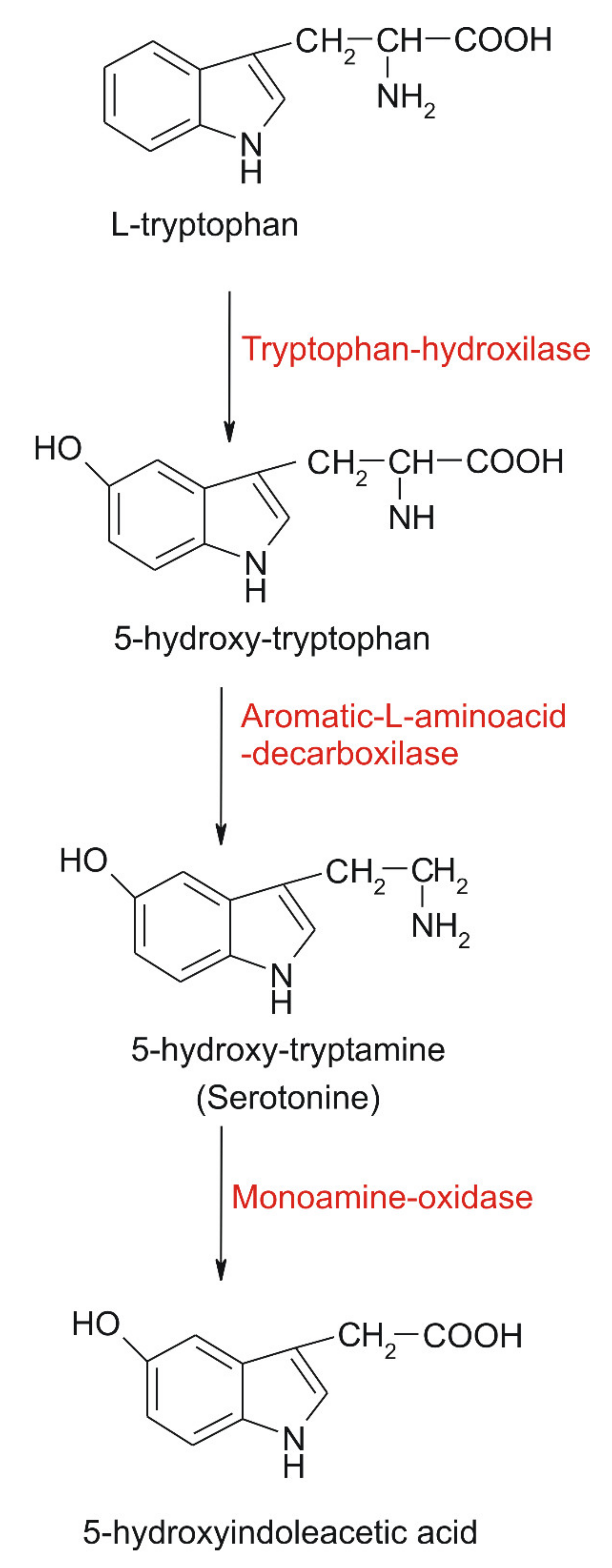
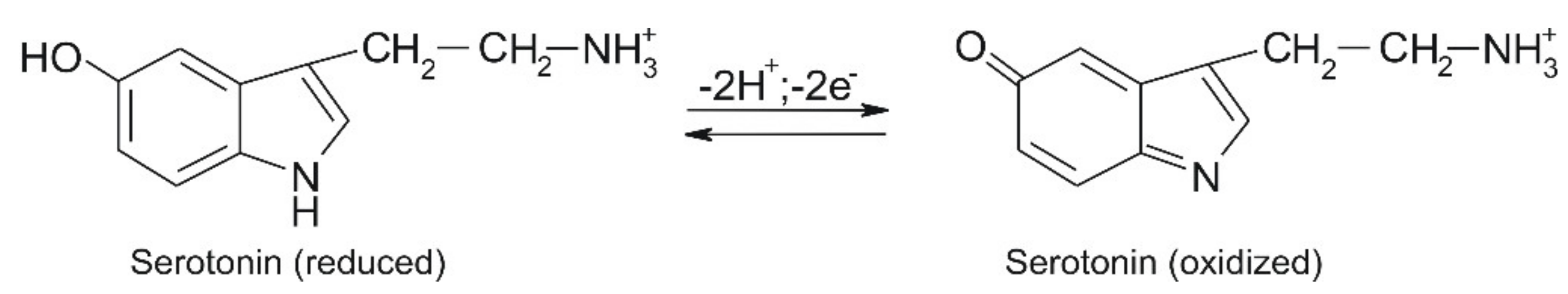
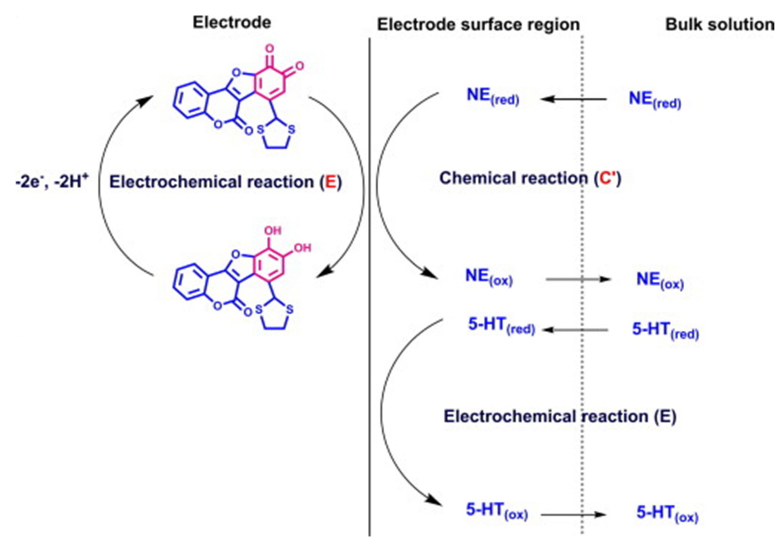
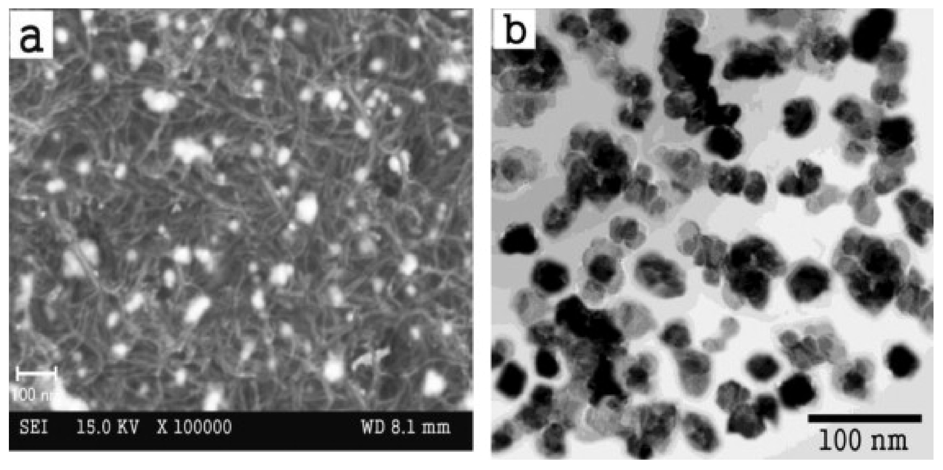
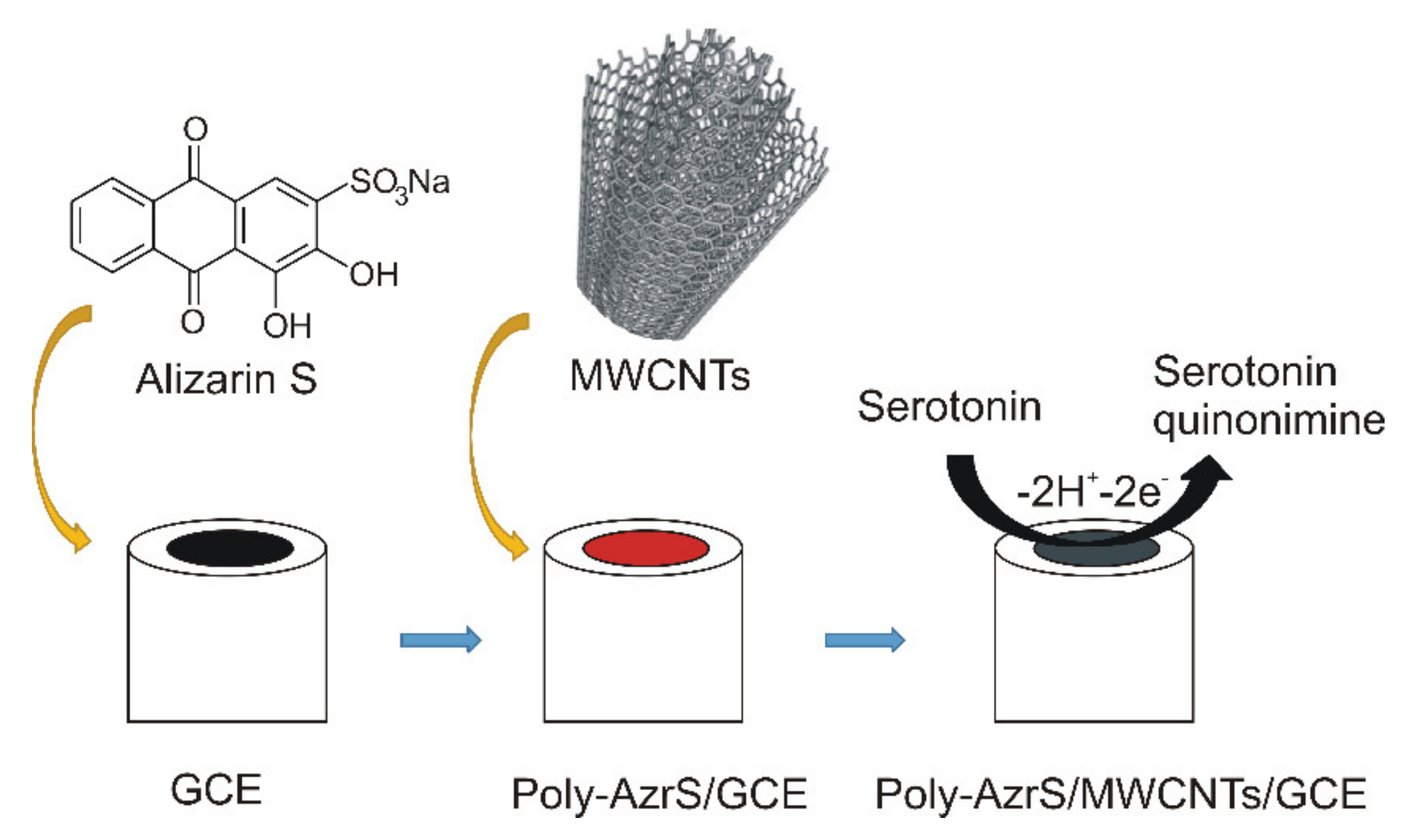
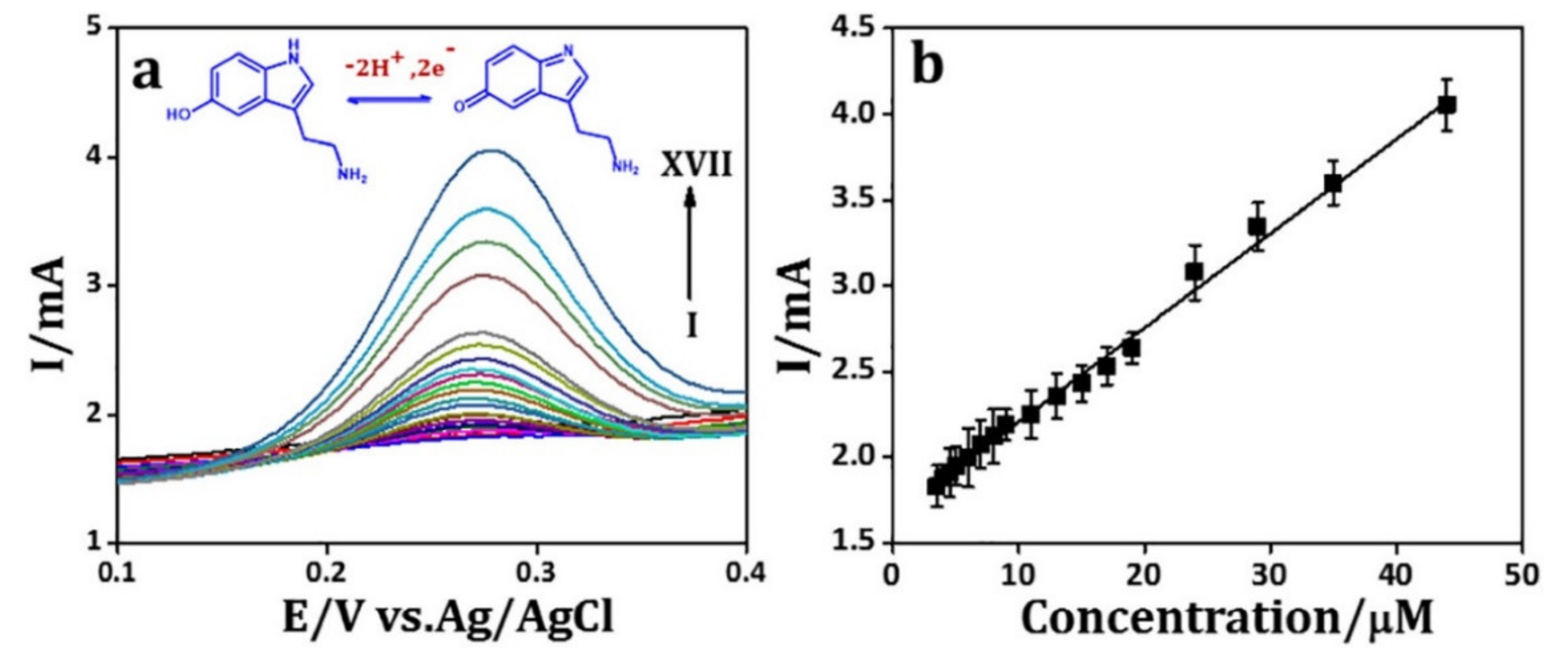
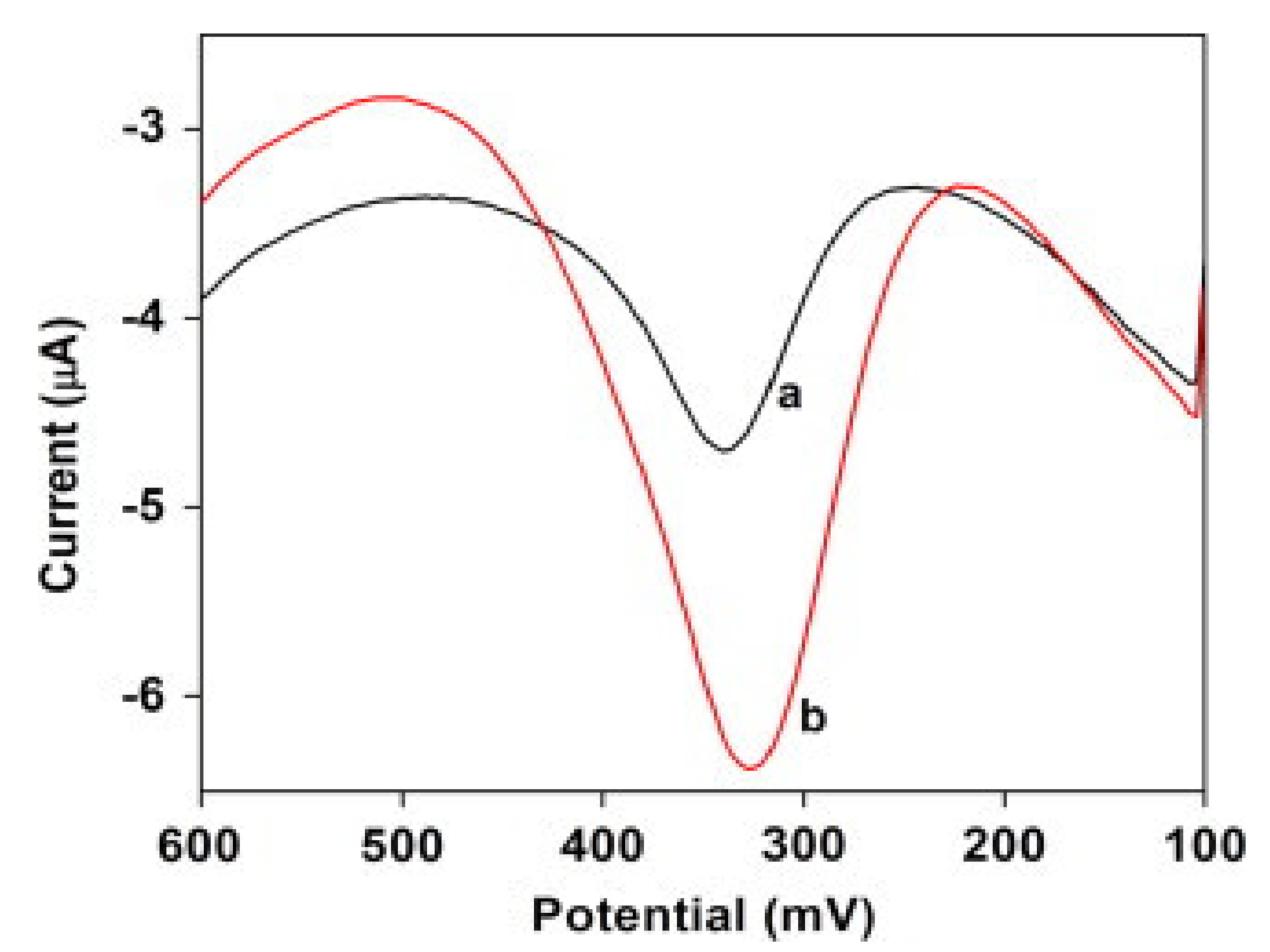
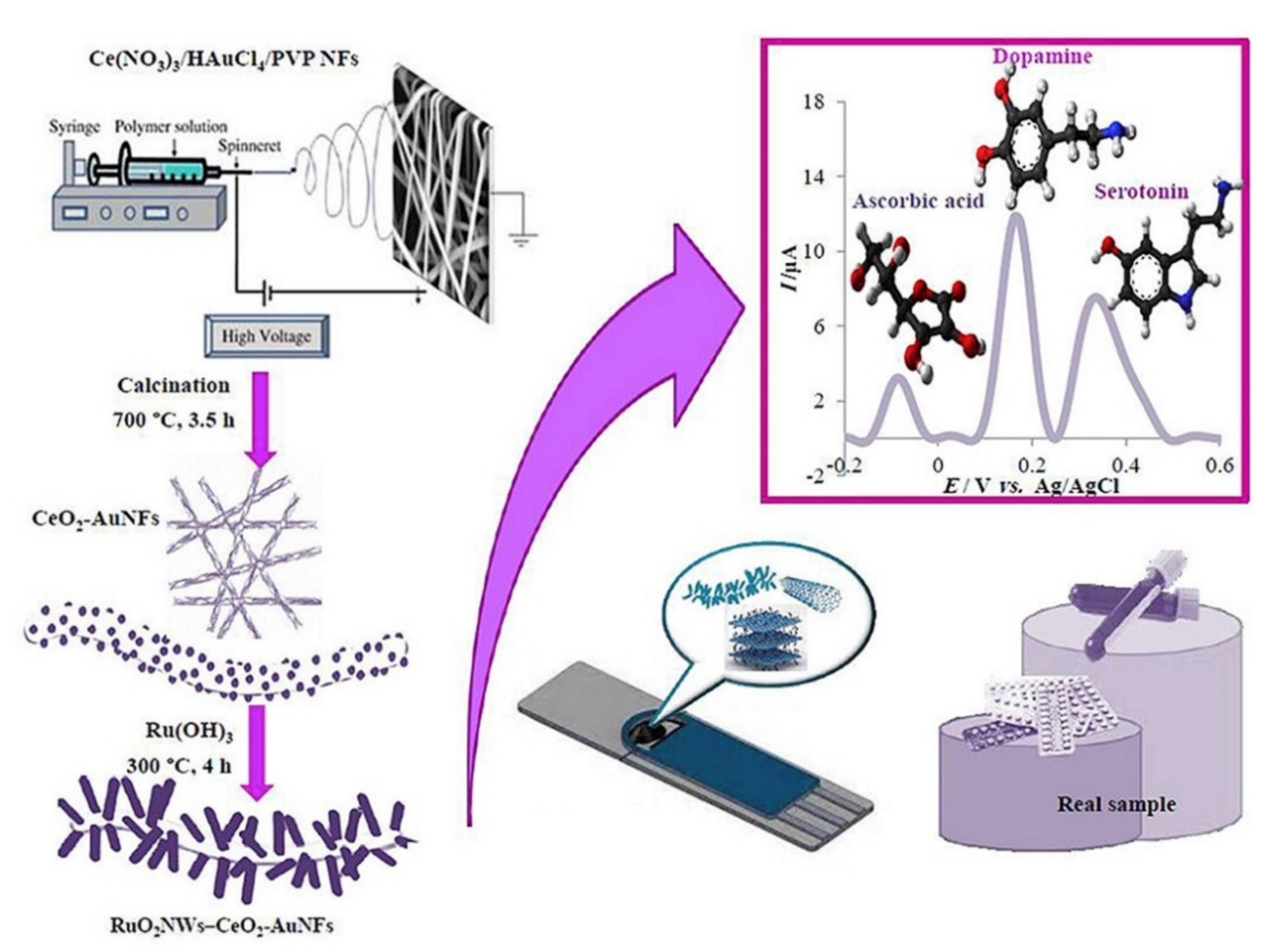
Table 1.
Electrochemical sensors used for serotonin detection.
| No | Modifying Material | Detection Technique | Linear Range | LOD | Sensitivity | Real Sample | Interference Studied | Reference |
|---|---|---|---|---|---|---|---|---|
| 1 | Glassy carbon electrode modified with carbon nanotubes with multiple walls functionalized with benzofuran derivatives and ionic liquid IL-DC-CNT/GC | DPV | 5–900 µM | 2 µM | 0.073 A/M | human blood serum | L-lysine, glucose, lactose, fructose, sucrose, L-asparagine, L-glutamic acid, L-glycine, L-cysteine, D-penicillamin, L-tryptophan, uric acid and ascorbic acid | [102] |
| 2 | Glassy carbon electrode modified with carbon nanotubes with multiple walls and Nafion/Ni(OH)2 Nafion/Ni(OH)2-MWNTs/GCE | DPV | 0.008–10 µM | 0.003 µM | 4.991 A/M | human blood serum | uric acid, ascorbic acid. 3,4-dihydroxyphenylacetic acid, citric acid, glutamic acid, glucose | [103] |
| 3 | Glassy carbon electrode modified with poly-alizarin red S and multi-wall carbon nanotubes poly-AzrS/MWCNTs/GCE | CV, DPV, EIS | 0.5–11 µM | 0.18 µM | 0.364 A/M | Human serum | epinephrine | [104] |
| 4 | Glassy carbon electrode modified with poly(L-arginine), reduced graphene oxide and gold nanoparticles GCE/P-Arg/ErGO/Au NP | DPV | 10–500 nM, 1–10 µM | 30 nM | 5.5758 A/M 1.3436 A/M | urine | glucose, urea, citric acid | [107] |
| 5 | Glassy carbon electrode modified with PEDOT-reduced graphene oxide and silver nanoparticles PEDOTNTs/rGO/Ag NPs/GCE | CV, DPV, CA | 1 nM–0.5 mM | 0.1 nM | 14.304 µA·mM−1/cm2 | bovine assayed multi-sera | ascorbic acid, uric acid, tyrosine | [108] |
| 6 | Glassy carbon electrode modified with silver selenide self-assembled on reduced grapheme rGO-Ag2Se/GCE | CV, EIS | 0.1–15 µM | 29.6 nM | 0.5133 A/M | Alzheimer’s patient’s serum | ascorbic acid, glucose | [109] |
| 7 | Screen-printed electrode modified with a mixture of multi-wall nanotubes, zinc oxide and chitosan MWNTs-ZnO/chitosan/SPE | CV, SWV | 0.05–1 µM | 0.01 µM | 1.892 A/M | rat cerebrospinal fluid | ascorbic acid, norepinephrine | [110] |
| 8 | Screen-printed electrode modified with a mixture of multi-wall nanotubes, aluminium oxide and chitosan MWNTs-Al2O3/chitosan/SPE | SWV | 0.01–1 µM | 0.005 µM | 3.76987 A/M | rat brain | glucose, citric acid, ascorbic acid, dopamine | [111] |
| 9 | Screen-printed electrode modified with polypyrrole, green iron oxide nanoparticles SPCE-PPy-Fe3O4NPs | CV, SWV | 0.007–0.1 µM | 0.020 µM | 6.36066 µA/μM | banana | ascorbic acid | [65] |
| 10 | Polymelamine modified edge plane pyrolitic graphite sensor EPPGS | CV, SWV, EIS | 0.1–100 µM | 30 nM | 0:088 µA/μM | urine | dopamine, serotonin, xanthine, hypoxanthine | [112] |
| 11 | Graphene encapsulated in an alloy of AuAg AuAg-GR | CV | 2.7 nM–4.82 µM | 1.6 nM | 0.766 A·μM−1 ·cm−2 | human serum | ascorbic acid, uric acid, glucose | [113] |
| 12 | Nanocomposite material SnO2-SnS2 | CV, SWV, EIS | 0.1–700 µM | 45 nM | 3.179 µA/μM | human blood | tryptophan, ascorbic acid | [119] |
| 13 | Hybrid structure CeO2 nanofibers and RuO2 nanowires RuO2NWs CeO2NFs | DPV | 0.01–150 µM | 2.4 nM | 4.0642 µA/μM | human cerebrospinal fluid, serum, blood plasma | Glucose, L-tryptophan, methionine, leucine, cysteine, glycine, folic acid, alanine, arginine, tyrosine, caffeine, L-dopa, uric acid | [120] |
| 14 | 3-amino-5-mercapto-1, 2, 4-triazole modified GCE AMTA/GCE | CV, DPV | 0.001–50 µM | 0.0132 nM | 0.05 µA/μM | blood plasma | urea, glucose, oxalate | [116] |
| 15 | Boron-doped diamond thin-film electrode | CV, HV | 0.01–50 µM | 10 nM | 25 nA/μM | - | histamine, 5-hydroxyindoleacetic acid | [121] |
| 16 | Carbon spheres/GCE | DPV | 40–750 µM | 700 nM | 0.09 µA/mM | pharmaceutics | uric acid, oxalate, glucose, L-cysteine, glutathione, folic acid, levodopa | [122] |
| 17 | PEDOT modified platinum electrode | CV, LSV | 20–100 µM | 71 nM | - | urine | ascorbic acid, glucose | [123] |
| 18 | Nafion membrane-coated colloidal gold SPE | DPV | 0.05–1.0 µM | 10.0 nM | 0.1495 µA/μM | platelet-rich plasma | dopamine, uric acid, ascorbic acid | [124] |
Hydrodynamic Voltammetry (HV); Linear Sweeping Voltammety (LSV); Chronoamperometry (CA).
Publisher’s Note: MDPI stays neutral with regard to jurisdictional claims in published maps and institutional affiliations. |
© 2021 by the authors. Licensee MDPI, Basel, Switzerland. This article is an open access article distributed under the terms and conditions of the Creative Commons Attribution (CC BY) license (http://creativecommons.org/licenses/by/4.0/).
Share and Cite
MDPI and ACS Style
Dăscălescu, D.; Apetrei, C. Nanomaterials Based Electrochemical Sensors for Serotonin Detection: A Review. Chemosensors 2021, 9, 14. https://0-doi-org.brum.beds.ac.uk/10.3390/chemosensors9010014
AMA Style
Dăscălescu D, Apetrei C. Nanomaterials Based Electrochemical Sensors for Serotonin Detection: A Review. Chemosensors. 2021; 9(1):14. https://0-doi-org.brum.beds.ac.uk/10.3390/chemosensors9010014
Chicago/Turabian StyleDăscălescu, Dorin, and Constantin Apetrei. 2021. "Nanomaterials Based Electrochemical Sensors for Serotonin Detection: A Review" Chemosensors 9, no. 1: 14. https://0-doi-org.brum.beds.ac.uk/10.3390/chemosensors9010014
Note that from the first issue of 2016, this journal uses article numbers instead of page numbers. See further details here.