Fluorescence-Based On-Resin Detection of Three Model Proteases


Department of Organic Chemistry, Faculty of Science, Palacký University, Tř. 17. Listopadu 12, 771 46 Olomouc, Czech Republic
*
Author to whom correspondence should be addressed.
Chemosensors 2021, 9(12), 359; https://0-doi-org.brum.beds.ac.uk/10.3390/chemosensors9120359
Submission received: 9 November 2021
/
Revised: 2 December 2021
/
Accepted: 7 December 2021
/
Published: 14 December 2021
(This article belongs to the Section Optical Chemical Sensors)
Abstract
:A new approach to on-resin detection of three model proteases (trypsin, chymotrypsin, and thrombin) has been developed, while at the same time already described methodology for simultaneous detection of two enzymes (trypsin and chymotrypsin) has been additionally generalized. Appropriate immobilized substrates, comprising specifically cleavable peptide sequences capped with fluorescent dyes, have been synthesized on Rink Amide PEGA resin or Amino PEGA resin modified with backbone amide linker (BAL). Resulting solid support-bound probes were then dispersed into Tris-HCl buffer solution (pH = 8.0) and subjected to enzymatic cleavage. Liberated fluorophores have been tracked by fluorescence measuring. The competitive activities of studied proteases towards the thrombin probe have been efficiently limited and controlled by employing a Bowman-Birk inhibitor into a system.
1. Introduction
Recent developments and advances in various scientific fields including medicine and pharmacy have resulted in a rapidly growing need for simultaneous detection of various proteases. Depending on the fact, whether a substrate is dissolved in a liquid phase or it is embedded to a solid support, we can distinguish between the two main operating modes of testing, namely homogeneous and heterogeneous enzyme assays [1]. While the former are predominately represented in practical applications [2,3,4,5,6,7,8], the latter also exhibit some advantages when it comes to flexibility of the protease synchronous screening systems. As various independent carrier-immobilized substrates with specifically cleavable sites for individual enzymes could be promptly put together in numerous combinations resulting in an immediate modification of corresponding sensor for synchronous protease detection, potential limitations from the perspective of multiplexed sensing capabilities [1] of soluble FRET-based probes might be efficiently overcome in this way.
Heterogeneous protease assays could be additionally divided into a few subgroups [9] including electrochemical assays [10], surface plasmon resonance (SPR) assays [11], surface-enhanced Raman spectroscopy (SERS) assays [12], as well as assays based on ELISA [13] and liquid crystals technology [14]. Although a certain number of studies describing various enzyme detection techniques based on immobilized substrates have been reported over the last couple of years [12,15,16,17,18,19,20,21], they have mostly been constructed in a way to screen only one enzyme at the time. On the other hand, simultaneous protease detection in heterogeneous mode has received negligible attention and thus remained almost completely unexplored until recently.
One such study was published by Liang et al. [22], who developed an electrochemical biosensor for simultaneous detection of trypsin and chymotrypsin based on DNA–peptide bioconjugates attached to the gold electrode. In the presence of suitable enzymes, the peptide fragments are liberated from the electrode surface and consequential diminishment of monitored current is detected.
A concept of a specifically cleavable peptide linkers embedded to a solid support was successfully implemented also in our research group [23], as we developed on-resin sensor for simultaneous detection of trypsin and chymotrypsin in a sample. During exposure of resin-anchored probes to corresponding proteases, peptide fragments bearing individual fluorophores are released into a buffer solution, while enzymatic cleavage is detected by measuring a change in fluorescence intensity.
Regardless, for the two last mentioned applications, potential use case of synchronous evaluation of multiple enzymes in heterogeneous manner seems to be severely underrated. To the best of our knowledge, there is no available report describing simultaneous on-resin detection of more than two proteases. As a result, we have attempted to develop an assay for synchronous screening of three proteases. Although we have failed in our original intention of single-step three protease detection, we are herein presenting a two-step methodology for unambiguous screening of trypsin, chymotrypsin, and thrombin as model enzymes in a mixture. Moreover, this is also the first known study dealing with multiple competitive protease detection in on-resin mode.
2. Materials and Methods
With the aim of on-resin enzyme assays, the trypsin and chymotrypsin probes were synthesized on Rink Amide PEGA resin (0.35 mmol/g, Novabiochem, Darmstadt, Germany), while the thrombin probe was prepared on Amino PEGA resin (0.42 mmol/g, Novabiochem, Darmstadt, Germany). For the purpose of complete characterization, soluble thrombin probe was synthesized also on Polystyrene Wang resin (0.9 mmol/g, AAPPTec, Louisville, KY, USA). The chemicals and solvents were obtained from the available commercial sources. Fluorescent dyes HN6 [24] and 7-diethylaminocoumarin-3-carboxylic acid (DEAC) [25] were synthesized according to published procedures. The detailed description of trypsin and chymotrypsin probes preparation, as well as their complete characterization can be found in our previous work [23].
2.1. Synthesis of Thrombin Probe on Amino PEGA Resin
Amino PEGA resin was weighted into 10 mL plastic syringe (B. Braun Melsungen AG, Melsungen, Germany) furnished with a sintered plastic filter (Torviq, Tucson, AZ, USA), prewashed with DCM (5 times), subsequently treated with 5% DIPEA in DCM, and finally subjected to solid-phase synthesis. The reactions were performed on Titer Plate Shaker (Thermo Fisher Scientific, Waltham, MA, USA), while after each transformation step, the solid support was manually washed with DMF (10 times) and DCM (10 times). Characterization and purity control of the intermediates were carried out by LC-MS, using ammonium acetate (10 mM) or formic acid (0.1% V/V) in ultrapure water and acetonitrile (gradient 20–80% during the first 4.5 min or 50–80% during the first 3 min) as a mobile phase. The comprehensive synthetic scheme (SM—Scheme S1), as well as LC-MS spectra of synthesized intermediates (SM—Figures S1–S10) can be found in Supplementary Materials. The detailed procedure of samples preparation and their characterization is available in our previously published article [23].
2.2. Loading and Yield Determination
Approximately 50 mg of Amino PEGA resin bearing N-(3-aminopropyl)-4-nitrobenzenesulfonamide moiety was subjected to chemical cleavage using 50% TFA in DCM (5 × 5 mL). After evaporation of volatile liquids, the obtained residue was dissolved in deuterated DMSO. With the aim of quantification of cleaved compound, a certain amount of 2-propanol (internal standard) was added, and 1H NMR was measured. The residual resin remained after its treatment with a cleavage cocktail was washed with DCM (10 times), diethyl ether (10 times), and finally dried to constant mass. Considering the amount of cleaved compound and the weight of dried solid support, a loading of 0.24 mmol/g was determined. By repeating the same procedure (after introduction of HN6 dye to the probe) and obtaining the final loading of 0.17 mmol/g, the overall yield of 71% after 17 reaction steps was established for thrombin probe synthesized on Amino PEGA resin.
2.3. Isolation and Characterization of Thrombin Probe
Soluble thrombin probe (ThrP) was synthesized on Polystyrene Wang resin, and then cleaved from the carrier using 50% TFA in DCM (SM—Scheme S2). The complete procedures of isolation and subsequent characterization are described in our previous work [23]. Instead of ammonium acetate, 0.1% (V/V) solution of formic acid in ultrapure water and acetonitrile was employed as a mobile phase during the purification process. The quantum yield of soluble thrombin probe (ThrP) (SM—Figure S11) was determined by standard protocol (Recording Fluorescence Quantum Yields—HORIBA Scientific) using Rhodamine B in distilled water as a reference. The fluorescence measurements were carried out on fluorescence spectrometer (Cary Eclipse, Agilent Technologies, Santa Carla, CA, USA).
2.4. Proteases and Inhibitor
Trypsin (bovine pancreas, ≥10,000 BAEE units/mg protein), α-chymotrypsin (bovine pancreas, ≥40 units/mg protein), thrombin (bovine plasma, ≥60 NIH units/mg protein), and Bowman-Birk inhibitor (trypsin-chymotrypsin inhibitor from Glycine max soybean) were obtained from Sigma-Aldrich (Steinheim, Germany) in the form of lyophilized white to off-white powders. The suitable quantity of individual protease was placed into Eppendorf safe-lock plastic tube (1.5 mL, Hamburg, Germany), then reconstituted in 1 mM HCl in ultrapure water (trypsin and chymotrypsin) or physiological saline solution—0.9% (w/V) NaCl in ultrapure water (thrombin), subsequently aliquoted, and finally stored at −80 °C. That way, obtained samples of reconstituted enzymes were utilized within two hours after defrosting. Bowman-Birk inhibitor was dissolved in Tris-HCl buffer shortly before its application.
2.5. In-Solution Protease Assays
Purified thrombin probe was dissolved in Tris-HCl buffer (0.1 M, 1.4 mL, pH = 8.0), and further diluted with 1 mM HCl (400 µL) and physiological saline solution (200 µL) containing a suitable protease. The enzyme assays were monitored by LC-MS. In all cases, the masses of cleaved peptide fragments were unambiguously confirmed (SM—Figures S15–S18).
2.6. On-Resin Protease Assays
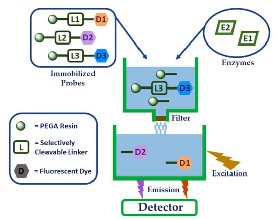
The on-resin protease assays were performed employing slightly modified testing procedure [23]. The appropriate amount of dried solid support (2.0–2.1 mg of ITP–immobilized trypsin probe and ICP–immobilized chymotrypsin probe; 1.0–1.1 mg of IThrP–immobilized thrombin probe) was placed into Eppendorf safe-lock plastic tube (2 mL). Then, 600 µL of Tris-HCl buffer (pH = 8.0) was added and resin was mechanically homogenized. Subsequently, additional 100 µL of Tris-HCl buffer with or without Bowman-Birk inhibitor, 200 µL of 1 mM HCl in ultrapure water, and 100 µL of physiological saline solution in ultrapure water, with or without enzymes were added. Resulting heterogeneous mixture was incubated on horizontal shaker (Benchmark Scientific, USA) (ν = 210 min−1) in incubator (Thermo Fisher Scientific, USA) at 37 °C during the time period of 15 min. After completion of incubation, the resin was filtered out while the fluorescence response of resulting filtrate was measured. All fluorescence data were obtained in five independent parallels. After discharging the highest and the lowest numbers in each set of five measurements, the average as well as standard deviation of the remaining three values within each individual set were calculated and graphically presented. The general graphical representation of enzyme on-resin assay is depicted in Supplementary Materials (SM—Figure S14).
2.7. Stability Testing of Amino PEGA Resin-Anchored Thrombin Probe
To evaluate the stability of freshly prepared immobilized thrombin probe (IThrP), dried resin (1.0–1.1 mg) was weighed into Eppendorf tube and stored at −80 °C or at lab temperature in darkness. At the time of evaluation, Tris-HCl buffer (700 μL), 1 mM HCl in ultrapure water (200 μL) and physiological saline solution (100 μL) were added, and resulting sample was at 37 °C incubated on horizontal shaker (ν = 210 min−1) for 15 min. Afterwards, the residual resin was filtered out and the fluorescence response of liberated fluorophore in the obtained filtrate was determined to establish a spontaneous fluorophore release throughout the time. Also in this case, all experiments were repeated five times. After withdrawal of both boundary values from each individual set, the average and standard deviation for the remaining three numbers were calculated.
3. Results and Discussion
3.1. Synthesis of Thrombin Probe
For the purpose of thrombin detection, a target immobilized probe IThrP comprising a thrombin cleavable amino acid sequence was synthesized on Amino PEGA resin modified with backbone amide linker (BAL) (Figure 1A). After binding of N-Fmoc-1,3-diaminopropane as the first building block, the nosyl moiety was temporarily introduced into a structure, enabling chemical cleavage and ensuing LC-MS characterization of synthesized materials. Subsequent attachment of a thrombin linker (Leu-Val-Pro-Arg-Gly-Ser) [26] was preceded by implementation of a poly(ethylene glycol)-based spacer (PEG) with the ability to facilitate an approach of a protease towards its recognizable site and thus improve the cleavability of a target peptide when subjected to enzyme assays. The fluorescent probe was capped with HN6 dye directly bound to sarcosine. Similar to our previous study [23], where Mitsunobu N-methylation was employed to prevent Rhodamine B spirolactam framework formation and consequent fluorescence quenching, a tertiary amide of HN6 dye as an anchoring moiety was found mandatory for retention of its fluorescent properties. Contrary to Mitsunobu N-methylation, where three reactions steps are needed, introduction of Fmoc-Sar-OH could be efficiently performed in a single step, resulting in better purity of a target product. In addition, potential denosylation process following N-methylation would also affect 4-nitrobenzenesulfonyl group bound to diaminopropane nitrogen and thus prevent further evaluation and purity control of the synthesized intermediates. After capping the amino acid sequence with HN6 dye and subsequent removal of temporarily bound nosyl segment using DBU and 2-mercaptoethanol in DMF, the resin-embedded peptide was treated with 50% TFA in DCM to remove tert-butyl (tBu) and 2,2,4,6,7-pentamethyldihydrobenzofuran-5-sulfonyl (Pbf) protecting groups from serine and arginine, respectively, and thus provide a final immobilized thrombin probe (IThrP) (Figure 1A). The complete synthetic scheme together with corresponding LC-MS analyses is available in Supplementary Materials (SM—Scheme S1 and Figures S1–S10).
With the intention of comprehensive compound characterization, the soluble (non-immobilized) thrombin probe (ThrP) (Figure 1B) was synthesized on cheaper and more available Polystyrene Wang resin (SM—Scheme S2). After introduction of diaminopropane moiety, a PEG spacer, a suitable amino acid sequence, and finally HN6 dye were bound in the appropriate order. Removal of acid labile protecting groups and chemical cleavage from the solid support was performed using 50% TFA in DCM.
3.2. In-Solution Enzymatic Cleavage
After isolation of a soluble thrombin probe (ThrP), enzyme assays were performed in Tris-HCl buffer (pH = 8.0) using thrombin (100 µg/mL) reconstituted in physiological saline solution. The probe was expectedly cleaved between N-terminus of glycine and C-terminus of arginine (Figure 2; SM—Figure S15). To evaluate potential cleavability of thrombin linker by the other two model proteases, ThrP was subjected to individual enzyme testing in Tris-HCl buffer (pH = 8.0) using trypsin (10 µg/mL) and chymotrypsin (100 µg/mL) reconstituted in 1 mM HCl. As can be seen (SM—Figures S16 and S17), ThrP was in both cases dismembered at the same cleavage site (between glycine and arginine) as in the case of thrombin (Figure 2; SM—Figure S18).
3.3. Fluorescence Spectral Properties of Thrombin Probe
Due to the fact that on-resin enzymatic cleavage evaluation was performed by monitoring a fluorescence response of liberated fluorophore into a buffer solution, a brief study of thrombin substrate fluorescence properties was carried out. Considering the fluorescence spectra of soluble thrombin probe ThrP (Figure 3), excitation wavelength of 600 nm was selected, while emission intensity read-out was set to 652 nm.
3.4. On-Resin Enzyme Studies
3.4.1. The Lowest Detectable Concentration of Thrombin
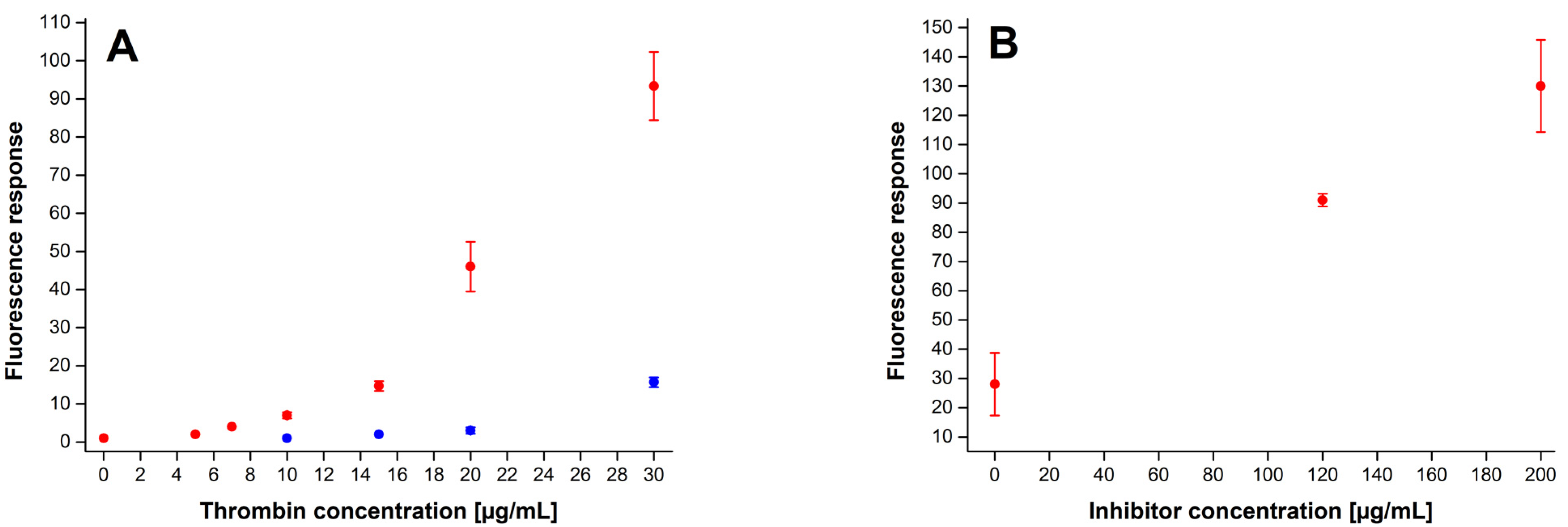
As already discussed in our previous work [23], from the perspective of on-resin assays, PEGA resin exhibited superior performance among studied hydrophilic solid supports. Consequently, all on-bead experiments were performed on the last mentioned type of resin. Immobilized thrombin probe (IThrP) dispersed in Tris-HCl buffer (pH = 8.0) was exposed to thrombin in various concentrations. After completion of incubation (15 min), the fluorescence response of a released fluorophore in buffer was measured, and in accordance with the obtained data, the lowest unambiguously detectable concentration of thrombin was found to be 20 µg/mL (Figure 4A; SM—Table S1). As thrombin linker was not cleavable solely by thrombin itself but also by trypsin and chymotrypsin, we were aware of the fact that utilization of a trypsin-chymotrypsin inhibitor was necessary to enable reliable detection of thrombin in the presence of two competitive proteases. For that purpose, Bowman-Birk inhibitor was employed. Considering the product specification (Bowman-Birk inhibitor; Sigma-Aldrich), as well as the maximal concentrations of trypsin (10 µg/mL) and chymotrypsin (100 µg/mL) used in this study, the uniform conditions for thrombin cleavage assays were established. Afterwards, determination of the lowest detectable concentration of thrombin was performed again, this time in the presence of Bowman-Birk inhibitor in concentration of 120 µg/mL. Surprisingly, the lowest unambiguously detectable concentration of thrombin in the presence of inhibitor was found nearly three times lower (7 µg/mL) than in its absence (20 µg/mL) (Figure 4A; SM—Table S1).
To confirm this hypothesis, the immobilized thrombin probe IThrP (1.0–1.1 mg) together with thrombin (30 µg/mL) and various concentrations of Bowman-Birk inhibitor (0, 120, 200 µg/mL) was incubated in Tris-HCl buffer (pH = 8.0) for 15 min. Comparing the average fluorescence responses of the samples incubated in the absence of inhibitor and those incubated in its presence (120 µg/mL), more than a threefold increase in fluorescence intensity was detected. Further increment of inhibitor concentration (200 µg/mL) resulted in an average fluorescence response higher for an additional nearly 43% (Figure 4B; SM—Table S2). The explanation for seemingly unexpected observations could be found in a study published by Lanchantin et al. [27], who reported formation of a complex between thrombin (human or bovine) and soybean trypsin inhibitor. Such enzyme-inhibitor complex has been proven enzymatically active towards fibrinogen Arg-Gly bonds, which is exactly the thrombin substrate cleavage site, utilized in our study.
3.4.2. Simultaneous Detection of Trypsin and Chymotrypsin in the Presence of Thrombin
Already reported on-resin methodology for simultaneous detection of trypsin–chymotrypsin pair [23] was additionally generalized and its applicability expanded by introduction of a third protease in a system. Immobilized trypsin (ITP) and immobilized chymotrypsin (ICP) probes (Figure 5) were subjected to enzyme assays applying various combinations of all three proteases, namely trypsin, chymotrypsin, and thrombin.
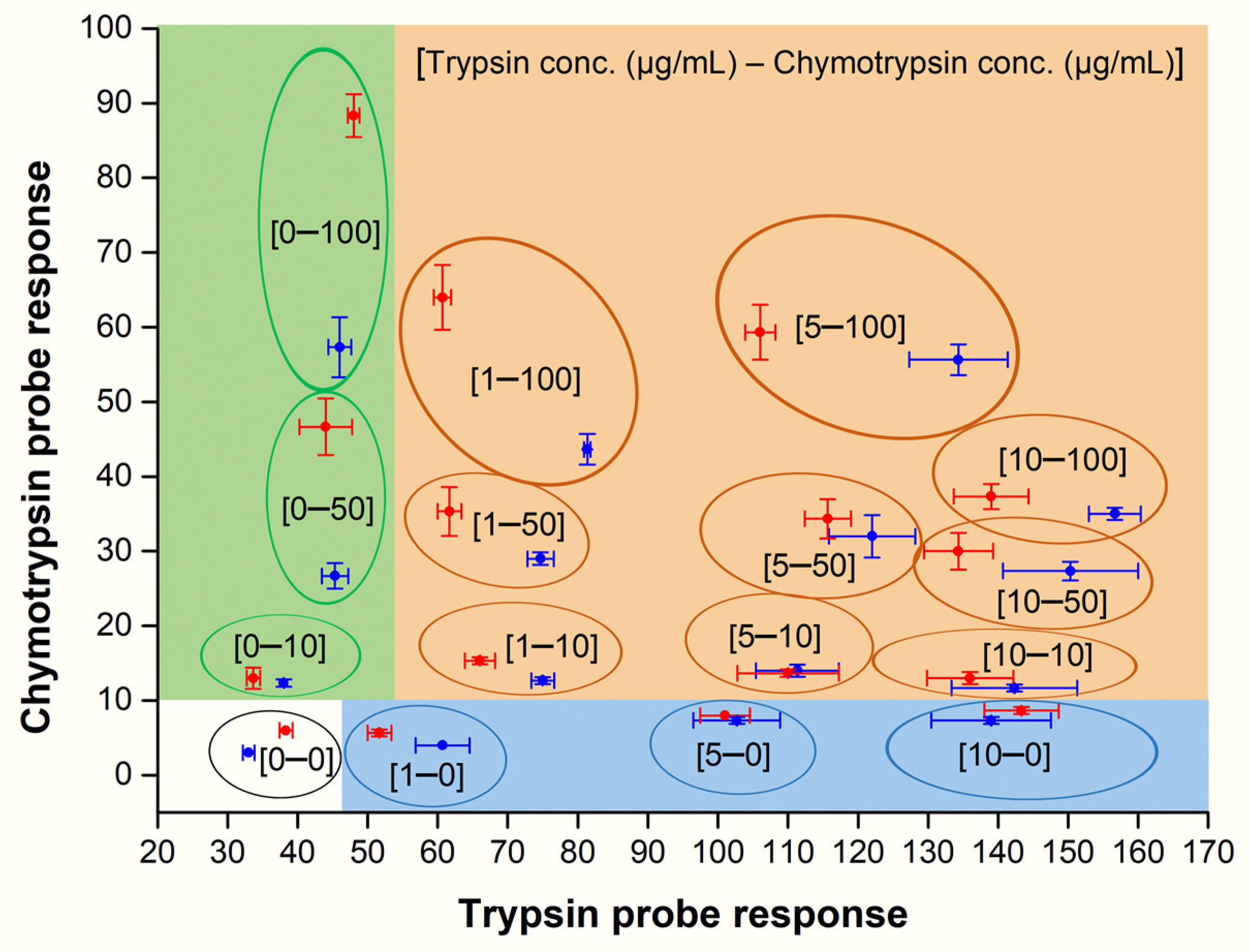
Responses for DEAC (ITP probe) and Rhodamine B (ICP probe) were measured for four different concentrations of trypsin (0, 1, 5, 10 µg/mL) and chymotrypsin (0, 10, 50, 100 µg/mL) in the presence (red points) and absence (blue points) of thrombin (50 µg/mL or 0 µg/mL). Comparing the average fluorescence intensities (Figure 6; SM—Table S3), the ICP response was considerably affected when chymotrypsin was present in at least medium concentration (50 µg/mL or higher) and concentration of trypsin was up to 1 µg/mL (the upper part of the green area and the ochre area close to the border with the green area). In addition, chymotrypsin concentration of at least 50 µg/mL in the presence of trypsin resulted in noticeable decline of ITP response (the upper part of the ochre area). In all other cases, the observed fluctuations in ITP and ICP fluorescence intensities caused by the potential presence of thrombin were small to negligible. Despite described discrepancies, all average fluorescence values reliably fell into appropriate areas—ochre (trypsin + chymotrypsin), green (only chymotrypsin), blue (only trypsin), and white (no trypsin and no chymotrypsin)—and thus any of the four possible combinations of the two last mentioned proteases could be unambiguously determined in the presence and absence of thrombin.
3.4.3. Detection of Thrombin in the Presence of Trypsin and Chymotrypsin
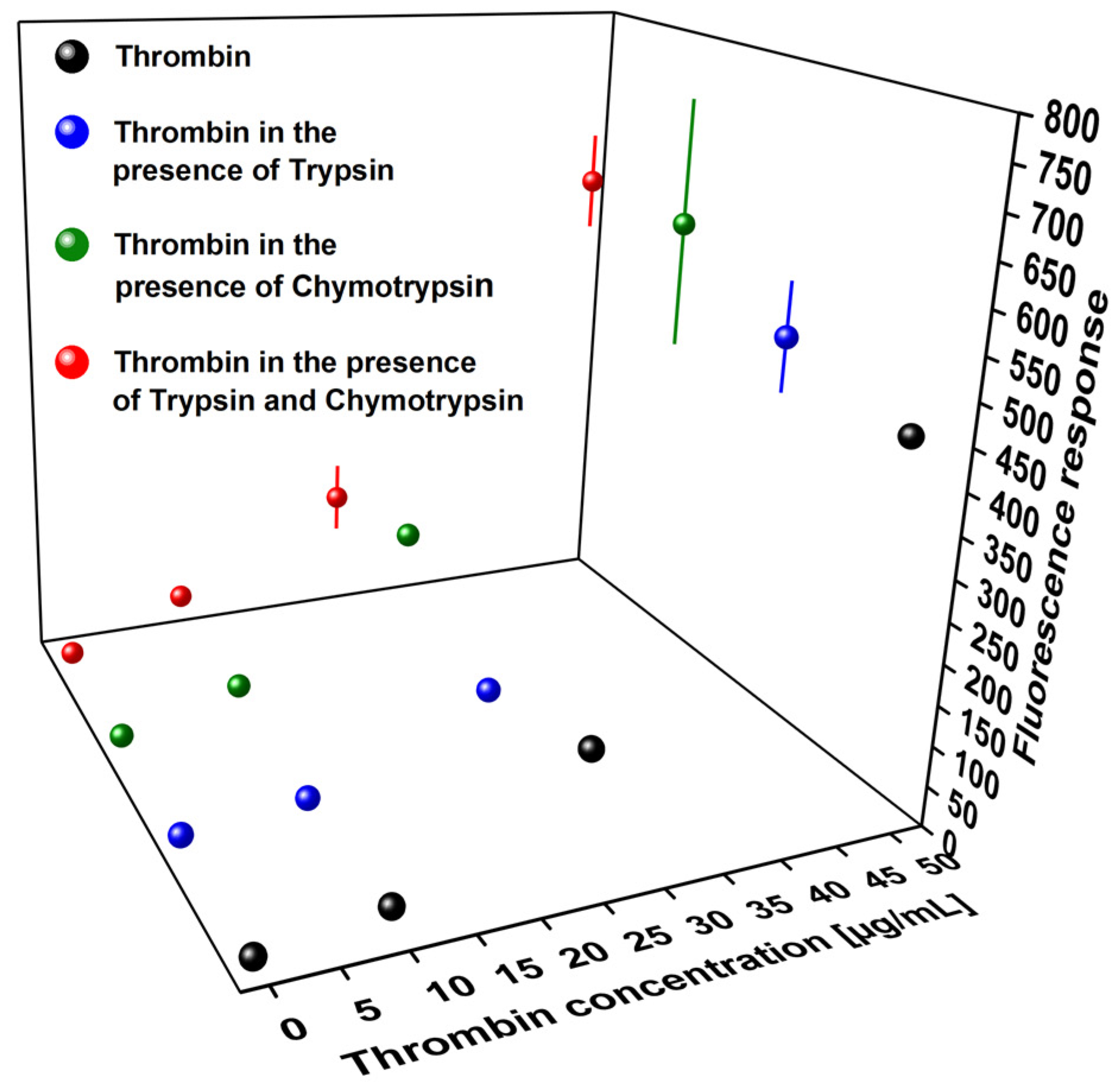
In the next step, a methodology for thrombin detection in the presence of trypsin and/or chymotrypsin was developed. Considering the lowest detectable concentration of thrombin, as well as amounts of another two proteases used in the above-described synchronous detection model, four different concentrations of thrombin (0, 10, 25, 50 µg/mL) and two for trypsin (0, 10 µg/mL) and chymotrypsin (0, 100 µg/mL) were taken into consideration. In all cases, Bowman-Birk inhibitor (120 µg/mL) was added to prevent unselective cleavage of thrombin linker by trypsin and/or chymotrypsin. The assays were carried out only in the presence of thrombin (black), thrombin in the presence of trypsin (10 µg/mL) (blue), thrombin in the presence of chymotrypsin (100 µg/mL) (green), as well as thrombin in the presence of both trypsin (10 µg/mL) and chymotrypsin (100 µg/mL) (red). As can be seen (Figure 7; SM—Table S4), no response was detected when thrombin was not present, indicating complete inhibition of trypsin and chymotrypsin species. On the other hand, presence of thrombin (10 µg/mL) in a mixture resulted in significant increase of HN6 (IThrP) fluorescence response (SM—Table S4), making the herein presented model reliable for detection of thrombin in the presence of trypsin and/or chymotrypsin.
3.4.4. Stability Testing
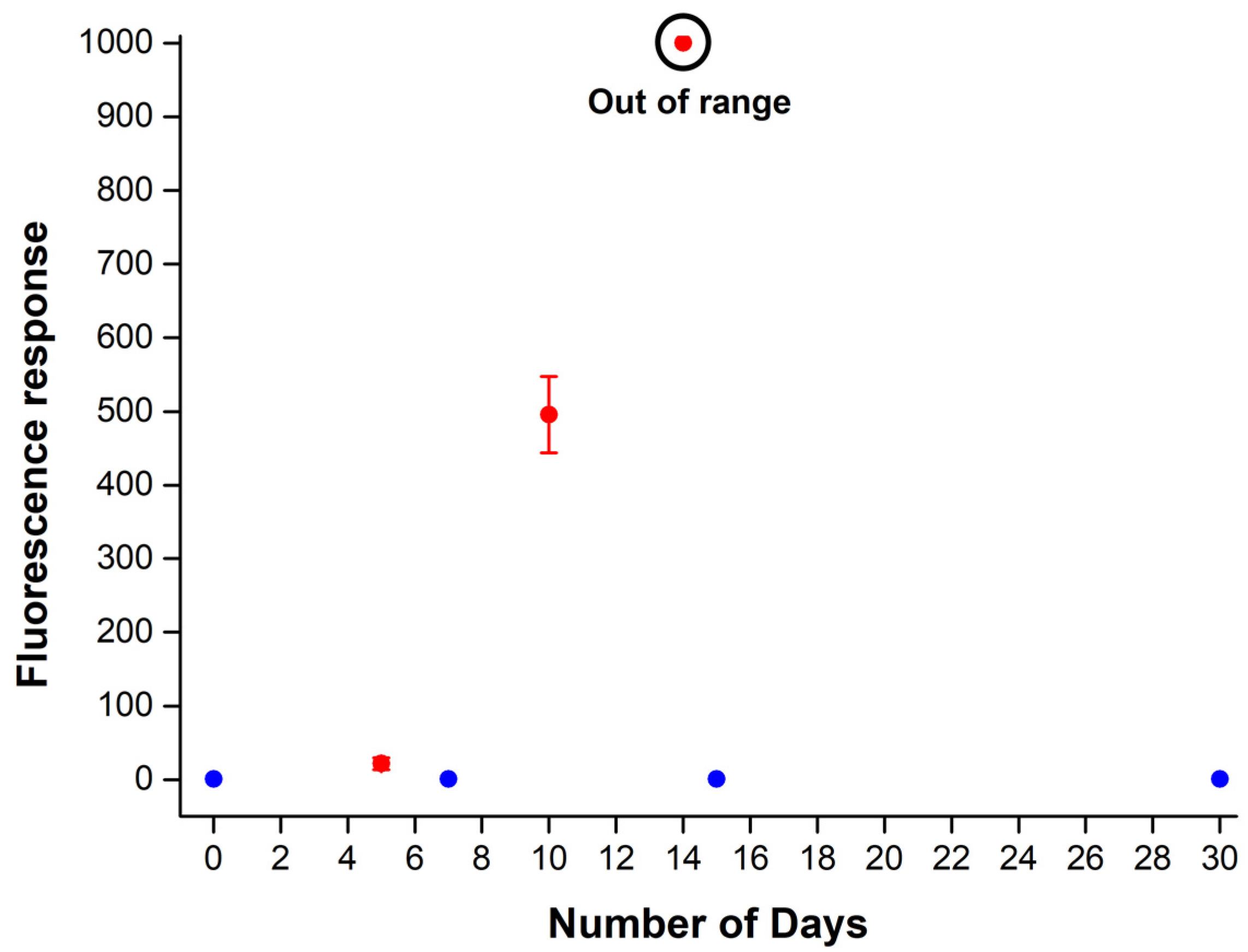
Following the procedure of ITP and ICP stability testing [23] that proved the necessity of resin embedded compounds to be stored at low temperature, IThrP was subjected to a similar experiment. As can be seen in Figure 8 (SM—Table S5), the immobilized thrombin probe kept at −80 °C exhibited extraordinary stability for the time period of at least 30 days (blue). Contrary to this, a significant increase in fluorescence intensity followed by its further exponential growth within the first 14 days of the experiment was detected not later than after only first five days of IThrP storing at laboratory temperature in darkness (red).
4. Conclusions
To summarize, an approach for simultaneous on-resin detection of three enzymes has been studied using trypsin, chymotrypsin, and thrombin as model proteases. The biological assays have been carried out in two separate parallels. While the model for synchronous screening of trypsin and chymotrypsin has been proven efficient also in the presence of the third protease (thrombin), an independent method for reliable thrombin determination employing newly designed Amino PEGA resin-immobilized substrate in the presence of two competitive proteases (trypsin and chymotrypsin) and Bowman-Birk inhibitor has been successfully developed and evaluated. Surprisingly, utilized trypsin-chymotrypsin inhibitor has not only effectively suppressed the activities of both competitive enzymes towards thrombin probe, but it has also additionally stimulated the thrombin cleavage of its target substrate. The methodology herein presented could potentially serve as an inspiration to chemical biologists in the development of novel tools for synchronous detection of various biologically compatible proteases.
Supplementary Materials
The following are available online at https://0-www-mdpi-com.brum.beds.ac.uk/article/10.3390/chemosensors9120359/s1, Scheme S1: Synthesis of immobilized thrombin probe (IThrP) on Amino PEGA resin (yield: 71%), Figure S1: LC-MS analysis of chemically cleaved compound from Resin 1, Figure S2: LC-MS analysis of chemically cleaved compound from Resin 2, Figure S3: LC-MS analysis of chemically cleaved compound from Resin 3, Figure S4: LC-MS analysis of chemically cleaved compound from Resin 4, Figure S5: LC-MS analysis of chemically cleaved compound from Resin 5, Figure S6: LC-MS analysis of chemically cleaved compound from Resin 6, Figure S7: LC-MS analysis of chemically cleaved compound from Resin 7, Figure S8: LC-MS analysis of chemically cleaved compound from Resin 8, Figure S9: LC-MS analysis of chemically cleaved compound from Resin 9, Figure S10: LC-MS analysis of chemically cleaved compound from Resin 10, Scheme S2: Synthesis of soluble thrombin probe (ThrP) on Polystyrene Wang resin, Figure S11: Thrombin probe (chemically cleaved from Polystyrene Wang resin), Figure S12: LC-MS analysis of purified thrombin probe, Figure S13: 1H NMR spectrum of purified thrombin probe, Figure S14: Graphical representation of on-resin enzyme assay, Figure S15: Enzymatically cleaved thrombin probe fragment, Figure S16: LC-MS analysis of thrombin probe fragment (trypsin cleavage), Figure S17: LC-MS analysis of thrombin probe fragment (chymotrypsin cleavage), Figure S18: LC-MS analysis of thrombin probe fragment (thrombin cleavage), Table S1: Fluorescence response—determination of the lowest detectable concentration of thrombin, Table S2: Fluorescence response—detection of thrombin in the presence of Bowman-Birk inhibitor, Table S3: Fluorescence response—simultaneous detection of trypsin and chymotrypsin in the presence of thrombin, Table S4: Fluorescence response—detection of thrombin in the presence of trypsin and chymotrypsin, Table S5: Fluorescence response—stability of Amino PEGA resin-anchored thrombin probe (IThrP).
Author Contributions
Conceptualization and methodology, D.M. and J.H.; Investigation, D.M.; Writing—original draft preparation, D.M. and J.H.; Writing—review and editing, D.M. and J.H.; Funding acquisition, J.H. All authors have read and agreed to the published version of the manuscript.
Funding
This work was supported by the Czech Science Foundation (GACR project 19-23972S).
Institutional Review Board Statement
Not applicable.
Informed Consent Statement
Not applicable.
Data Availability Statement
The data presented in this study are available in Supplementary Materials.
Conflicts of Interest
The authors declare no conflict of interest.
References
- Ong, I.L.H.; Yang, K.L. Recent developments in protease activity assays and sensors. Analyst 2017, 142, 1867–1881. [Google Scholar] [CrossRef] [PubMed] [Green Version]
- Okorochenkova, Y.; Porubský, M.; Benická, S.; Hlaváč, J. A novel three-fluorophore system as a ratiometric sensor for multiple protease detection. Chem. Commun. 2018, 54, 7589–7592. [Google Scholar] [CrossRef]
- Xu, J.; Fang, L.; Shi, M.; Huang, Y.; Yao, L.; Zhao, S.; Zhang, L.; Liang, H. A peptide-based four-color fluorescent polydopamine nanoprobe for multiplexed sensing and imaging of proteases in living cells. Chem. Commun. 2019, 55, 1651–1654. [Google Scholar] [CrossRef]
- Chen, X.; Zhang, Y.; Guan, X. Simultaneous detection of multiple proteases using a non-array nanopore platform. Nanoscale 2021, 13, 13658–13664. [Google Scholar] [CrossRef] [PubMed]
- Kominami, K.; Nagai, T.; Sawasaki, T.; Tsujimura, Y.; Yashima, K.; Sunaga, Y.; Tsuchimochi, M.; Nishimura, J.; Chiba, K.; Nakabayashi, J.; et al. In Vivo Imaging of Hierarchical Spatiotemporal Activation of Caspase-8 during Apoptosis. PLoS ONE 2012, 7, e50218. [Google Scholar] [CrossRef]
- Li, S.Y.; Liu, L.H.; Cheng, H.; Li, B.; Qiu, W.X.; Zhang, X.Z. A dual-FRET-based fluorescence probe for the sequential detection of MMP-2 and caspase-3. Chem. Commun. 2015, 51, 14520–14523. [Google Scholar] [CrossRef]
- Zhang, Y.; Chen, X.; Yuan, S.; Wang, L.; Guan, X. Joint Entropy-Assisted Graphene Oxide-Based Multiplexing Biosensing Platform for Simultaneous Detection of Multiple Proteases. Anal. Chem. 2020, 92, 15042–15049. [Google Scholar] [CrossRef]
- Bui, H.; Brown, C.W.; Buckhout-White, S.; Díaz, S.A.; Stewart, M.H.; Susumu, K.; Oh, E.; Ancona, M.G.; Goldman, E.R.; Medintz, I.L. Transducing Protease Activity into DNA Output for Developing Smart Bionanosensors. Small 2019, 15, 1–13. [Google Scholar] [CrossRef] [PubMed]
- Adem, S.; Jain, S.; Sveiven, M.; Zhou, X.; O’Donoghue, A.J.; Hall, D.A. Giant magnetoresistive biosensors for real-time quantitative detection of protease activity. Sci. Rep. 2020, 10, 1–10. [Google Scholar] [CrossRef]
- Park, S.; Kim, G.; Seo, J.; Yang, H. Ultrasensitive protease sensors using selective affinity binding, selective proteolytic reaction, and proximitY-dependent electrochemical reaction. Anal. Chem. 2016, 88, 11995–12000. [Google Scholar] [CrossRef]
- Chen, H.; Gal, Y.S.; Kim, S.H.; Choi, H.J.; Oh, M.C.; Lee, J.; Koh, K. Potassium ion sensing using a self-assembled calix[4]crown monolayer by surface plasmon resonance. Sens. Actuators B Chem. 2008, 133, 577–581. [Google Scholar] [CrossRef]
- Yang, L.; Wu, T.; Fu, C.; Chen, G.; Xu, S.; Xu, W. SERS determination of protease through a particle-on-a-film configuration constructed by electrostatic assembly in an enzymatic hydrolysis reaction. RSC Adv. 2016, 6, 90120–90125. [Google Scholar] [CrossRef]
- Yoon, H.K.; Yoo, T.H. A novel protease activity assay method based on an engineered autoinhibited protein using an enzyme-linked immunoassay. Analyst 2013, 138, 7164–7168. [Google Scholar] [CrossRef] [PubMed]
- Chen, C.H.; Yang, K.L. Oligopeptide immobilization strategy for improving stability and sensitivity of liquid-crystal protease assays. Sens. Actuators B Chem. 2014, 204, 734–740. [Google Scholar] [CrossRef]
- Chen, H.; Zhang, J.; Gao, Y.; Liu, S.; Koh, K.; Zhu, X.; Yin, Y. Sensitive cell apoptosis assay based on caspase-3 activity detection with graphene oxide-assisted electrochemical signal amplification. Biosens. Bioelectron. 2015, 68, 777–782. [Google Scholar] [CrossRef] [PubMed]
- Swisher, L.Z.; Prior, A.M.; Shishido, S.; Nguyen, T.A.; Hua, D.H.; Li, J. Quantitative electrochemical detection of cathepsin B activity in complex tissue lysates using enhanced AC voltammetry at carbon nanofiber nanoelectrode arrays. Biosens. Bioelectron. 2014, 56, 129–136. [Google Scholar] [CrossRef] [Green Version]
- Cao, Y.; Yu, J.; Bo, B.; Shu, Y.; Li, G. A simple and general approach to assay protease activity with electrochemical technique. Biosens. Bioelectron. 2013, 45, 1–5. [Google Scholar] [CrossRef]
- Xia, N.; Peng, P.; Wang, S.; Du, J.; Zhu, G.; Du, W.; Liu, L. A signal-on electrochemical strategy for protease detection based on the formation of ATCUN-Cu(II). Sens. Actuators B Chem. 2016, 232, 557–563. [Google Scholar] [CrossRef]
- Fan, G.-C.; Han, L.; Zhu, H.; Zhang, J.-R.; Zhu, J.-J. Ultrasensitive Photoelectrochemical Immunoassay for Matrix Metalloproteinase-2 Detection Based on CdS:Mn/CdTe Cosensitized TiO2 Nanotubes and Signal Amplification of SiO2@Ab2 Conjugates. Anal. Chem. 2014, 86, 12398–12405. [Google Scholar] [CrossRef]
- Mu, C.J.; LaVan, D.A.; Langer, R.S.; Zetter, B.R. Self-assembled gold nanoparticle molecular probes for detecting proteolytic activity in vivo. ACS Nano 2010, 4, 1511–1520. [Google Scholar] [CrossRef] [Green Version]
- Olson, E.S.; Jiang, T.; Aguilera, T.A.; Nguyen, Q.T.; Ellies, L.G.; Scadeng, M.; Tsien, R.Y. Activatable cell penetrating peptides linked to nanoparticles as dual probes for in vivo fluorescence and MR imaging of proteases. Proc. Natl. Acad. Sci. USA 2010, 107, 4311–4316. [Google Scholar] [CrossRef] [Green Version]
- Liang, R.P.; Tian, X.C.; Qiu, P.; Qiu, J.D. Multiplexed electrochemical detection of trypsin and chymotrypsin based on distinguishable signal nanoprobes. Anal. Chem. 2014, 86, 9256–9263. [Google Scholar] [CrossRef] [PubMed]
- Milićević, D.; Hlaváč, J. Immobilized fluorescent probes for simultaneous multiple protease detection. Chemosensors 2021, 9, 119. [Google Scholar] [CrossRef]
- Gong, Y.J.; Zhang, X.B.; Mao, G.J.; Su, L.; Meng, H.M.; Tan, W.; Feng, S.; Zhang, G. A unique approach toward near-infrared fluorescent probes for bioimaging with remarkably enhanced contrast. Chem. Sci. 2016, 7, 2275–2285. [Google Scholar] [CrossRef] [Green Version]
- He, G.; Guo, D.; He, C.; Zhang, X.; Zhao, X.; Duan, C. A color-tunable europium complex emitting three primary colors and white light. Angew. Chemie-Int. Ed. 2009, 48, 6132–6135. [Google Scholar] [CrossRef]
- Smith, D.B.; Johnson, K.S. Single-step purification of polypeptides expressed in Escherichia coli as fusions with glutathione S-transferase. Gene 1988, 67, 31–40. [Google Scholar] [CrossRef]
- Lanchantin, G.F.; Friedmann, J.A.; Hart, D.W. Interaction of soybean trypsin inhibitor with thrombin and its effect on prothrombin activation. J. Biol. Chem. 1969, 244, 865–875. [Google Scholar] [CrossRef]
Figure 1.
Amino PEGA resin-immobilized (IThrP) (A) and soluble (ThrP) (B) thrombin probe.

Figure 2.
Enzymatic cleavage of soluble thrombin probe (ThrP).

Figure 3.
Normalized excitation (blue) and emission (red) spectra of 3 × 10⁻⁶ M soluble thrombin probe (ThrP) in Tris-HCl buffer (pH = 8.0) measured at 37 °C.
Figure 3.
Normalized excitation (blue) and emission (red) spectra of 3 × 10⁻⁶ M soluble thrombin probe (ThrP) in Tris-HCl buffer (pH = 8.0) measured at 37 °C.

Figure 4.
Determination of the lowest detectable concentration of thrombin in the absence (blue) and presence (red) of Bowman-Birk inhibitor (120 µg/mL) (A). The dependence of fluorescence response on concentration of utilized Bowman-Birk inhibitor at the constant concentration of thrombin (30 µg/mL) (B). Immobilized thrombin probe (1.0–1.1 mg) was dispersed in Tris-HCl buffer (1.0 mL; pH = 8.0), and incubated for 15 min at 37 °C. Fluorescence emission was measured at 652 nm after excitation at 600 nm. The exc./ems. slit width was set to 5.0/5.0 nm.

Figure 5.
Rink Amide PEGA resin-immobilized trypsin (ITP) and chymotrypsin (ICP) probes.

Figure 6.
Simultaneous fluorescence-based detection of trypsin-chymotrypsin pair with synchronous usage of ITP and ICP bound to Rink Amide PEGA resin in the absence (blue points) and presence (red points) of thrombin (50 µg/mL). The immobilized probes (2.0–2.1 mg) were dispersed in Tris-HCl buffer (1.0 mL; pH = 8.0), and incubated for 15 min at 37 °C. Fluorescence emission was measured at 480 nm (ITP) and 590 nm (ICP) after uniform excitation at 410 nm. The exc./ems. slit width was set to 5.0/5.0 nm.

Figure 7.
Fluorescence-based detection of thrombin (black), thrombin in the presence of trypsin (blue), thrombin in the presence of chymotrypsin (green), and thrombin in the presence of trypsin and chymotrypsin (red). Immobilized thrombin probe (1.0–1.1 mg) was dispersed in Tris-HCl buffer (1.0 mL; pH = 8.0), and incubated in the presence of Bowman-Birk inhibitor (120 µg) for 15 min at 37 °C. Fluorescence emission was measured at 652 nm after excitation at 600 nm. The exc./ems. slit width was set to 5.0/5.0 nm.

Figure 8.
Fluorescence-based stability testing of IThrP anchored to Amino PEGA resin, stored at −80 °C (blue) and at laboratory temperature (red) for defined number of days. Immobilized probe IThrP was dispersed in Tris-HCl buffer (1.0 mL; pH = 8.0), and incubated for 15 min at 37 °C. Fluorescence emission was measured at 652 nm after excitation at 600 nm. The exc./ems. slit width was set to 5.0/5.0 nm.

Publisher’s Note: MDPI stays neutral with regard to jurisdictional claims in published maps and institutional affiliations. |
© 2021 by the authors. Licensee MDPI, Basel, Switzerland. This article is an open access article distributed under the terms and conditions of the Creative Commons Attribution (CC BY) license (https://creativecommons.org/licenses/by/4.0/).
Share and Cite
MDPI and ACS Style
Milićević, D.; Hlaváč, J. Fluorescence-Based On-Resin Detection of Three Model Proteases. Chemosensors 2021, 9, 359. https://0-doi-org.brum.beds.ac.uk/10.3390/chemosensors9120359
AMA Style
Milićević D, Hlaváč J. Fluorescence-Based On-Resin Detection of Three Model Proteases. Chemosensors. 2021; 9(12):359. https://0-doi-org.brum.beds.ac.uk/10.3390/chemosensors9120359
Chicago/Turabian StyleMilićević, David, and Jan Hlaváč. 2021. "Fluorescence-Based On-Resin Detection of Three Model Proteases" Chemosensors 9, no. 12: 359. https://0-doi-org.brum.beds.ac.uk/10.3390/chemosensors9120359
Note that from the first issue of 2016, this journal uses article numbers instead of page numbers. See further details here.