Next Issue
Volume 7, September
Previous Issue
Volume 7, March


Biomedicines, Volume 7, Issue 2 (June 2019) – 23 articles

Cover Story (view full-size image): 
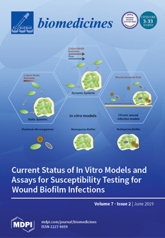
From static models in microtiter plates to dynamic models in flow cells, multiple systems have been developed to study the growth and susceptibility of biofilms to antimicrobials. In static systems, bacterial growth in media has a finite supply of nutrients, while in dynamic systems the nutrients are continually refreshed by the flow of sterile media. To resemble the physiological conditions of biofilm-infected chronic wounds, in vitro biofilm models aim to incorporate multiple clinically relevant wound microorganisms and simulate the composition of the wound exudate and wound bed. View this paper.
  • Issues are regarded as officially published after their release is announced to the table of contents alert mailing list.
  • You may sign up for e-mail alerts to receive table of contents of newly released issues.
  • PDF is the official format for papers published in both, html and pdf forms. To view the papers in pdf format, click on the "PDF Full-text" link, and use the free Adobe Reader to open them.
Order results
Result details
Section
Select all
Export citation of selected articles as:
24 pages, 2993 KiB  
Review
Protein Nanotubes: From Bionanotech towards Medical Applications
by Gerald F. Audette, Ayat Yaseen, Nicholas Bragagnolo and Raj Bawa
Biomedicines 2019, 7(2), 46; https://0-doi-org.brum.beds.ac.uk/10.3390/biomedicines7020046 - 23 Jun 2019
Cited by 11 | Viewed by 5531
Abstract
Nanobiotechnology involves the study of structures found in nature to construct nanodevices for biological and medical applications with the ultimate goal of commercialization. Within a cell most biochemical processes are driven by proteins and associated macromolecular complexes. Evolution has optimized these protein-based nanosystems [...] Read more.
Nanobiotechnology involves the study of structures found in nature to construct nanodevices for biological and medical applications with the ultimate goal of commercialization. Within a cell most biochemical processes are driven by proteins and associated macromolecular complexes. Evolution has optimized these protein-based nanosystems within living organisms over millions of years. Among these are flagellin and pilin-based systems from bacteria, viral-based capsids, and eukaryotic microtubules and amyloids. While carbon nanotubes (CNTs), and protein/peptide-CNT composites, remain one of the most researched nanosystems due to their electrical and mechanical properties, there are many concerns regarding CNT toxicity and biodegradability. Therefore, proteins have emerged as useful biotemplates for nanomaterials due to their assembly under physiologically relevant conditions and ease of manipulation via protein engineering. This review aims to highlight some of the current research employing protein nanotubes (PNTs) for the development of molecular imaging biosensors, conducting wires for microelectronics, fuel cells, and drug delivery systems. The translational potential of PNTs is highlighted. Full article
Show Figures

Figure 1

11 pages, 6458 KiB  
Article
Evidence for Contamination of Silica Microparticles in Advanced Platelet-Rich Fibrin Matrices Prepared Using Silica-Coated Plastic Tubes
by Tetsuhiro Tsujino, Akira Takahashi, Sadahiro Yamaguchi, Taisuke Watanabe, Kazushige Isobe, Yutaka Kitamura, Takaaki Tanaka, Koh Nakata and Tomoyuki Kawase
Biomedicines 2019, 7(2), 45; https://0-doi-org.brum.beds.ac.uk/10.3390/biomedicines7020045 - 19 Jun 2019
Cited by 28 | Viewed by 5980
Abstract
Platelet-rich fibrin (PRF) therapy has been widely applied in regenerative dentistry, and PRF preparation has been optimized to efficiently form fibrin clots using plain glass tubes. Currently, a shortage of commercially available glass tubes has forced PRF users to utilize silica-coated plastic tubes. [...] Read more.
Platelet-rich fibrin (PRF) therapy has been widely applied in regenerative dentistry, and PRF preparation has been optimized to efficiently form fibrin clots using plain glass tubes. Currently, a shortage of commercially available glass tubes has forced PRF users to utilize silica-coated plastic tubes. However, most plastic tubes are approved by regulatory authorities only for diagnostic use and remain to be approved for PRF therapy. To clarify this issue, we quantified silica microparticles incorporated into the PRF matrix. Blood samples were collected into three different brands of silica-containing plastic tubes and were immediately centrifuged following the protocol for advanced-PRF (A-PRF). Advanced-PRF-like matrices were examined using a scanning electron microscope (SEM), and silica microparticles were quantified using a spectrophotometer. Each brand used silica microparticles of specific size and appearance. Regardless of tube brands and individual donors, significant, but not accidental, levels of silica microparticles were found to be incorporated into the A-PRF-like matrix, which will be consequently incorporated into the implantation sites. Presently, from the increasing data for cytotoxicity of amorphous silica, we cannot exclude the possibility that such A-PRF-like matrices negatively influence tissue regeneration through induction of inflammation. Further investigation should be performed to clarify such potential risks. Full article
Show Figures

Graphical abstract

15 pages, 1021 KiB  
Review
The Role of Wnt and R-spondin in the Stomach During Health and Disease
by Anne-Sophie Fischer and Michael Sigal
Biomedicines 2019, 7(2), 44; https://0-doi-org.brum.beds.ac.uk/10.3390/biomedicines7020044 - 19 Jun 2019
Cited by 20 | Viewed by 7561
Abstract
The Wnt signaling pathway is one of the most prominent developmental signals. In addition to its functions in development, there is emerging evidence that it is also crucial for various organ functions in adult organisms, where Wnt signaling controls tissue stem cell behavior, [...] Read more.
The Wnt signaling pathway is one of the most prominent developmental signals. In addition to its functions in development, there is emerging evidence that it is also crucial for various organ functions in adult organisms, where Wnt signaling controls tissue stem cell behavior, proliferation and differentiation. Deregulation of Wnt signaling is involved in various pathological conditions and has been linked to malignant tissue transformation in different organ systems. The study of the Wnt signaling pathway has revealed a complex regulatory network that tightly balances the quality and strength of Wnt signaling in tissues. In this context, R-spondins are secreted proteins that stabilize Wnt receptors and enhance Wnt signaling. In this review we focus on new insights into the regulatory function of Wnt and R-spondin signaling in the stomach. In addition to its function in the healthy state, we highlight the connection between Wnt signaling and infection with Helicobacter pylori (H. pylori), a pathogen that colonizes the stomach and is the main risk factor for gastric cancer. In addition to experimental data that link Wnt signaling to carcinogenesis, we discuss that Wnt signaling is affected in a substantial proportion of patients with gastric cancer, and provide examples for potential clinical implications for altered Wnt signaling in gastric cancer. Full article
(This article belongs to the Special Issue Gastric Cancer Research: From Basic Science to the Clinic)
Show Figures

Graphical abstract

8 pages, 1009 KiB  
Article
Periodontitis Stage III–IV, Grade C and Correlated Factors: A Histomorphometric Study
by Barbara Buffoli, Gianluca Garzetti, Stefano Calza, Eleonora Scotti, Elisa Borsani, Veronica Cappa, Lia Rimondini and Magda Mensi
Biomedicines 2019, 7(2), 43; https://0-doi-org.brum.beds.ac.uk/10.3390/biomedicines7020043 - 12 Jun 2019
Cited by 2 | Viewed by 3813
Abstract
Background: Periodontitis is a disease that leads to serious functional and esthetic dysfunctions. Periodontitis exists in different forms, and its etiology is related to multiple component causes. Two key processes involved in the evolution of this pathology are angiogenesis and inflammatory infiltrate. The [...] Read more.
Background: Periodontitis is a disease that leads to serious functional and esthetic dysfunctions. Periodontitis exists in different forms, and its etiology is related to multiple component causes. Two key processes involved in the evolution of this pathology are angiogenesis and inflammatory infiltrate. The aim of this study was to understand if important factors such as smoking, gender, age, plaque, pus, and probing pocket depth could influence the histomorphological pattern of generalized stage III–IV, grade C periodontitis (GPIII–IVC), which is a particular form of periodontitis. Methods: Eighteen subjects with GPIII–IVC were enrolled in this study. The percentage of inflammatory cells and the vascular area were measured and evaluated in relation to each periodontal disease-associated factor. Results: Females showed a significant increase in the percentage of inflammatory cells compared to males (6.29% vs. 2.28%, p-value = 0.020) and it was higher in non-smokers than in smokers (4.56% vs. 3.14%, p-value = 0.048). Young patients showed a significant increase in vascular area percentage compared to older patients (0.60% vs. 0.46%, p-value = 0.0006) and this percentage was also higher in non-smokers compared to smokers (0.41% vs. 0.55%, p-value = 0.0008). The vascular area was also more than halved in subjects with residual plaque on tooth surfaces (0.74% vs. 0.36%, p-value = 0.0005). Conclusions: These results suggested that even if these factors are commonly related to the worsening of periodontal status, some of them (pus and periodontal probing depth (PPD)) do not affect the inflammatory and vascular patterns. Full article
Show Figures

Figure 1

24 pages, 1945 KiB  
Review
Neuromyelitis Optica Spectrum Disorder and Anti-MOG Syndromes
by Marco A. Lana-Peixoto and Natália Talim
Biomedicines 2019, 7(2), 42; https://0-doi-org.brum.beds.ac.uk/10.3390/biomedicines7020042 - 12 Jun 2019
Cited by 88 | Viewed by 13060
Abstract
Neuromyelitis optica spectrum disorder (NMOSD) and anti-myelin oligodendrocyte glycoprotein (anti-MOG) syndromes are immune-mediated inflammatory conditions of the central nervous system that frequently involve the optic nerves and the spinal cord. Because of their similar clinical manifestations and habitual relapsing course they are frequently [...] Read more.
Neuromyelitis optica spectrum disorder (NMOSD) and anti-myelin oligodendrocyte glycoprotein (anti-MOG) syndromes are immune-mediated inflammatory conditions of the central nervous system that frequently involve the optic nerves and the spinal cord. Because of their similar clinical manifestations and habitual relapsing course they are frequently confounded with multiple sclerosis (MS). Early and accurate diagnosis of these distinct conditions is relevant as they have different treatments. Some agents used for MS treatment may be deleterious to NMOSD. NMOSD is frequently associated with antibodies which target aquaporin-4 (AQP4), the most abundant water channel in the CNS, located in the astrocytic processes at the blood-brain barrier (BBB). On the other hand, anti-MOG syndromes result from damage to myelin oligodendrocyte glycoprotein (MOG), expressed on surfaces of oligodendrocytes and myelin sheaths. Acute transverse myelitis with longitudinally extensive lesion on spinal MRI is the most frequent inaugural manifestation of NMOSD, usually followed by optic neuritis. Other core clinical characteristics include area postrema syndrome, brainstem, diencephalic and cerebral symptoms that may be associated with typical MRI abnormalities. Acute disseminated encephalomyelitis and bilateral or recurrent optic neuritis are the most frequent anti-MOG syndromes in children and adults, respectively. Attacks are usually treated with steroids, and relapses prevention with immunosuppressive drugs. Promising emerging therapies for NMOSD include monoclonal antibodies and tolerization. Full article
(This article belongs to the Special Issue Multiple Sclerosis: Diagnosis and Treatment)
Show Figures

Figure 1

17 pages, 2777 KiB  
Article
Reporter Cell Assessment of TLR4-Induced NF-κB Responses to Cell-Free Hemoglobin and the Influence of Biliverdin
by Jill Sharma, Taylor Boyd, Claudia Alvarado, Edwin Gunn, Jaimie Adams, Traci Ness, Robert Dunwoody, John Lamb, Brittany House, James Knapp and Ronald Garner
Biomedicines 2019, 7(2), 41; https://0-doi-org.brum.beds.ac.uk/10.3390/biomedicines7020041 - 03 Jun 2019
Cited by 8 | Viewed by 4680
Abstract
Hemoglobin (Hb) released during red blood cell lysis can initiate TLR4-dependent signaling and trigger NF-κB activation in surrounding cells. Observations of chronic bleeding in various cancers leads us to hypothesize that Hb and Hb degradation products released from lysed RBC near cancer nests [...] Read more.
Hemoglobin (Hb) released during red blood cell lysis can initiate TLR4-dependent signaling and trigger NF-κB activation in surrounding cells. Observations of chronic bleeding in various cancers leads us to hypothesize that Hb and Hb degradation products released from lysed RBC near cancer nests might modulate local TLR4-positive cells. We addressed the hypothesis in vitro by measuring Hb- and biliverdin (Bv)-induced NF-κB signaling in an engineered human TLR4 reporter cell model (HEK-BlueTM hTLR4). Therein, TLR4 stimulation was assessed by measuring NF-κB-dependent secreted alkaline phosphatase (SEAP). hTLR4 reporter cells incubated with 8 ηM lipopolysaccharide (LPS) or 20-40 μM fungal mannoprotein (FM) produced significant amounts of SEAP. hTLR4 reporter cells also produced SEAP in response to human, but not porcine or bovine, Hb. HEK-Blue Null2TM reporter cells lacking TLR4 did not respond to LPS, FM, or Hb. Bv was non-stimulatory in reporter cells. When Bv was added to Hb-stimulated reporter cells, SEAP production was reduced by 95%, but when Bv was applied during LPS and FM stimulation, SEAP production was reduced by 33% and 27%, respectively. In conclusion, Hb initiated NF-κB signaling that was dependent upon TLR4 expression and that Bv can act as a TLR4 antagonist. Moreover, this study suggests that hemorrhage and extravascular hemolysis could provide competitive Hb and Bv signaling to nearby cells expressing TLR4, and that this process could modulate NF-κB signaling in TLR4-positive cancer cells and cancer-infiltrating leukocytes. Full article
(This article belongs to the Special Issue Roles of NF-κB in Cancer and Their Therapeutic Approaches 2019)
Show Figures

Figure 1

13 pages, 1246 KiB  
Article
Controlling Nuclear NF-κB Dynamics by β-TrCP—Insights from a Computational Model
by Uwe Benary and Jana Wolf
Biomedicines 2019, 7(2), 40; https://0-doi-org.brum.beds.ac.uk/10.3390/biomedicines7020040 - 27 May 2019
Cited by 9 | Viewed by 4054
Abstract
The canonical nuclear factor kappa-light-chain-enhancer of activated B cells (NF-κB) signaling pathway regulates central processes in mammalian cells and plays a fundamental role in the regulation of inflammation and immunity. Aberrant regulation of the activation of the transcription factor NF-κB is associated with [...] Read more.
The canonical nuclear factor kappa-light-chain-enhancer of activated B cells (NF-κB) signaling pathway regulates central processes in mammalian cells and plays a fundamental role in the regulation of inflammation and immunity. Aberrant regulation of the activation of the transcription factor NF-κB is associated with severe diseases such as inflammatory bowel disease and arthritis. In the canonical pathway, the inhibitor IκB suppresses NF-κB’s transcriptional activity. NF-κB becomes active upon the degradation of IκB, a process that is, in turn, regulated by the β-transducin repeat-containing protein (β-TrCP). β-TrCP has therefore been proposed as a promising pharmacological target in the development of novel therapeutic approaches to control NF-κB’s activity in diseases. This study explores the extent to which β-TrCP affects the dynamics of nuclear NF-κB using a computational model of canonical NF-κB signaling. The analysis predicts that β-TrCP influences the steady-state concentration of nuclear NF-κB, as well as changes characteristic dynamic properties of nuclear NF-κB, such as fold-change and the duration of its response to pathway stimulation. The results suggest that the modulation of β-TrCP has a high potential to regulate the transcriptional activity of NF-κB. Full article
(This article belongs to the Special Issue Roles of NF-κB in Cancer and Their Therapeutic Approaches 2019)
Show Figures

Graphical abstract

15 pages, 4751 KiB  
Article
Differential Expression of CD3, TNF-α, and VEGF Induced by Olanzapine on the Spleen of Adult Male Albino Rats and the Possible Protective Role of Vitamin C
by Sahar Youssef and Marwa Salah
Biomedicines 2019, 7(2), 39; https://0-doi-org.brum.beds.ac.uk/10.3390/biomedicines7020039 - 23 May 2019
Cited by 6 | Viewed by 4746
Abstract
Olanzapine is an antipsychotic drug effective in the treatment of stress-associated psychiatric illnesses, but its effect on the spleen remains unclear. Vitamin C is essential for the optimum function of the immune system. We aim to investigate the effect of Olanzapine on spleen [...] Read more.
Olanzapine is an antipsychotic drug effective in the treatment of stress-associated psychiatric illnesses, but its effect on the spleen remains unclear. Vitamin C is essential for the optimum function of the immune system. We aim to investigate the effect of Olanzapine on spleen structures and to assess the protective effect of vitamin C. Forty adult male albino rats were divided into four groups: group (I), a control; group (II), rats were given vitamin C at 40 mg/kg body weight; group (III), rats were given Olanzapine at 2 mg/kg body weight; and group (IV), rats were given vitamin C and Olanzapine at the same dose of group (II) and group (III) for one month. The hematoxylin and eosin (H&E) of the olanzapine treated group showed focal areas of cellular depletion and a decrease in the size of the white pulp. The red pulp was expanded and showed marked congestion and dilatation of blood sinusoids. Cluster of differentiation 3 (CD3) was significantly reduced, however both tumor necrosis factor alpha (TNF-α), and vascular endothelial growth factor (VEGF) were significantly higher. The administration of vitamin C repaired structural and immunohistochemical changes via increased CD3 and decreased TNF-α and VEGF. Therefore, the oxidative and the inflammatory pathways may be the possible mechanisms underlying olanzapine immunotoxicity. Vitamin C exerted immune modulator and antioxidant effects against olanzapine. Full article
Show Figures

Graphical abstract

17 pages, 805 KiB  
Review
Hosts for Hostile Protein Production: The Challenge of Recombinant Immunotoxin Expression
by Stefania Zuppone, Maria Serena Fabbrini and Riccardo Vago
Biomedicines 2019, 7(2), 38; https://0-doi-org.brum.beds.ac.uk/10.3390/biomedicines7020038 - 17 May 2019
Cited by 15 | Viewed by 4484
Abstract
For the recombinant expression of toxin-based drugs, a crucial step lies not only in the choice of the production host(s) but also in the accurate design of the protein chimera. These issues are particularly important since such products may be toxic to the [...] Read more.
For the recombinant expression of toxin-based drugs, a crucial step lies not only in the choice of the production host(s) but also in the accurate design of the protein chimera. These issues are particularly important since such products may be toxic to the expressing host itself. To avoid or limit the toxicity to productive cells while obtaining a consistent yield in chimeric protein, several systems from bacterial to mammalian host cells have been employed. In this review, we will discuss the development of immunotoxin (IT) expression, placing special emphasis on advantages and on potential drawbacks, as one single perfect host for every chimeric protein toxin or ligand does not exist. Full article
(This article belongs to the Special Issue Immunotoxins: Future Advances and Directions)
Show Figures

Figure 1

13 pages, 217 KiB  
Review
Evaluation of Angiogenesis Assays
by Zachary I. Stryker, Mehdi Rajabi, Paul J. Davis and Shaker A. Mousa
Biomedicines 2019, 7(2), 37; https://0-doi-org.brum.beds.ac.uk/10.3390/biomedicines7020037 - 16 May 2019
Cited by 68 | Viewed by 7651
Abstract
Angiogenesis assays allow for the evaluation of pro- or anti-angiogenic activity of endogenous or exogenous factors (stimulus or inhibitors) through investigation of their pro-or anti- proliferative, migratory, and tube formation effects on endothelial cells. To model the process of angiogenesis and the effects [...] Read more.
Angiogenesis assays allow for the evaluation of pro- or anti-angiogenic activity of endogenous or exogenous factors (stimulus or inhibitors) through investigation of their pro-or anti- proliferative, migratory, and tube formation effects on endothelial cells. To model the process of angiogenesis and the effects of biomolecules on that process, both in vitro and in vivo methods are currently used. In general, in vitro methods monitor specific stages in the angiogenesis process and are used for early evaluations, while in vivo methods more accurately simulate the living microenvironment to provide more pertinent information. We review here the current state of angiogenesis assays as well as their mechanisms, advantages, and limitations. Full article
14 pages, 1076 KiB  
Review
Salmonella-Based Targeted Cancer Therapy: Updates on A Promising and Innovative Tumor Immunotherapeutic Strategy
by Christian Ronquillo Pangilinan and Che-Hsin Lee
Biomedicines 2019, 7(2), 36; https://0-doi-org.brum.beds.ac.uk/10.3390/biomedicines7020036 - 02 May 2019
Cited by 26 | Viewed by 6097
Abstract
Presently, cancer is one of the leading causes of death in the world, primarily due to tumor heterogeneity associated with high-grade malignancy. Tumor heterogeneity poses a tremendous challenge, especially with the emergence of resistance not only to chemo- and radiation- therapies, but also [...] Read more.
Presently, cancer is one of the leading causes of death in the world, primarily due to tumor heterogeneity associated with high-grade malignancy. Tumor heterogeneity poses a tremendous challenge, especially with the emergence of resistance not only to chemo- and radiation- therapies, but also to immunotherapy using monoclonal antibodies. The use of Salmonella, as a highly selective and penetrative antitumor agent, has shown convincing results, thus meriting further investigation. In this review, the mechanisms used by Salmonella in combating cancer are carefully explained. In essence, Salmonella overcomes the suppressive nature of the tumor microenvironment and coaxes the activation of tumor-specific immune cells to induce cell death by apoptosis and autophagy. Furthermore, Salmonella treatment suppresses tumor aggressive behavior via inhibition of angiogenesis and delay of metastatic activity. Thus, harnessing the natural potential of Salmonella in eliminating tumors will provide an avenue for the development of a promising micro-based therapeutic agent that could be further enhanced to address a wide range of tumor types. Full article
(This article belongs to the Special Issue Bacterial Cancer Therapy)
Show Figures

Graphical abstract

26 pages, 417 KiB  
Review
Recent Advances in Non-Conventional Antimicrobial Approaches for Chronic Wound Biofilms: Have We Found the ‘Chink in the Armor’?
by Snehal Kadam, Saptarsi Shai, Aditi Shahane and Karishma S Kaushik
Biomedicines 2019, 7(2), 35; https://0-doi-org.brum.beds.ac.uk/10.3390/biomedicines7020035 - 30 Apr 2019
Cited by 54 | Viewed by 9148
Abstract
Chronic wounds are a major healthcare burden, with huge public health and economic impact. Microbial infections are the single most important cause of chronic, non-healing wounds. Chronic wound infections typically form biofilms, which are notoriously recalcitrant to conventional antibiotics. This prompts the need [...] Read more.
Chronic wounds are a major healthcare burden, with huge public health and economic impact. Microbial infections are the single most important cause of chronic, non-healing wounds. Chronic wound infections typically form biofilms, which are notoriously recalcitrant to conventional antibiotics. This prompts the need for alternative or adjunct ‘anti-biofilm’ approaches, notably those that account for the unique chronic wound biofilm microenvironment. In this review, we discuss the recent advances in non-conventional antimicrobial approaches for chronic wound biofilms, looking beyond standard antibiotic therapies. These non-conventional strategies are discussed under three groups. The first group focuses on treatment approaches that directly kill or inhibit microbes in chronic wound biofilms, using mechanisms or delivery strategies distinct from antibiotics. The second group discusses antimicrobial approaches that modify the biological, chemical or biophysical parameters in the chronic wound microenvironment, which in turn enables the disruption and removal of biofilms. Finally, therapeutic approaches that affect both, biofilm bacteria and microenvironment factors, are discussed. Understanding the advantages and limitations of these recent approaches, their stage of development and role in biofilm management, could lead to new treatment paradigms for chronic wound infections. Towards this end, we discuss the possibility that non-conventional antimicrobial therapeutics and targets could expose the ‘chink in the armor’ of chronic wound biofilms, thereby providing much-needed alternative or adjunct strategies for wound infection management. Full article
(This article belongs to the Special Issue Biofilms in Wounds: New Advances in Therapy and in Healing Management)
Show Figures

Graphical abstract

31 pages, 2227 KiB  
Review
Current Status of In Vitro Models and Assays for Susceptibility Testing for Wound Biofilm Infections
by Tania F. Bahamondez-Canas, Lara A. Heersema and Hugh D. C. Smyth
Biomedicines 2019, 7(2), 34; https://0-doi-org.brum.beds.ac.uk/10.3390/biomedicines7020034 - 30 Apr 2019
Cited by 46 | Viewed by 7706
Abstract
Biofilm infections have gained recognition as an important therapeutic challenge in the last several decades due to their relationship with the chronicity of infectious diseases. Studies of novel therapeutic treatments targeting infections require the development and use of models to mimic the formation [...] Read more.
Biofilm infections have gained recognition as an important therapeutic challenge in the last several decades due to their relationship with the chronicity of infectious diseases. Studies of novel therapeutic treatments targeting infections require the development and use of models to mimic the formation and characteristics of biofilms within host tissues. Due to the diversity of reported in vitro models and lack of consensus, this review aims to provide a summary of in vitro models currently used in research. In particular, we review the various reported in vitro models of Pseudomonas aeruginosa biofilms due to its high clinical impact in chronic wounds and in other chronic infections. We assess advances in in vitro models that incorporate relevant multispecies biofilms found in infected wounds, such as P. aeruginosa with Staphylococcus aureus, and additional elements such as mammalian cells, simulating fluids, and tissue explants in an attempt to better represent the physiological conditions found at an infection site. It is hoped this review will aid researchers in the field to make appropriate choices in their proposed studies with regards to in vitro models and methods. Full article
(This article belongs to the Special Issue Biofilms in Wounds: New Advances in Therapy and in Healing Management)
Show Figures

Graphical abstract

13 pages, 547 KiB  
Review
Oral and Psychological Alterations in Haemophiliac Patients
by Luca Fiorillo, Rosa De Stefano, Gabriele Cervino, Salvatore Crimi, Alberto Bianchi, Paola Campagna, Alan Scott Herford, Luigi Laino and Marco Cicciù
Biomedicines 2019, 7(2), 33; https://0-doi-org.brum.beds.ac.uk/10.3390/biomedicines7020033 - 20 Apr 2019
Cited by 48 | Viewed by 4669
Abstract
Haemophiliacs are hereditary coagulopathies whose basic anomaly consists of the quantitative or qualitative alteration of one or more plasma proteins in the coagulation system. The objective of this review is to analyse all risk factors, predispositions and alterations to the oral-maxillofacial district in [...] Read more.
Haemophiliacs are hereditary coagulopathies whose basic anomaly consists of the quantitative or qualitative alteration of one or more plasma proteins in the coagulation system. The objective of this review is to analyse all risk factors, predispositions and alterations to the oral-maxillofacial district in patients with haemophilia. The broader assessment also includes the psychological aspects that could affect the treatment and maintenance of oral conditions. The study takes into consideration all the works in the literature in the last 10 years. Works that present oral, dental and psychological changes in haemophilia patients have been combined. A total of 16 studies were analysed carefully evaluating and explaining all the alterations and risk factors that this disease provides. The aim of the review is to report all the anomalies reported in the literature for these patients, and to direct and update the clinician in the treatment of haemophilia patients. Full article
Show Figures

Figure 1

15 pages, 519 KiB  
Review
Women’s Health: Contemporary Management of MS in Pregnancy and Post-Partum
by Kelly Tisovic and Lilyana Amezcua
Biomedicines 2019, 7(2), 32; https://0-doi-org.brum.beds.ac.uk/10.3390/biomedicines7020032 - 19 Apr 2019
Cited by 14 | Viewed by 4918
Abstract
Multiple sclerosis (MS) primarily affects women in childbearing age and is associated with an increased risk of adverse post-partum outcomes. Relapses and now fetal exposure to disease modifying treatments in the early phase of pregnancy and thereafter are of concern. Safe and effective [...] Read more.
Multiple sclerosis (MS) primarily affects women in childbearing age and is associated with an increased risk of adverse post-partum outcomes. Relapses and now fetal exposure to disease modifying treatments in the early phase of pregnancy and thereafter are of concern. Safe and effective contraception is required for women who wish to delay or avoid pregnancy while on disease-modifying treatments. Counseling and planning is essential to assess the risk of both fetal and maternal complications, particularly now in the era of highly efficient and riskier therapies. The purpose of this review is to provide a practical framework using the available data surrounding pregnancy in MS with the goal of optimizing outcomes during this phase in MS. Full article
(This article belongs to the Special Issue Multiple Sclerosis: Diagnosis and Treatment)
Show Figures

Figure 1

19 pages, 1403 KiB  
Review
The Potential of the Cyclotide Scaffold for Drug Development
by Julio A. Camarero and Maria Jose Campbell
Biomedicines 2019, 7(2), 31; https://0-doi-org.brum.beds.ac.uk/10.3390/biomedicines7020031 - 19 Apr 2019
Cited by 44 | Viewed by 7559
Abstract
Cyclotides are a novel class of micro-proteins (≈30–40 residues long) with a unique topology containing a head-to-tail cyclized backbone structure further stabilized by three disulfide bonds that form a cystine knot. This unique molecular framework makes them exceptionally stable to physical, chemical, and [...] Read more.
Cyclotides are a novel class of micro-proteins (≈30–40 residues long) with a unique topology containing a head-to-tail cyclized backbone structure further stabilized by three disulfide bonds that form a cystine knot. This unique molecular framework makes them exceptionally stable to physical, chemical, and biological degradation compared to linear peptides of similar size. The cyclotides are also highly tolerant to sequence variability, aside from the conserved residues forming the cystine knot, and are orally bioavailable and able to cross cellular membranes to modulate intracellular protein–protein interactions (PPIs), both in vitro and in vivo. These unique properties make them ideal scaffolds for many biotechnological applications, including drug discovery. This review provides an overview of the properties of cyclotides and their potential for the development of novel peptide-based therapeutics. The selective disruption of PPIs still remains a very challenging task, as the interacting surfaces are relatively large and flat. The use of the cell-permeable highly constrained polypeptide molecular frameworks, such as the cyclotide scaffold, has shown great promise, as it provides unique pharmacological properties. The use of molecular techniques, such as epitope grafting, and molecular evolution have shown to be highly effective for the selection of bioactive cyclotides. However, despite successes in employing cyclotides to target PPIs, some of the challenges to move them into the clinic still remain. Full article
(This article belongs to the Special Issue Discovery and Development of Constrained Peptide Ligands)
Show Figures

Graphical abstract

15 pages, 3566 KiB  
Article
The Glycogen Synthase Kinase-3β Inhibitor LSN 2105786 Promotes Zebrafish Fin Regeneration
by Swapnalee Sarmah, Courtney Curtis, Jennifer Mahin, Mark Farrell, Thomas A. Engler, Manuel V. Sanchez-Felix, Masahiko Sato, Yanfai Linda Ma, Shaoyou Chu and James A. Marrs
Biomedicines 2019, 7(2), 30; https://0-doi-org.brum.beds.ac.uk/10.3390/biomedicines7020030 - 19 Apr 2019
Cited by 2 | Viewed by 4233
Abstract
The Wnt pathway has been shown to regulate bone homeostasis and to influence some bone disease states. We utilized a zebrafish model system to study the effects of a synthetic, orally bioavailable glycogen synthase kinase-3β (GSK3β) inhibitor LSN 2105786, which activates Wnt signaling [...] Read more.
The Wnt pathway has been shown to regulate bone homeostasis and to influence some bone disease states. We utilized a zebrafish model system to study the effects of a synthetic, orally bioavailable glycogen synthase kinase-3β (GSK3β) inhibitor LSN 2105786, which activates Wnt signaling during bone healing and embryogenesis. GSK3β inhibitor treatment was used to phenocopy GSK3β morpholino oligonucleotide (MO) knockdown in zebrafish embryos. Human and zebrafish synthetic mRNA injection were similarly effective at rescue of GSK3β MO knockdown. During caudal fin regeneration, bony rays are the first structure to differentiate in zebrafish fins, providing a useful model to study bone healing. Caudal fin regeneration experiments were conducted using various concentrations of a GSK3β inhibitor, examining duration and concentration dependence on regenerative outgrowth. Experiments revealed continuous low concentration (4–5 nM) treatment to be more effective at increasing regeneration than intermittent dosing. Higher concentrations inhibited fin growth, perhaps by excessive stimulation of differentiation programs. Increased Wnt responsive gene expression and differentiation were observed in response to GSK3b inhibitor treatment. Activating Wnt signaling also increased cell proliferation and osteoblast differentiation in fin regenerates. Together, these data indicate that bone healing in zebrafish fin regeneration was improved by activating Wnt signaling using GSK3b inhibitor treatment. In addition, caudal fin regeneration is useful to evaluate dose-dependent pharmacological efficacy in bone healing, various dosing regimens and possible toxicological effects of compounds. Full article
(This article belongs to the Special Issue Zebrafish Models for Development and Disease)
Show Figures

Graphical abstract

10 pages, 7318 KiB  
Article
Hepatoprotective Activity of InlB321/15, the HGFR Ligand of Bacterial Origin, in CCI4-Induced Acute Liver Injury Mice
by Yaroslava Chalenko, Konstantin Sobyanin, Elena Sysolyatina, Konstantin Midiber, Egor Kalinin, Alexandra Lavrikova, Lyudmila Mikhaleva and Svetlana Ermolaeva
Biomedicines 2019, 7(2), 29; https://0-doi-org.brum.beds.ac.uk/10.3390/biomedicines7020029 - 11 Apr 2019
Cited by 2 | Viewed by 3209
Abstract
HGF (hepatocyte growth factor)/HGFR (HGF receptor) signaling pathway is a key pathway in liver protection and regeneration after acute toxic damage. Listeria monocytogenes toxin InlB contains a HGFR-interacting domain and is a functional analog of HGF. The aim of this work was to [...] Read more.
HGF (hepatocyte growth factor)/HGFR (HGF receptor) signaling pathway is a key pathway in liver protection and regeneration after acute toxic damage. Listeria monocytogenes toxin InlB contains a HGFR-interacting domain and is a functional analog of HGF. The aim of this work was to evaluate the hepatoprotective activity of the InlB HGFR-interacting domain. The recombinant HGFR-interacting domain InlB321/15 was purified from E. coli. MTT (3-(4,5-dimethylthiazol-2-yl)-2,5-diphenyltetrazolium bromide) test was used to measure InlB321/15 mitogenic activity in HepG2 cells. Activation of MAPK- and PI3K/Akt-pathways was tracked with fluorescent microscopy, Western blotting, and ELISA. To evaluate hepatoprotective activity, InlB321/15 and recombinant human HGF (rhHGF) were intravenously injected at the same concentration of 2 ng·g−1 to BALB/c mice 2 h before liver injury with CCl4. InlB321/15 caused dose-dependent activation of MAPK- and PI3K/Akt-pathways and correspondent mitogenic effects. Both InlB321/15 and rhHGF improved macroscopic liver parameters (liver mass was 1.51, 1.27 and 1.15 g for the vehicle, InlB321/15 and rhHGF, respectively, p < 0.05), reduced necrosis (24.0%, 16.18% and 21.66% of the total area for the vehicle, InlB321/15 and rhHGF, respectively, p < 0.05). Obtained data suggest that InlB321/15 is a promising candidate for a tissue repair agent. Full article
Show Figures

Graphical abstract

19 pages, 3146 KiB  
Article
A High-Throughput Assay for Congenital and Age-Related Eye Diseases in Zebrafish
by Lindy K. Brastrom, C. Anthony Scott, Deborah V. Dawson and Diane C. Slusarski
Biomedicines 2019, 7(2), 28; https://0-doi-org.brum.beds.ac.uk/10.3390/biomedicines7020028 - 11 Apr 2019
Cited by 13 | Viewed by 4491
Abstract
Debilitating visual impairment caused by cataracts or microphthalmia is estimated to affect roughly 20 million people in the United States alone. According to the National Eye Institute, by 2050 that number is expected to more than double to roughly 50 million. The identification [...] Read more.
Debilitating visual impairment caused by cataracts or microphthalmia is estimated to affect roughly 20 million people in the United States alone. According to the National Eye Institute, by 2050 that number is expected to more than double to roughly 50 million. The identification of candidate disease-causing alleles for cataracts and microphthalmia has been accelerated with advanced sequencing technologies creating a need for verification of the pathophysiology of these genes. Zebrafish pose many advantages as a high-throughput model for human eye disease. By 5 days post-fertilization, zebrafish have quantifiable behavioral responses to visual stimuli. Their small size, many progeny, and external fertilization allows for rapid screening for vision defects. We have adapted the OptoMotor Response to assay visual impairment in zebrafish models of cataracts and microphthalmia. This research demonstrates an inexpensive, high-throughput method for analyzing candidate genes involved in visual impairment. Full article
(This article belongs to the Special Issue Zebrafish Models for Development and Disease)
Show Figures

Graphical abstract

16 pages, 4609 KiB  
Article
Platelet-Rich Plasma and Micrografts Enriched with Autologous Human Follicle Mesenchymal Stem Cells Improve Hair Re-Growth in Androgenetic Alopecia. Biomolecular Pathway Analysis and Clinical Evaluation
by Pietro Gentile, Maria G. Scioli, Alessandra Bielli, Barbara De Angelis, Ciro De Sio, Domenico De Fazio, Gabriele Ceccarelli, Angelo Trivisonno, Augusto Orlandi, Valerio Cervelli and Simone Garcovich
Biomedicines 2019, 7(2), 27; https://0-doi-org.brum.beds.ac.uk/10.3390/biomedicines7020027 - 08 Apr 2019
Cited by 98 | Viewed by 8044
Abstract
Platelet rich plasma (PRP) and Micrografts containing human follicle mesenchymal stem cells (HF-MSCs) were tried as a potential treatment for androgenetic alopecia (AGA). However, little to no work has yet to be seen wherein the bio-molecular pathway of HF-MSCs or PRP treatments were [...] Read more.
Platelet rich plasma (PRP) and Micrografts containing human follicle mesenchymal stem cells (HF-MSCs) were tried as a potential treatment for androgenetic alopecia (AGA). However, little to no work has yet to be seen wherein the bio-molecular pathway of HF-MSCs or PRP treatments were analyzed. The aims of this work are to report the clinical effectiveness of HF-MSCs and platelet-rich plasma evaluating and reviewing the most updated information related to the bio-molecular pathway. Twenty-one patients were treated with HF-MSCs injections and 57 patients were treated with A-PRP. The Wnt pathway and Platelet derived-growth factors effects were analyzed. 23 weeks after the last treatment with mean hair thickness increments (29 ± 5.0%) over baseline values for the targeted area. 12 weeks after the last injection with A-PRP mean hair count and hair density (31 ± 2%) increases significantly over baseline values. The increment of Wnt signaling in Dermal Papilla Cells evidently is one of the principal factors that enhances hair growth. Signaling from mesenchymal stem cells and platelet derived growth factors positively influences hair growth through cellular proliferation to prolong the anagen phase (FGF-7), inducing cell growth (ERK activation), stimulating hair follicle development (β-catenin), and suppressing apoptotic cues (Bcl-2 release and Akt activation). Full article
Show Figures

Figure 1

13 pages, 361 KiB  
Review
Stem Cell-Based Therapies for Multiple Sclerosis: Current Perspectives
by Fernando X. Cuascut and George J. Hutton
Biomedicines 2019, 7(2), 26; https://0-doi-org.brum.beds.ac.uk/10.3390/biomedicines7020026 - 30 Mar 2019
Cited by 39 | Viewed by 8688
Abstract
Multiple sclerosis (MS) is an inflammatory and neurodegenerative autoimmune disease of the central nervous system (CNS). Disease-modifying therapies (DMT) targeting inflammation have been shown to reduce disease activity in patients with relapsing–remitting MS (RRMS). The current therapeutic challenge is to find an effective [...] Read more.
Multiple sclerosis (MS) is an inflammatory and neurodegenerative autoimmune disease of the central nervous system (CNS). Disease-modifying therapies (DMT) targeting inflammation have been shown to reduce disease activity in patients with relapsing–remitting MS (RRMS). The current therapeutic challenge is to find an effective treatment to halt disease progression and reverse established neural damage. Stem cell-based therapies have emerged to address this dilemma. Several types of stem cells have been considered for clinical use, such as autologous hematopoietic (aHSC), mesenchymal (MSC), neuronal (NSC), human embryonic (hESC), and induced pluripotent (iPSC) stem cells. There is convincing evidence that immunoablation followed by hematopoietic therapy (aHSCT) has a high efficacy for suppressing inflammatory MS activity and improving neurological disability in patients with RRMS. In addition, MSC therapy may be a safe and tolerable treatment, but its clinical value is still under evaluation. Various studies have shown early promising results with other cellular therapies for CNS repair and decreasing inflammation. In this review, we discuss the current knowledge and limitations of different stem cell-based therapies for the treatment of patients with MS. Full article
(This article belongs to the Special Issue Multiple Sclerosis: Diagnosis and Treatment)
Show Figures

Figure 1

14 pages, 3538 KiB  
Article
Cytotoxicity of Extracts from New Zealand Surf Clams Against Organ Cancer Cell Lines
by Tinu Odeleye, William Lindsey White and Jun Lu
Biomedicines 2019, 7(2), 25; https://0-doi-org.brum.beds.ac.uk/10.3390/biomedicines7020025 - 30 Mar 2019
Cited by 3 | Viewed by 3240
Abstract
In this study, we examined the cytotoxic effects of four fractions from three species of New Zealand (NZ) surf clam on four common organ cancer cells. In most cases, a dose- and time-dependent inhibition on the proliferation of the cancer cells was observed. [...] Read more.
In this study, we examined the cytotoxic effects of four fractions from three species of New Zealand (NZ) surf clam on four common organ cancer cells. In most cases, a dose- and time-dependent inhibition on the proliferation of the cancer cells was observed. This was most significant in WiDr (colon) cells, where the percentages of viability reduced to as low as 6%, 5%, and 17% (at 1000 µg 72 h) by extracts from Diamond shell, Storm shell, and Tua tua species, respectively. A549 (lung) cells were the least susceptible to the treatment, with viability percentages at 82%, 15%, and 45%, under the same conditions. Induction of caspase-dependent apoptosis and alterations to the cell cycle further supported the observed morphological analysis. The ethanol, petroleum ether, and ethyl acetate fractions of NZ surf clam, rich in lipids and proteins, were more potent than their water-based counterpart. This is the first demonstration where extracts from NZ surf clams show the ability to inhibit the growth and proliferation of cancer cell lines. We suggest that NZ surf clam extracts have the potential to be further studied and developed as candidates for cancer supplementary management/treatment. Full article
Show Figures

Graphical abstract

21 pages, 524 KiB  
Concept Paper
The Transformative Possibilities of the Microbiota and Mycobiota for Health, Disease, Aging, and Technological Innovation
by Lucas Jones, Jessica Kumar, Adil Mistry, Thriveen Sankar Chittoor Mana, George Perry, V. Prakash Reddy and Mark Obrenovich
Biomedicines 2019, 7(2), 24; https://0-doi-org.brum.beds.ac.uk/10.3390/biomedicines7020024 - 28 Mar 2019
Cited by 27 | Viewed by 6098
Abstract
The gut microbiota is extremely important for the health of the host across its lifespan. Recent studies have elucidated connections between the gut microbiota and neurological disease and disorders such as depression, anxiety, Alzheimer’s disease (AD), autism, and a host of other brain [...] Read more.
The gut microbiota is extremely important for the health of the host across its lifespan. Recent studies have elucidated connections between the gut microbiota and neurological disease and disorders such as depression, anxiety, Alzheimer’s disease (AD), autism, and a host of other brain illnesses. Dysbiosis of the normal gut flora can have negative consequences for humans, especially throughout key periods during our lifespan as the gut microbes change with age in both phenotype and number of bacterial species. Neurologic diseases, mental disorders, and euthymic states are influenced by alterations in the metabolites produced by gut microbial milieu. We introduce a new concept, namely, the mycobiota and microbiota-gut-brain neuroendocrine axis and discuss co-metabolism with emphasis on means to influence or correct disruptions to normal gut flora throughout the lifespan from early development to old age. These changes involve inflammation and involve the permeability of barriers, such as the intestine blood barrier, the blood–brain barrier, and others. The mycobiota and microbiota–gut–brain axis offer new research horizons and represents a great potential target for new therapeutics, including approaches based around inflammatory disruptive process, genetically engineered drug delivery systems, diseased cell culling “kill switches”, phage-like therapies, medicinal chemistry, or microbial parabiosis to name a few. Full article
(This article belongs to the Special Issue The (Microbiota)-Gut-Brain Axis: Hype or Revolution?)
Show Figures

Figure 1

Previous Issue
Next Issue
Back to TopTop