
Recent Advances in Improved Anticancer Efficacies of Camptothecin Nano-Formulations: A Systematic Review

 ,
, 
 , and
, and
Abstract
:1. Introduction
1.1. Natural Nano-Formulations and Cancer Treatment
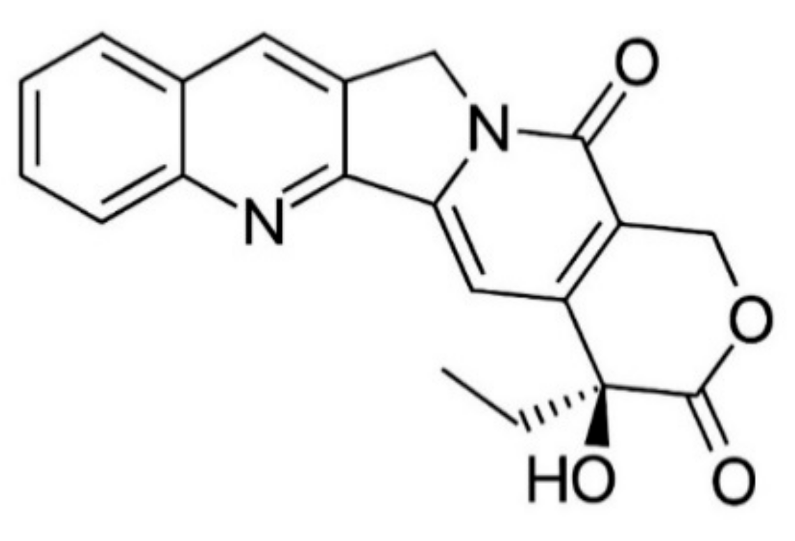
1.2. Camptothecin: Sources, Chemistry, and Pharmacology
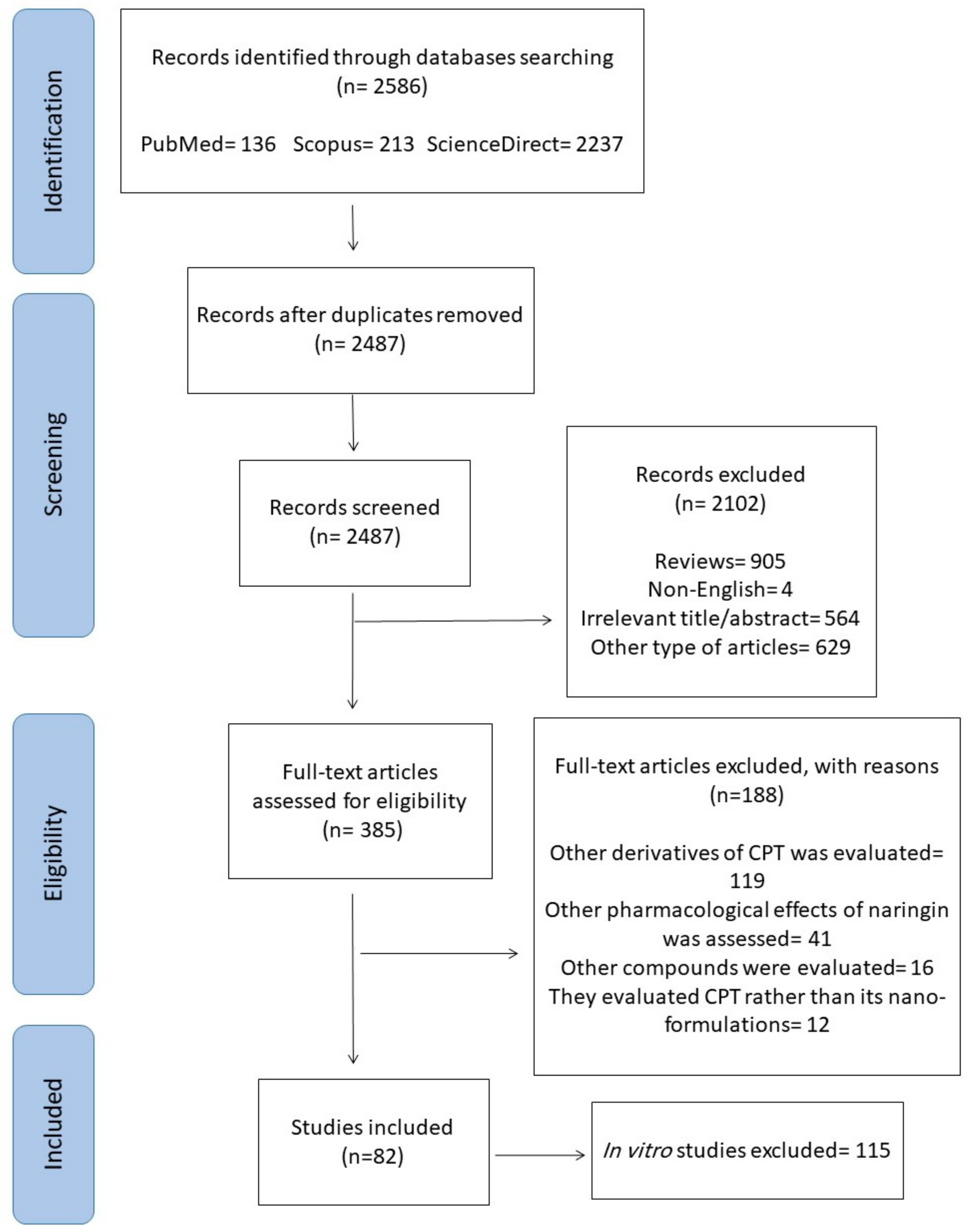
2. Methodology for Literature Search on Camptothecin Nano-Formulations and Cancer
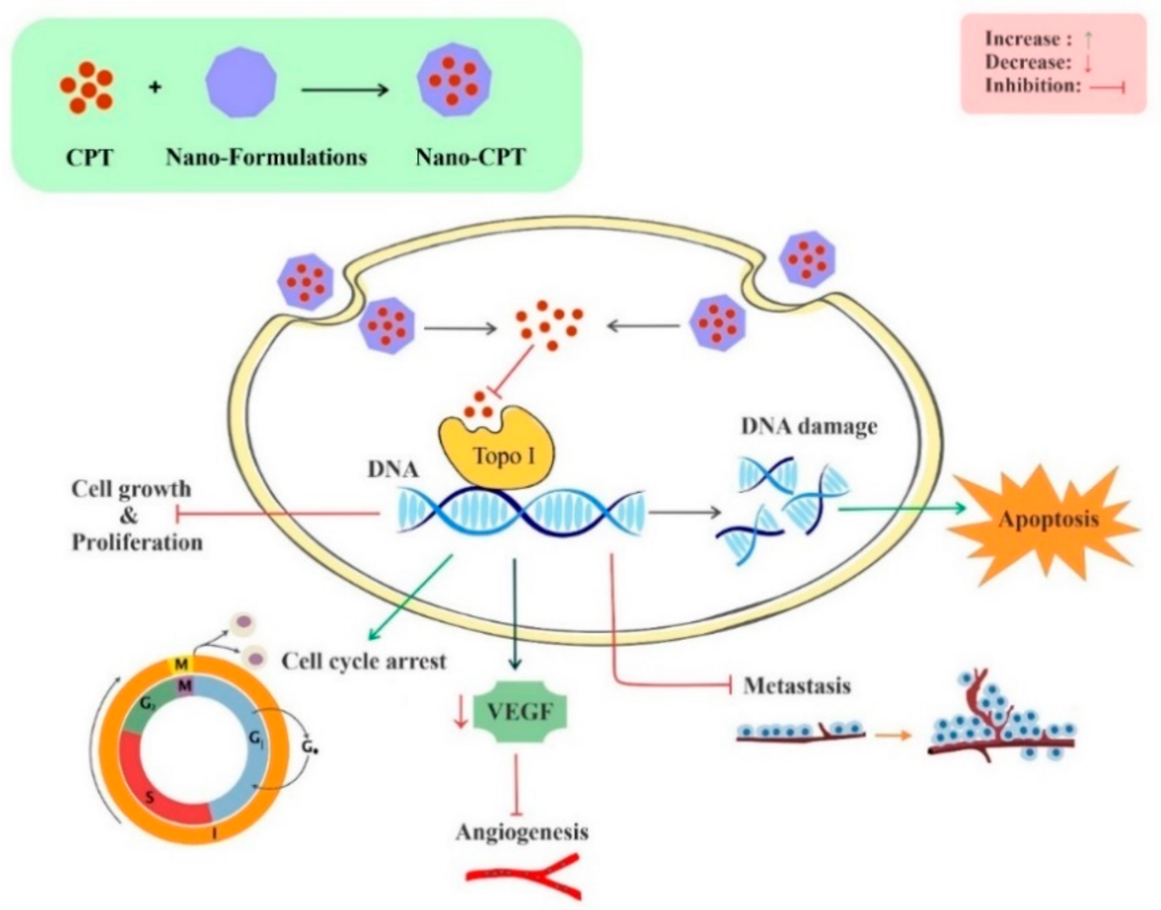
3. Anticancer Activities of Camptothecin Nano-Formulations
3.1. Bladder Cancer
3.2. Brain Cancer
3.3. Breast Cancer
3.4. Cervical Cancer
3.5. Colon Cancer
3.6. Liver Cancer
3.7. Lung Cancer
3.8. Ovarian Cancer
3.9. Pancreatic Cancer
3.10. Prostate Cancer
3.11. Skin Cancer
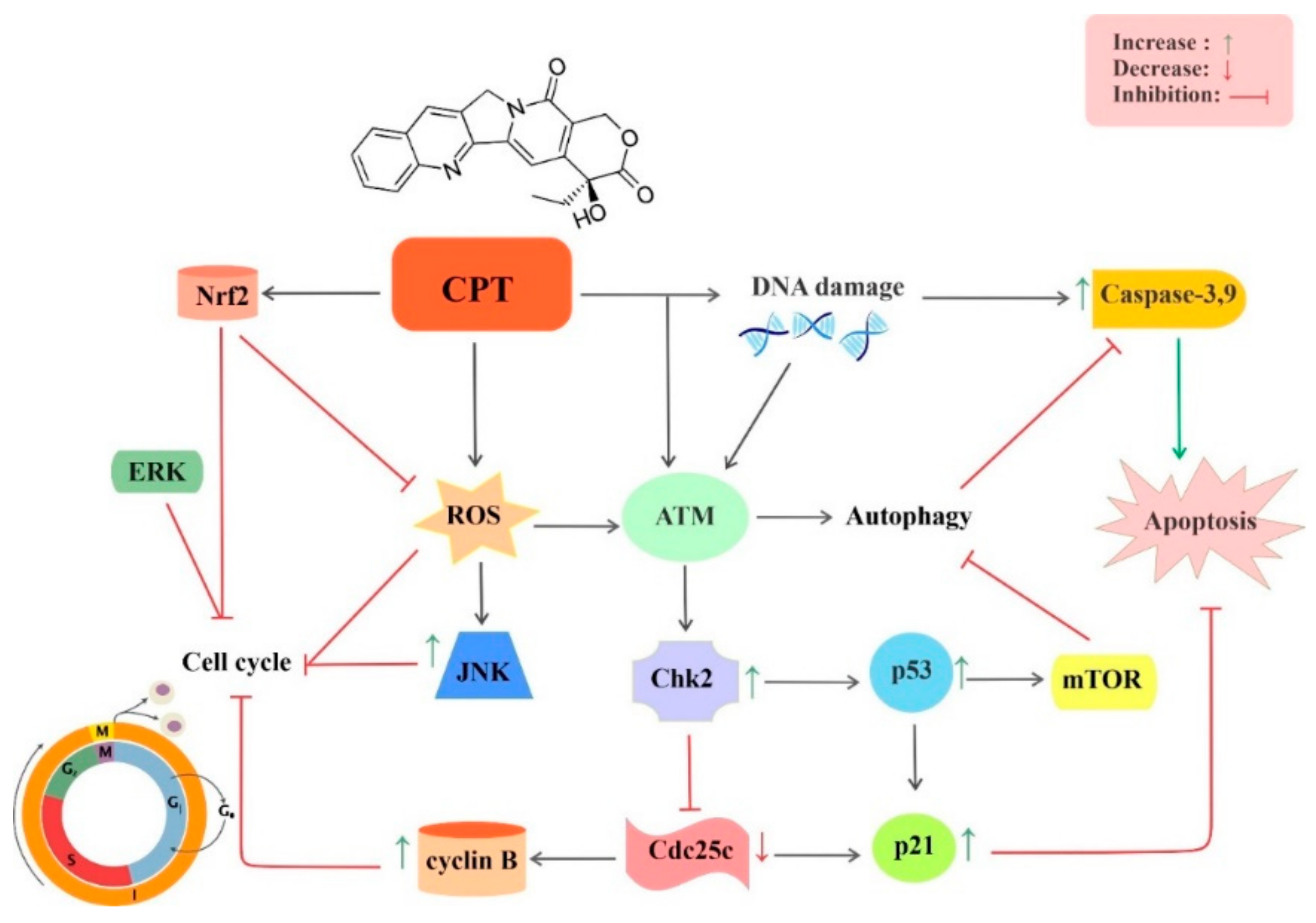
4. Pharmacokinetics and Toxicity of Nano-Camptothecin
5. Conclusions and Future Directions
Author Contributions
Funding
Institutional Review Board Statement
Informed Consent Statement
Acknowledgments
Conflicts of Interest
References
- Hanahan, D.; Weinberg, R.A. Hallmarks of cancer: The next generation. Cell 2011, 144, 646–674. [Google Scholar] [CrossRef] [PubMed] [Green Version]
- Seyfried, T.N.; Shelton, L.M. Cancer as a metabolic disease. Nutr. Metab. 2010, 7, 7. [Google Scholar] [CrossRef] [PubMed] [Green Version]
- Newman, D.J.; Cragg, G.M. Natural Products as Sources of New Drugs over the Nearly Four Decades from 01/1981 to 09/2019. J. Nat. Prod. 2020, 83, 770–803. [Google Scholar] [CrossRef] [PubMed]
- Cragg, G.M.; Pezzuto, J.M. Natural products as a vital source for the discovery of cancer chemotherapeutic and chemopreventive agents. Med. Princ. Pract. 2016, 25, 41–59. [Google Scholar] [CrossRef]
- Newman, D.J.; Cragg, G.M. Natural products as sources of new drugs over the 30 years from 1981 to 2010. J. Nat. Prod. 2012. [Google Scholar] [CrossRef] [Green Version]
- Bishayee, A.; Sethi, G. Bioactive natural products in cancer prevention and therapy: Progress and promise. Semin. Cancer Biol. 2016, 75, 311–335. [Google Scholar] [CrossRef]
- Mondal, A.; Gandhi, A.; Fimognari, C.; Atanasov, A.G.; Bishayee, A. Alkaloids for cancer prevention and therapy: Current progress and future perspectives. Eur. J. Pharmacol. 2019, 858, 172472. [Google Scholar] [CrossRef]
- Habli, Z.; Toumieh, G.; Fatfat, M.; Rahal, O.N.; Gali-Muhtasib, H. Emerging cytotoxic alkaloids in the battle against cancer: Overview of molecular mechanisms. Molecules 2017, 22, 250. [Google Scholar] [CrossRef]
- Cragg, G.M.; Newman, D.J. Plants as a source of anti-cancer agents. J. Ethnopharmacol. 2005, 100, 72–79. [Google Scholar] [CrossRef] [Green Version]
- Gunasekera, S.P.; Cordell, G.; Farnsworth, N.R. Anticancer indole alkaloids of Ervatamia heyneana. Phytochemistry 1980, 19, 1213–1218. [Google Scholar] [CrossRef]
- Davies, S.L.; Ferrer, E.; Moral, M.A. Chronicles in drug discovery. Drug News Perspect. 2006, 19, 295–298. [Google Scholar]
- Bertino, J.R. Irinotecan for colorectal cancer. Semin Oncol. 1997, 24, S18–S23. [Google Scholar]
- Hertzberg, R.P.; Caranfa, M.J.; Hecht, S.M. On the mechanism of topoisomerase I inhibition by camptothecin: Evidence for binding to an enzyme-DNA complex. Biochemistry 1989, 28, 4629–4638. [Google Scholar] [CrossRef]
- Pommier, Y. DNA topoisomerase I inhibitors: Chemistry, biology, and interfacial inhibition. Chem. Rev. 2009, 109, 2894–2902. [Google Scholar] [CrossRef] [Green Version]
- Pommier, Y. Topoisomerase I inhibitors: Camptothecins and beyond. Nat. Rev. Cancer 2006, 6, 789–802. [Google Scholar] [CrossRef]
- D’yakonov, V.A.; Dzhemileva, L.U.; Dzhemilev, U.M. Advances in the chemistry of natural and semisynthetic topoisomerase I/II inhibitors. Stud. Nat. Prod. Chem. 2017, 54, 21–86. [Google Scholar] [CrossRef]
- Pommier, Y.; Cushman, M. The indenoisoquinoline noncamptothecin topoisomerase I inhibitors: Update and perspectives. Mol. Cancer Ther. 2009, 8, 1008–1014. [Google Scholar] [CrossRef] [Green Version]
- Staker, B.L.; Feese, M.D.; Cushman, M.; Pommier, Y.; Zembower, D.; Stewart, L.; Burgin, A.B. Structures of three classes of anticancer agents bound to the human topoisomerase I−DNA covalent complex. J. Med. Chem. 2005, 48, 2336–2345. [Google Scholar] [CrossRef]
- Creaven, P.J.; PJ, C.; FM, M. Plasma camptothecin (NSC-100880) levels during a 5-day course of treatment: Relation to dose and toxicity. Cancer Chemother. Rep. 1972, 56, 573–578. [Google Scholar]
- Cheng, J.; Khin, K.T.; Davis, M.E. Antitumor activity of β-Cyclodextrin polymer-camptothecin conjugates. Mol. Pharm. 2004, 1, 183–193. [Google Scholar] [CrossRef]
- Orza, A.; Casciano, D.; Biris, A. Nanomaterials for targeted drug delivery to cancer stem cells. Drug Metab. Rev. 2014, 46, 191–206. [Google Scholar] [CrossRef] [PubMed]
- Feng, L.; Mumper, R.J. A critical review of lipid-based nanoparticles for taxane delivery. Cancer Lett. 2013, 334, 157–175. [Google Scholar] [CrossRef] [PubMed] [Green Version]
- Lagoa, R.; Silva, J.; Rodrigues, J.R.; Bishayee, A. Advances in phytochemical delivery systems for improved anticancer activity. Biotechnol. Adv. 2020, 38, 107382. [Google Scholar] [CrossRef] [PubMed]
- Kashyap, D.; Tuli, H.S.; Yerer, M.B.; Sharma, A.; Sak, K.; Srivastava, S.; Pandey, A.; Garg, V.K.; Sethi, G.; Bishayee, A. Natural product-based nanoformulations for cancer therapy: Opportunities and challenges. Semin. Cancer Biol. 2019, 69, 5–23. [Google Scholar] [CrossRef]
- Davatgaran-Taghipour, Y.; Masoomzadeh, S.; Farzaei, M.H.; Bahramsoltani, R.; Karimi-Soureh, Z.; Rahimi, R.; Abdollahi, M. Polyphenol nanoformulations for cancer therapy: Experimental evidence and clinical perspective. Int. J. Nanomed. 2017, 12, 2689. [Google Scholar] [CrossRef] [Green Version]
- Gokduman, K. Strategies targeting DNA topoisomerase I in cancer chemotherapy: Camptothecins, nanocarriers for camptothecins, organic non-camptothecin compounds and metal complexes. Curr. Drug Targets 2016, 17, 1928–1939. [Google Scholar] [CrossRef]
- Islam, M.T. Diterpenes and their derivatives as potential anticancer agents. Phyther. Res. 2017, 31, 691–712. [Google Scholar] [CrossRef]
- Pascolutti, M.; Quinn, R.J. Natural products as lead structures: Chemical transformations to create lead-like libraries. Drug Discov. Today 2014, 19, 215–221. [Google Scholar] [CrossRef] [Green Version]
- Ghanbari-Movahed, M.; Jackson, G.; Farzaei, M.H.; Bishayee, A. A Systematic Review of the Preventive and Therapeutic Effects of Naringin against Human Malignancies. Front. Pharmacol. 2021, 12, 250. [Google Scholar] [CrossRef]
- Sak, K. Cytotoxicity of dietary flavonoids on different human cancer types. Pharmacogn. Rev. 2014, 8, 122. [Google Scholar] [CrossRef] [Green Version]
- Newman, D.J.; Cragg, G.M. Natural products as sources of new drugs from 1981 to 2014. J. Nat. Prod. 2016, 79, 629–661. [Google Scholar] [CrossRef] [Green Version]
- JC Furtado, N.A.; Pirson, L.; Edelberg, H.; Miranda, L.M.; Loira-Pastoriza, C.; Preat, V.; Larondelle, Y.; André, C.M. Pentacyclic triterpene bioavailability: An overview of in vitro and in vivo studies. Molecules 2017, 22, 400. [Google Scholar] [CrossRef] [Green Version]
- Rahman, M.; Beg, S.; Ahmed, A.; Swain, S. Emergence of functionalized nanomedicines in cancer chemotherapy: Recent advancements, current challenges and toxicity considerations. Recent Pat. Nanomed. 2013, 3, 128–139. [Google Scholar] [CrossRef]
- Williams, J.; Lansdown, R.; Sweitzer, R.; Romanowski, M.; LaBell, R.; Ramaswami, R.; Unger, E. Nanoparticle drug delivery system for intravenous delivery of topoisomerase inhibitors. J. Control. Release 2003, 91, 167–172. [Google Scholar] [CrossRef]
- Martinez, A.; Benito-Miguel, M.; Iglesias, I.; Teijón, J.M.; Blanco, M.D. Tamoxifen-loaded thiolated alginate-albumin nanoparticles as antitumoral drug delivery systems. J. Biomed. Mater. Res. Part A 2012, 100, 1467–1476. [Google Scholar] [CrossRef]
- Lamb, Y.N.; Scott, L.J. Liposomal irinotecan: A review in metastatic pancreatic adenocarcinoma. Drugs 2017, 77, 785–792. [Google Scholar] [CrossRef]
- Zhuang, X.; Deng, Z.-B.; Mu, J.; Zhang, L.; Yan, J.; Miller, D.; Feng, W.; McClain, C.J.; Zhang, H.-G. Ginger-derived nanoparticles protect against alcohol-induced liver damage. J. Extracell. Vesicles 2015, 4, 28713. [Google Scholar] [CrossRef]
- Gunasekera, S.P.; Badawi, M.M.; Cordell, G.A.; Farnsworth, N.R.; Chitnis, M. Plant anticancer agents X. isolation of camptothecin and 9-methoxycamptothecin from Ervatamia heyneaya. J. Nat. Prod. 1979, 42, 475–477. [Google Scholar] [CrossRef]
- Govindachari, T.R.; Viswanathan, N. Alkaloids of Mappia foetida. Phytochemistry 1972, 11, 3529–3531. [Google Scholar] [CrossRef]
- Aimi, N.; Hoshino, H.; Nishimura, M.; Sakai, S.; Haginiwa, J. Chaboside, first natural glycocamptothecin found from Ophiorrhiza pumila. Tetrahedron Lett. 1990, 31, 5169–5172. [Google Scholar] [CrossRef]
- Wall, M.E.; Wani, M.C.; Cook, C.E.; Palmer, K.H.; McPhail, A.T.; Sim, G.A. Plant antitumor agents. I. The isolation and structure of camptothecin, a novel alkaloidal leukemia and tumor inhibitor from camptotheca acuminata1, 2. J. Am. Chem. Soc. 1966, 88, 3888–3890. [Google Scholar] [CrossRef]
- Cai, J.-C.; Hutchinson, C.R. Camptothecin. In The Alkaloids: Chemistry and Pharmacology; Elsevier: Amsterdam, The Netherlands, 1983; Volume 21, pp. 101–137. [Google Scholar] [CrossRef]
- Hsiang, Y.-H.; Liu, L.F. Identification of mammalian DNA topoisomerase I as an intracellular target of the anticancer drug camptothecin. Cancer Res. 1988, 48, 1722–1726. [Google Scholar] [PubMed]
- Li, Q.-Y.; Zu, Y.-G.; Shi, R.-Z.; Yao, L.-P. Review camptothecin: Current perspectives. Curr. Med. Chem. 2006, 13, 2021–2039. [Google Scholar] [CrossRef] [PubMed]
- Soepenberg, O.; Sparreboom, A.; Verweij, J. Clinical studies of camptothecin and derivatives. Alkaloids Chem. Biol. 2003, 60, 1–50. [Google Scholar] [CrossRef] [PubMed]
- Adams, V.R.; Burke, T.G. Camptothecins in Cancer Therapy; Springer: Berlin/Heidelberg, Germany, 2005; ISBN 1592598668. [Google Scholar] [CrossRef]
- Kacprzak, K.M. Chemistry and biology of camptothecin and its derivatives. In Natural Products; Springer: Berlin/Heidelberg, Germany, 2013; pp. 643–682. [Google Scholar] [CrossRef]
- Moher, D.; Liberati, A.; Tetzlaff, J.; Altman, D.G.; Group, P. Preferred reporting items for systematic reviews and meta-analyses: The PRISMA statement. PLoS Med. 2009, 6, e1000097. [Google Scholar] [CrossRef] [PubMed] [Green Version]
- Yen, H.C.; Cabral, H.; Mi, P.; Toh, K.; Matsumoto, Y.; Liu, X.; Koori, H.; Kim, A.; Miyazaki, K.; Miura, Y.; et al. Light-induced cytosolic activation of reduction-sensitive camptothecin-loaded polymeric micelles for spatiotemporally controlled in vivo chemotherapy. ACS Nano 2014, 8, 11591–11602. [Google Scholar] [CrossRef] [PubMed]
- Çırpanlı, Y.; Allard, E.; Passirani, C.; Bilensoy, E.; Lemaire, L.; Çalış, S.; Benoit, J.-P. Antitumoral activity of camptothecin-loaded nanoparticles in 9L rat glioma model. Int. J. Pharm. 2011, 403, 201–206. [Google Scholar] [CrossRef]
- Householder, K.T.; DiPerna, D.M.; Chung, E.P.; Wohlleb, G.M.; Dhruv, H.D.; Berens, M.E.; Sirianni, R.W. Intravenous delivery of camptothecin-loaded PLGA nanoparticles for the treatment of intracranial glioma. Int. J. Pharm. 2015, 479, 374–380. [Google Scholar] [CrossRef] [Green Version]
- Lu, L.; Zhao, X.; Fu, T.; Li, K.; He, Y.; Luo, Z.; Dai, L.; Zeng, R.; Cai, K. An iRGD-conjugated prodrug micelle with blood-brain-barrier penetrability for anti-glioma therapy. Biomaterials 2020, 230, 119666. [Google Scholar] [CrossRef]
- Kanazawa, T.; Taki, H.; Okada, H. Nose-to-brain drug delivery system with ligand/cell-penetrating peptide-modified polymeric nano-micelles for intracerebral gliomas. Eur. J. Pharm. Biopharm. 2020, 152, 85–94. [Google Scholar] [CrossRef]
- Taki, H.; Kanazawa, T.; Akiyama, F.; Takashima, Y.; Okada, H. Intranasal delivery of camptothecin-loaded tat-modified nanomicells for treatment of intracranial brain tumors. Pharmaceuticals 2012, 5, 1092–1102. [Google Scholar] [CrossRef]
- Lee, B.S.; Amano, T.; Wang, H.Q.; Pantoja, J.L.; Yoon, C.W.; Hanson, C.J.; Amatya, R.; Yen, A.; Black, K.L.; Yu, J.S. Reactive oxygen species responsive nanoprodrug to treat intracranial glioblastoma. ACS Nano 2013, 7, 3061–3077. [Google Scholar] [CrossRef]
- Zhang, F.; Zhu, G.; Jacobson, O.; Liu, Y.; Chen, K.; Yu, G.; Ni, Q.; Fan, J.; Yang, Z.; Xu, F.; et al. Transformative Nanomedicine of an Amphiphilic Camptothecin Prodrug for Long Circulation and High Tumor Uptake in Cancer Therapy. ACS Nano 2017, 11, 8838–8848. [Google Scholar] [CrossRef]
- Chen, M.; Zhang, Y.; Chen, Z.; Xie, S.; Luo, X.; Li, X. Synergistic antitumor efficacy of redox and pH dually responsive micelleplexes for co-delivery of camptothecin and genes. Acta Biomater. 2017, 49, 444–455. [Google Scholar] [CrossRef]
- Zhang, T.; Ma, X.; Bai, S.; Wang, Y.; Zhang, X.; Lu, Y.; Wen, F.; Xue, P.; Kang, Y.; Xu, Z. Reactive oxygen species-activatable camptothecin polyprodrug based dextran enhances chemotherapy efficacy by damaging mitochondria. J. Mater. Chem. B 2020, 8, 1245–1255. [Google Scholar] [CrossRef]
- Chen, Z.; He, N.; Chen, M.; Zhao, L.; Li, X. Tunable conjugation densities of camptothecin on hyaluronic acid for tumor targeting and reduction-triggered release. Acta Biomater. 2016, 43, 195–207. [Google Scholar] [CrossRef]
- Luo, X.; Chen, M.; Zhang, Y.; Chen, Z.; Li, X. Pharmacokinetics and antitumor efficacy of micelles assembled from multiarmed amphiphilic copolymers with drug conjugates in comparison with drug-encapsulated micelles. Eur. J. Pharm. Biopharm. 2016, 98, 9–19. [Google Scholar] [CrossRef]
- Luo, X.; Chen, M.; Chen, Z.; Xie, S.; He, N.; Wang, T.; Li, X. An implantable depot capable of in situ generation of micelles to achieve controlled and targeted tumor chemotherapy. Acta Biomater. 2018, 67, 122–133. [Google Scholar] [CrossRef]
- Hao, X.; Gai, W.; Wang, L.; Zhao, J.; Sun, D.; Yang, F.; Jiang, H.; Feng, Y. 5-Boronopicolinic acid-functionalized polymeric nanoparticles for targeting drug delivery and enhanced tumor therapy. Mater. Sci. Eng. C 2020, 119, 111553. [Google Scholar] [CrossRef]
- Hao, L.; Zhou, Q.; Piao, Y.; Zhou, Z.; Tang, J.; Shen, Y. Albumin-binding prodrugs via reversible iminoboronate forming nanoparticles for cancer drug delivery. J. Control. Release 2020, 330, 362–371. [Google Scholar] [CrossRef]
- Raja, S.T.K.; Prakash, T.; Gnanamani, A. Redox responsive albumin autogenic nanoparticles for the delivery of cancer drugs. Colloids Surf. B Biointerfaces 2017, 152, 393–405. [Google Scholar] [CrossRef]
- Cheng, Z.; Cheng, Y.; Chen, Q.; Li, M.; Wang, J.; Liu, H.; Li, M.; Ning, Y.; Yu, Z.; Wang, Y. Self-assembly of pentapeptides into morphology-adaptable nanomedicines for enhanced combinatorial chemo-photodynamic therapy. Nano Today 2020, 33, 100878. [Google Scholar] [CrossRef]
- Natesan, S.; Sugumaran, A.; Ponnusamy, C.; Thiagarajan, V.; Palanichamy, R.; Kandasamy, R. Chitosan stabilized camptothecin nanoemulsions: Development, evaluation and biodistribution in preclinical breast cancer animal mode. Int. J. Biol. Macromol. 2017, 104, 1846–1852. [Google Scholar] [CrossRef]
- Zhou, M.; Zhang, X.; Yang, Y.; Liu, Z.; Tian, B.; Jie, J.; Zhang, X. Carrier-free functionalized multidrug nanorods for synergistic cancer therapy. Biomaterials 2013, 34, 8960–8967. [Google Scholar] [CrossRef]
- Lu, Y.; Jia, D.; Ma, X.; Liang, M.; Hou, S.; Qiu, W.; Gao, Y.; Xue, P.; Kang, Y.; Xu, Z. Reduction-Responsive Chemo-Capsule-Based Prodrug Nanogel for Synergistic Treatment of Tumor Chemotherapy. ACS Appl. Mater. Interfaces 2021, 13, 8940–8951. [Google Scholar] [CrossRef]
- Bai, S.; Jia, D.; Ma, X.; Liang, M.; Xue, P.; Kang, Y.; Xu, Z. Cylindrical polymer brushes-anisotropic unimolecular micelle drug delivery system for enhancing the effectiveness of chemotherapy. Bioact. Mater. 2021, 6, 2894–2904. [Google Scholar] [CrossRef]
- Bai, S.; Ma, X.; Shi, X.; Shao, J.; Zhang, T.; Wang, Y.; Cheng, Y.; Xue, P.; Kang, Y.; Xu, Z. Smart Unimolecular Micelle-Based Polyprodrug with Dual-Redox Stimuli Response for Tumor Microenvironment: Enhanced in Vivo Delivery Efficiency and Tumor Penetration. ACS Appl. Mater. Interfaces 2019, 11, 36130–36140. [Google Scholar] [CrossRef]
- Ma, X.; Bai, S.; Zhang, X.; Ma, X.; Jia, D.; Shi, X.; Shao, J.; Xue, P.; Kang, Y.; Xu, Z. Enhanced tumor penetration and chemotherapy efficiency by covalent self-assembled nanomicelle responsive to tumor microenvironment. Biomacromolecules 2019, 20, 2637–2648. [Google Scholar] [CrossRef]
- Yang, G.-G.; Zhang, H.; Zhang, D.-Y.; Cao, Q.; Yang, J.; Ji, L.-N.; Mao, Z.-W. Cancer-specific chemotherapeutic strategy based on the vitamin k3 mediated ros regenerative feedback and visualized drug release in vivo. Biomaterials 2018, 185, 73–85. [Google Scholar] [CrossRef]
- Chen, K.-J.; Tang, L.; Garcia, M.A.; Wang, H.; Lu, H.; Lin, W.-Y.; Hou, S.; Yin, Q.; Shen, C.K.-F.; Cheng, J. The therapeutic efficacy of camptothecin-encapsulated supramolecular nanoparticles. Biomaterials 2012, 33, 1162–1169. [Google Scholar] [CrossRef] [Green Version]
- Tang, X.-J.; Han, M.; Yang, B.; Shen, Y.-Q.; He, Z.-G.; Xu, D.-H.; Gao, J.-Q. Nanocarrier improves the bioavailability, stability and antitumor activity of camptothecin. Int. J. Pharm. 2014, 477, 536–545. [Google Scholar] [CrossRef] [PubMed]
- Sun, R.; Luo, Q.; Li, X.; Huang, X.; Teng, L.; Shen, Z.; Zhu, W. Supramolecular PEGylation of Camptothecin for Cancer Therapy. Mater. Today Nano 2021, 14, 100115. [Google Scholar] [CrossRef]
- Shi, X.; Hou, M.; Ma, X.; Bai, S.; Zhang, T.; Xue, P.; Zhang, X.; Liu, G.; Kang, Y.; Xu, Z. Starburst Diblock Polyprodrugs: Reduction-Responsive Unimolecular Micelles with High Drug Loading and Robust Micellar Stability for Programmed Delivery of Anticancer Drugs. Biomacromolecules 2019, 20, 1190–1202. [Google Scholar] [CrossRef] [PubMed]
- Vinothini, K.; Rajendran, N.K.; Rajan, M.; Ramu, A.; Marraiki, N.; Elgorban, A.M. A magnetic nanoparticle functionalized reduced graphene oxide-based drug carrier system for a chemo-photodynamic cancer therapy. New J. Chem. 2020, 44, 5265–5277. [Google Scholar] [CrossRef]
- Xu, P.; Feng, Q.; Yang, X.; Liu, S.; Xu, C.; Huang, L.; Chen, M.; Liang, F.; Cheng, Y. Near infrared light triggered cucurbit [7] uril-stabilized gold nanostars as a supramolecular nanoplatform for combination treatment of cancer. Bioconjug. Chem. 2018, 29, 2855–2866. [Google Scholar] [CrossRef]
- Min, K.H.; Park, K.; Kim, Y.-S.; Bae, S.M.; Lee, S.; Jo, H.G.; Park, R.-W.; Kim, I.-S.; Jeong, S.Y.; Kim, K. Hydrophobically modified glycol chitosan nanoparticles-encapsulated camptothecin enhance the drug stability and tumor targeting in cancer therapy. J. Control. Release 2008, 127, 208–218. [Google Scholar] [CrossRef] [PubMed]
- Choi, K.Y.; Yoon, H.Y.; Kim, J.H.; Bae, S.M.; Park, R.W.; Kang, Y.M.; Kim, I.S.; Kwon, I.C.; Choi, K.; Jeong, S.Y.; et al. Smart nanocarrier based on PEGylated hyaluronic acid for cancer therapy. ACS Nano 2011, 5, 8591–8599. [Google Scholar] [CrossRef]
- Landgraf, M.; Lahr, C.A.; Kaur, I.; Shafiee, A.; Sanchez-Herrero, A.; Janowicz, P.W.; Ravichandran, A.; Howard, C.B.; Cifuentes-Rius, A.; McGovern, J.A.; et al. Targeted camptothecin delivery via silicon nanoparticles reduces breast cancer metastasis. Biomaterials 2020, 240, 119791. [Google Scholar] [CrossRef]
- Min, K.H.; Kim, J.H.; Bae, S.M.; Shin, H.; Kim, M.S.; Park, S.; Lee, H.; Park, R.W.; Kim, I.S.; Kim, K.; et al. Tumoral acidic pH-responsive MPEG-poly(β-amino ester) polymeric micelles for cancer targeting therapy. J. Control. Release 2010, 144, 259–266. [Google Scholar] [CrossRef]
- Zhai, S.; Hu, X.; Hu, Y.; Wu, B.; Xing, D. Visible light-induced crosslinking and physiological stabilization of diselenide-rich nanoparticles for redox-responsive drug release and combination chemotherapy. Biomaterials 2017, 121, 41–54. [Google Scholar] [CrossRef]
- Malhotra, S.; Dumoga, S.; Joshi, A.; Mohanty, S.; Singh, N. Polymeric micelles coated with hybrid nanovesicles enhance the therapeutic potential of the reversible topoisomerase inhibitor camptothecin in a mouse model. Acta Biomater. 2021, 121, 579–591. [Google Scholar] [CrossRef] [PubMed]
- Soukasene, S.; Toft, D.J.; Moyer, T.J.; Lu, H.; Lee, H.K.; Standley, S.M.; Cryns, V.L.; Stupp, S.I. Antitumor activity of peptide amphiphile nanofiber-encapsulated camptothecin. ACS Nano 2011, 5, 9113–9121. [Google Scholar] [CrossRef] [Green Version]
- Zhang, D.Y.; Zheng, Y.; Zhang, H.; Yang, G.G.; Tan, C.P.; He, L.; Ji, L.N.; Mao, Z.W. Folate receptor-targeted theranostic IrS:X nanoparticles for multimodal imaging-guided combined chemo-photothermal therapy. Nanoscale 2018, 10, 22252–22262. [Google Scholar] [CrossRef] [PubMed]
- Zhang, H.; Sun, Y.; Huang, R.; Cang, H.; Cai, Z.; Sun, B. pH-sensitive prodrug conjugated polydopamine for NIR-triggered synergistic chemo-photothermal therapy. Eur. J. Pharm. Biopharm. 2018, 128, 260–271. [Google Scholar] [CrossRef]
- Li, Y.; Liu, R.; Yang, J.; Ma, G.; Zhang, Z.; Zhang, X. Dual sensitive and temporally controlled camptothecin prodrug liposomes codelivery of siRNA for high efficiency tumor therapy. Biomaterials 2014, 35, 9731–9745. [Google Scholar] [CrossRef]
- Jiang, Q.; Chen, X.; Liang, H.; Nie, Y.; Jin, R.; Barz, M.; Yue, D.; Gu, Z. Multistage rocket: Integrational design of a prodrug-based siRNA delivery system with sequential release for enhanced antitumor efficacy. Nanoscale Adv. 2019, 1, 498–507. [Google Scholar] [CrossRef] [Green Version]
- Chan, M.H.; Lin, H.M. Preparation and identification of multifunctional mesoporous silica nanoparticles for invitro and invivo dual-mode imaging, theranostics, and targeted tracking. Biomaterials 2015, 46, 149–158. [Google Scholar] [CrossRef]
- Wang, Z.; Wu, H.; Liu, P.; Zeng, F.; Wu, S. A self-immolative prodrug nanosystem capable of releasing a drug and a NIR reporter for in vivo imaging and therapy. Biomaterials 2017, 139, 139–150. [Google Scholar] [CrossRef]
- Yoo, W.; Yoo, D.; Hong, E.; Jung, E.; Go, Y.; Singh, S.V.B.; Khang, G.; Lee, D. Acid-activatable oxidative stress-inducing polysaccharide nanoparticles for anticancer therapy. J. Control. Release 2018, 269, 235–244. [Google Scholar] [CrossRef]
- Jin, H.; Zhu, T.; Huang, X.; Sun, M.; Li, H.; Zhu, X.; Liu, M.; Xie, Y.; Huang, W.; Yan, D. ROS-responsive nanoparticles based on amphiphilic hyperbranched polyphosphoester for drug delivery: Light-triggered size-reducing and enhanced tumor penetration. Biomaterials 2019, 211, 68–80. [Google Scholar] [CrossRef]
- Schmid, D.; Fay, F.; Small, D.M.; Jaworski, J.; Riley, J.S.; Tegazzini, D.; Fenning, C.; Jones, D.S.; Johnston, P.G.; Longley, D.B. Efficient drug delivery and induction of apoptosis in colorectal tumors using a death receptor 5-targeted nanomedicine. Mol. Ther. 2014, 22, 2083–2092. [Google Scholar] [CrossRef] [PubMed] [Green Version]
- Ediriwickrema, A.; Zhou, J.; Deng, Y.; Saltzman, W.M. Multi-layered nanoparticles for combination gene and drug delivery to tumors. Biomaterials 2014, 35, 9343–9354. [Google Scholar] [CrossRef] [PubMed] [Green Version]
- Chen, Y.; Li, H.; Deng, Y.; Sun, H.; Ke, X.; Ci, T. Near-infrared light triggered drug delivery system for higher efficacy of combined chemo-photothermal treatment. Acta Biomater. 2017, 51, 374–392. [Google Scholar] [CrossRef]
- Zhang, J.; Guo, Y.; Pan, G.; Wang, P.; Li, Y.; Zhu, X.; Zhang, C. Injectable Drug-Conjugated DNA Hydrogel for Local Chemotherapy to Prevent Tumor Recurrence. ACS Appl. Mater. Interfaces 2020, 12, 21441–21449. [Google Scholar] [CrossRef]
- Jiang, M.; Mu, J.; Jacobson, O.; Wang, Z.; He, L.; Zhang, F.; Yang, W.; Lin, Q.; Zhou, Z.; Ma, Y. Reactive Oxygen Species Activatable Heterodimeric Prodrug as Tumor-Selective Nanotheranostics. ACS Nano 2020, 14, 16875–16886. [Google Scholar] [CrossRef]
- Botella, P.; Abasolo, I.; Fernández, Y.; Muniesa, C.; Miranda, S.; Quesada, M.; Ruiz, J.; Schwartz, S., Jr.; Corma, A. Surface-modified silica nanoparticles for tumor-targeted delivery of camptothecin and its biological evaluation. J. Control. Release 2011, 156, 246–257. [Google Scholar] [CrossRef]
- Yao, L.; Zhao, X.; Li, Q.; Zu, Y.; Fu, Y.; Zu, B.; Meng, X.; Liu, C. In vitro and in vivo evaluation of camptothecin nanosuspension: A novel formulation with high antitumor efficacy and low toxicity. Int. J. Pharm. 2012, 423, 586–588. [Google Scholar] [CrossRef]
- Ma, Y.; Mou, Q.; Sun, M.; Yu, C.; Li, J.; Huang, X.; Zhu, X.; Yan, D.; Shen, J. Cancer theranostic nanoparticles self-assembled from amphiphilic small molecules with equilibrium shift-induced renal clearance. Theranostics 2016, 6, 1703. [Google Scholar] [CrossRef]
- Yu, Y.; Yang, X.; Liu, M.; Nishikawa, M.; Tei, T.; Miyako, E. Anticancer drug delivery to cancer cells using alkyl amine-functionalized nanodiamond supraparticles. Nanoscale Adv. 2019, 1, 3406–3412. [Google Scholar] [CrossRef] [Green Version]
- Alibolandi, M.; Taghdisi, S.M.; Ramezani, P.; Shamili, F.H.; Farzad, S.A.; Abnous, K.; Ramezani, M. Smart AS1411-aptamer conjugated pegylated PAMAM dendrimer for the superior delivery of camptothecin to colon adenocarcinoma in vitro and in vivo. Int. J. Pharm. 2017, 519, 352–364. [Google Scholar] [CrossRef]
- Alibolandi, M.; Rezvani, R.; Farzad, S.A.; Taghdisi, S.M.; Abnous, K.; Ramezani, M. Tetrac-conjugated polymersomes for integrin-targeted delivery of camptothecin to colon adenocarcinoma in vitro and in vivo. Int. J. Pharm. 2017, 532, 581–594. [Google Scholar] [CrossRef] [PubMed]
- Babaei, M.; Abnous, K.; Taghdisi, S.M.; Taghavi, S.; Saljooghi, A.S.; Ramezani, M.; Alibolandi, M. Targeted rod-shaped mesoporous silica nanoparticles for the co-delivery of camptothecin and survivin shRNA in to colon adenocarcinoma in vitro and in vivo. Eur. J. Pharm. Biopharm. 2020, 156, 84–96. [Google Scholar] [CrossRef] [PubMed]
- Xiao, B.; Viennois, E.; Chen, Q.; Wang, L.; Han, M.K.; Zhang, Y.; Zhang, Z.; Kang, Y.; Wan, Y.; Merlin, D. Silencing of Intestinal Glycoprotein CD98 by Orally Targeted Nanoparticles Enhances Chemosensitization of Colon Cancer. ACS Nano 2018, 12, 5253–5265. [Google Scholar] [CrossRef] [PubMed]
- Ma, L.; Chen, Q.; Ma, P.; Han, M.K.; Xu, Z.; Kang, Y.; Xiao, B.; Merlin, D. IRGD-functionalized PEGylated nanoparticles for enhanced colon tumor accumulation and targeted drug delivery. Nanomedicine 2017, 12, 1991–2006. [Google Scholar] [CrossRef]
- Wen, C.; Cheng, R.; Gong, T.; Huang, Y.; Li, D.; Zhao, X.; Yu, B.; Su, D.; Song, Z.; Liang, W. β-Cyclodextrin-cholic acid-hyaluronic acid polymer coated Fe3O4-graphene oxide nanohybrids as local chemo-photothermal synergistic agents for enhanced liver tumor therapy. Colloids Surf. B Biointerfaces 2021, 199, 111510. [Google Scholar] [CrossRef]
- Peng, M.; Qin, S.; Jia, H.; Zheng, D.; Rong, L.; Zhang, X. Self-delivery of a peptide-based prodrug for tumor-targeting therapy. Nano Res. 2016, 9, 663–673. [Google Scholar] [CrossRef]
- Huang, Y.; Zhang, W.; Xu, Y.; Zhu, S.; Wu, Y.; Chen, T.; Xiao, Y.; Lu, W.; Zhang, X.; Yu, J. Dynamic core crosslinked camptothecin prodrug micelles with reduction sensitivity and boronic acid-mediated enhanced endocytosis: An intelligent tumor-targeted delivery nanoplatform. Int. J. Pharm. 2020, 580, 119250. [Google Scholar] [CrossRef]
- He, W.; Jiang, Y.; Li, Q.; Zhang, D.; Li, Z.; Luan, Y. A versatile strategy to create an active tumor-targeted chemo-photothermal therapy nanoplatform: A case of an IR-780 derivative co-assembled with camptothecin prodrug. Acta Biomater. 2019, 84, 356–366. [Google Scholar] [CrossRef]
- Konkimalla, V.B.; Efferth, T. Inhibition of epidermal growth factor receptor-overexpressing cancer cells by camptothecin, 20-(N, N-diethyl) glycinate. Biochem. Pharmacol. 2010, 80, 39–49. [Google Scholar] [CrossRef]
- Chen, Z.; Xia, T.; Zhang, Z.; Xie, S.; Wang, T.; Li, X. Enzyme-powered Janus nanomotors launched from intratumoral depots to address drug delivery barriers. Chem. Eng. J. 2019, 375, 122109. [Google Scholar] [CrossRef]
- Yao, X.; Li, M.; Li, B.; Xue, C.; Cai, K.; Zhao, Y.; Luo, Z. Tumor-targeted upconverting nanoplatform constructed by host-guest interaction for near-infrared-light-actuated synergistic photodynamic-/chemotherapy. Chem. Eng. J. 2020, 390, 124516. [Google Scholar] [CrossRef]
- Liu, J.; Jiang, Z.; Zhang, S.; Saltzman, W.M. Poly (ω-pentadecalactone-co-butylene-co-succinate) nanoparticles as biodegradable carriers for camptothecin delivery. Biomaterials 2009, 30, 5707–5719. [Google Scholar] [CrossRef] [Green Version]
- Zhang, W.; Wen, Y.; He, D.-X.; Wang, Y.-F.; Liu, X.-L.; Li, C.; Liang, X.-J. Near-infrared AIEgens as transformers to enhance tumor treatment efficacy with controllable self-assembled redox-responsive carrier-free nanodrug. Biomaterials 2019, 193, 12–21. [Google Scholar] [CrossRef]
- Lu, H.-Y.; Chang, Y.-J.; Fan, N.-C.; Wang, L.-S.; Lai, N.-C.; Yang, C.-M.; Wu, L.-C.; Ho, J.A. Synergism through combination of chemotherapy and oxidative stress-induced autophagy in A549 lung cancer cells using redox-responsive nanohybrids: A new strategy for cancer therapy. Biomaterials 2015, 42, 30–41. [Google Scholar] [CrossRef]
- Li, J.; Li, Y.; Wang, Y.; Ke, W.; Chen, W.; Wang, W.; Ge, Z. Polymer Prodrug-Based Nanoreactors Activated by Tumor Acidity for Orchestrated Oxidation/Chemotherapy. Nano Lett. 2017, 17, 6983–6990. [Google Scholar] [CrossRef]
- Wang, S.; Zhang, F.; Yu, G.; Wang, Z.; Jacobson, O.; Ma, Y.; Tian, R.; Deng, H.; Yang, W.; Chen, Z.Y.; et al. Zwitterionic-to-cationic charge conversion polyprodrug nanomedicine for enhanced drug delivery. Theranostics 2020, 10, 6629. [Google Scholar] [CrossRef]
- Yue, C.; Zhang, C.; Alfranca, G.; Yang, Y.; Jiang, X.; Yang, Y.; Pan, F.; de la Fuente, J.M.; Cui, D. Near-infrared light triggered ros-activated theranostic platform based on ce6-cpt-ucnps for simultaneous fluorescence imaging and chemo-photodynamic combined therapy. Theranostics 2016, 6, 456. [Google Scholar] [CrossRef]
- Yue, C.; Yang, Y.; Zhang, C.; Alfranca, G.; Cheng, S.; Ma, L.; Liu, Y.; Zhi, X.; Ni, J.; Jiang, W.; et al. ROS-responsive mitochondria-targeting blended nanoparticles: Chemo- and photodynamic synergistic therapy for lung cancer with on-demand drug release upon irradiation with a single light source. Theranostics 2016, 6, 2352. [Google Scholar] [CrossRef]
- Zheng, Y.; Ying, X.; Su, Y.; Jin, X.; Xu, Q.; Li, Y. Kinetically-stable Small-molecule Prodrug Nanoassemblies for Cancer Chemotherapy. Int. J. Pharm. 2021, 120369. [Google Scholar] [CrossRef]
- Chiang, C.-J.; Lin, C.-C.; Lu, T.-L.; Wang, H.-F. Functionalized nanoscale oil bodies for targeted delivery of a hydrophobic drug. Nanotechnology 2011, 22, 415102. [Google Scholar] [CrossRef] [PubMed] [Green Version]
- Wang, G.; Zhou, Z.; Zhao, Z.; Li, Q.; Wu, Y.; Yan, S.; Shen, Y.; Huang, P. Enzyme-Triggered Transcytosis of Dendrimer-Drug Conjugate for Deep Penetration into Pancreatic Tumors. ACS Nano 2020, 14, 4890–4904. [Google Scholar] [CrossRef] [PubMed]
- Johnston, M.C.; Nicoll, J.A.; Redmond, K.M.; Smyth, P.; Greene, M.K.; McDaid, W.J.; Chan, D.K.W.; Crawford, N.; Stott, K.J.; Fox, J.P. DR5-targeted, chemotherapeutic drug-loaded nanoparticles induce apoptosis and tumor regression in pancreatic cancer in vivo models. J. Control. Release 2020, 324, 610–619. [Google Scholar] [CrossRef] [PubMed]
- Wang, Y.; Wei, G.; Zhang, X.; Xu, F.; Xiong, X.; Zhou, S. A Step-by-Step Multiple Stimuli-Responsive Nanoplatform for Enhancing Combined Chemo-Photodynamic Therapy. Adv. Mater. 2017, 29, 1605357. [Google Scholar] [CrossRef] [PubMed]
- Zhang, Y.; Yang, D.; Chen, H.; Lim, W.Q.; Phua, F.S.Z.; An, G.; Yang, P.; Zhao, Y. Reduction-sensitive fluorescence enhanced polymeric prodrug nanoparticles for combinational photothermal-chemotherapy. Biomaterials 2018, 163, 14–24. [Google Scholar] [CrossRef]
- Yuan, X.; Liu, L.; Wang, W.; Gao, Y.; Zhang, D.; Jia, T.; Zeng, H.; Pan, G.; Yuan, Y. Development of (G3-C12)-mediated camptothecin polymeric prodrug targeting to Galectin-3 receptor against androgen-independent prostate cancer. Int. J. Pharm. 2020, 580, 119123. [Google Scholar] [CrossRef]
- Hu, Y.; Wu, S.; He, Y.; Deng, L. A redox prodrug micelle co-delivering camptothecin and curcumin for synergetic B16 melanoma cells inhibition. Chem. Eng. J. 2019, 362, 877–886. [Google Scholar] [CrossRef]
- Levi, F.; Vecchia, C.L.; Randimbison, L.; Franceschi, S. Incidence of infiltrating cancer following superficial bladder carcinoma. Int. J. Cancer 1993, 55, 419–421. [Google Scholar] [CrossRef]
- Durán, N.; Fávaro, W.J. Nanopharmaceuticals and their applications in bladder cancer therapy: A mini review. J. Braz. Chem. Soc. 2018, 29, 973–981. [Google Scholar] [CrossRef]
- Matsumura, Y. Preclinical and clinical studies of NK012, an SN-38-incorporating polymeric micelles, which is designed based on EPR effect. Adv. Drug Deliv. Rev. 2011, 63, 184–192. [Google Scholar] [CrossRef]
- Tzeng, S.Y.; Green, J.J. Therapeutic nanomedicine for brain cancer. Ther. Deliv. 2013, 4, 687–704. [Google Scholar] [CrossRef] [Green Version]
- Martínez-Vélez, N.; Gomez-Manzano, C.; Fueyo, J.; Patiño-García, A.; Alonso, M.M. Oncolytic Virotherapy for Gliomas: A Preclinical and Clinical Summary. In Gene Therapy in Neurological Disorders; Elsevier: Amsterdam, The Netherlands, 2018; pp. 357–384. [Google Scholar] [CrossRef]
- Cadoo, K.A.; Fornier, M.N.; Morris, P.G. Biological subtypes of breast cancer: Current concepts and implications for recurrence patterns. Q. J. Nucl. Med. Mol. Imaging Off. Publ. Ital. Assoc. Nucl. Med. 2013, 57, 312–321. [Google Scholar]
- Jain, V.; Kumar, H.; Anod, H.V.; Chand, P.; Gupta, N.V.; Dey, S.; Kesharwani, S.S. A review of nanotechnology-based approaches for breast cancer and triple-negative breast cancer. J. Control. Release 2020, 326, 628–647. [Google Scholar] [CrossRef]
- Zhang, W.; Lu, J.; Gao, X.; Li, P.; Zhang, W.; Ma, Y.; Wang, H.; Tang, B. Enhanced Photodynamic Therapy by Reduced Levels of Intracellular Glutathione Obtained by Employing a Nano-MOF with CuII as the Active Center. Angew. Chem. Int. Ed. 2018, 130, 4985–4990. [Google Scholar] [CrossRef]
- Fidler, M.M.; Gupta, S.; Soerjomataram, I.; Ferlay, J.; Steliarova-Foucher, E.; Bray, F. Cancer incidence and mortality among young adults aged 20–39 years worldwide in 2012: A population-based study. Lancet Oncol. 2017, 18, 1579–1589. [Google Scholar] [CrossRef] [Green Version]
- Pfaendler, K.S.; Tewari, K.S. Changing paradigms in the systemic treatment of advanced cervical cancer. Am. J. Obstet. Gynecol. 2016, 214, 22–30. [Google Scholar] [CrossRef] [Green Version]
- Van der Jeught, K.; Xu, H.-C.; Li, Y.-J.; Lu, X.-B.; Ji, G. Drug resistance and new therapies in colorectal cancer. World J. Gastroenterol. 2018, 24, 3834. [Google Scholar] [CrossRef]
- Alwhibi, M.S.; Khalil, M.I.M.; Ibrahim, M.M.; El-Gaaly, G.A.; Sultan, A.S. Potential antitumor activity and apoptosis induction of Glossostemon bruguieri root extract against hepatocellular carcinoma cells. Evid. Based Complement. Altern. Med. 2017, 2017, 7218562. [Google Scholar] [CrossRef] [Green Version]
- Jemal, A.; Thomas, A.; Murray, T.; Thun, M. Cancer statistics, 2002. CA A Cancer J. Clin. 2002, 52, 23–47. [Google Scholar] [CrossRef]
- Sangodkar, J.; Katz, S.; Melville, H.; Narla, G. Lung adenocarcinoma: Lessons in translation from bench to bedside. Mt. Sinai J. Med. A J. Transl. Pers. Med. 2010, 77, 597–605. [Google Scholar] [CrossRef]
- Cho, K.R.; Shih, I.-M. Ovarian cancer. Annu. Rev. Pathol. Mech. Dis. 2009, 4, 287–313. [Google Scholar] [CrossRef]
- Jessmon, P.; Boulanger, T.; Zhou, W.; Patwardhan, P. Epidemiology and treatment patterns of epithelial ovarian cancer. Expert Rev. Anticancer. Ther. 2017, 17, 427–437. [Google Scholar] [CrossRef]
- Manzur, A.; Oluwasanmi, A.; Moss, D.; Curtis, A.; Hoskins, C. Nanotechnologies in pancreatic cancer therapy. Pharmaceutics 2017, 9, 39. [Google Scholar] [CrossRef] [Green Version]
- Sielaff, C.M.; Mousa, S.A. Status and future directions in the management of pancreatic cancer: Potential impact of nanotechnology. J. Cancer Res. Clin. Oncol. 2018, 144, 1205–1217. [Google Scholar] [CrossRef]
- Ferlay, J.; Soerjomataram, I.; Dikshit, R.; Eser, S.; Mathers, C.; Rebelo, M.; Parkin, D.M.; Forman, D.; Bray, F. Cancer incidence and mortality worldwide: Sources, methods and major patterns in GLOBOCAN 2012. Int. J. Cancer 2015, 136, E359–E386. [Google Scholar] [CrossRef] [PubMed]
- de Oliveira Júnior, R.G.; Adrielly, A.F.C.; da Silva Almeida, J.R.G.; Grougnet, R.; Thiéry, V.; Picot, L. Sensitization of tumor cells to chemotherapy by natural products: A systematic review of preclinical data and molecular mechanisms. Fitoterapia 2018, 129, 383–400. [Google Scholar] [CrossRef] [PubMed]
- Siegel, R.; Naishadham, D. jemal A: Cancer statistics, 2013. CA Cancer J. Clin. 2013, 63, 11–30. [Google Scholar] [CrossRef] [Green Version]
- Ascierto, P.A.; Grimaldi, A.M.; Acquavella, N.; Borgognoni, L.; Calabrò, L.; Cascinelli, N.; Cesano, A.; Del Vecchio, M.; Eggermont, A.M.; Faries, M. Future perspectives in melanoma research. In Meeting Report from the “Melanoma Bridge. Napoli, 2nd–4th December 2012”; Springer: Berlin/Heidelberg, Germany, 2013. [Google Scholar] [CrossRef] [Green Version]
- Haupt, S.M.; Rubinstein, A. The colon as a possible target for orally administered peptide and protein drugs. Crit. Rev. Ther. Drug Carr. Syst. 2002, 19. [Google Scholar] [CrossRef]
- Hoffart, V.; Lamprecht, A.; Maincent, P.; Lecompte, T.; Vigneron, C.; Ubrich, N. Oral bioavailability of a low molecular weight heparin using a polymeric delivery system. J. Control. Release 2006, 113, 38–42. [Google Scholar] [CrossRef] [PubMed]
- Mi, Z.; Burke, T.G. Differential interactions of camptothecin lactone and carboxylate forms with human blood components. Biochemistry 1994, 33, 10325–10336. [Google Scholar] [CrossRef] [PubMed]
- Zhang, H.; Wang, X.; Dai, W.; Gemeinhart, R.A.; Zhang, Q.; Li, T. Pharmacokinetics and treatment efficacy of camptothecin nanocrystals on lung metastasis. Mol. Pharm. 2014, 11, 226–233. [Google Scholar] [CrossRef] [PubMed]
- Hong, T.; Li, S.M.; Huang, Y.; Shu, K.; Li, L.X. Evaluation of the Cytotoxicity and Immune and Subacute Toxicity of Camptothecin-loaded Nanoparticles. Pharm. Anal. Acta 2019, 10, 604. [Google Scholar]
- Tyner, K.M.; Schiffman, S.R.; Giannelis, E.P. Nanobiohybrids as delivery vehicles for camptothecin. J. Control. Release 2004, 95, 501–514. [Google Scholar] [CrossRef]
- Xu, B.; Ju, Y.; Cui, Y.; Song, G.; Iwase, Y.; Hosoi, A.; Morita, Y. tLyP-1–Conjugated Au-Nanorod@ SiO2 Core–Shell Nanoparticles for Tumor-Targeted Drug Delivery and Photothermal Therapy. Langmuir 2014, 30, 7789–7797. [Google Scholar] [CrossRef]


| Cancer Type | Animal Model | Nano-Formulation | Dose & Duration | Anticancer Effects | References |
|---|---|---|---|---|---|
| Bladder | Nude mice bearing subcutaneous AY27 xenografts | CPT-loaded micelles | 5–10 mg/mL for 16 days | ↑Tumor elimination, ↓tumor growth | [49] |
| Brain | 9 L tumor-bearing mice | CPT-loaded amphiphilic β-cyclodextrin NPs | 40 days | ↓Tumor progression, ↓tumor growth, ↓tumor volume, ↑median survival time | [50] |
| Brain | GL261-luc2 tumor-bearing C57BL/6 albino mice | CPT-loaded PLGA NPs | 10–20 mg/kg for 60 days | ↑Tumor elimination, ↓tumor progression, ↓tumor growth | [51] |
| Brain | Mice bearing intracranial Luc-U87 glioma | CPC@IR780 micelles and CPD@IR780 micelles | 3 mg/kg for 28 days | ↑Survival time, ↓tumor growth, ↓side effects | [52] |
| Brain | C6 tumor-bearing rats | CPT-loaded Bom/PEG-PCL-Tat micelles | 1.2 mg/kg for 60 days | ↑Therapeutic efficacy, ↑tumor elimination, ↓tumor progression, ↓tumor growth | [53] |
| Brain | Rats bearing intracranial C6 glioma tumors | CPT-loaded MPEG-PCL and CPT-loaded MPEG-PCL-Tat micelles | 1.2 mg/kg for 60 days | ↑Median survival, ↓tumor growth, ↓side effects | [54] |
| Brain | U-87 MG tumor-bearing nude mice | CPT-TEG-ALA | 4–16 mg/kg for 5 days | ↑Survival time, ↓tumor progression, ↓tumor growth, ↑therapeutic efficacy | [55] |
| Breast | 4T1 tumor-bearing BALB/c mice | CPT-ss-EB or CPT-cc-EB | 3 mg/kg for 2 weeks | ↓Tumor progression, ↓tumor growth, ↓side effects | [56] |
| Breast | 4T1 tumor-bearing mice | FP-CPT/TNF micelleplexes, FP-CPT micelles | 3.7 mg/kg for 25 days | ↑ Animal survival, ↓tumor growth, ↓tumor volume | [57] |
| Breast | 4T1 tumor-bearing BALB/c mice | DCPT micelles | 5 mg/kg for 11 days | ↓Tumor progression, ↓tumor growth, ↑therapeutic efficacy | [58] |
| Breast | 4T1 tumor-bearing mice | HA-ss-CPT micelles | 4 mg/kg for 60 days | ↑Tumor elimination, ↓tumor progression, ↓tumor growth | [59] |
| Breast | 4T1 tumor-bearing mice | DCM and DEM micelles | 4 mg/kg for 60 days | ↑Tumor elimination, ↓tumor progression, ↓tumor growth | [60] |
| Breast | 4T1 tumor-bearing Balb/c mice | PMCPT-co-electrospun fibers | 4.0 mg/kg for 30 days | ↑Therapeutic efficacy, ↑tumor elimination, ↓tumor progression, ↓tumor growth | [61] |
| Breast | 4T1 tumor-bearing mice | NP/CPT | 5 mg/kg for 16 days | ↑Tumor elimination, ↓tumor progression, ↓tumor growth | [62] |
| Breast | 4 T1 tumor-bearing mice | CPT-SS-APBA, BBSA/CPT-SS-APBA NPs | 5 mg/kg for 18 days | ↑Tumor elimination, ↓tumor progression, ↓tumor growth | [63] |
| Breast | 4T1 tumor-bearing mic | P@CH NPs | 20–65 days | ↓Tumor growth, ↓tumor metastases, ↓tumor recurrence | [64] |
| Breast | 4T1 breast tumor-bearing mice | ACI, and PCI | 0.9 mg/kg for 25 days | ↑Tumor elimination, ↓tumor growth, ↓tumor volume | [65] |
| Breast | 4T1 tumor-bearing BALB/c mice | CHI-CPT-NEs | 2.5 mg/kg for 4 weeks | ↑Therapeutic activity, ↑tumor elimination, ↓tumor growth | [66] |
| Breast | 4T1 tumor-bearing mice | MDNCs | 1 mg/kg for 2 weeks | ↑Tumor elimination, ↓tumor progression, ↓tumor growth | [67] |
| Breast | 4T1 tumor-bearing mice | HSD NGs | 5 mg/kg for 12 days | ↑Tumor accumulation, ↑apoptosis, ↓tumor progression, ↓tumor growth | [68] |
| Breast | MCF-7 tumor-bearing nude mice | CCO micelles | 5 mg/kg for 1–14 days | ↑Tumor elimination, ↓tumor progression, ↓tumor growth | [69] |
| Breast | MCF-7 tumor-bearing mice | CP micelles | 5 mg/kg for 12 days | ↑Inhibition rate, ↓tumor progression, ↓tumor growth | [70] |
| Breast | BALB/c nude mice bearing MCF-7 xenograft tumors | Cy@CPT or Ce@CPT micelles | 1g/kg for 1–10 days | ↓Tumor progression, ↓tumor growth | [71] |
| Breast | MCF-7 tumor-bearing nude mice | CPT@Ru-CD, VK3-CPT@RuxCD andVK3-CPT@Ru-CD | 10 mg/kg for 16 days | ↑ROS, ↑tumor elimination, ↓tumor progression, ↓tumor growth, ↓side effects | [72] |
| Breast | MCF-7 tumor-bearing mice | CPT-PGA encapsulated SNPs | 13.6 mg/kg for 10 days | ↑Tumor elimination, ↓tumor progression, ↓tumor growth | [73] |
| Breast | MCF-7 tumor-bearing nude mice | CPT/NS | 5 mg/kg for 16 days | ↑Tumor elimination, ↓tumor growth, ↓tumor volume | [74] |
| Breast | MCF-7 tumor-bearing mice | CSP-CPT | 10 mg/kg for 25 days | ↑Therapeutic activity, ↑tumor elimination, ↓tumor progression, ↓tumor growth | [75] |
| Breast | MCF-7 tumor-bearing nude mice | CCP UMs | 1 mg/mL for 15 days | ↑Tumor elimination, ↓tumor progression, ↓tumor growth | [76] |
| Breast | MCF-7 tumor-bearing rat | CPT-loaded MrGO-AA-g-4-HC | 5 µg/kg for 6 weeks | ↑Synergistic anti-tumor efficiency, ↑apoptosis, ↓tumor growth | [77] |
| Breast | MCF-7 tumor-bearing mouse | GNS-CB[7]-CPT | 300 μg for 1–15 days | ↑Tumor elimination, ↓tumor progression, ↓tumor growth | [78] |
| Breast | MDA-MB231 tumor-bearing nude mice | CPT-HGC NPs | 10–30 mg/kg for 35 days | ↑Survival rate, ↑tumor elimination, ↓tumor progression, ↓tumor growth | [79] |
| Breast | MDA-MB-231 tumor-bearing mice | CPT-P-HA-NPs | 10 mg/kg for 40 days | ↑Survival rate, ↓tumor progression, ↓tumor growth | [80] |
| Breast | MDA-MB-231BO tumor-bearing mice | pSiNP + CPT, pSiNP + CPT + Ab | 7.5 mg/kg for 15 weeks | ↓Tumor growth, ↓metastatic spread, ↓tumor size, ↑tumor elimination | [81] |
| Breast | MDA-MB231 tumor-bearing mice | CPT-pH-PMs | 5–10 mg/kg for 50 days | ↓Tumor progression, ↓tumor growth, ↓side effects, ↑survival rate | [82] |
| Breast | EMT6 tumor bearing Balb/c mice | CPT/DOX-NCM, and CPT/ DOX-CCM | 1 mg/kg for 24 days | ↑Tumor elimination, ↓tumor progression, ↓tumor growth | [83] |
| Breast | Ehrlich Ascites Carcinoma (EAC) mice models | CPT-loaded NPs (CPTNV-Pmic) | 2–3 mg/kg for 30 days | ↓Necrosis, ↑tumor elimination, ↓tumor progression, ↓tumor growth | [84] |
| Breast | BT-474 tumor-bearing athymic nude mice | CPT encapsulated in E3 PA nanofibers | 1.5 mg/kg for 40 days | ↓Tumor growth, ↑anti-tumor activity | [85] |
| Cervix | Mice bearing HeLa tumors | CPT@IrSx-PEG-FA NPs | 2 mg/mL for 2 weeks | ↑Tumor elimination, ↓tumor progression, ↓tumor growth | [86] |
| Cervix | HeLa tumor-bearing mice | PDA@PCPT NPs | 9.81 mg/kg for 2 weeks | ↑Therapeutic efficacy, ↑tumor elimination, ↓tumor progression, ↓tumor growth | [87] |
| Cervix | Hela tumor-bearing mice | CPT-PCBn based lipoplexes | 4 mg/kg for 28 days | ↑Tumor suppression, ↓tumor progression, ↓tumor growth | [88] |
| Cervix | HeLa tumor-bearing nude mice | RLS/siPLK1+ CPT | 14–26 days | ↓Tumor growth, ↓tumor volumes, ↓side effects, ↑apoptosis | [89] |
| Cervix | HeLa tumor-bearing nude mice | EuGd-SS-CPT-FA-MSNs | 23 days | ↑Tumor elimination, ↓tumor progression, ↓tumor growth | [90] |
| Cervix | HeLa tumor-bearing BALB/c-nu nude mice | CPT-DNS-DCM | 20 mg/kg for 12 days | ↑Tumor elimination, ↓tumor progression, ↓tumor growth | [91] |
| Colon | HCT116 tumor-bearing mice | CPT-ss-EB or CPT-cc-EB | 3 mg/kg for 2 weeks | ↓Tumor progression, ↓tumor growth, ↓side effects | [56] |
| Colon | SW620 tumor-bearing nude BALB/c mice | CPT-loaded CMD NPs | 10 mg/kg for 30 days | ↑Tumor elimination, ↓tumor progression, ↓tumor growth, ↑anticancer efficiency | [92] |
| Colon | HT29 tumor-bearing nude mice | CPT&Ce6, HBPTK-Ce6@CPT | 8 mg/kg for 25 days | ↑Tumor elimination, ↓tumor progression, ↓tumor growth, ↓tumor volume | [93] |
| Colon | Athymic mice bearing HCT116 xenografts | CPT-loaded NPs | 1.9 mg/kg for 22 days | ↑Tumor elimination, ↓tumor progression, ↓tumor growth | [94] |
| Colon | HCT116 tumor-bearing mice | CT MLNPs | 2.4 ng/kg for 15 days | ↑Tumor elimination, ↓tumor progression, ↓tumor growth | [95] |
| Colon | HCT-116 tumor-bearing nude mice | CPT + ICG NPs | 2.5 mg/kg for 55 days | ↓Tumor suppression, ↓tumor growth, ↓tumor volume | [96] |
| Colon | HCT 116 tumor-bearing nude BALB/c mice | CPT-DNA-Gel | 1.6 mg/kg for 10–60 days | ↓Tumor recurrence, ↑therapeutic efficacy, ↑apoptosis, ↓side effects, ↑tumor inhibition | [97] |
| Colon | HCT116 tumor-bearing mic | HRC@F127 NPs | 5 mg/kg for 40 days | ↓Tumor progression, ↓tumor growth, ↑therapeutic efficacy | [98] |
| Colon | HT-29.Fluc tumor-bearing mice | SNP-CPT and SNP-CPT-Cy5.5 NPs | 0.8 mg/kg for 22 days | ↑Tumor elimination, ↓tumor progression, ↓tumor growth | [99] |
| Colon | HCT-8 tumor- bearing BALB/c mice | Nano-CPT | 1–2 mg/kg for 5 weeks | ↓Tumor suppression, ↓tumor growth, ↓tumor volume | [100] |
| Colon | Nude mice bearing LoVo tumors | Gd(DTPA-CPT) NPs and Gd(DTPA)/CPT mixture | 0.02–0.10 mmol/kg for 0.5 h- 30 days | ↑Tumor elimination, ↓tumor growth, ↑anticancer efficiency | [101] |
| Colon | HT-29 tumor-bearing BALB/c mice | CPT@Dod-ND-SPs | 3 mg/kg for 18 days | ↓Tumor suppression, ↓tumor growth, ↓tumor volumes | [102] |
| Colon | C26 tumor-bearing BALB/C mice | PEG-PAMAM-CPT and Apt-PEG-PAMAM-CPT | 3 mg/kg for 20 days | ↑Tumor elimination, ↓tumor progression, ↓tumor growth | [103] |
| Colon | BALB/c mice bearing C26 tumors | PEG-PLGA-CPT-NPs or tet-PEG-PLGA- CPT-NPs | 10 mg/kg for 20 days | ↑Therapeutic index, ↑tumor elimination, ↓tumor progression, ↓tumor growth | [104] |
| Colon | C26 tumor-bearing Balb/c mice | PEG@MSNR-CPT, PEG@MSNR-CPT/Sur and Apt-PEG@ MSNR-CPT/Sur | 3 mg/kg for 35 days | ↑Tumor elimination, ↓tumor progression, ↓tumor growth | [105] |
| Colon | Colon-26 tumor-bearing mice | Fab’-siCD98/CPT-NPs | 1.5 mg/kg for 1–50 days | ↓Tumorigenesis, ↓tumor growth | [106] |
| Colon | Colon-26 tumor-bearing mice | PEG-CM-NPs and iRGD-PEG-CM-NPs | 3 mg/kg for 10 days | ↑Therapeutic efficacy, ↓tumor progression, ↓tumor growth | [107] |
| Liver | BEL-7402 tumor-bearing nude mice | MGO@CD-CA-HA/CPT | 0.1–1 mg/kg for 3 weeks | ↑Apoptosis, ↓tumor progression, ↓Tumor growth, ↑therapeutic efficacy, ↑cellular uptake | [108] |
| Liver | H22 tumor-bearing mice | RGD-prodrug | 10 mg/kg for 2 weeks | ↑Tumor elimination, ↓tumor growth, ↓tumor volume | [109] |
| Liver | HepG2 and H22 tumor-bearing mice | CCLM and CCLM without PBA | 5 mg/kg for 25 days | ↑Tumor elimination, ↓tumor progression, ↓tumor growth | [110] |
| Liver | balb/c mice bearing Hep1–6 tumor | IR780-LA/CPT-ss-CPT NPs | 5.74 μmol/kg for 15 days | ↑Synergistic chemo-photothermal therapy, ↑tumor elimination, ↓tumor growth | [111] |
| Liver | HepG2 tumor-bearing BALB/c nude mice | P(CPT-MAA) prodrug nanogels, P(CPT-MAA) nanogels without SeS | 5–10 mg/kg for 21 days | ↑Tumor elimination, ↓tumor growth, ↓side effects | [112] |
| Liver | H22 tumor-bearing mice | JP@EF and JNM@EF | 4.0 mg/kg for 40 days | ↑Tumor elimination, ↓tumor progression, ↓tumor growth | [113] |
| Liver | HepG2 tumor-bearing mice | UCNP@mSiO2-NBCCPT/β-CD-PEG, UCNP@mSiO2–NBCCPT @(DHMA)/β-CD-PEG, UCNP@mSiO2-NBCCPT@(DHMA)/β-CD-PEG-LA | 15 mg/kg for 21 days | ↓Tumor progression, ↓tumor growth, ↓tumor volume, ↑median survival time | [114] |
| Lung | LLC tumor-bearing mice | CPT-loaded PPBS NPs | 5–10 mg/kg for 25 days | ↑Tumor elimination, ↓tumor progression, ↓tumor growth | [115] |
| Lung | A549 tumor-bearing Balb/c mice | NCssG NPs and CssG NWs | 4.8 mg/kg for 16 days | ↑Tumor elimination, ↓tumor progression, ↓tumor growth, ↓tumor volume | [116] |
| Lung | A549 tumor-bearing nude mice | CPT-loaded GCMSNs | 0.1–0.4 mg for 22 days | ↑Tumor elimination, ↓tumor progression, ↓tumor growth, ↑synergistic therapeutic effects | [117] |
| Lung | BALB/c nude mice bearing A549 tumor | PCPT-V and GOD@PCPT-NR | 35 mg/kg for 1–50 days | ↓Tumor progression, ↓tumor growth, ↑therapeutic efficacy | [118] |
| Lung | A549 tumor-bearing mice | ZTC-NMs | 3 mg/kg for 1–30 days | ↓Tumor progression, ↓tumor growth | [119] |
| Lung | NCI-H460 tumor-bearing nude mice | Ce6-CPT-UCNPs | 2.5 mg/kg for 18 days | ↓Tumor recurrence, ↓metastasis, ↓tumor progression, ↓tumor growth | [120] |
| Lung | NCI-H460 tumor-bearing nude mice | ZnPc/CPT-TPPNPs or ZnPc/CPT-NH2NPs | 22.65 mg/kg for 16 days | ↓Tumor recurrence, ↑tumor elimination, ↓tumor progression, ↓tumor growth | [121] |
| Lung | LLC tumor-bearing mice | 2OA-CPT/NAs and OA-CPT/NAs | 10 mg/kg for 14 days | ↑Tumor elimination, ↓tumor progression, ↓tumor growth | [122] |
| Ovary | SKOV3 tumor-bearing nude mice | CPT-loaded NOBs | 0.5 mg/kg for 25 days | ↓Tumor suppression, ↓tumor growth, ↓tumor volumes | [123] |
| Pancreas | BxPC-3 tumor-bearing mice | GSHPTCPT, EGGPTCPT, and PEGPTCPT | 10 mg/kg for 12 h–14 days | ↓Tumor progression, ↓Tumor growth | [124] |
| Pancreas | PANC-1 and MIA PaCa-2 xenografts in SCID mice | CPT-loaded nude NPs and CPT-loaded αDR5-NPs | 2 mg/kg for 30 days | ↑Tumor regressions, ↓tumor growth | [125] |
| Pancreas | PANC-1 tumor-bearing nude mice | DPPSC + LL, and DPPSC + SL | 5 mg/kg for 3–42 days | ↓Tumor size, ↓tumor growth, ↑therapeutic efficacy | [126] |
| Prostate | U14 tumor-bearing mice | CPT-HA and CPT-HA@IR825 | 1 mg/mL for 12 days | ↑Tumor elimination, ↓tumor progression, ↓tumor growth, ↓tumor volume | [127] |
| Prostate | DU145 tumor-bearing nude mice | P(OEGMA-co-CPT-co-G3-C12) or P(OEGMA-co-CPT) | 0.8 mg/kg for 32 days | ↓Tumor size, ↑tumor elimination, ↓tumor progression, ↓tumor growth | [128] |
| Skin (Melanoma) | B16 tumor-bearing nude mice | PEG-SeSe-CPT and PEG-SeSe-CPT/CUR | 9.8–10 mg for 21 days | ↑Tumor elimination, ↓tumor growth, ↑synergistic therapy effect | [129] |
Publisher’s Note: MDPI stays neutral with regard to jurisdictional claims in published maps and institutional affiliations. |
© 2021 by the authors. Licensee MDPI, Basel, Switzerland. This article is an open access article distributed under the terms and conditions of the Creative Commons Attribution (CC BY) license (https://creativecommons.org/licenses/by/4.0/).
Share and Cite
Ghanbari-Movahed, M.; Kaceli, T.; Mondal, A.; Farzaei, M.H.; Bishayee, A. Recent Advances in Improved Anticancer Efficacies of Camptothecin Nano-Formulations: A Systematic Review. Biomedicines 2021, 9, 480. https://0-doi-org.brum.beds.ac.uk/10.3390/biomedicines9050480
Ghanbari-Movahed M, Kaceli T, Mondal A, Farzaei MH, Bishayee A. Recent Advances in Improved Anticancer Efficacies of Camptothecin Nano-Formulations: A Systematic Review. Biomedicines. 2021; 9(5):480. https://0-doi-org.brum.beds.ac.uk/10.3390/biomedicines9050480
Chicago/Turabian StyleGhanbari-Movahed, Maryam, Tea Kaceli, Arijit Mondal, Mohammad Hosein Farzaei, and Anupam Bishayee. 2021. "Recent Advances in Improved Anticancer Efficacies of Camptothecin Nano-Formulations: A Systematic Review" Biomedicines 9, no. 5: 480. https://0-doi-org.brum.beds.ac.uk/10.3390/biomedicines9050480