Article

CX-4945 and siRNA-Mediated Knockdown of CK2 Improves Cisplatin Response in HPV(+) and HPV(−) HNSCC Cell Lines

by
Janeen H. Trembley
1,2,3,*,†,
Bin Li
4,5,†,§,
Betsy T. Kren
1,3,
Amy A. Gravely
1,
Emiro Caicedo-Granados
3,4,5,‡,
Mark A. Klein
3,6,7,‡ and
Khalil Ahmed
1,2,3,5,‡
1
Minneapolis VA Health Care System Research Service, Minneapolis, MN 55417, USA
2
Department of Laboratory Medicine and Pathology, University of Minnesota, Minneapolis, MN 55455, USA
3
Masonic Cancer Center, University of Minnesota, Minneapolis, MN 55455, USA
4
Minneapolis VA Health Care System Otolaryngology Section, Minneapolis, MN 55417, USA
5
Department of Otolaryngology, University of Minnesota, Minneapolis, MN 55455, USA
6
Minneapolis VA Health Care System Hematology and Oncology Section, Minneapolis, MN 55455, USA
7
Department of Medicine, University of Minnesota, One Veterans Drive, Minneapolis, MN 55417, USA
*
Author to whom correspondence should be addressed.
Equal contributions by authors.
Co-senior authors.
§
Current address: Kaiser Permanente Roseville Medical Center, Department of Head and Neck Surgery, 1600 Eureka Rd, Medical Office Building D, Roseville, CA 95661, USA.
Submission received: 13 April 2021 / Revised: 7 May 2021 / Accepted: 13 May 2021 / Published: 18 May 2021
(This article belongs to the Special Issue CK2 Regulation of Cell Death and Targeting in Cancer Treatment)

Abstract

:
Head and neck squamous cell carcinoma (HNSCC) can be categorized into human papillomavirus (HPV) positive or negative disease. Elevated protein kinase CK2 level and activity have been historically observed in HNSCC cells. Previous studies on CK2 in HNSCC did not generally include consideration of HPV(+) and HPV(−) status. Here, we investigated the response of HPV(+) and HPV(−) HNSCC cells to CK2 targeting using CX-4945 or siRNA downregulation combined with cisplatin treatment. HNSCC cell lines were examined for CK2 expression levels and activity and response to CX-4945, with and without cisplatin. CK2 levels and NFκB p65-related activity were high in HPV(+) HNSCC cells relative to HPV(−) HNSCC cells. Treatment with CX-4945 decreased viability and cisplatin IC50 in all cell lines. Targeting of CK2 increased tumor suppressor protein levels for p21 and PDCD4 in most instances. Further study is needed to understand the role of CK2 in HPV(+) and HPV(−) HNSCC and to determine how incorporation of the CK2-targeted inhibitor CX-4945 could improve cisplatin response in HNSCC.

1. Introduction

Investigations over the past half century have shown that protein kinase CK2 is a multifaceted master regulator of cell function in both normal and disease states [1,2,3,4]. CK2 has enzymatic activity as a monomer (CK2α or CK2α’) or as a tetramer with two α and/or α’ catalytic subunits linked via two β subunits, which serve a regulatory role. This highly conserved kinase phosphorylates primarily serine and threonine amino acids. Phosphosites for CK2 number close to one thousand (phosphositeplus website [5]) and these substrate proteins are found in nuclear and cytoplasmic compartments, organelles, and subdomains; thus, the abundance of CK2 targets underpins its involvement in a large number of cellular activities [6]. In the context of cancer cells, CK2 functions in a global sense to promote cell growth and proliferation and suppress apoptosis [7]. In the majority of cancers, increased CK2 activity and expression relative to normal tissue is well documented [8,9]. In head and neck squamous cell carcinoma (HNSCC), data indicate that elevated CK2 is associated with aggressive tumor behavior and poor clinical outcome [9,10,11].
Human papillomavirus infection is a causative agent for HNSCC, and HNSCC is currently categorized into HPV(+) or HPV(−) disease in part due to differential survival prognoses [12,13,14,15,16]. CK2 is known to phosphorylate numerous viral proteins [17]. In the case of HPV, CK2 substrates include the replication factor E1 and the transforming protein E7, which inactivates the tumor suppressor pRb [18,19,20,21,22]. In addition, CK2 phosphorylation of the chromatin-associated bromodomain-containing protein 4 (Brd4) influences HPV and cellular transcription [23,24].
Given CK2 influence over the HPV lifecycle and the general lack of data on the efficacy of targeting CK2 in HPV(+) HNSCC, we investigated the potential effect of reducing CK2 activity or protein levels on the viability of HPV(+) vs. HPV(−) HNSCC cell lines. Further, due to the common use of cisplatin in HNSCC patient therapy, we also examined the response of multiple HPV(+) and HPV(+) cell lines to combined CK2 targeting and cisplatin treatment. Our data demonstrated that CK2 expression levels and NFκB p65-directed phosphorylation are higher in HPV(+) compared to HPV(−) HNSCC. Further, inhibition of CK2 with CX-4945 (Silmitasertib) effectively reduced the viability of both types of HNSCC cell lines, decreased the IC50 for cisplatin in all cell lines, and demonstrated synergy with cisplatin in 2 cell lines. Our results suggest that, despite additional roles for CK2 in the biology of HPV(+) HNSCC, blocking CK2 activity has good potential as a therapeutic strategy to improve cisplatin response in HNSCC.

2. Materials and Methods

2.1. Cell Lines, Culture, and Drugs

The cell lines UM-SCC-47 and 93-Vu-147T were from the stocks of Dr. Emiro Caicedo-Granados (UM-SCC-47 originally from Dr. Thomas Carey, University of Michigan, USA; 93-Vu-147T originally from Dr. John Lee, previously Sanford Health, South Dakota, USA; now Avera Cancer Institute, South Dakota, USA). UPCI-SCC-90 cells were obtained from Dr. John Lee (Avera Cancer Institute, South Dakota, USA). UM-SCC-6 cells were purchased from EMD Millipore (Temecula, CA, USA). UM-SCC-47, UM-SCC-6, 93-Vu-147T, and UPCI-SCC-90 sells were cultured in Hyclone Dulbecco’s modified Eagle’s medium—high glucose with sodium pyruvate (SH30243.01, Cytiva Lifesciences, Marlborough, MA, USA), 10% fetal bovine serum (FBS, Atlanta Biologicals, R&D Systems, Minneapolis, MN, USA), 1X non-essential amino acids (Specialty Media, ThermoFisher Scientific, Waltham, MA, USA), and 1% penicillin/streptomycin (ThermoFisher Scientific, Waltham, MA, USA). Detroit-562 and Fadu cells were purchased from ATCC (Manassas, VA, USA) and grown in Eagle’s minimum essential medium (SH30024.01, GE Healthcare, Chicago, IL, USA) with 10% FBS and 1% penicillin/streptomycin. Human epidermal keratinocytes (HEKn pooled) were purchased from ThermoFisher Scientific (A13401) and grown as recommended by the manufacturer. All cell lines were grown in an incubator at 37 °C with 5% CO2. All transformed cells were tested for mycoplasma and were maintained in culture for up to 2 months. CX-4945 was made up as a 40 mM stock in DMSO (AdooQ Bioscience, Irvine, CA, USA). Cisplatin was obtained from the Minneapolis VA hospital pharmacy at a concentration of 1 mg/mL.

2.2. siRNA Transfections

Standard chemistry siRNAs were obtained from Dharmacon (ThermoFisher Scientific, Waltham, MA, USA). The siCK2α sense strand sequence is 5′-auacaacccaaacuccacauuu-3′ [25]. The CK2α’ sense strand sequence is 5′-auacagcccaaacuccacauuu-3′ [25]. The CK2α and CK2α’ siRNAs were cotransfected in a ratio of 3:1. The control siRNA (siControl) used was siNon-targeting #2 (Dharmacon; D-001810-02). Transfections of siRNA were performed on 60 mm plates with cells at 40–50% confluence using Dharmafect 1 and 2 reagents using 10 µL of Dharmafect and 20 nM siRNA total concentration [26].

2.3. Combination Treatments and Viability Assays

For siRNA/cisplatin experiments, cells were trypsinized from 60 mm plates after 24 h of transfection, and transferred into 96-well plates (Primaria, Thermo Fisher Scientific; Detroit-562 3500 cells; Fadu 2500 cells). Cisplatin was added at 48 h of transfection in a 2-fold dilution series from 20 to 0.62 µM. Viability assays were performed at 96 h of siRNA transfection and 48 h of cisplatin treatment. Success of transfection was judged by visual confirmation on 96-well plates that siCK2 transfected cells (without cisplatin) were 2–4-fold less confluent with predominantly apoptotic morphology compared to siControl transfected cells, which were not apoptotic. Calculation of IC50 values used siControl as the comparator control treatment. For CX-4945/cisplatin experiments, cells were plated into 96-well Primaria plates (Detroit-562 4000 cells; Fadu 3000 cells; UM-SCC-6 6000 cells; UM-SCC-47 6000 cells; 93-Vu-147T 6000 cells). CX-4945 was added in a 2-fold dilution series from 50 to 0.39 µM for Detroit-562 and Fadu and from 100 to 0.78 µM in UM-SCC-6, UM-SCC-47, and 93-Vu-147T. Cisplatin was added in a 2-fold dilution series from 80 to 0.62 µM. DMSO was added to cisplatin treated cells at the same concentration as used for CX-4945 single treatments to control for DMSO effects. Viability was measured at 72 h of CX-4945 and 48 h of cisplatin treatment. Calculation of IC50 values used untreated cells as the comparator control treatment. CellTiter 96® Aqueous One or CellTiter-Blue® Assays (Promega Corp., Fitchburg, WI, USA) were used to assess cell viability following treatments. Assays were performed according to the manufacturer instructions [27]. Absorbance was measured at 490 nm for Aqueous One and at 560 nm excitation/590 nm emission for CellTiter Blue using a Molecular Devices 5 plate reader with absorbance values for media alone subtracted from the experimental values.

2.4. Cell Treatments and Immunoblot Analysis

Detroit-562 and Fadu cells were transfected as described above. After 24 h of transfection, cisplatin was added to cells to a final concentration of 1 µM for an additional 24 h. For CX-4945/cisplatin treatments, CX-4945 was added to cells for a total of 48 h and cisplatin for a total of 24 h. The drug concentrations (µM) for CX-4945/cisplatin for each cell line were as follows: Detroit-562 1.5/5; Fadu 2.5/5; all others 5/5. Cell pellets were processed in radioimmunoprecipitation assay (RIPA) buffer, and 20 µg of each lysate was subjected to electrophoresis using TGX 5–15% midi gel system (BioRad, Hercules, CA, USA) and wet tank transfer to nitrocellulose membrane, as described [28]. After transfer, the membranes were fully dried, rehydrated in nano-pure water, and blocked for 30 min with 5% nonfat milk (Bio-Rad 170-6404) or 5% bovine serum albumin (Sigma A-9647) in Tris buffered saline (TBS, pH 7.4) with 0.1% Tween 20 (TBS-T) at room temperature. Antibodies were diluted into fresh blocking buffer according to the manufacturer’s recommendations, and the membranes processed as described [28]. Antibodies used: CK2α (A300-197A) and CK2α’ (A300-199A) from Bethyl Laboratories (Montgomery, TX, USA); CK2α’ (CSNK2A2) from ABclonal (A1616; Woburn, MA, USA); CK2β (sc-46666), NFκB p65 P-S529 (sc-101751), RB (sc-102), and actin (sc-1616) from Santa Cruz Biotechnology (Santa Cruz, CA, USA); NFκB p65 (6956), Bax (2772), Bak (121505), p21 (2947), PDCD4 (9535), p53 (48818), AKT-1 (2967), and AKT-1 P-S129 (13461) from Cell Signaling (Danvers, MA, USA). Proteins were detected by enhanced chemiluminescence using Pierce SuperSignal West Pico Plus and Dura substrates (Pierce 34580, 34076). Chemiluminescent signal was detected using the LiCor Odyssey Fc instrument, with quantitation performed using Image Studio 5.2.

2.5. Statistical Analysis

Table 2 includes descriptive statistics. PDCD4 immunoblot data was analyzed by the Mann–Whitney U test (2-sided). Viability curves and IC50 calculations were performed using GraphPad Prism 9. Synergy calculations for combination index were performed using Compusyn [29].

3. Results

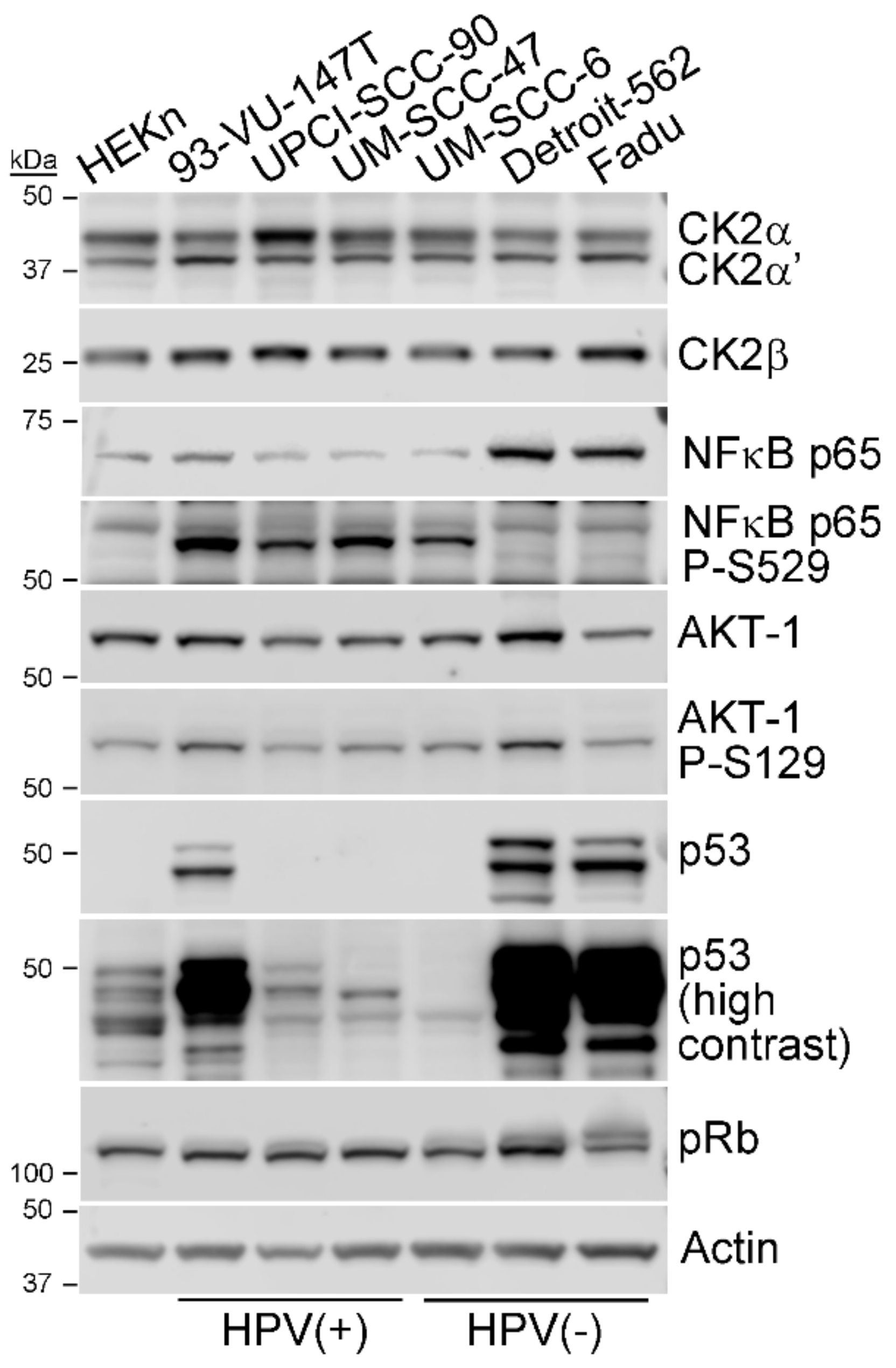
3.1. CK2 Expression and Activity in HPV+ and HPV- HNSCC Cell Lines

We evaluated CK2 levels and surrogate activity in representative HPV(+) and HPV(-) cell lines. In order to evaluate CK2 subunit expression in HNSCC cell lines, we employed 3 HPV+ and 3 HPV- cell lines and a non-transformed human epithelial keratinocyte cell line (Table 1). Under standard growth conditions, CK2 subunit protein expression was examined by immunoblot analysis. The three subunits of CK2 were well expressed in all cell lines (Figure 1), and steady-state expression levels were 1.4–1.9-fold higher in HPV+ cell lines compared to HPV- (Table 2; Table S1). We also investigated markers of CK2 activity; specifically, phosphorylation of the CK2-specific sites on NFκB p65 (S529) and AKT-1 (S129) [30,31]. CK2 phosphorylation of NFκB p65 at S529 was dramatically higher in HPV(+) compared to HPV(−) cell lines (Figure 1, Table 2). In contrast, AKT-1 phosphorylation at the CK2 site was roughly equivalent in both types of HNSCC cells. Expression of p53 was detected in all cell lines, with much higher levels in the cells with mutant p53. Overall levels of pRb were similar in all cell lines, with higher molecular weight species evident in the HPV(−) cell lines.

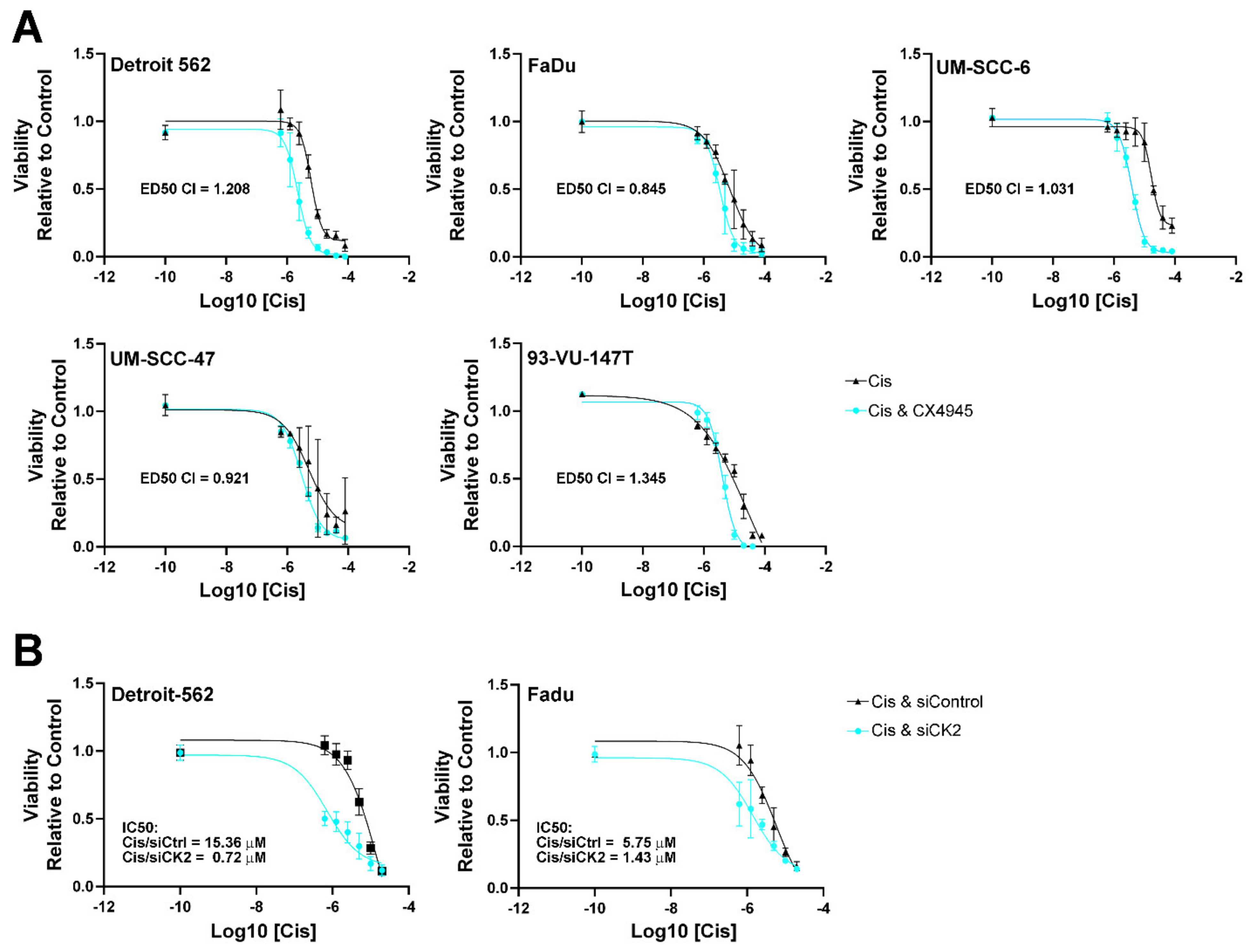
3.2. HNSCC Cell Viability Following Cisplatin Treatment and Reduced CK2 Activity or Expression

Given the widespread clinical use of cisplatin therapy for both HPV(+) and HPV(−) HNSCC cases and the general reliance of HNSCC cells on CK2 activity, we investigated the potential utility of combined CX-4945 treatment with cisplatin treatment [32,33]. Specifically, we evaluated the viability of HNSCC cells after cisplatin, CX-4945, and combined CX-4945 followed by cisplatin treatment. Adding CX-4945 to cisplatin treatment further reduced cell viability over cisplatin alone and significantly decreased the IC50 for cisplatin for all cell lines but UM-SCC-47. The fold decrease with the addition of CX-4945 ranged from 1.7 to 7.9 (Table 3 and Figure 2A). In reverse analysis, when we determined the effect of cisplatin addition to CX-4945, the effect was to slightly reduce the IC50 for CX-4945, from 1.1 to 2.0-fold (Table 3). These results suggest that under the treatment conditions that we used, CX-4945 treatment was the dominant effect reducing cell viability in both HPV(+) and HPV(−) cells. We analyzed this data for synergy and found that CX-4945 and cisplatin combined treatment was synergistic in the HPV(+) cell line UM-SCC-47 and the HPV(−) cell line Fadu (Figure 2A). CX-4945 and cisplatin combined treatment was additive in the HPV(−) cell line SCC-6 (Figure 2A).
We also examined the viability of Detroit-562 and Fadu cells following siRNA-mediated CK2 downregulation and cisplatin treatment. Cells were transfected with a constant amount of either CK2-targeting or control siRNAs and treated 2 days later with cisplatin for an additional 48 h. Similar to the combined effects of CX-4945 with cisplatin, the viability curve was shifted to the left with significantly reduced IC50s for cisplatin when CK2 expression was inhibited by 4 to 21-fold (Figure 2B).

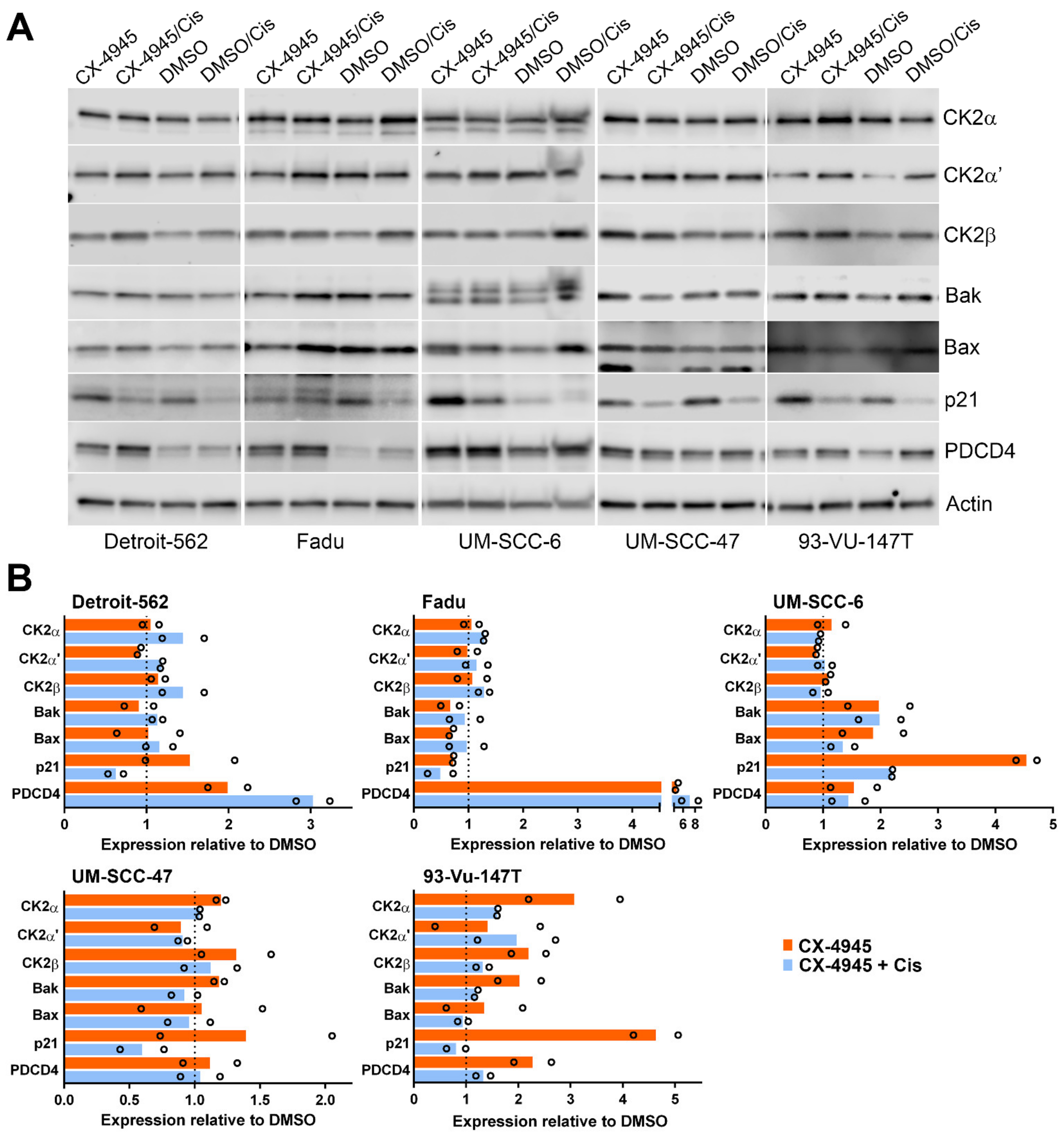
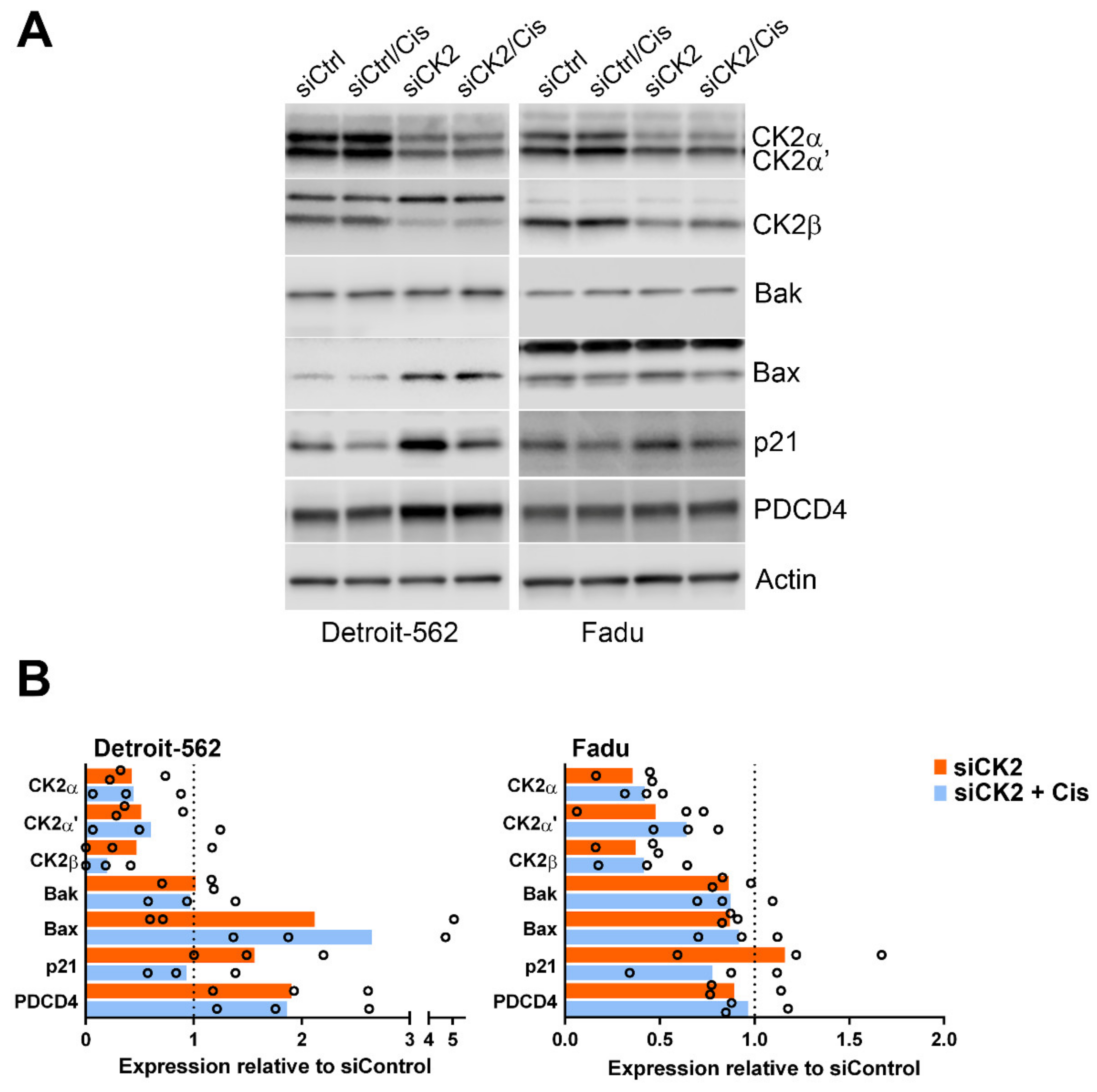
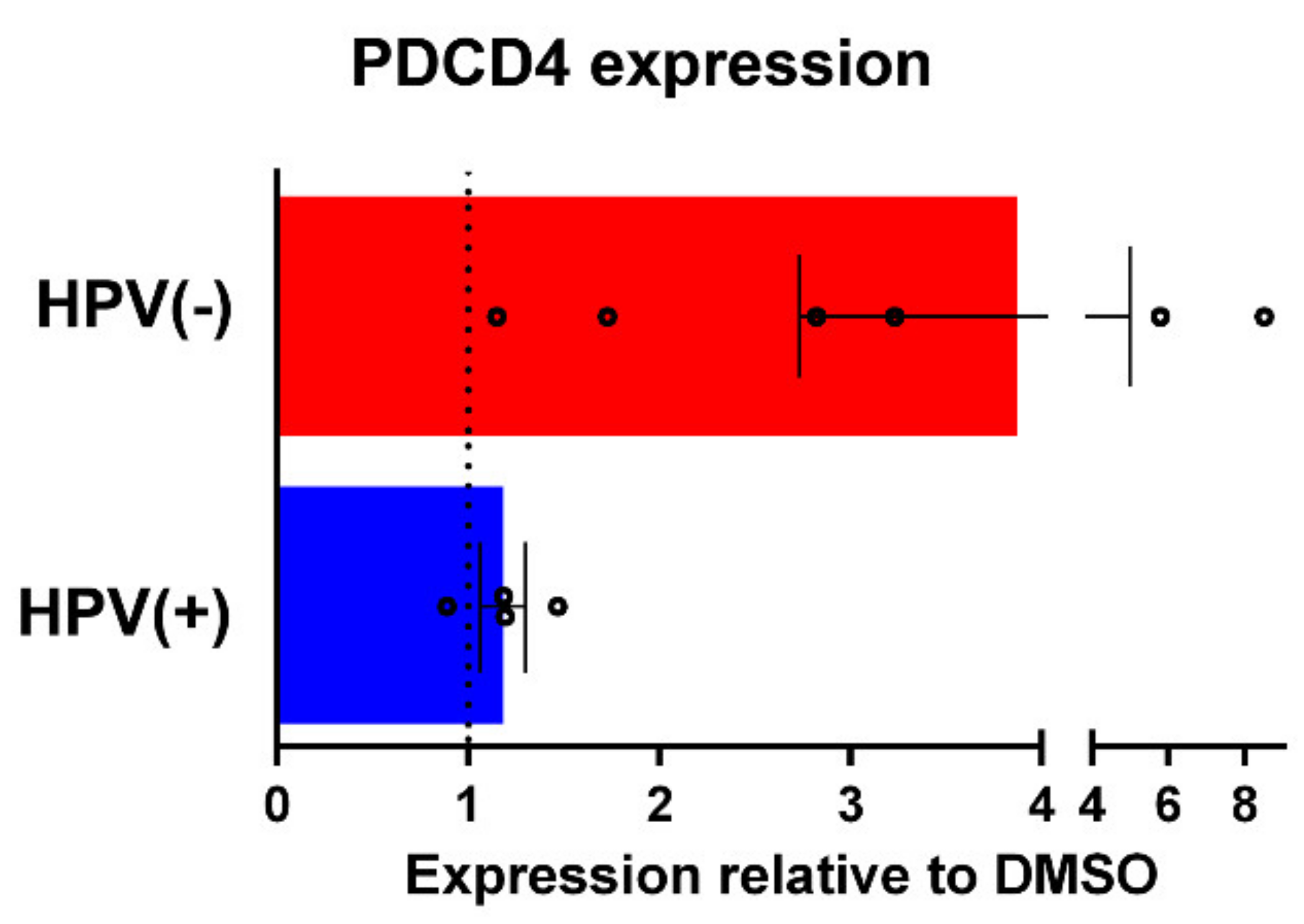
3.3. Signaling Response of HNSCC cells to CK2 Targeting Using CX-4945 or siRNA and Cisplatin Treatment
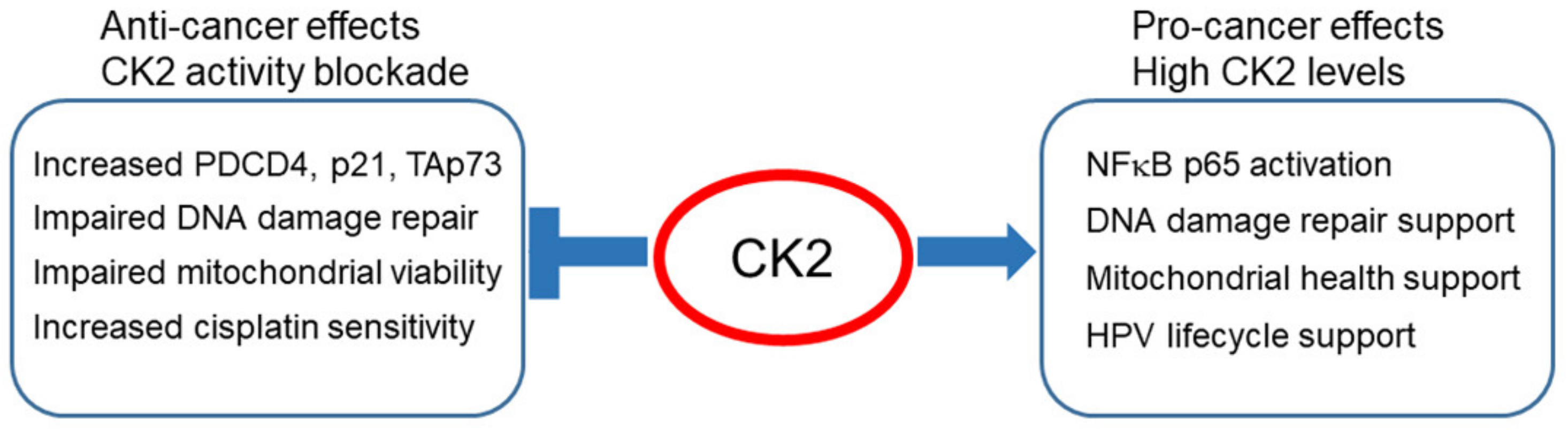
We further examined treatment response to CX-4945 treatment alone and in combination with cisplatin in 3 HPV(−) and 2 HPV(+) cell lines by immunoblot. We examined the cells, using sequential treatment, with CX-4945 treatment for 48 h and cisplatin treatment for 24 h. This relatively short treatment regimen was chosen to examine CX-4945-induced changes in HPV(−) and HPV(+) HNSCC cells prior to the full cascade of death signaling. Overall, CX-4945 treatment alone or combined with cisplatin caused no consistent change in the proapoptotic proteins Bak and Bax; although some induction of Bak and Bax was observed in UM-SCC-6 and 93-Vu-147T cells (Figure 3). CX-4945 caused induction of the cyclin dependent kinase inhibitor p21 from 1.4- to 4.6-fold in four of the cell lines; the presence of cisplatin generally reversed p21 induction in combined treatment. Cisplatin treatment alone caused reduction of p21 below 60% of control cells (Figure 3). Levels of the tumor suppressor programmed cell death 4 protein (PDCD4) were elevated 1.5- to 5-fold following CX-4945 inhibition in four of the five cell lines, with further increase upon cisplatin addition in two of these cell lines. Addition of cisplatin to CX-4945 treatment slightly increased CK2 protein levels in four of the five cell lines.
Detroit-562 and Fadu cells were also transfected with a siRNA cocktail to specifically knockdown CK2α and CK2α’ protein expression. Reduced expression of the CK2 subunits was confirmed (Figure 4). Many of the observations from CX-4945 treatments were replicated for Detroit-562, including induction of p21 and PDCD4 by loss of CK2 (Figure 4). One difference was that Bax levels increased following downregulation of CK2 combined with cisplatin as opposed to CX-4945 plus cisplatin treatment. The response of Fadu cells to siRNA-mediated CK2 loss with and without cisplatin was very similar to that for the use of CX-4945, with the exception that CK2 downregulation did not induce PDCD4. CK2 knockdown in these two cell lines only slightly induced PDCD4 mRNA (Detroit-562: 1.36 ± 0.49; Fadu: 1.09 ± 0.07).
Finally, we grouped the HPV(−) versus HPV(+) cell line immunoblot data together to evaluate any differences between HNSCC cells according to viral status. One striking difference was the large increase in PDCD4 level in HPV(−) cells compared to HPV(+) (Figure 5, p = 0.07) following combined CX-4945 and cisplatin treatment. No other notable differences were observed.

4. Discussion

Given the rise in HPV-related HNSCC, we undertook this examination of CK2 level in relation to HPV status and the effect of CK2 targeting as a cotreatment with cisplatin. We observed higher steady-state CK2 protein levels and kinase activity directed to NFκB p65 in HPV(+) cells relative to HPV(−). It appears that under conditions of HPV infection, CK2 protein levels are elevated by a currently unknown mechanism to meet the demands of viral replication. CK2 regulates HPV proteins such as E1 and E7 involved in the papillomavirus lifecycle. The HPV E7 oncoprotein promotes immortalization and transformation in infected cells through inactivation of pRb and related pathway proteins [34]. Previous studies have demonstrated key regulation of E7 function by CK2 [18,19,20,35]. For example, the phosphorylation of E7 by CK2 is essential to promote Rb-related p130 degradation and cell cycle S-phase entry [36,37]. CK2α is required for HPV DNA replication by regulating the stability and nuclear retention of E1, and CK2 has been proposed as a promising target for the development of antiviral drugs [21,38].
Cervical cancer is almost entirely associated with positive HPV status. In other work, it was shown that targeting of CK2 activity using an investigational peptide inhibitor is effective in treating cervical cancer [39,40]. This CK2 inhibitor, CIGB-300, was identified in a screen of peptides, which bind and block phosphorylation of an HPV16 E7 fusion protein [41]. Combinatorial use of CIGB-300 with cisplatin demonstrated a good synergy and/or additivity profile against a cervical cancer cell line, and improved survival in mouse xenograft studies [42]. These observations are analogous to those described here on the combinatorial treatment of HNSCC with CK2 inhibitor CX-4945 and cisplatin.
Treatment of malignant cells with cisplatin and/or radiation causes nuclear DNA damage and redox stress, mitochondrial DNA damage, and mitochondrial outer membrane permeabilization [43,44,45]. Over time, adaptation to cisplatin in malignant cells results in therapeutic failure and tumor recurrence in patients. We have previously shown in prostate cancer that CK2 inhibition has a negative impact on mitochondrial health through decreased membrane potential and Ca2+ flux [27,46]. CK2 has significant influence on numerous DNA repair and other pathways activated by radiation and cisplatin [27,47,48,49]; loss of CK2 improves sensitivity to cisplatin or radiation in numerous cancers, including head and neck cancer [42,50,51,52,53,54,55,56,57,58,59,60,61,62]. CX-4945 blocks DNA repair after cisplatin or gemcitabine treatment [51,63], and next generation platinum Pt(IV) prodrugs conjugated with CX-4945 have shown efficacy in other cancer types [64]. In a pilot study, metastatic HNSCC lesions were successfully treated in nude mice using a combination of cisplatin and tumor directed nanocapsules containing RNAi oligonucleotides against CK2 [65]. Together, these data suggest that incorporating CK2 blockade could improve or prolong the response to cisplatin therapy by hindering DNA repair and influencing mitochondrial health in both HPV(+) and HPV(−) HNSCC [66].
A key downstream target of CK2 activity in HNSCC is NFκB. The NFκB complex plays broad roles promoting proliferative and inflammatory pathways, and is aberrantly activated in numerous cancers, including HNSCC [65,67]. CK2 modulates IKKβ and IκBα phospho-states and degradation, promotes IKK-mediated phosphorylation of NFκB p65 at S536, and directly activates p65 by phosphorylation at S529 [65]. We have previously shown that CK2 knockdown modulates NFκB activity and sensitizes HPV(−) HNSCC cells to cisplatin [50]. Our data here show a new link between high CK2 levels and CK2-activated NFκB, but not CK2-activated AKT-1, in HPV(+) HNSCC cells. Transcriptome analysis demonstrated that NFκB and death signaling pathways differed according to HPV status [68]. Future studies could further investigate CK2 signaling in HPV(+) HNSCC in relation to NFκB activity and induction of cell death.
Loss of tumor suppressor gene or protein expression is proposed as part of HNSCC oncogenesis [14]. It was previously shown in HNSCC that molecular downregulation or kinase inhibition of CK2 enhanced levels of the tumor suppressor TAp73 and inhibited expression of cancer stem cell genes and side population [50,69]. The tumor suppressor PDCD4 protects cells from neoplastic transformation, exhibits reduced expression levels in malignant compared to non-transformed cells, and functions to inhibit protein translation [70,71,72]. PDCD4 protein expression is regulated by multiple microRNAs in HNSCC, and loss of PDCD4 renders cancer cells more resistant to cisplatin [73,74,75]. PDCD4 interacts with CK2 in the nucleus and is a predicted CK2 substrate [76,77]. Our results showed for the first time that PDCD4 protein levels were strongly induced in HNSCC by CX-4945 treatment, especially in the p53 mutant cell lines Detroit-562, Fadu and 93-Vu-147T. CK2 knockdown using siRNA inexplicably raised PDCD4 in Detroit-562 but not Fadu cells. At present it is unclear what molecular pathways or genetic characteristics influence PDCD4 abundance following interruption of CK2 activity or expression.
CX-4945 treatment or CK2 downregulation also strongly induced tumor suppressor p21 levels, a cyclin dependent kinase inhibitor promoting cell cycle arrest [78]. Induction of p21 was shown to occur when cisplatin-resistant HPV(−) HNSCC cells were resensitized using palbociclib and JQ1 [79]. Here we observed increased p21 protein after CX-4945 treatment in both HPV(+) and HPV(−) HNSCC. Elevation of p21 is due to CK2 knockdown in two of two cell lines and CX-4945 treatment in four of five cell lines. We noted a different CX-4945 related p21 molecular response in the Fadu cell line. The heterogeneity of malignancies observed in patients and the corresponding cancer cell lines has emerged as a fundamental tenet of cancer studies over the past many years. It is not unusual that the HNSCC cell lines we employed in this study occasionally showed different responses to experimental manipulation, and in fact this is to be expected. Thus, we conclude that in most HNSCC cell lines, blocking CK2 activity or expression allows elevation of p21. The recovery of PDCD4 protein levels and the induction of p21 and TAp73 may represent potential mechanisms by which reducing CK2 activity or expression levels improves cisplatin sensitivity in HNSCC cells.
Ongoing clinical trials continue to support the potential efficacy of CX-4945 in different therapeutic contexts in a range of malignancies (clinicaltrials.gov). The small molecule inhibitor CX-4945 is not solely selective for CK2 activity, due to some off-target inhibition of and/or interaction with several other kinases [80,81]. In light of the off-target effects of CX-4945, it is important to note that data from studies using CX-4945 significantly overlap with data generated in vivo and in vitro by knockdown of CK2 gene expression, as shown here and in numerous other cancer types [82,83,84,85,86,87,88,89,90,91,92]. A recent summary of an original research publication states that the development of a new highly selective chemical probe for CK2 “challenges the broad cancer essentiality of CK2” [93]. The chemical probe SGC-CK2-1 did not cause notable loss of proliferation in greater than 90% of more than 140 cancer cell lines, which was a surprising finding [94]. However, there was no discussion of published data on the effects of CK2 knockdown using RNA interference and CRISPR/Cas9 techniques. The strong foundation of evidence that molecular downregulation of CK2 protein levels induces loss of cancer cell viability and tumor cell death should not be discounted. Further research using SGC-CK2-1 will hopefully shed some light on this observed discrepancy.
Current therapy of HNSCC relies on surgery, radiation, and/or chemotherapy, typically cisplatin. A significant number of patients, especially those with HPV(−) disease, suffer poor outcomes after therapy; while patients who experience long-term benefits also develop persistent cisplatin-related toxicity. This study shows significantly reduced IC50 values for cisplatin in multiple HPV(+) and HPV(−) HNSCC cell lines under combined treatment with CX-4945 or after CK2 knockdown, suggesting a path toward a therapy strategy allowing reduced cisplatin usage. HNSCC is the 6th most common cancer in the world, and new treatment modalities that can improve outcomes and reduce toxicities are needed [14,95,96]. We summarized in Figure 6 the results presented here and in other published works related to loss of CK2 activity and potential pathways of achieving improved HNSCC response.

Future Directions and Limitations

Extended work could examine cisplatin treatment as the first drug since the CX-4945 treatment dominated the loss of viability in these studies, as suggested by the data in Table 3. The possible off-target effects of CX-4945 are well documented, and further investigation into the role of CK2 downregulation through the use of other inhibitors and/or CK2 molecular downregulation and would define the contribution of off-target kinases.

5. Conclusions

To our knowledge, this is the first report evaluating CK2 expression level in relation to HPV status in HNSCC cells. Treatment with CX-4945 resulted in significantly decreased IC50 values for combined treatment with cisplatin in both HPV(+) and HPV(−) HNSCC cell lines; synergy was observed in a subset of cell lines. Phase 1 and 2 clinical trials demonstrated that the oral CK2 inhibitor CX-4945 is safe for use in cancer patients, slowing disease progression and extending treatment benefit for some patients with advanced solid tumor cancers. Our results suggest that HNSCC patients could benefit from further investigation into the incorporation of CX-4945 into treatment strategies. In addition, the function of CK2 in HPV biology as it pertains to HNSCC requires further study given the emergence of HPV(+) HNSCC.

Supplementary Materials

The following are available online: https://0-www-mdpi-com.brum.beds.ac.uk/article/10.3390/biomedicines9050571/s1. Table S1, Quantitation of HNSCC cell line immunoblot signals.

Author Contributions

Conceptualization, J.H.T., B.L., B.T.K., K.A., M.A.K., E.C.-G.; methodology, B.L., J.H.T., B.T.K.; statistical analysis, B.L., B.T.K., A.A.G.; writing—original draft preparation, B.L., J.H.T.; writing—review and editing, K.A., M.A.K., E.C.-G., B.T.K., A.A.G.; funding acquisition, B.L., K.A.; project administration, E.C.-G., J.H.T., K.A.; investigation, B.L., J.H.T., B.T.K.; supervision, K.A., M.A.K., J.H.T., E.C.-G.; resources, K.A., B.L., E.C.-G.; visualization, J.H.T., B.L.; validation, J.H.T., A.A.G. All authors have read and agreed to the published version of the manuscript.

Funding

This work was supported by Merit Review research funds I01 BX003282 and I01 BX005091 awarded by the United States Department of Veterans Affairs Biomedical Laboratory Research and Development Service (K.A.); Lion’s award (B.L.).

Institutional Review Board Statement

Not applicable.

Informed Consent Statement

Not applicable.

Data Availability Statement

The data presented in this study are available on request from the corresponding author.

Acknowledgments

K.A. holds the title of Senior Research Career Scientist awarded by the U.S. Department of Veterans Affairs. We thank Daniel P. Shaughnessy for technical assistance.

Conflicts of Interest

The authors declare no conflict of interest.

Disclaimer

The contents do not represent the views of the U.S. Department of Veterans Affairs or the United States government.

References

  1. Silva-Pavez, E.; Tapia, J.C. Protein Kinase CK2 in Cancer Energetics. Front. Oncol. 2020, 10. [Google Scholar] [CrossRef] [PubMed]
  2. D’Amore, C.; Salizzato, V.; Borgo, C.; Cesaro, L.; Pinna, L.A.; Salvi, M. A Journey through the Cytoskeleton with Protein Kinase CK2. Curr. Protein Pept. Sci. 2019, 20, 547–562. [Google Scholar] [CrossRef] [PubMed]
  3. Castello, J.; Ragnauth, A.; Friedman, E.; Rebholz, H. CK2-An Emerging Target for Neurological and Psychiatric Disorders. Pharmaceuticals 2017, 10, 7. [Google Scholar] [CrossRef] [PubMed] [Green Version]
  4. Nuñez de Villavicencio-Diaz, T.; Rabalski, A.J.; Litchfield, D.W. Protein Kinase CK2: Intricate Relationships within Regulatory Cellular Networks. Pharmaceuticals 2017, 10, 27. [Google Scholar] [CrossRef] [PubMed] [Green Version]
  5. Hornbeck, P.V.; Zhang, B.; Murray, B.; Kornhauser, J.M.; Latham, V.; Skrzypek, E. PhosphoSitePlus, 2014: Mutations, PTMs and recalibrations. Nucleic Acids Res. 2015, 43, D512–D520. [Google Scholar] [CrossRef] [Green Version]
  6. Faust, M.; Montenarh, M. Subcellular localization of protein kinase CK2. A key to its function? Cell Tissue Res. 2000, 301, 329–340. [Google Scholar] [CrossRef]
  7. Ahmed, K.; Gerber, D.A.; Cochet, C. Joining the cell survival squad: An emerging role for protein kinase CK2. Trends Cell Biol. 2002, 12, 226–230. [Google Scholar] [CrossRef]
  8. Trembley, J.H.; Wang, G.; Unger, G.; Slaton, J.; Ahmed, K. CK2: A key player in cancer biology. Cell Mol. Life Sci. 2009, 66, 1858–1867. [Google Scholar] [CrossRef] [Green Version]
  9. Chua, M.M.J.; Lee, M.; Dominguez, I. Cancer-type dependent expression of CK2 transcripts. PLoS ONE 2017, 12, e0188854. [Google Scholar] [CrossRef] [PubMed] [Green Version]
  10. Gapany, M.; Faust, R.A.; Tawfic, S.; Davis, A.; Adams, G.L.; Ahmed, K. Association of elevated protein kinase CK2 activity with aggressive behavior of squamous cell carcinoma of the head and neck. Mol. Med. 1995, 1, 659–666. [Google Scholar] [CrossRef] [PubMed] [Green Version]
  11. Faust, R.A.; Gapany, M.; Tristani, P.; Davis, A.; Adams, G.L.; Ahmed, K. Elevated protein kinase CK2 activity in chromatin of head and neck tumors: Association with malignant transformation. Cancer Lett. 1996, 101, 31–35. [Google Scholar] [CrossRef]
  12. Pfister, D.G.; Fury, M.G. New chapter in our understanding of human papillomavirus-related head and neck cancer. J. Clin. Oncol. 2014, 32, 3349–3352. [Google Scholar] [CrossRef] [PubMed] [Green Version]
  13. Fakhry, C.; Zhang, Q.; Nguyen-Tan, P.F.; Rosenthal, D.; El-Naggar, A.; Garden, A.S.; Soulieres, D.; Trotti, A.; Avizonis, V.; Ridge, J.A.; et al. Human Papillomavirus and Overall Survival After Progression of Oropharyngeal Squamous Cell Carcinoma. J. Clin. Oncol. 2014, 32, 3365–3373. [Google Scholar] [CrossRef] [PubMed]
  14. Johnson, D.E.; Burtness, B.; Leemans, C.R.; Lui, V.W.Y.; Bauman, J.E.; Grandis, J.R. Head and neck squamous cell carcinoma. Nat. Rev. Dis Primers 2020, 6, 92. [Google Scholar] [CrossRef]
  15. Ang, K.K.; Harris, J.; Wheeler, R.; Weber, R.; Rosenthal, D.I.; Nguyen-Tan, P.F.; Westra, W.H.; Chung, C.H.; Jordan, R.C.; Lu, C.; et al. Human papillomavirus and survival of patients with oropharyngeal cancer. N. Engl. J. Med. 2010, 363, 24–35. [Google Scholar] [CrossRef] [PubMed] [Green Version]
  16. Gillison, M.L.; Koch, W.M.; Capone, R.B.; Spafford, M.; Westra, W.H.; Wu, L.; Zahurak, M.L.; Daniel, R.W.; Viglione, M.; Symer, D.E.; et al. Evidence for a causal association between human papillomavirus and a subset of head and neck cancers. J. Natl. Cancer Inst. 2000, 92, 709–720. [Google Scholar] [CrossRef]
  17. Borgo, C.; D’Amore, C.; Cesaro, L.; Sarno, S.; Pinna, L.A.; Ruzzene, M.; Salvi, M. How can a traffic light properly work if it is always green? The paradox of CK2 signaling. Crit. Rev. Biochem. Mol. Biol. 2021, 1–39. [Google Scholar] [CrossRef]
  18. Firzlaff, J.M.; Galloway, D.A.; Eisenman, R.N.; Luscher, B. The E7 protein of human papillomavirus type 16 is phosphorylated by casein kinase II. New Biol. 1989, 1, 44–53. [Google Scholar]
  19. Basukala, O.; Mittal, S.; Massimi, P.; Bestagno, M.; Banks, L. The HPV-18 E7 CKII phospho acceptor site is required for maintaining the transformed phenotype of cervical tumour-derived cells. PLoS Pathog. 2019, 15, e1007769. [Google Scholar] [CrossRef]
  20. Zine El Abidine, A.; Tomaić, V.; Bel Haj Rhouma, R.; Massimi, P.; Guizani, I.; Boubaker, S.; Ennaifer, E.; Banks, L. A naturally occurring variant of HPV-16 E7 exerts increased transforming activity through acquisition of an additional phospho-acceptor site. Virology 2017, 500, 218–225. [Google Scholar] [CrossRef] [PubMed]
  21. Piirsoo, A.; Piirsoo, M.; Kala, M.; Sankovski, E.; Lototskaja, E.; Levin, V.; Salvi, M.; Ustav, M. Activity of CK2α protein kinase is required for efficient replication of some HPV types. PLoS Pathog. 2019, 15, e1007788. [Google Scholar] [CrossRef]
  22. Rampias, T.; Sasaki, C.; Weinberger, P.; Psyrri, A. E6 and e7 gene silencing and transformed phenotype of human papillomavirus 16-positive oropharyngeal cancer cells. J. Natl. Cancer Inst. 2009, 101, 412–423. [Google Scholar] [CrossRef] [Green Version]
  23. Wu, S.Y.; Nin, D.S.; Lee, A.Y.; Simanski, S.; Kodadek, T.; Chiang, C.M. BRD4 Phosphorylation Regulates HPV E2-Mediated Viral Transcription, Origin Replication, and Cellular MMP-9 Expression. Cell Rep. 2016, 16, 1733–1748. [Google Scholar] [CrossRef] [PubMed] [Green Version]
  24. Wu, S.Y.; Lee, A.Y.; Lai, H.T.; Zhang, H.; Chiang, C.M. Phospho switch triggers Brd4 chromatin binding and activator recruitment for gene-specific targeting. Mol. Cell 2013, 49, 843–857. [Google Scholar] [CrossRef] [PubMed] [Green Version]
  25. Trembley, J.H.; Unger, G.M.; Korman, V.L.; Tobolt, D.K.; Kazimierczuk, Z.; Pinna, L.A.; Kren, B.T.; Ahmed, K. Nanoencapsulated anti-CK2 small molecule drug or siRNA specifically targets malignant cancer but not benign cells. Cancer Lett. 2012, 315, 48–58. [Google Scholar] [CrossRef] [PubMed] [Green Version]
  26. Ahmed, K.; Unger, G.M.; Kren, B.T.; Trembley, J.H. Targeting CK2 for cancer therapy using a nanomedicine approach. In Protein Kinase CK2 in Cellular Function in Normal and Disease States; Ahmed, K., Issinger, O.-G., Szyszka, R., Dhalla, N.S., Eds.; Advances in Biochemistry in Health and Disease; Springer: Cham, Switzerland, 2015; Volume 12, pp. 299–315. [Google Scholar]
  27. Qaiser, F.; Trembley, J.H.; Kren, B.T.; Wu, J.J.; Naveed, A.K.; Ahmed, K. Protein Kinase CK2 Inhibition Induces Cell Death via Early Impact on Mitochondrial Function. J. Cell Biochem. 2014, 115, 2103–2115. [Google Scholar] [CrossRef] [PubMed] [Green Version]
  28. Trembley, J.H.; Unger, G.M.; Tobolt, D.K.; Korman, V.L.; Wang, G.; Ahmad, K.A.; Slaton, J.W.; Kren, B.T.; Ahmed, K. Systemic administration of antisense oligonucleotides simultaneously targeting CK2alpha and alpha’ subunits reduces orthotopic xenograft prostate tumors in mice. Mol. Cell Biochem. 2011, 356, 21–35. [Google Scholar] [CrossRef] [PubMed] [Green Version]
  29. Chou, T.C. Preclinical versus clinical drug combination studies. Leuk Lymphoma 2008, 49, 2059–2080. [Google Scholar] [CrossRef]
  30. Ponce, D.P.; Maturana, J.L.; Cabello, P.; Yefi, R.; Niechi, I.; Silva, E.; Armisen, R.; Galindo, M.; Antonelli, M.; Tapia, J.C. Phosphorylation of AKT/PKB by CK2 is necessary for the AKT-dependent up-regulation of beta-catenin transcriptional activity. J. Cell. Physiol. 2011, 226, 1953–1959. [Google Scholar] [CrossRef]
  31. Wang, D.; Westerheide, S.D.; Hanson, J.L.; Baldwin, A.S. Tumor necrosis factor α-induced phosphorylation of RelA/p65 on Ser529 is controlled by casein kinase II. J. Biol. Chem. 2000, 275, 32592–32597. [Google Scholar] [CrossRef] [Green Version]
  32. Gillison, M.L.; Trotti, A.M.; Harris, J.; Eisbruch, A.; Harari, P.M.; Adelstein, D.J.; Jordan, R.C.K.; Zhao, W.; Sturgis, E.M.; Burtness, B.; et al. Radiotherapy plus cetuximab or cisplatin in human papillomavirus-positive oropharyngeal cancer (NRG Oncology RTOG 1016): A randomised, multicentre, non-inferiority trial. Lancet 2019, 393, 40–50. [Google Scholar] [CrossRef]
  33. Burtness, B.; Harrington, K.J.; Greil, R.; Soulieres, D.; Tahara, M.; de Castro, G., Jr.; Psyrri, A.; Baste, N.; Neupane, P.; Bratland, A.; et al. Pembrolizumab alone or with chemotherapy versus cetuximab with chemotherapy for recurrent or metastatic squamous cell carcinoma of the head and neck (KEYNOTE-048): A randomised, open-label, phase 3 study. Lancet 2019, 394, 1915–1928. [Google Scholar] [CrossRef]
  34. Eckhardt, M.; Zhang, W.; Gross, A.M.; Von Dollen, J.; Johnson, J.R.; Franks-Skiba, K.E.; Swaney, D.L.; Johnson, T.L.; Jang, G.M.; Shah, P.S.; et al. Multiple Routes to Oncogenesis Are Promoted by the Human Papillomavirus-Host Protein Network. Cancer Discov. 2018, 8, 1474–1489. [Google Scholar] [CrossRef] [Green Version]
  35. Nogueira, M.O.; Hosek, T.; Calcada, E.O.; Castiglia, F.; Massimi, P.; Banks, L.; Felli, I.C.; Pierattelli, R. Monitoring HPV-16 E7 phosphorylation events. Virology 2017, 503, 70–75. [Google Scholar] [CrossRef] [PubMed]
  36. Chien, W.M.; Parker, J.N.; Schmidt-Grimminger, D.C.; Broker, T.R.; Chow, L.T. Casein kinase II phosphorylation of the human papillomavirus-18 E7 protein is critical for promoting S-phase entry. Cell Growth Differ. 2000, 11, 425–435. [Google Scholar]
  37. Genovese, N.J.; Banerjee, N.S.; Broker, T.R.; Chow, L.T. Casein kinase II motif-dependent phosphorylation of human papillomavirus E7 protein promotes p130 degradation and S-phase induction in differentiated human keratinocytes. J. Virol. 2008, 82, 4862–4873. [Google Scholar] [CrossRef] [Green Version]
  38. Bouhaddou, M.; Memon, D.; Meyer, B.; White, K.M.; Rezelj, V.V.; Correa Marrero, M.; Polacco, B.J.; Melnyk, J.E.; Ulferts, S.; Kaake, R.M.; et al. The Global Phosphorylation Landscape of SARS-CoV-2 Infection. Cell 2020, 182, 685–712.e619. [Google Scholar] [CrossRef]
  39. Sarduy, M.R.; Garcia, I.; Coca, M.A.; Perera, A.; Torres, L.A.; Valenzuela, C.M.; Baladron, I.; Solares, M.; Reyes, V.; Hernandez, I.; et al. Optimizing CIGB-300 intralesional delivery in locally advanced cervical cancer. Br. J. Cancer 2015, 112, 1636–1643. [Google Scholar] [CrossRef] [PubMed] [Green Version]
  40. Solares, A.M.; Santana, A.; Baladron, I.; Valenzuela, C.; Gonzalez, C.A.; Diaz, A.; Castillo, D.; Ramos, T.; Gomez, R.; Alonso, D.F.; et al. Safety and preliminary efficacy data of a novel casein kinase 2 (CK2) peptide inhibitor administered intralesionally at four dose levels in patients with cervical malignancies. BMC Cancer 2009, 9, 146. [Google Scholar] [CrossRef] [Green Version]
  41. Perea, S.E.; Reyes, O.; Puchades, Y.; Mendoza, O.; Vispo, N.S.; Torrens, I.; Santos, A.; Silva, R.; Acevedo, B.; Lopez, E.; et al. Antitumor effect of a novel proapoptotic peptide that impairs the phosphorylation by the protein kinase 2 (casein kinase 2). Cancer Res. 2004, 64, 7127–7129. [Google Scholar] [CrossRef] [Green Version]
  42. Perera, Y.; Toro, N.D.; Gorovaya, L.; Fernandez, D.E.C.J.; Farina, H.G.; Perea, S.E. Synergistic interactions of the anti-casein kinase 2 CIGB-300 peptide and chemotherapeutic agents in lung and cervical preclinical cancer models. Mol. Clin. Oncol. 2014, 2, 935–944. [Google Scholar] [CrossRef] [Green Version]
  43. Sancho-Martinez, S.M.; Prieto-Garcia, L.; Prieto, M.; Lopez-Novoa, J.M.; Lopez-Hernandez, F.J. Subcellular targets of cisplatin cytotoxicity: An integrated view. Pharmacol. Ther. 2012, 136, 35–55. [Google Scholar] [CrossRef] [PubMed]
  44. Galluzzi, L.; Vitale, I.; Michels, J.; Brenner, C.; Szabadkai, G.; Harel-Bellan, A.; Castedo, M.; Kroemer, G. Systems biology of cisplatin resistance: Past, present and future. Cell Death Dis. 2014, 5, e1257. [Google Scholar] [CrossRef] [PubMed] [Green Version]
  45. Lee, S.Y.; Jeong, E.K.; Ju, M.K.; Jeon, H.M.; Kim, M.Y.; Kim, C.H.; Park, H.G.; Han, S.I.; Kang, H.S. Induction of metastasis, cancer stem cell phenotype, and oncogenic metabolism in cancer cells by ionizing radiation. Mol. Cancer 2017, 16, 10. [Google Scholar] [CrossRef] [Green Version]
  46. Afzal, M.; Kren, B.T.; Naveed, A.K.; Trembley, J.H.; Ahmed, K. Protein kinase CK2 impact on intracellular calcium homeostasis in prostate cancer. Mol. Cell. Biochem. 2020, 470, 131–143. [Google Scholar] [CrossRef]
  47. Yata, K.; Lloyd, J.; Maslen, S.; Bleuyard, J.Y.; Skehel, M.; Smerdon, S.J.; Esashi, F. Plk1 and CK2 act in concert to regulate Rad51 during DNA double strand break repair. Mol. Cell 2012, 45, 371–383. [Google Scholar] [CrossRef] [PubMed]
  48. Peng, B.; Wang, J.; Hu, Y.; Zhao, H.; Hou, W.; Zhao, H.; Wang, H.; Liao, J.; Xu, X. Modulation of LSD1 phosphorylation by CK2/WIP1 regulates RNF168-dependent 53BP1 recruitment in response to DNA damage. Nucleic Acids Res. 2015, 43, 5936–5947. [Google Scholar] [CrossRef] [PubMed] [Green Version]
  49. Herhaus, L.; Perez-Oliva, A.B.; Cozza, G.; Gourlay, R.; Weidlich, S.; Campbell, D.G.; Pinna, L.A.; Sapkota, G.P. Casein kinase 2 (CK2) phosphorylates the deubiquitylase OTUB1 at Ser16 to trigger its nuclear localization. Sci. Signal. 2015, 8, ra35. [Google Scholar] [CrossRef] [PubMed] [Green Version]
  50. Brown, M.S.; Diallo, O.T.; Hu, M.; Ehsanian, R.; Yang, X.; Arun, P.; Lu, H.; Korman, V.; Unger, G.; Ahmed, K.; et al. CK2 Modulation of NF-κB, TP53, and the Malignant Phenotype in Head and Neck Cancer by Anti-CK2 Oligonucleotides In vitro or In vivo via Sub–50-nm Nanocapsules. Clin. Cancer Res. 2010, 16, 2295–2307. [Google Scholar] [CrossRef] [PubMed] [Green Version]
  51. Siddiqui-Jain, A.; Bliesath, J.; Macalino, D.; Omori, M.; Huser, N.; Streiner, N.; Ho, C.B.; Anderes, K.; Proffitt, C.; O’Brien, S.E.; et al. CK2 inhibitor CX-4945 suppresses DNA repair response triggered by DNA-targeted anticancer drugs and augments efficacy: Mechanistic rationale for drug combination therapy. Mol. Cancer Ther. 2012, 11, 994–1005. [Google Scholar] [CrossRef] [Green Version]
  52. Zhang, F.; Yang, B.; Shi, S.; Jiang, X. RNA interference (RNAi) mediated stable knockdown of protein casein kinase 2-alpha (CK2α) inhibits migration and invasion and enhances cisplatin-induced apoptosis in HEp-2 laryngeal carcinoma cells. Acta Histochem. 2014, 116, 1000–1006. [Google Scholar] [CrossRef]
  53. So, K.S.; Rho, J.K.; Choi, Y.J.; Kim, S.Y.; Choi, C.M.; Chun, Y.J.; Lee, J.C. AKT/mTOR Down-regulation by CX-4945, a CK2 Inhibitor, Promotes Apoptosis in Chemorefractory Non-small Cell Lung Cancer Cells. Anticancer Res. 2015, 35, 1537–1542. [Google Scholar]
  54. Dutta, A.; Eckelmann, B.; Adhikari, S.; Ahmed, K.M.; Sengupta, S.; Pandey, A.; Hegde, P.M.; Tsai, M.-S.; Tainer, J.A.; Weinfeld, M.; et al. Microhomology-mediated end joining is activated in irradiated human cells due to phosphorylation-dependent formation of the XRCC1 repair complex. Nucleic Acids Res. 2016. [Google Scholar] [CrossRef] [Green Version]
  55. Wang, Z.Q.; Johnson, C.L.; Kumar, A.; Molkentine, D.P.; Molkentine, J.M.; Rabin, T.; Mason, K.A.; Milas, L.; Raju, U. Inhibition of P-TEFb by DRB Suppresses SIRT1/CK2alpha Pathway and Enhances Radiosensitivity of Human Cancer Cells. Anticancer Res. 2014, 34, 6981–6989. [Google Scholar]
  56. Olsen, B.B.; Wang, S.Y.; Svenstrup, T.H.; Chen, B.P.; Guerra, B. Protein kinase CK2 localizes to sites of DNA double-strand break regulating the cellular response to DNA damage. BMC Mol. Biol. 2012, 13, 7. [Google Scholar] [CrossRef] [PubMed] [Green Version]
  57. Parsons, J.L.; Dianova, I.I.; Finch, D.; Tait, P.S.; Strom, C.E.; Helleday, T.; Dianov, G.L. XRCC1 phosphorylation by CK2 is required for its stability and efficient DNA repair. DNA Repair 2010. [Google Scholar] [CrossRef]
  58. Kang, H.; Jung, J.W.; Kim, M.K.; Chung, J.H. CK2 Is the Regulator of SIRT1 Substrate-Binding Affinity, Deacetylase Activity and Cellular Response to DNA-Damage. PLoS ONE 2009, 4. [Google Scholar] [CrossRef] [Green Version]
  59. Yamane, K.; Kinsella, T.J. CK2 inhibits apoptosis and changes its cellular localization following ionizing radiation. Cancer Res. 2005, 65, 4362–4367. [Google Scholar] [CrossRef] [PubMed] [Green Version]
  60. Montenarh, M. Protein kinase CK2 in DNA damage and repair. Transl. Cancer Res. 2016, 5, 49–63. [Google Scholar] [CrossRef]
  61. Jin, C.; Song, P.; Pang, J. The CK2 inhibitor CX4945 reverses cisplatin resistance in the A549/DDP human lung adenocarcinoma cell line. Oncol. Lett. 2019, 18, 3845–3856. [Google Scholar] [CrossRef] [PubMed] [Green Version]
  62. Yang, B.; Yao, J.; Li, B.; Shao, G.; Cui, Y. Inhibition of protein kinase CK2 sensitizes non-small cell lung cancer cells to cisplatin via upregulation of PML. Mol. Cell Biochem. 2017, 436, 87–97. [Google Scholar] [CrossRef]
  63. Xu, W.; Chen, Q.; Wang, Q.; Sun, Y.; Wang, S.; Li, A.; Xu, S.; Roe, O.D.; Wang, M.; Zhang, R.; et al. JWA reverses cisplatin resistance via the CK2-XRCC1 pathway in human gastric cancer cells. Cell Death Dis. 2014, 5, e1551. [Google Scholar] [CrossRef] [PubMed] [Green Version]
  64. Chen, F.; Pei, S.; Wang, X.; Zhu, Q.; Gou, S. Emerging JWA-targeted Pt(IV) prodrugs conjugated with CX-4945 to overcome chemo-immune-resistance. Biochem. Biophys. Res. Commun. 2020, 521, 753–761. [Google Scholar] [CrossRef]
  65. Trembley, J.H.; Chen, Z.; Unger, G.; Slaton, J.; Kren, B.T.; van Waes, C.; Ahmed, K. Emergence of protein kinase CK2 as a key target in cancer therapy. BioFactors 2010, 36, 187–195. [Google Scholar] [CrossRef] [Green Version]
  66. Sewell, A.; Brown, B.; Biktasova, A.; Mills, G.B.; Lu, Y.; Tyson, D.R.; Issaeva, N.; Yarbrough, W.G. Reverse-phase protein array profiling of oropharyngeal cancer and significance of PIK3CA mutations in HPV-associated head and neck cancer. Clin. Cancer Res. 2014, 20, 2300–2311. [Google Scholar] [CrossRef] [PubMed] [Green Version]
  67. Dominguez, I.; Sonenshein, G.E.; Seldin, D.C. Protein kinase CK2 in health and disease: CK2 and its role in Wnt and NF-kappaB signaling: Linking development and cancer. Cell. Mol. Life Sci. 2009, 66, 1850–1857. [Google Scholar] [CrossRef]
  68. Yang, X.; Cheng, H.; Chen, J.; Wang, R.; Saleh, A.; Si, H.; Lee, S.; Guven-Maiorov, E.; Keskin, O.; Gursoy, A.; et al. Head and Neck Cancers Promote an Inflammatory Transcriptome through Coactivation of Classic and Alternative NF-κB Pathways. Cancer Immunol. Res. 2019, 7, 1760–1774. [Google Scholar] [CrossRef] [PubMed]
  69. Lu, H.; Yan, C.; Quan, X.X.; Yang, X.; Zhang, J.; Bian, Y.; Chen, Z.; van Waes, C. CK2 Phosphorylates and Inhibits TAp73 Tumor Suppressor Function to Promote Expression of Cancer Stem Cell Genes and Phenotype in Head and Neck Cancer. Neoplasia 2014, 16, 789–800. [Google Scholar] [CrossRef] [PubMed] [Green Version]
  70. Cmarik, J.L.; Min, H.; Hegamyer, G.; Zhan, S.; Kulesz-Martin, M.; Yoshinaga, H.; Matsuhashi, S.; Colburn, N.H. Differentially expressed protein Pdcd4 inhibits tumor promoter-induced neoplastic transformation. Proc. Natl. Acad. Sci. USA 1999, 96, 14037–14042. [Google Scholar] [CrossRef] [Green Version]
  71. Wang, Q.; Yang, H.S. The role of Pdcd4 in tumour suppression and protein translation. Biol. Cell 2018. [Google Scholar] [CrossRef]
  72. Matsuhashi, S.; Manirujjaman, M.; Hamajima, H.; Ozaki, I. Control Mechanisms of the Tumor Suppressor PDCD4: Expression and Functions. Int. J. Mol. Sci. 2019, 20, 2304. [Google Scholar] [CrossRef] [Green Version]
  73. Zhang, X.; Gee, H.; Rose, B.; Lee, C.S.; Clark, J.; Elliott, M.; Gamble, J.R.; Cairns, M.J.; Harris, A.; Khoury, S.; et al. Regulation of the tumour suppressor PDCD4 by miR-499 and miR-21 in oropharyngeal cancers. BMC Cancer 2016, 16, 86. [Google Scholar] [CrossRef]
  74. Sun, Z.; Li, S.; Kaufmann, A.M.; Albers, A.E. miR-21 increases the programmed cell death 4 gene-regulated cell proliferation in head and neck squamous carcinoma cell lines. Oncol. Rep. 2014, 32, 2283–2289. [Google Scholar] [CrossRef] [PubMed]
  75. Ren, W.; Wang, X.; Gao, L.; Li, S.; Yan, X.; Zhang, J.; Huang, C.; Zhang, Y.; Zhi, K. MiR-21 modulates chemosensitivity of tongue squamous cell carcinoma cells to cisplatin by targeting PDCD4. Mol. Cell Biochem. 2014, 390, 253–262. [Google Scholar] [CrossRef] [PubMed]
  76. Bian, Y.; Ye, M.; Wang, C.; Cheng, K.; Song, C.; Dong, M.; Pan, Y.; Qin, H.; Zou, H. Global screening of CK2 kinase substrates by an integrated phosphoproteomics workflow. Sci. Rep. 2013, 3, 3460. [Google Scholar] [CrossRef] [PubMed] [Green Version]
  77. Homma, M.K.; Shibata, T.; Suzuki, T.; Ogura, M.; Kozuka-Hata, H.; Oyama, M.; Homma, Y. Role for Protein Kinase CK2 on Cell Proliferation: Assessing CK2 Complex Components in the Nucleus During the Cell Cycle Progression. In Protein Kinase CK2 Cellular Function in Normal and Disease States; Ahmed, K., Issinger, O.-G., Szyszka, R., Eds.; Springer International Publishing: Cham, Switzerland, 2015; pp. 197–226. [Google Scholar]
  78. Parveen, A.; Akash, M.S.; Rehman, K.; Kyunn, W.W. Dual Role of p21 in the Progression of Cancer and Its Treatment. Crit. Rev. Eukaryot Gene Expr. 2016, 26, 49–62. [Google Scholar] [CrossRef]
  79. Robinson, A.M.; Rathore, R.; Redlich, N.J.; Adkins, D.R.; VanArsdale, T.; van Tine, B.A.; Michel, L.S. Cisplatin exposure causes c-Myc-dependent resistance to CDK4/6 inhibition in HPV-negative head and neck squamous cell carcinoma. Cell Death Dis. 2019, 10, 867. [Google Scholar] [CrossRef]
  80. Siddiqui-Jain, A.; Drygin, D.; Streiner, N.; Chua, P.; Pierre, F.; O’Brien, S.E.; Bliesath, J.; Omori, M.; Huser, N.; Ho, C.; et al. CX-4945, an orally bioavailable selective inhibitor of protein kinase CK2, inhibits prosurvival and angiogenic signaling and exhibits antitumor efficacy. Cancer Res. 2010, 70, 10288–10298. [Google Scholar] [CrossRef] [Green Version]
  81. Pierre, F.; Chua, P.C.; O’Brien, S.E.; Siddiqui-Jain, A.; Bourbon, P.; Haddach, M.; Michaux, J.; Nagasawa, J.; Schwaebe, M.K.; Stefan, E.; et al. Discovery and SAR of 5-(3-chlorophenylamino)benzo[c][2,6]naphthyridine-8-carboxylic acid (CX-4945), the first clinical stage inhibitor of protein kinase CK2 for the treatment of cancer. J. Med. Chem. 2011, 54, 635–654. [Google Scholar] [CrossRef]
  82. Ahmed, K.; Kren, B.T.; Abedin, M.J.; Vogel, R.I.; Shaughnessy, D.P.; Nacusi, L.; Korman, V.L.; Li, Y.; Dehm, S.M.; Zimmerman, C.L.; et al. CK2 targeted RNAi therapeutic delivered via malignant cell-directed tenfibgen nanocapsule: Dose and molecular mechanisms of response in xenograft prostate tumors. Oncotarget 2016, 7, 61789–61805. [Google Scholar] [CrossRef] [PubMed] [Green Version]
  83. Cannon, C.M.; Trembley, J.H.; Kren, B.T.; Unger, G.M.; O’Sullivan, M.G.; Cornax, I.; Modiano, J.F.; Ahmed, K. Therapeutic Targeting of Protein Kinase CK2 Gene Expression in Feline Oral Squamous Cell Carcinoma: A Naturally Occurring Large-Animal Model of Head and Neck Cancer. Hum. Gene Ther. Clin. Dev. 2017, 28, 80–86. [Google Scholar] [CrossRef] [PubMed]
  84. Kren, B.; Unger, G.; Abedin, M.; Vogel, R.; Henzler, C.; Ahmed, K.; Trembley, J. Preclinical evaluation of cyclin dependent kinase 11 and casein kinase 2 survival kinases as RNA interference targets for triple negative breast cancer therapy. Breast Cancer Res. 2015, 17, 19. [Google Scholar] [CrossRef] [PubMed] [Green Version]
  85. Unger, G.M.; Kren, B.T.; Korman, V.L.; Kimbrough, T.G.; Vogel, R.I.; Ondrey, F.G.; Trembley, J.H.; Ahmed, K. Mechanism and efficacy of sub-50-nm tenfibgen nanocapsules for cancer cell-directed delivery of anti-CK2 RNAi to primary and metastatic squamous cell carcinoma. Mol. Cancer Ther. 2014, 13, 2018–2029. [Google Scholar] [CrossRef] [Green Version]
  86. Takahashi, K.; Setoguchi, T.; Tsuru, A.; Saitoh, Y.; Nagano, S.; Ishidou, Y.; Maeda, S.; Furukawa, T.; Komiya, S. Inhibition of casein kinase 2 prevents growth of human osteosarcoma. Oncol. Rep. 2017, 37, 1141–1147. [Google Scholar] [CrossRef] [PubMed]
  87. Olsen, B.B.; Svenstrup, T.H.; Guerra, B. Downregulation of protein kinase CK2 induces autophagic cell death through modulation of the mTOR and MAPK signaling pathways in human glioblastoma cells. Int. J. Oncol. 2012, 41, 1967–1976. [Google Scholar] [CrossRef] [Green Version]
  88. Di Maira, G.; Gentilini, A.; Pastore, M.; Caligiuri, A.; Piombanti, B.; Raggi, C.; Rovida, E.; Lewinska, M.; Andersen, J.B.; Borgo, C.; et al. The protein kinase CK2 contributes to the malignant phenotype of cholangiocarcinoma cells. Oncogenesis 2019, 8, 61. [Google Scholar] [CrossRef] [Green Version]
  89. Kim, S.; Ham, S.; Yang, K.; Kim, K. Protein kinase CK2 activation is required for transforming growth factor β-induced epithelial-mesenchymal transition. Mol. Oncol. 2018, 12, 1811–1826. [Google Scholar] [CrossRef] [Green Version]
  90. Sass, G.; Klinger, N.; Sirma, H.; Hashemolhosseini, S.; Hellerbrand, C.; Neureiter, D.; Wege, H.; Ocker, M.; Tiegs, G. Inhibition of experimental HCC growth in mice by use of the kinase inhibitor DMAT. Int. J. Oncol 2011, 39, 433–442. [Google Scholar] [CrossRef]
  91. Wu, D.; Sui, C.; Meng, F.; Tian, X.; Fu, L.; Li, Y.; Qi, X.; Cui, H.; Liu, Y.; Jiang, Y. Stable knockdown of protein kinase CK2-alpha (CK2α) inhibits migration and invasion and induces inactivation of hedgehog signaling pathway in hepatocellular carcinoma Hep G2 cells. Acta Histochem. 2014, 116, 1501–1508. [Google Scholar] [CrossRef]
  92. Zhang, H.X.; Jiang, S.S.; Zhang, X.F.; Zhou, Z.Q.; Pan, Q.Z.; Chen, C.L.; Zhao, J.J.; Tang, Y.; Xia, J.C.; Weng, D.S. Protein kinase CK2α catalytic subunit is overexpressed and serves as an unfavorable prognostic marker in primary hepatocellular carcinoma. Oncotarget 2015, 6, 34800–34817. [Google Scholar] [CrossRef] [Green Version]
  93. Licciardello, M.P.; Workman, P. A New Chemical Probe Challenges the Broad Cancer Essentiality of CK2. Trends Pharmacol. Sci. 2021. [Google Scholar] [CrossRef] [PubMed]
  94. Wells, C.I.; Drewry, D.H.; Pickett, J.E.; Tjaden, A.; Krämer, A.; Müller, S.; Gyenis, L.; Menyhart, D.; Litchfield, D.W.; Knapp, S.; et al. Development of a potent and selective chemical probe for the pleiotropic kinase CK2. Cell Chem. Biol. 2021, 28, 546–558.e10. [Google Scholar] [CrossRef] [PubMed]
  95. Bray, F.; Ferlay, J.; Soerjomataram, I.; Siegel, R.L.; Torre, L.A.; Jemal, A. Global cancer statistics 2018: GLOBOCAN estimates of incidence and mortality worldwide for 36 cancers in 185 countries. CA Cancer J. Clin. 2018, 68, 394–424. [Google Scholar] [CrossRef] [PubMed] [Green Version]
  96. Ferlay, J.; Colombet, M.; Soerjomataram, I.; Mathers, C.; Parkin, D.M.; Piñeros, M.; Znaor, A.; Bray, F. Estimating the global cancer incidence and mortality in 2018: GLOBOCAN sources and methods. Int. J. Cancer 2019, 144, 1941–1953. [Google Scholar] [CrossRef] [PubMed] [Green Version]
Figure 1. Expression of CK2 subunits and key markers in untransformed cells and HNSCC cell lines. Immunoblot analysis of cultured cell lines, as indicated above the blots. CK2α and CK2α’ antibodies were combined for simultaneous detection of these 2 proteins. Proteins detected are indicated on the right side of the blots. Molecular mass markers are indicated on the left side of the blots. Actin signal was used as the loading control.
Biomedicines 09 00571 g001
Figure 2. Viability curves for cisplatin treatment alone or combined with reduced CK2 activity or expression in HPV(+) and HPV(−) HNSCC. Cells were treated and viability measured using MTT-related assays as described in Materials and Methods. Log10 cisplatin dosing is indicated on the X-axis and viability relative to control is indicated on the Y-axis. (A) Cisplatin anchored analysis viability curves for cisplatin alone or combined with CX-4945. Cisplatin treatment alone is indicated by black triangles, and combined CX-4945 plus cisplatin treatment is indicated by blue circles. N = 4. IC50 values are shown in Table 3. Combination Index for 50% loss of viability is indicated on each curve (ED50 CI). (B) Viability curves for cisplatin treatment in siCK2 or siControl transfected cells. Cisplatin/siControl treatment is indicated by black squares (Detroit-562) and black triangles (Fadu), and cisplatin/siCK2 treatment is indicated by blue circles. N = 4. IC50 values are indicated on each curve. 95% CI for Detroit-562: Cis + siControl (8.99, 31.22 µM), Cis + siCK2 (0.43, 1.2 µM). 95% CI for FaDu: Cis + siControl (3.47, 10.37), Cis + siCK2 (0.77, 2.79 µM).
Biomedicines 09 00571 g002
Figure 3. Immunoblot analysis of various signals following CX-4945 treatment alone or combined with cisplatin in HNSCC. Cells were treated and protein expression measured using immunoblot assays as described under Materials and Methods. The drug concentrations (µM) for CX-4945/cisplatin for each cell line were as follows: Detroit-562 1.5/5; Fadu 2.5/5; all others 5/5. (A) Representative blots from immunoblot analysis of HNSCC cells following CX-4945 treatment (48 h) with and without cisplatin (24 h). Proteins detected are indicated on the right side of the blots. Actin signal was used as the loading control. (B) Charts representing quantitation of protein signals relative to DMSO control treatment. Orange = CX-4945 treatment alone. Blue = CX-4945 and cisplatin treatment combined. Black open circles represent each data point from 2 biological replicate immunoblots.
Biomedicines 09 00571 g003
Figure 4. Immunoblot analysis of various signals following CK2 downregulation alone or combined with cisplatin in HNSCC. Cells were treated and protein expression measured using immunoblot assays as described under Materials and Methods. (A) Immunoblot analysis of Detroit-562 and Fadu cells following siRNA transfection (48 h) with and without cisplatin (24 h) carried out as described under Materials and Methods. CK2α and CK2α’ antibodies were combined for simultaneous detection of these 2 proteins. Proteins detected are indicated on the right side of the blots. Actin signal was used as the loading control. (B) Charts representing quantitation of protein signals relative to si-Control treatment. Orange = siCK2. Blue = siCK2 and cisplatin treatment combined. Black open circles represent each data point from 3 biological replicate experiments. siCtrl = siRNA for non-targeting control.
Biomedicines 09 00571 g004
Figure 5. Comparison of PDCD4 induction following combined CX-4945 and cisplatin treatment in HPV(−) vs. HPV(+) HNSCC cells. The mean and SEM of PDCD4 immunoblot signals relative to DMSO control treatment is depicted. Red = HPV(−). Blue = HPV(+). Circles represent each data point.
Biomedicines 09 00571 g005
Figure 6. Impact of CK2 level and activity in HNSCC. A summary of results presented here and previously published from this group and others is depicted. Increased cisplatin sensitivity following CX-4945 or CK2 downregulation is observed in both HPV(+) and HPV(−) HNSCC cells.
Biomedicines 09 00571 g006
Table 1. Characteristics of head and neck cell lines.
Cell LinesTissue OriginAge (yr)SexHPV StatusCDKN2A Status 1TP53 Status 1
HEKn 2Foreskin<1Male-ND 3ND 3
Detroit 562Pharynx (metastatic pleural effusion)ND 3Female-Homozygous mutantHomozygous mutant
FaduHypopharynx56Male-Homozygous mutantHeterozygous mutant, both alleles
UM-SCC-6Base of tongue32Male-Homozygous deletionWild-type
UM-SCC-47Lateral tongue53Male+Wild-typeWild-type
UPCI-SCC-90Base of tongue46Male+Wild-typeWild-type
VU-SCC-147T
(93-Vu-147T)
Floor of mouth58Male+Wild-typeMutant
1 As determined from: ATCC.org; Expasy.org; broadinstitute.org/ccle; cancer.sanger.ac.uk/cosmic. 2 Primary human epidermal keratinocytes from neonatal foreskin. 3 ND—not determined or known.
Table 2. Comparison of CK2 abundance and CK2-related activity immunoblot signals between HPV(+) and HPV(−).
StatusCK2αCK2α’CK2βNFκB p65 totalNFκB p65 P-S529p65 P-S529/TotalAKT-1 TotalAKT-1 P-S129AKT-1 P-S129/Total
HPV+1.00 ± 0.490.62 ± 0.142.92 ± 0.860.02 ± 0.012.18 ± 0.59145.97 ± 44.790.56 ± 0.041.12 ±0.232.02 ± 0.31
HPV-0.55 ± 0.100.423 ± 0.021.96 ± 0.230.03 ± 0.020.33 ± 0.5135.52 ± 67.140.51 ± 0.191.10 ± 0.562.07 ± 0.29
HPV+/HPV-1.821.4501.490.486.653.691.081.020.97
Mean signal, expressed relative to actin, for 3 each HPV(+) and HPV(−) cell lines ± standard deviation.
Table 3. IC50 calculations from single and combined treatments.
IC50 (µM)IC50 (µM)
Cisplatin Anchored AnalysisCX-4945 Anchored Analysis
Cell LineCisplatinCisplatin with CX-4945CX-4945CX-4945 with Cisplatin
Detroit 5628.032.421.921.46
(5.49, 11.97)(1.72, 3.46)(1.51, 2.45)(1.03, 2.09)
Fadu7.353.414.362.13
(5.06, 10.82)(2.51, 4.67)(2.88, 6.78)(1.54, 2.98)
UM-SCC-631.373.955.584.94
(18.99, 55.56)(3.00, 5.24)(4.24, 7.40)(3.75, 6.56)
UM-SCC-475.122.944.773.67
(2.20, 12.19)(2.39, 3.63)(3.82, 5.99)(2.98, 4.53)
93-Vu-147T9.524.055.75.07
(6.37, 14.23)(3.18, 5.19)(4.43, 7.37)(3.98, 6.48)
N = 4. 95% confidence intervals in parentheses.
Publisher’s Note: MDPI stays neutral with regard to jurisdictional claims in published maps and institutional affiliations.

