Next Issue
Volume 9, July
Previous Issue
Volume 9, May


Biomedicines, Volume 9, Issue 6 (June 2021) – 118 articles

Cover Story (view full-size image): 
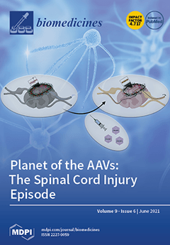
Spinal cord injury (SCI) is associated with pathological cascades leading to regeneration failure, and there is no adequate treatment for it. During the last few years, adeno-associated virus (AAV)-mediated gene therapies have received clinical interest for treating a large range of neurodegenerative and neuromuscular diseases. Currently, there are no clinical trials using AAV vectors in SCI treatment. In SCI preclinical studies using gene therapies, AAV viral vectors are used to break down scar tissue and enable new growth and reconnections between the spinal cord and muscles/spinal cord and brain, which control motor functions and sensation. View this paper
  • Issues are regarded as officially published after their release is announced to the table of contents alert mailing list.
  • You may sign up for e-mail alerts to receive table of contents of newly released issues.
  • PDF is the official format for papers published in both, html and pdf forms. To view the papers in pdf format, click on the "PDF Full-text" link, and use the free Adobe Reader to open them.
Order results
Result details
Section
Select all
Export citation of selected articles as:
17 pages, 4873 KiB  
Article
Exercise Training Enhances Myocardial Mitophagy and Improves Cardiac Function via Irisin/FNDC5-PINK1/Parkin Pathway in MI Mice
by Hangzhuo Li, Shuguang Qin, Qiaoqin Liang, Yue Xi, Wenyan Bo, Mengxin Cai and Zhenjun Tian
Biomedicines 2021, 9(6), 701; https://0-doi-org.brum.beds.ac.uk/10.3390/biomedicines9060701 - 21 Jun 2021
Cited by 23 | Viewed by 3631
Abstract
Myocardial infarction is the major cause of death in cardiovascular disease. In vitro and in vivo models are used to find the exercise mode which has the most significant effect on myocardial irisin/FNDC5 expression and illuminate the cardioprotective role and mechanisms of exercise-activated [...] Read more.
Myocardial infarction is the major cause of death in cardiovascular disease. In vitro and in vivo models are used to find the exercise mode which has the most significant effect on myocardial irisin/FNDC5 expression and illuminate the cardioprotective role and mechanisms of exercise-activated myocardial irisin/FNDC5-PINK1/Parkin-mediated mitophagy in myocardial infarction. The results indicated that expression of irisin/FNDC5 in myocardium could be up-regulated by different types of exercise and skeletal muscle electrical stimulation, which then promotes mitophagy and improves cardiac function and the effect of resistance exercise. Resistance exercise can improve cardiac function by activating the irisin/FNDC5-PINK1/Parkin-LC3/P62 pathway, regulating mitophagy and inhibiting oxidative stress. OPA1 may play an important role in the improvement of cardiac function and mitophagy pathway in myocardial infarction mice by irisin-mediated resistance exercise. Resistance exercise is expected to become an effective therapeutic way to promote myocardial infarction rehabilitation. Full article
(This article belongs to the Section Molecular and Translational Medicine)
Show Figures

Graphical abstract

16 pages, 2089 KiB  
Article
Optimization of Heparin Monitoring with Anti-FXa Assays and the Impact of Dextran Sulfate for Measuring All Drug Activity
by Jean Amiral, Cédric Amiral and Claire Dunois
Biomedicines 2021, 9(6), 700; https://0-doi-org.brum.beds.ac.uk/10.3390/biomedicines9060700 - 21 Jun 2021
Cited by 6 | Viewed by 2897
Abstract
Heparins, unfractionated or low molecular weight, are permanently in the spotlight of both clinical indications and laboratory monitoring. An accurate drug dosage is necessary for an efficient and safe therapy. The one-stage kinetic anti-FXa assays are the most widely and universally used with [...] Read more.
Heparins, unfractionated or low molecular weight, are permanently in the spotlight of both clinical indications and laboratory monitoring. An accurate drug dosage is necessary for an efficient and safe therapy. The one-stage kinetic anti-FXa assays are the most widely and universally used with full automation for large series, without needing exogenous antithrombin. The WHO International Standards are available for UFH and LMWH, but external quality assessment surveys still report a high inter-assay variability. This heterogeneity results from the following: assay formulation, designed without or with dextran sulfate to measure all heparin in blood circulation; calibrators for testing UFH or LMWH with the same curve; and automation parameters. In this study, various factors which impact heparin measurements are reviewed, and we share our experience to optimize assays for testing all heparin anticoagulant activities in plasma. Evidence is provided on the usefulness of low molecular weight dextran sulfate to completely mobilize all of the drug present in blood circulation. Other key factors concern the adjustment of assay conditions to obtain fully superimposable calibration curves for UFH and LMWH, calibrators’ formulations, and automation parameters. In this study, we illustrate the performances of different anti-FXa assays used for testing heparin on UFH or LMWH treated patients’ plasmas and obtained using citrate or CTAD anticoagulants. Comparable results are obtained only when the CTAD anticoagulant is used. Using citrate as an anticoagulant, UFH is underestimated in the absence of dextran sulfate. Heparin calibrators, adjustment of automation parameters, and data treatment contribute to other smaller differences. Full article
(This article belongs to the Special Issue Monitoring Anticoagulant)
Show Figures

Figure 1

9 pages, 532 KiB  
Review
Endothelial Dysfunction and Cardiovascular Disease: History and Analysis of the Clinical Utility of the Relationship
by Peter J. Little, Christopher D. Askew, Suowen Xu and Danielle Kamato
Biomedicines 2021, 9(6), 699; https://0-doi-org.brum.beds.ac.uk/10.3390/biomedicines9060699 - 20 Jun 2021
Cited by 36 | Viewed by 3640
Abstract
The endothelium is the single-cell monolayer that lines the entire vasculature. The endothelium has a barrier function to separate blood from organs and tissues but also has an increasingly appreciated role in anti-coagulation, vascular senescence, endocrine secretion, suppression of inflammation and beyond. In [...] Read more.
The endothelium is the single-cell monolayer that lines the entire vasculature. The endothelium has a barrier function to separate blood from organs and tissues but also has an increasingly appreciated role in anti-coagulation, vascular senescence, endocrine secretion, suppression of inflammation and beyond. In modern times, endothelial cells have been identified as the source of major endocrine and vaso-regulatory factors principally the dissolved lipophilic vosodilating gas, nitric oxide and the potent vascular constricting G protein receptor agonists, the peptide endothelin. The role of the endothelium can be conveniently conceptualized. Continued investigations of the mechanism of endothelial dysfunction will lead to novel therapies for cardiovascular disease. In this review, we discuss the impact of endothelial dysfunction on cardiovascular disease and assess the clinical relevance of endothelial dysfunction. Full article
Show Figures

Figure 1

16 pages, 2392 KiB  
Article
A Metabolic Change towards Fermentation Drives Cancer Cachexia in Myotubes
by Michele Mannelli, Tania Gamberi, Francesca Magherini and Tania Fiaschi
Biomedicines 2021, 9(6), 698; https://0-doi-org.brum.beds.ac.uk/10.3390/biomedicines9060698 - 20 Jun 2021
Cited by 8 | Viewed by 3237
Abstract
Cachexia is a disorder associated with several pathologies, including cancer. In this paper, we describe how cachexia is induced in myotubes by a metabolic shift towards fermentation, and the block of this metabolic modification prevents the onset of the cachectic phenotype. Cachectic myotubes, [...] Read more.
Cachexia is a disorder associated with several pathologies, including cancer. In this paper, we describe how cachexia is induced in myotubes by a metabolic shift towards fermentation, and the block of this metabolic modification prevents the onset of the cachectic phenotype. Cachectic myotubes, obtained by the treatment with conditioned medium from murine colon carcinoma cells CT26, show increased glucose uptake, decreased oxygen consumption, altered mitochondria, and increased lactate production. Interestingly, the block of glycolysis by 2-deoxy-glucose or lactate dehydrogenase inhibition by oxamate prevents the induction of cachexia, thus suggesting that this metabolic change is greatly involved in cachexia activation. The treatment with 2-deoxy-glucose or oxamate induces positive effects also in mitochondria, where mitochondrial membrane potential and pyruvate dehydrogenase activity became similar to control myotubes. Moreover, in myotubes treated with interleukin-6, cachectic phenotype is associated with a fermentative metabolism, and the inhibition of lactate dehydrogenase by oxamate prevents cachectic features. The same results have been achieved by treating myotubes with conditioned media from human colon HCT116 and human pancreatic MIAPaCa-2 cancer cell lines, thus showing that what has been observed with murine-conditioned media is a wide phenomenon. These findings demonstrate that cachexia induction in myotubes is linked with a metabolic shift towards fermentation, and inhibition of lactate formation impedes cachexia and highlights lactate dehydrogenase as a possible new tool for counteracting the onset of this pathology. Full article
(This article belongs to the Section Cancer Biology and Oncology)
Show Figures

Figure 1

14 pages, 1604 KiB  
Article
Syndecan-1 Expression Is Increased in the Aortic Wall of Patients with Type 2 Diabetes but Is Unrelated to Elevated Fasting Plasma Glucagon-Like Peptide-1
by Stelia Ntika, Linda M. Tracy, Anders Franco-Cereceda, Hanna M. Björck and Camilla Krizhanovskii
Biomedicines 2021, 9(6), 697; https://0-doi-org.brum.beds.ac.uk/10.3390/biomedicines9060697 - 20 Jun 2021
Cited by 4 | Viewed by 1982
Abstract
A reduced prevalence of a thoracic aortic aneurysm (thoracic AA) is observed in type 2 diabetes (T2D). Glucagon-like peptide-1 (GLP-1)/GLP-1-based anti-diabetic therapy has indicated protective effects in thoracic AA and regulates the processes controlling the vascular tissue expression of Syndecan-1 (Sdc-1). Sdc-1 expression [...] Read more.
A reduced prevalence of a thoracic aortic aneurysm (thoracic AA) is observed in type 2 diabetes (T2D). Glucagon-like peptide-1 (GLP-1)/GLP-1-based anti-diabetic therapy has indicated protective effects in thoracic AA and regulates the processes controlling the vascular tissue expression of Syndecan-1 (Sdc-1). Sdc-1 expression on macrophages infiltrating the aortic tissue contributes to a counter-regulatory response to thoracic AA formation in animal models through the interplay with inflammation/proteolytic activity. We hypothesized that elevated fasting plasma GLP-1 (fpGLP-1) increases the aortic Sdc-1 expression in T2D, which may contribute to a reduced prevalence of thoracic AA. Consequently, we determined whether T2D/thoracic AA associates with an altered Sdc-1 expression in the aortic tissue and the possible associations with fpGLP-1 and inflammation/proteolytic activity. From a cohort of surgical patients with an aortic valve pathology, we compared different disease groups (T2D/thoracic AA) with the same sub-cohort group of controls (patients without T2D and thoracic AA). The MMP-2 activity and Sdc-1, GLP-1R and CD68 expression were analyzed in the aortic tissue. GLP-1, Sdc-1 and cytokines were analyzed in the plasma. The aortic Sdc-1 expression was increased in T2D patients but did not correlate with fpGLP-1. Thoracic AA was associated with an increased aortic expression of Sdc-1 and the macrophage marker CD68. CD68 was not detected in T2D. In conclusion, an increased aortic Sdc-1 expression may contribute to a reduced prevalence of thoracic AA in T2D. Full article
(This article belongs to the Special Issue Aortic Aneurysms: Vascular Remodeling and Repair)
Show Figures

Figure 1

16 pages, 3480 KiB  
Article
Unexpected Role of Sterol Synthesis in RNA Stability and Translation in Leishmania
by Zemfira N. Karamysheva, Samrat Moitra, Andrea Perez, Sumit Mukherjee, Elena B. Tikhonova, Andrey L. Karamyshev and Kai Zhang
Biomedicines 2021, 9(6), 696; https://0-doi-org.brum.beds.ac.uk/10.3390/biomedicines9060696 - 19 Jun 2021
Cited by 3 | Viewed by 2264
Abstract
Leishmania parasites are trypanosomatid protozoans that cause leishmaniasis affecting millions of people worldwide. Sterols are important components of the plasma and organellar membranes. They also serve as precursors for the synthesis of signaling molecules. Unlike animals, Leishmania does not synthesize cholesterol but makes [...] Read more.
Leishmania parasites are trypanosomatid protozoans that cause leishmaniasis affecting millions of people worldwide. Sterols are important components of the plasma and organellar membranes. They also serve as precursors for the synthesis of signaling molecules. Unlike animals, Leishmania does not synthesize cholesterol but makes ergostane-based sterols instead. C-14-demethylase is a key enzyme involved in the biosynthesis of sterols and an important drug target. In Leishmania parasites, the inactivation of C-14-demethylase leads to multiple defects, including increased plasma membrane fluidity, mitochondrion dysfunction, hypersensitivity to stress and reduced virulence. In this study, we revealed a novel role for sterol synthesis in the maintenance of RNA stability and translation. Sterol alteration in C-14-demethylase knockout mutant leads to increased RNA degradation, reduced translation and impaired heat shock response. Thus, sterol biosynthesis in Leishmania plays an unexpected role in global gene regulation. Full article
(This article belongs to the Special Issue Parasitic Infection and Immunity)
Show Figures

Figure 1

15 pages, 2957 KiB  
Article
Distinct Roles of Vav Family Members in Adaptive and Innate Immune Models of Arthritis
by Javier Conde, Isabel Fernández-Pisonero, Myriam Cuadrado, Antonio Abad, Javier Robles-Valero and Xosé R. Bustelo
Biomedicines 2021, 9(6), 695; https://0-doi-org.brum.beds.ac.uk/10.3390/biomedicines9060695 - 19 Jun 2021
Cited by 1 | Viewed by 2091
Abstract
Genetic evidence suggests that three members of the VAV family (VAV1, VAV2 and VAV3) of signal transduction proteins could play important roles in rheumatoid arthritis. However, it is not known currently whether the inhibition of these proteins protects against this disease and, if [...] Read more.
Genetic evidence suggests that three members of the VAV family (VAV1, VAV2 and VAV3) of signal transduction proteins could play important roles in rheumatoid arthritis. However, it is not known currently whether the inhibition of these proteins protects against this disease and, if so, the number of family members that must be eliminated to get a therapeutic impact. To address this issue, we have used a collection of single and compound Vav family knockout mice in experimental models for antigen-dependent (methylated bovine serum albumin injections) and neutrophil-dependent (Zymosan A injections) rheumatoid arthritis in mice. We show here that the specific elimination of Vav1 is sufficient to block the development of antigen-induced arthritis. This protection is likely associated with the roles of this Vav family member in the development and selection of immature T cells within the thymus as well as in the subsequent proliferation and differentiation of effector T cells. By contrast, we have found that depletion of Vav2 reduces the number of neutrophils present in the joints of Zymosan A-treated mice. Despite this, the elimination of Vav2 does not protect against the joint degeneration triggered by this experimental model. These findings indicate that Vav1 is the most important pharmacological target within this family, although its main role is limited to the protection against antigen-induced rheumatoid arthritis. They also indicate that the three Vav family proteins do not play redundant roles in these pathobiological processes. Full article
(This article belongs to the Section Molecular and Translational Medicine)
Show Figures

Figure 1

17 pages, 17688 KiB  
Review
Histone H2B Mutations in Cancer
by Yi Ching Esther Wan and Kui Ming Chan
Biomedicines 2021, 9(6), 694; https://0-doi-org.brum.beds.ac.uk/10.3390/biomedicines9060694 - 19 Jun 2021
Cited by 4 | Viewed by 3692
Abstract
Oncohistones have emerged as a new area in cancer epigenetics research. Recent efforts to catalogue histone mutations in cancer patients have revealed thousands of histone mutations across different types of cancer. In contrast to previously identified oncohistones (H3K27M, H3G34V/R, and H3K36M), where the [...] Read more.
Oncohistones have emerged as a new area in cancer epigenetics research. Recent efforts to catalogue histone mutations in cancer patients have revealed thousands of histone mutations across different types of cancer. In contrast to previously identified oncohistones (H3K27M, H3G34V/R, and H3K36M), where the mutations occur on the tail domain and affect histone post-translational modifications, the majority of the newly identified mutations are located within the histone fold domain and affect gene expression via distinct mechanisms. The recent characterization of the selected H2B has revealed previously unappreciated roles of oncohistones in nucleosome stability, chromatin accessibility, and chromatin remodeling. This review summarizes recent advances in the study of H2B oncohistones and other emerging oncohistones occurring on other types of histones, particularly those occurring on the histone fold domain. Full article
(This article belongs to the Special Issue Paradigm Shift: From Genetics to Epigenetics)
Show Figures

Figure 1

21 pages, 407 KiB  
Review
Ovarian Cancer: Biomarkers and Targeted Therapy
by Mihaela Raluca Radu, Alina Prădatu, Florentina Duică, Romeo Micu, Sanda Maria Creţoiu, Nicolae Suciu, Dragoş Creţoiu, Valentin Nicolae Varlas and Viorica Elena Rădoi
Biomedicines 2021, 9(6), 693; https://0-doi-org.brum.beds.ac.uk/10.3390/biomedicines9060693 - 18 Jun 2021
Cited by 28 | Viewed by 5420
Abstract
Ovarian cancer is one of the most common causes of death in women as survival is highly dependent on the stage of the disease. Ovarian cancer is typically diagnosed in the late stage due to the fact that in the early phases is [...] Read more.
Ovarian cancer is one of the most common causes of death in women as survival is highly dependent on the stage of the disease. Ovarian cancer is typically diagnosed in the late stage due to the fact that in the early phases is mostly asymptomatic. Genomic instability is one of the hallmarks of ovarian cancer. While ovarian cancer is stratified into different clinical subtypes, there still exists extensive genetic and progressive diversity within each subtype. Early detection of the disorder is one of the most important steps that facilitate a favorable prognosis and a good response to medical therapy for the patients. In targeted therapies, individual patients are treated by agents targeting the changes in tumor cells that help them grow, divide and spread. Currently, in gynecological malignancies, potential therapeutic targets include tumor-intrinsic signaling pathways, angiogenesis, homologous-recombination deficiency, hormone receptors, and immunologic factors. Ovarian cancer is usually diagnosed in the final stages, partially due to the absence of an effective screening strategy, although, over the times, numerous biomarkers have been studied and used to assess the status, progression, and efficacy of the drug therapy in this type of disorder. Full article
10 pages, 1805 KiB  
Article
Fabrication of an Electrochemical Aptasensor Composed of Multifunctional DNA Three-Way Junction on Au Microgap Electrode for Interferon Gamma Detection in Human Serum
by Seungwoo Noh, Jinmyeong Kim, Chulhwan Park, Junhong Min and Taek Lee
Biomedicines 2021, 9(6), 692; https://0-doi-org.brum.beds.ac.uk/10.3390/biomedicines9060692 - 18 Jun 2021
Cited by 10 | Viewed by 4241
Abstract
Interferon gamma (IFN-γ) is an important cytokine with antiviral, antibacterial, and immunosuppressive properties. It has been used as a biomarker for the early detection of several diseases, including cancer, human immunodeficiency virus (HIV), tuberculosis, and paratuberculosis. In this study, we developed an electrochemical [...] Read more.
Interferon gamma (IFN-γ) is an important cytokine with antiviral, antibacterial, and immunosuppressive properties. It has been used as a biomarker for the early detection of several diseases, including cancer, human immunodeficiency virus (HIV), tuberculosis, and paratuberculosis. In this study, we developed an electrochemical biosensor composed of multifunctional DNA 3WJ to detect IFN-γ level with high sensitivity. Each multifunctional triple-stranded aptamer (MF-3WJ) was designed to have an IFN-γ aptamer sequence, anchoring region (thiol group), and 4C–C (cytosine–cytosine) mismatch sequence (signal generation), which could introduce silver ions. To generate the electrochemical signal, four Ag+ ions were intercalated (3wj b-3wj c) in the 4C–C mismatch sequence. MF-3WJ was assembled through the annealing step, and the assembly of MF-3WJ was confirmed by 8% tris–boric–EDTA native polyacrylamide gel electrophoresis. The Au microgap electrode was manufactured to load sample volumes of 5 µL. The reliability of electrochemical biosensor measurement was established by enabling the measurement of seven samples from one Au microgap electrode. MF-3WJ was immobilized on the Au microgap electrode. Then, cyclic voltammetry and electrochemical impedance spectroscopy were performed to confirm the electrochemical properties of MF-3WJ. To test the electrochemical biosensor’s ability to detect IFN-γ, the limit of detection (LOD) and selectivity tests were performed by square wave voltammetry. A linear region was observed in the concentration range of 1 pg/mL–10 ng/mL of IFN-γ. The LOD of the fabricated electrochemical biosensor was 0.67 pg/mL. In addition, for the clinical test, the LOD test was carried out for IFN-γ diluted in 10% human serum samples in the concentration range of 1 pg/mL–10 ng/mL, and the LOD was obtained at 0.42 pg/mL. Full article
(This article belongs to the Special Issue Biosensors at the Aid of Medicine)
Show Figures

Figure 1

10 pages, 1229 KiB  
Review
Can Optimizing the Mechanical Environment Deliver a Clinically Significant Reduction in Fracture Healing Time?
by Jan Barcik and Devakara R. Epari
Biomedicines 2021, 9(6), 691; https://0-doi-org.brum.beds.ac.uk/10.3390/biomedicines9060691 - 18 Jun 2021
Cited by 9 | Viewed by 2862
Abstract
The impact of the local mechanical environment in the fracture gap on the bone healing process has been extensively investigated. Whilst it is widely accepted that mechanical stimulation is integral to callus formation and secondary bone healing, treatment strategies that aim to harness [...] Read more.
The impact of the local mechanical environment in the fracture gap on the bone healing process has been extensively investigated. Whilst it is widely accepted that mechanical stimulation is integral to callus formation and secondary bone healing, treatment strategies that aim to harness that potential are rare. In fact, the current clinical practice with an initially partial or non-weight-bearing approach appears to contradict the findings from animal experiments that early mechanical stimulation is critical. Therefore, we posed the question as to whether optimizing the mechanical environment over the course of healing can deliver a clinically significant reduction in fracture healing time. In reviewing the evidence from pre-clinical studies that investigate the influence of mechanics on bone healing, we formulate a hypothesis for the stimulation protocol which has the potential to shorten healing time. The protocol involves confining stimulation predominantly to the proliferative phase of healing and including adequate rest periods between applications of stimulation. Full article
(This article belongs to the Special Issue New Ground-Breaking Strategy in Bone Regeneration)
Show Figures

Figure 1

18 pages, 28130 KiB  
Review
Immunotherapeutic Potential of m6A-Modifiers and MicroRNAs in Controlling Acute Myeloid Leukaemia
by Sunil Kumar, Ravinder Nagpal, Amit Kumar, Muhammad Umer Ashraf and Yong-Soo Bae
Biomedicines 2021, 9(6), 690; https://0-doi-org.brum.beds.ac.uk/10.3390/biomedicines9060690 - 18 Jun 2021
Cited by 21 | Viewed by 5997
Abstract
Epigenetic alterations have contributed greatly to human carcinogenesis. Conventional epigenetic studies have been predominantly focused on DNA methylation, histone modifications, and chromatin remodelling. Epitranscriptomics is an emerging field that encompasses the study of RNA modifications that do not affect the RNA sequence but [...] Read more.
Epigenetic alterations have contributed greatly to human carcinogenesis. Conventional epigenetic studies have been predominantly focused on DNA methylation, histone modifications, and chromatin remodelling. Epitranscriptomics is an emerging field that encompasses the study of RNA modifications that do not affect the RNA sequence but affect functionality via a series of RNA binding proteins called writer, reader and eraser. Several kinds of epi-RNA modifications are known, such as 6-methyladenosine (m6A), 5-methylcytidine (m5C), and 1-methyladenosine. M6A modification is the most studied and has large therapeutic implications. In this review, we have summarised the therapeutic potential of m6A-modifiers in controlling haematological disorders, especially acute myeloid leukaemia (AML). AML is a type of blood cancer affecting specific subsets of blood-forming hematopoietic stem/progenitor cells (HSPCs), which proliferate rapidly and acquire self-renewal capacities with impaired terminal cell-differentiation and apoptosis leading to abnormal accumulation of white blood cells, and thus, an alternative therapeutic approach is required urgently. Here, we have described how RNA m6A-modification machineries EEE (Editor/writer: Mettl3, Mettl14; Eraser/remover: FTO, ALKBH5, and Effector/reader: YTHDF-1/2) could be reformed into potential druggable candidates or as RNA-modifying drugs (RMD) to treat leukaemia. Moreover, we have shed light on the role of microRNAs and suppressors of cytokine signalling (SOCS/CISH) in increasing anti-tumour immunity towards leukaemia. We anticipate, our investigation will provide fundamental knowledge in nurturing the potential of RNA modifiers in discovering novel therapeutics or immunotherapeutic procedures. Full article
(This article belongs to the Special Issue Autoimmune Blistering Diseases)
Show Figures

Figure 1

27 pages, 21882 KiB  
Review
Natural Products, Alone or in Combination with FDA-Approved Drugs, to Treat COVID-19 and Lung Cancer
by Liyan Yang and Zhonglei Wang
Biomedicines 2021, 9(6), 689; https://0-doi-org.brum.beds.ac.uk/10.3390/biomedicines9060689 - 18 Jun 2021
Cited by 78 | Viewed by 9875
Abstract
As a public health emergency of international concern, the highly contagious coronavirus disease 2019 (COVID-19) pandemic has been identified as a severe threat to the lives of billions of individuals. Lung cancer, a malignant tumor with the highest mortality rate, has brought significant [...] Read more.
As a public health emergency of international concern, the highly contagious coronavirus disease 2019 (COVID-19) pandemic has been identified as a severe threat to the lives of billions of individuals. Lung cancer, a malignant tumor with the highest mortality rate, has brought significant challenges to both human health and economic development. Natural products may play a pivotal role in treating lung diseases. We reviewed published studies relating to natural products, used alone or in combination with US Food and Drug Administration-approved drugs, active against severe acute respiratory syndrome coronavirus 2 (SARS-CoV-2) and lung cancer from 1 January 2020 to 31 May 2021. A wide range of natural products can be considered promising anti-COVID-19 or anti-lung cancer agents have gained widespread attention, including natural products as monotherapy for the treatment of SARS-CoV-2 (ginkgolic acid, shiraiachrome A, resveratrol, and baicalein) or lung cancer (daurisoline, graveospene A, deguelin, and erianin) or in combination with FDA-approved anti-SARS-CoV-2 agents (cepharanthine plus nelfinavir, linoleic acid plus remdesivir) and anti-lung cancer agents (curcumin and cisplatin, celastrol and gefitinib). Natural products have demonstrated potential value and with the assistance of nanotechnology, combination drug therapies, and the codrug strategy, this “natural remedy” could serve as a starting point for further drug development in treating these lung diseases. Full article
Show Figures

Figure 1

16 pages, 4675 KiB  
Article
Allergen Exposure in Murine Neonates Promoted the Development of Asthmatic Lungs
by Jeng-Chang Chen, Cheng-Chi Chan, Nai-Chun Ting and Ming-Ling Kuo
Biomedicines 2021, 9(6), 688; https://0-doi-org.brum.beds.ac.uk/10.3390/biomedicines9060688 - 18 Jun 2021
Cited by 4 | Viewed by 2303
Abstract
We previously demonstrated that fetal allergen exposure caused T-helper 2 (Th2) cell sensitization. Although neonates are immunologically more mature than fetuses, asthmatic lungs were reportedly mitigated by neonatal allergen administration, mechanically referring to regulatory T-cells and TGF-β signaling but lacking the immunological [...] Read more.
We previously demonstrated that fetal allergen exposure caused T-helper 2 (Th2) cell sensitization. Although neonates are immunologically more mature than fetuses, asthmatic lungs were reportedly mitigated by neonatal allergen administration, mechanically referring to regulatory T-cells and TGF-β signaling but lacking the immunological profiles after neonatal exposure. To reappraise the immunological outcome of neonatal allergen exposure, we injected adjuvant-free ovalbumin intraperitoneally into 2-day-old BALB/c neonates, followed by aerosolized ovalbumin inhalation in adulthood. Mice were examined for the immunological profiles specifically after neonatal exposures, lung function and histology (hematoxylin-eosin or periodic acid Schiff staining), and gene expressions of intrapulmonary cytokines (IL-4, IL-5, IL-13 and IFN-γ) and chemokines (CCL17, CCL22, CCL11 and CCL24). Neonatal ovalbumin exposure triggered Th2-skewed sensitization and ovalbumin-specific IgE production. Subsequent ovalbumin inhalation in adulthood boosted Th2 immunity and caused asthmatic lungs with structural and functional alterations of airways. Gender difference mainly involved airway hyperresponsiveness and resistance with greater female susceptibility to methacholine bronchospastic stimulation. In lungs, heightened chemoattractant gene expressions were only granted to neonatally ovalbumin-sensitized mice with aerosolized ovalbumin stress in adulthood, and paralleled by upregulated Th2 cytokine genes. Thus, aeroallergen stress in atopic individuals might upregulate the expression of intrapulmonary chemoattractants to recruit Th2 cells and eosinophils into the lungs, pathogenically linked to asthma development. Conclusively, murine neonates were sensitive to allergen exposures. Exposure events during neonatal stages were crucial to asthma predisposition in later life. These findings from a murine model point to allergen avoidance in neonatal life, possibly even very early in utero, as the best prospect of primary asthma prevention. Full article
(This article belongs to the Topic Animal Model in Biomedical Research)
Show Figures

Figure 1

14 pages, 1014 KiB  
Review
The Role of Oxidative Stress in NAFLD–NASH–HCC Transition—Focus on NADPH Oxidases
by Daniela Gabbia, Luana Cannella and Sara De Martin
Biomedicines 2021, 9(6), 687; https://0-doi-org.brum.beds.ac.uk/10.3390/biomedicines9060687 - 17 Jun 2021
Cited by 52 | Viewed by 4992
Abstract
A peculiar role for oxidative stress in non-alcoholic fatty liver disease (NAFLD) and its transition to the inflammatory complication non-alcoholic steatohepatitis (NASH), as well as in its threatening evolution to hepatocellular carcinoma (HCC), is supported by numerous experimental and clinical studies. NADPH oxidases [...] Read more.
A peculiar role for oxidative stress in non-alcoholic fatty liver disease (NAFLD) and its transition to the inflammatory complication non-alcoholic steatohepatitis (NASH), as well as in its threatening evolution to hepatocellular carcinoma (HCC), is supported by numerous experimental and clinical studies. NADPH oxidases (NOXs) are enzymes producing reactive oxygen species (ROS), whose abundance in liver cells is closely related to inflammation and immune responses. Here, we reviewed recent findings regarding this topic, focusing on the role of NOXs in the different stages of fatty liver disease and describing the current knowledge about their mechanisms of action. We conclude that, although there is a consensus that NOX-produced ROS are toxic in non-neoplastic conditions due to their role in the inflammatory vicious cycle sustaining the transition of NAFLD to NASH, their effect is controversial in the neoplastic transition towards HCC. In this regard, there are indications of a differential effect of NOX isoforms, since NOX1 and NOX2 play a detrimental role, whereas increased NOX4 expression appears to be correlated with better HCC prognosis in some studies. Further studies are needed to fully unravel the mechanisms of action of NOXs and their relationships with the signaling pathways modulating steatosis and liver cancer development. Full article
(This article belongs to the Special Issue NASH as an Inducer of Hepatocellular Carcinoma (HCC))
Show Figures

Figure 1

15 pages, 2314 KiB  
Article
Calcium Regulation on the Atrial Regional Difference of Collagen Production Activity in Atrial Fibrogenesis
by Cheng-Chih Chung, Yung-Kuo Lin, Yao-Chang Chen, Yu-Hsun Kao, Yung-Hsin Yeh and Yi-Jen Chen
Biomedicines 2021, 9(6), 686; https://0-doi-org.brum.beds.ac.uk/10.3390/biomedicines9060686 - 17 Jun 2021
Cited by 6 | Viewed by 2194
Abstract
Background: Atrial fibrosis plays an important role in the genesis of heart failure and atrial fibrillation. The left atrium (LA) exhibits a higher level of fibrosis than the right atrium (RA) in heart failure and atrial arrhythmia. However, the mechanism for the high [...] Read more.
Background: Atrial fibrosis plays an important role in the genesis of heart failure and atrial fibrillation. The left atrium (LA) exhibits a higher level of fibrosis than the right atrium (RA) in heart failure and atrial arrhythmia. However, the mechanism for the high fibrogenic potential of the LA fibroblasts remains unclear. Calcium (Ca2+) signaling contributes to the pro-fibrotic activities of fibroblasts. This study investigated whether differences in Ca2+ homeostasis contribute to differential fibrogenesis in LA and RA fibroblasts. Methods: Ca2+ imaging, a patch clamp assay and Western blotting were performed in isolated rat LA and RA fibroblasts. Results: The LA fibroblasts exhibited a higher Ca2+ entry and gadolinium-sensitive current compared with the RA fibroblasts. The LA fibroblasts exhibited greater pro-collagen type I, type III, phosphorylated Ca2+/calmodulin-dependent protein kinase II (CaMKII), phosphorylated phospholipase C (PLC), stromal interaction molecule 1 (STIM1) and transient receptor potential canonical (TRPC) 3 protein expression compared with RA fibroblasts. In the presence of 1 mmol/L ethylene glycol tetra-acetic acid (EGTA, Ca2+ chelator), the LA fibroblasts had similar pro-collagen type I, type III and phosphorylated CaMKII expression compared with RA fibroblasts. Moreover, in the presence of KN93 (a CaMKII inhibitor, 10 μmol/L), the LA fibroblasts had similar pro-collagen type I and type III compared with RA fibroblasts. Conclusion: The discrepancy of phosphorylated PLC signaling and gadolinium-sensitive Ca2+ channels in LA and RA fibroblasts induces different levels of Ca2+ influx, phosphorylated CaMKII expression and collagen production. Full article
(This article belongs to the Section Molecular and Translational Medicine)
Show Figures

Figure 1

14 pages, 4436 KiB  
Article
Serum Insufficiency Induces RANKL-Independent Osteoclast Formation during Developing Ischemic ONFH
by Yi-min Hsiao, Chih-Chien Hu, Mei-Feng Chen, Chih-Hsiang Chang, Yu-Tien Chiu and Yuhan Chang
Biomedicines 2021, 9(6), 685; https://0-doi-org.brum.beds.ac.uk/10.3390/biomedicines9060685 - 17 Jun 2021
Cited by 4 | Viewed by 1892
Abstract
Blood supply interruption induces hypoxia and reduces serum provision to cause ischemia-induced osteonecrosis, including avascular osteonecrosis of the femoral head (ONFH). Oxygen deficiency (hypoxia) is known to induce different expression patterns in osteoblasts and osteoclasts, which have been extensively studied. However, the effects [...] Read more.
Blood supply interruption induces hypoxia and reduces serum provision to cause ischemia-induced osteonecrosis, including avascular osteonecrosis of the femoral head (ONFH). Oxygen deficiency (hypoxia) is known to induce different expression patterns in osteoblasts and osteoclasts, which have been extensively studied. However, the effects of serum insufficiency in nutrients, growth factors, and hormones on osteoblast and osteoclast activity in the damaged area and nearby regions remain poorly understood. In this study, the expression of osteoblast and osteoclast marker proteins was elucidated through in vitro and ex vivo studies. The results indicate that serum insufficiency accelerates the formation of monocyte-derived osteoclasts. The combined effect of serum insufficiency and hypoxia (mimicking ischemia) suppressed the activity of alkaline phosphatase and calcification in osteoblasts after the stimulation of osteogenic growth factors. Serum insufficiency increased the activity of tartrate-resistant acid phosphatase, expression of phosphorylated extracellular signal-regulated kinases, and production of reactive oxygen species in monocyte-derived osteoclasts in the absence of receptor activator of nuclear factor kappa-Β ligand stimulation. The findings indicate that changes in the expression of osteoblast and osteoclast markers in necrotic bone extracts were similar to those observed during an in vitro study. These results also suggest that serum insufficiency may be involved in the regulation of osteoclast formation in patients with ONFH. Full article
(This article belongs to the Section Molecular and Translational Medicine)
Show Figures

Graphical abstract

27 pages, 845 KiB  
Review
Neuropsychiatric and Cognitive Deficits in Parkinson’s Disease and Their Modeling in Rodents
by Mélina Decourt, Haritz Jiménez-Urbieta, Marianne Benoit-Marand and Pierre-Olivier Fernagut
Biomedicines 2021, 9(6), 684; https://0-doi-org.brum.beds.ac.uk/10.3390/biomedicines9060684 - 17 Jun 2021
Cited by 15 | Viewed by 3841
Abstract
Parkinson’s disease (PD) is associated with a large burden of non-motor symptoms including olfactory and autonomic dysfunction, as well as neuropsychiatric (depression, anxiety, apathy) and cognitive disorders (executive dysfunctions, memory and learning impairments). Some of these non-motor symptoms may precede the onset of [...] Read more.
Parkinson’s disease (PD) is associated with a large burden of non-motor symptoms including olfactory and autonomic dysfunction, as well as neuropsychiatric (depression, anxiety, apathy) and cognitive disorders (executive dysfunctions, memory and learning impairments). Some of these non-motor symptoms may precede the onset of motor symptoms by several years, and they significantly worsen during the course of the disease. The lack of systematic improvement of these non-motor features by dopamine replacement therapy underlines their multifactorial origin, with an involvement of monoaminergic and cholinergic systems, as well as alpha-synuclein pathology in frontal and limbic cortical circuits. Here we describe mood and neuropsychiatric disorders in PD and review their occurrence in rodent models of PD. Altogether, toxin-based rodent models of PD indicate a significant but non-exclusive contribution of mesencephalic dopaminergic loss in anxiety, apathy, and depressive-like behaviors, as well as in learning and memory deficits. Gene-based models display significant deficits in learning and memory, as well as executive functions, highlighting the contribution of alpha-synuclein pathology to these non-motor deficits. Collectively, neuropsychiatric and cognitive deficits are recapitulated to some extent in rodent models, providing partial but nevertheless useful options to understand the pathophysiology of non-motor symptoms and develop therapeutic options for these debilitating symptoms of PD. Full article
(This article belongs to the Special Issue Animal Models of Parkinson's Disease)
Show Figures

Figure 1

29 pages, 2501 KiB  
Article
Prevalence and Risk Factors of Infection with High Risk Human Papilloma Viruses among HIV-Positive Women with Clinical Manifestations of Tuberculosis in a Middle-Income Country
by Maria Isaguliants, Marina Nosik, Anastasia Karlsen, Natalia Petrakova, Marina Enaeva, Natalia Lebedeva, Daria Podchufarova, Vita Laga, Konstantin Gromov, Anatoly Nazarov, Sona Chowdhury, Mikhail Sinitsyn, Alexander Sobkin, Natalya Chistyakova, Svetlana Aleshina, Alexei Grabarnik and Joel M. Palefsky
Biomedicines 2021, 9(6), 683; https://0-doi-org.brum.beds.ac.uk/10.3390/biomedicines9060683 - 16 Jun 2021
Cited by 3 | Viewed by 3392
Abstract
Women living with HIV-1 are at high risk of infection with human papillomavirus of high carcinogenic risk (HR HPVs). M. tuberculosis (TB) promotes HPV infection and increases the risk to develop HPV-associated cancer. Our knowledge of persisting HR HPVs genotypes, and of the [...] Read more.
Women living with HIV-1 are at high risk of infection with human papillomavirus of high carcinogenic risk (HR HPVs). M. tuberculosis (TB) promotes HPV infection and increases the risk to develop HPV-associated cancer. Our knowledge of persisting HR HPVs genotypes, and of the factors promoting HR HPV infection in people living with HIV-1 with clinical TB manifestations is sparse. Here, we analyzed 58 women living with HIV-1 with clinical TB manifestations (WLWH with TB) followed up in specialized centers in Russia, a middle income country endemic for HIV-1 and TB, for the presence in cervical smears of DNA of twelve HR HPV genotypes. DNA encoding HPV16 E5, E6/E7 was sequenced. Sociodemographic data of patients was collected by questionnaire. All women were at C2-C3 stages of HIV-infection (by CDC). The majority were over 30 years old, had secondary education, were unemployed, had sexual partners, experienced 2–3 pregnancies and at least one abortion, and were smokers. The most prevalent was HPV16 detected in the cervical smears of 38% of study participants. Altogether 34.5% of study participants were positive for HR HPV types other than HPV16; however, but none of these types was seen in more than 7% of tested samples. Altogether, 20.7% of study participants were positive for several HR HPV types. Infections with HPVs other than HPV16 were common among WLWH with generalized TB receiving combined ART/TB-therapy, and associated with their ability to work, indirectly reflecting both their health and lifestyle. The overall prevalence of HR HPVs was associated with sexual activity of women reflected by the number of pregnancies, and of HPV 16, with young age; none was associated to CD4+-counts, route of HIV-infection, duration of life with HIV, forms of TB-infection, or duration of ART, characterizing the immune status. Thus, WLWH with TB—especially young—were predisposed to infection with HPV16, advancing it as a basis for a therapeutic HPV vaccine. Phylogenetic analysis of HPV16 E5, E6/E7 DNA revealed no common ancestry; sequences were similar to those of the European and American HPV16 strains, indicating that HPV vaccine for WLWH could be the same as HPV16 vaccines developed for the general population. Sociodemographic and health correlates of HR HPV prevalence in WLWH deserve further analysis to develop criteria/recommendations for prophylactic catch-up and therapeutic HPV vaccination of this highly susceptible and vulnerable population group. Full article
Show Figures

Figure 1

26 pages, 7141 KiB  
Article
Chronic Glaucoma Using Biodegradable Microspheres to Induce Intraocular Pressure Elevation. Six-Month Follow-Up
by Maria Jesus Rodrigo, David Garcia-Herranz, Manuel Subias, Teresa Martinez-Rincón, Silvia Mendez-Martínez, Irene Bravo-Osuna, Ana Carretero, Jesús Ruberte, Julián Garcia-Feijoo, Luis Emilio Pablo, Rocío Herrero-Vanrell and Elena Garcia-Martin
Biomedicines 2021, 9(6), 682; https://0-doi-org.brum.beds.ac.uk/10.3390/biomedicines9060682 - 16 Jun 2021
Cited by 13 | Viewed by 2559
Abstract
Background: To compare two prolonged animal models of glaucoma over 24 weeks of follow-up. A novel pre-trabecular model of chronic glaucoma was achieved by injection of biodegradable poly lactic-co-glycolic acid (PLGA) microspheres (10–20 µm) (Ms20/10) into the ocular anterior chamber to progressively increase [...] Read more.
Background: To compare two prolonged animal models of glaucoma over 24 weeks of follow-up. A novel pre-trabecular model of chronic glaucoma was achieved by injection of biodegradable poly lactic-co-glycolic acid (PLGA) microspheres (10–20 µm) (Ms20/10) into the ocular anterior chamber to progressively increase ocular hypertension (OHT). Methods: Rat right eyes were injected to induce OHT: 50% received a suspension of Ms20/10 in the anterior chamber at 0, 2, 4, 8, 12, 16 and 20 weeks, and the other 50% received a sclerosing episcleral vein injection biweekly (EPIm). Ophthalmological clinical signs, intraocular pressure (IOP), neuroretinal functionality measured by electroretinography (ERG), and structural analysis of the retina, retinal nerve fiber layer (RNFL) and ganglion cell layer (GCL) protocols using optical coherence tomography (OCT) and histological exams were performed. Results: Both models showed progressive neuroretinal degeneration (p < 0.05), and contralateral eye affectation. The Ms20/10 model showed a more progressive increase in IOP and better preservation of ocular surface. Although no statistical differences were found between models, the EPIm showed a tendency to produce thicker retinal and thinner GCL thicknesses, slower latency and smaller amplitude as measured using ERG, and more aggressive disturbances in retinal histology. In both models, while the GCL showed the greatest percentage loss of thickness, the RNFL showed the greatest and earliest rate of thickness loss. Conclusions: The intracameral model with biodegradable microspheres resulted more like the conditions observed in humans. It was obtained by a less-aggressive mechanism, which allows for adequate study of the pathology over longer periods. Full article
(This article belongs to the Topic Animal Model in Biomedical Research)
Show Figures

Graphical abstract

13 pages, 2083 KiB  
Article
Estrogen-Like Effect of Mitotane Explained by Its Agonist Activity on Estrogen Receptor-α
by Elisa Rossini, Edoardo Giacopuzzi, Fabrizio Gangemi, Mariangela Tamburello, Deborah Cosentini, Andrea Abate, Marta Laganà, Alfredo Berruti, Salvatore Grisanti and Sandra Sigala
Biomedicines 2021, 9(6), 681; https://0-doi-org.brum.beds.ac.uk/10.3390/biomedicines9060681 - 16 Jun 2021
Cited by 7 | Viewed by 2070
Abstract
Mitotane is the cornerstone of medical treatment of adrenocortical carcinoma. Estrogenic-like side effects frequently occur in patients, and previous studies explored the chemical nature of the interaction between estrogen receptor-α (ER-α) and toxic compounds, including the DDD derivatives. We used molecular docking and [...] Read more.
Mitotane is the cornerstone of medical treatment of adrenocortical carcinoma. Estrogenic-like side effects frequently occur in patients, and previous studies explored the chemical nature of the interaction between estrogen receptor-α (ER-α) and toxic compounds, including the DDD derivatives. We used molecular docking and molecular dynamics (MD) simulations to explore the possible interaction between mitotane and the ER-α receptor and the induced conformational changes. The ER-α expressing MCF-7 cells were exposed to mitotane with/without tamoxifen, and the cell viability/proliferation was evaluated by MTT assay and direct count. The transient ER-α silencing was performed using two ER-α siRNA (50 nM) and verified by Western blot. MDA-MB-231 cells were used as a negative control. Mitotane showed a similar docking configuration to 17β-estradiol and bisphenol A (BPA) and a significant binding affinity to ER-α. MD simulations showed that mitotane preserves the active conformation of ER-α more than both BPA and Bisphenol C, classifying it as an agonist. Exposure of MCF-7 cells to mitotane led to the concentration-dependent increase of cell viability and proliferation, which was reduced in the presence of tamoxifen and nullified by the transient ER-α knock-down. Integrating bioinformatics approaches with cell biology and pharmacological methods, we demonstrated that mitotane directly binds and activates ER-α. Full article
(This article belongs to the Special Issue Pathogenesis and Treatment of Adrenal Tumors)
Show Figures

Figure 1

13 pages, 17235 KiB  
Article
Altered Calcium Influx Pathways in Cancer-Associated Fibroblasts
by Francisco Sadras, Teneale A. Stewart, Mélanie Robitaille, Amelia A. Peters, Priyakshi Kalita-de Croft, Patsy S. Soon, Jodi M. Saunus, Sunil R. Lakhani, Sarah J. Roberts-Thomson and Gregory R. Monteith
Biomedicines 2021, 9(6), 680; https://0-doi-org.brum.beds.ac.uk/10.3390/biomedicines9060680 - 16 Jun 2021
Cited by 5 | Viewed by 2677
Abstract
Cancer-associated fibroblasts (CAFs) represent an important component of the tumour microenvironment and are implicated in disease progression. Two outstanding questions in cancer biology are how CAFs arise and how they might be targeted therapeutically. The calcium signal also has an important role in [...] Read more.
Cancer-associated fibroblasts (CAFs) represent an important component of the tumour microenvironment and are implicated in disease progression. Two outstanding questions in cancer biology are how CAFs arise and how they might be targeted therapeutically. The calcium signal also has an important role in tumorigenesis. To date, the role of calcium signalling pathways in the induction of the CAF phenotype remains unexplored. A CAF model was generated through exogenous transforming growth factor beta 1 (TGFβ1) stimulation of the normal human mammary fibroblast cell line, HMF3S (HMF3S-CAF), and changes in calcium signalling were investigated. Functional changes in HMF3S-CAF calcium signalling pathways were assessed using a fluorescent indicator, gene expression, gene-silencing and pharmacological approaches. HMF3S-CAF cells demonstrated functionally altered calcium influx pathways with reduced store-operated calcium entry. In support of a calcium signalling switch, two voltage-gated calcium channel (VGCC) family members, CaV1.2 and CaV3.2, were upregulated in HMF3S-CAFs and a subset of patient-derived breast CAFs. Both siRNA-mediated silencing and pharmacological inhibition of CaV1.2 or CaV3.2 significantly impaired CAF activation in HMF3S cells. Our findings show that VGCCs contribute to TGFβ1-mediated induction of HMF3S-CAF cells and both transcriptional interference and pharmacological antagonism of CaV1.2 and CaV3.2 inhibit CAF induction. This suggests a potential therapeutic role for targeting calcium signalling in breast CAFs. Full article
(This article belongs to the Special Issue Calcium Signaling and Its Dysregulation in Cancer)
Show Figures

Figure 1

13 pages, 2859 KiB  
Article
Utilizing the ABC Transporter for Growth Factor Production by fleQ Deletion Mutant of Pseudomonas fluorescens
by Benedict-Uy Fabia, Joshua Bingwa, Jiyeon Park, Nguyen-Mihn Hieu and Jung-Hoon Ahn
Biomedicines 2021, 9(6), 679; https://0-doi-org.brum.beds.ac.uk/10.3390/biomedicines9060679 - 16 Jun 2021
Cited by 3 | Viewed by 2762
Abstract
Pseudomonas fluorescens, a gram-negative bacterium, has been proven to be a capable protein manufacturing factory (PMF). Utilizing its ATP-binding cassette (ABC) transporter, a type I secretion system, P. fluorescens has successfully produced recombinant proteins. However, besides the target proteins, P. fluorescens also [...] Read more.
Pseudomonas fluorescens, a gram-negative bacterium, has been proven to be a capable protein manufacturing factory (PMF). Utilizing its ATP-binding cassette (ABC) transporter, a type I secretion system, P. fluorescens has successfully produced recombinant proteins. However, besides the target proteins, P. fluorescens also secretes unnecessary background proteins that complicate protein purification and other downstream processes. One of the background proteins produced in large amounts is FliC, a flagellin protein. In this study, the master regulator of flagella gene expression, fleQ, was deleted from P. fluorescens Δtp, a lipase and protease double-deletion mutant, via targeted gene knockout. FleQ directs flagella synthesis, so the new strain, P. fluorescens ΔfleQ, does not produce flagella-related proteins. This not only simplifies purification but also makes P. fluorescens ΔfleQ an eco-friendly expression host because it will not survive outside a controlled environment. Six recombinant growth factors, namely, insulin-like growth factors I and II, beta-nerve growth factor, fibroblast growth factor 1, transforming growth factor beta, and tumor necrosis factor beta, prepared using our supercharging method, were successfully secreted by P. fluorescens ΔfleQ. Our findings demonstrate the potential of P. fluorescens ΔfleQ, combined with our supercharging process, as a PMF. Full article
(This article belongs to the Special Issue Drug Transporters)
Show Figures

Graphical abstract

14 pages, 2753 KiB  
Article
Self-Supporting Hydrogels Based on Fmoc-Derivatized Cationic Hexapeptides for Potential Biomedical Applications
by Carlo Diaferia, Elisabetta Rosa, Enrico Gallo, Giovanni Smaldone, Mariano Stornaiuolo, Giancarlo Morelli and Antonella Accardo
Biomedicines 2021, 9(6), 678; https://0-doi-org.brum.beds.ac.uk/10.3390/biomedicines9060678 - 15 Jun 2021
Cited by 20 | Viewed by 3077
Abstract
Peptide-based hydrogels (PHGs) are biocompatible materials suitable for biological, biomedical, and biotechnological applications, such as drug delivery and diagnostic tools for imaging. Recently, a novel class of synthetic hydrogel-forming amphiphilic cationic peptides (referred to as series K), containing an aliphatic region and a [...] Read more.
Peptide-based hydrogels (PHGs) are biocompatible materials suitable for biological, biomedical, and biotechnological applications, such as drug delivery and diagnostic tools for imaging. Recently, a novel class of synthetic hydrogel-forming amphiphilic cationic peptides (referred to as series K), containing an aliphatic region and a Lys residue, was proposed as a scaffold for bioprinting applications. Here, we report the synthesis of six analogues of the series K, in which the acetyl group at the N-terminus is replaced by aromatic portions, such as the Fmoc protecting group or the Fmoc-FF hydrogelator. The tendency of all peptides to self-assemble and to gel in aqueous solution was investigated using a set of biophysical techniques. The structural characterization pointed out that only the Fmoc-derivatives of series K keep their capability to gel. Among them, Fmoc-K3 hydrogel, which is the more rigid one (G’ = 2526 Pa), acts as potential material for tissue engineering, fully supporting cell adhesion, survival, and duplication. These results describe a gelification process, allowed only by the correct balancing among aggregation forces within the peptide sequences (e.g., van der Waals, hydrogen bonding, and π–π stacking). Full article
(This article belongs to the Special Issue Hydrogels for Biomedical Application)
Show Figures

Figure 1

16 pages, 3061 KiB  
Article
PFOS Inhibited Normal Functional Development of Placenta Cells via PPARγ Signaling
by Jing Li, Xiaojie Quan, Saifei Lei, Zhenyao Huang, Qi Wang and Pengfei Xu
Biomedicines 2021, 9(6), 677; https://0-doi-org.brum.beds.ac.uk/10.3390/biomedicines9060677 - 15 Jun 2021
Cited by 8 | Viewed by 2995
Abstract
Perfluorooctane sulfonic acid (PFOS), a persistent environmental pollutant, has adverse effects on gestation pregnancy. Peroxisome proliferator-activated receptor γ (PPARγ) is involved in angiogenesis, metabolic processes, anti-inflammatory, and reproductive development. However, the function of PPARγ in PFOS evoked disadvantageous effects on the [...] Read more.
Perfluorooctane sulfonic acid (PFOS), a persistent environmental pollutant, has adverse effects on gestation pregnancy. Peroxisome proliferator-activated receptor γ (PPARγ) is involved in angiogenesis, metabolic processes, anti-inflammatory, and reproductive development. However, the function of PPARγ in PFOS evoked disadvantageous effects on the placenta remain uncertain. Here, we explored the role of PPARγ in PFOS-induced placental toxicity. Cell viability, cell migration, angiogenesis, and mRNA expression were monitored by CCK-8 assay, wound healing assay, tube formation assay, and real-time PCR, respectively. Activation and overexpression of PPARγ were conducted by rosiglitazone or pcDNA-PPARγ, and inhibition and knockdown of PPARγ were performed by GW9662 or si-PPARγ. Results revealed that PFOS decreased cell growth, migration, angiogenesis, and increased inflammation in human HTR-8/SVneo and JEG-3 cells. Placenta diameter and fetal weight decreased in mice treated with PFOS (12.5 mg/kg). In addition, rosiglitazone or pcDNA-PPARγ rescued cell proliferation, migration, angiogenesis, and decreased inflammation induced by PFOS in HTR8/SVneo and JEG-3 cells. Furthermore, GW9662 or si-PPARγ exacerbated the inhibition of cell viability, migration, angiogenesis, and aggravated inflammation induced by PFOS in HTR-8/SVneo and JEG-3 cells. Meanwhile, the results of mRNA expression level were consistent with the cell representation. In conclusion, our findings revealed that PFOS induced placenta cell toxicity and functional damage through PPARγ pathway. Full article
Show Figures

Figure 1

12 pages, 2207 KiB  
Article
Aging Additively Influences Insulin- and Insulin-Like Growth Factor-1-Mediated Endothelial Dysfunction and Antioxidant Deficiency in Spontaneously Hypertensive Rats
by Kunanya Masodsai, Yi-Yuan Lin, Sih-Yin Lin, Chia-Ting Su, Shin-Da Lee and Ai-Lun Yang
Biomedicines 2021, 9(6), 676; https://0-doi-org.brum.beds.ac.uk/10.3390/biomedicines9060676 - 15 Jun 2021
Cited by 1 | Viewed by 2126
Abstract
This study aimed to investigate the aging-related endothelial dysfunction mediated by insulin and insulin-like growth factor-1 (IGF-1) and antioxidant deficiency in hypertension. Male spontaneously hypertensive rats (SHRs) and age-matched normotensive Wistar–Kyoto rats (WKYs) were randomly divided into 24-week-old (younger) and 48-week-old (older) groups, [...] Read more.
This study aimed to investigate the aging-related endothelial dysfunction mediated by insulin and insulin-like growth factor-1 (IGF-1) and antioxidant deficiency in hypertension. Male spontaneously hypertensive rats (SHRs) and age-matched normotensive Wistar–Kyoto rats (WKYs) were randomly divided into 24-week-old (younger) and 48-week-old (older) groups, respectively. The endothelial function was evaluated by the insulin- and IGF-1-mediated vasorelaxation of aortic rings via the organ bath system. Serum levels of nitric oxide (NO), malondialdehyde (MDA), catalase, and total antioxidant capacity (TAC) were examined. The insulin- and IGF-1-mediated vasorelaxation was significantly impaired in both 24- and 48-week-old SHRs compared with age-matched WKYs and was significantly worse in the 48-week-old SHR than the 24-week-old SHR. After pretreatments of phosphoinositide 3-kinase (PI3K) or NO synthase (NOS) inhibitors, the insulin- and IGF-1-mediated vasorelaxation became similar among four groups. The serum level of MDA was significantly increased, while the NO, catalase, and TAC were significantly reduced in the 48-week-old SHR compared with the 24-week-old SHR. This study demonstrated that the process of aging additively affected insulin- and IGF-1-mediated endothelial dysfunction in SHRs, which could be partly attributed to the reduced NO production and antioxidant deficiency. Full article
(This article belongs to the Special Issue Oxidative Stress and Antioxidants in Cardiovascular Diseases)
Show Figures

Figure 1

20 pages, 1903 KiB  
Review
Mucus, Microbiomes and Pulmonary Disease
by Oliver W. Meldrum and Sanjay H. Chotirmall
Biomedicines 2021, 9(6), 675; https://0-doi-org.brum.beds.ac.uk/10.3390/biomedicines9060675 - 13 Jun 2021
Cited by 22 | Viewed by 4835
Abstract
The respiratory tract harbors a stable and diverse microbial population within an extracellular mucus layer. Mucus provides a formidable defense against infection and maintaining healthy mucus is essential to normal pulmonary physiology, promoting immune tolerance and facilitating a healthy, commensal lung microbiome that [...] Read more.
The respiratory tract harbors a stable and diverse microbial population within an extracellular mucus layer. Mucus provides a formidable defense against infection and maintaining healthy mucus is essential to normal pulmonary physiology, promoting immune tolerance and facilitating a healthy, commensal lung microbiome that can be altered in association with chronic respiratory disease. How one maintains a specialized (healthy) microbiome that resists significant fluctuation remains unknown, although smoking, diet, antimicrobial therapy, and infection have all been observed to influence microbial lung homeostasis. In this review, we outline the specific role of polymerizing mucin, a key functional component of the mucus layer that changes during pulmonary disease. We discuss strategies by which mucin feed and spatial orientation directly influence microbial behavior and highlight how a compromised mucus layer gives rise to inflammation and microbial dysbiosis. This emerging field of respiratory research provides fresh opportunities to examine mucus, and its function as predictors of infection risk or disease progression and severity across a range of chronic pulmonary disease states and consider new perspectives in the development of mucolytic treatments. Full article
(This article belongs to the Special Issue Pneumonia Basic Science)
Show Figures

Figure 1

12 pages, 655 KiB  
Review
Nutritional Supplements for the Treatment of Neuropathic Pain
by Khaled M. Abdelrahman and Kevin V. Hackshaw
Biomedicines 2021, 9(6), 674; https://0-doi-org.brum.beds.ac.uk/10.3390/biomedicines9060674 - 13 Jun 2021
Cited by 14 | Viewed by 6897
Abstract
Neuropathic pain affects 7–10% of the population and is often ineffectively and incompletely treated. Although the gold standard for treatment of neuropathic pain includes tricyclic antidepressants (TCAs), serotonin-noradrenaline reuptake inhibitors, and anticonvulsants, patients suffering from neuropathic pain are increasingly turning to nonpharmacologic treatments, [...] Read more.
Neuropathic pain affects 7–10% of the population and is often ineffectively and incompletely treated. Although the gold standard for treatment of neuropathic pain includes tricyclic antidepressants (TCAs), serotonin-noradrenaline reuptake inhibitors, and anticonvulsants, patients suffering from neuropathic pain are increasingly turning to nonpharmacologic treatments, including nutritional supplements for analgesia. So-called “nutraceuticals” have garnered significant interest among patients seeking to self-treat their neuropathic pain with readily available supplements. The supplements most often used by patients include vitamins such as vitamin B and vitamin D, trace minerals zinc and magnesium, and herbal remedies such as curcumin and St. John’s Wort. However, evidence surrounding the efficacy and mechanisms of these supplements in neuropathic pain is limited, and the scientific literature consists primarily of preclinical animal models, case studies, and small randomized controlled trials (RCTs). Further exploration into large randomized controlled trials is needed to fully inform patients and physicians on the utility of these supplements in neuropathic pain. In this review, we explore the basis behind using several nutritional supplements commonly used by patients with neuropathic pain seen in rheumatology clinics. Full article
(This article belongs to the Special Issue Neuropathic Pain: Therapy and Mechanisms)
12 pages, 2545 KiB  
Article
Context-Specific Osteogenic Potential of Mesenchymal Stem Cells
by Aleksandra Kostina, Arseniy Lobov, Daria Semenova, Artem Kiselev, Polina Klausen and Anna Malashicheva
Biomedicines 2021, 9(6), 673; https://0-doi-org.brum.beds.ac.uk/10.3390/biomedicines9060673 - 12 Jun 2021
Cited by 7 | Viewed by 2532
Abstract
Despite the great progress in the field of bone tissue regeneration, the early initiating mechanisms of osteogenic differentiation are not well understood. Cells capable of osteogenic transformation vary from mesenchymal stem cells of various origins to mural cells of vessels. The mechanisms of [...] Read more.
Despite the great progress in the field of bone tissue regeneration, the early initiating mechanisms of osteogenic differentiation are not well understood. Cells capable of osteogenic transformation vary from mesenchymal stem cells of various origins to mural cells of vessels. The mechanisms of pathological calcification are thought to be similar to those of bone formation. Notch signaling has been shown to play an important role in osteogenic differentiation, as well as in pathological calcification. Nevertheless, despite its known tissue- and context-specificity, the information about its role in the osteogenic differentiation of different cells is still limited. We compared mesenchymal stem cells from adipogenic tissue (MSCs) and interstitial cells from the aortic valve (VICs) by their ability to undergo Notch-dependent osteogenic differentiation. We showed differences between the two types of cells in their ability to activate the expression of proosteogenic genes RUNX2, BMP2, BMP4, DLX2, BGLAP, SPRY, IBSP, and SPP1 in response to Notch activation. Untargeted metabolomic profiling also confirms differences between MSCs and VICs in their osteogenic state. Analysis of the activity of RUNX2 and SPP1 promoters shows fine-tuned dose-dependency in response to Notch induction and suggests a direct link between the level of Notch activation, and the proostogenic gene expression and corresponding osteogenic induction. Our data suggest that osteogenic differentiation is a context-dependent process and the outcome of it could be cell-type dependent. Full article
Show Figures

Figure 1

12 pages, 1190 KiB  
Article
Low Hemoglobin-to-Red Cell Distribution Width Ratio Is Associated with Disease Progression and Poor Prognosis in Upper Tract Urothelial Carcinoma
by Yung-Chun Su, Sheng-Chen Wen, Ching-Chia Li, Hsiao-Chun Su, Hung-Lung Ke, Wei-Ming Li, Hsiang-Ying Lee, Chia-Yang Li, Sheau-Fang Yang, Hung-Pin Tu, Wen-Jeng Wu and Hsin-Chih Yeh
Biomedicines 2021, 9(6), 672; https://0-doi-org.brum.beds.ac.uk/10.3390/biomedicines9060672 - 11 Jun 2021
Cited by 13 | Viewed by 2239
Abstract
The importance of blood cell markers in patients with malignant tumors has been studied, but there are few studies on the prognostic value of hemoglobin-to-red cell distribution width ratio (HRR) in cancer. This is the first study to investigate the effect of preoperative [...] Read more.
The importance of blood cell markers in patients with malignant tumors has been studied, but there are few studies on the prognostic value of hemoglobin-to-red cell distribution width ratio (HRR) in cancer. This is the first study to investigate the effect of preoperative HRR on patients with upper tract urothelial carcinoma (UTUC). Our retrospective cohort study included 730 UTUC patients who underwent nephroureterectomy from 2000 to 2019. Clinicopathological parameters were compared according to HRR levels, and the relationship between blood cell markers (HRR, white blood cell [WBC] count, platelet count) and prognosis was evaluated using Kaplan–Meier method and Cox regression model. We found that patients with HRR ≤ 1.05 tended to have worse renal function, higher pathological stages, and more high-grade tumors. In univariate analysis, HRR ≤ 1.05, WBC > 8.65 × 10³ cells/μL and platelets >309 × 103 cells/μL were associated with poor progression-free survival (PFS), cancer-specific survival (CSS), and overall survival (OS). Multivariate analysis demonstrated that HRR ≤ 1.05 and WBC > 8.65 × 10³ cells/μL were independent prognostic factors for predicting deterioration of PFS, CSS, and OS. In conclusion, HRR and WBC are easy to obtain in clinical practice and are useful indicators to provide prognostic information before surgery for UTUC. Full article
(This article belongs to the Section Cancer Biology and Oncology)
Show Figures

Figure 1

Previous Issue
Next Issue
Back to TopTop