
Co-Players in Chronic Pain: Neuroinflammation and the Tryptophan-Kynurenine Metabolic Pathway


1
MTA-SZTE, Neuroscience Research Group, Semmelweis u. 6, H-6725 Szeged, Hungary
2
Interdisciplinary Excellence Centre, Department of Neurology, Faculty of Medicine, University of Szeged, H-6725 Szeged, Hungary
*
Author to whom correspondence should be addressed.
Biomedicines 2021, 9(8), 897; https://0-doi-org.brum.beds.ac.uk/10.3390/biomedicines9080897
Submission received: 3 June 2021
/
Revised: 18 July 2021
/
Accepted: 19 July 2021
/
Published: 26 July 2021
(This article belongs to the Special Issue Crosstalk between Depression, Anxiety, Dementia, and Chronic Pain: Comorbidity in Behavioral Neurology and Neuropsychiatry 2.0)
Abstract
:Chronic pain is an unpleasant sensory and emotional experience that persists or recurs more than three months and may extend beyond the expected time of healing. Recently, nociplastic pain has been introduced as a descriptor of the mechanism of pain, which is due to the disturbance of neural processing without actual or potential tissue damage, appearing to replace a concept of psychogenic pain. An interdisciplinary task force of the International Association for the Study of Pain (IASP) compiled a systematic classification of clinical conditions associated with chronic pain, which was published in 2018 and will officially come into effect in 2022 in the 11th revision of the International Statistical Classification of Diseases and Related Health Problems (ICD-11) by the World Health Organization. ICD-11 offers the option for recording the presence of psychological or social factors in chronic pain; however, cognitive, emotional, and social dimensions in the pathogenesis of chronic pain are missing. Earlier pain disorder was defined as a condition with chronic pain associated with psychological factors, but it was replaced with somatic symptom disorder with predominant pain in the Diagnostic and Statistical Manual of Mental Disorders, 5th Edition (DSM-5) in 2013. Recently clinical nosology is trending toward highlighting neurological pathology of chronic pain, discounting psychological or social factors in the pathogenesis of pain. This review article discusses components of the pain pathway, the component-based mechanisms of pain, central and peripheral sensitization, roles of chronic inflammation, and the involvement of tryptophan-kynurenine pathway metabolites, exploring the participation of psychosocial and behavioral factors in central sensitization of diseases progressing into the development of chronic pain, comorbid diseases that commonly present a symptom of chronic pain, and psychiatric disorders that manifest chronic pain without obvious actual or potential tissue damage.
1. Introduction
Chronic pain is an unpleasant sensory and emotional experience that persists or recurs more than three months and may extend beyond the expected time of healing [1,2]. Chronic pain occurs as a part of symptoms due to an underlying medical condition or remains despite successful treatment of the condition that originally caused it [3]. Chronic pain frequently becomes the sole or predominant clinical complaint [4]. The prevalence of chronic pain estimates as much as 20%, and the incidence reaches about 10% every year of the world adult population [5]. Nearly 10% of individuals with chronic pain was found to suffer from moderate to severe debilitating pain [6]. Furthermore, individuals with severe chronic pain are twice more likely to die of respiratory disease or heart disease than those with mild pain or without pain [5]. The Global Burden of Disease Research ranked low back pain and migraine first and second place of Years Lived with Disability (YLD), respectively, and thus, chronic pain imposes a substantial socioeconomic burden directly and indirectly on society [7].
The International Classification of Diseases, Eleventh Revision (ICD-11), classifies chronic pain into primary and secondary. Primary chronic pain is fibromyalgia or low-back pain; the secondary chronic pain occurs secondary to an underlying medical condition subcategorizing into cancer-related, post-trauma, neuropathic, headache and orofacial, visceral, and musculoskeletal pain. ICD-11 offers minimal options for recording psychological or social factors in chronic pain [8]. Meanwhile, the Diagnostic and Statistical Manual of Mental Disorders, 5th Edition (DSM-5) recognizes chronic pain in the diagnosis of somatic symptom disorder (SSD), having replaced pain disorder, a condition with chronic pain due to psychological factors [9]. SSD is caused by somatosensory amplification, which is associated with fibromyalgia [10]. The trend toward a neurological explanation obviously discounts cognitive, emotional, and social dimensions in the pathomechanism of chronic pain. Hyperalgesia is a condition of abnormally increased sensitivity to pain caused by injury to tissues or nerves. Nociceptive sensation is also caused by exposure to opioids used for pain treatment, which paradoxically makes individuals more sensitive to certain stimuli. Hyperalgesia is a challenging issue for pain specialists who treat patients at terminal care [11]. Chronic pain is often elicited by stimuli that previously did not provoke discomfort sensation. It is called allodynia. Allodynia is commonly observed in patients with neuropathies, fibromyalgia, migraine, complex regional pain syndrome, and postherpetic neuralgia [12]. Chronic pain may proceed to clinical conditions accompanied often by mood alterations, such as depression, anxiety, anger, cognitive disturbance including memory impairment, sleep disturbances, fatigue, loss of libido, and/or disability, called chronic pain syndrome (CPS). CPS appears to be linked to the dysfunction of the hypothalamic–pituitary–adrenal axis and the central nervous system (CNS), but exact mechanisms remain unknown [13].
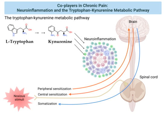
Neuroinflammation has been intricately linked to the pathogenesis of chronic pain. Chronic pain was proposed to be caused by the disturbance of peripheral nociception, neuropathy in the somatosensory system, motor system, central and peripheral nociplasticity, and/or psychosocial system [14]. Increasing evidence suggests that chronic inflammation is strongly tied to aberration in each mechanism of chronic pain. Furthermore, the tryptophan (TRP)–kynurenine (KYN) pathway and its metabolites were observed to play an important role in neuroinflammation and chronic pain [15]. This review article presents the components of the pain pathway; mechanisms of chronic pain based on the components; the development of chronic pain through peripheral and central sensitization; evidence of the presence of chronic neuroinflammation in each pain mechanism; the involvement of the TRP–KYN metabolic pathway; and the need of a psychogenic component in the pathogenesis of chronic pain.
2. The Pain Pathway, Mechanisms, Neuroinflammation, and Tryptophan Metabolism
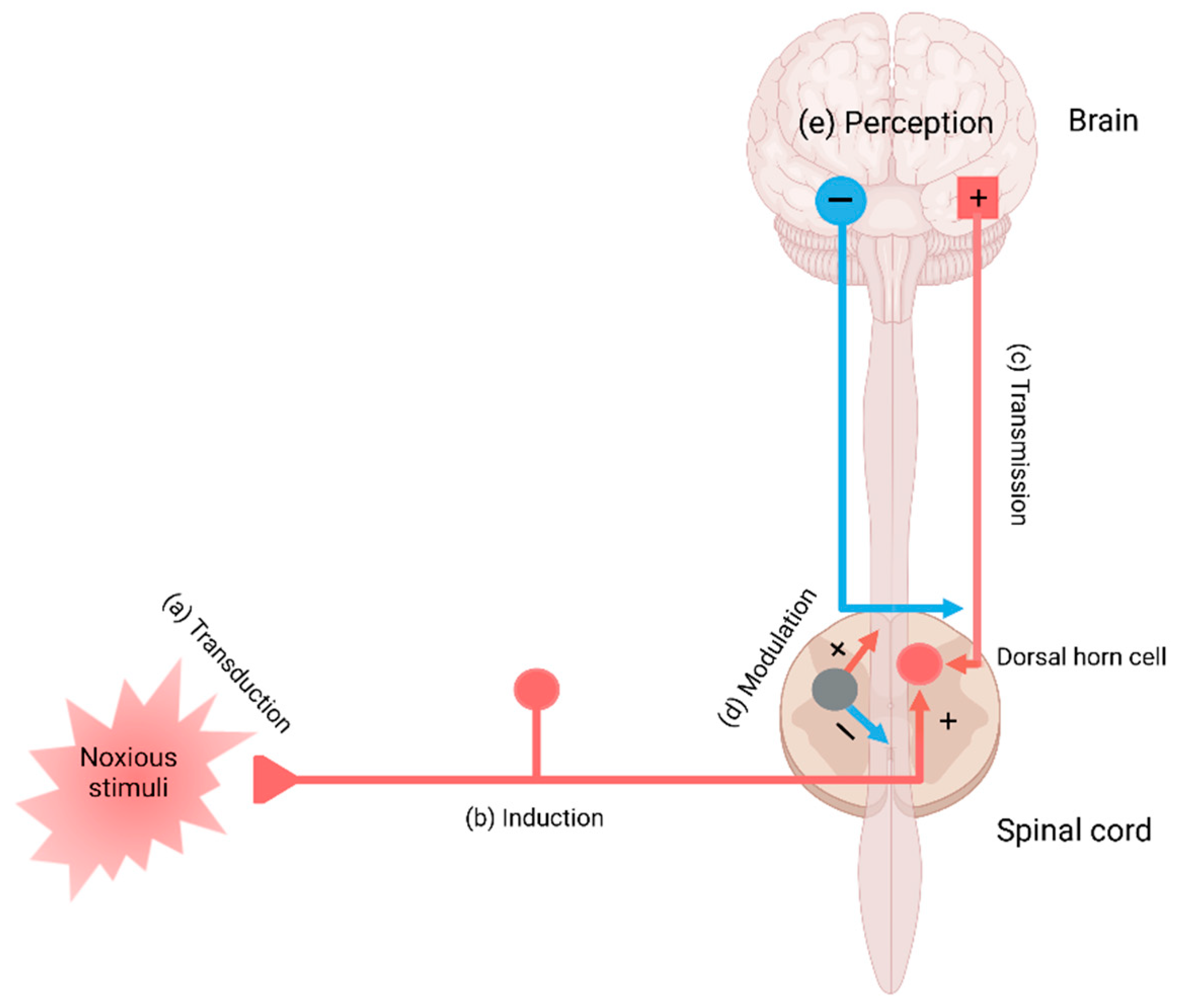
Pain perception is signaling through the pain pathway, whose components consist of transduction, conduction, transmission, modulation, and perception. Transduction is the process by which noxious or potentially damaging stimuli activate the nociceptors to convert to neural signals. Transmission refers to the signal transfer from the peripheral neurons to the second-order neurons in the spinal cord, which wire the signals to the thalamus and brain stem in the brain. Pain modulation takes place by inhibition of pain signaling in the spinal cord and the activation of the descending inhibitory fibers. The third-order neurons project to the somatosensory cortex, enabling the perception of pain. Perception is the subjective awareness in connection with arousal, physiological, and behavioral brain centers, involving the integration of psychological processes such as attention, expectation, and interpretation [16,17,18] (Figure 1).
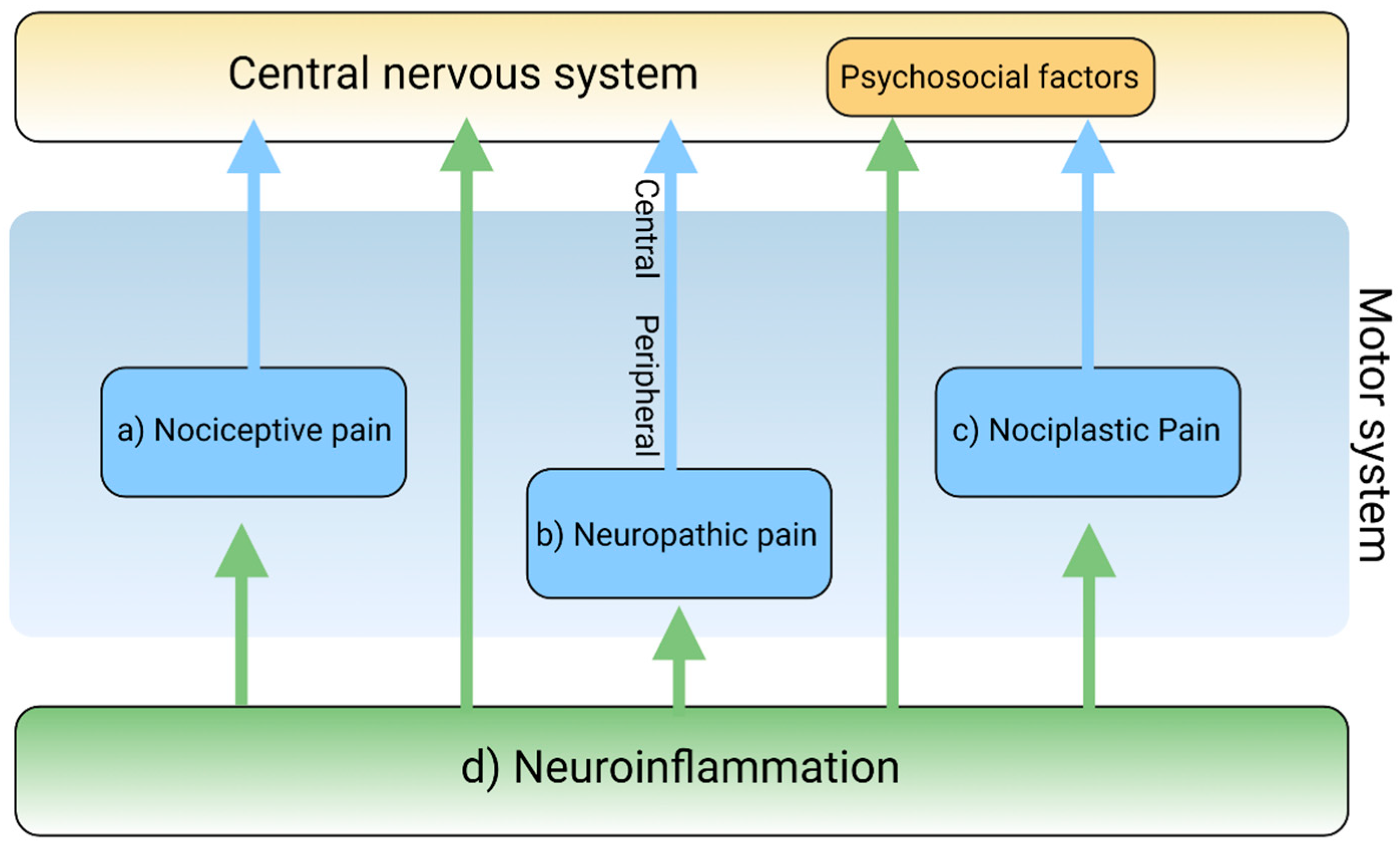
Pain is a complex and intricate process attributable to nociceptic, neuropathic, and/or neuroplastic mechanisms. The most common type of pain is nociceptive pain caused by damage or potentially harmful to peripheral tissues involving nociceptors responsible for transduction. Neuropathic pain is caused by lesions or diseases affecting the somatosensory nervous system responsible for the transmission of peripheral to central pain signals. Nociplastic pain refers to the condition caused by altered nociceptive processing without actual or potentially harmful tissue damage activating peripheral nociceptors (nociceptive pain) or without lesions or diseases of the somatosensory nervous system (neuropathic pain). Cortical perception is one of the main components in the pain pathway; however, the ICD-11 excludes psychogenic pain [19] (Figure 2). Thus, participation of cortical perception in chronic pain mechanisms remains ambiguous.
Inflammation is generally involved in the pathogenesis of various diseases and plays a key role in diseases that cause chronic pain [20]. Resident and recruited immune cells release inflammatory mediators at peripheral nerve innervating damaged or inflammatory tissue to trigger action potentials in sensor neurons or sensitize neurons by increasing transduction and excitability. Inflammatory mediators also act directly on peripheral nerves to damage peripheral transmission [21]. Immune cells infiltrate the spinal cord and the dorsal root ganglia to damage the central transmission and/or modulate pain sensitivity [22]. Activated immune cells release inflammatory cytokines, chemokines, and other factors that influence cognition, mood, and behaviors through immune-to-CNS signaling [16,23]. Accumulating evidence suggests that chronic dysregulation of the immune response is involved in the pathogenesis of psychiatric disorders such as mood disorders, substance abuse disorders, psychotic disorders, attention-deficit disorders, and autism spectrum disorders [24,25,26] (Figure 2).
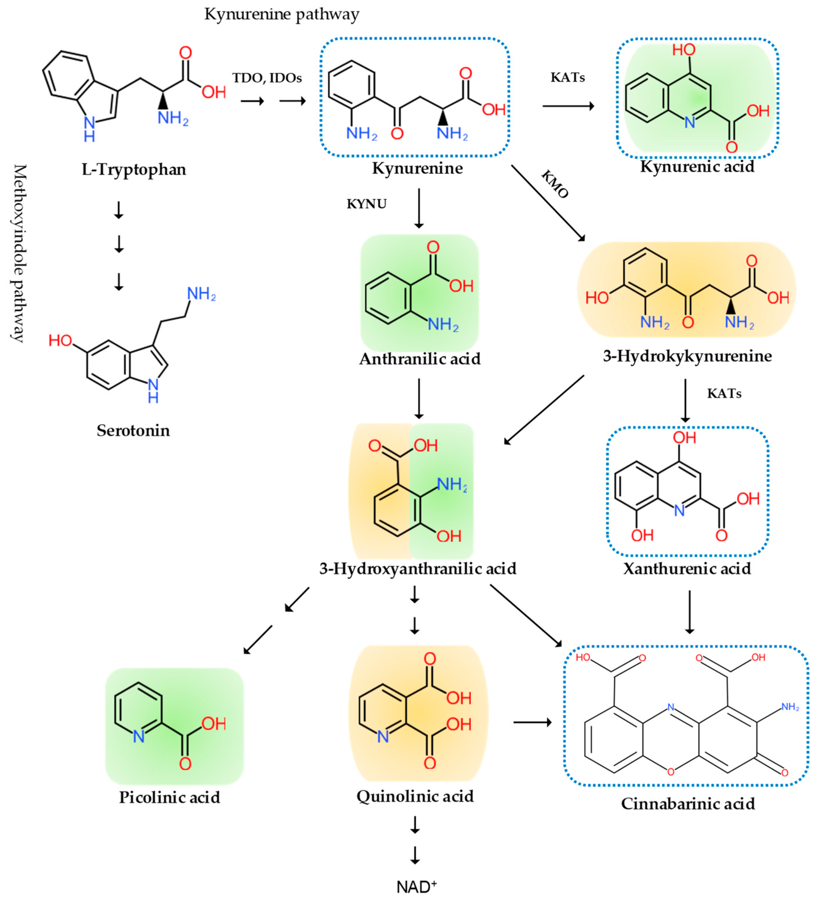
Inflammation is invariably linked to the activation of TRP metabolism [27,28]. The essential amino acid TRP is a precursor to serotonin, melatonin, and nicotinamide adenine dinucleotide (NAD+), among others. More than 95% of TRP is metabolized through the TRP–KYN pathway, synthesizing various bioactive metabolites such as neuroprotective antioxidants and neuroprotectants, toxic oxidants and neurotoxins, as well as immunomodulators. The disturbance of KYN metabolites has been linked to immune disorders, cancers, neurodegenerative diseases, and psychiatric disorders [29]. Furthermore, TRP–KYN metabolites are under extensive research in search of peripheral biomarkers as well as novel drug prototypes for a wide range of diseases [30,31,32,33,34,35,36]. Inflammation activates the TRP–KYN pathway, elevating the levels of oxidative compounds or neurotoxic ligands to receptors of the excitatory glutamatergic nervous system, which damage the peripheral nervous system or CNS through the broken blood–nerve or blood–brain barrier, respectively [16]. Furthermore, immunomodulators are known to trigger the shift of acute inflammatory status toward tolerogenic and chronic inflammation, perpetuating low-grade inflammation [28,37]. KYN is synthesized from TRP by the tryptophan 2,3-dioxygenase (TDO) in the liver and the indoleamine 2,3-dioxygenases (IDOs) in the brain and the immune system, which are induced by cortisol, and interferon (IFN)-α, IFN-γ, and tumor necrosis factor (TNF)-α, respectively [38]. Anthranilic acid (AA), 3-hydroxykynurenine (3-HK), or kynurenic acid (KYNA) are produced from KYN by the kynureninase (KYNU), the KYN-3-monooxygenase (KMO), or the kynurenine aminotransferases (KATs), respectively. The KATs also convert 3-HK to xanthurenic acid (XA). XA converts into cinnabarinic acid by autoxidation. AA and 3-HK convert into 3-hydroxyanthranilic acid (3-HAA) and then into picolinic acid and quinolinic acid (QA). QA converts into NADH, which is a feedback inhibitor of TDO [31] (Figure 3). Generally, 3-HK and QA are described as neurotoxic, while KYNA is considered to be neuroprotective. The 3-HK/KYNA ratio is often applied as an indicator of neurotoxicity. However, emerging evidence suggests that some metabolites of the TRP–KYN pathway possess Janus-face properties, depending on the dose or the situation. For example, KYNA is excitatory in lower concentrations but inhibitory in higher concentrations at α-amino-3-hydroxy-5-methyl-4-isoxazole propionic acid (AMPA) receptors. 3-HK is known to be an oxidant but observed to be an antioxidant in certain conditions [27,39].
The stress hormone cortisol, the strong immune activator lipopolysaccharide, proinflammatory cytokines, positive feedback loops, diminished levels of antioxidant system enzyme superoxide dismutase, and anti-inflammatory cytokines all lead to the potentiation of the TRP–KYN pathway [28]. Furthermore, the action of the KYN enzymes and metabolites are complicated by the interactions with adjacent biosystems such as the oxidative stress complex, the antioxidant enzyme systems, the serotonin neurotransmission, the glutamate neurotransmission, the tetrahydrobiopterin pathway, the cannabinoid system, and the aryl hydrocarbon receptor signaling [28,39].
3. Transduction and Nociceptive Pain
The transduction of the pain sensation takes place when noxious stimuli depolarize the afferent terminal of nociceptive myelinated A-beta (Aß) and A-delta (Aδ) fibers and unmyelinated C fibers through the terminal membrane proteins and voltage-gated ion channels converting them into electric signals in the neurons (Figure 1a).
Nociceptive pain is the most common pain that originates from a tissue injury or inflammation in which the nociceptor of peripheral sensory nerves detects noxious or potentially harmful stimuli [40]. In chronic pain, the peripheral nociceptors continue to transmit painful stimuli even after the original injury has healed [41]. Osteoarthritis is a classical nociceptive pain condition when abnormal loading of a damaged joint opens mechanogated ion channels on nociceptive nerve endings [42]. Overextending or tearing a ligament sensitizes nociceptors, which causes acute nociceptive pain, such as in the case of an ankle sprain. In addition to mechanical irritation or physical injury, the primary cells of the epidermis, keratinocytes, induce pain by releasing endogenous mediators, such as adenosine triphosphate (ATP), Interleukin (IL)-1 beta (β), prostaglandin E2, endothelin, and nerve growth factor. However, keratinocytes act in a dual matter in pain sensation: They release β–endorphin that help pleasurable feeling during modest sun-bathing but activate transient receptor potential cation channel subfamily V member 4 (TRPV4) and release pro-inflammatory cytokines, eliciting the pain sensation of a sunburn [43,44] (Figure 2a and Table 1).
Inflammation also activates nociceptors in the nerve endings. Inflammatory mediators bind to their receptors on nociceptive sensory neurons in the peripheral nervous system, resulting in pain [20]. Pro-inflammatory factors including TNF-γ and IL-1β secreted by monocytes and macrophages at the site of a peripheral injury facilitate pain transduction and conduction by modifying ion channels including transient receptor potential cation channel subfamily A member 1 (TRPA1), transient receptor potential cation channel subfamily V member 1 (TRPV1), and Nav1.7–1.9. However, those cells secret anti-inflammatory factors such as IL-10 and/or pro-resolution mediators, including resolvins, protectins, and maresins, to reduce nociception in the resolution phase of acute inflammation. Different phenotypes of macrophages, such as pro-inflammatory M1 and anti-inflammatory M2, contribute to the induction and resolution of pain, respectively [45]. Schwann cells of the peripheral nervous system also secret TNF-γ and IL-1β to sensitize nociceptors at axons in neuronal injury. Activated Schwann cells secrete matrix metalloprotease (MMP) 9 that help open the blood–nerve barrier, resulting in the recruitment of immune cells that release inflammatory cytokines [22,46]. Furthermore, nociceptive afferent sensory neurons directly modulate inflammation by releasing inflammatory mediators, such as substance P, calcitonin gene-related peptide (CGRP), neurokinin A, and endothelin-3. The process is called neurogenic inflammation (Figure 2d).
The disturbance of TRP metabolism is observed in neurogenic inflammation. The increased levels of the stress hormone cortisol and inflammatory cytokines such as IFN-α, IFN-γ, and TNF-α activate the TRP–KYN pathway producing higher levels of oxidant KYN metabolites which leak into the peripheral nervous system through the damaged gap junction following the immune reaction. The oxidative KYN metabolites 3-HK, 3-HA, and QA are harmful compounds to nerve endings of the afferent sensory neurons (Figure 3).
4. Conduction, Transmission, and Neuropathic Pain
In conduction, the electrical signals are conducted from the peripheral neurons to the central neurons where a network of interneurons facilitates or inhibits transmission to the second-order neurons in the dorsal horn [47]. The presynaptic terminals of C fibers release glutamate, substance P, and CGRP, which activate postsynaptic AMPA receptors, NK1 receptors, and CGRP receptors, respectively [48] (Figure 1b). In transmission, the activation of the postsynaptic receptors generates an action potential of the second-order neurons and interneurons, which relay signals through the contralateral spinothalamic tract to the thalamus; or the spinoreticular and spinomesencephalic tracts to the medulla and brain stem; or the spinohypothalamic tract to the hypothalamus [49] (Figure 1c).
Neuropathic pain originates from lesions or diseases of the somatosensory nervous system made up of peripheral and central components. Peripheral neuropathic pain is commonly caused by diabetic neuropathy, metabolic disorders, shingles, HIV-related distal symmetrical neuropathies, nutritional deficiencies, toxins such as arsenic and thallium, a paraneoplastic manifestation of cancer, immune-mediated inflammatory diseases such as Guillain-Barre syndrome, amyloidosis, Fabry’s disease, and nerve trunk injuries [50]. Presumably, burning and poorly localized pain is transmitted by C fibers, while sharp and lancinating pain is relayed by Aδ fibers [51]. Diabetic neuropathy is the most common neuropathy associated with severe pain, which presents a distal symmetrical polyneuropathy with numbness and loss of sensation in the distal extremities, often accompanied by peripheral vascular diseases, leading to infection and ultimately amputation [52]. Neuropathic pain is also caused by direct invasion to peripheral nerves by tumor, side effects of chemotherapy, radiation injury, or surgery [53] (Figure 2b and Table 1). Central neuropathic pain is a common sequela to injury to the CNS such as vascular accidents, including ischemic and hemorrhagic stroke, infections, including abscess, encephalitis, and myelitis, demyelinating diseases, including multiple sclerosis, tumors, and brain or spinal cord [54,55,56] (Figure 2b and Table 1). Mixed pain is a term never formally defined, but it indicates pain caused by a combination of nociceptive and neuropathic mechanisms observed in patients who suffer from osteoarthritis, sciatica, and cancer.
Neuropathic pain is often manifested as a part of the symptoms of psychological disorders. The lifetime and current prevalence of psychiatric disorders in patients with chronic peripheral pain were 39% and 20%, respectively [57]. Diseases that cause neuropathic pain include diabetes, herpes zoster infection, nerve compression, nerve trauma, channelopathies, and autoimmune diseases. The most common psychiatric disorders were generalized anxiety disorders and mood disorders [57]. Furthermore, antidepressants showed efficacy for neuropathic pain in patients with depression, suggesting neuropathic pain and depression have a bidirectional relationship [58]. Individuals with chronic neuropathic pain were associated with substance abuse or suicide ideation [59] (Figure 2b and Table 1).
Inflammation plays an important role in neuropathic pain. Around afferent peripheral nerves, monocytes and macrophages release pro-inflammatory factors, including TNF-γ and IL-1β, while they secrete anti-inflammatory factor IL-10 and pro-resolving lipid mediators at the resolution of acute inflammation [60]. T lymphocytes (T-cells) play an important role in neuropathic pain. T-cells secret a pro-inflammatory cytokine IL-17 and accumulate in the dorsal root ganglion (DRG) to release pro-analgesic leukocyte elastase, inducing mechanical allodynia. In the resolution phase, T-cells secrete anti-inflammatory cytokines IL-4 and IL-10. In response to noxious stimuli, the satellite glial cells (SGCs) are activated and proliferated at DRG to release pro-inflammatory cytokines TNF and IL-1β and a nociceptive neurotransmitter ATP signaling through P2 receptors [61]. SGCs also release MMPs that open the blood–nerve barriers, allowing entry of immune cells [62]. Bone marrow stem cells trigger analgesic actions by secreting anti-inflammatory cytokine-transforming growth factor-beta 1 by suppressing glial activation induced by nerve injury and migrating to DRG via a (C-X-C motif) chemokine ligand (CXCL) 12 chemotactic signal after intrathecal injection [63].
Spinal cord microglia play major roles in pathological pain. Following peripheral injury, ATP, colony-stimulating factor 1, chemokines including (C-C motif) chemokine ligand (CCL) 2 and fractalkine (CX3CL1), and proteases activate spinal microglia [64]. Meanwhile, the expression of the receptors for ATP and CX3CL1 increases, converging an intracellular signaling cascade, leading to the phosphorylation of p38 mitogen-activated protein (MAP) kinase, which, in turn, elevates production and release of TNF-γ, IL-1β, IL-18, brain-derived growth factor (BDNF), and prostaglandin E2. TNF-γ and Il-1β increase synaptic transmission and decrease inhibitory synaptic transmission of lamina II spinal cord neurons [22]. BDNF suppressed gamma-aminobutyric acid inhibitory synaptic transmission in projection to lamina I spinal cord neurons. Microglia release anti-inflammatory cytokine IL-10 in the resolution phase of inflammation [65].
An astrocyte is in contact with more than one million synapses, and thus, chronic pain in astrocyte activation is more persistent [66]. Astrocytes communicate with neurons through gap junction mediated by connexin-43 (Cx43). Cx43 is upregulated in astrocytes after nerve injury, serving as a paracrine modulator. The paracrine modulation results in elevating the release of glutamate, ATP, MMP2 and chemokines, including CCL2 and CXCL1. The chemokines function as neuromodulators that potentiate excitatory synaptic transmission. Meanwhile, following nerve injury, spinal cord neurons upregulate CXCL13 that activates astrocytes via C-C chemokine receptor type 5 to sustain neuropathic pain [67]. The spinal cord and cortical astrocytes upregulate thrombospondin 4 that leads to new synapsis formation and subsequent somatosensory cortical circuit rewiring, causing neuropathic pain [68]. Astrocytes cause neuronal hyperexcitability resulting from disturbance of homeostasis of extracellular potassium and glutamate. IFN-α produced by astrocytes inhibits nociceptive transmission in the spinal cord [66].
Oligodendrocytes form myelin sheath insulating axons in the CNS [69]. Little is known about their roles in pain. IL-33 produced from oligodendrocytes contributes to pain sensitivity via MAP kinases and nuclear factor kappa-light-chain-enhancer of activated B cells in chronic constriction injury model of nerve injury-induced neuropathic pain [70]. Diphtheria toxin ablation of oligodendrocytes leads to neuropathic pain, suggesting analgesic roles of the cells. Following nerve injury, T-cells infiltrate the spinal cord, contributing to the development of mechanical sensitivity. T-cells release pro-inflammatory cytokine TNF-γ, they secrete anti-inflammatory cytokines IL-4 and IL-10 in the resolution phase of inflammation [71]. Following chemotherapy, intrathecal injection of cytotoxic T-cells enhanced neuropathic pain, while the injection of regulatory T-cells diminished neuropathic pain [72] (Figure 2d).
The involvement of the TRP–KYN pathway was reported in inflammation-induced neuropathic pain. The enzyme activities of the TRP–KYN pathway were studied in a lipopolysaccharide-stimulated chronic constriction injury at the spinal cord and DRG levels of rats. The intrathecal administration of L-KYN and the intraperitoneal injection of L-KYN and an organic anion transport inhibitor probenecid significantly reversed tactile allodynia in L5-L6 spinal nerve root-ligated rats, suggesting that the N-methyl-D-aspartate (NMDA) receptor, an organic anion transport inhibitor agonist KYNA, mediates relieving the allodynia [73]. The increased ratio of QA/KYN and the mRNA expression of KMO, KYNU, and 3-hydroxyanthranilate dioxygenase (HAOO) was elevated in neuronal nuclear antigen-positive neurons of the contralateral hippocampal dentate gyrus in a neuropathic mouse model [74]. TDOIDO1 and 2, KMO, KYNU, and HAOO were found to be derived from cerebral microglial cells, and mRNA expression of IDO2, KMO, and HAOO were upregulated at the spinal cord after one week. Microglia inhibitor, minocycline, decreased the levels of IDO2 and KMO enzymes and tactile and thermal hypersensitivity; furthermore, IDO2 inhibitor 1-methyl-d-tryptophan and KMO inhibitor UPF 648 significantly decreased mechanical and thermal hypersensitivity [75]. This suggests the participation of IDO2 and KMO enzymes in the pathogenesis of neuropathic pain. The intracerebroventricular administration of KMO inhibitor Ro 61-8048 alleviated spared nerve injury-induced depressive-like behavior, and the intrathecal injection of Ro 61-8048 attenuated both the depressive-like behavior and mechanical allodynia in rats [76]. The NMDA receptor seems to play a major role in neuropathic pain and in the development of opioid tolerance. Dextromethorphan is an NMDA antagonist at high doses. Both animal and human studies showed that NMDA antagonist ketamine was beneficial for analgesics [77] (Figure 3).
5. Modulation and Nociplastic Pain
Modulation of pain transmission occurs at all levels of the pain pathway from peripheral to the brain, as well as from upward-to-downward pain regulations, involving both excitatory and inhibitory mechanisms that facilitate or suppress the responses of second-order neurons, respectively [48]. Peripheral pain modulation is achieved through local growth factor, hormonal, and peptide release, which alters signaling through neurotransmitter, ion, or receptor-based mechanisms. The pain modulation takes place neuronal signaling through corticospinal, corticoperiheral, and intraspinal pathways and neuroplasticity regulation [78] (Figure 1d and Figure 4).
Nociplastic pain is defined as pain that arises from altered nociception despite no clear evidence of actual or threatened tissue damage causing the activation of peripheral nociceptors or no clear evidence of diseased lesions of the somatosensory nervous system causing the pain [79,80]. Nociplastic pain is generally chronic and widespread and is caused by the disturbance of central pain processing mechanisms, such as elevated excitability of ascending and descending pain facilitatory pathways and/or reduced inhibition of the descending anti-nociceptive pathway [14,81,82] (Figure 4). The condition refers to central sensitization in which pain is elicited by innocuous stimuli or different kinds of stimuli, resulting in central hyperalgesia or allodynia, respectively [83]. The process involves increased activity of the insula, anterior cingulate cortex, and the prefrontal cortex, which becomes active during acute pain sensation as well as of the brain stem nuclei, dorsolateral frontal cortex, and parietal cortex, which do not participate during acute pain sensation [84] (Figure 4). Fatigue, negative affect, unrefreshing sleep, and cognitive dysfunction are common accompanying findings in centralized nociplastic pain [85]. This typical pattern of nociplastic pain is observed in fibromyalgia, a medical condition of unknown cause but known to be involved in genetic and environmental factors [86]. Temporomandibular disorder and nonspecific back pain are also characterized by central sensitization (Figure 2c and Table 1).
The inflammatory response is remarkable in nociplastic pain. The levels of proinflammatory cytokines including IL-6 and IL-8 were observed to be higher, while anti-inflammatory cytokines IL-1 receptor antagonist was higher and IL-4 was lower in patients with fibromyalgia. Several chemokine levels were elevated in fibromyalgia patients. They were monocyte recruiting such as protein eotaxin (CCL11), TARC (CCL17), and MDC (CCL22) and neutrophil chemoattractant MIG (CXCL9) and I-TAC (CXCL11) [87]. Furthermore, the disruption of the proinflammatory and anti-inflammatory cytokine network was considered to play a key role in the pathogenesis of central sensitization in fibromyalgia. Chronic inflammation has been considered to induce central pain in rheumatoid arthritis [88]. Thus, inflammation certainly contributes to the development of nociplastic pain, as in fibromyalgia (Figure 2d).
The alteration of TRP metabolism has been linked to nociplastic pain such as the temporomandibular disorders myalgia and fibromyalgia. The levels of TRP were observed to be significantly lower in the plasma of fibromyalgia patients compared to control, and the KYN/TRP ratio was negatively correlated with anxiety levels. The plasma TRP levels were negatively correlated with the wrist pain intensity, whereas the KYN/TRP ratio was positively correlated with the average and wrist pain intensity in temporomandibular disorders [89]. TRP depletion appears to be involved in the pathogenesis of fibromyalgia and temporomandibular disorders; however, little is known about the roles of NMDA receptor agonists 3-HK and QA and NMDA receptor antagonist KYNA. Furthermore, the direct link between KYNs and nociplastic pain has not been reported (Figure 3).
6. Cortical Perception and Psychogenic Pain
The perception of pain is processed in the brain and the spinal cord. The thalamus, sensorimotor cortex, insular cortex, and anterior cingulate decode signals of unpleasant sensation carried through ascending spinothalamic tract, whereas the amygdala and hypothalamus decode signals of urgency and intensity brought through ascending the spinobulbar tract. The third-order neurons transfer signals and communicate with the cortex centers. Overall, the integration of sensations, emotions, and cognition in the brain lead to the perception of pain [90] (Figure 1e). Psychogenic pain is pain without relevant anatomic tissue injury or inconsistent with functional causes in distribution and is considered to be caused by psychological factors such as depression, anxiety, and emotion [91]. Depression, anxiety, and cognitive disturbance are common symptoms that manifest in a wide range of diseases and comorbidity [92]. Individuals with depression and anxiety often experience psychogenic pain all over their bodies without any relevant physical cause [93,94]. Other psychiatric disorders frequently observed in individuals with chronic pain include substance abuse, somatoform disorder, and panic disorders [95]. Furthermore, chronic pain is associated with the disturbance of cognitive functions such as attention, working memory, reasoning ability, information processing, and verbal communication [96,97].
Inflammation is obviously involved in psychiatric disorders such as depression and anxiety. Meta-analyses reported strong evidence of significantly increased levels of c-reactive protein (CRP), IL-1, IL-6, TNF-α and soluble IL-2 receptor in the serum of major depressive disorder (MDD) patients [98,99,100,101,102]. A higher concentration of CCL2/MCP-1 was also reported in patients with MDD. CRP levels in blood, serum or plasma samples was significantly raised in generalized anxiety disorder (GAD) patients by meta-analysis, and IFN-γ and TNF-α levels were significantly increased in at least two or more studies [103]. Lower levels of IL-10 and higher ratios of TNF-α/IL-10, TNF-α/IL-4, IFN-γ/IL-10, and IFN-γ/IL-4 were observed in the serum of GAD patients, showing significantly increased pro- to anti-inflammatory cytokine ratios, which suggests a distinct cytokine imbalance [104] (Figure 2d).
Similarly, activation of the TRP–KYN pathway has been reported in depression and anxiety. Meta-analyses reported decreased TRP levels in plasma and decreased levels of KYN and KYNA in MDD patients, while antidepressant-free patients showed an increased level of QA. The postmortem brain tissues from patients with MDD showed the increased QA immunoreactivity in the prefrontal cortex and hippocampus [105,106]. Magnetic resonance spectroscopy suggested a higher turnover of cells with KYN and the 3-HAA/KYN ratio in adolescent depression. Those findings are in accordance with the activation of the TRP-KYN pathway toward 3-HK and QA branches by pro-inflammatory cytokines activating IDOs, and KMO, resulting in higher neurotoxic 3-HK and QA levels [107]. Decreased plasma KYN levels were observed in endogenous anxiety and normalized after treatment [108]. The alteration of the TRP-KYN pathway by stress or inflammation may cause serotonin and melatonin deficiency, making an individual more susceptible to anxiety (Figure 3).
7. Conclusions and Future Perspective
The pain pathway, pain mechanisms, inflammation, KYN metabolites and enzymes of the TRP–KYN pathway, and diseases associated with chronic pain are overviewed in this review article. Pain sensation can be attributed to damage and/or potential harm in various components of the pain pathway and corresponding pain mechanisms, involving inflammation and alteration of the TRP–KYN pathway [109]. Chronic inflammation triggers not only nociceptive pain but induces other pain mechanisms, including psychogenic pain. Thus, a search for unique inflammatory signatures and various interventional targets in chronic inflammation is currently under extensive research [110,111,112,113]. Intervention through the TRP–KYN pathway is under comprehensive research to alleviate oxidative stress and excitotoxicity in various illnesses [114,115,116,117,118,119,120,121]. Meanwhile, the effectiveness of motor cortex stimulation and spinal cord stimulation to alleviate chronic pain caused by various underlying conditions is under evaluation [122,123].
Chronic pain arises through a complex pathogenic process involving more components and developing into the pain continuum. Central sensitization, peripheral sensitization, and somatization are pathogenic processes of pain development in the pain continuum spanning components of the pain pathway and the pain mechanism, which is hardly understood without the presence of the cortical perception (Figure 5). The nociplastic mechanism of pain attempts to delineate pain without relevant cause or lesions of the somatosensory nervous system, such as altered perception of nociception. Chronic pain presented in fibromyalgia syndrome, chronic back pain, and complex regional pain syndrome is best understood in the framework of pain perception, including cognitive, emotional, and social components. Chronic pain experienced in psychiatric conditions, in particular, is not fully explainable in the view of the nociplastic pain mechanism. Pain sensation is developed through complex interactions with higher cortical centers governing mood, emotion, and cognition.
Animal studies are one of the most important arenas for pain research. The endothelin receptor mediates the alteration of astrocyte functions, leading to the alleviation of neuropathic pain [124]. The dissociative anesthetics ketamine induces analgesic effects in models of acute pain and relieves thermal and mechanical allodynia in a chronic neuropathic pain model [125]. The involvement of the serotonergic neurotransmission in analgesic actions has been studied using neuropathic pain models in rats [126]. The gender difference in pain sensation and emotional domain has been reported using the transgenic mouse model of Alzheimer’s disease [127].
More and more emerging findings shed light on the relationship between psychiatric symptoms and networks of the brain centers in neuropsychiatric disorders [128,129,130]. Stimulus-evoked functional magnetic resonance imaging (fMRI), task-free fMRI, and perfusion MRI revealed that chronic pains arise from pre-existing vulnerabilities and sustained abnormal input [131]. Neuroimaging techniques, including fMRI and positron emission tomography, may open the gate to understanding underlying mechanisms in signaling to the third-order neurons to the cortex in chronic pain sensation [132,133,134]. Pain relief can be achieved through accompanying symptoms such as cognition, mood, and sleep by pharmacotherapy and/or psychotherapy [132,135,136,137]. Therefore, psychogenic components of pain play an essential role in understanding the pathomechanism of chronic pain unless the nociplastic pain mechanism can sufficiently elucidate the reciprocal interaction with third-order neurons in the pathogenesis of chronic pain.
Author Contributions
Conceptualization, M.T. and L.V.; writing—original draft preparation, M.T.; writing—review and editing, M.T., N.T., F.T., Á.S. and L.V.; visualization, M.T. and Á.S.; supervision, L.V.; project administration, L.V.; funding acquisition, L.V. All authors have read and agreed to the published version of the manuscript.
Funding
This research was funded by GINOP 2.3.2-15-2016-00034, GINOP 2.3.2-15-2016-00048, TUDFO/47138-1/2019-ITM, and TKP2020 Thematic Excellence Programme 2020—The APC was funded by the University of Szeged Open Access Fund (4942).
Institutional Review Board Statement
Not applicable.
Informed Consent Statement
Not applicable.
Data Availability Statement
Not applicable.
Acknowledgments
The authors are grateful to Eleonóra Spekker for the technical assistance of graphic design.
Conflicts of Interest
The authors declare no conflict of interest.
Abbreviations
| AA | anthranilic acid |
| Aß fiber | A-beta fiber |
| Aδ fiber | A-delta fiber |
| AMPA | α-amino-3-hydroxy-5-methyl-4-isoxazole propionic acid |
| AS | ankle sprain |
| AT | Achilles tendinopathy |
| ATP | Adenosine triphosphate |
| BDNF | brain-derived growth factor |
| CCL | (C-C motif) chemokine ligand |
| CGRP | calcitonin gene-related peptide |
| CNS | central nervous system |
| CPS | chronic pain syndrome |
| CRP | c-reactive protein |
| CX3CL1 | fractalkine |
| Cx43 | connexin-43 |
| CXCL | (C-X-C motif) chemokine ligand |
| DRG | dorsal root ganglion |
| DSM-5 | Diagnostic and Statistical Manual of Mental Disorders, 5th Edition |
| FM | fibromyalgia |
| fMRI | functional magnetic resonance imaging |
| GAD | generalized anxiety disorder |
| 3-HAA | 3-hydroxyanthranilic acid |
| 3-HK | 3-hydroxykynurenine |
| HAOO | 3-hydroxyanthranilate dioxygenase |
| ICD-11 | 11th revision of the International Statistical Classification of Diseases and Related Health Problems |
| IDO | indolamine 2,3-dioxygenase |
| IFN | interferon |
| IL | interleukin |
| KAT | kynurenine aminotransferase |
| KMO | kynurenine 3-monooxygenase |
| KYN | kynurenine |
| KYNA | kynurenic acid |
| KYNU | kynureninase |
| LBP | low back pain |
| MAP | mitogen-activated |
| MDD | major depressive disorder |
| MMP | matrix metalloprotease |
| NAD+ | nicotinamide adenine dinucleotide |
| NMDA | N-methyl-D-aspartate |
| OA | osteoarthritis |
| QA | quinolinic acid |
| SGCs | satellite glial cells |
| SSD | somatic symptom disorder |
| T-cells | T lymphocytes |
| TDO | tryptophan 2,3-dioxygenase |
| TMD | temporomandibular joint disorder |
| TNF-γ | tumor necrosis factor gamma |
| TRP | tryptophan |
| TRPA1 | transient receptor potential cation channel, subfamily A member 1 |
| TRPV1 | transient receptor potential cation channel subfamily V member 1 |
| TRPV4 | transient receptor potential cation channel subfamily V member 4 |
| XA | xanthurenic acid |
References
- IASP. Definitions of Chronic Pain Syndromes. Available online: https://www.iasp-pain.org/Advocacy/icd.aspx?ItemNumber=5354#chronicpain (accessed on 18 December 2020).
- Di Lernia, D.; Lacerenza, M.; Ainley, V.; Riva, G. Altered Interoceptive Perception and the Effects of Interoceptive Analgesia in Musculoskeletal, Primary, and Neuropathic Chronic Pain Conditions. J. Pers. Med. 2020, 10, 201. [Google Scholar] [CrossRef]
- Medicina. Special Issue “Chronic Pain Management”. Available online: https://0-www-mdpi-com.brum.beds.ac.uk/journal/medicina/special_issues/chronic_pain_management (accessed on 18 December 2020).
- Mäntyselkä, P.; Kumpusalo, E.; Ahonen, R.; Kumpusalo, A.; Kauhanen, J.; Viinamäki, H.; Halonen, P.; Takala, J. Pain as a reason to visit the doctor: A study in Finnish primary health care. Pain 2001, 89, 175–180. [Google Scholar] [CrossRef]
- Mills, S.; Nicolson, K.P.; Smith, B.H. Chronic pain: A review of its epidemiology and associated factors in population-based studies. Br. J. Anaesth. 2019, 123, e273–e283. [Google Scholar] [CrossRef] [PubMed]
- Johnson, M.I. The Landscape of Chronic Pain: Broader Perspectives. Medicina 2019, 55, 182. [Google Scholar] [CrossRef] [PubMed] [Green Version]
- GBD 2016 Disease and Injury Incidence and Prevalence Collaborators. Global, regional, and national incidence, prevalence, and years lived with disability for 328 diseases and injuries for 195 countries, 1990–2016: A systematic analysis for the Global Burden of Disease Study 2016. Lancet 2017, 390, 1211–1259. [Google Scholar] [CrossRef] [Green Version]
- Treede, R.D.; Rief, W.; Barke, A.; Aziz, Q.; Bennett, M.I.; Benoliel, R.; Cohen, M.; Evers, S.; Finnerup, N.B.; First, M.B.; et al. Chronic pain as a symptom or a disease: The IASP Classification of Chronic Pain for the International Classification of Diseases (ICD-11). Pain 2019, 160, 19–27. [Google Scholar] [CrossRef] [Green Version]
- American Psychiatric Association. Diagnostic and Statistical Manual of Mental Disorders, 5th ed. Available online: https://0-doi-org.brum.beds.ac.uk/10.1176/appi.books.9780890425596 (accessed on 27 December 2020).
- Ciaramella, A.; Silvestri, S.; Pozzolini, V.; Federici, M.; Carli, G. A retrospective observational study comparing somatosensory amplification in fibromyalgia, chronic pain, psychiatric disorders and healthy subjects. Scand. J. Pain 2020. [Google Scholar] [CrossRef]
- Rivat, C.; Ballantyne, J. The dark side of opioids in pain management: Basic science explains clinical observation. Pain Rep. 2016, 1, e570. [Google Scholar] [CrossRef]
- He, Y.; Kim, P.Y. Allodynia. In StatPearls; StatPearls Publishing: Treasure Island, FL, USA, 2020. Available online: https://0-www-ncbi-nlm-nih-gov.brum.beds.ac.uk/books/NBK537129/ (accessed on 27 December 2020).
- Yasaei, R.; Peterson, E.; Saadabadi, A. Chronic Pain Syndrome. In StatPearls; StatPearls Publishing: Treasure Island, FL, USA. Available online: https://0-www-ncbi-nlm-nih-gov.brum.beds.ac.uk/books/NBK470523/ (accessed on 18 December 2020).
- Chimenti, R.L.; Frey-Law, L.A.; Sluka, K.A. A Mechanism-Based Approach to Physical Therapist Management of Pain. Phys. Ther. 2018, 98, 302–314. [Google Scholar] [CrossRef]
- Jovanovic, F.; Candido, K.D.; Knezevic, N.N. The Role of the Kynurenine Signaling Pathway in Different Chronic Pain Conditions and Potential Use of Therapeutic Agents. Int. J. Mol. Sci. 2020, 21, 6045. [Google Scholar] [CrossRef]
- Dantzer, R.; O’Connor, J.C.; Freund, G.G.; Johnson, R.W.; Kelley, K.W. From inflammation to sickness and depression: When the immune system subjugates the brain. Nat. Rev. Neurosci. 2008, 9, 46–56. [Google Scholar] [CrossRef] [Green Version]
- Lovelace, M.D.; Varney, B.; Sundaram, G.; Franco, N.F.; Ng, M.L.; Pai, S.; Lim, C.K.; Guillemin, G.J.; Brew, B.J. Current Evidence for a Role of the Kynurenine Pathway of Tryptophan Metabolism in Multiple Sclerosis. Front. Immunolog. 2016, 7, 246. [Google Scholar] [CrossRef] [PubMed] [Green Version]
- Ong, W.Y.; Stohler, C.S.; Herr, D.R. Role of the Prefrontal Cortex in Pain Processing. Mol. Neurobiol. 2019, 56, 1137–1166. [Google Scholar] [CrossRef] [PubMed] [Green Version]
- Trouvin, A.P.; Perrot, S. New concepts of pain. Best Pract. Res. Clin. Rheumatol. 2019, 33, 101415. [Google Scholar] [CrossRef] [PubMed]
- Pinho-Ribeiro, F.A.; Verri, W.A.; Chiu, I.M. Nociceptor Sensory Neuron-Immune Interactions in Pain and Inflammation. Trends Immunol. 2017, 38, 5–19. [Google Scholar] [CrossRef] [PubMed] [Green Version]
- Gonçalves dos Santos, G.; Delay, L.; Yaksh, T.L.; Corr, M. Neuraxial Cytokines in Pain States. Front Immunol. 2020, 10, 3061. [Google Scholar] [CrossRef] [Green Version]
- Ji, R.R.; Chamessian, A.; Zhang, Y.Q. Pain regulation by non-neuronal cells and inflammation. Science 2016, 354, 572–577. [Google Scholar] [CrossRef] [PubMed] [Green Version]
- Matsuda, M.; Huh, Y.; Ji, R.-R. Roles of Inflammation, Neurogenic inflammation, and Neuroinflammation in Pain. J. Anesth. 2019, 33, 131–139. [Google Scholar] [CrossRef]
- Misiak, B.; Frydecka, D.; Stanczykiewicz, B.; Samochowiec, J. Editorial: Peripheral Markers of Immune Response in Major Psychiatric Disorders: Where Are We Now and Where Do We Want to Be? Front. Psychiatry 2019, 10, 5. [Google Scholar] [CrossRef]
- Verlaet, A.A.J.; Maasakkers, C.M.; Hermans, N.; Savelkoul, H.F.J. Rationale for Dietary Antioxidant Treatment of ADHD. Nutrients 2018, 10, 405. [Google Scholar] [CrossRef] [Green Version]
- Fung, T.C.; Olson, C.A.; Hsiao, E.Y. Interactions between the microbiota, immune and nervous systems in health and disease. Nat. Neurosci. 2017, 20, 145–155. [Google Scholar] [CrossRef] [PubMed]
- Encyclopedia. The Tryptophan-Kynurenine Metabolic Pathway. Available online: https://encyclopedia.pub/8633 (accessed on 13 April 2021).
- Tanaka, M.; Tóth, F.; Polyák, H.; Szabó, Á.; Mándi, Y.; Vécsei, L. Immune Influencers in Action: Metabolites and Enzymes of the Tryptophan-Kynurenine Metabolic Pathway. Biomedicines 2021, 9, 734. [Google Scholar] [CrossRef] [PubMed]
- Tanaka, M.; Bohár, Z.; Vécsei, L. Are Kynurenines Accomplices or Principal Villains in Dementia? Maintenance of Kynurenine Metabolism. Molecules 2020, 25, 564. [Google Scholar] [CrossRef] [PubMed] [Green Version]
- Dezsi, L.; Tuka, B.; Martos, D.; Vecsei, L. Alzheimer’s disease, astrocytes and kynurenines. Curr. Alzheimer Res. 2015, 12, 462–480. [Google Scholar] [CrossRef] [Green Version]
- Török, N.; Tanaka, M.; Vécsei, L. Searching for Peripheral Biomarkers in Neurodegenerative Diseases: The Tryptophan-Kynurenine Metabolic Pathway. Int. J. Mol. Sci. 2020, 21, 9338. [Google Scholar] [CrossRef] [PubMed]
- Erabi, H.; Okada, G.; Shibasaki, C.; Setoyama, D.; Kang, D.; Takamura, M.; Yoshino, A.; Fuchikami, M.; Kurata, A.; Kato, T.A.; et al. Kynurenic acid is a potential overlapped biomarker between diagnosis and treatment response for depression from metabolome analysis. Sci. Rep. 2020, 10, 16822. [Google Scholar] [CrossRef]
- Carrillo-Mora, P.; Pérez-De la Cruz, V.; Estrada-Cortés, B.; Toussaint-González, P.; Martínez-Cortéz, J.A.; Rodríguez-Barragán, M.; Quinzaños-Fresnedo, J.; Rangel-Caballero, F.; Gamboa-Coria, G.; Sánchez-Vázquez, I.; et al. Serum Kynurenines Correlate with Depressive Symptoms and Disability in Poststroke Patients: A Cross-sectional Study. Neurorehabilit. Neural Repair 2020, 34, 936–944. [Google Scholar] [CrossRef]
- Tanaka, M.; Török, N.; Vécsei, L. Novel Pharmaceutical Approaches in Dementia. In NeuroPsychopharmacotherapy; Riederer, P., Laux, G., Nagatsu, T., Le, W., Riederer, C., Eds.; Springer: Cham, Switzerland, 2021; Available online: https://0-doi-org.brum.beds.ac.uk/10.1007/978-3-319-56015-1_444-1 (accessed on 1 June 2021).
- Ulivieri, M.; Wierońska, J.M.; Lionetto, L.; Martinello, K.; Cieslik, P.; Chocyk, A.; Curto, M.; Di Menna, L.; Iacovelli, L.; Traficante, A.; et al. The Trace Kynurenine, Cinnabarinic Acid, Displays Potent Antipsychotic-Like Activity in Mice and Its Levels Are Reduced in the Prefrontal Cortex of Individuals Affected by Schizophrenia. Schizophr. Bull. 2020, 46, 1471–1481. [Google Scholar] [CrossRef]
- Tanaka, M.; Vécsei, L. Monitoring the Redox Status in Multiple Sclerosis. Biomedicines 2020, 8, 406. [Google Scholar] [CrossRef] [PubMed]
- Tanaka, M.; Toldi, J.; Vécsei, L. Exploring the Etiological Links behind Neurodegenerative Diseases: Inflammatory Cytokines and Bioactive Kynurenines. Int. J. Mol. Sci. 2020, 21, 2431. [Google Scholar] [CrossRef] [PubMed] [Green Version]
- Török, N.; Maszlag-Török, R.; Molnár, K.; Szolnoki, Z.; Somogyvári, F.; Boda, K.; Tanaka, M.; Klivényi, P.; Vécsei, L. Single Nucleotide Polymorphisms of Indoleamine 2,3-Dioxygenase 1 Influenced the Age Onset of Parkinson’s Disease. Preprints 2020. [Google Scholar] [CrossRef]
- Tanaka, M.; Vécsei, L. Monitoring the Kynurenine System in Neurodegenerative and Psychiatric Illnesses: Concentrations, Ratios, or What Else? Adv. Clin. Exp. Med 2021, 30. in press. [Google Scholar]
- Armstrong, S.A.; Herr, M.J. Physiology, Nociception. In StatPearls; StatPearls Publishing: Treasure Island, FL, USA, 2020. Available online: https://0-www-ncbi-nlm-nih-gov.brum.beds.ac.uk/books/NBK551562/ (accessed on 27 December 2020).
- Tompkins, D.A.; Hobelmann, J.G.; Compton, P. Providing chronic pain management in the “Fifth Vital Sign” Era: Historical and treatment perspectives on a modern-day medical dilemma. Drug Alcohol Depend. 2017, 173 (Suppl. 1), S11–S21. [Google Scholar] [CrossRef]
- Fu, K.; Robbins, S.R.; McDougall, J.J. Osteoarthritis: The genesis of pain. Rheumatology 2018, 57 (Suppl. 4), iv43–iv50. [Google Scholar] [CrossRef] [Green Version]
- Talagas, M.; Lebonvallet, N.; Berthod, F.; Misery, L. Lifting the veil on the keratinocyte contribution to cutaneous nociception. Protein Cell 2020, 11, 239–250. [Google Scholar] [CrossRef] [PubMed] [Green Version]
- Moore, C.; Cevikbas, F.; Pasolli, H.A.; Chen, Y.; Kong, W.; Kempkes, C.; Parekh, P.; Lee, S.H.; Kontchou, N.A.; Yeh, I.; et al. UVB radiation generates sunburn pain and affects skin by activating epidermal TRPV4 ion channels and triggering endothelin-1 signaling. Proc. Natl. Acad. Sci. USA 2013, 110, E3225–E3234. [Google Scholar] [CrossRef] [Green Version]
- Hwang, S.-M.; Chung, G.; Kim, Y.H.; Park, C.-K. The Role of Maresins in Inflammatory Pain: Function of Macrophages in Wound Regeneration. Int. J. Mol. Sci. 2019, 20, 5849. [Google Scholar] [CrossRef] [Green Version]
- Calvo, M.; Dawes, J.M.; Bennett, D.L. The role of the immune system in the generation of neuropathic pain. Lancet Neurol. 2012, 11, 629–642. [Google Scholar] [CrossRef]
- Dubin, A.E.; Patapoutian, A. Nociceptors: The sensors of the pain pathway. J. Clin. Investig. 2010, 120, 3760–3772. [Google Scholar] [CrossRef] [PubMed] [Green Version]
- Yam, M.F.; Loh, Y.C.; Tan, C.S.; Khadijah Adam, S.; Abdul Manan, N.; Basir, R. General Pathways of Pain Sensation and the Major Neurotransmitters Involved in Pain Regulation. Int. J. Mol. Sci. 2018, 19, 2164. [Google Scholar] [CrossRef] [Green Version]
- Raney, E.B.; Thankam, F.G.; Dilisio, M.F.; Agrawal, D.K. Pain and the pathogenesis of biceps tendinopathy. Am. J. Transl. Res. 2017, 9, 2668–2683. [Google Scholar]
- Barohn, R.J.; Amato, A.A. Pattern-recognition approach to neuropathy and neuronopathy. Neurol. Clin. 2013, 31, 343–361. [Google Scholar] [CrossRef] [PubMed] [Green Version]
- Bourne, S.; Machado, A.G.; Nagel, S.J. Basic anatomy and physiology of pain pathways. Neurosurg. Clin. N. Am. 2014, 25, 629–638. [Google Scholar] [CrossRef] [PubMed]
- Zakin, E.; Abrams, R.; Simpson, D.M. Diabetic Neuropathy. Semin. Neurol. 2019, 39, 560–569. [Google Scholar] [CrossRef]
- Macone, A.; Otis, J.A.D. Neuropathic Pain. Semin. Neurol. 2018, 38, 644–653. [Google Scholar]
- Meacham, K.; Shepherd, A.; Mohapatra, D.P.; Haroutounian, S. Neuropathic Pain: Central vs. Peripheral Mechanisms. Curr. Pain. Headache Rep. 2017, 21, 28. [Google Scholar] [CrossRef] [PubMed]
- Watson, J.C.; Sandroni, P. Central Neuropathic Pain Syndromes. Mayo Clin. Proc. 2016, 91, 372–385. [Google Scholar] [CrossRef] [PubMed] [Green Version]
- Yoon, S.Y.; Oh, J. Neuropathic cancer pain: Prevalence, pathophysiology, and management. Korean J. Intern. Med. 2018, 33, 1058–1069. [Google Scholar] [CrossRef]
- Radat, F.; Margot-Duclot, A.; Attal, N. Psychiatric co-morbidities in patients with chronic peripheral neuropathic pain: A multicentre cohort study. Eur. J. Pain 2013, 17, 1547–1557. [Google Scholar] [CrossRef]
- Obata, H. Analgesic Mechanisms of Antidepressants for Neuropathic Pain. Int. J. Mol. Sci. 2017, 18, 2483. [Google Scholar] [CrossRef] [Green Version]
- Hooten, W.M. Chronic Pain and Mental Health Disorders: Shared Neural Mechanisms, Epidemiology, and Treatment. Mayo Clin. Proc. 2016, 91, 955–970. [Google Scholar] [CrossRef] [Green Version]
- Liu, J.A.; Yu, J.; Cheung, C.W. Immune Actions on the Peripheral Nervous System in Pain. Int. J. Mol. Sci. 2021, 22, 1448. [Google Scholar] [CrossRef]
- Inoue, K.; Tsuda, M. Nociceptive signaling mediated by P2X3, P2X4 and P2X7 receptors. Biochem. Pharmacol. 2020, 114309. [Google Scholar] [CrossRef]
- Rempe, R.G.; Hartz, A.; Bauer, B. Matrix metalloproteinases in the brain and blood-brain barrier: Versatile breakers and makers. J. Cereb. Blood. Flow Metab. 2016, 36, 1481–1507. [Google Scholar] [CrossRef] [Green Version]
- Huh, Y.; Ji, R.R.; Chen, G. Neuroinflammation, Bone Marrow Stem Cells, and Chronic Pain. Front. Immunol. 2017, 21, 1014. [Google Scholar] [CrossRef] [Green Version]
- Ji, R.R.; Nackley, A.; Huh, Y.; Terrando, N.; Maixner, W. Neuroinflammation and Central Sensitization in Chronic and Widespread Pain. Anesthesiology 2018, 129, 343–366. [Google Scholar] [CrossRef]
- Chen, G.; Zhang, Y.Q.; Qadri, Y.J.; Serhan, C.N.; Ji, R.R. Microglia in Pain: Detrimental and Protective Roles in Pathogenesis and Resolution of Pain. Neuron 2018, 100, 1292–1311. [Google Scholar] [CrossRef] [Green Version]
- Ji, R.R.; Donnelly, C.R.; Nedergaard, M. Astrocytes in chronic pain and itch. Nat. Rev. Neurosc. 2019, 20, 667–685. [Google Scholar] [CrossRef]
- Jiang, B.C.; Cao, D.L.; Zhang, X.; Zhang, Z.J.; He, L.N.; Li, C.H.; Zhang, W.W.; Wu, X.B.; Berta, T.; Ji, R.R.; et al. CXCL13 drives spinal astrocyte activation and neuropathic pain via CXCR5. J. Clin. Investig. 2016, 126, 745–761. [Google Scholar] [CrossRef] [PubMed] [Green Version]
- Kim, S.K.; Hayashi, H.; Ishikawa, T.; Shibata, K.; Shigetomi, E.; Shinozaki, Y.; Inada, H.; Roh, S.E.; Kim, S.J.; Lee, G.; et al. Cortical astrocytes rewire somatosensory cortical circuits for peripheral neuropathic pain. J. Clinvinvest. 2016, 126, 1983–1997. [Google Scholar] [CrossRef] [PubMed]
- Malta, I.; Moraes, T.; Rodrigues, G.; Franco, P.; Galdino, G. The role of oligodendrocytes in chronic pain: Cellular and molecular mechanisms. J. Physiol. Pharmacol. 2019, 70. [Google Scholar] [CrossRef]
- Popiolek-Barczyk, K.; Mika, J. Targeting the Microglial Signaling Pathways: New Insights in the Modulation of Neuropathic Pain. Curr. Med. Chem. 2016, 23, 2908–2928. [Google Scholar] [CrossRef] [PubMed] [Green Version]
- Kany, S.; Vollrath, J.T.; Relja, B. Cytokines in Inflammatory Disease. Int. J. Mol. Sci. 2019, 20, 6008. [Google Scholar] [CrossRef] [Green Version]
- Starobova, H.; Vetter, I. Pathophysiology of Chemotherapy-Induced Peripheral Neuropathy. Front. Mol. Neurosci. 2017, 10, 174. [Google Scholar] [CrossRef] [PubMed]
- Pineda-Farias, J.B.; Perez-Severiano, F.; Gonzalez-Esquivel, D.F.; Barragan-Iglesias, P.; Bravo-Hernandez, M.; Cervantes-Duran, C.; Aguilera, P.; Ríos, C.; Granados-Soto, V. The L-kynurenine-probenecid combination reduces neuropathic pain in rats. Eur. J. Pain 2013, 17, 1365–1373. [Google Scholar] [CrossRef] [PubMed]
- Laumet, G.; Zhou, W.; Dantzer, R.; Edralin, J.D.; Huo, X.; Budac, D.P.; O’Connor, J.C.; Lee, A.W.; Heijnen, C.J.; Kavelaars, A. Upregulation of neuronal kynurenine 3-monooxygenase mediates depression-like behavior in a mouse model of neuropathic pain. Brain Behav. Immun. 2017, 66, 94–102. [Google Scholar] [CrossRef] [PubMed]
- Rojewska, E.; Ciapała, K.; Piotrowska, A.; Makuch, W.; Mika, J. Pharmacological Inhibition of Indoleamine 2,3-Dioxygenase-2 and Kynurenine 3-Monooxygenase, Enzymes of the Kynurenine Pathway, Significantly Diminishes Neuropathic Pain in a Rat Model. Front. Pharmacol. 2018, 9, 724. [Google Scholar] [CrossRef] [Green Version]
- Rojewska, E.; Piotrowska, A.; Makuch, W.; Przewlocka, B.; Mika, J. Pharmacological kynurenine 3-monooxygenase enzyme inhibition significantly reduces neuropathic pain in a rat model. Neuropharmacology 2016, 102, 80–91. [Google Scholar] [CrossRef]
- Aiyer, R.; Mehta, N.; Gungor, S.; Gulati, A. A Systematic Review of NMDA Receptor Antagonists for Treatment of Neuropathic Pain in Clinical Practice. Clin. J. Pain 2018, 34, 450–467. [Google Scholar] [CrossRef]
- Zheng, H.; Lim, J.Y.; Seong, J.Y.; Hwang, S.W. The Role of Corticotropin-Releasing Hormone at Peripheral Nociceptors: Implications for Pain Modulation. Biomedicines 2020, 8, 623. [Google Scholar] [CrossRef]
- IASP. IASP Terminology. Available online: https://www.iasp-pain.org/Education/Content.aspx?ItemNumber=1698#Nociplasticpain (accessed on 27 December 2020).
- Aydede, M.; Shriver, A. Recently introduced definition of “nociplastic pain” by the International Association for the Study of Pain needs better formulation. Pain 2018, 159, 1176–1177. [Google Scholar] [CrossRef]
- Meeus, M.; Nijs, J. Central sensitization: A biopsychosocial explanation for chronic widespread pain in patients with fibromyalgia and chronic fatigue syndrome. Clin. Rheumatol. 2007, 26, 465–473. [Google Scholar] [CrossRef] [PubMed] [Green Version]
- Meeus, M.; Nijs, J.; Van de Wauwer, N.; Toeback, L.; Truijen, S. Diffuse noxious inhibitory control is delayed in chronic fatigue syndrome: An experimental study. Pain 2008, 139, 439–448. [Google Scholar] [CrossRef] [PubMed]
- Shin, H.-J.; Na, H.-S.; Do, S.-H. Magnesium and Pain. Nutrients 2020, 12, 2184. [Google Scholar] [CrossRef] [PubMed]
- Seifert, F.; Maihöfner, C. Central mechanisms of experimental and chronic neuropathic pain: Findings from functional imaging studies. Cell. Mol. Life Sci. 2009, 66, 375. [Google Scholar] [CrossRef] [PubMed]
- Schrepf, A.; Williams, D.A.; Gallop, R.; Naliboff, B.D.; Basu, N.; Kaplan, C.; Harper, D.E.; Landis, J.R.; Clemens, J.Q.; Strachan, E.; et al. Sensory sensitivity and symptom severity represent unique dimensions of chronic pain: A MAPP Research Network study. Pain 2018, 159, 2002–2011. [Google Scholar] [CrossRef] [PubMed]
- Journal of Clinical Medicine. Special Issue “New Frontiers in the Diagnosis, Prediction, Prevention, and Management of Fibromyalgia”. Available online: https://0-www-mdpi-com.brum.beds.ac.uk/journal/jcm/special_issues/NF_Fibromyalgia (accessed on 27 December 2020).
- Rodriguez-Pintó, I.; Agmon-Levin, N.; Howard, A.; Shoenfeld, Y. Fibromyalgia and cytokines. Immunol. Lett. 2014, 161, 200–203. [Google Scholar] [CrossRef]
- Hong, J.I.; Park, I.Y.; Kim, H.A. Understanding the Molecular Mechanisms Underlying the Pathogenesis of Arthritis Pain Using Animal Models. Int. J. Mol. Sci. 2020, 21, 533. [Google Scholar] [CrossRef] [Green Version]
- Barjandi, G.; Louca Jounger, S.; Löfgren, M.; Bileviciute-Ljungar, I.; Kosek, E.; Ernberg, M. Plasma tryptophan and kynurenine in females with temporomandibular disorders and fibromyalgia-An exploratory pilot study. J. Oral Rehabil. 2020, 47, 150–157. [Google Scholar] [CrossRef]
- Bushnell, M.C.; Ceko, M.; Low, L.A. Cognitive and emotional control of pain and its disruption in chronic pain. Nat. Rev. Neurosci. 2013, 14, 502–511. [Google Scholar] [CrossRef] [Green Version]
- Yao, Z.F.; Hsieh, S. Neurocognitive Mechanism of Human Resilience: A Conceptual Framework and Empirical Review. Int. J. Environ. Res. Public Health 2019, 16, 5123. [Google Scholar] [CrossRef] [PubMed] [Green Version]
- Tanaka, M.; Vécsei, L. Editorial of Special Issue “Crosstalk between Depression, Anxiety, and Dementia: Comorbidity in Behavioral Neurology and Neuropsychiatry”. Biomedicines 2021, 9, 517. [Google Scholar] [CrossRef] [PubMed]
- Bransfield, R.C.; Friedman, K.J. Differentiating Psychosomatic, Somatopsychic, Multisystem Illnesses, and Medical Uncertainty. Healthcare 2019, 7, 114. [Google Scholar] [CrossRef] [Green Version]
- Defrin, R.; Amanzio, M.; de Tommaso, M.; Dimova, V.; Filipovic, S.; Finn, D.P.; Gimenez-Llort, L.; Invitto, S.; Jensen-Dahm, C.; Lautenbacher, S.; et al. Experimental pain processing in individuals with cognitive impairment: Current state of the science. Pain 2015, 156, 1396–1408. [Google Scholar] [CrossRef] [PubMed]
- Fu, X.; Zhang, F.; Liu, F.; Yan, C.; Guo, W. Editorial: Brain and Somatization Symptoms in Psychiatric Disorders. Front. Psychiatry 2019, 10, 146. [Google Scholar] [CrossRef]
- Jacobsen, H.B.; Stiles, T.C.; Stubhaug, A.; Landrø, N.I.; Hansson, P. Comparing objective cognitive impairments in patients with peripheral neuropathic pain or fibromyalgia. Sci. Rep. 2021, 11, 673. [Google Scholar] [CrossRef]
- Gimenez-Llort, L.; Serrano, A.; Roquer, A.; Moriana, I.; Pajuelos, L.; Monllau, A.; Sanchez, M. Loose Verbal Communication of Pain in the Elderly People with Dementia. Int. Psychogeriatr. 2019, 31, 31. [Google Scholar]
- Howren, M.B.; Lamkin, D.M.; Suls, J. Associations of depression with C-reactive protein, IL-1, and IL-6: A meta-analysis. Psychosom. Med. 2009, 71, 171–186. [Google Scholar] [CrossRef] [Green Version]
- Dowlati, Y.; Herrmann, N.; Swardfager, W.; Liu, H.; Sham, L.; Reim, E.K.; Lanctôt, K.L. A meta-analysis of cytokines in major depression. Biol. Psychiatry 2010, 67, 446–457. [Google Scholar] [CrossRef]
- Liu, Y.; Ho, R.C.; Mak, A. Interleukin (IL)-6, tumour necrosis factor alpha (TNF-α) and soluble interleukin-2 receptors (sIL-2R) are elevated in patients with major depressive disorder: A meta-analysis and meta-regression. J. Affec. Disord. 2012, 139, 230–239. [Google Scholar] [CrossRef]
- Valkanova, V.; Ebmeier, K.P.; Allan, C.L. CRP, IL-6 and depression: A systematic review and meta-analysis of longitudinal studies. J. Affect. Disord. 2013, 150, 736–744. [Google Scholar] [CrossRef] [PubMed]
- Haapakoski, R.; Mathieu, J.; Ebmeier, K.P.; Alenius, H.; Kivimäki, M. Cumulative meta-analysis of interleukins 6 and 1β, tumour necrosis factor α and C-reactive protein in patients with major depressive disorder. Brain Behav. Immun. 2015, 49, 206–215. [Google Scholar] [CrossRef] [PubMed] [Green Version]
- Costello, H.; Gould, R.L.; Abrol, E.; Howard, R. Systematic review and meta-analysis of the association between peripheral inflammatory cytokines and generalized anxiety disorder. BMJ Open 2019, 9, e027925. [Google Scholar] [CrossRef] [Green Version]
- Hou, R.; Garne, M.; Holmes, C.; Osmond, C.; Teeling, J.; Lau, L.; Baldwin, D.S. Peripheral inflammatory cytokines and immune balance in Generalised Anxiety Disorder: Case-controlled study. Brain Behav. Immun. 2017, 62, 212–218. [Google Scholar] [CrossRef] [Green Version]
- Ogawa, S.; Fujii, T.; Koga, N.; Hori, H.; Teraishi, T.; Hattori, K.; Noda, T.; Higuchi, T.; Motohashi, N.; Kunugi, H. Plasma L-tryptophan concentration in major depressive disorder: New data and meta-analysis. J. Clin. Psychiatry 2014, 75, e906-15. [Google Scholar] [CrossRef]
- Ogyu, K.; Kubo, K.; Noda, Y.; Iwata, Y.; Tsugawa, S.; Omura, Y.; Wada, M.; Tarumi, R.; Plitman, E.; Moriguchi, S.; et al. Kynurenine pathway in depression: A systematic review and meta-analysis. Neurosci. Biobehav. Rev. 2018, 90, 16–25. [Google Scholar] [CrossRef]
- Réus, G.; Jansen, K.; Titus, S.; Carvalho, A.F.; Gabbay, V.; Quevedo, J. Kynurenine pathway dysfunction in the pathophysiology and treatment of depression: Evidences from animal and human studies. J. Psychiatric Res. 2015, 68, 316–328. [Google Scholar] [CrossRef] [PubMed] [Green Version]
- Orlikov, A.B.; Prakhye, I.B.; Ryzov, I.V. Kynurenine in blood plasma and DST in patients with endogenous anxiety and endogenous depression. Biol. Psychiatry 1994, 36, 97–102. [Google Scholar] [CrossRef]
- Tanaka, M.; Török, N.; Vécsei, L. Editorial: Are 5-HT1 receptor agonists effective anti-migraine drugs? Opin. Pharmacother. 2021, 12, 1–5. [Google Scholar]
- González-Sanmiguel, J.; Schuh, C.M.A.P.; Muñoz-Montesino, C.; Contreras-Kallens, P.; Aguayo, L.G.; Aguayo, S. Complex Interaction between Resident Microbiota and Misfolded Proteins: Role in Neuroinflammation and Neurodegeneration. Cells 2020, 9, 2476. [Google Scholar] [CrossRef]
- Diez-Iriepa, D.; Chamorro, B.; Talaván, M.; Chioua, M.; Iriepa, I.; Hadjipavlou-Litina, D.; López-Muñoz, F.; Marco-Contelles, J.; Oset-Gasque, M.J. Homo-Tris-Nitrones Derived from α-Phenyl-N-tert-butylnitrone: Synthesis, Neuroprotection and Antioxidant Properties. Int. J. Mol. Sci. 2020, 21, 7949. [Google Scholar] [CrossRef] [PubMed]
- Hunt, C.; Macedo e Cordeiro, T.; Suchting, R.; de Dios, C.; Cuellar Leal, V.A.; Soares, J.C.; Dantzer, R.; Teixeira, A.L.; Selvaraj, S. Effect of immune activation on the kynurenine pathway and depression symptoms—A systematic review and meta-analysis. Neurosci. Biobeha. Rev. 2020, 118, 514. [Google Scholar] [CrossRef] [PubMed]
- Pérez-Pérez, A.; Sánchez-Jiménez, F.; Vilariño-García, T.; Sánchez-Margalet, V. Role of Leptin in Inflammation and Vice Versa. Int. J Mol. Sci. 2020, 21, 5887. [Google Scholar] [CrossRef]
- Jiménez-Jiménez, F.J.; Alonso-Navarro, H.; García-Martín, E.; Agúndez, J.A.G. Anti-Inflammatory Effects of Amantadine and Memantine: Possible Therapeutics for the Treatment of Covid-19? J. Pers. Med. 2020, 10, 217. [Google Scholar] [CrossRef] [PubMed]
- Abdul Aziz, N.U.; Chiroma, S.M.; Mohd Moklas, M.A.; Adenan, M.I.; Ismail, A.; Hidayat Baharuldin, M.T. Antidepressant-Like Properties of Fish Oil on Postpartum Depression-Like Rats Model: Involvement of Serotonergic System. Brain Sci. 2020, 10, 733. [Google Scholar] [CrossRef]
- Zhang, Y.; Li, L.; Zhang, J. Curcumin in antidepressant treatments: An overview of potential mechanisms, pre-clinical/clinical trials and ongoing challenges. Basic Clin. Pharmacol. Toxicol. 2020. [Google Scholar] [CrossRef]
- Leonel Javeres, M.N.; Habib, R.; Judith, N.; Iqbal, M.; Nepovimova, E.; Kuca, K.; Batool, S.; Nurulain, S.M. Analysis of PON1 gene polymorphisms (rs662 and rs854560) and inflammatory markers in organophosphate pesticides exposed cohorts from two distinct populations. Environ. Res. 2020, 191, 110210. [Google Scholar] [CrossRef]
- Małgorzata, P.; Paweł, K.; Iwona, M.L.; Brzostek, T.; Andrzej, P. Glutamatergic dysregulation in mood disorders: Opportunities for the discovery of novel drug targets. Expert Opin. Ther. Targets 2020, 24, 1187–1209. [Google Scholar] [CrossRef] [PubMed]
- Koola, M.M. Galantamine-Memantine combination in the treatment of Alzheimer’s disease and beyond. Psychiatry Res. 2020, 293, 113409. [Google Scholar] [CrossRef]
- Tanaka, M.; Bohár, Z.; Martos, D.; Telegdy, G.; Vécsei, L. Antidepressant-like effects of kynurenic acid in a modified forced swim test. Pharmacol. Rep. 2020, 72, 449–455. [Google Scholar] [CrossRef] [Green Version]
- Negro, A.; Martelletti, P. Novel synthetic treatment options for migraine. Expert Opin. Pharmacother. 2020, 28, 1–16. [Google Scholar]
- Sokal, P.; Harat, M.; Zieliński, P.; Furtak, J.; Paczkowski, D.; Rusinek, M. Motor cortex stimulation in patients with chronic central pain. Adv. Clin. Exp. Med. 2015, 24, 289–296. [Google Scholar] [CrossRef] [Green Version]
- Harat, A.; Sokal, P.; Zieliński, P.; Harat, M.; Rusicka, T.; Herbowski, L. Assessment of economic effectiveness in treatment of neuropathic pain and refractory angina pectoris using spinal cord stimulation. Adv. Clin. Exp. Med. 2012, 21, 653–663. [Google Scholar]
- Koyama, Y. Endothelin ETB Receptor-Mediated Astrocytic Activation: Pathological Roles in Brain Disorders. Int. J. Mol. Sci. 2021, 22, 4333. [Google Scholar] [CrossRef]
- Doncheva, N.D.; Vasileva, L.; Saracheva, K.; Dimitrova, D.; Getova, D. Study of antinociceptive effect of ketamine in acute and neuropathic pain models in rats. Adv. Clin. Exp. Med. 2019, 28, 573–579. [Google Scholar] [CrossRef]
- Muchacki, R.; Szkilnik, R.; Malinowska-Borowska, J.; Żelazko, A.; Lewkowicz, Ł.; Nowak, P.G. Impairment in Pain Perception in Adult Rats Lesioned as Neonates with 5.7-Dihydroxytryptamine. Adv. Clin. Exp. Med. 2015, 24, 419–427. [Google Scholar] [CrossRef] [PubMed]
- Cañete, T.; Gimenez-Llort, L. Preserved thermal pain in 3xTg-AD mice with increased sensory-discriminative pain sensitivity in females but affective-emotional dimension in males as early sex-specific AD. Front. Aging Neurosci. 2021, 13, 323. [Google Scholar] [CrossRef]
- Kowalska, K.; Krzywoszański, Ł.; Droś, J.; Pasińska, P.; Wilk, A.; Klimkowicz-Mrowiec, A. Early Depression Independently of Other Neuropsychiatric Conditions, Influences Disability and Mortality after Stroke (Research Study—Part of PROPOLIS Study). Biomedicines 2020, 8, 509. [Google Scholar] [CrossRef]
- Cantón-Habas, V.; Rich-Ruiz, M.; Romero-Saldaña, M.; Carrera-González, M.P. Depression as a Risk Factor for Dementia and Alzheimer’s Disease. Biomedicines 2020, 8, 457. [Google Scholar] [CrossRef] [PubMed]
- Park, S.; Bak, A.; Kim, S.; Nam, Y.; Kim, H.; Yoo, D.-H.; Moon, M. Animal-Assisted and Pet-Robot Interventions for Ameliorating Behavioral and Psychological Symptoms of Dementia: A Systematic Review and Meta-Analysis. Biomedicines 2020, 8, 150. [Google Scholar] [CrossRef] [PubMed]
- Davis, K.D.; Moayedi, M. Central mechanisms of pain revealed through functional and structural MRI. J. Neuroimmune Pharmacol. 2013, 8, 518–534. [Google Scholar] [CrossRef] [PubMed]
- Balogh, L.; Tanaka, M.; Török, N.; Vécsei, L.; Taguchi, S. Crosstalk between Existential Phenomenological Psychotherapy and Neurological Sciences in Mood and Anxiety Disorders. Biomedicines 2021, 9, 340. [Google Scholar] [CrossRef]
- Kim, J.; Kim, Y.-K. Crosstalk between Depression and Dementia with Resting-State fMRI Studies and Its Relationship with Cognitive Functioning. Biomedicines 2021, 9, 82. [Google Scholar] [CrossRef]
- Komatsu, H.; Watanabe, E.; Fukuchi, M. Psychiatric Neural Networks and Precision Therapeutics by Machine Learning. Biomedicines 2021, 9, 403. [Google Scholar] [CrossRef] [PubMed]
- Bannister, K.; Kucharczyk, M.; Dickenson, A.H. Hopes for the future of pain control. Pain Ther. 2017, 6, 117–128. [Google Scholar] [CrossRef] [PubMed] [Green Version]
- Kordestani-Moghadam, P.; Assari, S.; Nouriyengejeh, S.; Mohammadipour, F.; Pourabbasi, A. Cognitive impairments and associated structural brain changes in metabolic syndrome and implications of neurocognitive intervention. J. Obes. Metab. Syndr. 2021, 29, 174–179. [Google Scholar] [CrossRef] [PubMed]
- Kordestani-Moghadam, P.; Nasehi, M.; Vaseghi, S.; Khodagholi, F.; Zarrindast, M.R. The role of sleep disturbances in depressive-like behavior with emphasis on α-ketoglutarate dehydrogenase activity in rats. Physiol. Behav. 2020, 224, 113023. [Google Scholar] [CrossRef] [PubMed]
Figure 1.
The main components in the pain pathway: (a) transduction, (b) induction, (c) transmission, (d) modulation, and (e) perception. Created with BioRender.com.
Figure 1.
The main components in the pain pathway: (a) transduction, (b) induction, (c) transmission, (d) modulation, and (e) perception. Created with BioRender.com.

Figure 2.
The main mechanisms of pain and the involvement of neuroinflammation. The mechanisms of pain are classified into (a) nociceptive, (b) neuropathic, and (c) nociplastic pain. Neuroinflammation (d) is involved in each pain mechanism. Created with BioRender.com.
Figure 2.
The main mechanisms of pain and the involvement of neuroinflammation. The mechanisms of pain are classified into (a) nociceptive, (b) neuropathic, and (c) nociplastic pain. Neuroinflammation (d) is involved in each pain mechanism. Created with BioRender.com.

Figure 3.
The tryptophan (TRP)–kynurenine (KYN) metabolic pathway and its metabolites. The pathway varies from the type of cells. Some enzyme is missing in some cells, not producing some metabolites. The TRP–KYN metabolic pathway synthesizes various metabolites, including oxidants (orange color), antioxidants (green color), and immunomodulators (blue dotted line). TDO: tryptophan 2,3-dioxygenase; IDO: indoleamine 2, 3-dioxygenase; KYNU: kynureninase; KMO: kynurenine-3-monooxygenase; KATs: kynurenine aminotransferases.

Figure 4.
Pain pathway and pain modulation. Nociplastic pain is caused by the disturbance of central pain processing mechanisms, such as elevated excitability of ascending and descending pain facilitatory pathways and/or reduced inhibition of the descending anti-nociceptive pathway. Created with BioRender.com.

Figure 5.
The continuum of pain sensitization and somatization. Chronic pain arises through a complex pathogenic process involving more components and developing into the pain continuum. Central sensitization, peripheral sensitization, and somatization are pathogenic processes of pain development spanning components of the pain pathway and the pain mechanism, which is hardly understood without the presence of the cortical perception. Acute pain may develop into chronic pain. AS: ankle sprain, OA: osteoarthritis, AT; Achilles tendinopathy; LBT: low back pain, TMD: temporomandibular joint disorder, FM: fibromyalgia.


Table 1.
Pain pathway components, pain mechanisms, and representative diseases.
| Pain Pathway Components | Pain Mechanisms | Diseases, Disorders, and Injuries |
|---|---|---|
| Transduction | Nociceptive pain | Ankle sprain and osteoarthritis |
| Conduction transmission | Neuropathic pain | Diabetic neuropathy, shingles, nutritional deficiencies, toxins, cancer, Guillain-Barre syndrome, amyloidosis, Fabry’s disease, and nerve trunk injuries |
| Modulation | Nociplastic pain | Fibromyalgia temporomandibular disorders, and nonspecific back pain |
| Perception | Psychogenic pain | Depression, anxiety, and cognitive impairment |
Publisher’s Note: MDPI stays neutral with regard to jurisdictional claims in published maps and institutional affiliations. |
© 2021 by the authors. Licensee MDPI, Basel, Switzerland. This article is an open access article distributed under the terms and conditions of the Creative Commons Attribution (CC BY) license (https://creativecommons.org/licenses/by/4.0/).
Share and Cite
MDPI and ACS Style
Tanaka, M.; Török, N.; Tóth, F.; Szabó, Á.; Vécsei, L. Co-Players in Chronic Pain: Neuroinflammation and the Tryptophan-Kynurenine Metabolic Pathway. Biomedicines 2021, 9, 897. https://0-doi-org.brum.beds.ac.uk/10.3390/biomedicines9080897
AMA Style
Tanaka M, Török N, Tóth F, Szabó Á, Vécsei L. Co-Players in Chronic Pain: Neuroinflammation and the Tryptophan-Kynurenine Metabolic Pathway. Biomedicines. 2021; 9(8):897. https://0-doi-org.brum.beds.ac.uk/10.3390/biomedicines9080897
Chicago/Turabian StyleTanaka, Masaru, Nóra Török, Fanni Tóth, Ágnes Szabó, and László Vécsei. 2021. "Co-Players in Chronic Pain: Neuroinflammation and the Tryptophan-Kynurenine Metabolic Pathway" Biomedicines 9, no. 8: 897. https://0-doi-org.brum.beds.ac.uk/10.3390/biomedicines9080897
Note that from the first issue of 2016, this journal uses article numbers instead of page numbers. See further details here.