
ArtiSential® Articulated Wrist-Like Instruments and Their First Application in Pediatric Minimally Invasive Surgery: Case Reports and Literature Review of the Most Commonly Available Robot-Inspired Devices

 ,
, 
Abstract
:1. Introduction
2. Case Reports
2.1. Thoracoscopic Thymectomy
2.1.1. Case Presentation
2.1.2. Surgical Treatment
2.2. Hepatic Lymphangioma Debulking
2.2.1. Case Presentation
2.2.2. Surgical Treatment
3. Discussion
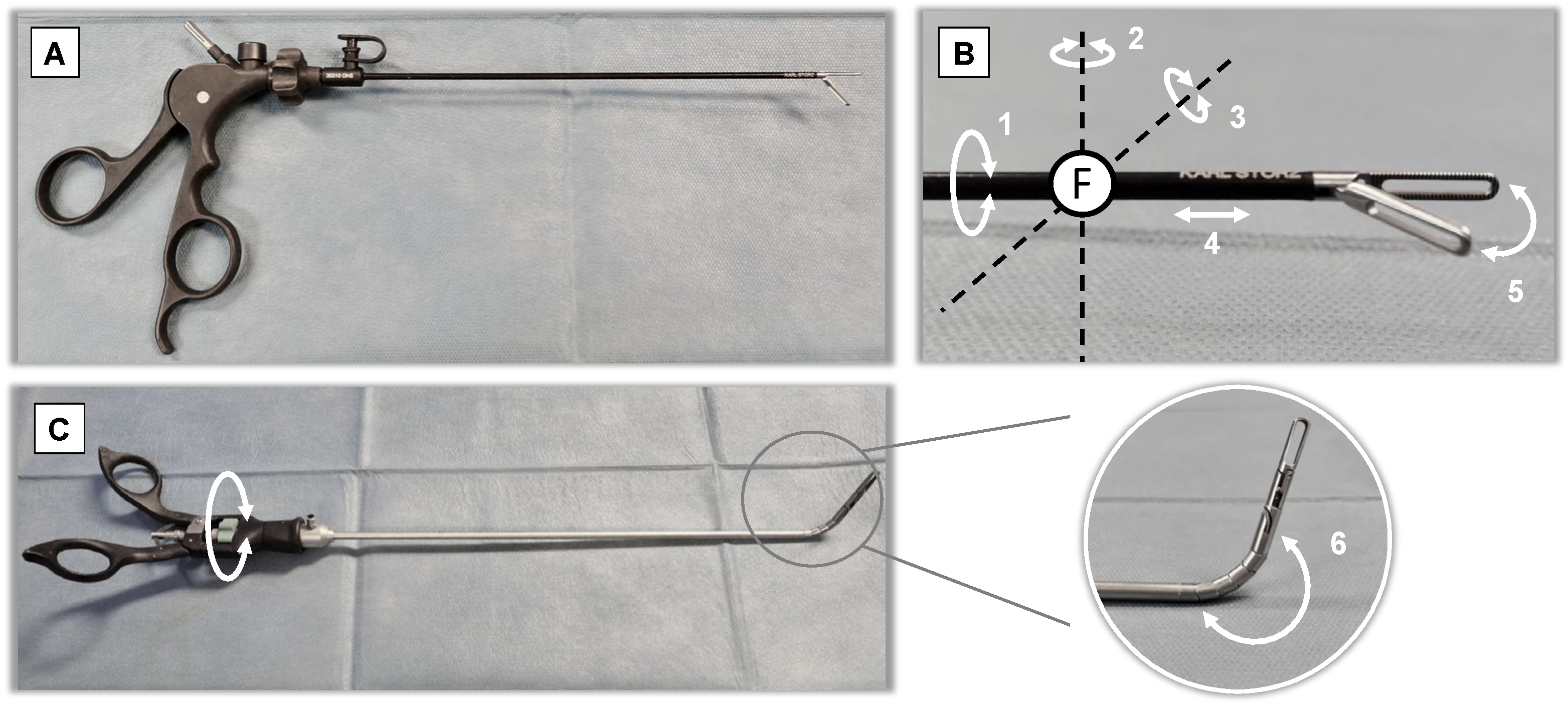
- (a)
- Type of wrist-like mechanism
- (b)
- Type of movements control at the handle
- (c)
- Direction of wrist movement compared to one imparted to the handle
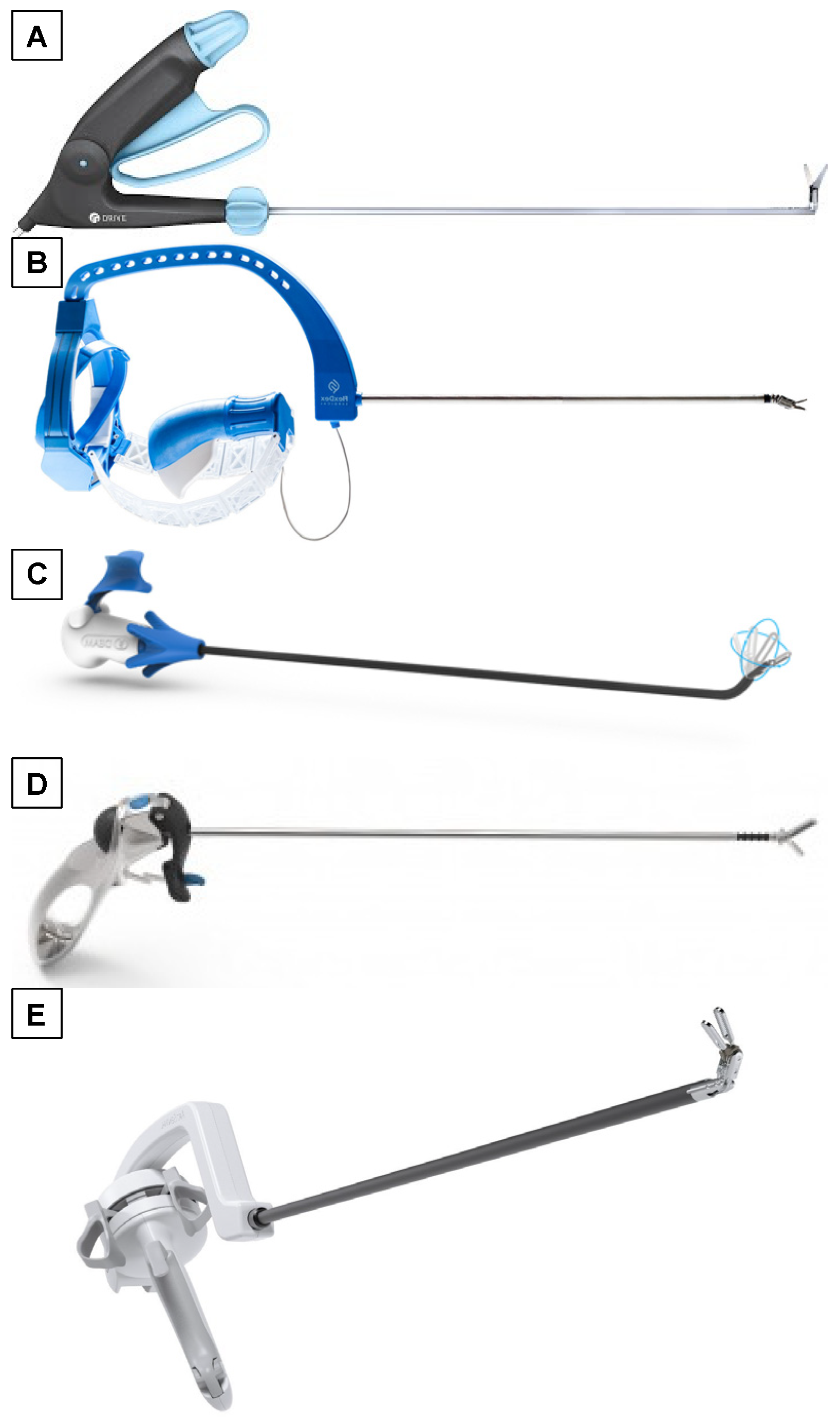
3.1. Radius Surgical System® (Tuebingen Scientific Medical GmbH, Tübingen, Germany)
3.2. FlexDex® (FlexDex Surgical Inc., Brighton, MI, USA)
3.3. LaproFlex® (DEAM, Roden, The Netherlands)
3.4. Intuitool® (UNeMed Corporation, Omaha, NE, USA)
3.5. ArtiSential® (LIVSMED, San Diego, CA, USA)
4. Conclusions
Author Contributions
Funding
Institutional Review Board Statement
Informed Consent Statement
Data Availability Statement
Conflicts of Interest
References
- Anderson, P.L.; Lathrop, R.A.; Webster, R.J., III. Robot-like dexterity without computers and motors: A review of hand-held lapa-roscopic instruments with wrist-like tip articulation. Expert Rev. Med. Devices 2016, 13, 661–672. [Google Scholar] [CrossRef] [PubMed]
- Frede, T.; Hammady, A.; Klein, J.; Teber, D.; Inaki, N.; Waseda, M.; Buess, G.; Rassweiler, J. The Radius Surgical System–A New Device for Complex Minimally Invasive Procedures in Urology? Eur. Urol. 2007, 51, 1015–1022. [Google Scholar] [CrossRef] [PubMed]
- Hirano, Y.; Inaki, N.; Ishikawa, N.; Watanabe, G. Laparoscopic Treatment for Esophageal Achalasia and Gastro-Esophago-reflex Disease Using Radius Surgical System. Indian J. Surg. 2012, 75, 160–162. [Google Scholar] [CrossRef] [PubMed] [Green Version]
- Inaki, N.; Waseda, M.; Schurr, M.O.; Braun, M.; Buess, G.F. Experimental results of mesh fixation by a manual manipulator in a laparoscopic inguinal hernia repair model. Surg. Endosc. 2007, 21, 197–201. [Google Scholar] [CrossRef] [PubMed]
- Awtar, S.; Trutna, T.T.; Nielsen, J.M.; Abani, R.; Geiger, J. FlexDex™: A minimally invasive surgical tool with enhanced dexterity and intuitive control. J. Med. Device. 2010, 4, 035003. [Google Scholar] [CrossRef]
- García-Jiménez, M.L.; Castro-Diez, L.; Aguirrezabalaga-González, J.; Noguera-Aguilar, J.F. Robotic-like suturing with FlexDex Surgical System® for difficult laparoscopic suture. Sutura laparoscópica mecanizada con FlexDex Surgical System® para ubicaciones anatómicamente difíciles. Cir. Esp. 2021, 99, 222–228. [Google Scholar] [CrossRef] [PubMed]
- Khan, M.F.; Murphy, E.; Moynihan, A.; Cahill, R.A. Closure of the retroperitoneal space in laparoscopic anterior resection with FLEXDEX™. Tech. Coloproctology 2019, 23, 1177–1178. [Google Scholar] [CrossRef] [PubMed]
- Gorgen, A.; Araldi, M.; Paludo, A.D.O.; Da Silva, A.; Ghissi, A.; Fernandes, A.; Tavares, P.; Rosito, T.; Cabral, R. Laparoscopic pediatric pyeloplasty using the Flexdex® articulating needle driver: Step-by-step video. J. Pediatr. Urol. 2019, 15, 421–422. [Google Scholar] [CrossRef] [PubMed]
- Khan, M.F.; Murphy, E.; Cahill, R.A. Training pathway for a novel smart surgical system (FlexDex™)-a video vignette. Colorectal Dis. 2020, 22, 469–470. [Google Scholar] [CrossRef] [PubMed]
- Lacitignola, L.; Trisciuzzi, R.; Imperante, A.; Fracassi, L.; Crovace, A.M.; Staffieri, F. Comparison of Laparoscopic Steerable Instru-ments Performed by Expert Surgeons and Novices. Vet. Sci. 2020, 7, 135. [Google Scholar] [CrossRef] [PubMed]
- Rousek, J.B.; Brown-Clerk, B.; Lowndes, B.R.; Balogh, B.J.; Hallbeck, S. Optimizing integration of electrosurgical hand controls within a laparoscopic surgical tool. Minim. Invasive Ther. Allied Technol. 2011, 21, 222–233. [Google Scholar] [CrossRef] [PubMed]
- Trejo, A.; Jung, M.C.; Oleynikov, D.; Hallbeck, M.S. Effect of handle design and target location on insertion and aim with a lapa-roscopic surgical tool. Appl. Ergon. 2007, 38, 745–753. [Google Scholar] [CrossRef] [PubMed]
- Darwich, I.; Scheidt, M.; Koliesnikov, Y.; Willeke, F. Laparoscopic low anterior resection performed using ArtiSential® in an obese male patient with a narrow pelvis—a video vignette. Color. Dis. 2021, 23, 757–758. [Google Scholar] [CrossRef] [PubMed]
- Trevis, J.; Chilvers, N.; Freystaetter, K.; Dunning, J. Surgeon-Powered Robotics in Thoracic Surgery; An Era of Surgical Innovation and Its Benefits for the Patient and Beyond. Front. Surg. 2020, 7. [Google Scholar] [CrossRef] [PubMed]
- Darwich, I.; Abuassi, M.; Weiss, C.; Stephan, D.; Willeke, F. The Artisential® Articulated Laparoscopic Forceps: A Dry Lab Study to Examine Dexterity and Learning Effects in Operators with Different Levels of Laparoscopic Experience. Surg. Technol. Int. 2021. [Google Scholar] [CrossRef] [PubMed]
- Darwich, I.; Aliyev, R.; Koliesnikov, Y.; Willeke, F. Laparoscopic ventral mesh rectopexy performed with ArtiSential®: A video vignette. Tech. Coloproctology 2021, 10, 1–2. [Google Scholar] [CrossRef]
- Kim, A.; Lee, C.M.; Park, S. Is it Beneficial to Utilize an Articulating Instrument in Single-Port Laparoscopic Gastrectomy? J. Gastric Cancer 2021, 21, 38–48. [Google Scholar] [CrossRef] [PubMed]
- Jin, H.Y.; Lee, C.S.; Lee, Y.S. Single Incision Laparoscopic Appendectomy Using a New Multi-joint Articulating Instrument. J. Gastrointest. Surg. 2021, 1–2. [Google Scholar] [CrossRef]
- Jelínek, F.; Gerboni, G.; Henselmans, P.W.; Pessers, R.; Breedveld, P. Attaining high bending stiffness by full actuation in steerable minimally invasive surgical instruments. Minim. Invasive Allied Technol. 2015, 24, 77–85. [Google Scholar] [CrossRef] [PubMed]


Publisher’s Note: MDPI stays neutral with regard to jurisdictional claims in published maps and institutional affiliations. |
© 2021 by the authors. Licensee MDPI, Basel, Switzerland. This article is an open access article distributed under the terms and conditions of the Creative Commons Attribution (CC BY) license (https://creativecommons.org/licenses/by/4.0/).
Share and Cite
Parente, G.; Thomas, E.; Cravano, S.; Di Mitri, M.; Vastano, M.; Gargano, T.; Cerasoli, T.; Ruspi, F.; Libri, M.; Lima, M. ArtiSential® Articulated Wrist-Like Instruments and Their First Application in Pediatric Minimally Invasive Surgery: Case Reports and Literature Review of the Most Commonly Available Robot-Inspired Devices. Children 2021, 8, 603. https://0-doi-org.brum.beds.ac.uk/10.3390/children8070603
Parente G, Thomas E, Cravano S, Di Mitri M, Vastano M, Gargano T, Cerasoli T, Ruspi F, Libri M, Lima M. ArtiSential® Articulated Wrist-Like Instruments and Their First Application in Pediatric Minimally Invasive Surgery: Case Reports and Literature Review of the Most Commonly Available Robot-Inspired Devices. Children. 2021; 8(7):603. https://0-doi-org.brum.beds.ac.uk/10.3390/children8070603
Chicago/Turabian StyleParente, Giovanni, Eduje Thomas, Sara Cravano, Marco Di Mitri, Marzia Vastano, Tommaso Gargano, Tosca Cerasoli, Francesca Ruspi, Michele Libri, and Mario Lima. 2021. "ArtiSential® Articulated Wrist-Like Instruments and Their First Application in Pediatric Minimally Invasive Surgery: Case Reports and Literature Review of the Most Commonly Available Robot-Inspired Devices" Children 8, no. 7: 603. https://0-doi-org.brum.beds.ac.uk/10.3390/children8070603