
MIS-C-Implications for the Pediatric Surgeon: An Algorithm for Differential Diagnostic Considerations


Abstract
:1. Introduction
2. Methods
3. Results
3.1. Patient 1
3.2. Patient 2
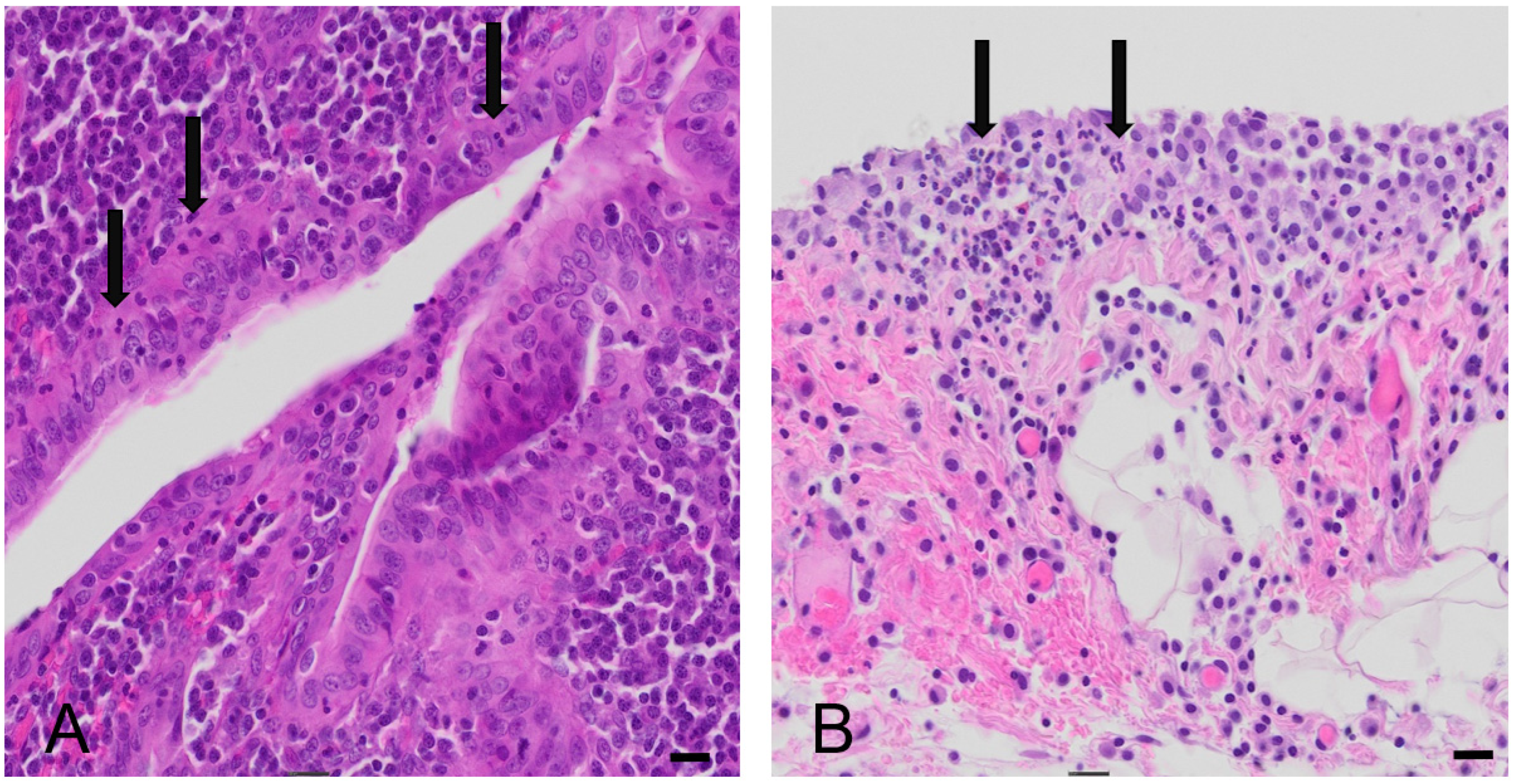
3.3. Patient 3
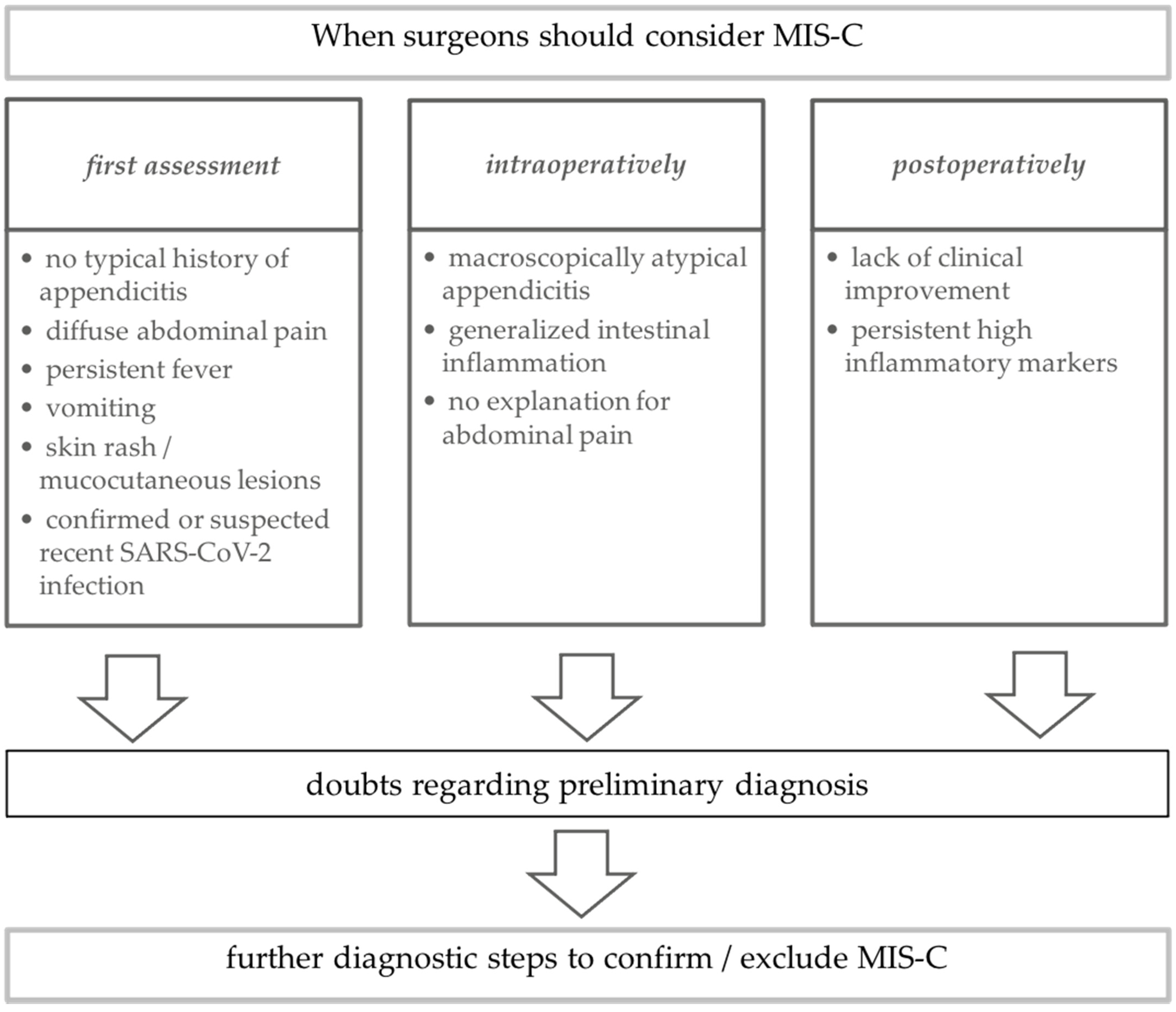
4. Discussion
Author Contributions
Funding
Institutional Review Board Statement
Informed Consent Statement
Data Availability Statement
Conflicts of Interest
References
- Riphagen, S.; Gomez, X.; Gonzalez-Martinez, C.; Wilkinson, N.; Theocharis, P. Hyperinflammatory shock in children during COVID-19 pandemic. Lancet 2020, 395, 1607–1608. [Google Scholar] [CrossRef]
- Verdoni, L.; Mazza, A.; Gervasoni, A.; Martelli, L.; Ruggeri, M.; Ciuffreda, M.; Bonanomi, E.; D’Antiga, L. An outbreak of severe Kawasaki-like disease at the Italian epicentre of the SARS-CoV-2 epidemic: An observational cohort study. Lancet 2020, 395, 1771–1778. [Google Scholar] [CrossRef]
- Licciardi, F.; Pruccoli, G.; Denina, M.; Parodi, E.; Taglietto, M.; Rosati, S.; Montin, D. SARS-CoV-2-Induced Kawasaki-Like Hyperinflammatory Syndrome: A Novel COVID Phenotype in Children. Pediatrics 2020, 146, e20201711. [Google Scholar] [CrossRef] [PubMed]
- Coronavirus Disease 2019 (COVID-19): Epidemiology Update. Available online: https://health-infobase.canada.ca/COVID-19/epidemiological-summary-COVID-19-cases.html (accessed on 22 February 2021).
- European Centre for Disease Prevention and Control. COVID-19 Situation Dashboard. Available online: https://qap.ecdc.europa.eu/public/extensions/COVID-19/COVID-19.html#global-overview-tab (accessed on 22 February 2021).
- The Novel Coronavirus Pneumonia Emergency Response Epidemiology Team. The Epidemiological Characteristics of an Outbreak of 2019 Novel Coronavirus Diseases (COVID-19) in China. Zhonghua Liu Xing Bing Xue Za Zhi 2020, 41, 145–151. [Google Scholar]
- Pakistan Cases Details. COVID-19 Dashboard. Available online: https://covid.gov.pk/stats/pakistan (accessed on 22 February 2021).
- Feldstein, L.R.; Rose, E.B.; Horwitz, S.M.; Collins, J.P.; Newhams, M.M.; Son, M.B.F.; Newburger, J.W.; Kleinman, L.C.; Heidemann, S.M.; Martin, A.A.; et al. Multisystem Inflammatory Syndrome in U.S. Children and Adolescents. N. Engl. J. Med. 2020, 383, 334–346. [Google Scholar] [CrossRef]
- Belot, A.; Antona, D.; Renolleau, S.; Javouhey, E.; Hentgen, V.; Angoulvant, F.; Delacourt, C.; Iriart, X.; Ovaert, C.; Bader-Meunier, B.; et al. SARS-CoV-2-related paediatric inflammatory multisystem syndrome, an epidemiological study, France, 1 March to 17 May 2020. Eurosurveillance 2020, 25, 2001010. [Google Scholar] [CrossRef]
- Jiang, L.; Tang, K.; Levin, M.; Irfan, O.; Morris, S.K.; Wilson, K.; Klein, J.D.; Bhutta, Z.A. COVID-19 and multisystem inflammatory syndrome in children and adolescents. Lancet Infect. Dis. 2020, 20, e276–e288. [Google Scholar] [CrossRef]
- Gottlieb, M.; Bridwell, R.; Ravera, J.; Long, B. Multisystem inflammatory syndrome in children with COVID-19. Am. J. Emerg. Med. 2021, 49, 148–152. [Google Scholar] [CrossRef] [PubMed]
- Abrams, J.Y.; Godfred-Cato, S.E.; Oster, M.E.; Chow, E.J.; Koumans, E.H.; Bryant, B.; Leung, J.W.; Belay, E.D. Multisystem Inflammatory Syndrome in Children Associated with Severe Acute Respiratory Syndrome Coronavirus 2: A Systematic Review. J. Pediatrics 2020, 226, 45–54.e1. [Google Scholar] [CrossRef]
- Radia, T.; Williams, N.; Agrawal, P.; Harman, K.; Weale, J.; Cook, J.; Gupta, A. Multi-system inflammatory syndrome in children & adolescents (MIS-C): A systematic review of clinical features and presentation. Paediatr. Respir. Rev. 2020, 38, 51–57. [Google Scholar] [PubMed]
- Henderson, L.A.; Canna, S.W.; Friedman, K.G.; Gorelik, M.; Lapidus, S.K.; Bassiri, H.; Behrens, E.M.; Ferris, A.; Kernan, K.F.; Schulert, G.S.; et al. American College of Rheumatology Clinical Guidance for Multisystem Inflammatory Syndrome in Children Associated With SARS–CoV-2 and Hyperinflammation in Pediatric COVID-19: Version 1. Arthritis Rheumatol. 2020, 72, 1791–1805. [Google Scholar] [CrossRef]
- Kaushik, A.; Gupta, S.; Sood, M.; Sharma, S.; Verma, S. A Systematic Review of Multisystem Inflammatory Syndrome in Children Associated With SARS-CoV-2 Infection. Pediatric Infect. Dis. J. 2020, 39, e340–e346. [Google Scholar] [CrossRef] [PubMed]
- Aronoff, S.C.; Hall, A.; Del Vecchio, M.T. The Natural History of SARS-CoV-2 Related Multisystem Inflammatory Syndrome in Children (MIS-C): A Systematic Review. J. Pediatric Infect. Dis. Soc. 2020, 9, 746–751. [Google Scholar] [CrossRef] [PubMed]
- Whittaker, E.; Bamford, A.; Kenny, J.; Kaforou, M.; Jones, C.E.; Shah, P.; Ramnarayan, P.; Fraisse, A.; Miller, O.; Davies, P.; et al. Clinical Characteristics of 58 Children With a Pediatric Inflammatory Multisystem Syndrome Temporally Associated with SARS-CoV-2. JAMA 2020, 324, 259–269. [Google Scholar] [CrossRef]
- Cheung, E.W.; Zachariah, P.; Gorelik, M.; Boneparth, A.; Kernie, S.G.; Orange, J.S.; Milner, J.D. Multisystem Inflammatory Syndrome Related to COVID-19 in Previously Healthy Children and Adolescents in New York City. JAMA 2020, 324, 294–296. [Google Scholar] [CrossRef] [PubMed]
- World Health Organization. Multisystem Inflammatory Syndrome in Children and Adolescents Temporally Related to COVID-19. Available online: https://www.who.int/news-room/commentaries/detail/multisystem-inflammatory-syndrome-in-children-and-adolescents-with-COVID-19 (accessed on 23 April 2021).
- Centers for Disease Control and Prevention. Multisystem Inflammatory Syndrome in Children (MIS-C) Associated with Coronavirus Disease 2019 (COVID-19). Available online: https://www.cdc.gov/mis-c/hcp/ (accessed on 23 April 2021).
- Royal College of Pediatrics and Child Health. Guidance: Paediatric Multisystem Inflammatory Syndrome Temporally Associated with COVID-19. Available online: https://www.rcpch.ac.uk/resources/paediatric-multisystem-inflammatory-syndrome-temporally-associated-COVID-19-pims-guidance (accessed on 23 April 2021).
- Morparia, K.; Park, M.J.; Kalyanaraman, M.; McQueen, D.; Bergel, M.; Phatak, T. Abdominal Imaging Findings in Critically Ill Children With Multisystem Inflammatory Syndrome Associated With COVID-19. Pediatric Infect. Dis. J. 2021, 40, e82–e83. [Google Scholar] [CrossRef] [PubMed]
- Valitutti, F.; Verde, A.; Pepe, A.; Sorrentino, E.; Veneruso, D.; Ranucci, G.; Orlando, F.; Mastrominico, A.; Grella, M.G.; Mandato, C. Multisystem inflammatory syndrome in children. An emerging clinical challenge for pediatric surgeons in the COVID 19 era. J. Pediatric Surg. Case Rep. 2021, 69, 101838. [Google Scholar] [CrossRef] [PubMed]
- Anderson, J.E.; Campbell, J.A.; Durowoju, L.; Greenberg, S.L.M.; Rice-Townsend, S.E.; Gow, K.W.; Avansino, J. COVID-19-associated multisystem inflammatory syndrome in children (MIS-C) presenting as appendicitis with shock. J. Pediatric Surg. Case Rep. 2021, 71, 101913. [Google Scholar] [CrossRef]
- Lishman, J.; Kohler, C.; de Vos, C.; van der Zalm, M.M.; Itana, J.; Redfern, A.; Smit, L.; Rabie, H. Acute Appendicitis in Multisystem Inflammatory Syndrome in Children With COVID-19. Pediatric Infect. Dis. J. 2020, 39, e472–e473. [Google Scholar] [CrossRef] [PubMed]
- Garnett, G.M.; Kimball, S.; Melish, M.E.; Thompson, K.S.; Puapong, D.P.; Johnson, S.M.; Woo, R.K. Appendicitis as the presenting manifestation of Kawasaki disease. Pediatric Surg. Int. 2014, 30, 549–552. [Google Scholar] [CrossRef]
- Ulloa-Gutierrez, R.; Gutierrez-Alvarez, R.; Avila-Aguero, M.L. Kawasaki disease mimicking an acute appendicitis. J. Pediatric 2004, 144, 691. [Google Scholar] [CrossRef] [PubMed]
- Harwood, R.; Partridge, R.; Minford, J.; Almond, S. Paediatric abdominal pain in the time of COVID-19: A new diagnostic dilemma. J. Surg. Case Rep. 2020, 2020, rjaa337. [Google Scholar] [CrossRef] [PubMed]
- Jackson, R.J.; Chavarria, H.D.; Hacking, S.M. A Case of Multisystem Inflammatory Syndrome in Children Mimicking Acute Appendicitis in a COVID-19 Pandemic Area. Cureus 2020, 12, e10722. [Google Scholar] [CrossRef] [PubMed]
- Gerall, C.D.; Duron, V.P.; Griggs, C.L.; Kabagambe, S.K.; Maddocks, A.B.; DeFazio, J.R. Multisystem Inflammatory Syndrome in Children Mimicking Surgical Pathologies: What Surgeons Need to Know about MIS-C. Ann. Surg. 2020, 273, e146–e148. [Google Scholar] [CrossRef]
- Meyer, J.S.; Robinson, G.; Moonah, S.; Levin, D.; McGahren, E.; Herring, K.; Poulter, M.; Waggoner-Fountain, L.; Shirley, D.A. Acute appendicitis in four children with SARS-CoV-2 infection. J. Pediatric Surg. Case Rep. 2021, 64, 101734. [Google Scholar] [CrossRef]
- Khesrani, L.S.; Chana, K.; Sadar, F.Z.; Dahdouh, A.; Ladjadj, Y.; Bouguermouh, D. Intestinal ischemia secondary to COVID-19. J. Pediatric Surg. Case Rep. 2020, 61, 101604. [Google Scholar] [CrossRef]


| WHO | CDC | Royal College | P 1 | P 2 | P 3 | |
|---|---|---|---|---|---|---|
| Systematic inflammation | fever ≥ 3 days and elevated inflammatory parameters (ESR, CRP, PCT) | fever ≥ 38 °C for ≥ 24 h, elevated inflammatory parameters | persistent fever > 38.5°, lymphadenopathy, elevated inflammatory parameters | yes | yes | yes |
| Skin and mucosa | rash or bilateral conjunctivitis or mucocutaneous signs | rash, muco-cutaneous lesions | rash, conjunctivitis/swollen hands and feet | yes | yes | no |
| Fluid balance | hypotension or shock | hypotension or shock | hypotension | yes | no | no |
| Heart | laboratory or echocardio-graphical findings of myocardial dysfunction, pericarditis, valvulitis, coronary abnormalities | laboratory findings of cardiac dysfunction, myocarditis | laboratory, ECG or echocardiographical findings of myocardial dysfunction, pericarditis, valvulitis, coronary abnormalities | yes | yes | yes |
| Coagulation | evidence of coagulopathy (PT, PTT, D-dimers) | Elevated d-dimers and fibrinogen | High d-dimers, abnormal fibrinogen | yes | yes | yes |
| Gastro-intestinal tract | diarrhea, vomiting, abdominal pain | diarrhea, vomiting | diarrhea, vomiting, abdominal pain, ultrasound with colitis, ileitis, lymphadenopathyascites, hepato-splenomegaly | yes | yes | yes |
| Other organ systems | acute kidney injury | yes | yes | no | ||
| exclusion of microbial cause | no alternative plausible diagnosis | exclusion of microbial cause including toxic shock syndromes | yes | yes | yes | |
| Evidence of SARS-CoV-2 | PCR, antigen test, serology or likely contact with SARS-CoV-2 positive person | positive PCR, serology or antigen test or exposure within 4 weeks prior to onset of symptoms | positive SARS-CoV-2-PCR | yes | yes | yes |
Publisher’s Note: MDPI stays neutral with regard to jurisdictional claims in published maps and institutional affiliations. |
© 2021 by the authors. Licensee MDPI, Basel, Switzerland. This article is an open access article distributed under the terms and conditions of the Creative Commons Attribution (CC BY) license (https://creativecommons.org/licenses/by/4.0/).
Share and Cite
Manz, N.; Höfele-Behrendt, C.; Bielicki, J.; Schmid, H.; Matter, M.S.; Bielicki, I.; Holland-Cunz, S.; Gros, S.J. MIS-C-Implications for the Pediatric Surgeon: An Algorithm for Differential Diagnostic Considerations. Children 2021, 8, 712. https://0-doi-org.brum.beds.ac.uk/10.3390/children8080712
Manz N, Höfele-Behrendt C, Bielicki J, Schmid H, Matter MS, Bielicki I, Holland-Cunz S, Gros SJ. MIS-C-Implications for the Pediatric Surgeon: An Algorithm for Differential Diagnostic Considerations. Children. 2021; 8(8):712. https://0-doi-org.brum.beds.ac.uk/10.3390/children8080712
Chicago/Turabian StyleManz, Nora, Claudia Höfele-Behrendt, Julia Bielicki, Hanna Schmid, Matthias S. Matter, Isabella Bielicki, Stefan Holland-Cunz, and Stephanie J. Gros. 2021. "MIS-C-Implications for the Pediatric Surgeon: An Algorithm for Differential Diagnostic Considerations" Children 8, no. 8: 712. https://0-doi-org.brum.beds.ac.uk/10.3390/children8080712