
Treatment of an Avulsed and Ankylosed Incisor through Single Tooth Alveolar Osteotomy and Conventional Orthodontic Mechanisms

 and
and 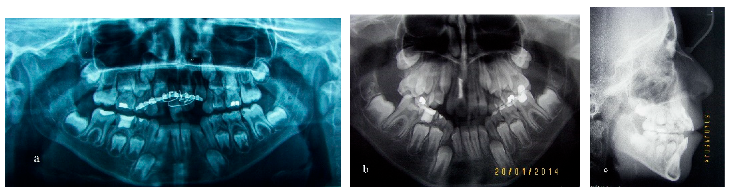
Abstract
:1. Introduction
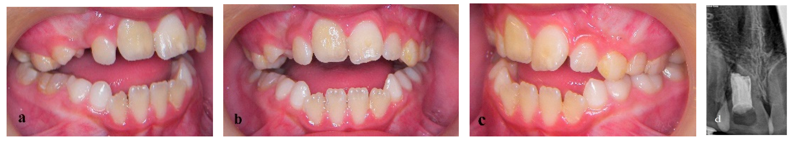
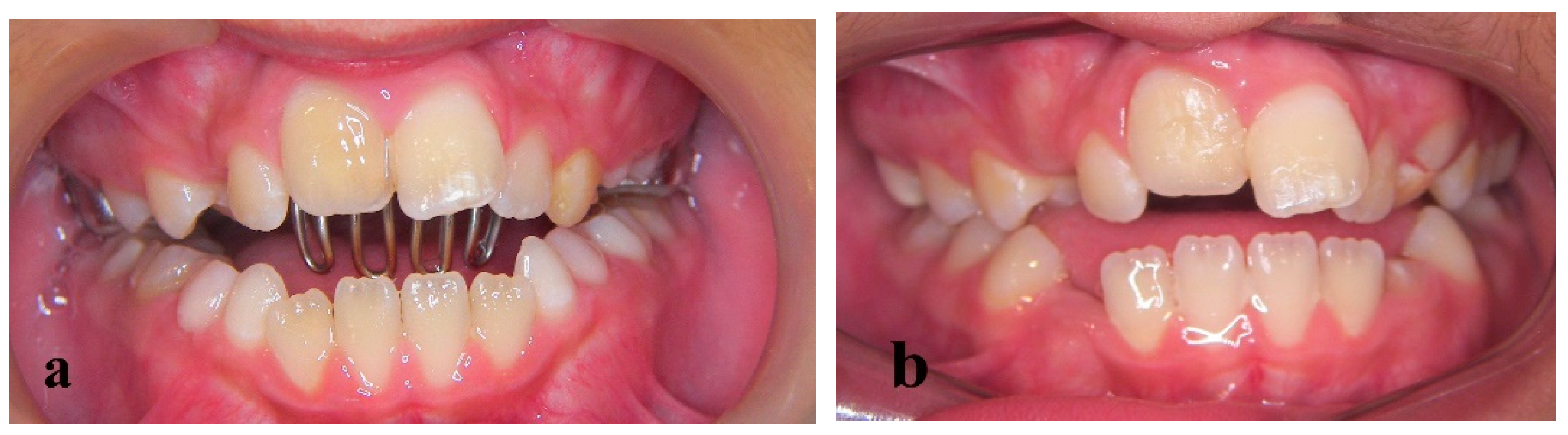
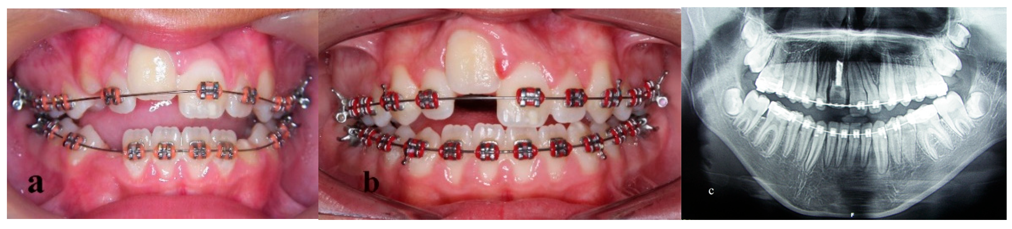
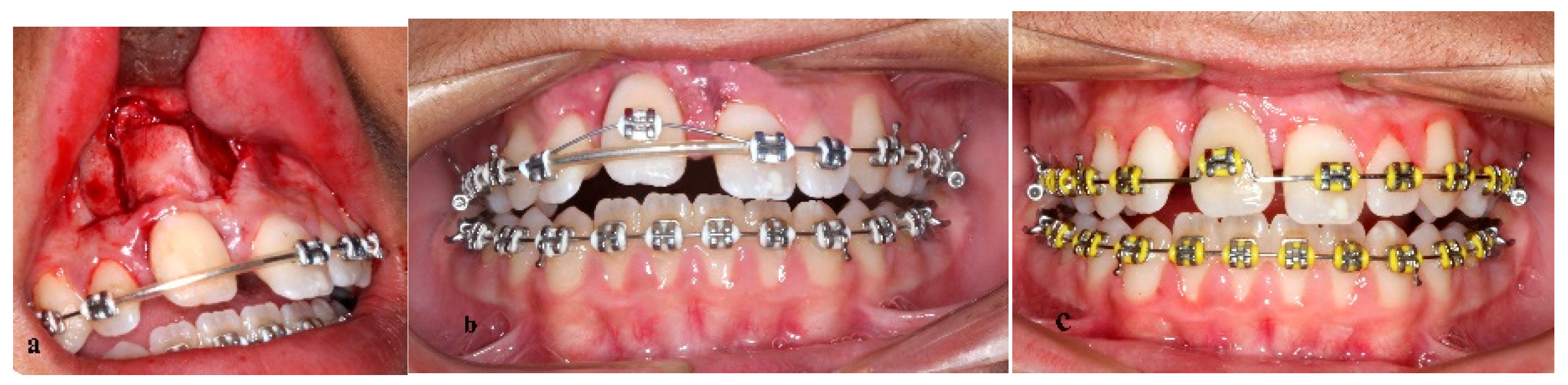
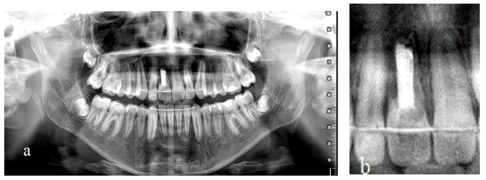
2. Materials and Methods
3. Results
4. Discussion
5. Conclusions
Author Contributions
Funding
Institutional Review Board Statement
Informed Consent Statement
Data Availability Statement
Conflicts of Interest
References
- Forsberg, C.M.; Tedestam, G. Etiological and predisposing factors related to traumatic injuries to permanent teeth. Swed. Dent. J. 1993, 17, 183–190. [Google Scholar] [PubMed]
- Bauss, O.; Freitag, S.; Rohling, J.; Rahman, A. Influence of overjet and lip coverage on the prevalence and severity of incisor trauma. J. Orofac. Orthop. 2008, 69, 402–410. [Google Scholar] [CrossRef] [PubMed]
- Andreasen, J.O.; Andreasen, F.M. Textbook and Color Atlas of Traumatic Injuries to the Tooth, 3rd ed.; Munksgaard: Copenhagen, Denmark, 1994; pp. 587–663. [Google Scholar]
- Delmar, D.A. Ankylosis of teeth in the developing dentition. Quintessence Int. 1986, 17, 303–308. [Google Scholar]
- Takahashi, T.; Takagi, T.; Moriyama, K. Orthodontic treatment of a traumatically intruded tooth with ankylosis by traction after surgical luxation. Am. J. Orthod. Dentofac. Orthop. 2005, 127, 233–241. [Google Scholar] [CrossRef] [PubMed]
- Malmgren, B. Ridge Preservation/Decoronation. J. Endod. 2013, 39, S67–S72. [Google Scholar] [CrossRef]
- Epker, B.N.; Paulus, P.J. Surgical-orthodontic correction of adult malocclusions: Single-tooth dento-osseous osteotomies. Am. J. Orthod. 1978, 74, 551–563. [Google Scholar] [CrossRef]
- Medeiros, P.; Bezerra, A. Treatment of an ankylosed central incisor by single-tooth dento-osseous osteotomy. Am. J. Orthod. Dentofac. Orthop. 1997, 112, 496–501. [Google Scholar] [CrossRef]
- Isaacson, R.J.; Strauss, R.A.; Bridges-Poquis, A.; Peluso, A.R.; Lindauer, S.J. Moving an ankylosed central incisor using orthodontics, surgery and distraction osteogenesis. Angle Orthod. 2001, 71, 411–418. [Google Scholar]
- Alcan, T. A miniature tooth-borne distractor for the alignment of ankylosed teeth. Angle Orthod. 2006, 76, 77–83. [Google Scholar]
- Kofod, T.; Wurtz, V.; Melsen, B. Treatment of an ankylosed central incisor by single tooth dento-osseous osteotomy and a simple distraction device. Am. J. Orthod. Dentofac. Orthop. 2005, 127, 72–80. [Google Scholar] [CrossRef]
- Chang, H.Y.; Chang, Y.L.; Chen, H.L. Treatment of a severely ankylosed central incisor and a missing lateral incisor by distraction osteogenesis and orthodontic treatment. Am. J. Orthod. Dentofac. Orthop. 2010, 138, 829–838. [Google Scholar] [CrossRef]
- Gomes, J.C.; Gomes, C.C.; Bolognese, A.M. Clinical and histological alterations in the surrounding periodontium of dog’s teeth submitted for an intrusive luxation. Dent. Traumatol. 2008, 24, 332–336. [Google Scholar] [CrossRef]
- Yang, Y.; Bai, Y.; Li, S.; Li, J.; Gao, W.; Ru, N. Effect of early orthodontic force on periodontal healing after autotransplantation of permanent incisors in beagle dogs. J. Periodontol. 2012, 83, 235–241. [Google Scholar] [CrossRef]
- Ferreira, M.M.; Ferreira, H.M.; Botelho, F.; Carrilho, E. Autotransplantation combined with orthodontic treatment: A case involving the maxillary central incisors with root resorption after traumatic injury. Restor. Dent. Endod. 2015, 40, 236–240. [Google Scholar] [CrossRef]
- Fields, H.W.; Christensen, J. Orthodontic Procedures after Trauma. J. Endod. 2013, 39, S78–S87. [Google Scholar] [CrossRef]
- Ebrahim, F.H.; Kulkarni, G. Fixed Orthodontic Appliances in the Management of Severe Dental Trauma in Mixed Dentition: A Case Report. J. Can. Dent. Assoc. 2013, 79, d131. [Google Scholar]
- Tome, W.; Uematsu, S.; Yamashiro, T. Multidisciplinary treatment for a patient with traumatically intruded permanent canine and premolar. Aust. Dent. J. 2015, 60, 536–539. [Google Scholar] [CrossRef]
- Kindelan, S.A.; Day, P.F.; Kindelan, J.D.; Spencer, J.R.; Duggal, M.S. Dental trauma: An overview of its influence on the management of orthodontic treatment: Part 1. J. Orthod. 2008, 35, 68–78. [Google Scholar] [CrossRef]
- Wickwire, N.A.; McNeil, M.H.; Norton, L.A.; Duell, R.C. The effects of tooth movement upon endodontically treated teeth. Angle Orthod. 1974, 44, 235–242. [Google Scholar]
- Hunter, M.L.; Hunter, B.; Kingdon, A.; Addy, M.; Dummer, P.M.; Shaw, W.C. Traumatic injury to maxillary incisor teeth in a group of South Wales school children. Endod. Dent. Traumatol. 1990, 6, 260–264. [Google Scholar] [CrossRef]
- Remington, D.N.; Joondeph, D.R.; Artun, J.; Riedel, R.A.; Chapko, M.K. Long-term evaluation of root resorption occurring during orthodontic treatment. Am. J. Orthod. Dentofac. Orthop. 1989, 96, 43–46. [Google Scholar] [CrossRef]
- Spurrier, S.W.; Hall, S.H.; Joondeph, D.R.; Shapiro, P.A.; Riedel, R.A. A comparison of apical root resorption during orthodontic treatment in endodontically treated and vital teeth. Am. J. Orthod. Dentofac. Orthop. 1990, 97, 130–134. [Google Scholar] [CrossRef]
- Esteves, T.; Ramos, A.L.; Pereira, C.M.; Hidalgo, M.M. Orthodontic root resorption of endodontically treated teeth. J. Endod. 2007, 33, 119–122. [Google Scholar] [CrossRef] [PubMed]
- Andersson, L.; Andreasen, J.O.; Day, P.; Heithersay, G.; Trope, M.; DiAngelis, A.J.; Kenny, D.J.; Sigurdsson, A.; Bourguignon, C.; Flores, M.T.; et al. International Association of Dental Traumatology guidelines for the management of traumatic dental injuries: 2—Avulsion of permanent teeth. Dent. Traumatol. 2012, 28, 88–96. [Google Scholar] [CrossRef]
- Adnan, S.; Lone, M.M.; Khan, F.R.; Hussain, S.M.; Nagi, S.E. Which is the most recommended medium for the storage and transport of avulsed teeth? A systematic review. Dent. Traumatol. 2018, 34, 59–70. [Google Scholar] [CrossRef] [Green Version]
- De Brier, N.O.D.; Borra, V.; Singletary, E.M.; Zideman, D.A.; De Buck, E.; International Liaison Committee on Resuscitation First Aid Task Force. Storage of an avulsed tooth prior to replantation: A systematic review and meta-analysis. Dent. Traumatol. 2020, 36, 453–476. [Google Scholar] [CrossRef]
- Zaleckiene, V.; Peciuliene, V.; Brukiene, V.; Drukteinis, S. Traumatic dental injuries: Etiology, prevalence and possible outcomes. Stomatologija 2014, 16, 7–14. [Google Scholar] [PubMed]
- Malmgren, O.; Malmgren, B.; Goldson, L. Orthodontic management of the traumatized dentition. In Textbook and Colour Atlas of Traumatic Injuries to the Teeth; Andreasen, J., Andreasen, F., Eds.; Munksgard: Copenhagan, Denmark, 1994. [Google Scholar]
- American Academy of Pediatric Dentistry (AAPD). Guidelines on Management of Acute Dental Trauma; American Academy of Pediatric Dentistry: Chicago, IL, USA, 2011. [Google Scholar]
- Barrett, E.J.; Kenny, D.J. Survival of avulsed permanent maxillary incisors in children following delayed replantation. Endod. Dent. Traumatol. 1997, 13, 269–275. [Google Scholar] [CrossRef]
- Andreasen, J.O.; Malmgren, B.; Bakland, L.K. Tooth avulsion in children: To replant or not. Endod. Top. 2006, 14, 28–34. [Google Scholar] [CrossRef]
- Cohenca, N.; Stabholz, A. Decoronation: A conservative method to treat ankylosed teeth for preservation of alveolar ridge prior to permanent prosthetic reconstruction—literature review and case presentation. Dent. Traumatol. 2006, 23, 87–94. [Google Scholar] [CrossRef]
- Hagg, U.; Taranger, J. Skeletal stages of the hand and wrist as indicators of the pubertal growth spurt. Acta Odontol. Scand. 1980, 38, 187–200. [Google Scholar] [CrossRef]
- Jensen, S.S. Timing of implant placement after traumatic dental injury. Dent. Traumatol. 2019, 35, 376–379. [Google Scholar] [CrossRef] [Green Version]
- Toledano-Serrabona, J.; Sánchez-Garcés, M.Á.; Sánchez-Torres, A.; Gay-Escoda, C. Alveolar distraction osteogenesis for dental implant treatments of the vertical bone atrophy: A systematic review. Med. Oral Patol. Oral Cir. Bucal 2019, 24, e70–e75. [Google Scholar] [CrossRef]
- Kinzinger, G.S.; Janicke, S.; Riediger, D.; Diedrich, P.R. Orthodontic fine adjustment after vertical callus distraction of an ankylosed incisor using the floating bone concept. Am. J. Orthod. Dentofac. Orthop. 2003, 124, 582–590. [Google Scholar] [CrossRef]
- Kim, Y.; Park, S.B.; Son, W.S.; Kim, S.; Kim, Y.D.; Mah, J. Treatment of an ankylosed maxillary incisor by intraoral alveolar bone distraction osteogenesis. Am. J. Orthod. Dentofac. Orthop. 2010, 138, 215–220. [Google Scholar] [CrossRef]
- Doshi, U.H.; Mahindra, R.K.; Gaffar, K.A.A. Modified hyrax screw for treatment of ankylosed incisor by applying alveolar distraction and floating bone concept. J. World Fed. Orthod. 2013, 2, e31–e36. [Google Scholar] [CrossRef]
- Senışık, N.E.; Koçer, G.; Kaya, B.Ü. Ankylosed maxillary incisor with severe root resorption treated with a single-tooth dento-osseous osteotomy, vertical alveolar distraction osteogenesis, and mini-implant anchorage. Am. J. Orthod. Dentofac. Orthop. 2014, 146, 371–384. [Google Scholar] [CrossRef]
- Dolanmaz, D.; Karaman, A.; Pampu, A.; Topkara, A. Orthodontic treatment of an ankylosed maxillary central incisor through osteogenic distraction. Angle Orthod. 2010, 80, 391–395. [Google Scholar] [CrossRef]
- Mayne, R.; Mayne, R.; Saranathan, S.; Spencer, K. Single tooth osteotomy and distraction in the treatment of an ankylosed central incisor—A case report. Aust. Orthod. J. 2017, 33, 280–291. [Google Scholar] [CrossRef]
- Im, J.J.; Kye, M.K.; Hwang, K.G.; Park, C.J. Miniscrew-anchored alveolar distraction for the treatment of the ankylosed maxillary central incisor. Dent. Traumatol. 2010, 26, 285–288. [Google Scholar] [CrossRef]


Publisher’s Note: MDPI stays neutral with regard to jurisdictional claims in published maps and institutional affiliations. |
© 2022 by the authors. Licensee MDPI, Basel, Switzerland. This article is an open access article distributed under the terms and conditions of the Creative Commons Attribution (CC BY) license (https://creativecommons.org/licenses/by/4.0/).
Share and Cite
Vasoglou, G.; Markomanolaki, C.C.; Vasoglou, M.; Markomanolakis, A. Treatment of an Avulsed and Ankylosed Incisor through Single Tooth Alveolar Osteotomy and Conventional Orthodontic Mechanisms. Children 2022, 9, 732. https://0-doi-org.brum.beds.ac.uk/10.3390/children9050732
Vasoglou G, Markomanolaki CC, Vasoglou M, Markomanolakis A. Treatment of an Avulsed and Ankylosed Incisor through Single Tooth Alveolar Osteotomy and Conventional Orthodontic Mechanisms. Children. 2022; 9(5):732. https://0-doi-org.brum.beds.ac.uk/10.3390/children9050732
Chicago/Turabian StyleVasoglou, Georgios, Chrysi Christina Markomanolaki, Michail Vasoglou, and Andreas Markomanolakis. 2022. "Treatment of an Avulsed and Ankylosed Incisor through Single Tooth Alveolar Osteotomy and Conventional Orthodontic Mechanisms" Children 9, no. 5: 732. https://0-doi-org.brum.beds.ac.uk/10.3390/children9050732