Review

A Comprehensive Review of Recent Advancements in Cancer Immunotherapy and Generation of CAR T Cell by CRISPR-Cas9

1
Biotechnology, Medical School, University of Pécs, 7624 Pecs, Hungary
2
Department of Genetic Engineering and Biotechnology, Faculty of Biological Science and Technology, Jashore University of Science and Technology (JUST), Jashore 7408, Bangladesh
3
ABEx Bio-Research Center, East Azampur, Dhaka 1230, Bangladesh
4
Department of Biochemistry and Molecular Biology, Life Science Faculty, Bangabandhu Sheikh Mujibur Rahman Science and Technology University, Gopalganj 8100, Bangladesh
5
Agricultural Biotechnology, Hungarian University of Agriculture and Life Sciences, 2100 Godollo, Hungary
6
Department of Biotechnology and Genetic Engineering, Bangabandhu Sheikh Mujibur Rahman Science and Technology University, Gopalganj 8100, Bangladesh
7
Global Biotechnology & Biomedical Research Network (GBBRN), Department of Biotechnology and Genetic Engineering, Faculty of Biological Sciences, Islamic University, Kushtia 7003, Bangladesh
8
Department of Pathology, College of Korean Medicine, Kyung Hee University, Seoul 02447, Korea
9
Korean Medicine-Based Drug Repositioning Cancer Research Center, College of Korean Medicine, Kyung Hee University, Seoul 02447, Korea
10
Pharmaceutical Biotechnology Laboratory, Department of Genetic Engineering and Biotechnology, Faculty of Biological Science and Technology, Jashore University of Science and Technology (JUST), Jashore 7408, Bangladesh
*
Authors to whom correspondence should be addressed.
These authors contributed equally to the work.
Submission received: 15 November 2021 / Revised: 9 December 2021 / Accepted: 15 December 2021 / Published: 23 December 2021
(This article belongs to the Section Biological Processes and Systems)

Abstract

:
The mechanisms involved in immune responses to cancer have been extensively studied for several decades, and considerable attention has been paid to harnessing the immune system’s therapeutic potential. Cancer immunotherapy has established itself as a promising new treatment option for a variety of cancer types. Various strategies including cancer vaccines, monoclonal antibodies (mAbs), adoptive T-cell cancer therapy and CAR T-cell therapy have gained prominence through immunotherapy. However, the full potential of cancer immunotherapy remains to be accomplished. In spite of having startling aspects, cancer immunotherapies have some difficulties including the inability to effectively target cancer antigens and the abnormalities in patients’ responses. With the advancement in technology, this system has changed the genome-based immunotherapy process in the human body including the generation of engineered T cells. Due to its high specificity, CRISPR-Cas9 has become a simple and flexible genome editing tool to target nearly any genomic locus. Recently, the CD19-mediated CAR T-cell (chimeric antigen receptor T cell) therapy has opened a new avenue for the treatment of human cancer, though low efficiency is a major drawback of this process. Thus, increasing the efficiency of the CAR T cell (engineered T cells that induce the chimeric antigen receptor) by using CRISPR-Cas9 technology could be a better weapon to fight against cancer. In this review, we have broadly focused on recent immunotherapeutic techniques against cancer and the use of CRISPR-Cas9 technology for the modification of the T cell, which can specifically recognize cancer cells and be used as immune-therapeutics against cancer.

1. Introduction

Cancer is the major increasing curse for the human population throughout the world and is the second leading cause of death after cardiovascular disease. Thus, researchers are trying to ascertain safe and efficient therapies for cancer treatment, among them, “immunotherapy” (i.e., enhancing the immunity of the body to fight against cancer) is the foremost therapeutic approach [1]. Basically, it provides much potential and is a more stable treatment strategy than other traditional cancer treatments. During the last decades, this cutting age approach has provided a significant response against cancer [2]. In immunotherapy techniques, T cells, NK cells and dendritic cells are directly involved to fight against the rapidly proliferating cancer cells, but they need modification to work effectively against cancer cells [3]. Immune checkpoint inhibitors have been shown to have exceptional effectiveness against a wide spectrum of solid and hematological malignancies by restoring malfunctioning or exhausted T cells [3,4].
Tumor-infiltrating lymphocyte (TIL), chimeric antigen receptor (CAR) T cell and engineered natural killer cell (NK) therapy, as well as immunomodulators such as engineered T-cell receptor (TCR) and antibody therapy, are the common immunotherapy strategies for the effective treatment of cancer [5,6]. The future of cancer treatment can be accelerated by CAR T (chimeric antigen receptor T)-cell immunotherapy, in which cells are generated by modifying T cells’ receptors. These modified T cells express functionalized proteins known as CAR T receptors, which assist the CAR-expressing immune cell in effectively recognizing tumor antigens [7]. In recent times, many research activities have demonstrated that B-cell lymphoma patients have shown decisive responses by using CAR T-cell therapies, using CD19-directed chimeric antigen receptor T cells (CART19) [8,9]. In the USA, the FDA authorized three CD19-directed genetically engineered autologous T cells named as Kymriah, Yescarta and liso-cel (Lisocabtagene maraleucel) that were applied in patients who possessed acute lymphoblastic leukemia (ALL) and certain types of non-Hodgkin lymphoma (NHL), in some respects [10]. Liso-cel (Lisocabtagene maraleucel) has outstanding features: it is genetically engineered, has a specific structure, autologous cellular immunotherapy and, most interestingly, is CD19-directed [11]. The liso-cel CAR is mainly composed of diverse components such as a single-chain variable fragment, transmembrane domain of CD28, G4 hinge region of an immunoglobulin, activation domain (CD3ζ) and 4-1BB co-stimulatory domain [12]. However, CAR liso-cel possesses a non-functional receptor and is enabled to be co-expressed with the CAR receptor, and it actively binds to the CD19 marker of B-cell lymphoma. Here, they can inhibit the random proliferation of B cells’ activation, and directly show the cytotoxic effect on harmful target cells [13]. A recent study from Abramson et al., 2020, showed the safety and potential activity of CAR T liso-cel for patients with large B-cell lymphomas, along with the positive aspects of liso-cel treatment, which were a low incidence of severe cytokine release syndrome and neurological problems [14]. Moreover, cancer patients have shown hopeful results by following chimeric antigen receptor (CAR) T-cell treatment and engineered T-cell receptor (TCR) technology [15,16]. All of these successful results were reported during B-cell malignancies’ treatment. Solid tumors that contain complex inhibitory receptors can hardly be removed. The activity of T cells is hampered by exhaustion when patients are affected with chronic infections and cancers. Generally, the resistant properties of T cells stimulate the induction of CAR T cells, which can inhibit the proliferation of solid tumors [17].
The CRISPR-Cas9 technique can be used as a genetic alteration and multiple genomes editing tool that can enhance the efficacy of CAR T cells so they can effectively recognize the solid tumor antigens [18,19]. The CRISPR-Cas9 system exhibits a great result in cancer treatment by editing the cancer genomes, removing or reducing the activity of carcinogenic viral infections [20]. Unlike other gene editing techniques (TALENs or ZFNs), CRISPR-Cas9 utilizes single guided RNA (sgRNA) to recognize the target and then modify the DNA in a very precise manner [21]. It is very important to design the CAR T cells precisely so that their therapeutic capability becomes more selective and specific, otherwise, they may generate cytokine release syndrome, which could result in a serious consequence for the patient [22]. For this reason, we described here the highly efficient genome editing tool CRISPR-Cas9 for increasing the function of T cells against cancer.
In this review article, we have broadly explained the importance of diverse immunotherapy strategies including chimeric antigen receptor (CAR) T-cell therapy as well as the tumor-infiltrating lymphocyte (TIL), engineered T-cell receptor (TCR) therapy and engineered natural killer (NK) cell therapy. In addition, we have demonstrated the potential of several immunomodulators, antibody-mediated therapy, cancer vaccine formation, nanoparticle-based cancer immunotherapy and inhibition of T cell exhaustion. Most importantly, we have strongly focused on the strategy of T cell modification through using the CRISPR-Cas9 genome editing tool.

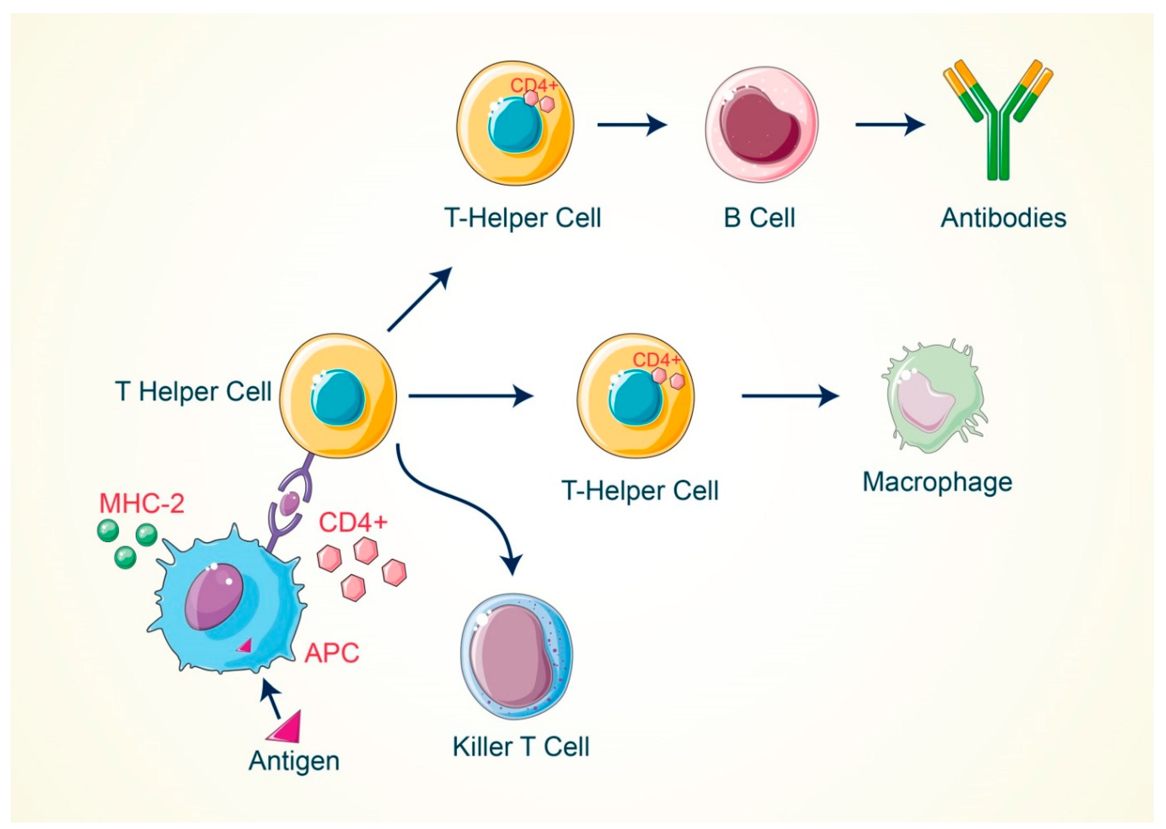
2. Activation of T Cell by Antigenic Response

An adult human has around 4 × 10¹¹ T cells in blood circulation [23], which contain numerous T-cell receptors (TCR) [24] on their surface. In the blood stream, T cells are assigned to immunity at the cell level of the tissue, while B cells have dual responsibility for both tissue and bodily fluid immunity. A diverse group of cells such as T helper cells and cytotoxic T cells belong to the T cell group as a whole and execute different types of function for regulating the immune system in the body [25]. A T cell is activated by an antigen-mediated process that is responsible for cellular progression and the transformation of primary T cells into matured cells via the T-cell receptor (TCR) [26]. TCR and antigen interaction is needed for T cell activation and multiplication, but phosphorylation processes are critical for early signal transduction, which requires an extra signal, known as co-stimulation [27]. Most potent co-stimulatory signals come from the nonpolymorphic surface receptor of a T cell, which is denoted as CD28 [27,28]. It was also demonstrated that anti-CD28 antibodies can inhibit T cell activation and proliferation [29,30,31,32]. B7-1 and B7-2 ligands for CD28 are expressed on antigen-presenting cells and are amplified when these cells come into contact with pathogens that activate Toll-like receptors or other receptors’ signals responsible for sensing those pathogens [33,34]. During immunological responses, inhibitory molecules such as CTLA4 and PD1 are produced and serve as a “checkpoint” to reduce T cell hyperactivation [35]. A complex of three sets (ε–δ, γ–δ and ζ– ζ) of dimeric CD3 chains transmits signals from the polymorphic TCR [36]. CD3 chains include immunoreceptor tyrosine-based activation motives that are phosphorylated by the SRC family kinase of lymphocyte specific protein kinases (LCK) [37]. The CD45 protein signaling from the surface displays phosphatase activity, which inhibits the LCK function of the resting T cell [38]. CD45 removes the inhibitory phosphate from LCK after activation of the T cell [39], then LCK permits phosphorylation of ZAP70, which binds to the CD3 ζ-chain and recruits PLCγ and the linker for activation of T cells (LAT) [40]. Following adequate co-stimulation, signaling cascades are initiated, which influence ca2+ release and activate GTPase RAS and, finally, regulate the transcription for T cell function [41]. Antigen-presenting cells (APC) have a protein on their cell surface that attaches to the T cell receptors (TCRs), and this complex of protein and receptor is known as signal phase-1 in the case of T cell activation. Moreover, the major histocompatibility complex (MHC) of the antigen-presenting cells (APC) (Figure 1) attaches to the T-cell receptors [42]. Signal phase-2 of the T cell activation happens when the co-stimulatory proteins attach to the co-activating molecules of the T cell on the surface of the APC. In this cascade, the most significant protein B7 (B-lymphocyte activation protein B7) executes an important role by activating the T cell as B7 protein attaches to the co-stimulatory proteins of the T cell. T cells cannot damage the body’s own proteins as B7 protein is not produced in the normal cells of the body [43]. The co-stimulatory response is more effective for the activation/inactivation of the T cell response. Here, B7–CD28 interaction leads to the activation of the T cell, where B7 is found on the APC surface and CD28 is a marker of the T cell. On the contrary, B7–CTLA-4 interaction regulates the suppression of the T cell [44], and, therefore, co-stimulatory molecules play a more vital role in the activation of T cells.
When MHC-I attaches to the cytotoxic (CD8) T cells, then it is activated and plays a crucial role in the lysis of target cells. On the other hand, the helper (CD4) T cells are activated by attaching the MHC-II, which results in multiple downstream effects [45]. The body’s immunity is regulated by CD4 cells as well as complex and normal immune activities. A group of scientists have demonstrated that CD4 cells are reduced in numbers or do not work in AIDS patients. In contrast, CD4 cells can overproduce inflammatory cytokines when they are more effective [46].

3. Significance of T Cell in Cancer Treatment

In the human body there are three types of immune cells, known as the T cells (thymus-dependent cells), the B cells (bone marrow-mediated cells) and the natural killer cells (NK cells), which fight to damage cancer cells [47]. T lymphocytes are originated by the bone marrow and their maturation process is regulated in the thymus, which plays significant role in body’s immunity and fight against cancer cells. Importantly, there are diverse types of T cells that are responsible for damaging cancer cells, such as the cytotoxic T cells, helper T cells and regulator T cells [48]. However, Cytotoxic T cells (CTL) play a pivotal role in the minimization of a malignant tumor or cancer: they release particular cytokines such as TNF-alpha and IFN-gamma, and, hence, both are involved in the activation of cellular macrophages, which attack the tumor cells and clean them from the body [49]. Conversely, CTL are directly involved in the damage of cancer cells via the secretion of lytic granules, mainly perforin and granzymes, after the recognition of faulty cancerous protein [50]. Notably, helper T cells (Th cells) indirectly boost the functions of other immune cells to damage the cancer cells [51].
T-cell therapy for cancer treatment mainly relies on the ability to genetically engineer cells with targeted antigen specificity and then induce the cell to proliferate by preserving their effector function and homing abilities. Mouse models have usually been used for the identification and preclinical optimization of tumor therapy; nevertheless, no mouse models have been found that can be used as good predictors of a successful human response to immunotherapy. In recent times, substantial limitations in some mouse models have been found that have important indications for T-cell cancer therapy. In particular cases, it has been reported that many mouse tumor antigens do not represent human tumor antigens [52], which has led to strategies to generate transferred human T cells with the appropriate specificity of tumor. T-cell therapy can be used as an efficient treatment for viral infections and the early stages of cancer. Antiviral immunological processes are complicated and involve both adaptive and innate responses mediated by CD8+T cells, whose specific immunosurveillance functions have been examined in a variety of clinical and experimental conditions. The rapid, powerful, cytotoxic and non-cytolytic antiviral actions of CD8+T cells are certainly crucial to defending the host from various viral diseases [53]. Recent studies focused on cellular immunology and tumor biology have shown new strategies for adoptive T-cell therapy. For example, the use of engineered T cells has been tested as a strategy to increase the function of memory T cells and effector T cells through manipulating the host to suppress the immune toxic effect in the tumor microenvironment, with a promising result in the early stages of clinical trials [54]. Many research studies have reported that using the T cell for cancer treatment shows effective results and this plays a significant role in cancer therapy [54]. Adoptive T-cell therapy is used for the goal of eliminating a tumor and its recurrence, which consists of various mature T cells. Lymphocytes with inhibitory effects on the growth of cancer cells were observed in many patients who are potential candidates for immunotherapy [55]. The main advantages of using T cells for immunotherapy for cancer treatment instead of other cytolytic cells, such as NK cells, is that T cells have the ability to bind specifically on target tumor cells by the recognition of differentially expressed tumor proteins, which are present on the cell surface. As the T cells have a long clonal life span, this allows for both immunoprophylactic and therapeutic scenarios, and is also a great advantage for cancer treatment. Moreover, T cells can be well suited for genetic manipulation, which enables the evaluation of genetically-enhanced T cells for cancer and other diseases.

4. The Causes of T Cell Failure for Recognizing Cancer Antigens

The immune system plays a vital role against cancer cells. It always searches for diseased and imperfect cells for killing as its target cells. Most cancer patients possess a weak immune system, which is why specific cancers cannot be detected by the body’s immune cells [56]. As T cells possess weak activity in some cases, cancer cells may evade the defensive immunity and be the primary cause of cancer development. The excessive occurrence of free radicals and protein oxidation in the body causes DNA damage [57], which also reduces the potential activity of T cells to effectively recognize the cancer antigens. For example, the CHOP protein (C/EBP-homologous protein) is involved in the cellular stress response and myeloid immune cell response [58]. Additionally, the tissue of ovarian cancer patients expresses more CHOP than normal healthy tissues. CHOP proteins can alter the immune system to form cancer [59]. The activities of the CHOP proteins of T cells have been determined by researchers on mice models via a comprehensive set of laboratory trials [60]. Moreover, the CHOP level is increased when a T cell is activated and the CHOP also causes a negative T cell regulation process. A research study found that antitumor CD8+T cell immunity is boosted when the CHOP gene in T cells is deleted, and, thus, T cell-based immunotherapy became more successful in immunologically-developed mice models [59]. In normal conditions, the CHOP helps to balance the antitumor T-cell response [61]. Moreover, tumors can alter the general activities of the CHOP to decrease T-cell immunity. A small number of immune systems permit cancer cells to bypass the antitumor immunity activities of T cells, which can cause cancer outgrowth and development [62]. There are several others factors that inhibit the T cell’s immunity such as the low production of (IL-2) Interleukin-2, an abundance of proinflammatory cytokines, the high-grade chronic infection, improper function of Treg cells and T cell exhaustion [62,63]. Through the mechanism of T cell exhaustion, dysfunction of the T cells occurs, which mainly happens in various types of chronic diseases and also in cancers [64]. Furthermore, this is also a condition of poor effector action and the sustainable expression of inhibitory receptors. The exhausting of T cells inhibits the optimal monitoring of infections and tumors [65]. Exhausted T cells detect inhibitory receptor overexpression in the tumor microenvironment, which reduces effector cytokine production as well as the cytolytic activities of cancer cells in the body, allowing them to remove cancer cells from the body [66]. The activity of exhausted T cells is lost, which is identified by an altered transcriptional program. PD-1, Cytotoxic T-lymphocyte antigen 4 (CTLA-4), lymphocyte activated gene-3 (LAG-3) and domain-containing protein-3 have been reported as playing an important role in T cell exhaustion [67]. T cell exhaustion can be reversed by blocking the PD-1 or CTLA-4 checkpoint. By following this system, the T cells become resistant to the different inhibitory pathways [67].

5. The Current Immunotherapies That Are Used in Cancer Treatment

Current cancer immunotherapy has the potential to inhibit the growth of cancerous cells by enhancing the body’s immunity. Tumor antigens are found on the cell surface of tumor cells that encounter the antibody, but the self-antibody is able to destroy the cancer cells. In this manner, we need modified immunological components that potentially encounter the tumor cells and are able to manage the metastasis of cancer [64]. Here, we illustrate the major approaches of cancer immunotherapy and demonstrate the mechanisms as to how they are followed to treat a wide variety of cancers and summarized (Table 1).

5.1. Adoptive Cell Therapy

Adoptive cell therapy is a form of treatment strategy that uses the cells of our immune system to eradicate the cancer of our body, mainly known as cellular immunotherapy. Cellular immunotherapy approaches directly involve the isolating of our own immune cells and simply increasing their numbers, whereas others require gene therapy for genetically engineering immune effector cells to enhance their anticancer capabilities [66]. Significant cancer immunotherapy approaches are illustrated here as tumor-infiltrating lymphocyte (TIL), engineered T-cell receptor (TCR), chimeric antigen receptor (CAR) T-cell and engineered natural killer cell (NK) therapy.

5.1.1. Tumor-Infiltrating Lymphocyte (TIL)

Naturally occurring T cells are powerful immune cells in our immune system that are engaged to fight against cancer cells, but the “killer-like T cells” are enabled to recognize and alleviate the cancer cells in a very particular way. To effectively kill cancer cells and activity for a durable period in order to sustain an effective antitumor response, killer T cells need to be harvested by ex vivo expansion from cancer patients to activate and expand the T cell numbers. Potentially, the huge amount of activated T cells are re-infused into patients and can directly encounter and destroy tumor cells [68]. Recent clinical reports proposed that tumor-infiltrating lymphocytes (TILs) have been extensively used for patients who developed solid tumors, metastatic melanoma, ovarian cancer, renal cell carcinoma, colorectal cancer, pancreatic cancer, and so on [69]. A clinical research study of the use of tumor-infiltrating lymphocytes (TILs) on cutaneous melanoma patients reported that TIL grade is a strong predictor of survival and sentinel lymph node (SLN) condition in patients with melanoma, and patients with a significant TIL impact have an increased survival rate [70]. Donastas et al., 2019 [71], noted that tumor-infiltrating lymphocytes (TILs) showed potential anticancer activity on HLA-matched ovarian cancer cell lines, which was prior or post resistant to chemotherapy. In addition, many other research studies have demonstrated that tumor-infiltrating lymphocytes (TILs) show strong anticancer activity on both the breast cancer and triple negative breast cancer research model [72,73].

5.1.2. Engineered T-Cell Receptor (TCR) Therapy

In some cases, T cells are unable to counter the advanced forms of cancer, so we requisition engineered T-cell receptors for action against solid tumors. This approach takes T cells from cancer patients and introduces the engineered novel receptors that enable them to target specific cancer antigens and tumor lysis, and eradicate the tumor cells. Here we declare that engineered T cells demonstrate their outstanding functions and their longevity in the tumor microenvironment [74]. Engineered T-cell receptors (TCRs) are composed of the α and β chains that are associated with δ, ε and γ chains, and the largest signaling regions of this receptor are known as the ζ chains. These novel T-cell receptors encounter the tumor antigens via the MHC-I/MHC-II and, with the advantage of engineered T cells, are able to identify suitable target antigens, overcome immunosuppressive tumor microenvironments, prevent antigen escape and reduce toxicities [75].
Adoptive immunotherapy with TCR-engineered T cells has been identified as a significant strategy for cancer treatment, with promising results from recent clinical trials [76]. This evolution was also demonstrated in clinical trials involving MART-1 TCR-engineered T cells in 2009 and 2014 [77,78]. Johnson et al. demonstrated that 19% of patients who were treated with T cells engineered with the gp100 TCR had an effective anticancer response [77]. Many other research studies have identified that patients with metastatic melanoma, multiple myeloma, colorectal carcinoma and synovial sarcoma possessed significant survival rates after treatment with TCR-engineered T cells [79,80,81].

5.1.3. Engineered Natural Killer (NK)-Cell Therapy

NK cell therapy involves augmenting the capability of NK cell antitumor responses via the introduction of antigen specificity by using genetic modification. Herein, CAR NK cells’ basic structural framework has similarity to CAR T cells, mainly CAR composed of extracellular, hinge, transmembrane as well as intracellular domains. The extracellular domain of CARs is ScFvs, which interact with the hinge domain and the transmembrane domains connected between the hinge and intracellular domains, such as CD3ζ and/or CD28. In addition to that fact, CAR-mediated NK cells require co-stimulatory molecules such as CD28, 4-1BB and CD134 in order to increase the proliferation and cytotoxicity effect against the solid tumor [82]. These cutting-edge approaches (CAR NK therapy) might be potential substitutes for the existing time-consuming technology CAR T-cell therapy. Here, CAR NK-cell therapy is more significant due to some basic criteria, for example, it is less expensive, easy to be isolated and secretes safer cytokines (IFN-γ and GM-CSF) [83]. Throughout the past years, many research groups have developed NK-92 cells for expressing different CARs, including CD19 and CD20 on B-cell leukemia/lymphoid, CD38 and CS-1 on pancreatic cancer and HER-2 for endothelial malignancy [84,85,86,87]. Additionally, CAR-modified NK-92 cells can always be injected orally, enabling them to migrate to targeted tumor tissue and exhibit their actual impact through a vaccine-like technique [88]. In addition, although transduction efficiency varies widely with primary NK cells, the translation of NK-92 cells is more consistent, contributing significantly to the homogeneity of the cell line. NK-92 cells have an average transduction efficiency from around 50%, even though non-viral techniques such as electrophoresis or nucleofection are used [89,90].

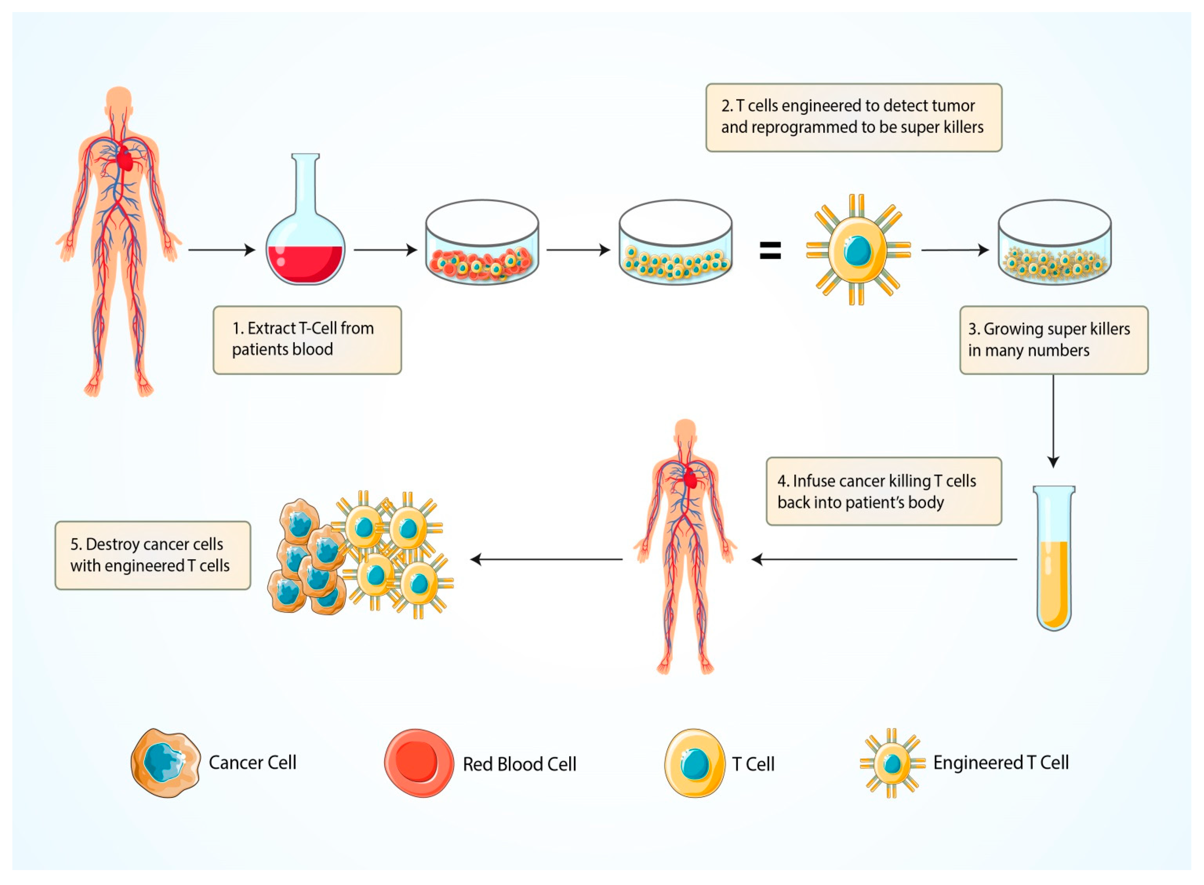
5.1.4. Chimeric Antigen Receptor (CAR) T-Cell Therapy

CAR T-cell therapy depends on efficient, stable and safe gene transfer platforms. Cancer patient T cells can be isolated through leukapheresis and are harvested and genetically modified by ex vivo expansion through viral and non-viral transfection methods. The CAR T cell consists of the extracellular antigen-binding moieties that could be a single-chain fragment variable (scFv) portion consisting of the variable heavy (VH) and variable light (VL) chains of an antibody, and fused by a peptide spacer. It is interlinked to an intracellular signaling molecule/domain, i.e., TCR CD3ζ signaling chain or immune receptor tyrosine-based activation motif (ITAM)-containing protein that are bound in tandem with co-stimulation signals such as CD28 or 4-1BB [91]. At the start of CAR T-cell therapy the focus is on the recognition of unprocessed antigens and carbohydrate, as well as glycolipid structures that are present on the tumor cells surface. However, both types of CAR T cells, such as CD4+ and/or CD8+ T cells, are recruited and redirected to the target site of the cancer without the expression of MHC-I and MHC-II. These two effective T cells perform the major killing mechanism by cytolysis via perforin and granzyme secretion and, in some cases, follow the death mechanism by expressing the Fas/Fas-ligand (Fas-L) and/or TNF/TNF-receptor (TNF-R) [92]. Feins S et al. (2019) also illustrated that CAR T cells act against the CD19 protein of tumor cells of acute lymphoblastic leukemia and can diffuse large B-cell lymphoma, and, thus, this T-cell therapy can be used for the treatment of cancers. Personalized cancer immunotherapy using engineered chimeric antigen receptor (CAR) gene-transduced T-cell (CAR T) therapies has shown significant potential in the development of highly personalized interventional cancer immunotherapy. The first FDA-approved CAR T therapy to treat re-occurring or refractory B-cell acute lymphoblastic leukemia in the US has recently been approved, named as Novartis’ Kymriah (tisagenlecleucel), which highlights the potential of CAR T-cell-based immunotherapy for the treatment of cancer [93]. With the ability to target different components of the tumor ecosystem, CAR T cells are an effective instrument to target different components of the tumor environment, including malignant cells and their microenvironment [94,95,96].

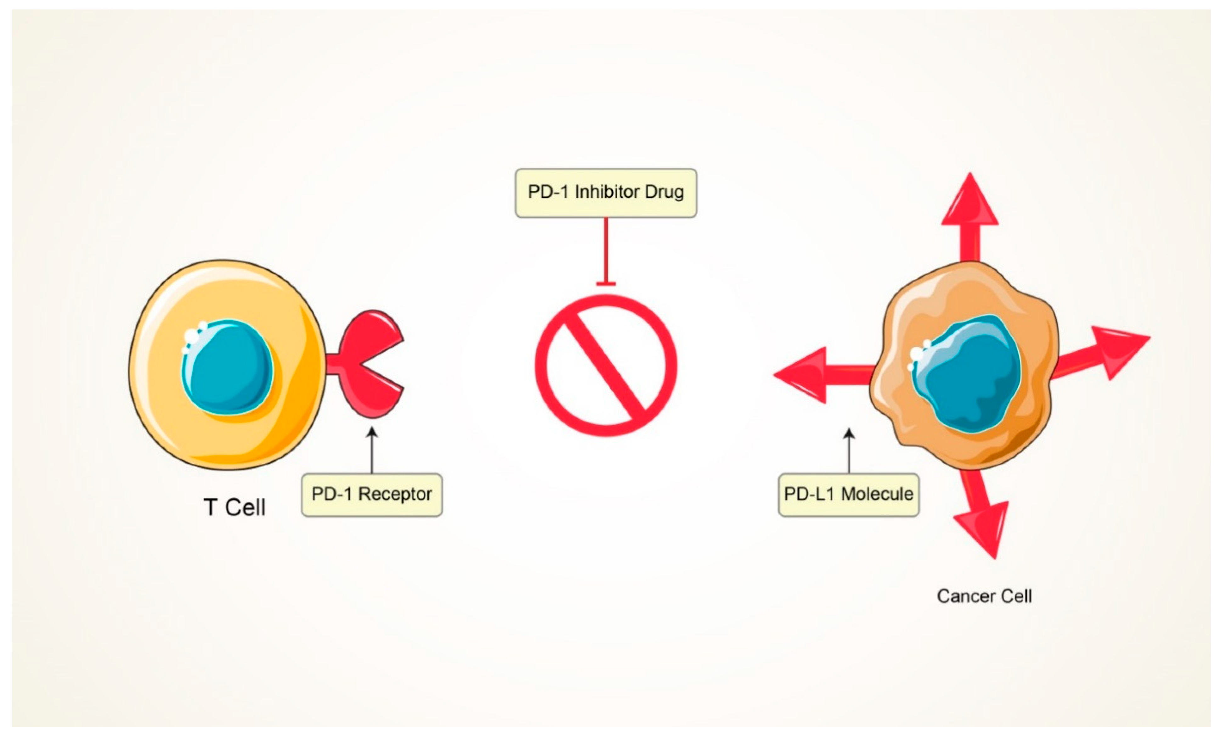
5.2. Immunomodulators

Immunomodulators are directly interlinked to the modification of the immune system and along with these stimulatory molecules enable checkpoint blockers [97], enhance cytokines secretion and act as an agonist for blocking cancer progression and enhancing the potential activity of immune cells. Basically, cytotoxic T lymphocyte-associated antigen 4 (CTLA-4) manifests on the surface of T cells and regulatory T cells (Tregs) and counteracts the activity of the T-cell co-stimulatory receptor (CD28). After recognition of the antigen, the CD28 molecules strongly amplify TCR signaling for the T cells activation. Here, we can clearly say that CD28 and/or CTLA-4 represent identical ligands such as CD80 and CD86 [98]. Notably, CTLA-4 has a higher affinity for both ligands (CD80 and CD86) and expression of the ligands on the surface of T cells dampens the activation of T cells by outcompeting CD28, as well as actively delivering inhibitory signals to the T cell [99]. On the contrary, programmed death-1 (PD-1)/programmed death ligand-1 (PDL-1) play crucial roles in tumor progression and the survival of tumors in the tumor microenvironment. Mainly, PD-1 is found on several immune cells such as T cells, B cells, dendritic cells as well as monocytes [100]. Tumor cells and antigen-presenting cells express PDL-1 and interact with the T cells’ PD-1 and may cause T cell dysfunction, neutralization and exhaustion. It has been reported that PDL-1 is expressed on tumor cells and can escape the cytotoxic T cell-mediated cell killing and develop a tumor mass in the body [101]. However, when checkpoint inhibitors inhibit the pathway of PD-1/PDL-1 and CTL-4, they are capable of helping T cells to inhibit the advancement of tumor mass.
Cytokines are proteins or proteolytic enzymes secreted by immune cells that act as mediators of immunity and directly modulate immunomodulatory cells. Immunostimulatory cytokines such as IFNs, IL-2, IL-12, IL-15 and IL-18 are known to enhance immune responses against cancer [102]. The major cytokine IL-2 is able to promote the expansion of helper T cells (CD4+ T) and NK cells and facilitates the synthesis of Ig-type antibodies. The crucial role of IL-2 is in the Fas-mediated activation of CD4+ T cells that induce the cancerous cell’s death [103]. The research study by Dranoff G [104] reported that two major cytokines, namely IL-4 and IL-7, directly engage in the enhancement of the T cell’s function, but IL-4 activates eosinophil and eradicates the cancer cells from the body. IL-15 is needed for the activation, proliferation and cytotoxic action of NK cells and CD8+ T cells, which are able to release cytokines such as IFN-γ, leading to potential antitumor activity. Importantly, IFN-α is a specialized cytokine to use for the treatment of several malignancies and solid tumors through the maturation of dendritic cells and T lymphocytes [105]. Many research studies have now established our understanding of the immunological system and clinical evidence is available for the regulation of the immunologic reaction to malignancies. The role of bacille Calmette–Guerin (BCG) in the management of superficial bladder cancer has been demonstrated [106]. Intravenous BCG instillation causes an inflammatory reaction to the bladder epithelium that leads to inflammation cell recruitment and cytokine production [107]. Metastatic melanoma and renal cell cancer patients who receive IL-2 experience long-term responses, potentially cures [108]. Chemotherapy long-term survivors frequently relapse, while IL-2 responders always remain disease-free. Thus, immunomodulation appears to have been successful in priming the immune system to reject any recurrent tumor cells prior to the onset of clinical disease [109].

5.3. Antibody-Mediated Therapy

Antibodies are proteinaceous compounds that act against cell surface markers or antigens and protect us from several threats produced by B cells. Antibodies such as monoclonal antibodies (MAbs) have a significant cytotoxic effect against tumor cell surface antigens and modify the signal transduction cascade pathway within the tumor cells through the complement-dependent cytotoxicity (CDC) and/or antibody-dependent cellular cytotoxicity (ADCC) [110]. In the CDC pathway, MAbs directly modulate the classic complement pathway, when the complement component C1 recognizes the fragment crystallizable region (Fc region) of antibodies and activates C3a, C3b. The C3a recruit immune effector cells at the complement activation site, but C5 convertase is activated by the complement component C3b via the activation of an alternative pathway that forms the membrane attack complex (MAC) to lysis the tumor cells [111]. The process of ADCC involves specific antibodies binding to the immune effector cells such as monocytes, NK cells and other leukocytes to kill the antigen specific tumor cells. Therapeutic antibodies engage with the Fc receptor of the immune effector cells via the Fc region of antibodies, for example, the MAbs Fc region attach the FcγRIII receptors on NK cells for activation and trigger the NK cell to secrete perforin and granzyme resulting in the death of cancer cells. Moreover, NK cells release proinflammatory cytokines due to the recruitment of adaptive immune cells and the inhibitory action plays against the cancer cells [112]. The research study by Christian P. Pallasch and his colleagues noted that the antibody-mediated therapy showed significant anticancer properties in the tumor microenvironment that possessed resistance activity toward several chemotherapeutic drugs [113]. Monoclonal antibody-mediated treatment induced tumor regression through apoptosis [114]. Jessica Marlind et al., 2008, [115] reported that in breast cancer treatment, the antibody-mediated insertion of IL-2 effectively enhanced the efficiency of chemotherapeutics drugs. In addition, several research studies have shown that the antibody-mediated therapy exhibits new promise for the potential treatment of pancreatic cancer and hepatocellular cancer [116,117].

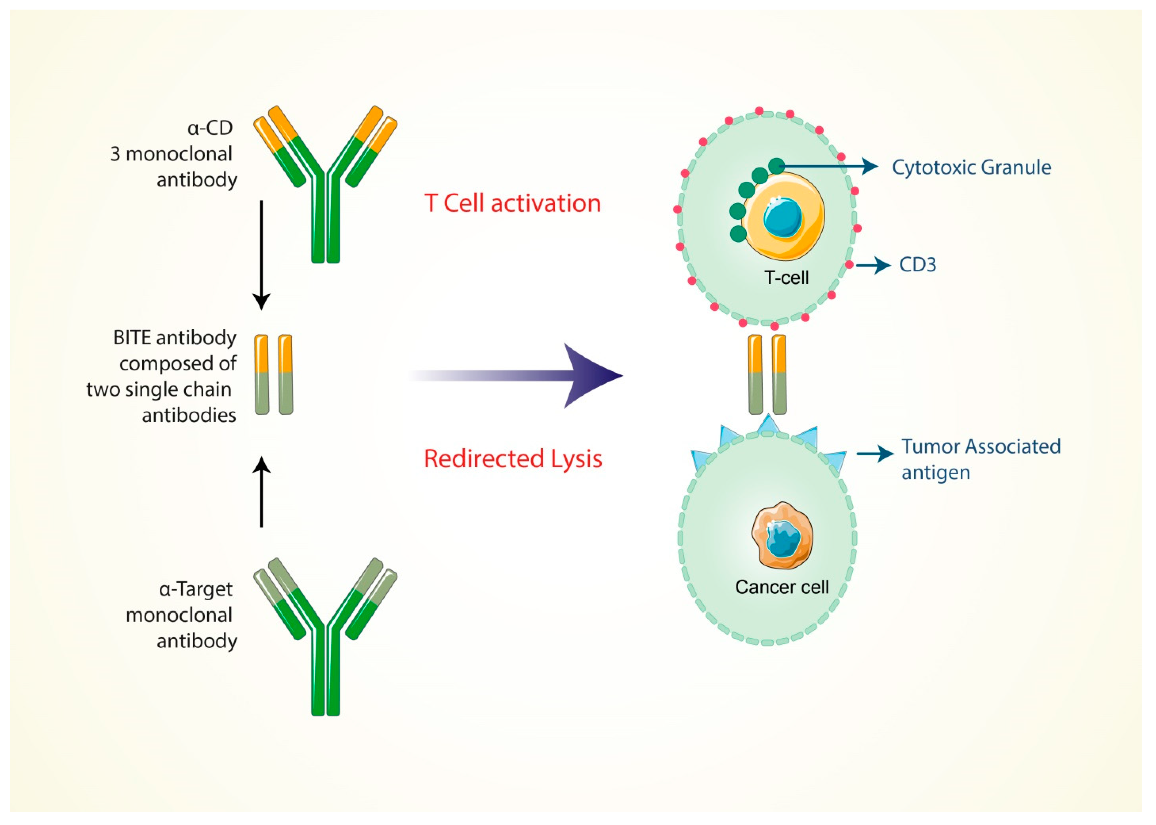
5.4. Formation of Bi-Specific T Cell-Engaging Antibodies for Cancer Therapy

Many research activities have indicated that in both early and late phases of the diseases, T cells are capable of controlling the enhancement of tumor development and progression in cancer patients [118]. However, there are some difficulties because the T cell responses for tumor specific antigens are complex to develop and maintain in cancer patients. Moreover, when immunoediting is taking place, the responses are confined by some of the immune regulatory mechanisms for the selective tumor cells [119]. The production of antibodies is an effective strategy to assemble T cells for the immune-therapeutic treatment of cancers, and these types of antibodies are expressed both on the surface specific antigen of every type of cancer cells and for the CD3 complex on T cells. These types of antibodies are efficient at engaging with any kind of cytotoxic T cells to damage the cancer cells [120].
Independently, the production of antibodies can enhance the specificity of the T-cell receptor, co-stimulation or presentation of peptide antigens. Various research activities have demonstrated that, among bi-specific antibodies derived from T cells, the BITE (bi-specific T-cell engager) antibodies show a promising efficacy in the treatment of both bulky and minimal residual disease [120], and now it is FDA approved for treating several cancers. Kufer and colleagues’ research project showed that the special design of the CD3/target antigen-associated bi-specific antibodies has a very high rate of efficiency and can also involve CD8+ and CD4+T cells to redirect the cancer cell lysis process to the target (E–T ratio) ratios [121]. When the bi-specific antibodies bind only the CD3-specific branch, this shows low efficiency as the monovalent antibodies cannot trigger the T-cell signaling by CD3 (Figure 2). Contrary to this, the “bi-specific T-cell engager” (BITE) antibody is activated to the T cell through the multivalent strategy by the target cells [122,123]. In an in vitro study, the BITE antibody was found to be highly effective at redirecting the damaging response of cancer cells. Additionally, the drug’s potent antitumor activity has been demonstrated in a variety of animal models [124,125].
The BITE antibodies (MT110) were tested in a clinical trial with an ovarian cancer patient who, at late stage, had developed ovarian cancer cell metastasis [126]. Moreover, they were tested as a stage 2 clinical trial of cancer patients who had b-precursor acute lymphoblastic leukemia (B-All) and showed minimum accessorial disease in the patient’s bone marrow [127]. Another BITE antibody, named as MTT110, which is now in the clinical development phase, is bi-specific only, especially for the CD3 complex and the epithelial cell adhesion molecule (EpCAM) [128]. It is currently being clinically tested in patients with lung and gastrointestinal cancer as a part of the clinical trial one. The expression efficiency of the epithelial cell adhesion molecule (EpCAM) has, therefore, been shown to be high on several squamous cell carcinoma types, human adenocarcinoma and cancer stem cells. It has certain limitations because the animal model did not identify the CD3 modulation of T cells by treating BITE antibodies to suppress the cytotoxic granules of T cells [129].

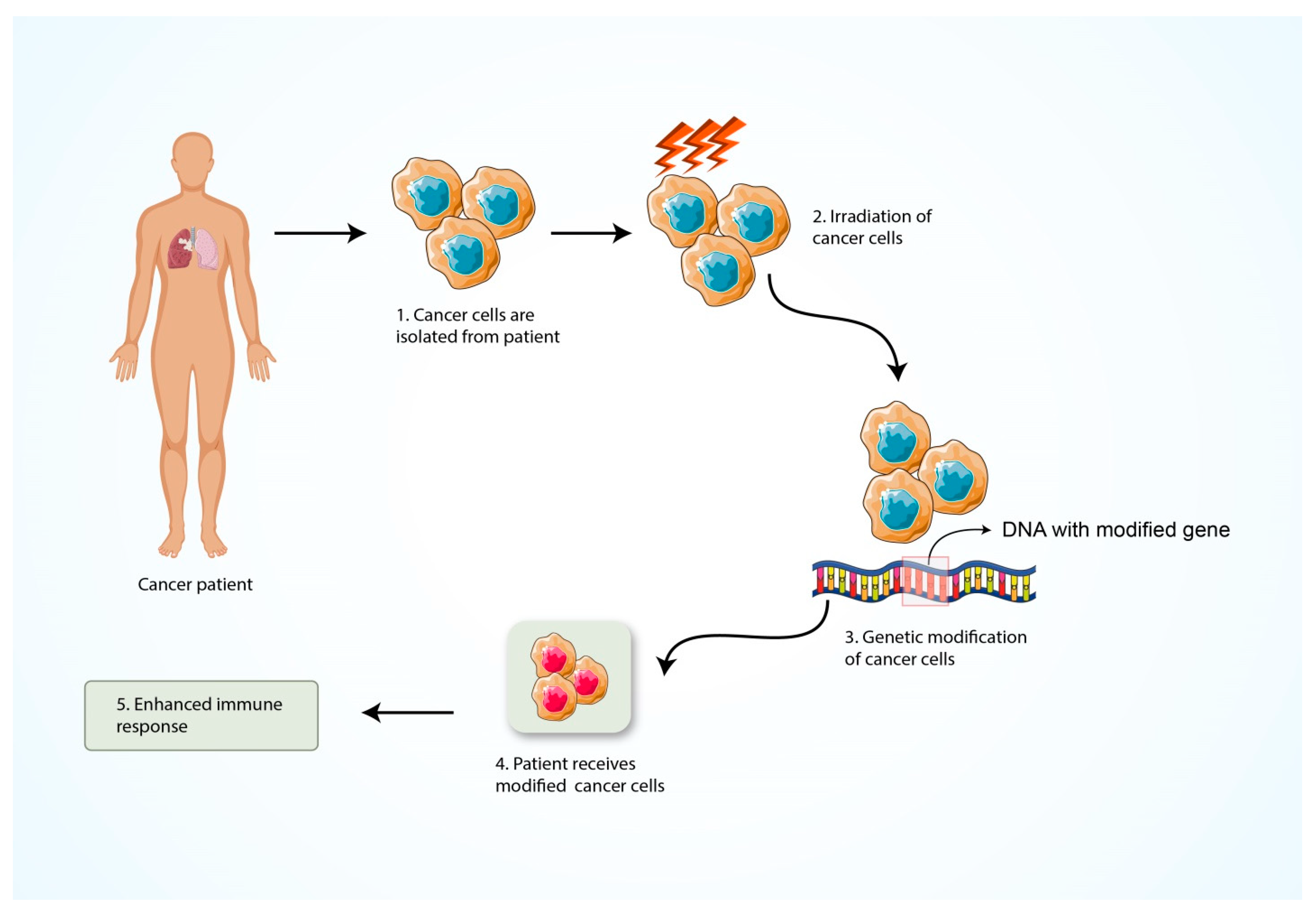
5.5. Formation of Cancer Vaccine

Cancer treatment vaccines enhance the immune system’s capability to signify and damage the cancer antigens more effectively. Cancer cell’s possess specific molecules, named cancer specific antigens, for every type of cancer cell on their cell surface, but they are absent in healthy cells. Generally, cancer vaccines can be produced for individual cancer patients. These categories of cancer vaccines are composed from the tumor sample of the individual cancer patient (Figure 3). For that, surgery is required to find a large enough sample of the tumor cells to make the vaccine against these cancer cells. There are two types of vaccine in cancer treatment. They are the personalized cancer vaccine and the vaccine to target specific cancer antigens [130].
Even if cancers can be originated by common mechanisms that are regulated by the mutated genes in the process of cell transformation (i.e., p53, ras), they pass through excessive random mutations in other genes. The expression of foreign antigens is regulated by these mutations and by making a molecular “fingerprint” that absolutely distinguishes the patient’s tumor. Whereas the mutations of genes are regulated indiscriminately, the antigenic fingerprint of one person’s cancer is generally not the same as another person’s cancer. In this way, the particular cancers among the identical pathological group are independent based on their antigens. These essential characteristics demand that the immune system of individual patient’s has to be treated to identify the specific cancer antigens and cancer cells. The novel approaches for generating cancer immune-therapeutics are known as the autologous tumor cell vaccines, allogeneic tumor cell vaccines, dendritic cell vaccines, protein- or peptide-based cancer vaccines, genetic vaccines and the whole tumor lysates [131,132].
The cancer vaccine possesses an effective activity to provide immunity towards the cancer antigens. The response stimulates the activity of antigen-presenting cells (APCs) to trigger the functions of T helper cells to provide the antibody against the cancer antigens and also activate the effector or cytotoxic T cells. However, cancer vaccines have some limitations: because the tumor cells or cancer cells express random specific types of antigens, these antigen’s type is not fixed. For that reason, cancer vaccines are not so effective for inhibiting the growth of the cancer cells.

5.6. Nanoparticle-Based Cancer Immunotherapy

The contribution of nanoparticle (NP) technology for diagnostics and therapy has been improving medicine for many years now. Nanoparticles (NPs) can be employed as adjuvants or as carriers to carry molecules to a specified destination, equipped with certain ligands that encourage specific use, and they can be applied in an immune-modulating activity. Nanoparticles (NPs), which can be classified into three types: micelles, liposomes and inorganic NPs, are the most preferred delivery platform [133].
Matured dendritic cells (DCs) are represented in the antigens of tumor cells towards the effector T cells. Without maturation, DCs cannot present their antigens to T cells and tumor cells can escape. Immunostimulatory spherical nucleic acids (IS-LSNAs) were created to be able to target TLRs 7/8. Plasmacytoid dendritic cells uptake IS-LSNAs and this can generate an NF-κB pathway to activate immature DCs. An organic nanoparticle called liposome was used to carry the IS-LSNAs loaded with antigens to target specific DCs in the tumor microenvironment [134]. Liu et al. synthesized amphiphilic vaccines (amph-vaccines) consisting of an antigen that was linked to a lipophilic albumin-binding tail, while the lipid head was bound to single-stranded oligonucleotides containing unmethylated cytosine–guanine motifs (CpG DNAs) that were capable of interacting with TLR9. Liu et al. produced vaccines that were amphiphilic in nature (amph-vaccines). These consist of two parts, a lipophilic albumin-binding tail and lipid-based CpG DNAs to interact with TLR9. The amph-vaccines codelivery method successfully transmits peptide antigens and CpG DNAs through a nanocarrier and enhances the T cell responses and antitumor efficiency through effective lymph node stimulation [135]. An anti-CTLA-4 antibody combined with a radiotherapy approach can generate a strong immune stimulation. Song et al. developed a liposomal delivery method to provide catalase and H2O2 to reverse hypoxia in the tumorous location and to improve the radio-immunotherapy’s and anti-CTLA-4 immunotherapy’s immunological impacts [136]. NK-92, activated NK cells, fusogenic liposome encapsulating doxorubicin and DOX@NKsomes have been developed by Pitchaimani et al. All protein receptors such as CD 56, NKG-2D and NKp30 are preserved in the liposome-fusing NK cell. Consequently, they can identify tumor cells using NK cell markers and merge with the tumor cells to release doxorubicin to the cytoplasm of the tumor cell [137].
Table 1. Antigens and type of cancers they cause. Tumor antigens’ classification depends on their molecular structure and their origin, and, based on these properties, we can classify the tumor antigen into following categories of tumor-associated antigens (TAAs): (i) mutation products of specific genes (In breast cancer, the mutation of Her2 and Ras causes 10% of the carcinomas); (ii) oncogenic virus antigens; (iii) abnormal cellular proteins; (iv) types of oncofetal antigens; (v) classes of glycolipids and glycoproteins; (vi) types of differentiated tissue-specific antigens.
AntigensType of Cancer They Cause References
NY-ESO-1Esophageal Squamous Cell carcinoma [138]
MAGEA-A3Melanoma[139]
WT1Acute Myelocytic leukemia[140]
hTERTViral mediated Cancer[141]
TyrosinaseBrain and Skin Cancer[142]
gp 100Melanoma[143]
MART-1 Melanoma[143]
Melan AMelanoma[143]
β cateninMelanoma[143]
MUC1 Breast Cancer[144]
CEAColon Cancer, Lung Cancer[145]
Mam-ABreast Cancer[146]
Sialyl-TnBreast, Gastric, Lung, Colon, Esophageal, Prostate and Endometrial Cancer[147]
α-fetoproteinHepatic Cancer[148]
CA-125Ovarian Cancer[149]
Ras, SrcExhibited in Several Cancer Types[150]
MesothelinMalignant Pleural Mesothelioma, Ovarian and Pancreatic Cancer[151]
PSMAProstate Cancer[151]
TPD52Prostate, Breast and Ovarian Cancer[151]
PSAProstate Cancer[151]
PAPProstate Cancer[151]

5.7. Inhibition of T Cell Exhaustion

T-cell exhaustion is known as T-cell dysfunction that occurs in many chronic infections and cancer, and which is also a condition of poor effector action and the sustainable expression of inhibitory receptors [65]. The exhaustion of T cells inhibits the optimal monitoring of infections and tumors so that the over expression of inhibitory receptors is found in the tumor microenvironment, which reduces the effector cytokine production and also cytolytic activities. The mechanisms for these T cell abnormalities are not clearly known. In order to solve these abnormalities, the gene expression was observed in the virus-specific CDRT cell line of mice that were functionally weakened and chronically infected with lymphocytic choriomeningitis virus (LCMV). It was shown that PD-1 (programmed death 1) was upregulated by the exhausted T cell [152].
By coating themselves in PD-L1 (known as B7&-H1), the T cell is blocked by antibodies (Figure 4). Thus, by blocking the PD-1/PD-L1 inhibitory pathway, the CD8 T cell is restored and they are now able to proliferate, secrete cytokines and destroy the infected cell [153]. In contrast, T cell exhaustion can also be eliminated through engineered T cells in the event of lethal diseases and cancer [154]. The expression of PD-1 can also be degenerated by genome engineering strategies instead of blocking the PD-1/PD-L1 mechanism. The process can be completed by the CRISPR/Cas9 system [155]. By using the CRISPR/Cas9 system, it was reported that the expression of PD-1 on the primary human cells was reduced. Thus, it shows an effective strategy to generate CAR T cells that are resistant to exhaustion [63]. Thus, by following the two abovementioned methods, the exhausted T cell can be restored. This cancer treatment, by restoring these exhausted T cells, could be an inspiring event that can provide a hopeful result and could become an important breakthrough in cancer immunotherapy.

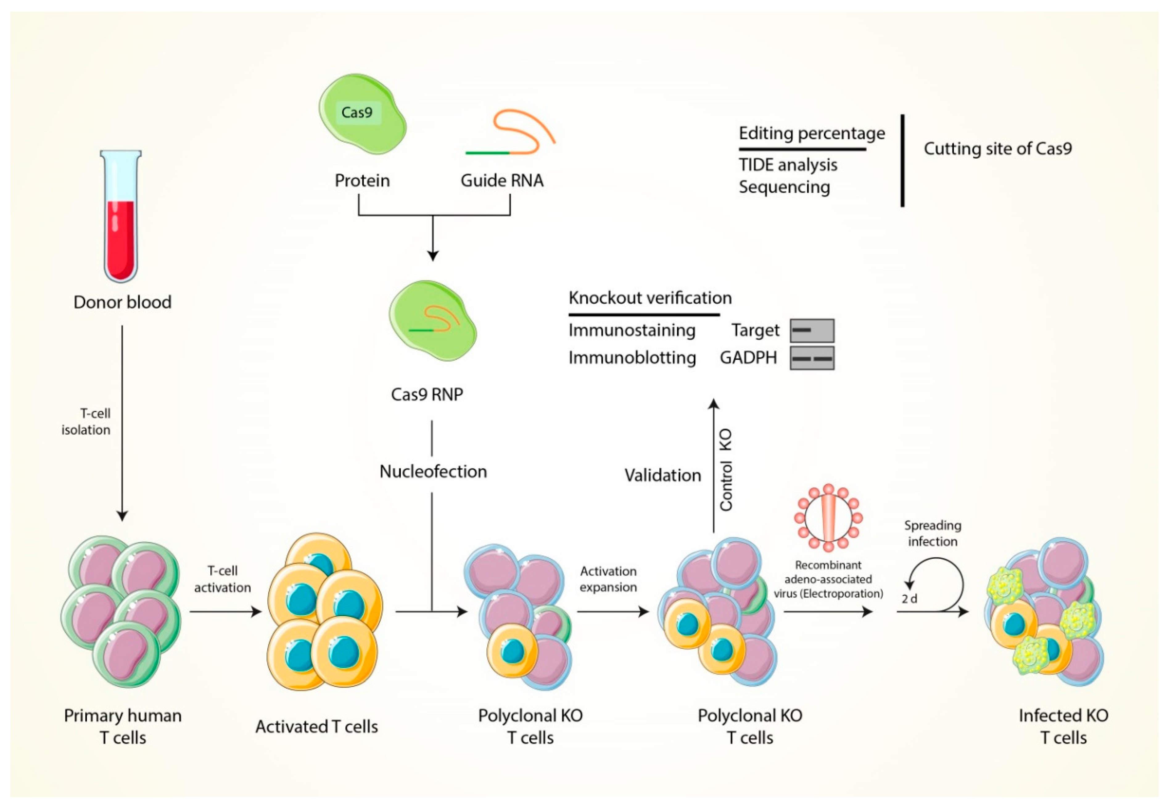
6. The Strategy of T Cell Modification through Using the CRISPR-Cas9 Genome Editing Tool

By the above demonstration from many research findings, the estimation can be generalized that the modification of T cell receptors could be a possible solution as an effective immunotherapy cancer treatment in which the genetically engineered T cells can express the genes of the chimeric antigen receptor (CAR) proteins [156]. The genetic construct of CAR introduced to T cells by transfection with viral vectors, mRNA or plasmid can be performed using a gene editing tool (CRISPR-Cas9) to guide T cells that are aligned with the surface TAAs of the tumor cells (Table 1), and the recent development have evolved in molecular biology and gene editing tools. The only first-generation CAR (CD3ζ signaling domain) evolved to second-generation (CD3ζ with 41BB or CD28 domains) and third-generation (CD3ζ with 41BB and CD28 domains) CARs, which have improved T cell targeting and proliferation for the addition of co-stimulatory endo domains (Table 2). In the human leucocyte antigen components, CARs are designed to target particular peptides, which may enable the targeting of tumor-associated antigens (TAA) or tumor specific antigens (TSA) (Figure 5) [157].
These modified T cells, which have tumor targeting receptors, were shown to be very promising in clinical trials for multiple leukemia and lymphoma and could be used to treat solid cancer [96]. Genetic modification to generate this potential chimeric antigen receptor can be carried out only by a specific editing tool such as CRISPR-Cas9 (Table 3) [158]. This is because the alternatives to CRISPR-Cas9 are zinc-finger nucleases (ZFNs) and transcription activator-like effector nucleases (TALENs), and the major drawback of ZFNs and TALENS is that the recognition of the target DNA sequence is determined by protein sequences [159]. For each particular target DNA sequence, therefore, a laborious and complex process and the optimization of proteins are needed, and it is difficult to deliver many of these proteins in cells for simultaneous multiplexed genetic modification [160]. The CRISPR system was identified inside bacteria when it was invaded with foreign DNA. Type II CRISPR strategies combine invading DNA sequences and CRISPR repeat sequences that are encoded by a bacterial genome. This CRISPR repeat DNA sequence is transcribed and forms crRNAs [161], it includes a protospacer, which is a variable RNA sequence that transcribes and forms crRNAs [162], a variable RNA sequence from the invading DNA and CRISPR arrays of the host DNA. Every crRNA hybridizes with a second RNA known as the CRISPR RNA (tracrRNA) and then they form a complex with Cas9 endonuclease protein, which cuts the DNA [161]. The crRNA protospacer directs Cas9 through additional target DNA sequences and cleaves the DNA if they are adjacent to short sequences recognized as protospacer adjacent motifs (PAMs). It was first revealed that the Streptococcus pyogenes Cas9 protein (SpCas 9) would associate with the tracrRNA–crRNA RNA complex to induce a double stranded breakage (DSB) in a target DNA sequence [162]. This experiment also showed that Cas9 does not need an RNA complex to bind and break a certain DNA sequence, rather a constructed single guide RNA can easily achieve this method (sgRNA). The above studies laid the foundation for CRISPR/Cas9 to be used in the field of genome engineering involving genetic modification and control of gene expression, epigenetic alteration and the imaging of the genome [163,164,165,166,167]. By using a plasmid encoding Cas9 and sgRNA, CRISPR/Cas9 can be applied directly to human cells, and there have also been studies of gene editing with non-integrated viruses such as adenovirus and adenovirus-associated virus (AAV) [19,128,129]. New developments of smaller Cas proteins have allowed and improved the combination of this technology with vectors that have increased their optimization, such as AAV vectors. Efficient gene editing in human cells was also used in CRISPR delivery via Cas9 (Ribonucleoproteins, RNP) [168]. Lentiviral and adenoviral transmission of CRISPR components to primary T cells resulted in genetic change in T cells.
Table 2. Diverse malignancies with their target antigens and their endo domains. Summary of solid tumor antigens targeted by CAR T-cell therapy.
Target AntigenMalignanciesEndo DomainsReferences
Epidermal growth factor receptorGastric cancerCD28+CD3ζ, 4-1BB[169]
HER2Sarcoma, Glioblastoma, OsteosarcomaCD28-CD3ζ[170]
IL13Rα2GlioblastomaCD3ζ[171]
GD2NeuroblastomaCD3ζ[172,173]
FAPColon and ovarian cancerCD8α, CD3ζ, 4-1BB[174]
MSLNPancreatic cancer, Malignant pleural mesotheliomaCD3ζ and 4-1BB[175]
CD171Refractory neuroblastomaCD3ζ[176]
EGFRvIIIGliomaCD28+CD3ζ, 4-1BB[177]
Carbonic anhydrase IXMetastatic renal cell carcinomaFcRγ[169]
Alpha-folate receptorOvarianFcRγ[178]
Carcinoembryonic antigenLiver metastasisCD28+CD3ζ[179]
ErbB2+MUC1Breast cancerCD28, CD3ζ[173]
Vascular endothelial growth factor receptorMelanomaCD28, CD3ζ[180]
HER2+CD19MedulloblastomaCD28, CD3ζ[174]
NKG2DBreast cancerCD28+CD3ζ[181]
Table 3. The tabular representation of the current immunotherapy techniques that are used for cancer treatment worldwide.
S.N.Immunotherapy TechniquesTheir Mechanism of Action Advantages LimitationsReferences
01Tumor-infiltrating lymphocyte (TIL)Harvesting the Killer T cells in ex vivo expansion that have already been infiltrated from cancer patients, as well as activating and expanding the T cell population, is required for efficiently killing cancer cells. A large number of activated T cells are re-infused into patient’s body allowing them to destroy tumor cells. TILs have been widely used to treat solid tumors, metastatic melanoma, pancreatic cancer, colorectal cancer and various other cancers. TILs are efficacious for melanoma patients. They are able to focus properly on tumor antigens.Therapeutic delays owing to lengthy ex vivo expansion. MHC- I may be downregulated by tumor cells.[169,182]
02Engineered T-cell receptor (TCR) therapyAfter isolating T cells from cancer patients, the engineered receptors are introduced into patient’s body for targeting specific cancer antigen and eradication of tumor cells. Modified T cells have exceptional activities and function in terms of having greater lifetime in tumor microenvironment. Enhance functionality, polarization and working efficiency. In most cases, it is monoclonal specificity. Inconceivable toxic effects may be found. [170,182]
03Engineered natural killer (NK) cell therapyAugmenting the capability of NK cell antitumor responses via the introduction of antigen specificity by using genetic modification. CAR-mediated NK cell requires co-stimulatory molecule such as CD28, 4-1BB, CD134 in order to increase the proliferation and cytotoxicity effect against the solid tumor. Less expensive, easy to be isolated, secreted safer cytokines (IFN-γ and GM-CSF).Always have to be injected orally. [82,83]
04Chimeric antigen receptor (CAR) T-cell therapyThe CAR T-cell treatment is used to recognize unprocessed antigens that are represented on the surface of tumor cells. The Car T cells are attracted and guided to cancer target site without MHC molecule’s expression. CD4+ and CD8+ T cells carry out the primary killing methods as cytolysis by secretion of granzyme and perforin. Large-scale production within short time, do not depend on MHC molecule, recognizes any surface antigens (Carbohydrate, protein and glycolipid). CAR T cell can only target cell surface antigens.
Lethal toxicity may be found owing to cytokine storm.
[175,182]
05ImmunomodulatorsEnhancing cytokines’ secretion and act as an agonist for blocking the cancer progression and enhancing the potential activity of immune cells.Can effectively suppress the cancer cells. Inability to predict treatment efficacy and patient response.[97]
06Antibody-mediated therapyPossesses cytotoxic effect against a tumor cell surface antigen and modifies the signal transduction cascade pathway within the tumor cells through the complement-dependent cytotoxicity (CDC) and/or antibody-dependent cellular cytotoxicity (ADCC).Showed significant anticancer properties in the tumor microenvironment.Inadequate pharmacokinetics and tissue accessibility as well as impaired interactions with the immune system.[110]
07Formation of bi-specific T-cell-engaging antibodies for cancer therapyEnhance the specificity of the T-cell receptor, co-stimulation or presentation of peptide antigens independently.Demonstrated promising efficacy in the cancer treatment.When the bi-specific antibodies bind only the CD3-specific branch it shows low efficiency.[120,121]
08Formation of cancer vaccineEnhance the immune system’s capability to signify and damage the cancer antigens more effectively. Possesses an effective activity to provide the immunity towards the cancer antigens. The efficacy of cancer vaccines has been limited. [130]
09Nanoparticle-based cancer immunotherapyThey can be employed as adjuvants or as carriers to carry molecules to a specified destination, equipped with certain ligands that encourage specific use and they can be applied in an immune-modulating activity. Good pharmacokinetics, precise targeting of tumor cells, reduction of side effects and drug resistance. Nanoparticles have no common feature other than their size.[133]
10Inhibition of T cell exhaustionInhibition of T cell exhaustion increase the efficacy of T cells in which it can effectively recognize and kill the cancer antigens. It shows an effective strategy to generate the CAR T cells that are resistant to exhaustion. Can provide a hopeful result and it may become an important breakthrough in cancer immunotherapy. More complex technique and also it is patient specific.[63]
However, these techniques cannot directly insert and interrupt the basic genetic elements at the location level and have not been successful in genetic modification [183,184]. The lentiviral CRISPR kit, which is Jurkat T cell-based, has recently been created, with the aim to be versatile and simple to operate, and this toolbox can provide an additional advantage for research on T-cell signal transductions and programmability with different variants of Cas9 proteins [185]. There is also a study on the gene editing of Cas9 RNP cells at a particular site [155]. However, in vitro-produced sgRNAs and Cas9 protein (recombinant) form the Cas9 RNPs, and this synthetic Cas9 RNPs was introduced inside CD4 T cells and resulted in specific genetic modification because the treated T cells did not express PD-1 and CXCR4 proteins. In particular, the successful implantation of DNA inserts by the incorporation of an HDR (homology directed repair) template at the Cas9 cleavage position was assured by the deep sequencing. There are existing methods to introduce the CRISPR/Cas9 system into the T cells, but each method has its own pros and cons (Table 3). For example, the lentiviral method was used to target the CCR5 gene of T cells in humans for Cas9 delivery, but the main drawback of this method is the low knockout efficacies [186,187]. Moreover, it is not ideal to have permanent expression of gene editing factors, which might generate higher off target consequences [168]. In addition, it has recently been revealed that Cas9 antibodies are present in the serum of cord blood in a great number of healthy donors, which might trigger an immune response and cause an undesirable effect [188,189,190,191]. Thus, transient expression is desirable in the case of CRISPR/Cas9 system delivery. These can be applied by the non-viral transfection of T cells, for example, by electroporation, liposome-mediated nanoparticles, ligand fusion tags or cell penetrating peptides. All other approaches have not yet been utilized in T cells, except for electroporation. Electroporation is less productive and 95% of T cells die because of a cytotoxic effect, but they can retain their expansion potential to increase viable cell numbers after CRISPR/Cas9-based genome editing [192,193]. As a result, electroporation-based methods might be the most optimum to utilize Cas9-mediated genome modifications in T cells for therapeutic reasons given the temporary existence of a CRISPR/Cas9 system and the retention of T cell growth capacity.
In 55% of the targeted T cells, there were indels and 20% exogenous DNA [155]. In another study by Hendel et al., it was shown that modification of the CCR5 gene by Cas9 mRNA in T cells with synthetic sgRNAs can result in a greater success rate where around 49% of target T cells were modified at their targeted site and were evaluated with the TIDE method by hybridizing indels with fluorescent molecules [194]. There is also a report on the procedure of disrupting DNA in T cells to synthesize clinically standard CAR T cells [195]. Freshly isolated human T cells were stimulated with anti-CD3/CD28 beads and lentivirally transformed for stable expression of the CAR-Transgene at a period of 1 day after stimulation, and T cells were electroporated simultaneously on days 3 and 4 with RNA encodes Cas9 and SgRNA to cut PD1, HLA-1 and TCR genes [196]. The output using this technique was based on the donor sequence, with results showing >70% CAR transduction ability in several production models and also >60% dual knockout efficiency, which have been briefly demonstrated in Figure 6. This development process resulted in CAR T cells that were unique to CD19 targets, resistant to host rejections and unable to cause GVHD, for this reason emphasizing CRISPR/Cas9-mediated universal CAR T cell generation. Similar findings were recorded using a different CRISPR/Cas9 strategy with Cas9 RNPs targeting the B2m, PD1 and TCR genes [197]. With a single electroporation of different Cas9mRNAs, an effective gene alteration can be achieved [198], and this could be less time-consuming and the success rate could increase. Therefore, these gene editing techniques based on CRISPR/Cas9 can inhibit TLA-4 or PD-1 cells from T cells, and then target tumor cells for successful immunotherapy.

7. Concluding Remarks

Overall, the new emerging CAR T-cell therapy will be a novel strategy that will increase the activity of T cells and can be worked as an immune-therapeutic approach in the treatment of cancer. In this review, we have shown the modification of the T-cell for therapeutic purposes in cancer treatment with the high efficiency genome editing tool CRISPR Cas9; specifically, its application in therapeutic processes against cancer and the development process of T-cell therapy in the next generation metamorphosis drugs. The CD19-mediated chimeric antigen receptor T-cell therapy will become a novel strategy in the treatment of human cancers. In chimeric antigen receptor (CAR) T cells, an extracellular single-chain variable fragment (ScFv) is specific to the antigens on tumor cells and the T-cell activation is initiated by an intracellular chimeric signaling domain. Then, the T cells are able to damage the tumor cells.
Many cancer research activities focus on the role of T cells in both early and late phages of disease where T cells show their functions for controlling the enhancement of tumor development and progression in cancer patients. However, there have been some abnormalities for generating T cell responses against the tumor specific antigens, which include them being difficult to develop and maintain in cancer patients. An effective strategy used to add T cells for cancer therapeutic treatments are antibodies. These antibodies are activated only for the surface specific antigen on cancer cells and for the CD3 complex on T cells. These types of antibodies are efficient in engaging with any kind of T cells that are cytotoxic to cancer cells in order to damage the cancer cells. However, to use the CRISPR/Cas9 technology to mediate immunotherapy, we have broadly demonstrated the CRISPR/Cas9-mediated genome engineering strategy, which exhibits a significant impact and the therapeutic potential of this technology and, thus, represents a very important area that will help biological researchers in their future study about this novel strategy for cancer immunotherapy.

Author Contributions

Conceptualization by M.A.S., P.B., D.D. and M.A.I.; Writing and main draft preparation by M.A.S., P.B., D.D., M.A.K., M.A.I., M.I.A.T., M.Y.B. and P.P. Figures drawing by M.H.R. and T.I.R.; Review and editing by M.A.R., M.N.H. and B.K.; Visualization and supervision by M.A.R. and B.K. The Project Funded by B.K. All authors have read and agreed to the published version of the manuscript.

Funding

This research was supported by the Basic Science Research Program through the National Research Foundation of Korea (NRF) funded by the Ministry of Education (NRF-2020R1I1A2066868), the National Research Foundation of Korea (NRF) grant funded by the Korea government (MSIT) (No. 2020R1A5A2019413), a grant of the Korea Health Technology R&D Project through the Korea Health Industry Development Institute (KHIDI), funded by the Ministry of Health & Welfare, Republic of Korea (grant number: HF20C0116), and a grant of the Korea Health Technology R&D Project through the Korea Health Industry Development Institute (KHIDI), funded by the Ministry of Health & Welfare, Republic of Korea (grant number: HF20C0038).

Institutional Review Board Statement

Not applicable.

Informed Consent Statement

Not applicable.

Data Availability Statement

Not applicable.

Conflicts of Interest

The authors’ have no competing interest at all with the others.

References

  1. Porter, D.L.; Levine, B.L.; Kalos, M.; Bagg, A.; June, C.H. Chimeric antigen receptor-modified T cells in chronic lymphoid leukemia. N. Engl. J. Med. 2011, 365, 725–733. [Google Scholar] [CrossRef] [Green Version]
  2. Riley, R.S.; June, C.H.; Langer, R.; Mitchell, M.J. Delivery technologies for cancer immunotherapy. Nat. Rev. Drug Discov. 2019, 18, 175–196. [Google Scholar] [CrossRef]
  3. Mellman, I.; Coukos, G.; Dranoff, G. Cancer immunotherapy comes of age. Nature 2011, 480, 480–489. [Google Scholar] [CrossRef]
  4. Topalian, S.L.; Taube, J.M.; Anders, R.A.; Pardoll, D.M. Mechanism-driven biomarkers to guide immune checkpoint blockade in cancer therapy. Nat. Rev. Cancer 2016, 16, 275–287. [Google Scholar] [CrossRef]
  5. Crowther, M.D.; Svane, I.M.; Met, Ö. T-Cell Gene Therapy in Cancer Immunotherapy: Why It Is No Longer Just CARs on The Road. Cells 2020, 9, 1588. [Google Scholar] [CrossRef]
  6. Mahoney, K.M.; Rennert, P.D.; Freeman, G.J. Combination cancer immunotherapy and new immunomodulatory targets. Nat. Rev. Drug Discov. 2015, 14, 561–584. [Google Scholar] [CrossRef]
  7. Kochenderfer, J.N.; Wilson, W.H.; Janik, J.E.; Dudley, M.E.; Stetler-Stevenson, M.; Feldman, S.A.; Maric, I.; Raffeld, M.; Nathan, D.A.; Lanier, B.J.; et al. Eradication of B-lineage cells and regression of lymphoma in a patient treated with autologous T cells genetically engineered to recognize CD19. Blood 2010, 116, 4099–4102. [Google Scholar] [CrossRef]
  8. Li, A.M.; Hucks, G.E.; Dinofia, A.M.; Seif, A.E.; Teachey, D.T.; Baniewicz, D.; Callahan, C.; Fasano, C.; McBride, B.; Gonzalez, V.J.B. Checkpoint inhibitors augment CD19-directed chimeric antigen receptor (CAR) T cell therapy in relapsed B-cell acute lymphoblastic leukemia. Blood 2018, 132, 556. [Google Scholar] [CrossRef]
  9. Wei, G.; Ding, L.; Wang, J.; Hu, Y.; Huang, H.J.E.H. Advances of CD19-directed chimeric antigen receptor-modified T cells in refractory/relapsed acute lymphoblastic leukemia. Exp. Hematol. Oncol. 2017, 6, 1–7. [Google Scholar] [CrossRef] [Green Version]
  10. Mollanoori, H.; Shahraki, H.; Rahmati, Y.; Teimourian, S. CRISPR/Cas9 and CAR-T cell, collaboration of two revolutionary technologies in cancer immunotherapy, an instruction for successful cancer treatment. Hum. Immunol. 2018, 79, 876–882. [Google Scholar] [CrossRef]
  11. Havard, R.; Stephens, D.M. Anti-CD19 Chimeric Antigen Receptor T Cell Therapies: Harnessing the Power of the Immune System to Fight Diffuse Large B Cell Lymphoma. Curr. Hematol. Malig. Rep. 2018, 13, 534–542. [Google Scholar] [CrossRef]
  12. Kallam, A.; Vose, J.M. Recent Advances in CAR-T Cell Therapy for Non-Hodgkin Lymphoma. Clin. Lymphoma Myeloma Leuk. 2019, 19, 751–757. [Google Scholar] [CrossRef]
  13. Ogasawara, K.; Dodds, M.; Mack, T.; Lymp, J.; Dell’Aringa, J.; Smith, J. Population Cellular Kinetics of Lisocabtagene Maraleucel, an Autologous CD19-Directed Chimeric Antigen Receptor T-Cell Product, in Patients with Relapsed/Refractory Large B-Cell Lymphoma. Clin. Pharmacokinet. 2021. [Google Scholar] [CrossRef]
  14. Abramson, J.S.; Palomba, M.L.; Gordon, L.I.; Lunning, M.A.; Wang, M.; Arnason, J.; Mehta, A.; Purev, E.; Maloney, D.G.; Andreadis, C.; et al. Lisocabtagene maraleucel for patients with relapsed or refractory large B-cell lymphomas (TRANSCEND NHL 001): A multicentre seamless design study. Lancet 2020, 396, 839–852. [Google Scholar] [CrossRef]
  15. Ishizaki, Y.; Yukaya, N.; Kusuhara, K.; Kira, R.; Torisu, H.; Ihara, K.; Sakai, Y.; Sanefuji, M.; Pipo-Deveza, J.R.; Silao, C.L.; et al. PD1 as a common candidate susceptibility gene of subacute sclerosing panencephalitis. Hum. Genet. 2010, 127, 411–419. [Google Scholar] [CrossRef]
  16. Chevolet, I.; Speeckaert, R.; Schreuer, M.; Neyns, B.; Krysko, O.; Bachert, C.; Hennart, B.; Allorge, D.; van Geel, N.; Van Gele, M.; et al. Characterization of the in vivo immune network of IDO, tryptophan metabolism, PD-L1, and CTLA-4 in circulating immune cells in melanoma. Oncoimmunology 2015, 4, e982382. [Google Scholar] [CrossRef] [Green Version]
  17. Srivastava, S.; Riddell, S.R. Chimeric Antigen Receptor T Cell Therapy: Challenges to Bench-to-Bedside Efficacy. J. Immunol. 2018, 200, 459–468. [Google Scholar] [CrossRef]
  18. Mali, P.; Yang, L.; Esvelt, K.M.; Aach, J.; Guell, M.; DiCarlo, J.E.; Norville, J.E.; Church, G.M. RNA-guided human genome engineering via Cas9. Science 2013, 339, 823–826. [Google Scholar] [CrossRef] [Green Version]
  19. Cong, L.; Ran, F.A.; Cox, D.; Lin, S.; Barretto, R.; Habib, N.; Hsu, P.D.; Wu, X.; Jiang, W.; Marraffini, L.A.; et al. Multiplex genome engineering using CRISPR/Cas systems. Science 2013, 339, 819–823. [Google Scholar] [CrossRef] [Green Version]
  20. Chen, S.; Sun, H.; Miao, K.; Deng, C.X. CRISPR-Cas9: From Genome Editing to Cancer Research. Int. J. Biol. Sci. 2016, 12, 1427–1436. [Google Scholar] [CrossRef]
  21. Hsu, P.D.; Lander, E.S.; Zhang, F. Development and applications of CRISPR-Cas9 for genome engineering. Cell 2014, 157, 1262–1278. [Google Scholar] [CrossRef] [Green Version]
  22. Frey, N.; Porter, D. Cytokine Release Syndrome with Chimeric Antigen Receptor T Cell Therapy. Biol. Blood Marrow Transplant. J. Am. Soc. Blood Marrow Transplant. 2019, 25, e123–e127. [Google Scholar] [CrossRef] [Green Version]
  23. Jenkins, M.K.; Chu, H.H.; McLachlan, J.B.; Moon, J.J. On the composition of the preimmune repertoire of T cells specific for Peptide-major histocompatibility complex ligands. Annu. Rev. Immunol. 2010, 28, 275–294. [Google Scholar] [CrossRef]
  24. Varma, R. TCR triggering by the pMHC complex: Valency, affinity, and dynamics. Sci. Signal. 2008, 1, pe21. [Google Scholar] [CrossRef]
  25. Kleiveland, C.R. Peripheral blood mononuclear cells. Impact Food Bioact. Health 2015, 161–167. [Google Scholar] [CrossRef] [Green Version]
  26. Huppa, J.B.; Davis, M.M. T-cell-antigen recognition and the immunological synapse. Nat. Rev. Immunol. 2003, 3, 973–983. [Google Scholar] [CrossRef]
  27. Mueller, D.L.; Jenkins, M.K.; Schwartz, R.H. Clonal expansion versus functional clonal inactivation: A costimulatory signalling pathway determines the outcome of T cell antigen receptor occupancy. Annu. Rev. Immunol. 1989, 7, 445–480. [Google Scholar] [CrossRef]
  28. Martin, P.J.; Ledbetter, J.A.; Morishita, Y.; June, C.H.; Beatty, P.G.; Hansen, J.A. A 44 kilodalton cell surface homodimer regulates interleukin 2 production by activated human T lymphocytes. J. Immunol. 1986, 136, 3282–3287. [Google Scholar]
  29. Gmünder, H.; Lesslauer, W. A 45-kDa human T-cell membrane glycoprotein functions in the regulation of cell proliferative responses. Eur. J. Biochem. 1984, 142, 153–160. [Google Scholar] [CrossRef]
  30. June, C.H.; Ledbetter, J.A.; Linsley, P.S.; Thompson, C.B. Role of the CD28 receptor in T-cell activation. Immunol. Today 1990, 11, 211–216. [Google Scholar] [CrossRef] [Green Version]
  31. Lesslauer, W.; Koning, F.; Ottenhoff, T.; Giphart, M.; Goulmy, E.; van Rood, J.J. T90/44 (9.3 antigen). A cell surface molecule with a function in human T cell activation. Eur. J. Immunol. 1986, 16, 1289–1296. [Google Scholar] [CrossRef] [PubMed] [Green Version]
  32. Hansen, J.A.; Martin, P.J.; Nowinski, R.C.J.I. Monoclonal antibodies identifying a novel T-cell antigen and Ia antigens of human lymphocytes. Immunogenetics 1980, 10, 247–260. [Google Scholar] [CrossRef]
  33. Hara, T.; Fu, S.M.; Hansen, J.A. Human T cell activation. II. A new activation pathway used by a major T cell population via a disulfide-bonded dimer of a 44 kilodalton polypeptide (9.3 antigen). J. Exp. Med. 1985, 161, 1513–1524. [Google Scholar] [CrossRef]
  34. Jin, B.; Sun, T.; Yu, X.H.; Yang, Y.X.; Yeo, A.E. The effects of TLR activation on T-cell development and differentiation. Clin. Dev. Immunol. 2012, 2012, 836485. [Google Scholar] [CrossRef]
  35. Sharpe, A.H.; Freeman, G.J. The B7-CD28 superfamily. Nat. Rev. Immunol. 2002, 2, 116–126. [Google Scholar] [CrossRef]
  36. Buchbinder, E.I.; Desai, A. CTLA-4 and PD-1 Pathways: Similarities, Differences, and Implications of Their Inhibition. Am. J. Clin. Oncol. 2016, 39, 98–106. [Google Scholar] [CrossRef] [PubMed] [Green Version]
  37. Cantrell, D.A. T-cell antigen receptor signal transduction. Immunology 2002, 105, 369–374. [Google Scholar] [CrossRef] [PubMed] [Green Version]
  38. Palacios, E.H.; Weiss, A. Function of the Src-family kinases, Lck and Fyn, in T-cell development and activation. Oncogene 2004, 23, 7990–8000. [Google Scholar] [CrossRef] [Green Version]
  39. Alexander, D.R. The CD45 tyrosine phosphatase: A positive and negative regulator of immune cell function. Semin. Immunol. 2000, 12, 349–359. [Google Scholar] [CrossRef]
  40. Rossy, J.; Williamson, D.J.; Gaus, K. How does the kinase Lck phosphorylate the T cell receptor? Spatial organization as a regulatory mechanism. Front. Immunol. 2012, 3, 167. [Google Scholar] [CrossRef] [Green Version]
  41. Macián, F.; García-Cózar, F.; Im, S.H.; Horton, H.F.; Byrne, M.C.; Rao, A. Transcriptional mechanisms underlying lymphocyte tolerance. Cell 2002, 109, 719–731. [Google Scholar] [CrossRef] [Green Version]
  42. Salsman, J.; Dellaire, G. Precision genome editing in the CRISPR era. Biochem. Cell Biol. Biochim. Et Biol. Cell. 2017, 95, 187–201. [Google Scholar] [CrossRef]
  43. Smith-Garvin, J.E.; Koretzky, G.A.; Jordan, M.S. T cell activation. Annu. Rev. Immunol. 2009, 27, 591–619. [Google Scholar] [CrossRef] [PubMed]
  44. Crow, M.K. Costimulatory molecules and T-cell-B-cell interactions. Rheum. Dis. Clin. N. Am. 2004, 30, 175–191. [Google Scholar] [CrossRef]
  45. Fukunaga, A.; Miyamoto, M.; Cho, Y.; Murakami, S.; Kawarada, Y.; Oshikiri, T.; Kato, K.; Kurokawa, T.; Suzuoki, M.; Nakakubo, Y.; et al. CD8+ tumor-infiltrating lymphocytes together with CD4+ tumor-infiltrating lymphocytes and dendritic cells improve the prognosis of patients with pancreatic adenocarcinoma. Pancreas 2004, 28, e26–e31. [Google Scholar] [CrossRef] [PubMed]
  46. Oshikiri, T.; Miyamoto, M.; Shichinohe, T.; Suzuoki, M.; Hiraoka, K.; Nakakubo, Y.; Shinohara, T.; Itoh, T.; Kondo, S.; Katoh, H. Prognostic value of intratumoral CD8+ T lymphocyte in extrahepatic bile duct carcinoma as essential immune response. J. Surg. Oncol. 2003, 84, 224–228. [Google Scholar] [CrossRef]
  47. Vivier, E.; Ugolini, S.; Blaise, D.; Chabannon, C.; Brossay, L. Targeting natural killer cells and natural killer T cells in cancer. Nat. Rev. Immunol. 2012, 12, 239–252. [Google Scholar] [CrossRef] [PubMed]
  48. Speiser, D.E.; Ho, P.C.; Verdeil, G. Regulatory circuits of T cell function in cancer. Nat. Rev. Immunol. 2016, 16, 599–611. [Google Scholar] [CrossRef]
  49. Farhood, B.; Najafi, M.; Mortezaee, K. CD8(+) cytotoxic T lymphocytes in cancer immunotherapy: A review. J. Cell. Physiol. 2019, 234, 8509–8521. [Google Scholar] [CrossRef]
  50. Martínez-Lostao, L.; Anel, A.; Pardo, J. How Do Cytotoxic Lymphocytes Kill Cancer Cells? Clin. Cancer Res. Off. J. Am. Assoc. Cancer Res. 2015, 21, 5047–5056. [Google Scholar] [CrossRef] [Green Version]
  51. Schultze, J.L.; Michalak, S.; Seamon, M.J.; Dranoff, G.; Jung, K.; Daley, J.; Delgado, J.C.; Gribben, J.G.; Nadler, L.M. CD40-activated human B cells: An alternative source of highly efficient antigen presenting cells to generate autologous antigen-specific T cells for adoptive immunotherapy. J. Clin. Investig. 1997, 100, 2757–2765. [Google Scholar] [CrossRef] [PubMed]
  52. Boon, T.; Cerottini, J.C.; Van den Eynde, B.; van der Bruggen, P.; Van Pel, A. Tumor antigens recognized by T lymphocytes. Annu. Rev. Immunol. 1994, 12, 337–365. [Google Scholar] [CrossRef] [PubMed]
  53. Poccia, F.; Agrati, C.; Martini, F.; Capobianchi, M.R.; Wallace, M.; Malkovsky, M. Antiviral reactivities of gammadelta T cells. Microbes Infect. 2005, 7, 518–528. [Google Scholar] [CrossRef] [PubMed]
  54. Li, D.; Li, X.; Zhou, W.L.; Huang, Y.; Liang, X.; Jiang, L.; Yang, X.; Sun, J.; Li, Z.; Han, W.D.; et al. Genetically engineered T cells for cancer immunotherapy. Signal Transduct. Target. Ther. 2019, 4, 35. [Google Scholar] [CrossRef] [PubMed]
  55. Southam, C.M.; Brunschwig, A.; Levin, A.G.; Dizon, Q.S. Effect of leukocytes on transplantability of human cancer. Cancer 1966, 19, 1743–1753. [Google Scholar] [CrossRef]
  56. Cao, Y.; Trillo-Tinoco, J.; Sierra, R.A.; Anadon, C.; Dai, W.; Mohamed, E.; Cen, L.; Costich, T.L.; Magliocco, A.; Marchion, D.; et al. ER stress-induced mediator C/EBP homologous protein thwarts effector T cell activity in tumors through T-bet repression. Nat. Commun. 2019, 10, 1280. [Google Scholar] [CrossRef] [Green Version]
  57. Meydan, İ.; Kizil, G.; Demir, H.; Toptanci, B.C.; Kizil, M. In vitro DNA damage, protein oxidation protective activity and antioxidant potentials of almond fruit (Amygdalus trichamygdalus) parts (hull and drupe) using soxhlet ethanol extraction. Adv. Tradit. Med. 2020, 20, 571–579. [Google Scholar] [CrossRef]
  58. Chen, D.S.; Mellman, I. Oncology meets immunology: The cancer-immunity cycle. Immunity 2013, 39, 1–10. [Google Scholar] [CrossRef] [Green Version]
  59. Yang, Y.; Liu, L.; Naik, I.; Braunstein, Z.; Zhong, J.; Ren, B. Transcription Factor C/EBP Homologous Protein in Health and Diseases. Front. Immunol. 2017, 8, 1612. [Google Scholar] [CrossRef]
  60. Nagarsheth, N.; Wicha, M.S.; Zou, W. Chemokines in the cancer microenvironment and their relevance in cancer immunotherapy. Nat. Rev. Immunol. 2017, 17, 559–572. [Google Scholar] [CrossRef] [Green Version]
  61. Araki, K.; Morita, M.; Bederman, A.G.; Konieczny, B.T.; Kissick, H.T.; Sonenberg, N.; Ahmed, R. Translation is actively regulated during the differentiation of CD8(+) effector T cells. Nat. Immunol. 2017, 18, 1046–1057. [Google Scholar] [CrossRef] [Green Version]
  62. Wherry, E.J. T cell exhaustion. Nat. Immunol. 2011, 12, 492–499. [Google Scholar] [CrossRef]
  63. Wherry, E.J.; Kurachi, M. Molecular and cellular insights into T cell exhaustion. Nat. Rev. Immunol. 2015, 15, 486–499. [Google Scholar] [CrossRef]
  64. Hong, W.X.; Haebe, S.; Lee, A.S.; Westphalen, C.B.; Norton, J.A.; Jiang, W.; Levy, R. Intratumoral Immunotherapy for Early-stage Solid Tumors. Clin. Cancer Res. Off. J. Am. Assoc. Cancer Res. 2020, 26, 3091–3099. [Google Scholar] [CrossRef] [PubMed] [Green Version]
  65. Pauken, K.E.; Wherry, E.J. Overcoming T cell exhaustion in infection and cancer. Trends Immunol. 2015, 36, 265–276. [Google Scholar] [CrossRef] [Green Version]
  66. Met, Ö.; Jensen, K.M.; Chamberlain, C.A.; Donia, M.; Svane, I.M. Principles of adoptive T cell therapy in cancer. Semin. Immunopathol. 2019, 41, 49–58. [Google Scholar] [CrossRef] [PubMed]
  67. Jiang, Y.; Li, Y.; Zhu, B. T-cell exhaustion in the tumor microenvironment. Cell Death Dis. 2015, 6, e1792. [Google Scholar] [CrossRef] [Green Version]
  68. Vonderheide, R.H.; June, C.H. Engineering T cells for cancer: Our synthetic future. Immunol. Rev. 2014, 257, 7–13. [Google Scholar] [CrossRef] [PubMed] [Green Version]
  69. Andersen, R.; Donia, M.; Westergaard, M.C.; Pedersen, M.; Hansen, M.; Svane, I.M. Tumor infiltrating lymphocyte therapy for ovarian cancer and renal cell carcinoma. Hum. Vaccines Immunother. 2015, 11, 2790–2795. [Google Scholar] [CrossRef] [Green Version]
  70. Azimi, F.; Scolyer, R.A.; Rumcheva, P.; Moncrieff, M.; Murali, R.; McCarthy, S.W.; Saw, R.P.; Thompson, J.F. Tumor-infiltrating lymphocyte grade is an independent predictor of sentinel lymph node status and survival in patients with cutaneous melanoma. J. Clin. Oncol. Off. J. Am. Soc. Clin. Oncol. 2012, 30, 2678–2683. [Google Scholar] [CrossRef]
  71. Sakellariou-Thompson, D.; Forget, M.A.; Hinchcliff, E.; Celestino, J.; Hwu, P.; Jazaeri, A.A.; Haymaker, C.; Bernatchez, C. Potential clinical application of tumor-infiltrating lymphocyte therapy for ovarian epithelial cancer prior or post-resistance to chemotherapy. Cancer Immunol. Immunother. CII 2019, 68, 1747–1757. [Google Scholar] [CrossRef]
  72. Buisseret, L.; Garaud, S.; de Wind, A.; Van den Eynden, G.; Boisson, A.; Solinas, C.; Gu-Trantien, C.; Naveaux, C.; Lodewyckx, J.N.; Duvillier, H.; et al. Tumor-infiltrating lymphocyte composition, organization and PD-1/ PD-L1 expression are linked in breast cancer. Oncoimmunology 2017, 6, e1257452. [Google Scholar] [CrossRef]
  73. Chung, Y.R.; Kim, H.J.; Jang, M.H.; Park, S.Y. Prognostic value of tumor infiltrating lymphocyte subsets in breast cancer depends on hormone receptor status. Breast Cancer Res. Treat. 2017, 161, 409–420. [Google Scholar] [CrossRef]
  74. Ping, Y.; Liu, C.; Zhang, Y. T-cell receptor-engineered T cells for cancer treatment: Current status and future directions. Protein Cell 2018, 9, 254–266. [Google Scholar] [CrossRef] [Green Version]
  75. Fesnak, A.D.; June, C.H.; Levine, B.L. Engineered T cells: The promise and challenges of cancer immunotherapy. Nat. Rev. Cancer 2016, 16, 566–581. [Google Scholar] [CrossRef]
  76. Rosenberg, S.A.; Restifo, N.P. Adoptive cell transfer as personalized immunotherapy for human cancer. Science 2015, 348, 62–68. [Google Scholar] [CrossRef] [Green Version]
  77. Johnson, L.A.; Morgan, R.A.; Dudley, M.E.; Cassard, L.; Yang, J.C.; Hughes, M.S.; Kammula, U.S.; Royal, R.E.; Sherry, R.M.; Wunderlich, J.R.; et al. Gene therapy with human and mouse T-cell receptors mediates cancer regression and targets normal tissues expressing cognate antigen. Blood 2009, 114, 535–546. [Google Scholar] [CrossRef] [PubMed] [Green Version]
  78. Chodon, T.; Comin-Anduix, B.; Chmielowski, B.; Koya, R.C.; Wu, Z.; Auerbach, M.; Ng, C.; Avramis, E.; Seja, E.; Villanueva, A.; et al. Adoptive transfer of MART-1 T-cell receptor transgenic lymphocytes and dendritic cell vaccination in patients with metastatic melanoma. Clin. Cancer Res. Off. J. Am. Assoc. Cancer Res. 2014, 20, 2457–2465. [Google Scholar] [CrossRef] [PubMed] [Green Version]
  79. Robbins, P.F.; Morgan, R.A.; Feldman, S.A.; Yang, J.C.; Sherry, R.M.; Dudley, M.E.; Wunderlich, J.R.; Nahvi, A.V.; Helman, L.J.; Mackall, C.L.; et al. Tumor regression in patients with metastatic synovial cell sarcoma and melanoma using genetically engineered lymphocytes reactive with NY-ESO-1. J. Clin. Oncol. Off. J. Am. Soc. Clin. Oncol. 2011, 29, 917–924. [Google Scholar] [CrossRef] [PubMed]
  80. Rapoport, A.P.; Stadtmauer, E.A.; Binder-Scholl, G.K.; Goloubeva, O.; Vogl, D.T.; Lacey, S.F.; Badros, A.Z.; Garfall, A.; Weiss, B.; Finklestein, J.; et al. NY-ESO-1-specific TCR-engineered T cells mediate sustained antigen-specific antitumor effects in myeloma. Nat. Med. 2015, 21, 914–921. [Google Scholar] [CrossRef] [Green Version]
  81. Robbins, P.F.; Kassim, S.H.; Tran, T.L.; Crystal, J.S.; Morgan, R.A.; Feldman, S.A.; Yang, J.C.; Dudley, M.E.; Wunderlich, J.R.; Sherry, R.M.; et al. A pilot trial using lymphocytes genetically engineered with an NY-ESO-1-reactive T-cell receptor: Long-term follow-up and correlates with response. Clin. Cancer Res. Off. J. Am. Assoc. Cancer Res. 2015, 21, 1019–1027. [Google Scholar] [CrossRef] [PubMed] [Green Version]
  82. Rezvani, K. Adoptive cell therapy using engineered natural killer cells. Bone Marrow Transplant. 2019, 54, 785–788. [Google Scholar] [CrossRef] [PubMed]
  83. Hu, W.; Wang, G.; Huang, D.; Sui, M.; Xu, Y. Cancer Immunotherapy Based on Natural Killer Cells: Current Progress and New Opportunities. Front. Immunol. 2019, 10, 1205. [Google Scholar] [CrossRef] [PubMed]
  84. Romanski, A.; Uherek, C.; Bug, G.; Seifried, E.; Klingemann, H.; Wels, W.S.; Ottmann, O.G.; Tonn, T. CD19-CAR engineered NK-92 cells are sufficient to overcome NK cell resistance in B-cell malignancies. J. Cell. Mol. Med. 2016, 20, 1287–1294. [Google Scholar] [CrossRef]
  85. Jiang, H.; Zhang, W.; Shang, P.; Zhang, H.; Fu, W.; Ye, F.; Zeng, T.; Huang, H.; Zhang, X.; Sun, W.; et al. Transfection of chimeric anti-CD138 gene enhances natural killer cell activation and killing of multiple myeloma cells. Mol. Oncol. 2014, 8, 297–310. [Google Scholar] [CrossRef]
  86. Chu, J.; Deng, Y.; Benson, D.M.; He, S.; Hughes, T.; Zhang, J.; Peng, Y.; Mao, H.; Yi, L.; Ghoshal, K.; et al. CS1-specific chimeric antigen receptor (CAR)-engineered natural killer cells enhance in vitro and in vivo antitumor activity against human multiple myeloma. Leukemia 2014, 28, 917–927. [Google Scholar] [CrossRef] [PubMed] [Green Version]
  87. Schönfeld, K.; Sahm, C.; Zhang, C.; Naundorf, S.; Brendel, C.; Odendahl, M.; Nowakowska, P.; Bönig, H.; Köhl, U.; Kloess, S.; et al. Selective inhibition of tumor growth by clonal NK cells expressing an ErbB2/HER2-specific chimeric antigen receptor. Mol. Ther. J. Am. Soc. Gene Ther. 2015, 23, 330–338. [Google Scholar] [CrossRef] [Green Version]
  88. Rezvani, K.; Rouce, R.; Liu, E.; Shpall, E. Engineering Natural Killer Cells for Cancer Immunotherapy. Mol. Ther. J. Am. Soc. Gene Ther. 2017, 25, 1769–1781. [Google Scholar] [CrossRef]
  89. Boissel, L.; Betancur-Boissel, M.; Lu, W.; Krause, D.S.; Van Etten, R.A.; Wels, W.S.; Klingemann, H. Retargeting NK-92 cells by means of CD19- and CD20-specific chimeric antigen receptors compares favorably with antibody-dependent cellular cytotoxicity. Oncoimmunology 2013, 2, e26527. [Google Scholar] [CrossRef] [Green Version]
  90. Rubnitz, J.E.; Inaba, H.; Ribeiro, R.C.; Pounds, S.; Rooney, B.; Bell, T.; Pui, C.H.; Leung, W. NKAML: A pilot study to determine the safety and feasibility of haploidentical natural killer cell transplantation in childhood acute myeloid leukemia. J. Clin. Oncol. Off. J. Am. Soc. Clin. Oncol. 2010, 28, 955–959. [Google Scholar] [CrossRef] [Green Version]
  91. Miliotou, A.N.; Papadopoulou, L.C. CAR T-cell Therapy: A New Era in Cancer Immunotherapy. Curr. Pharm. Biotechnol. 2018, 19, 5–18. [Google Scholar] [CrossRef] [PubMed]
  92. Feins, S.; Kong, W.; Williams, E.F.; Milone, M.C.; Fraietta, J.A. An introduction to chimeric antigen receptor (CAR) T-cell immunotherapy for human cancer. Am. J. Hematol. 2019, 94, S3–S9. [Google Scholar] [CrossRef] [Green Version]
  93. Prasad, V. Immunotherapy: Tisagenlecleucel—The first approved CAR-T-cell therapy: Implications for payers and policy makers. Nat. Rev. Clin. Oncol. 2018, 15, 11–12. [Google Scholar] [CrossRef] [PubMed]
  94. Fujiwara, H. Adoptive immunotherapy for hematological malignancies using T cells gene-modified to express tumor antigen-specific receptors. Pharmaceuticals 2014, 7, 1049–1068. [Google Scholar] [CrossRef] [PubMed]
  95. Park, J.H.; Geyer, M.B.; Brentjens, R.J. CD19-targeted CAR T-cell therapeutics for hematologic malignancies: Interpreting clinical outcomes to date. Blood 2016, 127, 3312–3320. [Google Scholar] [CrossRef] [PubMed]
  96. Maus, M.V.; Grupp, S.A.; Porter, D.L.; June, C.H. Antibody-modified T cells: CARs take the front seat for hematologic malignancies. Blood 2014, 123, 2625–2635. [Google Scholar] [CrossRef] [PubMed]
  97. Marin-Acevedo, J.A.; Soyano, A.E.; Dholaria, B.; Knutson, K.L.; Lou, Y. Cancer immunotherapy beyond immune checkpoint inhibitors. J. Hematol. Oncol. 2018, 11, 8. [Google Scholar] [CrossRef]
  98. Pardoll, D.M. The blockade of immune checkpoints in cancer immunotherapy. Nat. Rev. Cancer 2012, 12, 252–264. [Google Scholar] [CrossRef] [Green Version]
  99. Sasidharan Nair, V.; Elkord, E. Immune checkpoint inhibitors in cancer therapy: A focus on T-regulatory cells. Immunol. Cell Biol. 2018, 96, 21–33. [Google Scholar] [CrossRef]
  100. Jiang, Y.; Chen, M.; Nie, H.; Yuan, Y. PD-1 and PD-L1 in cancer immunotherapy: Clinical implications and future considerations. Hum. Vaccines Immunother. 2019, 15, 1111–1122. [Google Scholar] [CrossRef]
  101. Alsaab, H.O.; Sau, S.; Alzhrani, R.; Tatiparti, K.; Bhise, K.; Kashaw, S.K.; Iyer, A.K. PD-1 and PD-L1 Checkpoint Signaling Inhibition for Cancer Immunotherapy: Mechanism, Combinations, and Clinical Outcome. Front. Pharmacol. 2017, 8, 561. [Google Scholar] [CrossRef] [PubMed]
  102. Weiss, J.M.; Subleski, J.J.; Wigginton, J.M.; Wiltrout, R.H. Immunotherapy of cancer by IL-12-based cytokine combinations. Expert Opin. Biol. Ther. 2007, 7, 1705–1721. [Google Scholar] [CrossRef] [PubMed]
  103. Waldmann, T.A. Cytokines in Cancer Immunotherapy. Cold Spring Harb. Perspect. Biol. 2018, 10. [Google Scholar] [CrossRef] [PubMed]
  104. Dranoff, G. Cytokines in cancer pathogenesis and cancer therapy. Nat. Rev. Cancer 2004, 4, 11–22. [Google Scholar] [CrossRef]
  105. Berraondo, P.; Sanmamed, M.F.; Ochoa, M.C.; Etxeberria, I.; Aznar, M.A.; Pérez-Gracia, J.L.; Rodríguez-Ruiz, M.E.; Ponz-Sarvise, M.; Castañón, E.; Melero, I. Cytokines in clinical cancer immunotherapy. Br. J. Cancer 2019, 120, 6–15. [Google Scholar] [CrossRef] [Green Version]
  106. Parekh, D.J.; Bochner, B.H.; Dalbagni, G. Superficial and muscle-invasive bladder cancer: Principles of management for outcomes assessments. J. Clin. Oncol. Off. J. Am. Soc. Clin. Oncol. 2006, 24, 5519–5527. [Google Scholar] [CrossRef]
  107. Bettex-Galland, M.; Studer, U.E.; Walz, A.; Dewald, B.; Baggiolini, M. Neutrophil-activating peptide-1/interleukin-8 detection in human urine during acute bladder inflammation caused by transurethral resection of superficial cancer and bacillus Calmette-Guérin administration. Eur. Urol. 1991, 19, 171–175. [Google Scholar] [CrossRef]
  108. Rosenberg, S.A.; Yang, J.C.; White, D.E.; Steinberg, S.M. Durability of complete responses in patients with metastatic cancer treated with high-dose interleukin-2: Identification of the antigens mediating response. Ann. Surg. 1998, 228, 307–319. [Google Scholar] [CrossRef]
  109. Thotathil, Z.; Jameson, M.B. Early experience with novel immunomodulators for cancer treatment. Expert Opin. Investig. Drugs 2007, 16, 1391–1403. [Google Scholar] [CrossRef]
  110. Scott, A.M.; Wolchok, J.D.; Old, L.J. Antibody therapy of cancer. Nat. Rev. Cancer 2012, 12, 278–287. [Google Scholar] [CrossRef]
  111. Hafeez, U.; Gan, H.K.; Scott, A.M. Monoclonal antibodies as immunomodulatory therapy against cancer and autoimmune diseases. Curr. Opin. Pharmacol. 2018, 41, 114–121. [Google Scholar] [CrossRef] [PubMed]
  112. Adams, G.P.; Weiner, L.M. Monoclonal antibody therapy of cancer. Nat. Biotechnol. 2005, 23, 1147–1157. [Google Scholar] [CrossRef] [PubMed]
  113. Pallasch, C.P.; Leskov, I.; Braun, C.J.; Vorholt, D.; Drake, A.; Soto-Feliciano, Y.M.; Bent, E.H.; Schwamb, J.; Iliopoulou, B.; Kutsch, N.; et al. Sensitizing protective tumor microenvironments to antibody-mediated therapy. Cell 2014, 156, 590–602. [Google Scholar] [CrossRef] [Green Version]
  114. Trauth, B.C.; Klas, C.; Peters, A.M.; Matzku, S.; Möller, P.; Falk, W.; Debatin, K.M.; Krammer, P.H. Monoclonal antibody-mediated tumor regression by induction of apoptosis. Science 1989, 245, 301–305. [Google Scholar] [CrossRef] [PubMed] [Green Version]
  115. Mårlind, J.; Kaspar, M.; Trachsel, E.; Sommavilla, R.; Hindle, S.; Bacci, C.; Giovannoni, L.; Neri, D. Antibody-mediated delivery of interleukin-2 to the stroma of breast cancer strongly enhances the potency of chemotherapy. Clin. Cancer Res. Off. J. Am. Assoc. Cancer Res. 2008, 14, 6515–6524. [Google Scholar] [CrossRef] [PubMed] [Green Version]
  116. Dakhel, S.; Padilla, L.; Adan, J.; Masa, M.; Martinez, J.M.; Roque, L.; Coll, T.; Hervas, R.; Calvis, C.; Messeguer, R.; et al. S100P antibody-mediated therapy as a new promising strategy for the treatment of pancreatic cancer. Oncogenesis 2014, 3, e92. [Google Scholar] [CrossRef] [Green Version]
  117. Xiao, Z.; Chung, H.; Banan, B.; Manning, P.T.; Ott, K.C.; Lin, S.; Capoccia, B.J.; Subramanian, V.; Hiebsch, R.R.; Upadhya, G.A.; et al. Antibody mediated therapy targeting CD47 inhibits tumor progression of hepatocellular carcinoma. Cancer Lett. 2015, 360, 302–309. [Google Scholar] [CrossRef] [Green Version]
  118. Krishnamurthy, A.; Jimeno, A. Bispecific antibodies for cancer therapy: A review. Pharmacol. Ther. 2018, 185, 122–134. [Google Scholar] [CrossRef]
  119. Runcie, K.; Budman, D.R.; John, V.; Seetharamu, N. Bi-specific and tri-specific antibodies- the next big thing in solid tumor therapeutics. Mol. Med. 2018, 24, 50. [Google Scholar] [CrossRef]
  120. Dreier, T.; Lorenczewski, G.; Brandl, C.; Hoffmann, P.; Syring, U.; Hanakam, F.; Kufer, P.; Riethmuller, G.; Bargou, R.; Baeuerle, P.A. Extremely potent, rapid and costimulation-independent cytotoxic T-cell response against lymphoma cells catalyzed by a single-chain bispecific antibody. Int. J. Cancer 2002, 100, 690–697. [Google Scholar] [CrossRef]
  121. Brischwein, K.; Parr, L.; Pflanz, S.; Volkland, J.; Lumsden, J.; Klinger, M.; Locher, M.; Hammond, S.A.; Kiener, P.; Kufer, P.; et al. Strictly target cell-dependent activation of T cells by bispecific single-chain antibody constructs of the BiTE class. J. Immunother. 2007, 30, 798–807. [Google Scholar] [CrossRef] [PubMed]
  122. Roumenina, L.T.; Daugan, M.V.; Petitprez, F.; Sautès-Fridman, C.; Fridman, W.H. Context-dependent roles of complement in cancer. Nat. Rev. Cancer 2019, 19, 698–715. [Google Scholar] [CrossRef] [PubMed] [Green Version]
  123. Baeuerle, P.A.; Kufer, P.; Bargou, R. BiTE: Teaching antibodies to engage T-cells for cancer therapy. Curr. Opin. Mol. Ther. 2009, 11, 22–30. [Google Scholar]
  124. Bargou, R.; Leo, E.; Zugmaier, G.; Klinger, M.; Goebeler, M.; Knop, S.; Noppeney, R.; Viardot, A.; Hess, G.; Schuler, M.; et al. Tumor regression in cancer patients by very low doses of a T cell-engaging antibody. Science 2008, 321, 974–977. [Google Scholar] [CrossRef]
  125. Hoffman, L.M.; Gore, L. Blinatumomab, a Bi-Specific Anti-CD19/CD3 BiTE(®) Antibody for the Treatment of Acute Lymphoblastic Leukemia: Perspectives and Current Pediatric Applications. Front. Oncol. 2014, 4, 63. [Google Scholar] [CrossRef] [Green Version]
  126. Brischwein, K.; Schlereth, B.; Guller, B.; Steiger, C.; Wolf, A.; Lutterbuese, R.; Offner, S.; Locher, M.; Urbig, T.; Raum, T.; et al. MT110: A novel bispecific single-chain antibody construct with high efficacy in eradicating established tumors. Mol. Immunol. 2006, 43, 1129–1143. [Google Scholar] [CrossRef]
  127. Maetzel, D.; Denzel, S.; Mack, B.; Canis, M.; Went, P.; Benk, M.; Kieu, C.; Papior, P.; Baeuerle, P.A.; Munz, M.; et al. Nuclear signalling by tumour-associated antigen EpCAM. Nat. Cell Biol. 2009, 11, 162–171. [Google Scholar] [CrossRef]
  128. Ran, F.A.; Cong, L.; Yan, W.X.; Scott, D.A.; Gootenberg, J.S.; Kriz, A.J.; Zetsche, B.; Shalem, O.; Wu, X.; Makarova, K.S.; et al. In vivo genome editing using Staphylococcus aureus Cas9. Nature 2015, 520, 186–191. [Google Scholar] [CrossRef]
  129. Swiech, L.; Heidenreich, M.; Banerjee, A.; Habib, N.; Li, Y.; Trombetta, J.; Sur, M.; Zhang, F. In vivo interrogation of gene function in the mammalian brain using CRISPR-Cas9. Nat. Biotechnol. 2015, 33, 102–106. [Google Scholar] [CrossRef]
  130. Saxena, M.; van der Burg, S.H.; Melief, C.J.M.; Bhardwaj, N. Therapeutic cancer vaccines. Nat. Rev. Cancer 2021, 21, 360–378. [Google Scholar] [CrossRef] [PubMed]
  131. Guo, C.; Manjili, M.H.; Subjeck, J.R.; Sarkar, D.; Fisher, P.B.; Wang, X.-Y. Therapeutic cancer vaccines: Past, present, and future. Adv. Cancer Res. 2013, 119, 421–475. [Google Scholar] [PubMed] [Green Version]
  132. Jain, K.K. Personalized cancer vaccines. Expert Opin. Biol. Ther. 2010, 10, 1637–1647. [Google Scholar] [CrossRef] [PubMed]
  133. Gao, A.; Hu, X.L.; Saeed, M.; Chen, B.F.; Li, Y.P.; Yu, H.J. Overview of recent advances in liposomal nanoparticle-based cancer immunotherapy. Acta Pharmacol. Sin. 2019, 40, 1129–1137. [Google Scholar] [CrossRef] [Green Version]
  134. Guan, C.; Chernyak, N.; Dominguez, D.; Cole, L.; Zhang, B.; Mirkin, C.A. RNA-Based Immunostimulatory Liposomal Spherical Nucleic Acids as Potent TLR7/8 Modulators. Small 2018, 14, e1803284. [Google Scholar] [CrossRef] [PubMed]
  135. Liu, H.; Moynihan, K.D.; Zheng, Y.; Szeto, G.L.; Li, A.V.; Huang, B.; Van Egeren, D.S.; Park, C.; Irvine, D.J. Structure-based programming of lymph-node targeting in molecular vaccines. Nature 2014, 507, 519–522. [Google Scholar] [CrossRef] [Green Version]
  136. Song, X.; Xu, J.; Liang, C.; Chao, Y.; Jin, Q.; Wang, C.; Chen, M.; Liu, Z. Self-Supplied Tumor Oxygenation through Separated Liposomal Delivery of H(2)O(2) and Catalase for Enhanced Radio-Immunotherapy of Cancer. Nano Lett. 2018, 18, 6360–6368. [Google Scholar] [CrossRef]
  137. Pitchaimani, A.; Nguyen, T.D.T.; Aryal, S. Natural killer cell membrane infused biomimetic liposomes for targeted tumor therapy. Biomaterials 2018, 160, 124–137. [Google Scholar] [CrossRef]
  138. Auci, D.L.; Cecil, D.L.; Herendeen, D.; Broussard, E.K.; Liao, J.B.; Holt, G.E.; Disis, M.L. Clinical application of plasmid-based cancer vaccines. In Gene Therapy of Cancer; Elsevier: Amsterdam, The Netherlands, 2014; pp. 335–343. [Google Scholar]
  139. Oka, Y.; Tsuboi, A.; Elisseeva, O.A.; Udaka, K.; Sugiyama, H. WT1 as a novel target antigen for cancer immunotherapy. Curr. Cancer Drug Targets 2002, 2, 45–54. [Google Scholar] [CrossRef]
  140. Kita, K.; Nakase, K.; Miwa, H.; Masuya, M.; Nishii, K.; Morita, N.; Takakura, N.; Otsuji, A.; Shirakawa, S.; Ueda, T.; et al. Phenotypical characteristics of acute myelocytic leukemia associated with the t(8;21)(q22;q22) chromosomal abnormality: Frequent expression of immature B-cell antigen CD19 together with stem cell antigen CD34. Blood 1992, 80, 470–477. [Google Scholar] [CrossRef]
  141. Robbins, P.F.; Kawakami, Y. Human tumor antigens recognized by T cells. Curr. Opin. Immunol. 1996, 8, 628–636. [Google Scholar] [CrossRef]
  142. Hegedűs, C.; Kovács, K.; Polgár, Z.; Regdon, Z.; Szabó, É.; Robaszkiewicz, A.; Forman, H.J.; Martner, A.; Virág, L. Redox control of cancer cell destruction. Redox Biol. 2018, 16, 59–74. [Google Scholar] [CrossRef] [PubMed]
  143. Cho, Y.; Miyamoto, M.; Kato, K.; Fukunaga, A.; Shichinohe, T.; Kawarada, Y.; Hida, Y.; Oshikiri, T.; Kurokawa, T.; Suzuoki, M.; et al. CD4+ and CD8+ T cells cooperate to improve prognosis of patients with esophageal squamous cell carcinoma. Cancer Res. 2003, 63, 1555–1559. [Google Scholar] [PubMed]
  144. Snyder, L.A.; Goletz, T.J.; Gunn, G.R.; Shi, F.F.; Harris, M.C.; Cochlin, K.; McCauley, C.; McCarthy, S.G.; Branigan, P.J.; Knight, D.M. A MUC1/IL-18 DNA vaccine induces anti-tumor immunity and increased survival in MUC1 transgenic mice. Vaccine 2006, 24, 3340–3352. [Google Scholar] [CrossRef]
  145. Xiang, R.; Silletti, S.; Lode, H.N.; Dolman, C.S.; Ruehlmann, J.M.; Niethammer, A.G.; Pertl, U.; Gillies, S.D.; Primus, F.J.; Reisfeld, R.A. Protective immunity against human carcinoembryonic antigen (CEA) induced by an oral DNA vaccine in CEA-transgenic mice. Clin. Cancer Res. 2001, 7, 856s–864s. [Google Scholar] [PubMed]
  146. Tiriveedhi, V.; Tucker, N.; Herndon, J.; Li, L.; Sturmoski, M.; Ellis, M.; Ma, C.; Naughton, M.; Lockhart, A.C.; Gao, F.; et al. Safety and preliminary evidence of biologic efficacy of a mammaglobin-a DNA vaccine in patients with stable metastatic breast cancer. Clin. Cancer Res. 2014, 20, 5964–5975. [Google Scholar] [CrossRef] [PubMed] [Green Version]
  147. Loureiro, L.R.; Carrascal, M.A.; Barbas, A.; Ramalho, J.S.; Novo, C.; Delannoy, P.; Videira, P.A. Challenges in Antibody Development against Tn and Sialyl-Tn Antigens. Biomolecules 2015, 5, 1783–1809. [Google Scholar] [CrossRef] [Green Version]
  148. He, Y.; Hong, Y.; Mizejewski, G.J. Engineering α-fetoprotein-based gene vaccines to prevent and treat hepatocellular carcinoma: Review and future prospects. Immunotherapy 2014, 6, 725–736. [Google Scholar] [CrossRef] [Green Version]
  149. Yin, B.W.; Lloyd, K.O. Molecular cloning of the CA125 ovarian cancer antigen: Identification as a new mucin, MUC16. J. Biol. Chem. 2001, 276, 27371–27375. [Google Scholar] [CrossRef] [Green Version]
  150. Amara, S.; Tiriveedhi, V. The Five Immune Forces Impacting DNA-Based Cancer Immunotherapeutic Strategy. Int. J. Mol. Sci. 2017, 18, 650. [Google Scholar] [CrossRef] [Green Version]
  151. Aldrich, J.F.; Lowe, D.B.; Shearer, M.H.; Winn, R.E.; Jumper, C.A.; Kennedy, R.C. Vaccines and immunotherapeutics for the treatment of malignant disease. Clin. Dev. Immunol. 2010, 2010, 697158. [Google Scholar] [CrossRef]
  152. Wang, J.C.; Xu, Y.; Huang, Z.M.; Lu, X.J. T cell exhaustion in cancer: Mechanisms and clinical implications. J. Cell. Biochem. 2018, 119, 4279–4286. [Google Scholar] [CrossRef]
  153. Barber, D.L.; Wherry, E.J.; Masopust, D.; Zhu, B.; Allison, J.P.; Sharpe, A.H.; Freeman, G.J.; Ahmed, R. Restoring function in exhausted CD8 T cells during chronic viral infection. Nature 2006, 439, 682–687. [Google Scholar] [CrossRef] [PubMed]
  154. Yong, C.S.M.; Dardalhon, V.; Devaud, C.; Taylor, N.; Darcy, P.K.; Kershaw, M.H. CAR T-cell therapy of solid tumors. Immunol. Cell Biol. 2017, 95, 356–363. [Google Scholar] [CrossRef]
  155. Schumann, K.; Lin, S.; Boyer, E.; Simeonov, D.R.; Subramaniam, M.; Gate, R.E.; Haliburton, G.E.; Ye, C.J.; Bluestone, J.A.; Doudna, J.A.; et al. Generation of knock-in primary human T cells using Cas9 ribonucleoproteins. Proc. Natl. Acad. Sci. USA 2015, 112, 10437–10442. [Google Scholar] [CrossRef] [PubMed] [Green Version]
  156. Gao, Q.; Dong, X.; Xu, Q.; Zhu, L.; Wang, F.; Hou, Y.; Chao, C.C. Therapeutic potential of CRISPR/Cas9 gene editing in engineered T-cell therapy. Cancer Med. 2019, 8, 4254–4264. [Google Scholar] [CrossRef] [Green Version]
  157. Willemsen, R.A.; Debets, R.; Hart, E.; Hoogenboom, H.R.; Bolhuis, R.L.; Chames, P. A phage display selected fab fragment with MHC class I-restricted specificity for MAGE-A1 allows for retargeting of primary human T lymphocytes. Gene Ther. 2001, 8, 1601–1608. [Google Scholar] [CrossRef] [PubMed] [Green Version]
  158. Wu, H.Y.; Cao, C.Y. The application of CRISPR-Cas9 genome editing tool in cancer immunotherapy. Brief. Funct. Genom. 2019, 18, 129–132. [Google Scholar] [CrossRef] [Green Version]
  159. Piganeau, M.; Ghezraoui, H.; De Cian, A.; Guittat, L.; Tomishima, M.; Perrouault, L.; René, O.; Katibah, G.E.; Zhang, L.; Holmes, M.C.; et al. Cancer translocations in human cells induced by zinc finger and TALE nucleases. Genome Res. 2013, 23, 1182–1193. [Google Scholar] [CrossRef] [Green Version]
  160. Provasi, E.; Genovese, P.; Lombardo, A.; Magnani, Z.; Liu, P.Q.; Reik, A.; Chu, V.; Paschon, D.E.; Zhang, L.; Kuball, J.; et al. Editing T cell specificity towards leukemia by zinc finger nucleases and lentiviral gene transfer. Nat. Med. 2012, 18, 807–815. [Google Scholar] [CrossRef]
  161. Deltcheva, E.; Chylinski, K.; Sharma, C.M.; Gonzales, K.; Chao, Y.; Pirzada, Z.A.; Eckert, M.R.; Vogel, J.; Charpentier, E. CRISPR RNA maturation by trans-encoded small RNA and host factor RNase III. Nature 2011, 471, 602–607. [Google Scholar] [CrossRef] [Green Version]
  162. Jinek, M.; Chylinski, K.; Fonfara, I.; Hauer, M.; Doudna, J.A.; Charpentier, E. A programmable dual-RNA-guided DNA endonuclease in adaptive bacterial immunity. Science 2012, 337, 816–821. [Google Scholar] [CrossRef]
  163. Cheng, A.W.; Wang, H.; Yang, H.; Shi, L.; Katz, Y.; Theunissen, T.W.; Rangarajan, S.; Shivalila, C.S.; Dadon, D.B.; Jaenisch, R. Multiplexed activation of endogenous genes by CRISPR-on, an RNA-guided transcriptional activator system. Cell Res. 2013, 23, 1163–1171. [Google Scholar] [CrossRef] [PubMed]
  164. DiCarlo, J.E.; Norville, J.E.; Mali, P.; Rios, X.; Aach, J.; Church, G.M. Genome engineering in Saccharomyces cerevisiae using CRISPR-Cas systems. Nucleic Acids Res. 2013, 41, 4336–4343. [Google Scholar] [CrossRef] [Green Version]
  165. Hwang, W.Y.; Fu, Y.; Reyon, D.; Maeder, M.L.; Tsai, S.Q.; Sander, J.D.; Peterson, R.T.; Yeh, J.R.; Joung, J.K. Efficient genome editing in zebrafish using a CRISPR-Cas system. Nat. Biotechnol. 2013, 31, 227–229. [Google Scholar] [CrossRef]
  166. Hilton, I.B.; D’Ippolito, A.M.; Vockley, C.M.; Thakore, P.I.; Crawford, G.E.; Reddy, T.E.; Gersbach, C.A. Epigenome editing by a CRISPR-Cas9-based acetyltransferase activates genes from promoters and enhancers. Nat. Biotechnol. 2015, 33, 510–517. [Google Scholar] [CrossRef] [PubMed] [Green Version]
  167. Kearns, N.A.; Pham, H.; Tabak, B.; Genga, R.M.; Silverstein, N.J.; Garber, M.; Maehr, R. Functional annotation of native enhancers with a Cas9-histone demethylase fusion. Nat. Methods 2015, 12, 401–403. [Google Scholar] [CrossRef] [Green Version]
  168. Kim, S.; Kim, D.; Cho, S.W.; Kim, J.; Kim, J.S. Highly efficient RNA-guided genome editing in human cells via delivery of purified Cas9 ribonucleoproteins. Genome Res. 2014, 24, 1012–1019. [Google Scholar] [CrossRef] [Green Version]
  169. Lamers, C.H.; Klaver, Y.; Gratama, J.W.; Sleijfer, S.; Debets, R. Treatment of metastatic renal cell carcinoma (mRCC) with CAIX CAR-engineered T-cells-a completed study overview. Biochem. Soc. Trans. 2016, 44, 951–959. [Google Scholar] [CrossRef]
  170. Kohn, D.B.; Dotti, G.; Brentjens, R.; Savoldo, B.; Jensen, M.; Cooper, L.J.; June, C.H.; Rosenberg, S.; Sadelain, M.; Heslop, H.E. CARs on track in the clinic. Mol. Ther. 2011, 19, 432–438. [Google Scholar] [CrossRef] [PubMed]
  171. Morgan, R.A.; Yang, J.C.; Kitano, M.; Dudley, M.E.; Laurencot, C.M.; Rosenberg, S.A. Case report of a serious adverse event following the administration of T cells transduced with a chimeric antigen receptor recognizing ERBB2. Mol. Ther. 2010, 18, 843–851. [Google Scholar] [CrossRef]
  172. Caruana, I.; Savoldo, B.; Hoyos, V.; Weber, G.; Liu, H.; Kim, E.S.; Ittmann, M.M.; Marchetti, D.; Dotti, G. Heparanase promotes tumor infiltration and antitumor activity of CAR-redirected T lymphocytes. Nat. Med. 2015, 21, 524–529. [Google Scholar] [CrossRef] [Green Version]
  173. Wilkie, S.; van Schalkwyk, M.C.; Hobbs, S.; Davies, D.M.; van der Stegen, S.J.; Pereira, A.C.; Burbridge, S.E.; Box, C.; Eccles, S.A.; Maher, J. Dual targeting of ErbB2 and MUC1 in breast cancer using chimeric antigen receptors engineered to provide complementary signaling. J. Clin. Immunol. 2012, 32, 1059–1070. [Google Scholar] [CrossRef]
  174. Grada, Z.; Hegde, M.; Byrd, T.; Shaffer, D.R.; Ghazi, A.; Brawley, V.S.; Corder, A.; Schönfeld, K.; Koch, J.; Dotti, G.; et al. TanCAR: A Novel Bispecific Chimeric Antigen Receptor for Cancer Immunotherapy. Mol. Ther. Nucleic Acids 2013, 2, e105. [Google Scholar] [CrossRef]
  175. Thomas, R.; Al-Khadairi, G.; Roelands, J.; Hendrickx, W.; Dermime, S.; Bedognetti, D.; Decock, J. NY-ESO-1 Based Immunotherapy of Cancer: Current Perspectives. Front. Immunol. 2018, 9, 947. [Google Scholar] [CrossRef] [PubMed]
  176. Park, J.R.; Digiusto, D.L.; Slovak, M.; Wright, C.; Naranjo, A.; Wagner, J.; Meechoovet, H.B.; Bautista, C.; Chang, W.C.; Ostberg, J.R.; et al. Adoptive transfer of chimeric antigen receptor re-directed cytolytic T lymphocyte clones in patients with neuroblastoma. Mol. Ther. 2007, 15, 825–833. [Google Scholar] [CrossRef]
  177. Morgan, R.A.; Johnson, L.A.; Davis, J.L.; Zheng, Z.; Woolard, K.D.; Reap, E.A.; Feldman, S.A.; Chinnasamy, N.; Kuan, C.T.; Song, H.; et al. Recognition of glioma stem cells by genetically modified T cells targeting EGFRvIII and development of adoptive cell therapy for glioma. Hum. Gene Ther. 2012, 23, 1043–1053. [Google Scholar] [CrossRef] [PubMed] [Green Version]
  178. Kershaw, M.H.; Westwood, J.A.; Parker, L.L.; Wang, G.; Eshhar, Z.; Mavroukakis, S.A.; White, D.E.; Wunderlich, J.R.; Canevari, S.; Rogers-Freezer, L.; et al. A phase I study on adoptive immunotherapy using gene-modified T cells for ovarian cancer. Clin. Cancer Res. 2006, 12, 6106–6115. [Google Scholar] [CrossRef] [Green Version]
  179. Katz, S.C.; Burga, R.A.; McCormack, E.; Wang, L.J.; Mooring, W.; Point, G.R.; Khare, P.D.; Thorn, M.; Ma, Q.; Stainken, B.F.; et al. Phase I Hepatic Immunotherapy for Metastases Study of Intra-Arterial Chimeric Antigen Receptor-Modified T-cell Therapy for CEA+ Liver Metastases. Clin. Cancer Res. 2015, 21, 3149–3159. [Google Scholar] [CrossRef] [Green Version]
  180. Chinnasamy, D.; Tran, E.; Yu, Z.; Morgan, R.A.; Restifo, N.P.; Rosenberg, S.A. Simultaneous targeting of tumor antigens and the tumor vasculature using T lymphocyte transfer synergize to induce regression of established tumors in mice. Cancer Res. 2013, 73, 3371–3380. [Google Scholar] [CrossRef] [Green Version]
  181. Moon, E.K.; Carpenito, C.; Sun, J.; Wang, L.C.; Kapoor, V.; Predina, J.; Powell, D.J., Jr.; Riley, J.L.; June, C.H.; Albelda, S.M. Expression of a functional CCR2 receptor enhances tumor localization and tumor eradication by retargeted human T cells expressing a mesothelin-specific chimeric antibody receptor. Clin. Cancer Res. 2011, 17, 4719–4730. [Google Scholar] [CrossRef] [PubMed] [Green Version]
  182. Papaioannou, N.E.; Beniata, O.V.; Vitsos, P.; Tsitsilonis, O.; Samara, P. Harnessing the immune system to improve cancer therapy. Ann. Transl. Med. 2016, 4, 261. [Google Scholar] [CrossRef] [Green Version]
  183. Wang, W.; Ye, C.; Liu, J.; Zhang, D.; Kimata, J.T.; Zhou, P. CCR5 gene disruption via lentiviral vectors expressing Cas9 and single guided RNA renders cells resistant to HIV-1 infection. PLoS ONE 2014, 9, e115987. [Google Scholar] [CrossRef] [Green Version]
  184. Li, J.F.; Norville, J.E.; Aach, J.; McCormack, M.; Zhang, D.; Bush, J.; Church, G.M.; Sheen, J. Multiplex and homologous recombination-mediated genome editing in Arabidopsis and Nicotiana benthamiana using guide RNA and Cas9. Nat. Biotechnol. 2013, 31, 688–691. [Google Scholar] [CrossRef]
  185. Chi, S.; Weiss, A.; Wang, H. A CRISPR-Based Toolbox for Studying T Cell Signal Transduction. BioMed Res. Int. 2016, 2016, 5052369. [Google Scholar] [CrossRef] [PubMed] [Green Version]
  186. Karthik, L.; Kumar, G.; Keswani, T.; Bhattacharyya, A.; Chandar, S.S.; Bhaskara Rao, K.V. Protease inhibitors from marine actinobacteria as a potential source for antimalarial compound. PLoS ONE 2014, 9, e90972. [Google Scholar] [CrossRef] [Green Version]
  187. Li, C.; Guan, X.; Du, T.; Jin, W.; Wu, B.; Liu, Y.; Wang, P.; Hu, B.; Griffin, G.E.; Shattock, R.J.; et al. Inhibition of HIV-1 infection of primary CD4+ T-cells by gene editing of CCR5 using adenovirus-delivered CRISPR/Cas9. J. Gen. Virol. 2015, 96, 2381–2393. [Google Scholar] [CrossRef] [PubMed]
  188. Miller, F.D.; Pozniak, C.D.; Walsh, G.S. Neuronal life and death: An essential role for the p53 family. Cell Death Differ. 2000, 7, 880–888. [Google Scholar] [CrossRef]
  189. Poletti, V.; Mavilio, F. Interactions between Retroviruses and the Host Cell Genome. Mol. Therapy. Methods Clin. Dev. 2018, 8, 31–41. [Google Scholar] [CrossRef] [Green Version]
  190. Simhadri, V.L.; McGill, J.; McMahon, S.; Wang, J.; Jiang, H.; Sauna, Z.E. Prevalence of Pre-existing Antibodies to CRISPR-Associated Nuclease Cas9 in the USA Population. Mol. Ther. Methods Clin. Dev. 2018, 10, 105–112. [Google Scholar] [CrossRef]
  191. Charlesworth, C.T.; Deshpande, P.S.; Dever, D.P.; Camarena, J.; Lemgart, V.T.; Cromer, M.K.; Vakulskas, C.A.; Collingwood, M.A.; Zhang, L.; Bode, N.M.; et al. Identification of preexisting adaptive immunity to Cas9 proteins in humans. Nat. Med. 2019, 25, 249–254. [Google Scholar] [CrossRef] [PubMed]
  192. Freen-van Heeren, J.J.; Popović, B.; Guislain, A.; Wolkers, M.C. Human T cells employ conserved AU-rich elements to fine-tune IFN-γ production. Eur. J. Immunol. 2020, 50, 949–958. [Google Scholar] [CrossRef] [Green Version]
  193. Chicaybam, L.; Sodre, A.L.; Curzio, B.A.; Bonamino, M.H. An efficient low cost method for gene transfer to T lymphocytes. PLoS ONE 2013, 8, e60298. [Google Scholar] [CrossRef]
  194. Hendel, A.; Bak, R.O.; Clark, J.T.; Kennedy, A.B.; Ryan, D.E.; Roy, S.; Steinfeld, I.; Lunstad, B.D.; Kaiser, R.J.; Wilkens, A.B.; et al. Chemically modified guide RNAs enhance CRISPR-Cas genome editing in human primary cells. Nat. Biotechnol. 2015, 33, 985–989. [Google Scholar] [CrossRef]
  195. Ren, J.; Liu, X.; Fang, C.; Jiang, S.; June, C.H.; Zhao, Y. Multiplex Genome Editing to Generate Universal CAR T Cells Resistant to PD1 Inhibition. Clin. Cancer Res. 2017, 23, 2255–2266. [Google Scholar] [CrossRef] [PubMed] [Green Version]
  196. Li, Y.; Kurlander, R.J. Comparison of anti-CD3 and anti-CD28-coated beads with soluble anti-CD3 for expanding human T cells: Differing impact on CD8 T cell phenotype and responsiveness to restimulation. J. Transl. Med. 2010, 8, 104. [Google Scholar] [CrossRef] [PubMed] [Green Version]
  197. Liu, X.; Zhang, Y.; Cheng, C.; Cheng, A.W.; Zhang, X.; Li, N.; Xia, C.; Wei, X.; Liu, X.; Wang, H. CRISPR-Cas9-mediated multiplex gene editing in CAR-T cells. Cell Res. 2017, 27, 154–157. [Google Scholar] [CrossRef] [PubMed]
  198. Ren, J.; Zhang, X.; Liu, X.; Fang, C.; Jiang, S.; June, C.H.; Zhao, Y. A versatile system for rapid multiplex genome-edited CAR T cell generation. Oncotarget 2017, 8, 17002–17011. [Google Scholar] [CrossRef] [PubMed] [Green Version]
Figure 1. Diagrammatic recognition process of cancer antigens via T cells. APC directly recognize the cancerous antigens on the surface on rapidly proliferative cells and activate signal phase-1. Following, signal phase-2 is activated by attaching the MHC-II with T-cell receptors and activating the T cell.
Processes 10 00016 g001
Figure 2. Formation of bi-specific T-cell engaging antibodies for cancer therapy. Bi-specific antibodies are specific for the CD3 and CD19 marker of cancer cells.
Processes 10 00016 g002
Figure 3. The steps for the formation of cancer vaccine. A surface protein found on the surface of cancer cells and vaccine developed depending on the structure of that protein. Here, the DNA with modified gene will be inserted into the cancer cells by using irradiation of the cancer cells.
Processes 10 00016 g003
Figure 4. The mechanism of returning the exhausted T cell to an activated T cell against cancer cell. By showing a molecule called PD-L1, the normal cells send white flag to the immune system. The signal is detected by T cells via PD-1 protein. The protein binds with PD-L1 and prevents T cells from attracting body’s own cells.
Processes 10 00016 g004
Figure 5. The mechanistic demonstration for the production of high efficiency cancer killing T cells. T cells are modified in order to make them more effective to kill cancer cells. For this purpose, the extract of T cells is taken from patients. T cells are engineered so that they can recognize tumor cells and, then, they are reprogrammed to generate super killer cells. The super killer T cells are expanded in vitro into multiple numbers. These T cells are infused back into patient’s body to work against cancer cells. Finally, the engineered T cells are able to destroy cancer cells.
Processes 10 00016 g005
Figure 6. The estimated strategy of T cell modification through using the CRISPR-Cas9 genome editing tool. Here, the donor blood cells have to be extracted to collect primary human T cells, then these cells are transformed into polyphenols KO T cells by our mostly desired CRISPR-Cas9 genome editing tool. After that, these genetically modified polyclonal T cells are validated by knockout verification and finally injected into the cancer patient body.
Processes 10 00016 g006
Publisher’s Note: MDPI stays neutral with regard to jurisdictional claims in published maps and institutional affiliations.

