
Development of Core-Shell Rh@Pt and Rh@Ir Nanoparticle Thin Film Using Atomic Layer Deposition for HER Electrocatalysis Applications

 , , and
Abstract
:1. Introduction
2. Experimental Section
2.1. Preparation of Rh@Pt and Rh@Ir Bi-Layer Electrocatalysts
2.2. Material Characterization
2.3. HER Electrocatalytic Measurement
3. Results and Discussion
4. Conclusions
Author Contributions
Funding
Acknowledgments
Conflicts of Interest
References
- Joy, J.; Mathew, J.; George, S.C. Nanomaterials for photoelectrochemical water splitting—Review. Int. J. Hydrogen Energy 2018, 43, 4804–4817. [Google Scholar] [CrossRef]
- Ifkovits, Z.P.; Evans, J.M.; Meier, M.C.; Papadantonakis, K.M.; Lewis, N.S. Decoupled electrochemical water-splitting systems: A review and perspective. Energy Environ. Sci. 2021, 14, 4740–4759. [Google Scholar] [CrossRef]
- Körner, A.; Tam, C.; Bennett, S.; Gagné, J. Technology Roadmap-Hydrogen and Fuel Cells; International Energy Agency: Paris, France, 2015. [Google Scholar]
- Zheng, X.; Yao, Y.; Ye, W.; Gao, P.; Liu, Y. Building up bimetallic active sites for electrocatalyzing hydrogen evolution reaction under acidic and alkaline conditions. Chem. Eng. J. 2021, 413, 128027. [Google Scholar] [CrossRef]
- Patil, S.B.; Basavarajappa, P.S.; Ganganagappa, N.; Jyothi, M.S.; Raghu, A.V.; Reddy, K.R. Recent advances in non-metals-doped TiO2 nanostructured photocatalysts for visible-light driven hydrogen production, CO2 reduction and air purification. Int. J. Hydrogen Energy 2019, 44, 13022–13039. [Google Scholar] [CrossRef]
- Li, X.; Zhao, L.; Yu, J.; Liu, X.; Zhang, X.; Liu, H.; Zhou, W. Water Splitting: From Electrode to Green Energy System. Nano-Micro Lett. 2020, 12, 131. [Google Scholar] [CrossRef]
- Xie, Y.-X.; Cen, S.-Y.; Ma, Y.-T.; Chen, H.-Y.; Wang, A.-J.; Feng, J.-J. Facile synthesis of platinum-rhodium alloy nanodendrites as an advanced electrocatalyst for ethylene glycol oxidation and hydrogen evolution reactions. J. Colloid Interface Sci. 2020, 579, 250–257. [Google Scholar] [CrossRef]
- Li, Y.; Sun, Y.; Qin, Y.; Zhang, W.; Wang, L.; Luo, M.; Yang, H.; Guo, S. Recent Advances on Water-Splitting Electrocatalysis Mediated by Noble-Metal-Based Nanostructured Materials. Adv. Energy Mater. 2020, 10, 1903120. [Google Scholar] [CrossRef]
- Wu Hao, B.; Lou Xiong, W. Metal-organic frameworks and their derived materials for electrochemical energy storage and conversion: Promises and challenges. Sci. Adv. 2017, 3, eaap9252. [Google Scholar] [CrossRef] [Green Version]
- Kenry; Liu, B. When In Situ Techniques Meet Nickel-Based Electrocatalyst in Hydrogen Evolution Reaction. Chem 2017, 3, 19–21. [Google Scholar] [CrossRef] [Green Version]
- Duan, H.; Li, D.; Tang, Y.; He, Y.; Ji, S.; Wang, R.; Lv, H.; Lopes, P.P.; Paulikas, A.P.; Li, H.; et al. High-Performance Rh2P Electrocatalyst for Efficient Water Splitting. J. Am. Chem. Soc. 2017, 139, 5494–5502. [Google Scholar] [CrossRef] [Green Version]
- Lai, W.-H.; Zhang, L.-F.; Hua, W.-B.; Indris, S.; Yan, Z.-C.; Hu, Z.; Zhang, B.; Liu, Y.; Wang, L.; Liu, M.; et al. General π-Electron-Assisted Strategy for Ir, Pt, Ru, Pd, Fe, Ni Single-Atom Electrocatalysts with Bifunctional Active Sites for Highly Efficient Water Splitting. Angew. Chem. Int. Ed. 2019, 58, 11868–11873. [Google Scholar] [CrossRef] [PubMed]
- Wang, C.; Shang, H.; Wang, Y.; Xu, H.; Li, J.; Du, Y. Interfacial electronic structure modulation enables CoMoOx/CoOx/RuOx to boost advanced oxygen evolution electrocatalysis. J. Mater. Chem. A 2021, 9, 14601–14606. [Google Scholar] [CrossRef]
- Xiong, B.; Chen, L.; Shi, J. Anion-Containing Noble-Metal-Free Bifunctional Electrocatalysts for Overall Water Splitting. ACS Catal. 2018, 8, 3688–3707. [Google Scholar] [CrossRef]
- Chi, J.-Q.; Zeng, X.-J.; Shang, X.; Dong, B.; Chai, Y.-M.; Liu, C.-G.; Marin, M.; Yin, Y. Embedding RhPx in N, P Co-Doped Carbon Nanoshells Through Synergetic Phosphorization and Pyrolysis for Efficient Hydrogen Evolution. Adv. Funct. Mater. 2019, 29, 1901790. [Google Scholar] [CrossRef]
- Qin, Q.; Jang, H.; Chen, L.; Nam, G.; Liu, X.; Cho, J. Low Loading of RhxP and RuP on N, P Codoped Carbon as Two Trifunctional Electrocatalysts for the Oxygen and Hydrogen Electrode Reactions. Adv. Energy Mater. 2018, 8, 1801478. [Google Scholar] [CrossRef]
- Bian, T.; Xiao, B.; Sun, B.; Huang, L.; Su, S.; Jiang, Y.; Xiao, J.; Yuan, A.; Zhang, H.; Yang, D. Local epitaxial growth of Au-Rh core-shell star-shaped decahedra: A case for studying electronic and ensemble effects in hydrogen evolution reaction. Appl. Catal. B Environ. 2020, 263, 118255. [Google Scholar] [CrossRef]
- Zhang, B.; Zhu, C.; Wu, Z.; Stavitski, E.; Lui, Y.H.; Kim, T.-H.; Liu, H.; Huang, L.; Luan, X.; Zhou, L.; et al. Integrating Rh Species with NiFe-Layered Double Hydroxide for Overall Water Splitting. Nano Lett. 2020, 20, 136–144. [Google Scholar] [CrossRef]
- Tian, L.; Li, Z.; Xu, X.; Zhang, C. Advances in noble metal (Ru, Rh, and Ir) doping for boosting water splitting electrocatalysis. J. Mater. Chem. A 2021, 9, 13459–13470. [Google Scholar] [CrossRef]
- Zhang, Q.; Lee, I.; Joo, J.B.; Zaera, F.; Yin, Y. Core–Shell Nanostructured Catalysts. Acc. Chem. Res. 2013, 46, 1816–1824. [Google Scholar] [CrossRef]
- Xin, Y.; Li, S.; Qian, Y.; Zhu, W.; Yuan, H.; Jiang, P.; Guo, R.; Wang, L. High-entropy alloys as a platform for catalysis: Progress, challenges, and opportunities. ACS Catal. 2020, 10, 11280–11306. [Google Scholar] [CrossRef]
- Joo, J.; Jin, H.; Oh, A.; Kim, B.; Lee, J.; Baik, H.; Joo, S.H.; Lee, K. An IrRu alloy nanocactus on Cu2−xS@IrSy as a highly efficient bifunctional electrocatalyst toward overall water splitting in acidic electrolytes. J. Mater. Chem. A 2018, 6, 16130–16138. [Google Scholar] [CrossRef]
- Alayoglu, S.; Nilekar, A.U.; Mavrikakis, M.; Eichhorn, B. Ru-Pt core-shell nanoparticles for preferential oxidation of carbon monoxide in hydrogen. Nat. Mater. 2008, 7, 333–338. [Google Scholar] [CrossRef] [PubMed]
- Nilekar, A.U.; Alayoglu, S.; Eichhorn, B.; Mavrikakis, M. Preferential CO Oxidation in Hydrogen: Reactivity of Core−Shell Nanoparticles. J. Am. Chem. Soc. 2010, 132, 7418–7428. [Google Scholar] [CrossRef] [PubMed]
- Chen, M.-T.; Zhang, R.-L.; Feng, J.-J.; Mei, L.-P.; Jiao, Y.; Zhang, L.; Wang, A.-J. A facile one-pot room-temperature growth of self-supported ultrathin rhodium-iridium nanosheets as high-efficiency electrocatalysts for hydrogen evolution reaction. J. Colloid Interface Sci. 2022, 606, 1707–1714. [Google Scholar] [CrossRef]
- Xu, H.; Zhao, Y.; He, G.; Chen, H. Race on engineering noble metal single-atom electrocatalysts for water splitting. Int. J. Hydrogen Energy 2022, 47, 14257–14279. [Google Scholar] [CrossRef]
- Weber, M.J.; Mackus, A.J.; Verheijen, M.A.; van der Marel, C.; Kessels, W.M. Supported core/shell bimetallic nanoparticles synthesis by atomic layer deposition. Chem. Mater. 2012, 24, 2973–2977. [Google Scholar] [CrossRef]
- Weber, M.; Verheijen, M.; Bol, A.; Kessels, W. Sub-nanometer dimensions control of core/shell nanoparticles prepared by atomic layer deposition. Nanotechnology 2015, 26, 094002. [Google Scholar] [CrossRef] [Green Version]
- Grillo, F.; Van Bui, H.; Moulijn, J.A.; Kreutzer, M.T.; Van Ommen, J.R. Understanding and controlling the aggregative growth of platinum nanoparticles in atomic layer deposition: An avenue to size selection. J. Phys. Chem. Lett. 2017, 8, 975–983. [Google Scholar] [CrossRef] [Green Version]
- Kim, D.H.; Park, J.C.; Park, J.; Cho, D.-Y.; Kim, W.-H.; Shong, B.; Ahn, J.-H.; Park, T.J. Wafer-Scale Growth of a MoS2 Monolayer via One Cycle of Atomic Layer Deposition: An Adsorbate Control Method. Chem. Mater. 2021, 33, 4099–4105. [Google Scholar] [CrossRef]
- Lu, J.; Elam, J.W.; Stair, P.C. Atomic layer deposition—Sequential self-limiting surface reactions for advanced catalyst “bottom-up” synthesis. Surf. Sci. Rep. 2016, 71, 410–472. [Google Scholar] [CrossRef] [Green Version]
- Johnson, R.W.; Hultqvist, A.; Bent, S.F. A brief review of atomic layer deposition: From fundamentals to applications. Mater. Today 2014, 17, 236–246. [Google Scholar] [CrossRef]
- Liu, M.; Li, X.; Karuturi, S.K.; Tok, A.I.Y.; Fan, H.J. Atomic layer deposition for nanofabrication and interface engineering. Nanoscale 2012, 4, 1522–1528. [Google Scholar] [CrossRef] [PubMed]
- Karuturi, S.K.; Liu, L.J.; Su, L.T.; Niu, W.B.; Tok, A.L.Y. Atomic Layer Deposition of Inverse Opals for Solar Cell Applications. Adv. Mater. Res. 2013, 789, 3–7. [Google Scholar] [CrossRef]
- Karuturi, S.K.; Cheng, C.; Liu, L.; Tat Su, L.; Fan, H.J.; Tok, A.I.Y. Inverse opals coupled with nanowires as photoelectrochemical anode. Nano Energy 2012, 1, 322–327. [Google Scholar] [CrossRef]
- Zou, Y.; Cheng, C.; Guo, Y.; Ong, A.J.; Goei, R.; Li, S.; Tok, A.I.Y. Atomic layer deposition of rhodium and palladium thin film using low-concentration ozone. RSC Adv. 2021, 11, 22773–22779. [Google Scholar] [CrossRef]
- Zou, Y.; Li, J.; Cheng, C.; Wang, Z.; Ong, A.J.; Goei, R.; Li, X.; Li, S.; Tok, A.I.Y. Atomic layer deposition of palladium thin film from palladium (II) hexafluoroacetylacetonate and ozone reactant. Thin Solid Film. 2021, 738, 138955. [Google Scholar] [CrossRef]
- Li, X.; Puttaswamy, M.; Wang, Z.; Kei Tan, C.; Grimsdale, A.C.; Kherani, N.P.; Tok, A.I.Y. A pressure tuned stop-flow atomic layer deposition process for MoS2 on high porous nanostructure and fabrication of TiO2/MoS2 core/shell inverse opal structure. Appl. Surf. Sci. 2017, 422, 536–543. [Google Scholar] [CrossRef]
- Hu, H.; Qian, D.; Lin, P.; Ding, Z.; Cui, C. Oxygen vacancies mediated in-situ growth of noble-metal (Ag, Au, Pt) nanoparticles on 3D TiO2 hierarchical spheres for efficient photocatalytic hydrogen evolution from water splitting. Int. J. Hydrogen Energy 2020, 45, 629–639. [Google Scholar] [CrossRef]
- Aaltonen, T.; Ritala, M.; Leskelä, M. ALD of rhodium thin films from Rh (acac) 3 and oxygen. Electrochem. Solid-State Lett. 2005, 8, C99. [Google Scholar] [CrossRef]
- Aaltonen, T.; Ritala, M.; Sajavaara, T.; Keinonen, J.; Leskelä, M. Atomic layer deposition of platinum thin films. Chem. Mater. 2003, 15, 1924–1928. [Google Scholar] [CrossRef]
- Hämäläinen, J.; Munnik, F.; Ritala, M.; Leskela, M. Atomic layer deposition of platinum oxide and metallic platinum thin films from Pt (acac) 2 and ozone. Chem. Mater. 2008, 20, 6840–6846. [Google Scholar] [CrossRef]
- Dendooven, J.; Ramachandran, R.K.; Devloo-Casier, K.; Rampelberg, G.; Filez, M.; Poelman, H.; Marin, G.B.; Fonda, E.; Detavernier, C. Low-temperature atomic layer deposition of platinum using (methylcyclopentadienyl) trimethylplatinum and ozone. J. Phys. Chem. C 2013, 117, 20557–20561. [Google Scholar] [CrossRef]
- Aaltonen, T.; Ritala, M.; Sammelselg, V.; Leskelä, M. Atomic layer deposition of iridium thin films. J. Electrochem. Soc. 2004, 151, G489. [Google Scholar] [CrossRef]
- Park, N.-Y.; Kim, M.; Kim, Y.-H.; Ramesh, R.; Nandi, D.K.; Tsugawa, T.; Shigetomi, T.; Suzuki, K.; Harada, R.; Kim, M. Atomic Layer Deposition of Iridium Using a Tricarbonyl Cyclopropenyl Precursor and Oxygen. Chem. Mater. 2022, 34, 1533–1543. [Google Scholar] [CrossRef]
- Lu, J. Atomic Lego Catalysts Synthesized by Atomic Layer Deposition. Acc. Mater. Res. 2022, 3, 358–368. [Google Scholar] [CrossRef]
- Yi, Y.; Weinberg, G.; Prenzel, M.; Greiner, M.; Heumann, S.; Becker, S.; Schlögl, R. Electrochemical corrosion of a glassy carbon electrode. Catal. Today 2017, 295, 32–40. [Google Scholar] [CrossRef]
- Guo, Y.; Zou, Y.; Cheng, C.; Wang, L.; Made, R.I.; Goei, R.; Tan, K.W.; Li, S.; Tok, A.I.Y. Noble metal alloy thin films by atomic layer deposition and rapid Joule heating. Sci. Rep. 2022, 12, 2522. [Google Scholar] [CrossRef]
- Noda, T.; Inagaki, M. The structure of glassy carbon. Bull. Chem. Soc. Jpn. 1964, 37, 1534–1538. [Google Scholar] [CrossRef]
- Jenkins, G.; Kawamura, K. Structure of glassy carbon. Nature 1971, 231, 175–176. [Google Scholar] [CrossRef]
- Innocent, A.J.; Hlatshwayo, T.T.; Njoroge, E.; Ntsoane, T.; Madhuku, M.; Ejeh, E.; Mlambo, M.; Ismail, M.; Theron, C.; Malherbe, J.B. Evaluation of diffusion parameters and phase formation between tungsten films and glassy carbon. Vacuum 2020, 175, 109245. [Google Scholar] [CrossRef]
- Alayoglu, S.; Eichhorn, B. Rh–Pt bimetallic catalysts: Synthesis, characterization, and catalysis of core—Shell, alloy, and monometallic nanoparticles. J. Am. Chem. Soc. 2008, 130, 17479–17486. [Google Scholar] [CrossRef] [PubMed]
- Jiao, Y.; Zheng, Y.; Jaroniec, M.; Qiao, S.Z. Design of electrocatalysts for oxygen- and hydrogen-involving energy conversion reactions. Chem. Soc. Rev. 2015, 44, 2060–2086. [Google Scholar] [CrossRef] [PubMed]
- Ooka, H.; Huang, J.; Exner, K.S. The Sabatier Principle in Electrocatalysis: Basics, Limitations, and Extensions. Front. Energy Res. 2021, 9, 155. [Google Scholar] [CrossRef]
- Zhou, W.; Zhou, Y.; Yang, L.; Huang, J.; Ke, Y.; Zhou, K.; Li, L.; Chen, S. N-doped carbon-coated cobalt nanorod arrays supported on a titanium mesh as highly active electrocatalysts for the hydrogen evolution reaction. J. Mater. Chem. A 2015, 3, 1915–1919. [Google Scholar] [CrossRef]
- Hu, C.; Zhang, L.; Gong, J. Recent progress made in the mechanism comprehension and design of electrocatalysts for alkaline water splitting. Energy Environ. Sci. 2019, 12, 2620–2645. [Google Scholar] [CrossRef]
- Kundu, M.K.; Mishra, R.; Bhowmik, T.; Barman, S. Rhodium metal-rhodium oxide (Rh-Rh2O3) nanostructures with Pt-like or better activity towards hydrogen evolution and oxidation reactions (HER, HOR) in acid and base: Correlating its HOR/HER activity with hydrogen binding energy and oxophilicity of the catalyst. J. Mater. Chem. A 2018, 6, 23531–23541. [Google Scholar] [CrossRef]
- Zhang, N.; Shao, Q.; Pi, Y.; Guo, J.; Huang, X. Solvent-Mediated Shape Tuning of Well-Defined Rhodium Nanocrystals for Efficient Electrochemical Water Splitting. Chem. Mater. 2017, 29, 5009–5015. [Google Scholar] [CrossRef]
- Kutyła, D.; Salcı, A.; Kwiecińska, A.; Kołczyk-Siedlecka, K.; Kowalik, R.; Żabiński, P.; Solmaz, R. Catalytic activity of electrodeposited ternary Co–Ni–Rh thin films for water splitting process. Int. J. Hydrogen Energy 2020, 45, 34805–34817. [Google Scholar] [CrossRef]


| Precursor | Sublimation (°C) | Tube Line (°C) | Deposition (°C) |
|---|---|---|---|
| Rh(acac)3 | 170 | 175 | 210 |
| Pt(acac)2 | 150 | 150 | 140 |
| Ir(acac)3 | 185 | 185 | 180 |
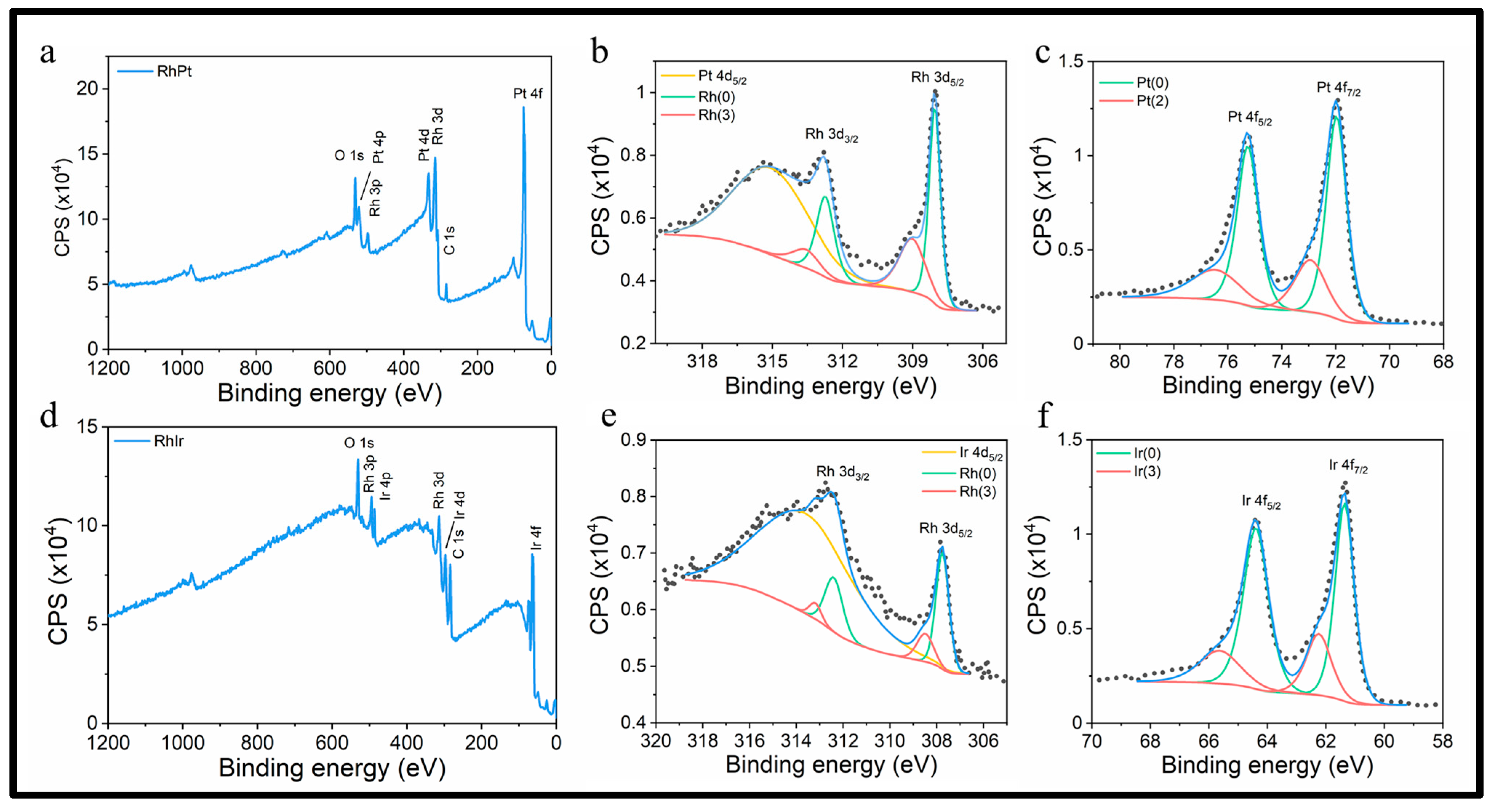
| Spin Orbit | BE (eV) | Spin Orbit | BE (eV) | Spin Orbit | BE (eV) |
|---|---|---|---|---|---|
| Rh 3d5/2 | 308.0 | Pt 4f7/2 | 72.0 | Ir 4f7/2 | 61.3 |
| Rh 3d3/2 | 312.7 | Pt 4f5/2 | 75.3 | Ir 4f5/2 | 64.5 |
| RhOx 3d5/2 | 309.0 | PtOx 4f7/2 | 72.9 | IrOx 4f7/2 | 62.2 |
| RhOx 3d3/2 | 313.7 | PtOx 4f5/2 | 76.2 | IrOx 4f5/2 | 65.4 |
Publisher’s Note: MDPI stays neutral with regard to jurisdictional claims in published maps and institutional affiliations. |
© 2022 by the authors. Licensee MDPI, Basel, Switzerland. This article is an open access article distributed under the terms and conditions of the Creative Commons Attribution (CC BY) license (https://creativecommons.org/licenses/by/4.0/).
Share and Cite
Zou, Y.; Goei, R.; Ong, S.-A.; ONG, A.J.; Huang, J.; TOK, A.I.Y. Development of Core-Shell Rh@Pt and Rh@Ir Nanoparticle Thin Film Using Atomic Layer Deposition for HER Electrocatalysis Applications. Processes 2022, 10, 1008. https://0-doi-org.brum.beds.ac.uk/10.3390/pr10051008
Zou Y, Goei R, Ong S-A, ONG AJ, Huang J, TOK AIY. Development of Core-Shell Rh@Pt and Rh@Ir Nanoparticle Thin Film Using Atomic Layer Deposition for HER Electrocatalysis Applications. Processes. 2022; 10(5):1008. https://0-doi-org.brum.beds.ac.uk/10.3390/pr10051008
Chicago/Turabian StyleZou, Yiming, Ronn Goei, Su-Ann Ong, Amanda Jiamin ONG, Jingfeng Huang, and Alfred Iing Yoong TOK. 2022. "Development of Core-Shell Rh@Pt and Rh@Ir Nanoparticle Thin Film Using Atomic Layer Deposition for HER Electrocatalysis Applications" Processes 10, no. 5: 1008. https://0-doi-org.brum.beds.ac.uk/10.3390/pr10051008