Article

Propagation and Molecular Characterization of Fowl Adenovirus Serotype 8b Isolates in Chicken Embryo Liver Cells Adapted on Cytodex™ 1 Microcarrier Using Stirred Tank Bioreactor

1
Faculty of Veterinary Medicine, University Putra Malaysia, Serdang 43400, Selangor, Malaysia
2
Department of Animal Science and Technology, Federal University of Technology, Owerri 460114, Imo State, Nigeria
3
Institute of Bioscience, University Putra Malaysia, Serdang 43400, Selangor, Malaysia
4
Faculty of Biotechnology and Biomolecular Sciences, University Putra Malaysia, Serdang 43400, Selangor, Malaysia
*
Author to whom correspondence should be addressed.
Submission received: 9 July 2020 / Revised: 6 August 2020 / Accepted: 11 August 2020 / Published: 1 September 2020
(This article belongs to the Special Issue Bioreactor System: Design, Modeling and Continuous Production Process)

Abstract

:
Large volume production of vaccine virus is essential for prevention and control of viral diseases. The objectives of this study were to propagate Fowl adenovirus (FAdV) isolate (UPM08136) in chicken embryo liver (CEL) cells adapted to Cytodex™ 1 microcarriers using stirred tank bioreactor (STB) and molecularly characterize the virus. CEL cells were prepared and seeded onto prepared Cytodex™ 1 microcarriers and incubated first in stationary phase for 3 h and in STB at 37 °C, 5% CO2, and 20 rpm for 24 h. The CEL cells were infected with FAdV isolate (UPM08136) passage 5 (UPM08136CELP5) or passage 20 (UPM08136CELP20) and monitored until cell detachment. Immunofluorescence, TCID50, sequencing, alignment of hexon and fiber genes, and phylogenetic analysis were carried out. CEL cells were adapted well to Cytodex™ 1 microcarriers and successfully propagated the FAdV isolates in STB with virus titer of 107.5 (UPM08136CELP5B1) and 106.5 (UPM08136CELP20B1) TCID50/mL. These isolates clustered with the reference FAdV serotype 8b in the same evolutionary clade. The molecular characteristics remained unchanged, except for a point substitution at position 4 of the hexon gene of UPM08136CELP20B1, suggesting that propagation of the FAdV isolate in STB is stable and suitable for large volume production and could be a breakthrough in the scale-up process.

1. Introduction

Fowl adenovirus (FAdV) is associated with inclusion body hepatitis (IBH) in chickens which is responsible for heavy losses in poultry industries worldwide [1,2,3]. Vaccination has proven to be the most effective tool in the control and prevention of viral diseases in animals and humans [4,5]. Despite the successes of vaccines against a wide range of animal diseases, developing a vaccine is a very challenging process, and one of the major drawbacks to the availability of vaccines over the years has been technical manufacturing scale-up obstacles [6]. Availability of vaccines in large volumes to meet the ever-growing production animal population could be easier through cell culture-based production than the traditional embryonated egg culture [7]. Cell culture also offers advantages like reduced contamination, product purity, ability to use wild type viruses efficiently, reduced immunogenic changes, large volume production, and quick response during pandemic such as influenza outbreaks or the recent COVID-19 pandemic [8,9]. Cell culture involves harvesting cells from the organs of animals, insects, or from plants, growing them in vitro [10] and then maintaining them in medium for use to express enzymes, antibodies, therapeutics, and to grow viruses for vaccine development [11,12]. Anchorage-dependent cells like liver cells require surfaces to attach for stabilization to enable proliferation. The early protocol for cell culture made use of glass slides, petri dishes, and other glass-based surfaces for attachment of cells for growth, but due to difficulty encountered in primary cells not being able to attach to glass surfaces, George Gey in 1956 used rat tail collagen to coat glass surfaces which pioneered the development of plastic flasks, dishes, and 96-well plates, which were all commercially available by 1960 [13]. Tissue culture flasks have offered a lot of advantages and are still widely used in cell biology and vaccine development to date [13]. Although the use of tissue culture flasks provided a solution to the issue of cell culture contaminations and improved cell proliferation, its limitation with regards to large volume production of vaccines has been inability to provide enough space [14]. Several platforms for large volume proliferation of cells has been experimented with at different stages, but one of the breakthroughs for the industrial application of mammalian cells has been the invention of microcarriers for vaccine production to support growth of adherent or anchorage-dependent cells (ADC) [15]. A variety of microcarriers are available for vaccine development, but spherical bead type microcarriers such as Cytodex™ 1 are suitable especially for stirred tank bioreactors [16]. It is a multipurpose microcarrier which can be used to grow a variety of cells. While culturing influenza virus vaccine in stirred tank bioreactor on Vero cells, Cytodex™ 1 was used and optimized to the production capacity of 6000 L [16].
For a successful suspension culture of adherent cells with microcarriers as attachment surface, in vivo conditions which promote growth and differentiation of target cell types should be replicated as closely as possible in vitro. A wide variety of bioreactors has been developed by researchers to provide an in vitro environment that recapitulates the in vivo environment as accurately as possible [17]. These include continuous stirred tank bioreactors, which are used in 70% of all fermentation and bioprocess operations [18]. Bioreactors are complex and expensive devices and are not optimized to provide an ideal environment for all mammalian cell growth due to high local fluid shear and bubble aeration. Each bioprocess operation requires optimization with respect to a specific set of parameters, such as cell growth, cell yield, and specific productivity, because there is no universal approach to optimize conditions for all animal cell culture systems [19]. A stirred tank bioreactor was used to propagate Infectious bursal disease (IBD) virus in BGM70 cell line [20], Modified vaccinia Ankara (MVA) virus in avian AGE1.CR.pIX cell line [21], and bacteriophages in E. coli [22]. It was expected that primary chicken embryo liver (CEL) cells could be adapted to Cytodex™ 1 microcarrier culture and used to propagate FAdV isolate in a stirred tank bioreactor. The objectives of this study were therefore to adapt primary CEL cells to Cytodex™ 1 Microcarrier and propagate FAdV isolates in the microcarrier adapted CEL cells in a bioreactor and determine the molecular characteristics of the virus.

2. Materials and Methods

2.1. Ethics Statement

The design of this experiment including the embryonated chicken egg utilization protocol was done following the guidelines and ethics of the UPM institutional animal care and use committee (IACUC) and was approved with ref number; UPM/IACUC/AUP-R086/2018.

2.2. Virus Isolates

Fowl adenovirus serotype 8b isolate, which was passaged in CEL cells for 5× (UPM08136CELP5) with Genebank accession number MT561443 and passaged in CEL cells 20× (UPM08136CELP20) with Genebank accession number MT561445, was used in this study. Each of the isolates was freeze-thawed three times and centrifuged at 1500 rpm for 5 min and the supernatant filtered through 0.45 µm syringe filter. Filtered isolates were used as inocula for this study.

2.3. Siliconization of Glassware

All glassware used in this study was first siliconized by applying dichlorodimethylsilane (Sigma-Aldrich, Shanghai, China) with ≥99.5% concentration on all surfaces that came in contact with microcarrier beads [23]. After siliconization, the glasswares were then sterilized by autoclaving.

2.4. Setting up the BIOSTAT® B Bioreactor

A B Braun Biostat® B Fermentation Cell Culture Bioreactor (Type 8840334) (Sartorius, Goettingen, Germany), which is a continuous stirred tank bioreactor (STB), was used in this study. The BIOSTAT® B bioreactor vessel (chamber) was disconnected from the control unit, washed thoroughly, and dried. The inner chamber was then siliconized and dried. The bioreactor was then assembled according to manufacturer’s recommendation and reconnected to the control unit. The pH and temperature regulators were then calibrated. After calibration, the unit was disconnected from the control unit and all the openings in the chamber and all the flow tubes were closed with aluminium foil. The prepared chamber was then sterilized by autoclaving at 121 °C 103.42 kpa for 15 min. After sterilization, the chamber was then reconnected to the control unit ready for use.

2.5. Preparation of Cytodex™ 1 Microcarrier Beads

Cytodex™ 1 was prepared according to manufacturer’s recommendation to obtain a concentration of 3 g/L. Three grams of Cytodex™ 1 was measured into a siliconized conical flask and suspended in 150 mL of CaCl2 and MgCl2 free PBS (Nacalai tesque, Kyoto, Japan) and hydrated for 3 h at room temperature after which PBS is decanted. The microcarriers were washed in fresh 150 mL of PBS for 30 min and PBS decanted. The washing process was repeated, after which fresh 200 mL of PBS was added and the microcarriers in PBS were sterilized by autoclaving. After sterilization, the PBS was decanted and 150 mL of DMEM media was added to dislodge retained PBS. The media was decanted before cells were seeded onto the microcarrier beads. The surface provided by the microcarrier is shown in Table 1.

2.6. Preparation of Chicken Embryo Liver Cells

The chicken embryo liver (CEL) cells were prepared using 15-day-old specific pathogen-free (SPF) chicken embryonated eggs (CEE) [24]. The liver from the embryo was aseptically harvested and macerated in sterile petri dish with sterile forceps. Macerated liver was transferred into sterile conical flask and cells dissociated with sodium bicarbonate-free Trypsin-EDTA (0.25% Trypsin and 2.25 mM EDTA) (Corning, Manassas, VA, USA) in a two-step dissociation. Liver from 2 SPF CEE was dissociated initially with 12 mL of Trypsin-EDTA and pipetted into centrifuge 50 mL tube, and dissociation was repeated subsequently with 8 mL Trypsin-EDTA. Dissociated liver cells were centrifuged (Hettich Zentrifugen, Tuttlingen, Germany) at 1500 rpm for 5 min, after which Trypsin-EDTA was discarded and the CEL cells were suspended in fresh DMEM media supplemented with 10% FBS and 2% (v/v) antibiotics (penicillin–streptomycin) [25]. The CEL cells were seeded onto already prepared Cytodex™ 1 microcarriers.

2.7. Adaptation of CEL Cells to Cytodex™ 1 Microcarrier Culture

Freshly prepared CEL cells were seeded onto the prepared Cytodex™ 1 microcarriers and incubated in CO2 incubator (ThermoForma, Champaign, IL, USA) at 37 °C and 5% CO2 for 1 h. The culture was made up to 1/3 of the final volume and incubated for additional 2 h. The sample was drawn into tissue culture flask and observed under the inverted light microscope (Leica Microsystems, Wetzlar, Germany) for cell attachment and confluency and images for each passage were captured using an inverse microscope (Nikon Eclipse TS100, Tokyo, Japan) with the aid of NIS Elements imaging system (Nikon Digital sight DS-U2, Tokyo, Japan) powered by PowerLogic computer with 17” monitor (PowerLogic, Brownsville, TX, USA). The culture was then transferred into the bioreactor [20]. The cell volume is shown in Table 1.

2.8. Propagation of FAdV in Cytodex™ 1 Microcarrier Adapted CEL Cells in STB

The microcarriers with CEL cells attached were transferred into the bioreactor chamber using peristaltic pump drive (Longer Pump, Baoding, China). The bioreactor was preset at 37 °C, pH 7.4, dissolved oxygen (DO) 40% air saturation [20]. The rotor spinning was set at 10 rpm and increased to 20 rpm after 3 h and the culture made up to final volume with DMEM media supplemented with 10% FBS and 2% penicillin–streptomycin [26]. After 24 h of incubation in STB, cells were inoculated with 10 mL of FAdV inoculum (Table 1) and the rotor was increased to 30 rpm and observed closely for pH changes. Sterile NaOH and HCl were used for pH controls. Samples were drawn at 24 and 48 h post-inoculation and observed for cytopathic effects (CPE). At 48 h post-inoculation, the culture was harvested. Harvesting of virus culture was achieved by increasing the stirring to 500 rpm for 2 min to enable complete detachment of cells after which the microcarrier beads were allowed to sediment. Harvested cells with virus was removed from bioreactor by reverse flow using the peristaltic pump drive (Longer Pump, Baoding, China) into a siliconized, sterile bottle while the sedimented microcarrier beads were collected separately, washed and kept for reuse. The culture was freeze-thawed 3 times at −20 °C after which aliquots were centrifuged in 50 mL tubes at 1500 rpm for 5 min. The supernatant was extracted, filtered through 0.45 µm syringe filter and stored at −80 °C. The titer of each FAdV inoculum is shown in Table 1.

2.9. Determination of the Infective Dose (TCID50) of the Passage Isolates

Ten-fold serial dilution of each isolate was carried out [27]. One 96-well tissue culture plate for each isolate to be tested was placed under UV light overnight and 0.2 mL of CEL cells, prepared as earlier described (2.5), was seeded into each well and incubated in CO2 incubator (ThermoForma, Champaign, IL, USA) at 37 °C and 5% CO2. The plates were observed daily until the cell became 100% confluent. At confluency, the plates were washed twice with PBS and infected with 0.1 mL of serially diluted isolate. Each dilution was used for 8 wells in a column (Columns 1–10). Columns 11 and 12 were infected with sterile distilled water as control. Infected plates were incubated for 1 h and 0.2 mL of DMEM media supplemented with 2% FBS was added to each well, re-incubated in CO2 incubator (ThermoForma, Champaign, IL, USA) at 37 °C and 5% CO2 and observed for CPE for 7 days. The number of wells in each column with more than 50% CPE was recorded and TCID50 calculated as described by Reeds and Muench [28].

2.10. Indirect Immunofluorescence Assay

CEL cells were prepared as previously described and seeded onto sterile cover slips inside 6-well tissue culture plates and incubated until confluency. The medium was discarded and washed 2× with PBS after which each well was infected with 0.2 mL of respective bioreactor passage isolates (UPM08136CELP5B1 or UPM08136CELP20B1) and incubated at 37 °C and 5% CO2 for 1 h for adsorption. Then, 1.8 mL of DMEM maintenance media was added while the uninfected control was maintained with only media and all incubated at 37 °C and 5% CO2 and observed for CPE for 48 h. On CPE, the wells were fixed and inactivated with 4% formalin for 30 min at room temperature, after which they were washed 2× with ice cold PBS containing 0.5% Tween 20 for 5 min and plates incubated in 0.5% Triton x-100 in PBST for 15 min to permeabilize the cells followed by rinsing with PBST 3× for 5 min. Unspecific bindings were blocked with blocking buffer (5% BSA in PBST) for 1 h at room temperature and rinsed 3× with PBST for 5 min. Forty microlitres of monoclonal FAdV hexon protein primary antibody (Santa Cruz, Dallas, TX, USA) was added into the cells, allowed to stand, and incubated at 4 °C in humidity chamber overnight after which the wells are rinsed 2× with PBST for 5 min. Cells were suspended in 40 µL of mice anti-chicken IgY-FC-FITC-conjugated secondary antibody (Santa Cruz, Dallas, TX, USA) 1:1000 dilution was added and incubated at room temperature (RT) for 1 h in a dark room. The plates were rinsed with PBST and 20 µL of diamidino-2-phenylindole dihydrochloride (DAPI) added for 10 min at room temperature. The cover slip was rinsed, dried, mounted on clean glass slides with dibutylphthalate polystyrene Xylene (DPX), and observed with fluorescence microscope [20].

2.11. DNA Extraction and PCR Amplification of Hexon and Fiber Genes

DNA from UPM08136CELP5B1 and UPM08136CELP20B1 was extracted using an innuPREP Virus DNA Kit (analytikjena, Jena, Germany) and the concentration measured at 70 dilution factor, using ultraviolet–visible spectrophotometer (UV-1601, PC, Shimadzu Europa, Duisburg, Germany). HexA1/HexB1 primers [29] were used to amplify hexon gene while fbrF/fbrR primers Fbr F-5′ ACCGATTACGGCCGACGAAC -3′ and Fbr R-5′ -GAGCGTTGGCTGTGCTTAGG -3′ designed from KU517714.1 reference strain for this study were used for fiber gene amplification with sensoquest labcycler gradient (Biomedizinische Elektronik, Goettingen, Germany) in a 25 µL reaction using MyTaq Red Mix (Bioline, London, UK). The amplification conditions were 95 °C for 2 min 1×, 95 °C for 1 min 35×, 55 °C for 1 min 35×, 72 °C for 2 min 35× and 72 °C 1 min 1× for hexon gene and 95 °C for 2 min 1×, 95 °C for 1 min 35×, 50 °C for 1 min 35×, 72 °C for 3 min 35×, and 72 °C for 2 min 1× for fiber genes. The PCR products were separated by electrophoresis using EPS-300 X (C.B.S. Scientific, San Diego, CA, USA) in 1% agarose gel (GeneDireX, Taoyuan, Taiwan) at 70 V and 400 mA for 50 min, stained with redsafe stain (iNtRON Biotechnology, Burlington, MA, USA) and visualised through UV transillumination (Syngene, Cambridge, UK).

2.12. Sequencing, Blast, Alignment, and Phylogentic Analysis

The PCR products were purified using MEGAquick-spin™ Total fragment DNA purification kit (iNtRON Biotechnology, Burlington, MA, USA) and sequenced (Apical Scientific, Serdang, Malaysia). Consensus sequences were assembled using contig assembly application and multiple sequence alignment to determine changes in the genes were carried out using BioEdit software v 7.2.5 [30,31] and sequences were subjected to NCBI Blast. Amino acids were deduced by using ExPasy software (www.expasy.ch/tools/dna.html) [32]. With the sequences and 37 published strains for hexon (Table 2) with NCBI Genebank links to the sequences in Table S1 and 27 published strains for fiber (Table 3) with NCBI Genebank links to the sequences in Table S2, phylogenetic trees were constructed by neighbor-joining method [33] using MEGA X software v 10.0.5 [32].

3. Results

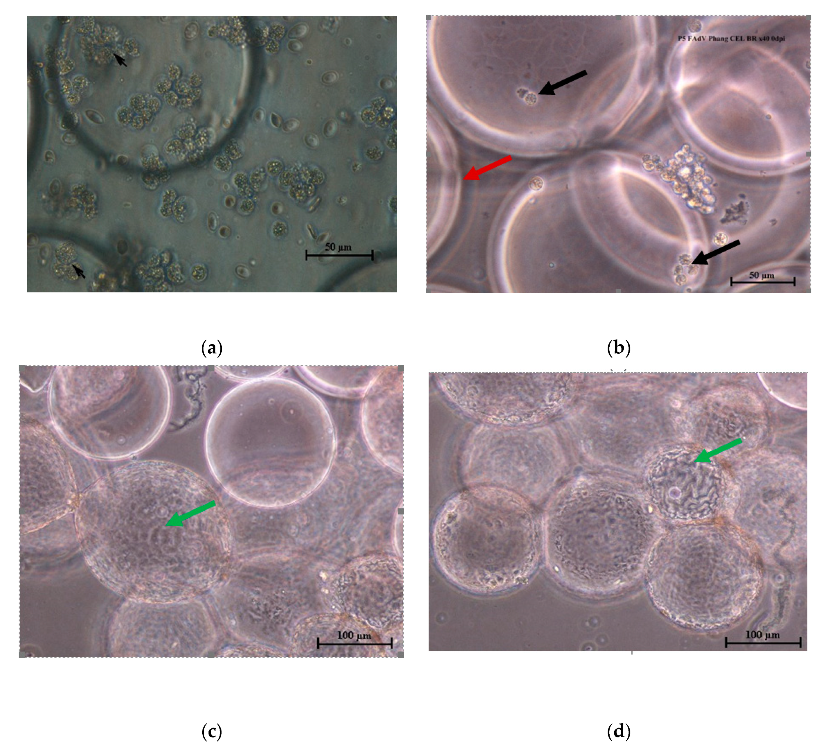
3.1. Adaptation of CEL Cells on Cytodex™ 1 Microcarriers

The CEL cells attached and adapted to the Cytodex™ 1 microcarriers within 3 h of incubation (Figure 1a,b) and grew confluent on the microcarriers beads after 24 h incubation in the bioreactor (Figure 1c,d).

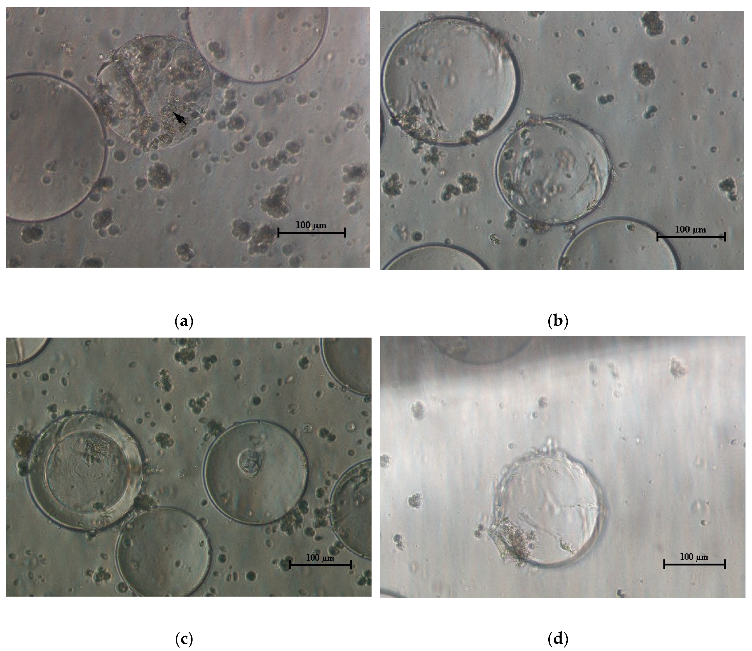
3.2. Propagation of FAdV in CEL Cells Adapted on Cytodex™ 1 Microcarrier

The FAdV UPM08136CELP5 (Figure 2a) and UPM08136CELP20 (Figure 2c) isolates caused CPE and detachment of cells from microcarriers from 24 h post-inoculation. At 48 h, most of the cells had detached from the microcarrier beads (Figure 2b,d) indicating suitability of the Cytodex™ 1 and STB for propagation of FAdV serotype 8b.

3.3. Infective Dose (TCID50) of UPM08136P5B1 and UPM08136P20B1 Isolates

The UPM08136CELP5B1 showed higher virus titer of 1 × 107.5 TCID50/mL in a final volume of 500 mL when compared to UPM08136CELP20B1 with virus titer of 1 × 106.5 TCID50/mL in a final volume of 600 mL. These could be due to final culture volume or infectivity of the isolate (Table 4).

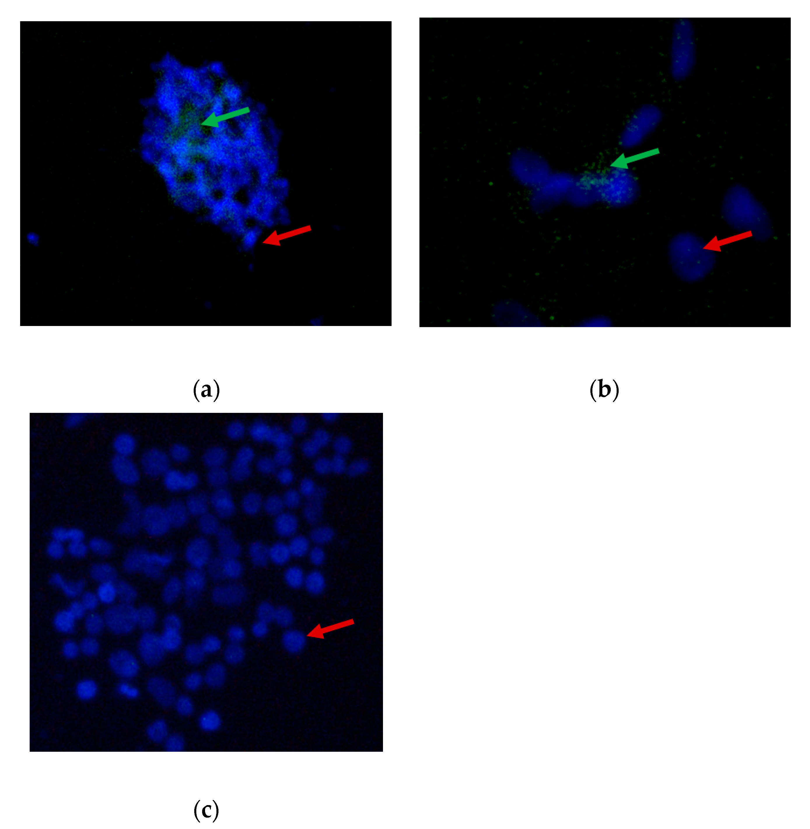
3.4. Indirect Immunofluorescence Assay of FAdV Isolates Propagated in CEL Cells Adapted Cytodex™ 1 Microcarrier in a Stirred Tank Bioreactor

There were green particles indicating presence of FAdV antigens (Figure 3a,b). In contrast, in the control, there were only blue particles, denoting a negative result (Figure 3c). The isolates also showed affinity for the CEL cells and caused cell depletion after 48 h post-infection (Figure 3a,b), while the uninfected control shows intact monolayer (Figure 3c).

3.5. Polymerase Chain Reaction Amplification of Hexon and Fiber Genes of the Propagated FAdV Isolates in CEL Cells Adapted on Cytodex™ 1 Microcarrier

The UPM08136CELP5B1 and UPM08136CELP20B1 yielded 900 bp (Figure 4a) and 940 bp (Figure 4b), which corresponds to the hexon and fiber genes of FAdV, respectively.

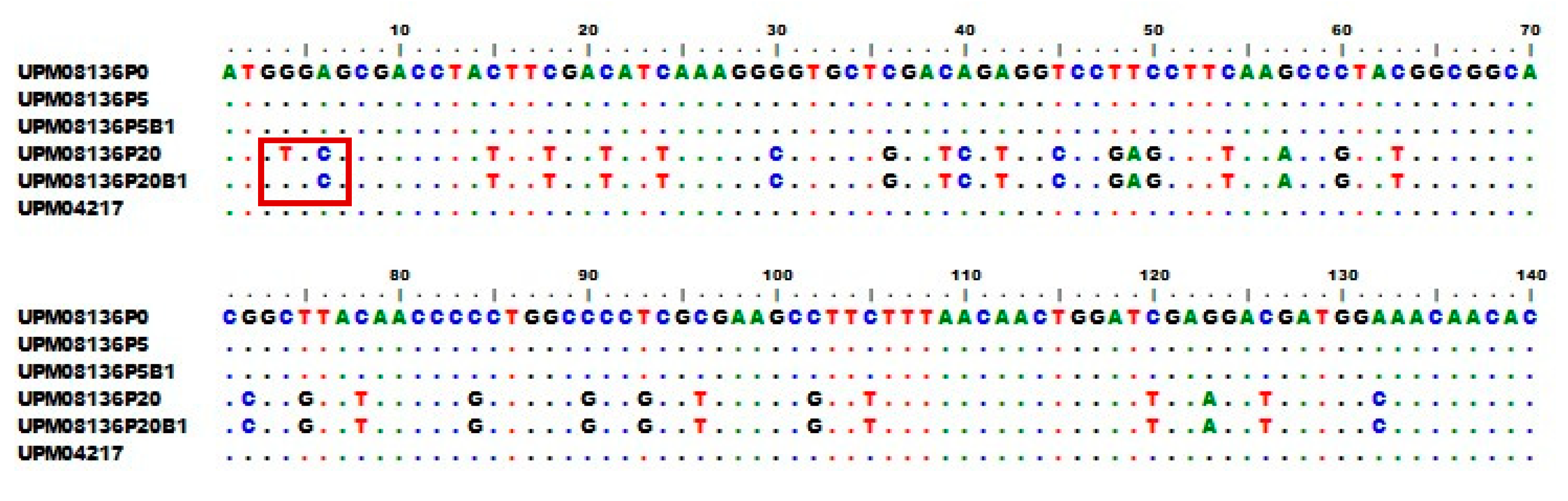
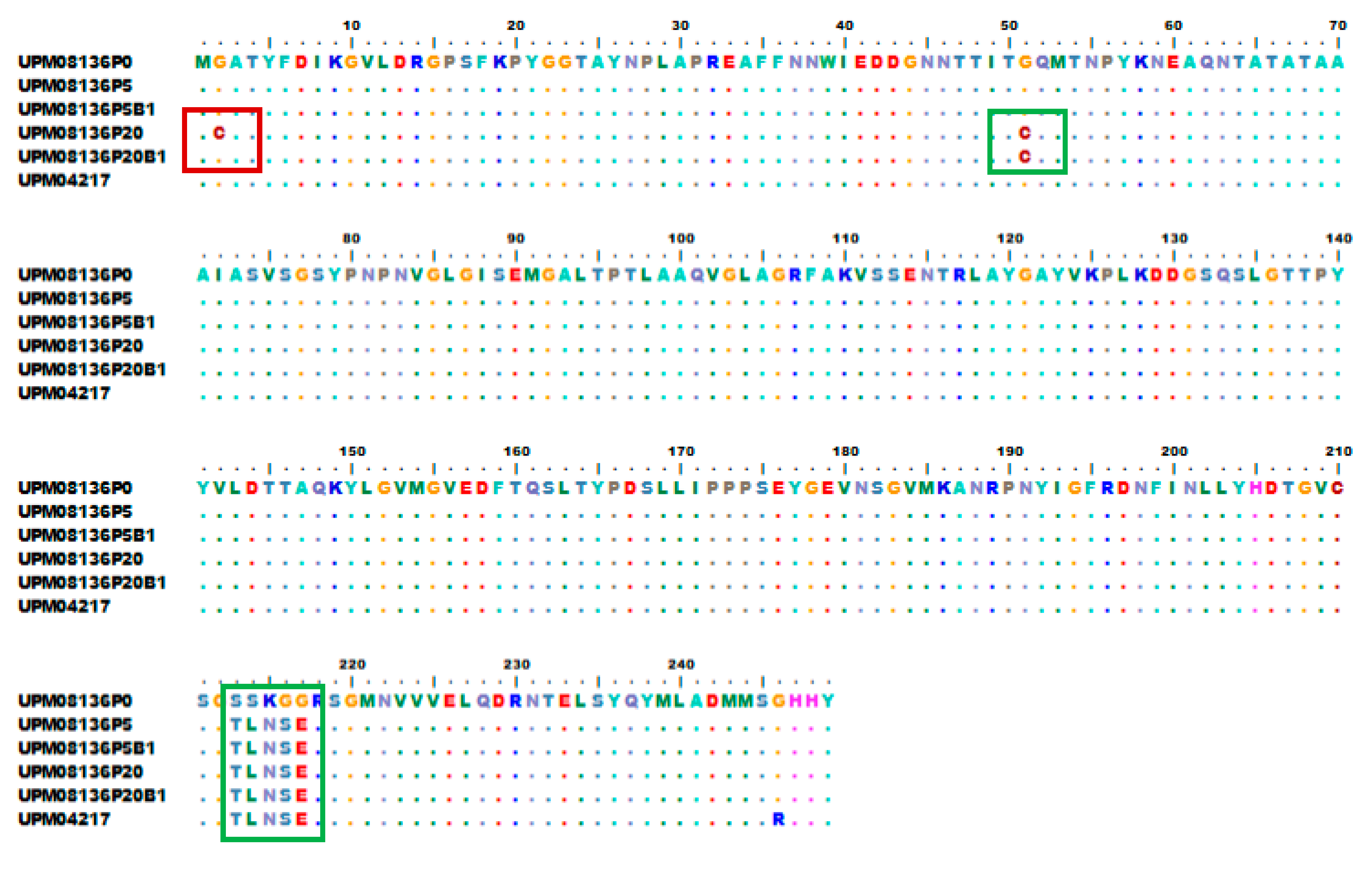
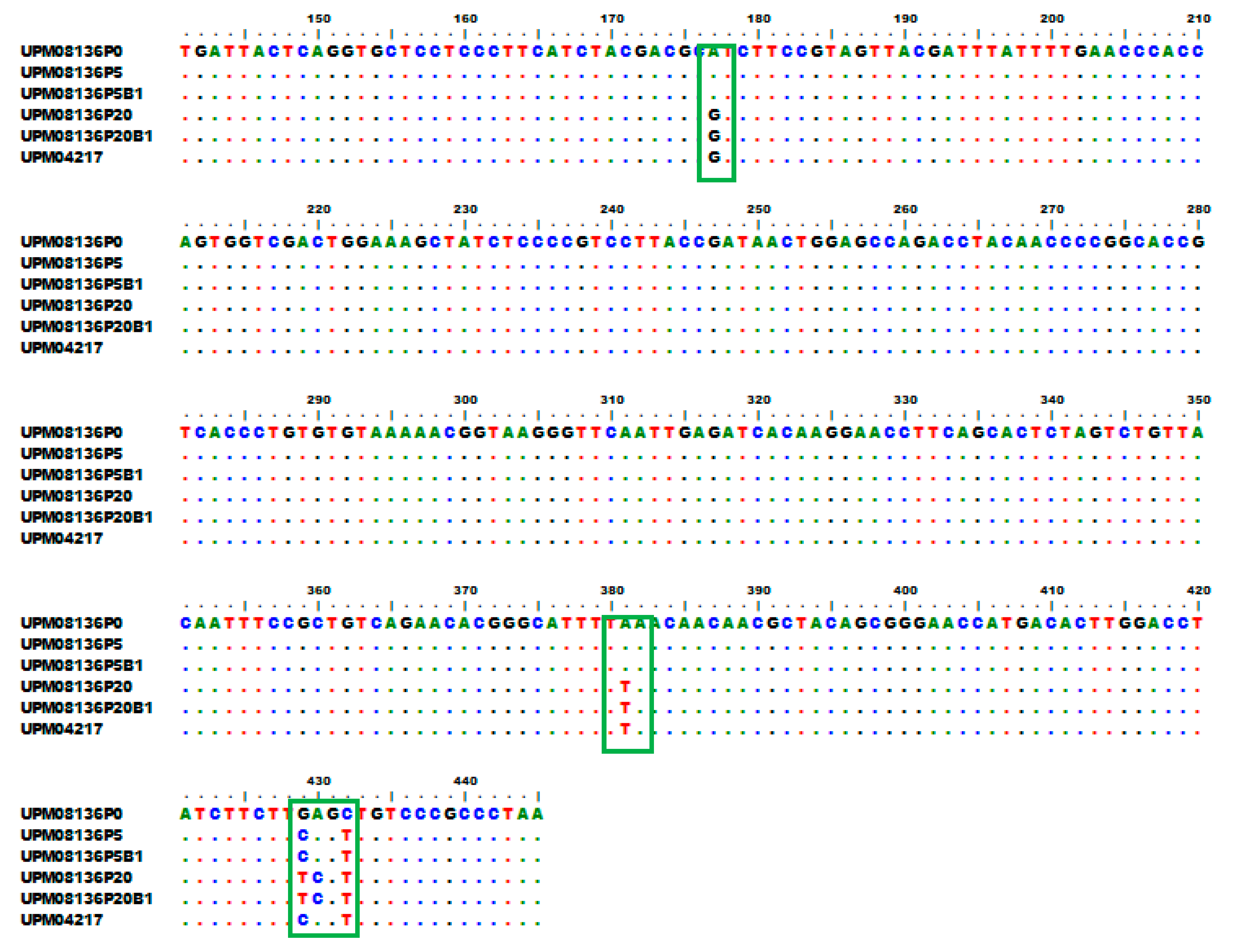
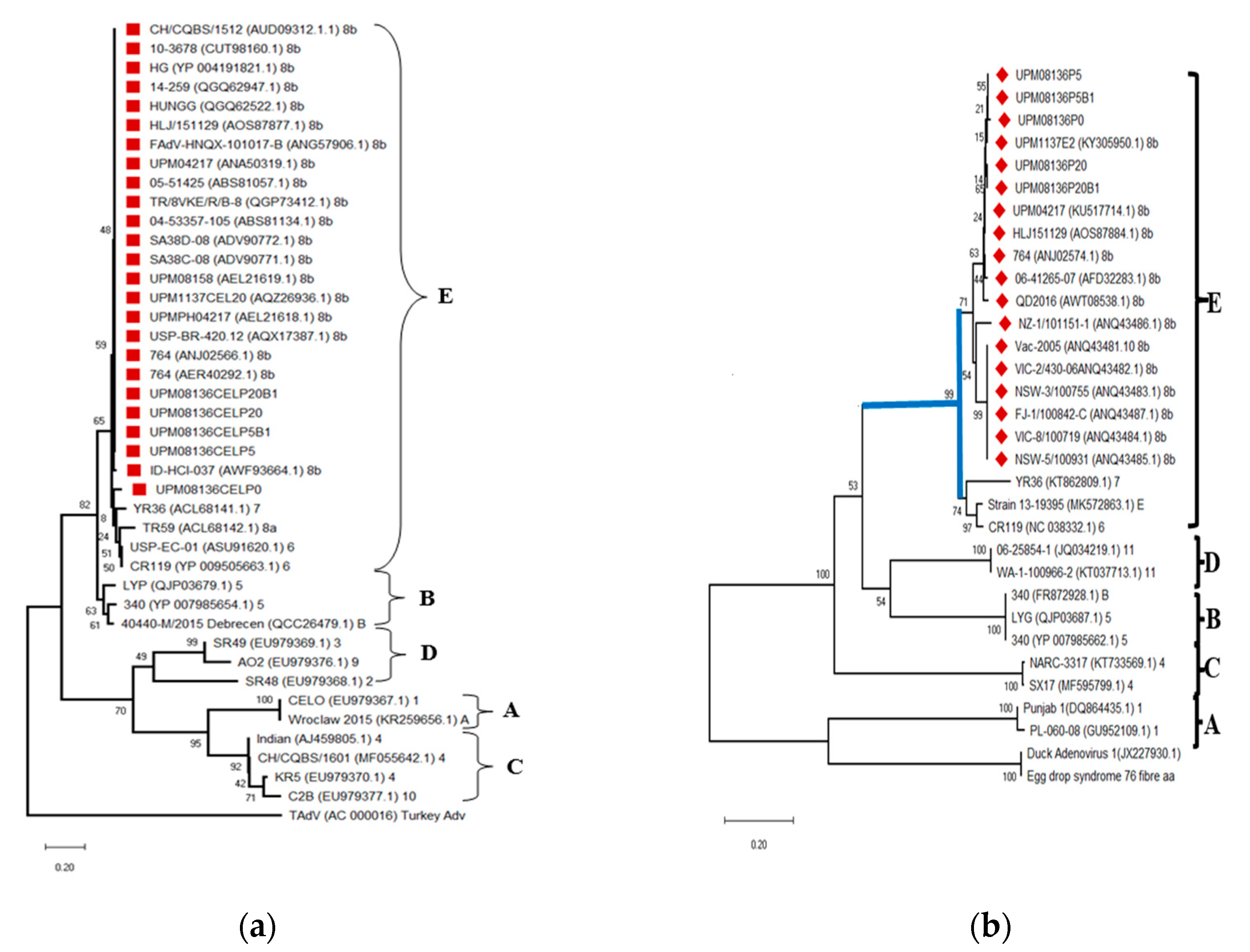
3.6. Multisequence Alignment and Phylogenetic Analysis of UPM08136CELP5B1 and UPM08136CELP20B1 with Reference Strains

The translation of the nucleotides of hexon gene yielded 244–245 amino acid residues while the translation of the nucleotides of fiber gene yielded 148–149 amino acid residues. The nucleotide sequences and their corresponding amino acids were uploaded to Genebank and were assigned accession numbers, UPM08136CELP5B1 (MT561443 and MT561447) and UPM08136CELP20B1 (MT561445 and MT561449) for the sequences of hexon gene and fiber genes, respectively. There was no change in the hexon gene of UPM08136CELP5B1 compared to UPM08136CELP5. There was a G►4T substitution (Figure 5) in the hexon gene of UPM08136CELP20B1 compared to UPM08136CELP20, which was the only change found in their hexon gene. This translated to an amino acid change from cysteine to glycine at position 2 (Figure 6) in the red box, which alone may not have effect on the stability of the virus. There was no change observed in the nucleotide sequences (Figure 7) and amino acid residues (Figure 8) of fiber gene of the bioreactor isolates compared to the flask isolates, suggesting suitability in FAdV serotype 8b propagation.
The hexon and fiber genes of UPM08136CELP5B1 and UPM08136CELP20B1 were 99–100% identical to UPM04217 and other FAdV serotype 8b isolates in Genebank, which confirms their non-variability with the flask isolates. The phylogenetic tree results based on the amino acid residues of hexon gene showed that all the test isolates clustered together in the same evolutionary clade with other Universiti Putra Malaysia (UPM) strains, the Australian vaccine strain, and other FAdV serotype 8b strains. Isolates in the FAdV genus branched out from the root away from the Duck and Turkey adenovirus strains (Figure 9a). The fiber amino acid phylogenetic tree results also showed that all the test isolates clustered together in the same evolutionary clade with other UPM strains and other FAdV serotype 8b strains. All the isolates in the FAdV genus branched out from the root away from the Duck adenovirus 1 strains (Figure 9b).

4. Discussion

The CEL cells attached to the surface of the microcarrier beads within 3 h of incubation which indication adaptation of the cells to microcarrier culture. Most animal cells are anchorage dependent and can usually survive and are most productive when attached to a surface. Even suspension-type cell, like hybridomas, tends to give increased product yields when attached [34]. Attachment of cells to a surface is a critical need and one of the most essential points in the optimal functioning of all anchorage-dependent cells [35,36]. Attachment to the microcarrier is indicative of CEL cells adaptation to microcarrier culture. It proves that Cytodex™ 1 microcarrier is suitable for growth of CEL cells. Cytodex™ 1 was engineered to meet all properties essential for a successful microcarrier and has been used to culture more than sixty different cell lines successfully [37]. It is also cost effective as it can be reused [20]. The cells grew confluent in the cells within 24 h which shows that proliferation of cells in the microcarrier was successful. For virus propagation, cells are usually needed, and it is more needed in higher volumes for vaccine production. This is because cell concentration is of significant importance in determining viral titer [38], and viral titer has control on the efficacy of vaccines, which makes the need for a high volume of cells for volume production of vaccine an ongoing requirement.
The infected CEL cells exhibited cytopathic effects synonymous with FAdV on CEL cells which included rounding and detachment of cells [24,39] from the microcarrier beads. CPE is a sign of pathogenicity of virus on cells [40] and confirms infection of the cells. This means that CEL cells grown in the Cytodex™ 1 microcarrier are suitable for propagation of FAdV in a continuous stirred tank bioreactor system. This corroborates the reports of Lawal et al. [20] and Blumi [16] who used Cytodex™ 1 to propagate Infectious bursal disease virus (IBDV) and Influenza virus, and Tapia et al. [21] who used stirred tank bioreactor to propagate modified vaccinia ankara (MVA) virus in avian AGE1.CR.pIX cell line. Mancuso et al. [22] also reported the successful use of E. coli to grow bacteriophages in stirred tank bioreactor. Stirred tank bioreactors are used in 70% of all fermentation and bioprocess operations and have been found to be suitable for propagation of virus vaccine candidates for large volume production [18]. The experimental volume was low especially when 6000 L was reported in influenza virus propagation [16], but it is a huge improvement when compared to flask propagation. UPM08136CELP20B1 had a lower titer than the flask propagated isolate which was similar to the report of Lawal et al. [20]. This could be attributed to the isolate having a lowered infectivity than the earlier passages. The titer values obtained in the study were 106.5/mL and 107.5/mL TCID50 which were similar to the report of Chen et al. [41] who obtained a titer of 7.6 log10 TCID50/mL of influenza vaccine virus grown in Vero cells and 106.4 TCID50/mL reported by Lawal et al. [20] in vvIBDV passaged once in BGM-70 cell line before bioreactor passage in Cytodex™ 1 adapted BGM-70 cell line. The result is also comparable to 106 TCID50 reported by Trabelsi et al. [42] in Measles vaccine virus grown in mammalian cells in microcarrier.
The amplification of the hexon gene of UPM08136CELP5B1 and UPM08136CELP20B1 yielded a partial hexon DNA 738–750 bp in size, which corresponds to the report of Meulemans et al. [29] who initially published HexA1/HexB1 primers. The primer had been used by several researchers for identification and studies of FAdV [43,44,45,46,47,48]. The amplification of the fiber gene using the novel fbrF/fbrR primers yielded partial fiber gene, mainly the knob region. Evaluation of fiber gene of FAdV has become necessary because through the interaction of the fiber knob with host cells, the fiber gene is responsible for tissue tropism which is also very important in the virulence of FAdV [49]. In fact, it had been reported that fiber alone could determine virulence of FAdV 8 [50]. The primer pair used in this study was designed in our laboratory and first published in this report. Analysis of fiber gene of FAdV had been carried out by previous researchers for identification, characterization and pathogenicity studies [25,51,52,53].
The NCBI blastn and blastp for nucleotide and protein sequences respectively of hexon and fiber genes showed 99–100% homology to FAdV serotype 8b in Genebank. This was synonymous with other FAdV sequences of Malaysian origin which suggests that the organisms in our study belong to the same serotype with those reported earlier in the country. Since the development of genomic analysis technology, researchers rely on NCBI Genebank blast as one of the repositories for virus taxonomical classification, species identification; and nucleotide and amino acid sequences identity search [54,55,56]. The major primary sequence databases are the International Nucleotide Sequence Database Collaboration (INSDC) databases, which includes GenBank (NCBI), European Nucleotide Archive (ENA), and the DNA Data Bank of Japan (DDBJ) [57], and Uniprot [58] which constitutes the main repositories of nucleotide and protein sequence data. In line with this, the sequences of hexon and fiber gene of isolates from this study were deposited in Genebank.
There were no changes observed in the hexon and fibre genes of UPM08136CELP5B1. There was also no change in the fiber gene of UPM08136CELP20B1. This means that the propagation of this FAdV isolate in CEL cells adapted in Cytodex™ 1 microcarrier in stirred tank bioreactor is suitable and had no adverse effect on the virus. This could be a model for adoption in the propagation of FAdV, especially serotype 8b when large volume of virus is needed. The bioreactor passage did not affect the virus so much to disturb its normal conformity. This is similar to the report of Lawal et al. [20] who found that vvIBD virus propagated in stirred tank bioreactor did not show molecular change in their VP2 protein. Other researchers did not report any change in the characteristics of the bioreactor propagated viruses which suggests that use of bioreactor for propagation of vaccine virus is suitable for large volume production of vaccines [41,59,60]. However, there was a point substitution in the hexon gene of UPM08136CELP20B1 which led to change in the amino acid residues at position 2 from cysteine to glycine. This alone would not affect the stability of the virus. However, following this change, it has become pertinent that all viruses propagated in a stirred tank bioreactor for a live-attenuated vaccine should be monitored for molecular changes in their antigenic proteins to forestall reversion to virulence. All the bioreactor propagated FAdV clustered together with serotype 8b viruses of Malaysian origin and those from other sources. This shows that they are FAdV serotype 8b. The closeness of the flask and bioreactor isolates in the phylogram of hexon and fibre genes shows their similarity which confirms the suitability of propagating FAdV serotype 8b in CEL cells adapted Cytodex™ 1 microcarrier using stirred tank bioreactor. As the data on substrate requirement, metabolism, and specific CEL cells needs and bioreactor conditions for growth of primary CEL cells were not readily available, further studies should explore to establish the optimal growth conditions for CEL cells in stirred tank bioreactor and other conditions most appropriate for the propagation of FAdV in CEL cells adapted in microcarrier for maximum yield, to standardize the scaling-up and industrialization of FAdV vaccine production. In this way scale up for production of FAdV vaccine up to 5000–10,000 L could be achieved as this study could only achieve 600 mL volume. Due to paucity of literature on this area, it is possible that adaptation of primary CEL cells in Cytodex™ 1 microcarrier and using it in stirred tank bioreactor to propagate FAdV serotype 8b is a novel finding. To our knowledge, there are no reports on propagation of FAdV serotype 8b in primary CEL cells using the bioreactor technology. This finding could therefore be a breakthrough in the scale up process of FAdV vaccines and could be useful for researchers interested in scaling up their developed FAdV serotype 8b vaccines. It will offer opportunity for large scale production and commercialization of the FAdV vaccine which will aid the prevention and control of IBH and other FAdV infections worldwide.

5. Conclusions

Chicken embryo liver cells were adapted well to Cytodex™ 1 microcarriers and used to propagate Fowl adenovirus serotype 8b isolates in a stirred tank bioreactor. These bioreactor isolates were similar to their corresponding flask isolates in their molecular characteristics, which show that bioreactor propagation of Cytodex™ 1 adapted chicken embryo liver cells was well tolerated. This could be a breakthrough in FAdV serotype 8b vaccine scale up process.

Supplementary Materials

The following are available online at https://0-www-mdpi-com.brum.beds.ac.uk/2227-9717/8/9/1065/s1, Table S1: NCBI Genebank link for reference adenovirus strains used for phylogenetic analysis of hexon gene, Table S2: NCBI Genebank link for reference adenovirus strains used for phylogenetic analysis of fibre gene.

Author Contributions

Conceptualization, M.H.-B.; methodology, C.C.U.; formal analysis, C.C.U., M.H.-B., M.I.N., and A.R.O.; investigation, C.C.U.; resources, M.H.-B.; writing—original draft preparation, C.C.U.; writing—review and editing, C.C.U., M.H.-B., M.I.N., A.R.O., and A.I.; visualization, C.C.U., M.H.-B.; supervision, M.H.-B., M.I.N., A.R.O., and A.I.; funding acquisition, M.H.-B. All authors have read and agreed to the published version of the manuscript.

Funding

This research was funded by UNIVERSITI PUTRA MALAYSIA., grant number vot 6300899.

Conflicts of Interest

The authors declare no conflict of interest. The funders had no role in the design of the study; in the collection, analyses, or interpretation of data; in the writing of the manuscript; or in the decision to publish the results.

References

  1. Dar, A.; Gomis, S.; Shirley, I.; Mutwiri, G.; Brownlie, R.; Potter, A.; Gerdts, V.; Tikoo, S.K. Pathotypic and Molecular Characterization of A Fowl Adenovirus Associated With Inclusion Body Hepatitis in Saskatchewan Chickens. Avian Dis. 2012, 56, 73–81. [Google Scholar] [CrossRef]
  2. Hair-Bejo, M. Inclusion Body Hepatitis in A Flock of Commercial Broiler Chickens. J. Vet. Malays. 2005, 17, 23–26. [Google Scholar]
  3. Norina, L.; Norsharina, A.; Nurnadiah, A.H.; Redzuan, I.; Ardy, A.; Nor-Ismaliza, I. Avian Adenovirus Isolated from Broiler Affected with Inclusion Body Hepatitis. Malays. J. Vet. Res. 2016, 7, 121–126. [Google Scholar]
  4. García, M. Current and Future Vaccines and Vaccination Strategies Against Infectious Laryngotracheitis (ILT) Respiratory Disease of Poultry. Vet. Microbiol. 2017, 206, 157–162. [Google Scholar] [CrossRef] [PubMed]
  5. Minor, P.D. Live attenuated vaccines: Historical Successes and Current Challenges. Virology 2015, 479–480, 379–392. [Google Scholar] [CrossRef] [PubMed] [Green Version]
  6. Hinman, A.R.; Orenstein, W.A.; Santoli, J.M.; Rodewald, L.E.; Cochi, S.L. Vaccine Shortages: History, Impact, and Prospects for the Future. Annu. Rev. Public Health 2006, 27, 235–259. [Google Scholar] [CrossRef]
  7. Whitford, W.G.; Fairbank, A. Considerations in Scale-Up of Viral Vaccine Production Bioprocess International. 2011. Available online: https://bioprocessintl.com/manufacturing/monoclonal-antibodies/considerations-in-scale-up-of-viral-vaccine-production-320990/ (accessed on 25 May 2019).
  8. Haredy, A.M.; Takenaka, N.; Yamada, H.; Sakoda, Y.; Okamatsu, M.; Yamamoto, N.; Omasa, T.; Ohtake, H.; Mori, Y.; Kida, H.; et al. An MDCK Cell Culture-Derived Formalin-Inactivated Influenza Virus Whole-Virion Vaccine from an Influenza Virus Library Confers Cross-Protective Immunity by Intranasal Administration in Mice. Clin. Vaccine Immunol. 2013, 20, 998–1007. [Google Scholar] [CrossRef] [Green Version]
  9. Whitford, W. Using Disposables in Cell-Culture–Based Vaccine Production. BioProcess Int. 2010, 8, S20–S27. [Google Scholar]
  10. Swain, P.; Nanda, P.K.; Natak, S.K.; Mishra, S.S. Basic techniques and limitations in establishing cell culture: A mini review. Adv. Anim. Vet. Sci. 2014, 2, 1–10. [Google Scholar] [CrossRef]
  11. Moreira, A.R. The Evolution of Protein Expression and Cell Culture. BioPharm Int. 2007, 20. Available online: http://www.biopharminternational.com/evolution-protein-expression-and-cell-culture (accessed on 14 May 2019).
  12. Marquis, C.P. Mammalian Cell Culture. Biotechnology 2019, 1. Available online: https://www.eolss.net/Sample-Chapters/C17/E6-58-01-04.pdf (accessed on 14 May 2019).
  13. Ryan, J.A. Evolution of Cell Culture Surfaces. BioFiles 2008, 3.8, 21. [Google Scholar]
  14. Merten, O.W. Advances in Cell Culture: Anchorage Dependence. Philos. Trans. R. Soc. Lond. Ser. B Biol. Sci. 2015, 370, 20140040. [Google Scholar] [CrossRef] [PubMed] [Green Version]
  15. Popovic, M.K.; Poertner, R. Bioreactors and Cultivation Systems for Cell and Tissue Culture. In Encyclopedia of Life Support Systems; Doelle, H.W., Rokem, S., Berovic, M., Eds.; Eolss Publications: Oxford, UK, 2011. [Google Scholar] [CrossRef]
  16. Blüml, G. Microcarrier Cell Culture Technology. In Animal Cell Biotechnology; Methods in Biotechnology; Pörtner, R., Ed.; Humana Press: Louisville, KY, USA, 2007; Volume 24. [Google Scholar] [CrossRef]
  17. Kluge, J.A.; Leisk, G.G.; Kaplan, D.L. Mechanical Determinants of Tissue Development. In Principles of Regenerative Medicine; Academic Press: Cambridge, MA, USA, 2008; pp. 480–497. [Google Scholar] [CrossRef]
  18. Ali, S.; Rafique, A.; Ahmed, M.; Sakander, S. Different Types of Industrial Fermenters and Their Associated Operations for The Mass Production of Metabolites. Eur. J. Pharm. Med. Res. 2018, 5, 109–119. [Google Scholar]
  19. Surabattula, R.R.; Sambasiva, K.R.S.; Polavarapu, R. An Optimized Process for Expression, Scale-Up and Purification of Recombinant Erythropoietin Produced in Chinese Hamster Ovary Cell Culture. Res. Biotechnol. 2011, 2, 58–74. [Google Scholar]
  20. Lawal, N.; Hair-Bejo, M.; Arshad, S.S.; Omar, A.R.; Ideris, A. Propagation and Molecular Characterization of Bioreactor Adapted Very Virulent Infectious Bursal Disease Virus Isolates of Malaysia. J. Pathog. 2018. [Google Scholar] [CrossRef]
  21. Tapia, F.; Jordan, I.; Genzel, Y.; Reichl, U. Efficient and Stable Production of Modified Vaccinia Ankara Virus in Two-Stage Semi-Continuous and in Continuous Stirred Tank Cultivation Systems. PLoS ONE 2017, 12, e0182553. [Google Scholar] [CrossRef] [Green Version]
  22. Mancuso, F.; Shi, J.; Malik, D. High Throughput Manufacturing of Bacteriophages Using Continuous Stirred Tank Bioreactors Connected in Series to Ensure Optimum Host Bacteria Physiology for Phage Production. Viruses 2018, 10, 537. [Google Scholar] [CrossRef] [Green Version]
  23. Noteboom, W.D.; Will, P.C. Siliconizing Glassware to Be Used for Suspension Cell Culture. J. Tissue Cult. Methods 1982, 7, 9–11. [Google Scholar] [CrossRef]
  24. Soumyalekshmi, S.; Ajith, M.K.; Chandraprakash, M. Isolation of Fowl Adenovirus in Chicken Embryo Liver Cell Culture and Its Detection by Hexon Gene-Based PCR. Indian J. Sci. Technol. 2014, 2, 33–36. [Google Scholar]
  25. Sohaimi, N.M.; Bejo, M.H.; Omar, A.R.; Ideris, A.; Isa, N.M. Hexon and Fiber Gene Changes in an Attenuated Fowl Adenovirus Isolate from Malaysia in Embryonated Chicken Eggs and Its Infectivity in Chickens. J. Vet. Sci. 2018, 19, 759. [Google Scholar] [CrossRef] [PubMed]
  26. Zhihong, C.; Yinliang, C.; Yuan, S.; Xuepin, Y.; Guangzi, Y. Microcarrier Culture of Fish Cells and Viruses in Cell Culture Bioreactor. In Biochemical Engineering for 2001; Furusaki, S., Endo, I., Matsuno, R., Eds.; Springer: Tokyo, Japan, 1992; pp. 335–338. [Google Scholar]
  27. Möller, L.; Schünadel, L.; Nitsche, A.; Schwebke, I.; Hanisch, M.; Laue, M. Evaluation of Virus Inactivation by Formaldehyde to Enhance Biosafety of Diagnostic Electron Microscopy. Viruses 2015, 7, 666–679. [Google Scholar] [CrossRef] [PubMed] [Green Version]
  28. Reed, L.J.; Muench, H.A. A Simple Method of Estimating Fifty Percent Endpoints. Am. J. Hyg. 1938, 27, 493–497. [Google Scholar]
  29. Meulemans, G.; Boschmans, M.; van den Berg, T.P.; Decaesstecker, M. Polymerase Chain Reaction Combined with Restriction Enzyme Analysis for Detection and Differentiation of Fowl Adenoviruses. Avian Pathol. 2001, 30, 655–660. [Google Scholar] [CrossRef] [PubMed]
  30. Maza, F.; Maldonado, J.; Vásquez-Dean, J.; Mandakovic, D.; Gaete, A.; Cambiazo, V.; González, M. Soil Bacterial Communities from the Chilean Andean Highlands: Taxonomic Composition and Culturability. Front. Bioeng. Biotechnol. 2019, 7. [Google Scholar] [CrossRef] [PubMed]
  31. Hall, T.A. Bioedit: A User-Friendly Biological Sequence Alignment Editor and Analysis Program for Windows 95/98/NT. Nucleic Acids Symp. Ser. 1999, 41, 95–98. [Google Scholar]
  32. Kumar, V.; Singh, G.; Sangwan, P.; Verma, A.K.; Agrawal, S. Cloning, Sequencing, and In Silico Analysis of-Propeller Phytase Bacillus licheniformis Strain PB-13. Biotechnol. Res. Int. 2014. [Google Scholar] [CrossRef]
  33. Saitou, N.; Nei, M. The Neighbor-Joining Method: A New Method for Reconstructing Phylogenetic Trees. Mol. Biol. Evol. 1987, 4, 406–425. [Google Scholar]
  34. Nilsson, K. Microcarrier Cell Culture. Biotechnol. Genet. Eng. Rev. 1988, 6, 404–439. [Google Scholar] [CrossRef]
  35. van Wezel, A.L. Cultivation of Anchorage Dependent Cells and Their Applications. J. Chem. Technol. Biotechnol. 1982, 32, 318–323. [Google Scholar] [CrossRef]
  36. Frauenschuh, S.; Reichmann, E.; Ibold, Y.; Goetz, P.M.; Sittinger, M.; Ringe, J. A Microcarrier-Based Cultivation System for Expansion of Primary Mesenchymal Stem Cells. Biotechnol. Prog. 2007, 23, 187–193. [Google Scholar] [CrossRef] [PubMed]
  37. Hirtenstein, M.; Clark, J.; Lindgren, G.; Vretblad, P. Microcarriers for Animal Cell Culture: A Brief Review of Theory and Practice. Dev. Biol. Stand. 1980, 46, 109–116. [Google Scholar] [PubMed]
  38. Mendonça, R.Z.; de Oliveira, M.I.; de Andrade Vaz-de-Lima, L.R.; Mendonça, R.M.Z.; Andrade, G.P.; Pereira, C.A.; Hoshino-Shimizu, S. Effect of Cell Culture System on the Production of Human Viral Antigens. J. Bras. Patol. Med. Lab. 2004, 40, 147–151. [Google Scholar] [CrossRef] [Green Version]
  39. Asthana, M.; Singh, V.K.; Kumar, R.; Chandra, R. Isolation, Cloning and In Silico Study of Hexon Gene of Fowl Adenovirus 4 (FAV4) Isolates Associated with Hydro Pericardium Syndrome in Domestic Fowl. J Proteom. Bioinform 2011, 4, 190–195. [Google Scholar] [CrossRef] [Green Version]
  40. Heaton, N.S. Revisiting the Concept of a Cytopathic Viral Infection. PLoS Pathog 2017, 13, e1006409. [Google Scholar] [CrossRef] [PubMed]
  41. Chen, A.; Poh, S.L.; Dietzsch, C.; Roethl, E.; Yan, M.L.; Ng, S.K. Serum-free Microcarrier Based Production of Replication Deficient Influenza Vaccine Candidate Virus Lacking NS1 Using Vero Cells. BMC Biotechnol. 2011, 11, 81. [Google Scholar] [CrossRef] [Green Version]
  42. Trabelsi, K.; Majoul, S.; Rourou, S.; Kallel, H. Development of a measles vaccine production process in MRC-5 cells grown on Cytodex1 microcarriers and in a stirred bioreactor. Appl. Microbiol. Biotechnol. 2014, 93, 1031–1040. [Google Scholar] [CrossRef]
  43. Jordan, B.A.; Blake, L.; Bisnath, J.; Ramgattie, C.; Carrington, C.V.; Oura, C.A.L. Identification of Four Serotypes of Fowl Adenovirus in Clinically Affected Commercial Poultry Co-Infected with Chicken Infectious Anaemia Virus in Trinidad and Tobago. Transbound. Emerg. Dis. 2019, 66, 1341–1348. [Google Scholar] [CrossRef]
  44. De la Torre, D.I.; Nuñez, L.F.; Astolfi-Ferreira, C.S.; Ferreira, A.J.P. Enteric Virus Diversity Examined by Molecular Methods in Brazilian Poultry Flocks. Vet. Sci. 2018, 5, 38. [Google Scholar] [CrossRef] [Green Version]
  45. Li, X.; Xia, W.; Mao, S.; Lu, S.; Mo, K.; Liao, M.; Zhou, J.; Zheng, X. Isolation, Identification and Whole Genome Sequence Analysis of Serotype 4 Fowl Adenovirus Zhejiang Strain. Anim. Sci. Vet. Med. 2019, 45, 635–646. [Google Scholar] [CrossRef]
  46. Pauly, M.; Akoua-Koffi, C.; Buchwald, N.; Schubert, G.; Weiss, S.; Couacy-Hymann, E.; Anoh, A.E.; Mossoun, A.; Calvignac-Spencer, S.; Leendertz, S.A.; et al. Adenovirus in Rural Côte D‘Ivoire: High Diversity and Cross-Species Detection. EcoHealth 2015, 12, 441–452. [Google Scholar] [CrossRef] [PubMed] [Green Version]
  47. Counihan, K.L.; Skerratt, L.F.; Franson, J.C.; Hollmén, T.E. Phylogenetic and Pathogenic Characterization of Novel Adenoviruses Isolated from Long-Tailed Ducks (Clangula hyemalis). Virology 2015, 485, 393–401. [Google Scholar] [CrossRef] [PubMed] [Green Version]
  48. Ohizumi, T.; Nakamura, K.; Yamamoto, Y.; Mase, M.; Yamada, M. Detection of Fowl Adenovirus DNA from Formalin-Fixed and Paraffin-Embedded Sections by PCR and Classification of Serotypes by Sequencing of PCR Products. Avian Dis. 2012, 56, 741–743. [Google Scholar] [CrossRef] [PubMed]
  49. Marek, A.; Schulz, E.; Hess, C.; Hes, M. Comparison of the Fibers of Fowl Adenovirus a Serotype 1 Isolates from Chickens with Gizzard Erosions in Europe and Apathogenic Reference Strains. J. Vet. Diagn. Investig. 2010, 22, 937–941. [Google Scholar] [CrossRef] [Green Version]
  50. Pallister, J.; Wright, P.; Sheppard, M. A Single Gene Encoding the Fiber is Responsible for Variations in Virulence in the Fowl Adenoviruses. J. Virol. 1996, 70, 5115–5122. [Google Scholar] [CrossRef] [Green Version]
  51. Mase, M.; Nakamura, K.; Imada, T. Characterization of Fowl Adenovirus Serotype 4 Isolated from Chickens with Hydropericardium Syndrome Based on Analysis of the Short Fiber Protein Gene. J. Vet. Diagn. Investig. 2010, 22, 218–223. [Google Scholar] [CrossRef] [Green Version]
  52. Zhao, J.; Zhong, Q.; Zhao, Y.; Hu, Y.-X.; Zhang, G.-Z. Pathogenicity and Complete Genome Characterization of Fowl Adenoviruses Isolated from Chickens Associated with Inclusion Body Hepatitis and Hydropericardium Syndrome in China. PLoS ONE 2015, 10, e0133073. [Google Scholar] [CrossRef]
  53. Slaine, P.D.; Ackford, J.G.; Kropinski, A.M.; Kozak, R.A.; Krell, P.J.; Nagy, E. Molecular Characterization of Pathogenic and Nonpathogenic Fowl Aviadenovirus Serotype 11 Isolates. Can. J. Microbiol. 2016, 62, 993–1002. [Google Scholar] [CrossRef]
  54. Kang, S.; Kim, Y.-C. Identification of Viral Taxon-Specific Genes (VTSG): Application toCaliciviridae. Genom. Inf. 2018, 16, e23. [Google Scholar] [CrossRef] [Green Version]
  55. Stano, M.; Beke, G.; Klucar, L. viruSITE—Integrated Database for Viral Genomics. Database 2016. [Google Scholar] [CrossRef]
  56. Goodacre, N.; Aljanahi, A.; Nandakumar, S.; Mikailov, M.; Khan, A.S. A Reference Viral Database (RVDB) To Enhance Bioinformatics Analysis of High-Throughput Sequencing for Novel Virus Detection. mSphere 2018, 3, e00069-18. [Google Scholar] [CrossRef] [PubMed] [Green Version]
  57. Cochrane, G.; Karsch-Mizrachi, I.; Takagi, T. International Nucleotide Sequence Database Collaboration. The International Nucleotide Sequence Database Collaboration. Nucleic Acids Res. 2016, 44, D48–D50. [Google Scholar] [CrossRef] [PubMed]
  58. Consortium, U. UniProt: A hub for protein information. Nucleic Acids Res. 2015, 43, D204–D212. [Google Scholar] [CrossRef] [PubMed]
  59. Yang, J.; Guertin, P.; Jia, G.; Lv, Z.; Yang, H.; Ju, D. Large-Scale Microcarrier Culture of HEK293T Cells and Vero Cells in Single-Use Bioreactors. AMB Express 2019, 9, 70. [Google Scholar] [CrossRef] [PubMed] [Green Version]
  60. Grein, T.A.; Weidner, T.; Czermak, P. Concepts for the Production of Viruses and Viral Vectors in Cell Culture. In New Insights into Cell Culture Technology; Gowder, S.J.T., Ed.; IntechOpen: London, UK, 2017. [Google Scholar] [CrossRef]
Figure 1. Inverted microscopic image of chicken embryo liver (CEL) cells attachment on the surface of Cytodex™ 1 microcarrier indicating adaptation of the cells to microcarrier culture. (a,b) CEL cells attached to Cytodex™ 1 microcarrier at 3 h of incubation. (c,d) CEL cells confluent on Cytodex™ 1 microcarrier beads at 24 h of incubation in bioreactor. Black arrow shows cells attached to Cytodex™ 1 microcarrier, red arrow shows normal Cytodex™ 1 microcarrier beads without any cells attached, and green arrow shows confluent Cytodex™ 1 microcarrier beads.
Processes 08 01065 g001
Figure 2. Inverted microscopic image of Cytodex™ 1 microcarrier beads with attached CEL cells infected with FAdV showing detachment of cells from the microcarrier beads. (a) Infected with UPM08136CELP5 at 24 h post-inoculation (pi). (b) Infected with UPM08136CELP5 48 h pi. (c) Infected with UPM08136CELP20 at 24 h pi. (d) Infected with UPM08136CELP20 at 48 h pi.
Processes 08 01065 g002
Figure 3. Indirect immunofluorescence images of CEL cells adapted Cytodex™ 1 microcarrier infected with FAdV isolates and propagated in a stirred tank bioreactor after 48 h incubation. (a) UPM08136CELP5B1 ×40; (b) UPM08136CELP20B1 ×40; (c) uninfected Control ×40. Red arrow indicates CEL cell nucleus; green arrow indicates FAdV in the cytoplasm of CEL cells.
Processes 08 01065 g003
Figure 4. Transillumination image of PCR fragments showing agarose gel electrophoresis bands of (a) hexon and (b) fiber genes. Lane 1, FAdV Hexon positive control; Lane 2, UPM08136CELP5B1; lane 3, UPM08136PCELP20B1; Lane 4 negative control; M, molecular weight DNA marker (1kb DNA ladder, Promega). Red labels show the band sizes.
Processes 08 01065 g004
Figure 5. Multisequence alignment comparing the hexon gene nucleotide sequences of original, flask, and bioreactor propagated isolates of UPM08136 and UPM04217 reference isolate from Genebank. There was a point substitution from T to G at position 4 between bioreactor UPM08136CELP20B1 and flask UPM08136CELP20 (red box). There was no change between UPM08136CELP5B1 and UPM08136CELP5.
Processes 08 01065 g005
Figure 6. Multisequence alignment comparing the amino acid residues of hexon gene of the original, flask and bioreactor propagated isolates of UPM08136 and UPM04217 reference isolate from Genebank. There was a C to G substitution at position 2 (Red box) which is the only change observed between the bioreactor and flask isolates tested. Other changes (green box) remained the same between the flask and bioreactor isolates.
Processes 08 01065 g006
Figure 7. Multisequence alignment comparing the nucleotide sequences of fiber gene of the original, flask and bioreactor propagated isolates of UPM08136 and UPM04217 reference isolate from Genebank. There were no changes between the flask and bioreactor isolates observed in the fiber gene of the test FAdV strains. The changes in the CEL passages (green boxes) remained the same.
Processes 08 01065 g007
Figure 8. Multisequence alignment comparing the fiber gene amino acid residues of original, flask and bioreactor propagated isolates of UPM08136 and UPM04217 reference isolate from Genebank. There were no changes between the flask and bioreactor isolates observed in the fiber gene of the test FAdV strains. The changes in the CEL passages (green boxes) remained the same.
Processes 08 01065 g008
Figure 9. Phylogenetic tree based on amino acid residues of (a) hexon and (b) fiber genes of the bioreactor propagated FAdV isolates and reference strains from GeneBank. The analysis involved 42 and 32 amino acid residues of hexon and fiber genes, respectively. The evolutionary history was inferred using the neighbor-joining method. Evolutionary analyses were conducted in MEGA X. Each serotype is indicated by name while the FAdV serotype 8b is labeled in red. FAdV isolates branched off in the same clade while all study isolates clustered together with other serotype 8b isolates.
Processes 08 01065 g009
Table 1. Microcarrier concentration, virus titer, and CEL cells volume used for the bioreactor propagation.
No.IsolateMicrocarrier Concentration (/mL)Surface AreaVirus Innoculum Titre (/mL)Inoculation Volume (mL)CEL Cells Inoculated (/mL)
1UPM08136 P58.6 × 10617.6 m2107.67103.1 × 107
2UPM08136 P208.6 × 10617.6 m2105.5103.1 × 107
Table 2. The aviadenovirus reference strains obtained from Genebank used in the construction of phylogenetic tree for hexon gene analysis.
NoIsolate NameSerotypeOriginAccession NumberUploaded By
1UPM042178bMalaysiaANA50319.1Juliana et al., 2016
2UPM081588bMalaysiaAEL21619.1Juliana et al., 2011
3UPM1137CEL208bMalaysiaAQZ26936.1Norfitriah et al., 2017
4UPM081588bMalaysiaAEL21619.1Juliana et al., 2011
5Celo1AustraliaEU979367.1Steer et al., 2009
6SR482AustraliaEU979368.1Steer et al., 2009
7SR493AustraliaEU070369.1Steer et al., 2009
8KR54AustraliaEU979370.1Steer et al., 2009
93405AustraliaYP 007985654.1Marek et al., 2013
10CR1196AustraliaYP 009505663.1Steer et al., 2009
11YR367AustraliaACL68141.1Steer et al., 2009
12AO29AustraliaEU979376.1Steer et al., 2009
13C2B10AustraliaEU979377.1Steer et al., 2009
14Wroclaw 20151PolandKR259656.1Wieliczko et al., 2015
15USP-EC-016EcuadorASU91620.1De la Torre et al., 2017
16TR598bAustraliaACL68142.1Steer et al., 2008
1740440-M/20155HungaryQCC26479.1Kajan et al., 2018
18LYP5ChinaQJP03679.1Chen, 2019
19CH/CQBS/16014ChinaMF055642Xia et al., 2017
20CH/CBQS/15128bChinaAUD09312.1Xia et al., 2017
2105-514258bCanadaABS81057.1Ojkic et al., 2007
22Indian4IndiaAJ459805Barua et al., 2002
2310-36788bPolandCUT98160Schachner et al., 2016
24HG8bCanadaYP 004191821.1Grgic et al., 2011
2514-2598bFranceQGQ62947.1Schachner et al., 2019
26HUNGG8bHungaryQGQ62522.1Schachner et al., 2019
27HLJ/1511298bChinaAOS87877.1Wang et al., 2016
28FAdV-HNQX-101017-B8bChinaANG57906.1Li et al., 2016
29TR/8VKE/R/B-88bTurkeyQGP73412.1Sahindokuyucu et al., 2019
3004-53357-1058BCanadaABS81134.1Ojkic et al., 2007
31SA38D-088bSouth AfricaADV90772.1Joubert et al., 2010
32SA38C-088bSouth AfricaADV90771.1Joubert et al., 2010
33USP-BR-420.128bBrazilAQX17387.1De la Torre et al., 2016
347648bUnited KingdomANJ02566.1Marek et al., 2016
357648bCanadaAER40292.1Dar et al., 2012
36ID-HCI-0378bPeruAWF93664.1Caballero-Garcia et al., 2018
37Turkey Adv3United KingdomAC_000016Davison et al., 2003
Table 3. The aviadenovirus reference strains obtained from Genebank used in the construction of phylogenetic tree for fibre gene analysis.
NoIsolate NameSerotypeOriginAccession NumberUploaded By
1UPM042178bMalaysiaKU517714.1Juliana et al., 2015
2UPM1137E28bMalaysiaKY305950.1Norfitriah et al., 2017
3PL-060-081PolandGU952109.1Domanska-Blicharz et al., 2011
4YR367AustriaKT862809.1Marek et al., 2015
5CR1196USANC_038332.1Schachner et al., 2018
6NARC-33174PakistanKT733569.1Jabeen et al., 2015
7SX174ChinaMF595799.1Feng et al., 2017
8Punjab11IndiaDQ864435.1Bhan et al., 2006
906-25854-111CanadaJQ034219.1Grgic et al., 2014
10WA-1/100966-211AustraliaKT037713.1Steer et al., 2015
113405AustriaFR872928.1Marek et al., 2015
12HLJ1511298bChinaAOS87884.1Wang et al., 2016
137648bUnited KingdomANJ02574.1Marek et al., 2015
1406-41265-078bCanadaAFD32283.1Grgic et al., 2014
15QD20168bChinaAWT08538.1Hu et al.,2017
16NZ-1/101151-18bNew ZealandANQ43486.1Steer et al., 2015
17Vac-20058bAustraliaANQ43481.1Steer et al., 2015
18VIC-2/430-68bAustraliaANQ43482.1Steer et al., 2015
19NSW-3/1009318bAustraliaANQ43483.1Steer et al., 2015
20FJ-1/100842-C8bFijiANQ43487.1Steer et al., 2015
21VIC-8/1007198bAustraliaANQ43484.1Steer et al., 2015
22NSW-5/1009318bAustraliaANQ43485.1Steer et al., 2015
23Strain 13-19395-GermanyMK572863.1Schachner et al., 2019
24LYG5ChinaQJP03687.1Chen, 2019
253405Northern IrelandYP 007985662.1Marek et al.,2013
26127DAdV 1RussiaZ86065.1Akopian et al., 1997
27D11-JW-010DAdV 1South KoreaJX227930Cha et al., 2012
Table 4. Final volume and virus titer (TCID50) of FAdV propagated in CEL cells adapted on Cytodex™ 1 Microcarriers in a Stirred Tank Bioreactor.
NoFAdV IsolateInitial Titre (/mL)Final Titre (/mL)Final Volume (/mL)
1UPM08136CELP5B1105.5107.5500
2UPM08136CELP20B1107.67106.5600

