Article

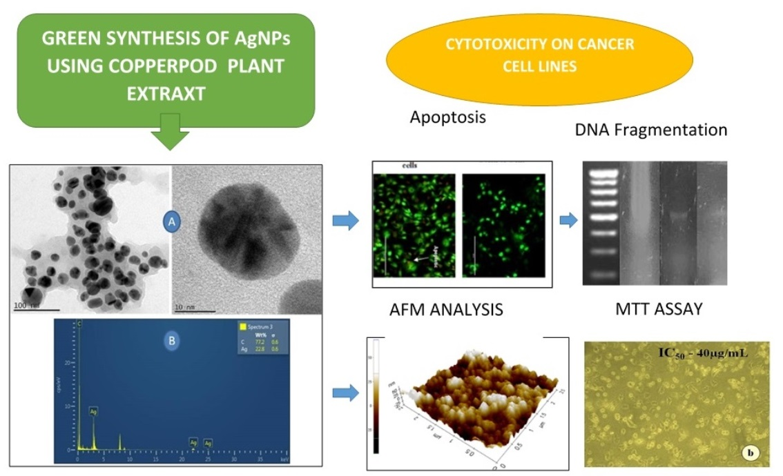
Copperpod Plant Synthesized AgNPs Enhance Cytotoxic and Apoptotic Effect in Cancer Cell Lines

by
Balashanmugam Pannerselvam
1,
Devasena Thiyagarajan
1,
Annamalai Pazhani
2,
Kalaichelvan Pudupalayam Thangavelu
2,
Hyung Joo Kim
3 and
Senthil Kumaran Rangarajulu
3,*
1
Centre for Nanoscience and Technology, Anna University, Chennai 600025, India
2
Centre for Advanced Studies in Botany, University of Madras, Chennai 600025, India
3
Department of Biological Engineering, Konkuk University, Seoul 05029, Korea
*
Author to whom correspondence should be addressed.
Submission received: 10 April 2021 / Revised: 15 May 2021 / Accepted: 17 May 2021 / Published: 18 May 2021
(This article belongs to the Special Issue Green Synthesis of Metallic Nanomaterials and Their Applications)

Abstract

:
The utilization of biological resources on the manufacture of nano silver has attracted the interest of researchers to develop an eco-friendly, cost-effective technology in nanomaterials production. In the present study, plant-mediated silver nanoparticles (AgNPs) were synthesized using aqueous leaf extracts of the Copperpod plant, which was well characterized. The ultraviolet-visible spectrophotometric study showed a maximum absorbance peak at 425 nm, and the observation of transmission electron microscopic features revealed that the nanoparticles size ranged between 20 and 70 nm. The synthesized AgNPs were tested for in vitro cytotoxic effects against cancerous cells, such as HepG2, A549 and MCF-7 cells. The findings showed that the IC50 values of AgNPs against cancerous cells viz., HepG2, MCF-7 and A549 cells, were observed to be 69 µg/mL, 62 µg/mL and 53 µg/mL, respectively. In addition, the apoptosis property was analysed using propidium iodide and acridine orange-ethidium bromide via the DNA fragmentation technique. Thus, the outcomes of the current analysis presume that the plant mediated AgNPs obtained from a synthesized Copperpod plant possess significant anti-cancer properties against various cancerous cells.

Graphical Abstract

1. Introduction

Nanoscience is a division in science that deals with the report on the manipulation of materials at the atomic and molecular level. In general, nanostructures are materials considered to have a diameter size in the range of 1 to 100 nm. Recently, several forms of nanomaterials have been investigated as it is considered to be an emerging area with promising prospective applications in diverse fields [1,2]. The major reason behind the vast applications of nanostructures is their reduced size and high-level surface to volume proportion [2,3,4]. Several nanoparticles have been found to have different applications in various fields. Nano-based structures provide excellent benefits to the environment as well as in the field of biomedical applications, such as drug delivery, diagnostic tools, treatment of cancer, biosensors, photothermal therapy, antibacterial and as antifungal agents, etc. [5,6]. Among numerous nanostructures, the application of metal-based nanoparticles, especially silver and silver-based nanomaterials, have provided a breakthrough in the field of biomedicine [7,8]. Among the various forms of nanostructures that are established and characterized, AgNPs have been found to have significant applications due to their inherent properties in the solid states that show exceptional physicochemical properties and good stability [9,10,11,12]. Although the significance of silver and silver-based compounds was recognized much earlier, these materials were not well exploited before, and they were only applied for antimicrobial activity, as antioxidants, for wound healing, for anticancer and anti-inflammatory properties, etc. This suggests that AgNPs are a promising candidate in the field of medical science, due to their vast applications [13,14,15] in the preparation of biopharmaceutical and biocosmetic products [7,12,16,17].
Recently, there has been a steady increase in scientific reports on illness and death due to cancer, which requires effective treatments for its inhibition and control. The traditional approaches employed for the diagnosis and treatment of cancer cells have failed due to their inability to target the root cause of its lethal effects on normal cells and curb the severe side effects [18,19]. To overcome this, several researchers and scientists are working to exploit various forms of nanostructured novel drug materials for targeting a wide range of cancer cells [20,21]. These nanomaterials are the active candidate that can directly deliver medications to a precise site of the targeted cancer cells with reduced side effects [22,23]. Among them, silver nanoparticles have been in the spotlight of various researchers due to their versatile applications in various fields; hence, there is a huge interest in developing a new protocol by exploring different biological resources for the synthesis of nanoparticles of different sizes and shapes [24,25,26,27]. Most of the processes involved in nanoparticle synthesis employ physical and chemical methods, which utilize various chemicals that are considered hazardous to both biological and natural environments. Therefore, there is a dire need for an eco-friendly synthesis process that does not affect the biological systems and the natural environment. Recent reports suggest that the use of biological methods of nanoparticles synthesis is less toxic, safe and environmentally friendly [28,29].
The application of nanoparticles as a prospective factor for preventing cancer has been explored broadly in the last decade. Several investigators have been analysing various anti-cancer mediators of different metallic nanoparticles synthesized using several methods [22,30]. However, due to the consumption of toxic chemicals and the production of hazardous by-products in the process of nanoparticles synthesis, the nanoparticles synthesized through biological sources are most preferred. Plant-mediated nanoparticle synthesis was the preferred method over various other biologically mediated processes due to their active plant components that are exploited as key sources of green synthesis [31,32]. This approach also has an advantage over other methods since it does not require complex activities of intracellular production and upkeep of microorganisms, which are found to be tedious [16,33].
Previously, plant extracts were utilized as potential capping (stabilizing) agents in the production of AgNPs [8,34,35]. In addition, many plant extracts possess a broad-range of bioactive compounds, namely alkaloids, flavonoids, phenols, carbohydrates, amino acids, terpenes, etc., which act as bio-reductants in the production of nanomaterials [36,37]. The chemical profile of such a kind of bioactive compounds has been extracted from the Copperpod plant and evaluated for their bioactivity [1,38,39]. However, in our earlier preliminary studies, the AgNPs synthesized using Peltophorum pterocarpum (Copperpod) leaf extracts was evaluated for their in vitro wound recovery and antimicrobic properties for cotton fabrics coated with selective pathogens [40,41]. However, the extracts obtained from the leaves of the Copperpod plant are yet to be extensively studied for their potential in the green synthesis of nano silver and its application in different fields. In addition, the effects of biogenic nanoparticles on cancer cells are also essential to understand the cytotoxic potentials of the synthesized AgNPs. Hence, in the present study, the in vitro cytotoxic property of AgNPs synthesized using Copperpod plant was investigated on animal cell lines to study their biocompatibility and anti-cancer properties.

2. Materials and Methods

2.1. Collection and Extraction of Plant Materials

For the present study, the leaves of the Copperpod plant (Peltophorum pterocarpum (DC.) K. Heyne), were collected at the campus of the Centre for Advanced Studies in Botany, University of Madras, Chennai (latitude 13.010° and longitude 80.239°), India. Based on the standard manuals, identification of the collected plant was performed at the school’s laboratory and later recorded. The voucher specimens were serially numbered and recorded as KA7. The aqueous extract from the leaves of Copperpod was prepared, and the filtrate was screened for further processing of AgNPs [40,42]. The collected mature leaves were washed in distilled water and air-dried under shade. Then, the leaves were pulverized into a powder using a blender and stored in the sealed bottle. Five grams of dried leaf power was combined with 100 mL of glass-distilled water. It was kept at 55 °C for 15 min in a water bath and then cool down to room temperature. The extract was then filtered using Whatman’s No.1 filter paper, and the filtrate was stored at 4 °C for further study.

2.2. Synthesis and Characterization of AgNPs

Then, 1 mM of silver nitrate was added separately to 1 mL of leaf extract in test tubes, which were incubated at room temperature in dark conditions for 24 h. During preparation, each extract with silver nitrate was continuously monitored for colour change, and the observations were recorded. The aqueous leaf extracts of the respective plants were kept as a control. The test tube that showed a change in the colour of the reaction mixture was recorded and further subjected to a UV-visible spectrophotometer, and the maximum absorption spectra was determined [42].

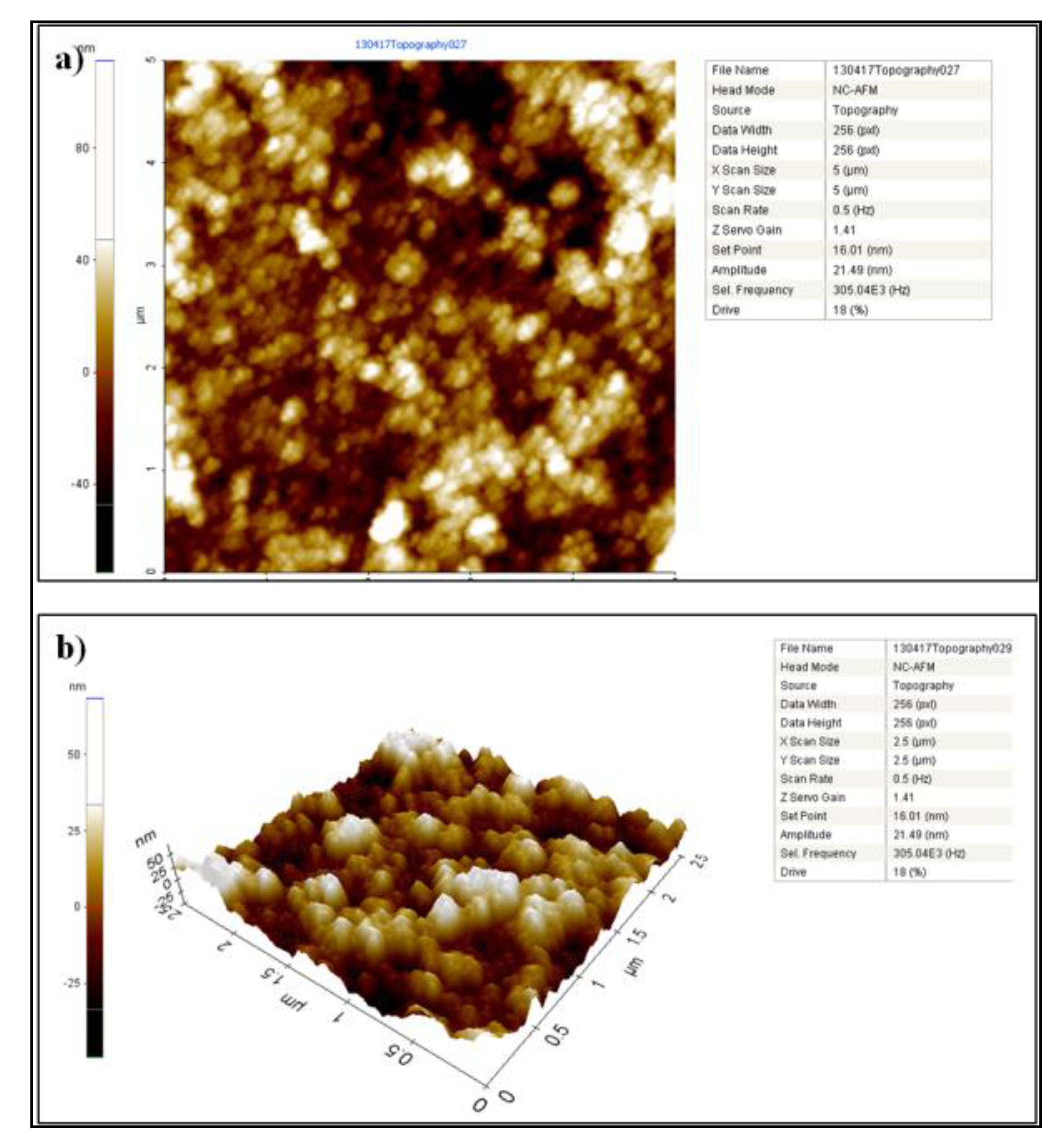
2.3. Characterization of Synthesized AgNPs

The plant biogenic AgNPs were characterized by various techniques, such as UV-visible spectroscopy (U2900, Hitachi, Tokyo, Japan), X-ray diffraction (XRD) (Philips X-ray diffractometer, London, UK), and transmission electron microscopy (TEM) (FEI Tecnai, Oregon, USA) equipped with an energy-dispersive X-ray (EDX). The XRD study of AgNPs produced using the Copperpod leaf extract was performed to investigate the crystalline nature of the nanoparticle formed. For this study, the AgNPs formed using Copperpod leaf extract were made into dried powder and then coated onto the XRD grid. The grid was then analysed using an XRD spectrum at a high voltage of 40 kV with Cu-Ka radiations by a Philips X-ray diffractometer [43]. In addition, the concentration of AgNPs synthesized from the aqueous leaf extract of Copperpod was determined using Inductively Coupled Plasma–Atomic Emission Spectrometer (ICP–AES, Elementar, Frankfurt, Germany). The surface properties of Biogenic AgNPs formed using leaf extracts were investigated using an atomic force microscope (XE-70 Park system, Mannheim, Germany) [44]. The quantitative analyses of Biosynthesized AgNPs were performed using Inductively Coupled Plasma Optical Emission Spectrometry (ICP-OES, Elementar, Frankfurt, Germany). The elemental concentration of the silver was obtained from the synthesized AgNPs stabilized at room temperature for two months. The two-month-old AgNPs samples were evaluated by ICP-OES Perkin Elmer optima 5300 DV.

2.4. In Vitro Cytotoxicity of Biogenic AgNPs

2.4.1. Cell Culture and Maintenance

The following cell lines, that is, Vero (African monkey kidney), HepG2 (hepatocellular carcinoma), A549 (lung cancer) and MCF-7 (breast cancer) cells, were procured from the National Centre for Cell Science (NCCS), in Pune, India, and cultured in Dulbecco’s modified Eagle’s medium (DMEM) accompanied with necessary growth factors. The subculture was performed after reaching 95% confluence by the trypsinization process, and the cells were replenished with fresh growth medium once every 3 days.

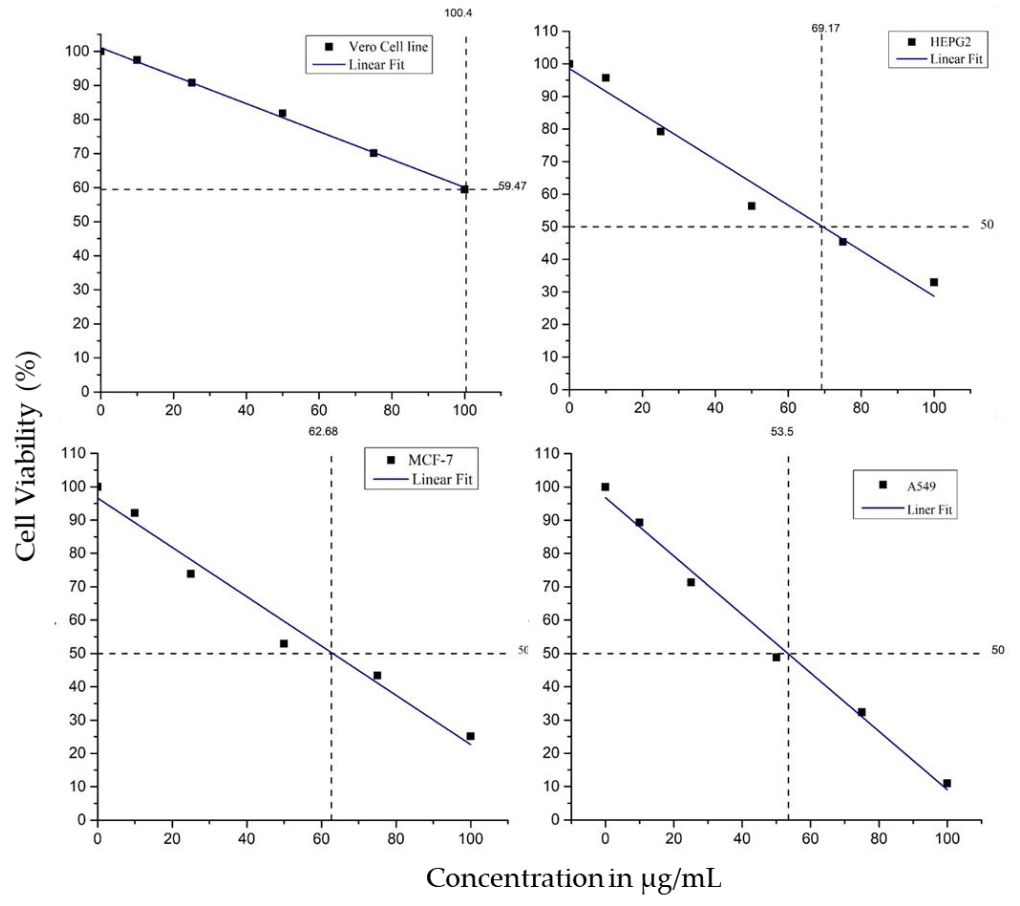
2.4.2. Cell Viability Assay

The comparative study on the assessment of nanotoxicity was evaluated between the normal-control cells (Vero cells) and cancer cells with an MTT (3-(4,5-dimethylthiazol-2-yl)-2,5-diphenyltetrazolium bromide) assay as described by Mosmann [45], albeit with some slight modifications. The cell lines of Vero cells (control-normal, non-cancerous cells) and 3 different cancer cells (HepG2, A549 and MCF-7) were separately seeded in a sterile 96 well flat bottom plates. They were then developed at 37 °C in a moistened incubator with 5% CO2 supply and allowed to reach 95% confluence. After achieving 95% confluence, the cells were replenished with a fresh medium and treated with different concentrations of biogenic AgNPs (10 to 100 µg/mL), separately. The treated cells were then incubated in a humidified incubator with 5% CO2 at 37 °C for 24 h. After incubation, the treated and untreated cells were washed in phosphate-buffered saline (pH 7.4). Each well of the 96 plates was loaded with 20 µL of MTT reagent (5 mg/mL in PBS) and allowed to incubate for 4 h. The wells were then loaded with 100 µL of dimethyl sulfoxide (DMSO), and observations were made for the formation of the purple-coloured formazan crystals [13,46]. The plates were then subjected to absorbance reading at 570 nm using a microplate ELISA reader. The IC50 values were plotted by using a software program (Origin pro-9.0).

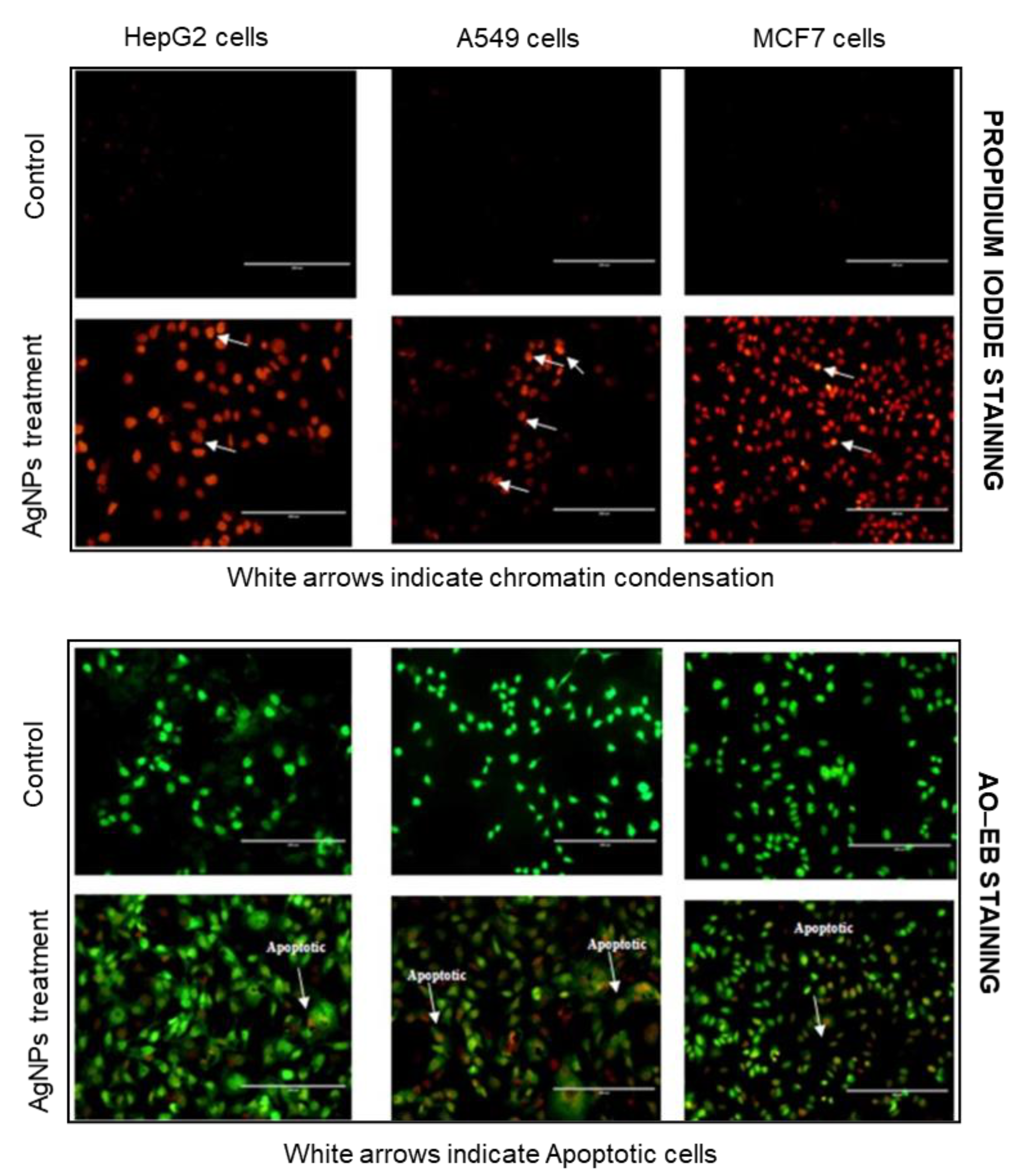
2.4.3. Propidium Iodide (PI) Staining

For PI staining, the tumour cells, HepG2, A549 and MCF-7, were separately grown in 24 well plates (1 × 10⁵ cells per well) as described above, and the cells were treated using IC50 concentration of biogenic AgNPs. The treated cells were then washed in phosphate-buffered saline and fixed in methanol:acetic acid (3:1, v/v) for 10 min. The fixed cells were then stained with propidium iodide (50 μg/mL in PBS) and incubated for another 20 min [47]. The cells were then observed for morphology of apoptotic cells under a FLoid® cell imaging station (Life Technologies)

2.4.4. Acridine Orange–Ethidium Bromide Dual Staining

The extent of apoptosis in the cells treated with biogenic AgNPs was analysed microscopically using an acridine orange/ethidium bromide (AO/EB) dual staining method. Dual staining was performed to detect the chromatin condensation of dead apoptotic cells by staining them with a fluorescence stain. The tumour cells, including HepG2, A549 and MCF-7, were separately grown in 24 well plates (1 × 10⁵ cells per well) as described above, and the cells were treated with an IC50 concentration of biogenic AgNPs. The treated cells were washed with phosphate-buffered saline, and the dual stain (AO/EB) (1 mg/mL AO and 1 mg/mL EB in PBS) was then added, and the mixture incubated for 5 min. The cells were again washed with PBS buffer, and the excess stain was removed and then visualized under a FLoid® cell imaging station (Life Technologies) with an excitation filter of 482 nm [47].

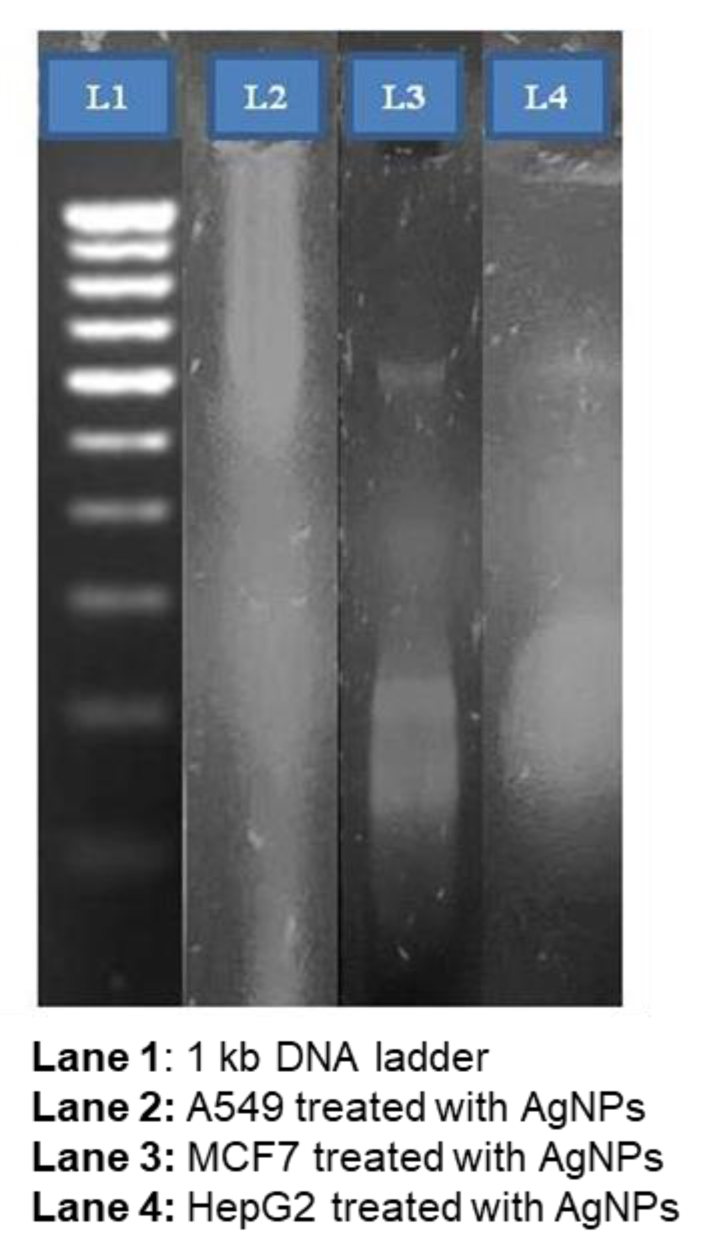
2.4.5. DNA Fragmentation Analysis

The DNA fragmentation analysis is considered as an important feature of cell apoptosis. The effect of apoptosis in the cells treated with biogenic AgNPs was analyzed using a DNA fragmentation technique, where the characteristic ladder of fragmented genomic DNA was visualized using agarose gel electrophoresis [48]. For this analysis, the IC50 concentration of biogenic AgNPs was used for HepG2, A549 and MCF-7 cells. After incubation, the cells treated with lysis buffer and extracted with phenol/chloroform/isoamyl alcohol mixture were further precipitated with ethanol and re-suspended using TE buffer containing RNase A. The extracted DNA was electrophoresed with 1% Agarose electrophoresis, and the untreated cells were used as controls for the experiments. The electrogram was stained with ethidium bromide and visualized under UV light. The gel was then photographed with gel documentation equipment. The separation of the DNA fragments was described using a standard molecular weight marker ladder.

3. Results and Discussions

3.1. Synthesis and Characterization of AgNPs

The plant leaf extracts obtained from Copperpod were employed for AgNPs synthesis. The optimal situation for producing AgNPs was observed to be 1 mg/mL plant concentration, 7 pH, 1 mM silver nitrate concatenation and an incubation temperature of 37 °C. The stability of produced AgNPs found to be even up to 3 months was also proved [40]. The formation of AgNPs was visually confirmed by recording the colour change from the initial one to the brown colour. The colour change was because of the addition of plant extracts to the aqueous mixture of AgNO3. The increase in the colour intensity with time showed the enhanced synthesis of AgNPs. In addition, the development of AgNPs was con-firmed by UV-visible spectrophotometric analysis showing a maximum absorption spectrum of 425 nm, while AgNO3 showed no peaks between 300 nm and 700 nm (Figure 1). The characteristic peak in the absorption range between 400 nm and 450 nm is a validation of the presence of surface plasma resonance in AgNPs. The present study demonstrates a single plasma resonance peak at 425 nm, which presented the evidence of biosynthesis in AgNPs [35].
The crystalline nature of biogenic AgNPs was analysed through XRD analysis. The results of the XRD spectrum revealed the presence of three distinct peaks representing Bragg reflections of 27.9°, 32.3°, 47.5°, 55.0°, 57.8°, 67.6°, 74.6°, 76.9° and 85.7°, relating to (1 1 1), (2 0 0), (2 2 0), (3 1 1), (2 2 2), (4 0 0), (3 3 1), (4 2 0) and (4 2 2) planes, respectively. These distinct peaks corresponding to the face-centered cubic (fcc) shape of the tested samples confirm the crystalline nature of the biogenic AgNPs (Figure 2), which was consistent with the database of the Joint Committee on Powder Diffraction Standards (JCPDS) (file No. 04-0783). The results of this study were supported by an earlier report [49] highlighting that the XRD spectrum of silver chloride nanoparticles displays solid peaks at 2θ values of 27.9°, 32.3°, 46.3°, 55.0°, 57.6°, 67.6°, 74.6°, 76.9° and 85.7°, relating to (1 1 1), (2 0 0), (2 2 0), (3 1 1), (2 2 2), (4 0 0), (3 3 1), (4 2 0) and (4 2 2) planes, respectively. The size and shape of the biogenic AgNPs were determined using TEM analysis. The TEM analysis revealed the presence of AgNPs, which were globular in structure with an median size varying from 20 to 60 nm; in addition, the presence of silver peaks in the EDX spectrum confirms the formation of AgNPs (Figure 3a,b). The surface properties of the synthesized biogenic AgNPs were analysed using an atomic force microscope. The results showed a three-dimensional morphology of the synthesized AgNPs having a particle size of approximately 40 nm, with a spherical shape (Figure 4).
In the present report, Copperpod was investigated for the synthesis of AgNPs and was found to be stable for up to 3 months. The utilization of plant and plant-based elements for the manufacture of AgNPs has been exploited by several researchers. However, the mechanism of AgNPs synthesis is not clear; however, most researchers have suggested that plant metabolites, such as alkaloids, phenols, tannins, glycosides, enzymes, polysaccharides, proteins and vitamins, may play a major role in the reduction of silver nitrate to AgNPs with good stability [50,51]. Such kinds of phytochemical compounds, including phenolic compounds, flavonoids, xanthoproteins, tannins, saponins, carboxylic acids, coumarins and carbohydrates, which are obtained from the Copperpod plant, are reported to have a high potential in regard to antimicrobial activity [39]. The biogenic synthesis of AgNPs was performed using an aqueous leaf extract of Copperpod, which was then treated with a 1mM solution of silver nitrate to induce the reduction process. The visible colour change to a brown colour is attributed to the excitation phenomenon of SPR bands, which is a characteristic feature of synthesized AgNPs. Carmona et al. [52] and Banerjee et al. [53] observed similar colour changes during the particle-formation process when the leaf extract was treated with silver nitrate. Thus, the reduction process from silver nitrate to AgNPs is coupled with the shift in the pigment of the liquid, especially due to the phenomenon of surface plasmon absorption of AgNPs. Moteriya and Chanda also suggested that the colour change from a colourless solution to a brown colour during the formation of AgNPs using the extract of C. pulcherrima was mainly due to a surface plasmon resonance phenomenon [54].
The inductively coupled plasma optical emission spectrometry (ICP-OES) was utilized to establish the concentration of the synthesized AgNPs [55]. The results showed that the concentration of AgNPs was 280.5 mg/L. The elemental wavelength of AgNPs was 328.068. Mukherjee et al. [56] reported that the concentrations of a silver solution of bio-synthesized (b-AgNPs) and chemically produced (c-AgNPs) nanoparticles can be quantitatively verified by ICP-OES analysis [57,58]. They showed that the concentration of b-AgNPs and c-AgNPs was 332.8 mg/mL and 383.7 mg/mL, respectively. However, in the present study, the concentration of AgNPs was found to be 280.5 mg/L. Similar findings were reported by other researchers who determined the concentration of AgNPs synthesized using an aqueous extract via the ICP-OES technique [59,60]. The crystalline nature of biogenic AgNPs synthesized from the aqueous leaf extract of Copperpod was analysed by XRD studies, revealing three distinct peaks of 1 1 1, 2 0 0 and 2 2 0, representing Bragg reflections that are reliable with the JCPDS database file No. 04-0783. In consistence with our results, Mishra et al. [61], Khatami et al. [62] and Nasar et al. [63] also recorded similar results of an XRD pattern showing a face-centred cubic (fcc) shape of silver ions.
The biogenic AgNPs synthesized from the aqueous leaf extracts of Copperpod were further characterized by TEM analysis. In the present study, HRTEM analysis confirmed that the shape of the synthesized AgNPs was spherical by an median size varying between 20 nm and 60 nm. The energy dispersive X-ray analysis confirmed the presence of elemental silver ions in the AgNPs synthesized using the aqueous leaf extracts of Copperpod. These results are consistent with the earlier studies performed by Amin et al. [64] and Khalil et al. [65] who also noticed the main peaks representing silver ions via the EDX analysis, thus confirming the synthesis of AgNPs for different plant extracts.
The surface properties of biogenic AgNPs were analysed using an atomic force microscope. The results revealed the presence of spherical nanoparticles with an approximate size of 40 nm. Daphedar and Taranath [66] studied the particle structure and dimension of the AgNPs synthesized using an aqueous leaf extract of Getonia floribunda by AFM. They reported that the synthesized AgNPs were found to be monodispersed spherical-shaped particles with an average size ranging between 10 and 25 nm. Similar findings were similarly described by several scholars who have previously explored the morphology and size of the AgNPs using AFM analysis [67,68].

3.2. In Vitro Cytotoxicity Analysis of Biogenic AgNPs by MTT Assay

The in vitro cytotoxic impact of AgNPs on normal Vero cells and cancerous cells, such as HepG2, A549 and MCF-7 cells, was performed with an MTT assay. The IC50 value of AgNPs on Vero cells was found to be 90 µg/mL (Figure 5). The morphological changes in the cells treated with AgNPs are indicated in Figure 4, where the cells are observed to be shrunken and rounded compared to normal untreated cells. Similarly, the cancerous cells treated with AgNPs showed significant morphological modifications via a phase contrast microscope, as in Figure 6. The IC50 values of AgNPs against cancerous cells, viz., HepG2, A549 and MCF-7 cells, were observed to be 47 µg/mL, 40 µg/mL and 44 µg/mL, respectively.
Recently, cancer-based research associated with nanoparticles has emerged as one of the potential areas of research among the scientific community. Several researchers have focused their studies towards validating the impacts of the various forms of nanoparticles on various cancerous cells [69,70].
Moldovan et al. [71] studied the in vitro cytotoxic properties of the biologically synthesized AgNPs from a Ligustrum ovalifolium extract against ovarian carcinoma cells, i.e., A2780 and A2780-Cis, in a dose-dependent manner. They found that when the concentration of AgNPs is increased from 0.01 to 180 μg/mL, the cells’ viability gradually decreases. The IC50 value of biologically synthesized AgNPs against A2780 and A2780-Cis was determined as 7 µg/mL and 14.04 µg/mL, respectively. In the present study, the in vitro cytotoxic effect of AgNPs synthesized from an aqueous leaf extract showing a monodisperse property, which was tested against normal Vero cells and cancerous cells, such as HepG2, A549 and MCF-7 cells, via an MTT assay. The results showed that the biogenic AgNPs have a potential cytotoxic property against all the tested cells. The treated cells showed apparent morphological changes, such as rounded cells, plasma membrane, and chromatin condensation, as compared to normal cells. The IC50 values of AgNPs against cancerous cells were recorded and found to be 69 µg/mL, 62 µg/mL and 53 µg/mL against HepG2, MCF-7 and A549 cells, respectively. The MTT viability assay can enhance knowledge on cell metabolic actions, survival and death. In addition, it is an essential technique used to analyse cytotoxicity, which clarifies the cellular reactions to toxic substances [72]. The AgNPs toxicity results clearly showed a dose-dependent response with exposure time in all the tested cancer cells. This demonstrates a significant increase in cell death with increased dosage of the nanoparticle treatment. The results reveal that the phytosynthesized AgNPs had a higher cytotoxicity on cancer cells as compared to the control.
Similar findings were observed by Venugopal et al. [73] who synthesized AgNPs using Piper nigrum extract and tested the cytotoxic effectiveness against MCF-7 and HEP-2 cancer cells. They evaluated the cytotoxic activity of AgNPs in a dose-dependent approach using concentrations ranging from 10 to 100 μg and found significant activities against both cancerous cells, i.e., MCF-7 and Hep-2 cells, when related to Piper nigrum extract. Satpathy et al. [74] studied the in vitro cytotoxic effect of AgNPs synthesized using Pueraria tuberosa aqueous extract. A few other reports also suggested that AgNPs had the tendency to reduce the cell’s sustainability in a dose-dependent approach, whereby it suppresses the cell proliferation of selective cells via arrest of the G2/M phase [47,75].

3.3. Propidium Iodide and Acridine Orange-Ethidium Bromide (AO-EB) Fluorescent Staining

The changes in the morphologic characteristics of cancerous cells, i.e., HepG2, A549 and MCF-7 cells, that were treated with AgNPs were observed under a fluorescent microscope. The AgNPs-treated cells stained with PI revealed the presence of condensation and fragmentation of dead cells (Figure 7). The extent of the apoptotic properties of AgNPs against cancerous cells, such as HepG2, A549 and MCF-7 cells, was analysed with AO–EB dual staining. The fluorescent images of AgNPs-treated cancerous cells, i.e., HepG2, A549, and MCF-7 cells, with corresponding IC50 concentration was analysed using dual staining (AO-EB), which clearly show early apoptotic cells and shrinkage of cells. The presence of orange-coloured cells indicates late apoptotic cells due to the condensation of nuclei, thus confirming the cell’s apoptotic property. The untreated (control) cells showed a characteristic bright green colour when stained with AO–EB, confirming the viability of the cells (Figure 7).
Mittal et al. [13] reviewed the antiproliferative impact of AgNPs made from a medicinal plant (Potentilla fulgens leaf extract) against MCF-7 and U-87 cell lines. The synthesized AgNPs were found to have good activity against both cells, i.e., MCF-7 and U-87, showing an IC50 value of 4.91 and 8.23 μg/mL, respectively. Recently, Chokkalingam et al. [76] synthesized AgNPs from Lycium chinense extract and tested for its cytotoxicity property against the MCF7 cell line. The apoptotic property of biogenic AgNPs synthesized from aqueous leaf extract of Copperpod against normal HepG2, A549 and MCF-7 cells was assessed by propidium iodide and AO–EB dual staining. The AgNPs-treated cancerous cells stained with PI showed the characteristic presence of condensation and fragmentations of dead cells. Similarly, the AgNPs-treated cells stained with an AO–EB dual stain revealed the characteristic appearance of chromatin condensation, a bright green nuclei dot. The presence of late apoptotic cells was observed in the cells treated with AgNPs, showing a characteristic observation of cell shrinkage with membrane blabbing when stained with acridine orange and ethidium bromide; thus, confirming the apoptotic activity of synthesized AgNPs. These observations are consistent with the earlier findings [77,78,79].

3.4. DNA Fragmentation Analysis

The apoptosis property of biogenic AgNPs against cancerous cells was analysed by the genomic DNA fragmentation technique, where the genomic DNA of treated cells was extracted and observed by agarose electrophoresis. The IC50 concentration of biogenic AgNPs was used to treat against cancerous cells, such as HepG2, A549 and MCF-7 cells, and the genomic DNA was extracted via electrophoresis using 2% agarose gel. The results showed that the biogenic AgNPs induced DNA fragmentation of the treated cells (Figure 8) compared to the DNA ladder control cells, which did not show any fragmentation, and this was well-evidenced in our earlier publication [80]. The ladder pattern of DNA damage in the treated cells showed the development of a long smear, whereas the control showed the least damage with no ladder pattern. The early study on the DNA fragmentation in control experiments on HepG2 also confirmed that DNA fragmentation is an important functional key which initiates the process of apoptosis [80,81]. The cells treated with AgNPs showed apoptosis due to the reduction in the percentage of cells in the G1 phase, followed by cell cycle arrest at G2 during mitosis. The occurrence of apoptosis in cancer cells generally occurs with the generation of ROS and mitochondrial disfunction which was induced by the cytotoxicity of AgNPs [82]. The DNA damage activity relates to ROS, which act as a signal molecule in the support of cell cycle development. In the progression of DNA damage, the cell is shrunken due to its irregular reduction in size, which finally results in apoptosis. The present results are consistent with studies conducted by Fard et al. [83] who studied the apoptotic property of biogenic AgNPs synthesized using Artemisia oliveriana extract via DNA fragmentation techniques. Similar results were likewise stated by many investigators who have investigated the apoptotic property of AgNPs using a DNA fragmentation assay [78,84].

4. Conclusions

The interest in AgNPs has significantly grown due to their tuneable properties and potential applications. Plant-based green synthesis approaches present a non-toxic, cost-effective and ecologically safe technology for the large-scale development of AgNPs. Thus, in the current report, the biogenic AgNPs were synthesized employing an aqueous extract of the Copperpod plant. The selected plant extract is known for its high productivity as a reducing and stabilizing agent for the biogenesis of highly stable AgNPs. The analytical method of characterization demonstrated that the synthesized biogenic AgNPs were evenly distributed with no accumulation. The unusual physio-chemical properties of the biogenic AgNPs occurred due to the presence of a broad range of bio-functional compounds in the plant extract which was utilized as a strong reductant during nanoparticle synthesis. In addition, phyto-extracts are the most valuable resource of bioactive elements for the evolution of novel chemotherapeutic agents. The biogenic particles also showed a promising cell cytotoxic activity against the tested cancerous cells, such as HepG2, A549, and MCF-7 cells. The present results indicate that the synthesized biogenic AgNPs are likely to have significant cytotoxic properties, which can be used in various bio-medical-related applications soon.

Author Contributions

Conceptualization, reviewing and editing, S.K.R.; investigation, writing—original draft, methodology, and supervision B.P. and D.T.; formal analysis, A.P.; resources, K.P.T.; supervision, H.J.K. All authors have read and agreed to the published version of the manuscript.

Funding

This research received no external funding.

Institutional Review Board Statement

Not applicable.

Informed Consent Statement

Not applicable.

Data Availability Statement

Not applicable.

Conflicts of Interest

The authors declare no conflict of interest.

Abbreviations

AgNPsSilver Nanoparticles
UV-visUltraviolet visible
TEMTransmission electron microscopy
HRTEMHigh-resolution transmission electron microscopy
AFMAtomic force microscopy
EDXEnergy disperse X-ray
XRDX-Ray Diffraction
HepG2Hepatocellular carcinoma cells
A549Lung cancer cells
MCF7Human breast cancer cells
ELISAEnzyme-linked immunosorbent assay
DMEMDulbecco‘s modified eagle media
CO2Carbon dioxide
NCCSNational centre for cell science
MTT3-(4,5-dimethylthiazol-2-yl)-2,5-diphenyltetrazolium bromide
ICP-AESInductively Coupled Plasma—Atomic Emission Spectrometer
ICP-OESInductively coupled plasma optical emission spectrometry
AO/EBAcridine orange and ethidium bromide
JCPDSJoint committee on powder diffraction standards
DMSODimethyl sulfoxide
PBSPhosphate-buffered saline
PIPropidium iodide
fccFace-centred cubic
kbKilo-base
µgMicrogram
nmNano-meter
mlMilligram
hHours
IC50The half maximal inhibitory concentration
G2/MGrowth 2/mitosis

References

  1. Ba, T.C.; Nguyen, T.H.L.; Nguyen, T.H.A.; Tran, D.C.; Nguyen, T.T.; Dao, D.T.; Le, T.H.N.; Tram, V.S.; Ninh, T.S.; Domenico, V.D.; et al. Chemical constituents of Peltophorum pterocarpum stems. Vietnam J. Chem. 2020, 58, 569–574. [Google Scholar]
  2. Salata, O.V. Applications of nanoparticles in biology and medicine. J. Nanobiotechnol. 2004, 2, 3. [Google Scholar] [CrossRef] [PubMed] [Green Version]
  3. Hajipour, M.J.; Fromm, K.M.; Ali, A.A.; Aberasturi, D.J.D.; Larramendi, I.R.D.; Rojo, T.; Serpooshan, V.; Parak, W.J.; Mahmoudi, M. Antibacterial properties of nanoparticles. Trends Biotechnol. 2021, 30, 499–511. [Google Scholar] [CrossRef] [PubMed] [Green Version]
  4. Ali, M.S.; Altaf, M.; Al-Lohedan, H.A. Green synthesis of biogenic silver nanoparticles using Solanum tuberosum extract and their interaction with human serum albumin: Evidence of “corona” formation through a multi-spectroscopic and molecular docking analysis. J. Photochem. Photobiol. B Biol. 2017, 173, 108–119. [Google Scholar] [CrossRef]
  5. Moritz, M.; Geszke-Moritz, M. The newest achievements in synthesis, immobilization and practical applications of antibacterial nanoparticles. Chem. Eng. J. 2013, 228, 596–613. [Google Scholar] [CrossRef]
  6. Ramos, A.P.; Cruz, M.A.E.; Tovani, C.B.; Ciancaglini, P. Biomedical applications of nanotechnology. Biophys. Rev. 2017, 9, 79–89. [Google Scholar] [CrossRef]
  7. Siva, S.S.; Huizhen, L.; Zhijun, Z.; Minaxi, S.; Zeba, U.; Tianyu, H.; Vasudeva, R.N.; Xin, W.; Vijai, K.G. Recent advances in essential oils-based metal nanoparticles: A review on recent developments and biopharmaceutical applications. J. Mol. Liquids 2021, 333, 115951. [Google Scholar]
  8. Paiva-Santos, A.C.; Herdade, A.M.; Guerra, C.; Peixoto, D.; Pereira-Silva, M.; Zeinali, M.; Mascarenhas-Melo, F.; Paranhos, A.; Veiga, F. Plant-mediated green synthesis of metal-based nanoparticles for dermopharmaceutical and cosmetic applications. Int. J. Pharm. 2021, 597, 120311. [Google Scholar] [CrossRef]
  9. Ueda, M.; Yokota, T.; Honda, M.; Lim, P.N.; Osaka, N.; Makita, M.; Nishikawa, Y.; Kasuga, T.; Aizawa, M. Regulating size of silver nanoparticles on calcium carbonate via ultrasonic spray for effective antibacterial efficacy and sustained release. Mater. Sci. Eng. C. 2021, 125, 112083. [Google Scholar] [CrossRef]
  10. Taiba, N.; Tayyiba, D. The role of some important metal oxide nanoparticles for wastewater and antibacterial applications: A review. Environ. Chem. Ecotoxicol. 2021, 3, 59–75. [Google Scholar]
  11. Saravanan, A.; Senthil Kumar, B.; Karishma, S.; Dai-Viet, N.V.; Jeevanantham, S.; Yaashikaa, P.R.; George, C.S. A review on biosynthesis of metal nanoparticles and its environmental applications. Chemosphere 2021, 264, 128580. [Google Scholar] [CrossRef]
  12. Behzad, F.; Naghib, S.M.; Kouhbanani, M.A.J.; Tabatabaei, S.N.; Zare, Y.; Rhee, K.Y. An overview of the plant-mediated green synthesis of noble metal nanoparticles for antibacterial applications. J. Ind. Eng. Chem. 2021, 94, 92–104. [Google Scholar] [CrossRef]
  13. Mittal, A.K.; Tripathy, D.; Choudhary, A.; Ali, P.K.; Chatterjee, A.; Singh, I.P.; Banerjee, U.C. Bio-synthesis of silver nanoparticles using Potentilla fulgens Wall. ex Hook. and its therapeutic evaluation as anticancer and antimicrobial agent. Mater. Sci. Eng. C Mater. Biol. Appl. 2015, 53, 120–127. [Google Scholar] [CrossRef]
  14. Kuppusamy, P.; Yusoff, M.M.; Maniam, G.P.; Govindan, N. Biosynthesis of metallic nanoparticles using plant derivatives and their new avenues in pharmacological applications—An updated report. Saudi. Pharm. J. 2016, 24, 473–484. [Google Scholar] [CrossRef]
  15. Nakkala, J.R.; Mata, R.; Gupta, A.K.; Sadras, S.R. Biological activities of green silver nanoparticles synthesized with Acorous calamus rhizome extract. Eur. J. Med. Chem. 2014, 85, 784–794. [Google Scholar] [CrossRef]
  16. Ahamed, M.; AlSalhi, M.S.; Siddiqui, M.K.J. Silver nanoparticle applications and human health. Clin. Chim. Acta 2010, 411, 1841–1848. [Google Scholar] [CrossRef]
  17. Chen, X.; Schluesener, H.J. Nanosilver: A nanoproduct in medical application. Toxicol. Lett. 2008, 176. [Google Scholar] [CrossRef]
  18. Milad, A.; Ali, Z.; Farid, H.; Ebrahim, R.M.; Fardin, H.; Maliheh, E.; Kiavash, H.; Reza, M.; Masoud, N. Curcumin in cancer therapy: A novel adjunct for combination chemotherapy with paclitaxel and alleviation of its adverse effects. Life Sci. 2020, 256, 117984. [Google Scholar]
  19. Jyoti, S.; Suaib, L.; Abha, M. Emerging role of phytochemicals in targeting predictive, prognostic, and diagnostic biomarkers of lung cancer. Food Chem. Toxicol. 2020, 144, 111592. [Google Scholar]
  20. Yonglu, L.; Xiaodong, Z.; Qiang, C. Bio-based nanomaterials for cancer therapy. Nano Today 2021, 38, 101134. [Google Scholar]
  21. Mehran, A.; Rajender, S.V. Phytosynthesis and modification of metal and metal oxide nanoparticles/nanocomposites for antibacterial and anticancer activities: Recent advances. Sust. Chem. Pharm. 2021, 21, 100412. [Google Scholar]
  22. Quan, L.; Yanhong, D.; Jianye, F.; Meng, Q.; Zhe, S.; Dickson, A.; Jianlong, K.; Zhongjian, X.; Taojian, F.; Shiyun, B.; et al. Nano-immunotherapy: Unique mechanisms of nanomaterials in synergizing cancer immunotherapy. Nano Today 2021, 36, 101023. [Google Scholar]
  23. Mohammad, R.S.; Sheyda, R.; Ivan, M.K.; Mostafa, A.; Willis, C.A.M.; Mareike, M.; Mohammad, H.G.; Mohammad, R. Targeting non-apoptotic cell death in cancer treatment by nanomaterials: Recent advances and future outlook. Nanomed. Nanotechnol. Biol. Med. 2020, 29, 102243. [Google Scholar]
  24. Dos Santos, C.A.; Seckler, M.M.; Ingle, A.P.; Gupta, I.; Gladiero, S.; Gladiero, M.; Gade, A.; Rai, M. Silver nanoparticles: Therapeutical uses, toxicity, and safety issues. J. Pharm. Sci. 2014, 103, 1931–1944. [Google Scholar] [CrossRef]
  25. Rizzello, L.; Pompa, P.P. Nanosilver-based antibacterial drugs and devices: Mechanisms, methodological drawbacks, and guidelines. Chem. Soc. Rev. 2009, 43, 1501–1518. [Google Scholar] [CrossRef]
  26. Sharma, V.K.; Yngard, R.A.; Lin, Y. Silver nanoparticles: Green synthesis and their antimicrobial activities. Adv. Colloid. Interface Sci. 2009, 145, 83–96. [Google Scholar] [CrossRef]
  27. Mostafa, F.A.H.; Gamal, A.G.; Sedky, H.A.H. A review of green methods for phyto-fabrication of hematite (α-Fe2O3) nanoparticles and their characterization, properties, and applications. Heliyon 2021, 7, e05806. [Google Scholar]
  28. Xu, Z.P.; Zeng, Q.H.; Lu, G.Q.; Yu, A.B. Inorganic nanoparticles as carriers for efficient cellular delivery. Chem. Eng. Sci. 2006, 61, 1027–1040. [Google Scholar] [CrossRef]
  29. Jing, H.; Yunlei, X. When nano meets plants: A review on the interplay between nanoparticles and plants. Nano Today 2021, 38, 101143. [Google Scholar]
  30. Muhammad, S.R.R.; Riaz, R.; Hafiza, M.M.; Yongai, X.; Xun, S.; Nazim, H.; Qinchang, Z.; Zhendan, H. Gut microbiota targeted nanomedicine for cancer therapy: Challenges and future considerations. Trends Food Sci. Technol. 2021, 107, 240–251. [Google Scholar]
  31. Arul, K.M.; Balashanmugam, P.; Javee, A.; Rajenderan, M.; Devasena, T. Facile green synthesis and characterization of Gloriosa superba L. tuber extract-capped silver nanoparticles (GST-AgNPs) and its potential antibacterial and anticancer activities against A549 human cancer cells. Environ. Nanotechnol. Monitoring Manag. 2021, 15, 10046. [Google Scholar]
  32. Violeta, M.L.; Heriberto, E.G.; Lucía, Z.F.L.; Erika, L.S.B.; Alfredo, N.R.; Rubén, D.C.N.; Gabriel, A.N.; Ignacio, A.R. Study of the effect of the different parts of Morinda citrifolia L. (noni) on the green synthesis of silver nanoparticles and their antibacterial activity. Appl. Surf. Sci. 2021, 537, 147855. [Google Scholar]
  33. Mohanpuria, P.; Rana, N.K.; Yadav, S.K. Biosynthesis of nanoparticles: Technological concepts and future applications. J. Nanoparticle Res. 2008, 10, 507–517. [Google Scholar] [CrossRef]
  34. Shohreh, F.; Fatemeh, A.; Mansour, G. Synthesis and therapeutic potential of silver nanomaterials derived from plant extracts. Ecotoxicol. Environ. Safety 2019, 168, 260–278. [Google Scholar]
  35. Balashanmugam, P.; Prabhu, D.; Devasena, T.; Hak, J.S.; Kim, K.W.; Jung, Y.S.; Song, H.J.; Kim, H.J.; Kumaran, R.S. Facile synthesis of silver nanoparticles using Asian spider flower and its in vitro cytotoxic activity against human breast carcinoma cells. Processes 2020, 8, 430. [Google Scholar]
  36. Harish, C.; Pragati, K.; Elza, B.; Saurabh, Y. Medicinal plants: Treasure trove for green synthesis of metallic nanoparticles and their biomedical applications. Biocatal. Agricult. Biotechnol. 2020, 24, 101518. [Google Scholar]
  37. Kathiravan, G.; Yamini, K.R.; Rajagopal, K.; Anandan, S.; Kim, K.W.; Jung, Y.S.; Song, H.J.; Kim, H.J.; Kumaran, R.S. Phytogenic synthesis of nano silver from Madagascar Periwinkle extracts and their angiogenic activities in Zebrafish embryos (ZFE). Nanosci. Nanotechnol. Lett. 2020, 12, 79–87. [Google Scholar] [CrossRef]
  38. Li, Y.C.; Kuo, P.C.; Yang, M.L.; Chen, T.Y.; Hwang, T.L.; Chiang, C.C.; Thang, T.D.; Tuan, N.N.; Tzen, J.T.C. Chemical constituents of the leaves of Peltophorum pterocarpum and their bioactivity. Molecules 2019, 24, 240. [Google Scholar] [CrossRef] [Green Version]
  39. Sukumaran, S.; Kiruba, S.; Mahesh, M.; Nisha, S.R.; Miller, P.Z.; Ben, C.P.; Jeeva, S. Phytochemical constituents and antibacterial efficacy of the flowers of Peltophorum pterocarpum (DC.) Baker ex Heyne. Asian Pacific J. Trop. Med. 2011, 4, 735–738. [Google Scholar] [CrossRef] [Green Version]
  40. Annamalai, P.; Balashanmugam, P.; Kalaichelvan, P.T. In vitro wound healing and antimicrobial property of cotton fabrics coated optimized silver nanoparticles synthesized using Peltophorum pterocarpum leaf extracts. Asian J. Pharm. Clin. Res. 2019, 12, 216–222. [Google Scholar] [CrossRef]
  41. Annamalai, P.; Balashanmugam, P.; Kalaichelvan, P.T. Biogenic synthesis silver nanoparticles using Peltophorum pterocarpum leaf extracts and its antimicrobial efficacy against selective pathogens. Int. J. App. Pharm. 2018, 10, 112–118. [Google Scholar] [CrossRef] [Green Version]
  42. Balashanmugam, P.; Kalaichelvan, P.T. Biosynthesis characterization of silver nanoparticles using Cassia roxburghii DC. aqueous extract and coated on cotton cloth for effective antibacterial activity. Int. J. Nanomed. 2015, 10, 87–97. [Google Scholar] [CrossRef] [PubMed] [Green Version]
  43. Singh, K.; Panghal, M.; Kadyan, S.; Chaudhary, U.; Yadav, J.P. Green silver nanoparticles of Phyllanthus amarus: As an antibacterial agent against multi drug resistant clinical isolates of Pseudomonas aeruginosa. J. Nanobiotechnol. 2014, 12, 40. [Google Scholar] [CrossRef] [PubMed] [Green Version]
  44. Balashanmugam, P.; Balakumaran, M.D.; Murugan, R.; Dhanapal, K.; Kalaichelvan, P.T. Phytogenic synthesis of silver nanoparticles, optimization and evaluation of in vitro antifungal activity against human and plant pathogens. Mibiol. Res. 2016, 192, 52–64. [Google Scholar] [CrossRef] [PubMed]
  45. Mosmann, T. Rapid colorimetric assay for cellular growth and survival: Application to proliferation and cytotoxicity assays. J. Immunol. Methods 1983, 65, 55–63. [Google Scholar] [CrossRef]
  46. Almutary, A.; Sanderson, B.J.S. The MTT and crystal violet assays: Potential confounders in nanoparticle toxicity testing. Int. J. Toxicol. 2016, 35, 454–462. [Google Scholar] [CrossRef]
  47. Devanesan, S.; Alsalhi, M.S.; Vishnubalaji, R.; Alfuraydi, A.A.; Alajez, N.M.; Alfayez, M.; Murugan, K.; Sayed, S.R.M.; Nicoletti, M.; Benelli, G. Rapid biological synthesis of silver nanoparticles using plant seed extracts and their cytotoxicity on colorectal cancer cell lines. J. Clust. Sci. 2017, 28, 595–605. [Google Scholar] [CrossRef]
  48. Dey, A.; Mukhopadhyay, A.K.; Gangadharan, S.; Sinha, M.K.; Basu, D.; Bandyopadhyay, N.R. Nanoindentation study of microplasma sprayed hydroxyapatite coating. Ceram. Int. 2009, 35, 2295–2304. [Google Scholar] [CrossRef]
  49. Paulkumar, K.; Rajeshkumar, S.; Gnanajobitha, G.; Vanaja, M.; Malarkodi, C.; Annadurai, G. Biosynthesis of silver chloride nanoparticles using Bacillus subtilis MTCC 3053 and assessment of its antifungal activity. ISRN Nanomater. 2013, 317963. [Google Scholar] [CrossRef] [Green Version]
  50. Kulkarni, N.; Muddapur, U. Biosynthesis of metal nanoparticles: A review. J. Nanotechnol. 2014, 510246. [Google Scholar] [CrossRef] [Green Version]
  51. Mohamad, N.A.N.; Arham, N.A.; Jai, J.; Hadi, A. Plant extract as reducing agent in synthesis of metallic nanoparticles: A review. Nanosci. Nanotechnol. Nanoeng. 2014, 832, 350–355. [Google Scholar] [CrossRef]
  52. Carmona, E.R.; Benito, N.; Plaza, T.; Recio-Sánchez, G. Green synthesis of silver nanoparticles by using leaf extracts from the endemic Buddleja globosa Hope. Green Chem. Lett. Rev. 2017, 10, 250–256. [Google Scholar] [CrossRef] [Green Version]
  53. Banerjee, P.; Satapathy, M.; Mukhopahayay, A.; Das, P. Leaf extract mediated green synthesis of silver nanoparticles from widely available Indian plants: Synthesis, characterization, antimicrobial property and toxicity analysis. Bioresour. Bioprocess. 2014, 1, 3. [Google Scholar] [CrossRef] [Green Version]
  54. Moteriya, P.; Chanda, S. Biosynthesis of silver nanoparticles formation from Caesalpinia pulcherrima stem metabolites and their broad spectrum biological activities. J. Genet. Eng. Biotechnol. 2018, 16, 105–113. [Google Scholar] [CrossRef]
  55. Rónavári, A.; Kovács, D.; Igaz, N.; Vagvolgyi, C.; Boros, I.M.; Konya, Z.; Pfeiffer, I.; Kiricsi, M. Biological activity of green-synthesized silver nanoparticles depends on the applied natural extracts: A comprehensive study. Int. J. Nanomed. 2017, 12, 871–883. [Google Scholar] [CrossRef] [Green Version]
  56. Mukherjee, S.; Chowdhury, D.; Kotcherlakota, R.; Patra, S.; Vinothkumar, B.; Bhadra, M.P.; Sreedhar, B.; Patra, C.R. Potential theranostics application of bio-synthesized silver nanoparticles (4-in-1 system). Theranostics 2014, 4, 316–335. [Google Scholar] [CrossRef] [Green Version]
  57. Mukherjee, S.; Sushma, V.; Patra, S.; Barui, A.K.; Bhadra, M.P.; Sreedhar, B.; Patra, C.R. Green chemistry approach for the synthesis and stabilization of biocompatible gold nanoparticles and their potential applications in cancer therapy. Nanotechnology 2012, 23, 455103. [Google Scholar] [CrossRef]
  58. Mukherjee, S.; Vinothkumar, B.; Prashanthi, S.; Bangal, P.R.; Sreedhar, B.; Patra, C.R. Potential therapeutic and diagnostic applications of one-step in situ biosynthesized gold nanoconjugates (2-in-1 system) in cancer treatment. RSC Adv. 2013, 3, 2318–2329. [Google Scholar] [CrossRef]
  59. Jadhav, K.; Dhamecha, D.; Bhattacharya, D.; Patil, M. Green and ecofriendly synthesis of silver nanoparticles: Characterization, biocompatibility studies and gel formulation for treatment of infections in burns. J. Photochem. Photobiol. B. Biol. 2016, 155, 109–115. [Google Scholar] [CrossRef]
  60. Krishnaraj, C.; Ji, B.J.; Harper, S.L.; Yun, S.I. Plant extract-mediated biogenic synthesis of silver, manganese dioxide, silver-doped manganese dioxide nanoparticles and their antibacterial activity against food- and water-borne pathogens. Bioprocess. Biosyst. Eng. 2016, 39, 759–772. [Google Scholar] [CrossRef]
  61. Mishra, P.M.; Sahoo, S.K.; Naik, G.K.; Parida, K. Biomimetic synthesis, characterization and mechanism of formation of stable silver nanoparticles using Averrhoa carambola L. leaf extract. Mater. Lett. 2015, 160, 566–571. [Google Scholar] [CrossRef]
  62. Khatami, M.; Nejad, M.S.; Almani, P.G.N.; Salari, S. Plant-mediated green synthesis of silver nanoparticles using Trifolium resupinatum seed exudate and their antifungal efficacy on Neofusicoccum parvum and Rhizoctonia solani. IET Nanobiotechnol. 2016, 10, 237–243. [Google Scholar] [CrossRef]
  63. Nasar, M.Q.; Khalil, A.T.; Ali, M.; Shah, M.; Ayaz, M.; Shinwari, Z.K. Phytochemical analysis, Ephedra procera C. A. Mey. mediated green synthesis of silver nanoparticles, their cytotoxic and antimicrobial potentials. Medicina 2019, 55, 369. [Google Scholar] [CrossRef] [Green Version]
  64. Amin, M.; Anwar, F.; Janjua, M.R.S.A.; Iqbal, M.A.; Rashid, U. Green synthesis of silver nanoparticles through reduction with Solanum xanthocarpum L. berry extract: Characterization, antimicrobial and urease inhibitory activities against Helicobacter pylori. Int. J. Mol. Sci. 2012, 13, 9923–9941. [Google Scholar] [CrossRef]
  65. Khalil, M.M.H.; Ismail, E.H.; El-Baghdady, K.Z.; Mohamed, D. Green synthesis of silver nanoparticles using olive leaf extract and its antibacterial activity. Arab. J. Chem. 2014, 7, 1131–1139. [Google Scholar] [CrossRef] [Green Version]
  66. Daphedar, A.; Taranath, T.C. Characterization and cytotoxic effect of biogenic silver nanoparticles on mitotic chromosomes of Drimia polyantha (Blatt. & McCann) Stearn. Toxicol. Rep. 2018, 5, 910–918. [Google Scholar]
  67. Ajitha, B.; Reddy, Y.A.K.; Reddy, P.S. Green synthesis and characterization of silver nanoparticles using Lantana camara leaf extract. Mater. Sci. Eng. C. 2015, 49, 373–381. [Google Scholar] [CrossRef]
  68. Yasir, M.; Singh, J.; Tripathi, M.K.; Singh, P.; Shrivastava, R. Green synthesis of silver nanoparticles using leaf extract of common arrowhead houseplant and its anticandidal activity. Pharmacogn. Mag. 2017, 13, S840–S844. [Google Scholar]
  69. Abbasi, B.H.; Nazir, M.; Muhammad, W.; Hashmi, S.S.; Abbasi, R.; Rahman, L.; Hano, C. A comparative evaluation of the antiproliferative activity against HepG2 liver carcinoma cells of plant-derived silver nanoparticles from basil extracts with contrasting anthocyanin contents. Biomolecules 2019, 9, 320. [Google Scholar] [CrossRef] [Green Version]
  70. Singh, A.K.; Tiwari, R.; Singh, V.K.; Singh, P.; Khadim, S.R.; Singh, U.; Laxmi; Srivastava, V.; Hasan, S.H.; Asthana, R.K. Green synthesis of gold nanoparticles from Dunaliella salina, its characterization and in vitro anticancer activity on breast cancer cell line. J. Drug Deliv. Sci. Technol. 2019, 51, 164–176. [Google Scholar] [CrossRef]
  71. Moldovan, B.; Sincari, V.; Perde-Schrepler, M.; David, L. Biosynthesis of Silver Nanoparticles Using Ligustrum Ovalifolium Fruits and Their Cytotoxic Effects. Nanomaterials 2018, 8, 627. [Google Scholar] [CrossRef] [PubMed] [Green Version]
  72. Asha, R.P.V.; Kah, M.G.L.; Hande, M.P.; Valiyaveettil, S. Cytotoxicity and genotoxicity of silver nanoparticles in human cells. ACS Nano 2009, 3, 279–290. [Google Scholar]
  73. Venugopal, K.; Rather, H.A.; Rajagopal, K.; Shanthi, M.P.; Sheriff, K.; Illiyas, M.; Rather, R.A.; Manikandan, E.; Uvarajan, S.; Bhaskar, M.; et al. Synthesis of silver nanoparticles (Ag NPs) for anticancer activities (MCF 7 breast and A549 lung cell lines) of the crude extract of Syzygium aromaticum. J. Photochem. Photobiol. B Biol. 2017, 167, 282–289. [Google Scholar] [CrossRef] [PubMed]
  74. Satpathy, S.; Patra, A.; Ahirwar, B.; Hussain, M.D. Antioxidant and anticancer activities of green synthesized silver nanoparticles using aqueous extract of tubers of Pueraria tuberosa. Artif. Cells Nanomed. Biotechnol. 2018, 46, S71–S85. [Google Scholar] [CrossRef] [Green Version]
  75. Zhao, X.; Zhou, L.; Rajoka, M.S.R.; Yuan, L.; Jiang, C.; Shao, D.; Zhu, J.; Shi, J.; Huang, Q.; Yang, H.; et al. Fungal silver nanoparticles: Synthesis, application and challenges. Crit. Rev. Biotechnol. 2018, 38, 817–835. [Google Scholar] [CrossRef]
  76. Chokkalingam, M.; Singh, P.; Huo, Y.; Soshnikova, V.; Ahn, S.; Kang, J.; Mathiyalagan, R.; Kim, Y.J.; Yang, D.C. Facile synthesis of Au and Ag nanoparticles using fruit extract of Lycium chinense and their anticancer activity. J. Drug Deliv. Sci. Technol. 2019, 4, 308–315. [Google Scholar] [CrossRef]
  77. Alsalhi, M.S.; Devanesan, S.; Alfuraydi, A.A.; Vshnubalaji, R.; Munusamy, M.A.; Murugan, K.; Nivoletti, M.; Benelli, G. Green synthesis of silver nanoparticles using Pimpinella anisum seeds: Antimicrobial activity and cytotoxicity on human neonatal skin stromal cells and colon cancer cells. Int. J. Nanomed. 2016, 11, 4439–4449. [Google Scholar] [CrossRef] [Green Version]
  78. Mata, R.; Nakkala, J.R.; Sadras, S.R. Biogenic silver nanoparticles from Abutilon indicum: Their antioxidant, antibacterial and cytotoxic effects in vitro. Coll. Surf. B. Biointerfaces 2015, 128, 276–286. [Google Scholar] [CrossRef]
  79. Saravanakumar, K.; Chelliah, R.; Shanmugam, S.; Varukattu, N.B.; Oh, D.H.; Kathiresan, K.; Wang, M.H. Green synthesis and characterization of biologically active nanosilver from seed extract of Gardenia jasminoides Ellis. J. Photochem. Photobiol. B. Biol. 2018, 185, 126–135. [Google Scholar] [CrossRef]
  80. Balashanmugam, P.; Durai, P.; Balakumaran, M.D.; Kalaichelvan, P.T. Phytosynthesized gold nanoparticles from C. roxburghii DC. leaf and their toxic effects on normal and cancer cell lines. J. Photochem. Photobiol. B Biol. 2016, 165, 163–173. [Google Scholar] [CrossRef]
  81. Hanna, D.H.; Saad, G.R. Nanocurcumin: Preparation, characterization and cytotoxic effects towards human laryngeal cancer cells. RSC Adv. 2020, 10, 20724–20737. [Google Scholar] [CrossRef]
  82. Sangour, M.H.; Ali, I.M.; Atwan, Z.W.; Ali, A.A.A.L.A.A. Effect of Ag nanoparticles on viability of MCF-7 and Vero cell lines and gene expression of apoptotic genes. Egypt. J. Med. Hum. Genet. 2021, 22, 9. [Google Scholar] [CrossRef]
  83. Fard, N.N.; Noorbazargan, H.; Mirzaie, A.; Hedayati Ch, M.; Moghimiyan, Z.; Rahimi, A. Biogenic synthesis of AgNPs using Artemisia oliveriana extract and their biological activities for an effective treatment of lung cancer. Artif. Cells Nanomed. Biotechnol. 2018, 46, S1047–S1058. [Google Scholar] [CrossRef] [Green Version]
  84. Krishnaraj, C.; Muthukumaran, P.; Ramachandran, R.; Balakumaran, M.D.; Kalaichelvan, P.T. Acalypha indica Linn: Biogenic synthesis of silver and gold nanoparticles and their cytotoxic effects against MDA-MB-231, human breast cancer cells. Biotechnol. Rep. 2014, 4, 42–49. [Google Scholar] [CrossRef] [Green Version]
Figure 1. UV-visible spectroscopic analysis of biogenic AgNPs.
Processes 09 00888 g001
Figure 2. XRD analysis of biogenic AgNPs showing three distinct peaks, representing Bragg reflections of 27.9°, 32.3°, 47.5°, 55.0°, 57.8°, 67.6°, 74.6°, 76.9° and 85.7°.
Processes 09 00888 g002
Figure 3. (A) and (B). TEM and EDX analysis of biogenic AgNPs. (A) AgNPs size determination using TEM analysis; (B) silver peaks were observed in the EDX of AgNPs.
Processes 09 00888 g003
Figure 4. Topographical image (a) and 3D image (b) of biogenic AgNPs using AFM analysis.
Processes 09 00888 g004
Figure 5. In vitro cytotoxic effect of AgNPs on normal Vero cells and cancerous cells, such as HepG2, A549 and MCF-7 cells, were analysed with an MTT assay showing IC50 values.
Processes 09 00888 g005
Figure 6. In vitro cytotoxic effect of AgNPs against normal Vero cells and cancerous cells, such as HepG2, A549 and MCF-7 cells, with an MTT assay. The untreated control (a) and NPs treatment (b) of 4 different cell lines were shown.
Processes 09 00888 g006
Figure 7. Propidium iodide and Acridine orange-ethidium bromide (AO–EB) staining of the biogenic AgNPs-treated HepG2, A549 and MCF-7 cells.
Processes 09 00888 g007
Figure 8. DNA fragmentation analysis of biogenic AgNPs on different cancerous cells.
Processes 09 00888 g008
Publisher’s Note: MDPI stays neutral with regard to jurisdictional claims in published maps and institutional affiliations.

