
Bullous Pemphigoid in Patients Receiving Immune-Checkpoint Inhibitors and Psoriatic Patients—Focus on Clinical and Histopathological Variation

 , , , and
Abstract
:1. Introduction
1.1. Psoriasis
1.2. Immune Checkpoint Blockade (ICB) and Autoimmune Blistering Skin Diseases (AIBS)
2. Materials and Methods
2.1. Patients and Inclusion Criteria
2.2. Chart Review
2.3. Histopathology
2.4. Statistical Analysis
2.5. Microscope and Camera
2.6. Literature Review
3. Results
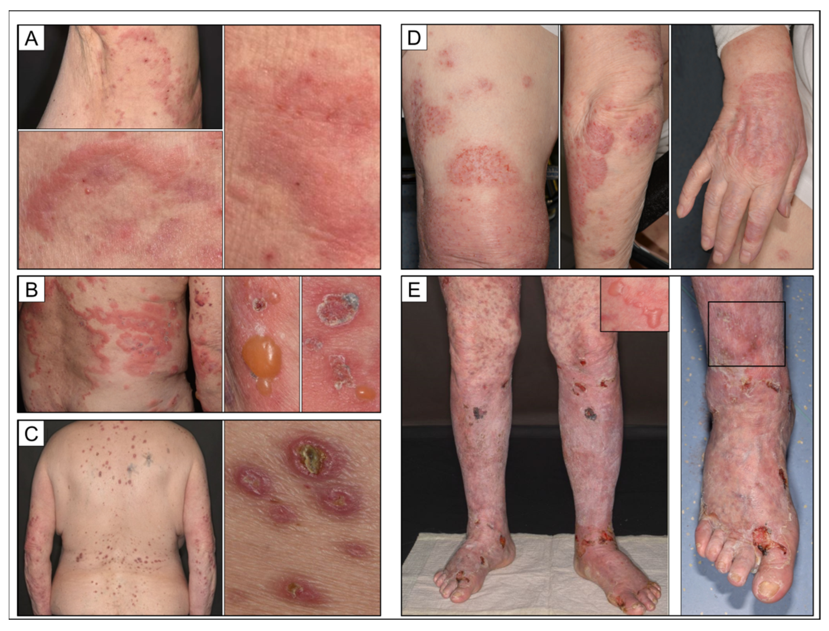
3.1. Clinical Characteristics
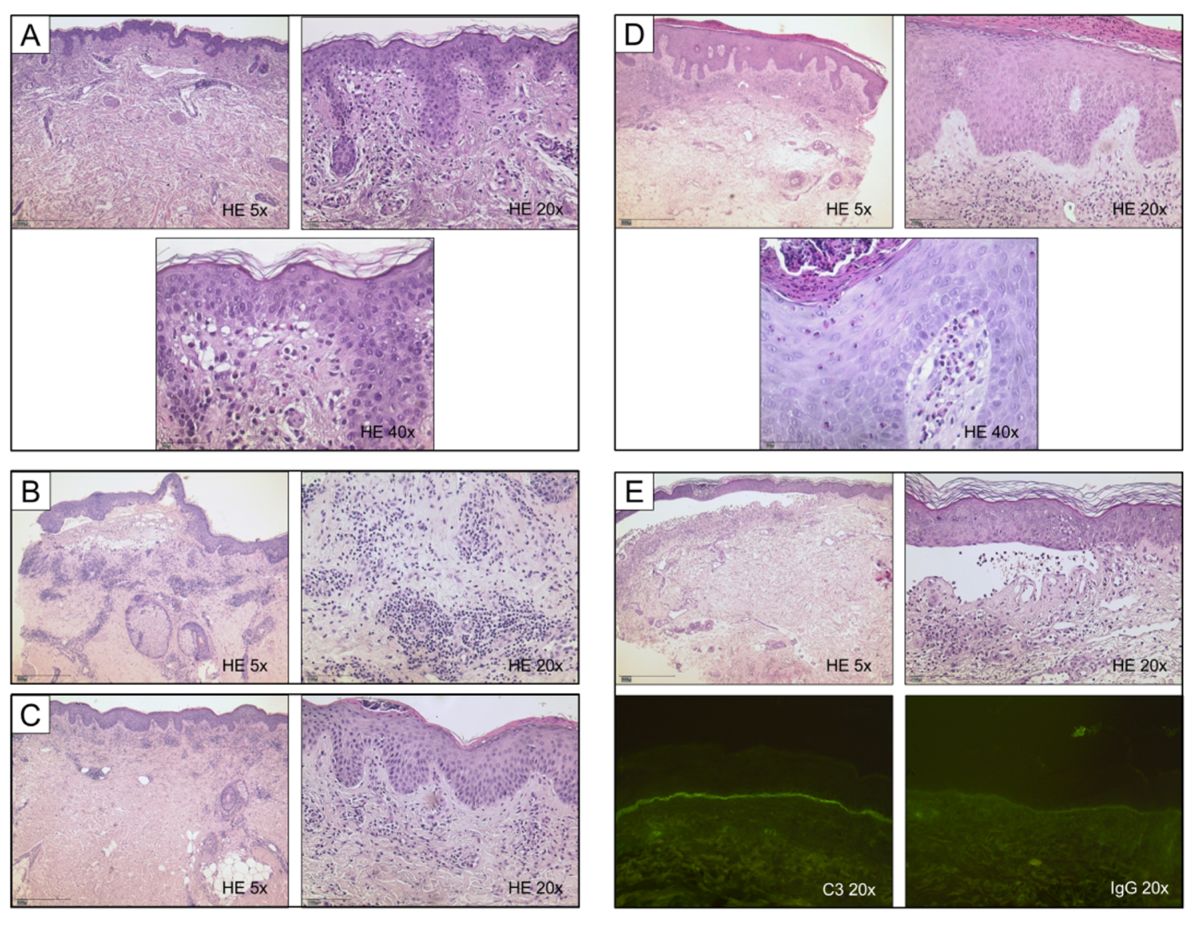
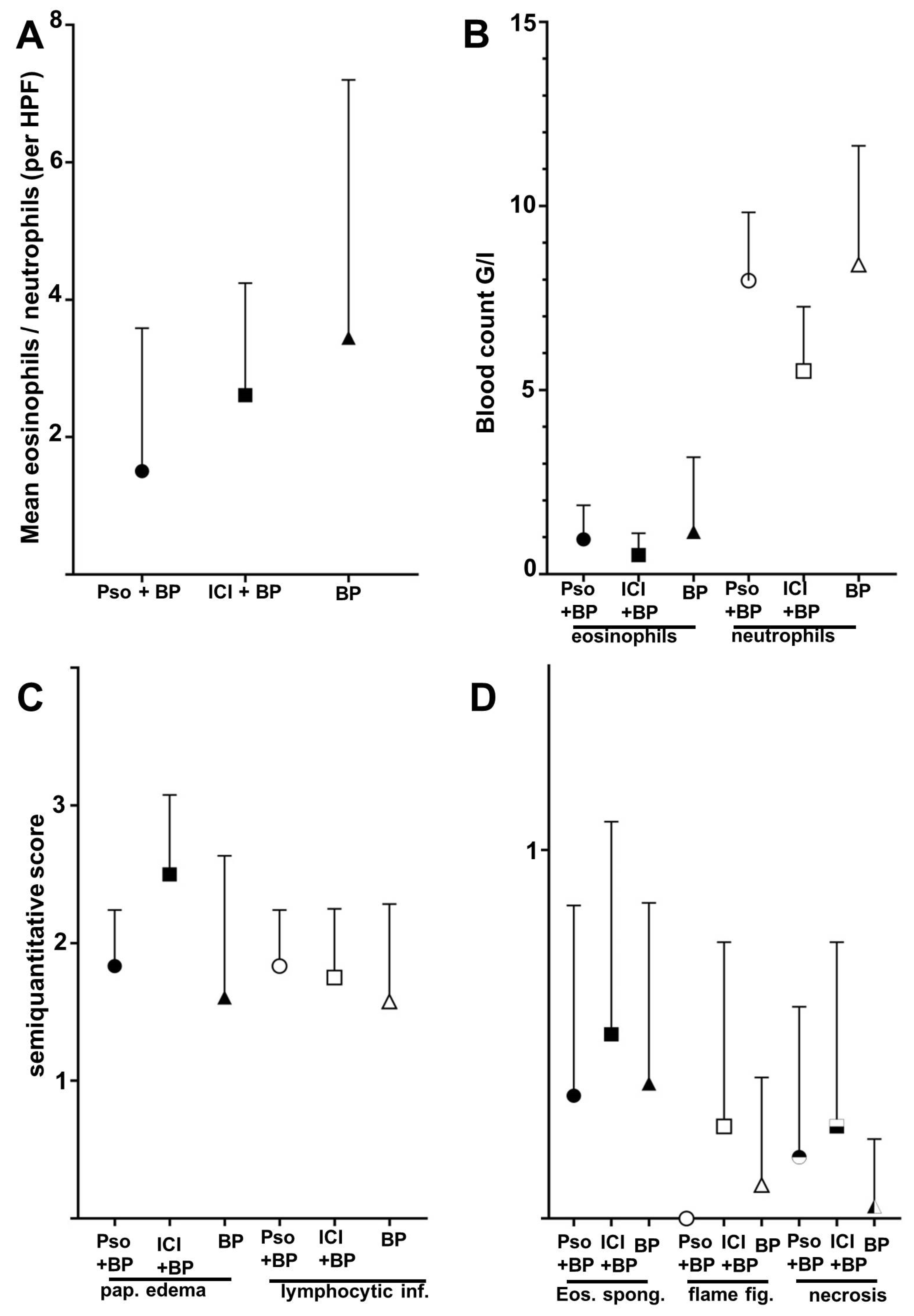
3.2. Dermatopathological Characteristics
3.3. Literature Review
4. Discussion
5. Conclusions
Author Contributions
Funding
Institutional Review Board Statement
Informed Consent Statement
Data Availability Statement
Acknowledgments
Conflicts of Interest
References
- Patterson, J.W.; Weedon, D. Weedon’s Skin Pathology, 4th ed.; Churchill Livingstone Elsevier: Edinburgh, UK, 2016; ISBN 978-0-7020-6200-1. [Google Scholar]
- Bolognia, J.L.; Schaffer, J.; Cerroni, L. Dermatology, 3rd ed.; Elsevier: Edinburgh, UK, 2012; ISBN 978-0-7020-6275-9. [Google Scholar]
- Kridin, K.; Ludwig, R.J. The Growing Incidence of Bullous Pemphigoid: Overview and Potential Explanations. Front. Med. 2018, 5, 220. [Google Scholar] [CrossRef] [PubMed] [Green Version]
- Ohata, C.; Ohyama, B.; Nagata, H.; Furumura, M.; Nakama, T. Comparative Study of Direct Immunofluorescence in Discoid Lupus Erythematosus and Bullous Pemphigoid. Am. J. Dermatopathol. 2016, 38, 121–123. [Google Scholar] [CrossRef] [PubMed]
- Lucariello, R.J.; Villablanca, S.E.; Mascaró, J.M.; Reichel, M. Association between bullous pemphigoid and malignancy: A meta-analysis. Australas. J. Dermatol. 2018, 59, 253–260. [Google Scholar] [CrossRef] [PubMed]
- Chuang, T.Y.; Korkij, W.; Soltani, K.; Clayman, J.; Cook, J. Increased frequency of diabetes mellitus in patients with bullous pemphigoid: A case-control study. J. Am. Acad. Dermatol. 1984, 11, 1099–1102. [Google Scholar] [CrossRef]
- Lambadiari, V.; Kountouri, A.; Kousathana, F.; Korakas, E.; Kokkalis, G.; Theotokoglou, S.; Palaiodimou, L.; Katsimbri, P.; Ikonomidis, I.; Theodoropoulos, K.; et al. The association of bullous pemphigoid with dipeptidyl-peptidase 4 inhibitors: A ten-year prospective observational study. BMC Endocr. Disord. 2021, 21, 23. [Google Scholar] [CrossRef]
- Aggarwal, P. Disproportionality analysis of bullous pemphigoid adverse events with PD-1 inhibitors in the FDA adverse event reporting system. Expert Opin. Drug Saf. 2019, 18, 623–633. [Google Scholar] [CrossRef] [PubMed]
- Amin, M.; Lee, E.B.; Tsai, T.-F.; Wu, J.J. Psoriasis and Co-morbidity. Acta Derm. Venereol. 2020, 100, adv00033. [Google Scholar] [CrossRef] [Green Version]
- Grattan, C.E. Evidence of an association between bullous pemphigoid and psoriasis. Br. J. Dermatol. 1985, 113, 281–283. [Google Scholar] [CrossRef] [PubMed]
- Kirtschig, G.; Chow, E.T.; Venning, V.A.; Wojnarowska, F.T. Acquired subepidermal bullous diseases associated with psoriasis: A clinical, immunopathological and immunogenetic study. Br. J. Dermatol. 1996, 135, 738–745. [Google Scholar] [CrossRef]
- Tsai, T.-F.; Wang, T.-S.; Hung, S.-T.; Tsai, P.I.-C.; Schenkel, B.; Zhang, M.; Tang, C.-H. Epidemiology and comorbidities of psoriasis patients in a national database in Taiwan. J. Dermatol. Sci. 2011, 63, 40–46. [Google Scholar] [CrossRef]
- Barnadas, M.A.; Gilaberte, M.; Pujol, R.; Agustí, M.; Gelpí, C.; Alomar, A. Bullous pemphigoid in a patient with psoriasis during the course of PUVA therapy: Study by ELISA test. Int. J. Dermatol. 2006, 45, 1089–1092. [Google Scholar] [CrossRef] [PubMed]
- Kridin, K.; Ludwig, R.J.; Schonmann, Y.; Damiani, G.; Cohen, A.D. The Bidirectional Association Between Bullous Pemphigoid and Psoriasis: A Population-Based Cohort Study. Front. Med. 2020, 7, 511. [Google Scholar] [CrossRef]
- Sticherling, M. Psoriasis and autoimmunity. Autoimmun. Rev. 2016, 15, 1167–1170. [Google Scholar] [CrossRef]
- Dainichi, T.; Kabashima, K. Interaction of Psoriasis and Bullous Diseases. Front. Med. 2018, 5, 222. [Google Scholar] [CrossRef] [PubMed] [Green Version]
- Yasukawa, S.; Dainichi, T.; Kokuba, H.; Moroi, Y.; Urabe, K.; Hashimoto, T.; Furue, M. Bullous pemphigoid followed by pustular psoriasis showing Th1, Th2, Treg and Th17 immunological changes. Eur. J. Dermatol. 2009, 19, 69–71. [Google Scholar] [CrossRef] [PubMed]
- Phan, K.; Goyal, S.; Murrell, D.F. Association between bullous pemphigoid and psoriasis: Systematic review and meta-analysis of case-control studies. Australas. J. Dermatol. 2019, 60, 23–28. [Google Scholar] [CrossRef] [Green Version]
- Ohata, C.; Ishii, N.; Koga, H.; Fukuda, S.; Tateishi, C.; Tsuruta, D.; Furumura, M.; Hashimoto, T. Coexistence of autoimmune bullous diseases (AIBDs) and psoriasis: A series of 145 cases. J. Am. Acad. Dermatol. 2015, 73, 50–55. [Google Scholar] [CrossRef]
- Inamura, E.; Tsujiwaki, M.; Ujiie, H.; Nishie, W.; Hata, H.; Shimizu, H.; Iwata, H. Bullous pemphigoid associated with psoriasis showing marked neutrophilic infiltrates. J. Dtsch. Dermatol. Ges. 2021, 19, 105–108. [Google Scholar] [CrossRef]
- Niebel, D.; Wenzel, J. Immunpathologie von kutanen Medikamentennebenwirkungen. Pathologe 2018, 39, 563–570. [Google Scholar] [CrossRef]
- Curry, J.L.; Tetzlaff, M.T.; Nagarajan, P.; Drucker, C.; Diab, A.; Hymes, S.R.; Duvic, M.; Hwu, W.-J.; Wargo, J.A.; Torres-Cabala, C.A.; et al. Diverse types of dermatologic toxicities from immune checkpoint blockade therapy. J. Cutan. Pathol. 2017, 44, 158–176. [Google Scholar] [CrossRef]
- Voudouri, D.; Nikolaou, V.; Laschos, K.; Charpidou, A.; Soupos, N.; Triantafyllopoulou, I.; Panoutsopoulou, I.; Aravantinos, G.; Syrigos, K.; Stratigos, A. Anti-PD1/PDL1 induced psoriasis. Curr. Probl. Cancer 2017, 41, 407–412. [Google Scholar] [CrossRef] [PubMed]
- Tsiogka, A.; Bauer, J.W.; Patsatsi, A. Bullous Pemphigoid Associated with Anti-programmed Cell Death Protein 1 and Anti-programmed Cell Death Ligand 1 Therapy: A Review of the Literature. Acta Derm. Venereol. 2021, 101, adv00377. [Google Scholar] [CrossRef] [PubMed]
- Pujalte-Martin, M.; Rocher, F.; Cardot-Leccia, N.; Giacchero, D.; Borchiellini, D. Immune checkpoint inhibitor-induced bullous pemphigoid: Towards a new class of drug-drug interaction? Eur. J. Cancer 2020, 138, 122–124. [Google Scholar] [CrossRef]
- Kridin, K.; Hundt, J.E.; Ludwig, R.J.; Amber, K.T.; Bitan, D.T.; Cohen, A.D. Melanoma is associated with an increased risk of bullous pemphigoid: A large population-based longitudinal study. Arch. Dermatol. Res. 2021, 314, 77–83. [Google Scholar] [CrossRef] [PubMed]
- Wongvibulsin, S.; Pahalyants, V.; Kalinich, M.; Murphy, W.; Yu, K.-H.; Wang, F.; Chen, S.T.; Reynolds, K.; Kwatra, S.G.; Semenov, Y.R. Epidemiology and risk factors for the development of cutaneous toxicities in patients treated with immune-checkpoint inhibitors: A United States population-level analysis. J. Am. Acad. Dermatol. 2022, 86, 563–572. [Google Scholar] [CrossRef]
- Lopez, A.T.; Khanna, T.; Antonov, N.; Audrey-Bayan, C.; Geskin, L. A review of bullous pemphigoid associated with PD-1 and PD-L1 inhibitors. Int. J. Dermatol. 2018, 57, 664–669. [Google Scholar] [CrossRef]
- Adachi, E.; Honda, T.; Nonoyama, S.; Irie, H.; Yamamura, K.; Otsuka, A.; Kabashima, K. Severe bullous pemphigoid in a metastatic lung cancer patient treated with pembrolizumab. J. Dermatol. 2019, 46, e232–e233. [Google Scholar] [CrossRef]
- Anastasopoulou, A.; Papaxoinis, G.; Diamantopoulos, P.; Christofidou, E.; Benopoulou, O.; Stratigos, A.; Gogas, H. Bullous Pemphigoid-like Skin Lesions and Overt Eosinophilia in a Patient with Melanoma Treated with Nivolumab: Case Report and Review of the Literature. J. Immunother. 2018, 41, 164–167. [Google Scholar] [CrossRef]
- Aoki, Y.; Miyagawa, F.; Miyashita, K.; Nishimura, Y.; Hashimoto, T.; Asada, H. Nivolumab-associated bullous pemphigoid reactions involving multiple epitopes. Eur. J. Dermatol. 2019, 29, 552–554. [Google Scholar] [CrossRef]
- Bandino, J.P.; Perry, D.M.; Clarke, C.E.; Marchell, R.M.; Elston, D.M. Two cases of anti-programmed cell death 1-associated bullous pemphigoid-like disease and eruptive keratoacanthomas featuring combined histopathology. J. Eur. Acad. Dermatol. Venereol. 2017, 31, e378–e380. [Google Scholar] [CrossRef]
- Beck, K.M.; Dong, J.; Geskin, L.J.; Beltrani, V.P.; Phelps, R.G.; Carvajal, R.D.; Schwartz, G.; Saenger, Y.M.; Gartrell, R.D. Disease stabilization with pembrolizumab for metastatic acral melanoma in the setting of autoimmune bullous pemphigoid. J. Immunother. Cancer 2016, 4, 20. [Google Scholar] [CrossRef] [PubMed] [Green Version]
- Cardona, A.F.; Ruiz-Patiño, A.; Zatarain-Barron, Z.L.; Ariza, S.; Ricaurte, L.; Rolfo, C.; Arrieta, O. Refractory Bullous Pemphigoid in a Patient with Metastatic Lung Adenocarcinoma Treated with Pembrolizumab. Case Rep. Oncol. 2021, 14, 386–390. [Google Scholar] [CrossRef] [PubMed]
- Carlos, G.; Anforth, R.; Chou, S.; Clements, A.; Fernandez-Peñas, P. A case of bullous pemphigoid in a patient with metastatic melanoma treated with pembrolizumab. Melanoma Res. 2015, 25, 265–268. [Google Scholar] [CrossRef] [PubMed]
- Cosimati, A.; Rossi, L.; Didona, D.; Forcella, C.; Didona, B. Bullous pemphigoid in elderly woman affected by non-small cell lung cancer treated with pembrolizumab: A case report and review of literature. J. Oncol. Pharm. Pract. 2021, 27, 727–733. [Google Scholar] [CrossRef]
- Cuenca-Barrales, C.; Espadafor-López, B.; Martínez-López, A.; Cancela-Díez, B.; Ruiz-Villaverde, R. Bullous pemphigoid in a patient treated with nivolumab. Dermatol. Ther. 2019, 32, e13030. [Google Scholar] [CrossRef]
- Damsky, W.; Kole, L.; Tomayko, M.M. Development of bullous pemphigoid during nivolumab therapy. JAAD Case Rep. 2016, 2, 442–444. [Google Scholar] [CrossRef] [Green Version]
- Fontecilla, N.M.; Khanna, T.; Bayan, C.-A.Y.; Antonov, N.A.; Geskin, L.J. Bullous Pemphigoid Associated with a New Combination Checkpoint Inhibitor Immunotherapy. J. Drugs Dermatol. 2019, 18, 103–104. [Google Scholar]
- Garje, R.; Chau, J.J.; Chung, J.; Wanat, K.; Zakharia, Y. Acute Flare of Bullous Pemphigus with Pembrolizumab Used for Treatment of Metastatic Urothelial Cancer. J. Immunother. 2018, 41, 42–44. [Google Scholar] [CrossRef]
- Grän, F.; Goebeler, M.; Gesierich, A. Bullous pemphigoid developing upon immune checkpoint inhibition with nivolumab. Eur. J. Dermatol. 2019, 29, 448–449. [Google Scholar] [CrossRef]
- Grimaux, X.; Delva, R.; Jadaud, E.; Croue, A. Nivolumab-induced bullous pemphigoid after radiotherapy and abscopal effect. Australas. J. Dermatol. 2019, 60, e235–e236. [Google Scholar] [CrossRef]
- Hanley, T.; Papa, S.; Saha, M. Bullous pemphigoid associated with ipilimumab therapy for advanced metastatic melanoma. JRSM Open 2018, 9, 2054270418793029. [Google Scholar] [CrossRef] [PubMed]
- Hara, K.; Yamasaki, K.; Yamada, S.; Yatera, K. Bullous Pemphigoid in a Patient Treated with Pembrolizumab. Intern. Med. 2020, 59, 139–140. [Google Scholar] [CrossRef] [Green Version]
- Hirotsu, K.; Chiou, A.S.; Chiang, A.; Kim, J.; Kwong, B.Y.; Pugliese, S. Localized bullous pemphigoid in a melanoma patient with dual exposure to PD-1 checkpoint inhibition and radiation therapy. JAAD Case Rep. 2017, 3, 404–406. [Google Scholar] [CrossRef] [PubMed] [Green Version]
- Honigman, A.D.; Lai, F.; Elakis, J.; Prall, O.; Goh, M.; McCormack, C. Pembrolizumab-induced sarcoid granulomatous panniculitis and bullous pemphigoid in a single patient. Clin. Case Rep. 2019, 7, 773–775. [Google Scholar] [CrossRef] [PubMed] [Green Version]
- Hwang, S.J.E.; Carlos, G.; Chou, S.; Wakade, D.; Carlino, M.S.; Fernandez-Penas, P. Bullous pemphigoid, an autoantibody-mediated disease, is a novel immune-related adverse event in patients treated with anti-programmed cell death 1 antibodies. Melanoma Res. 2016, 26, 413–416. [Google Scholar] [CrossRef]
- Jour, G.; Glitza, I.C.; Ellis, R.M.; Torres-Cabala, C.A.; Tetzlaff, M.T.; Li, J.Y.; Nagarajan, P.; Huen, A.; Aung, P.P.; Ivan, D.; et al. Autoimmune dermatologic toxicities from immune checkpoint blockade with anti-PD-1 antibody therapy: A report on bullous skin eruptions. J. Cutan. Pathol. 2016, 43, 688–696. [Google Scholar] [CrossRef] [PubMed]
- Kaul, S.; Wang, A.; Grushchak, S.; Albrecht, J. Pembrolizumab-induced reactivation of bullous pemphigoid. Int. J. Dermatol. 2021, 60, 757–758. [Google Scholar] [CrossRef]
- Kluger, N.; Harjama, L.; Makkonen, T.; Vehmanen, L.; Lappalainen, K. Nivolumab-associated pemphigoid-like blistering dermatosis after sun exposure. Dermatol. Ther. 2020, 33, e13970. [Google Scholar] [CrossRef]
- Kosche, C.; Owen, J.L.; Sadowsky, L.M.; Choi, J.N. Bullous dermatoses secondary to anti-PD-L1 agents: A case report and review of the literature. Dermatol. Online J. 2019, 25, 6. [Google Scholar] [CrossRef]
- Kuwatsuka, Y.; Iwanaga, A.; Kuwatsuka, S.; Okubo, Y.; Murayama, N.; Ishii, N.; Hashimoto, T.; Utani, A. Bullous pemphigoid induced by ipilimumab in a patient with metastatic malignant melanoma after unsuccessful treatment with nivolumab. J. Dermatol. 2018, 45, e21–e22. [Google Scholar] [CrossRef] [Green Version]
- Kwon, C.W.; Land, A.S.; Smoller, B.R.; Scott, G.; Beck, L.A.; Mercurio, M.G. Bullous pemphigoid associated with nivolumab, a programmed cell death 1 protein inhibitor. J. Eur. Acad. Dermatol. Venereol. 2017, 31, e349–e350. [Google Scholar] [CrossRef] [PubMed]
- Kwon, C.W.; Murthy, R.K.; Kudchadkar, R.; Stoff, B.K. Pembrolizumab-induced lichen planus pemphigoides in a patient with metastatic Merkel cell carcinoma. JAAD Case Rep. 2020, 6, 1045–1047. [Google Scholar] [CrossRef] [PubMed]
- Leavitt, E.; Holland, V. A case of atezolizumab-induced photodistributed bullous pemphigoid. Dermatol. Ther. 2019, 32, e12924. [Google Scholar] [CrossRef] [PubMed]
- Lee, H.; Chung, J.H.; Jo, S.J. Pruritic bullous skin eruption in a male patient receiving immunotherapy for oropharyngeal cancer. Int. J. Dermatol. 2020, 59, 685–686. [Google Scholar] [CrossRef] [PubMed]
- Lomax, A.J.; Ge, L.; Anand, S.; McNeil, C.; Lowe, P. Bullous pemphigoid-like reaction in a patient with metastatic melanoma receiving pembrolizumab and previously treated with ipilimumab. Australas. J. Dermatol. 2016, 57, 333–335. [Google Scholar] [CrossRef] [PubMed]
- Lopez, A.T.; Geskin, L. A Case of Nivolumab-Induced Bullous Pemphigoid: Review of Dermatologic Toxicity Associated with Programmed Cell Death Protein-1/Programmed Death Ligand-1 Inhibitors and Recommendations for Diagnosis and Management. Oncologist 2018, 23, 1119–1126. [Google Scholar] [CrossRef] [PubMed] [Green Version]
- Maya, Y.; Moriuchi, R.; Takashima, Y.; Hotta, M.; Izumi, H.; Shimizu, S. Bullous pemphigoid in a metastatic lung cancer patient associated with nivolumab. Int. J. Womens Dermatol. 2020, 6, 456–457. [Google Scholar] [CrossRef]
- Mochel, M.C.; Ming, M.E.; Imadojemu, S.; Gangadhar, T.C.; Schuchter, L.M.; Elenitsas, R.; Payne, A.S.; Chu, E.Y. Cutaneous autoimmune effects in the setting of therapeutic immune checkpoint inhibition for metastatic melanoma. J. Cutan. Pathol. 2016, 43, 787–791. [Google Scholar] [CrossRef]
- Munera-Campos, M.; Plana-Pla, A.; Rivera, N.; Boada, A.; Ferrándiz, C. Nivolumab-Induced Bullous Pemphigoid Managed without Drug Withdrawal. Indian J. Dermatol. 2020, 65, 214–216. [Google Scholar] [CrossRef]
- Muto, Y.; Namikawa, K.; Fujiwara, Y.; Mori, T.; Nakano, E.; Takahashi, A.; Yamazaki, N. Bullous pemphigoid induced by pembrolizumab in a patient with non-small-cell lung cancer who achieved durable complete response despite treatment cessation and long-term corticosteroid administration: A case report. J. Dermatol. 2020, 47, e9–e11. [Google Scholar] [CrossRef]
- Naidoo, J.; Schindler, K.; Querfeld, C.; Busam, K.; Cunningham, J.; Page, D.B.; Postow, M.A.; Weinstein, A.; Lucas, A.S.; Ciccolini, K.T.; et al. Autoimmune Bullous Skin Disorders with Immune Checkpoint Inhibitors Targeting PD-1 and PD-L1. Cancer Immunol. Res. 2016, 4, 383–389. [Google Scholar] [CrossRef] [PubMed] [Green Version]
- Nakai, K.; Sato, Y.; Kubo, S.; Tsuruta, D. PD-L1 positive macrophages were increased in a case of pembrolizumab-induced bullous pemphigoid. J. Dermatol. 2021, 48, e146–e148. [Google Scholar] [CrossRef] [PubMed]
- Niebel, D.; Brägelmann, C.; Wenzel, J.; Bieber, T. Unusual flaccid blistering with mucosal involvement upon immune checkpoint inhibition. J. Dtsch. Dermatol. Ges. 2020, 18, 149–152. [Google Scholar] [CrossRef] [PubMed]
- Palla, A.R.; Smith, E.; Doll, D. Bullous pemphigoid associated with the 480-mg nivolumab dose in a patient with metastatic renal cell carcinoma. Immunotherapy 2019, 11, 1187–1192. [Google Scholar] [CrossRef]
- Parakh, S.; Nguyen, R.; Opie, J.M.; Andrews, M.C. Late presentation of generalised bullous pemphigoid-like reaction in a patient treated with pembrolizumab for metastatic melanoma. Australas. J. Dermatol. 2017, 58, e109–e112. [Google Scholar] [CrossRef]
- Qiu, C.; Shevchenko, A.; Hsu, S. Bullous pemphigoid secondary to pembrolizumab mimicking toxic epidermal necrolysis. JAAD Case Rep. 2020, 6, 400–402. [Google Scholar] [CrossRef]
- Ridpath, A.V.; Rzepka, P.V.; Shearer, S.M.; Scrape, S.R.; Olencki, T.E.; Kaffenberger, B.H. Novel use of combination therapeutic plasma exchange and rituximab in the treatment of nivolumab-induced bullous pemphigoid. Int. J. Dermatol. 2018, 57, 1372–1374. [Google Scholar] [CrossRef]
- Rofe, O.; Bar-Sela, G.; Keidar, Z.; Sezin, T.; Sadik, C.D.; Bergman, R. Severe bullous pemphigoid associated with pembrolizumab therapy for metastatic melanoma with complete regression. Clin. Exp. Dermatol. 2017, 42, 309–312. [Google Scholar] [CrossRef]
- Russo, I.; Sacco, G.; Frega, S.; Polo, V.; Pasello, G.; Alaibac, M. Immunotherapy-related skin toxicity: Bullous pemphigoid in a lung adenocarcinoma patient treated with the anti-PDL1 antibody atezolizumab. Eur. J. Dermatol. 2017, 27, 205–208. [Google Scholar] [CrossRef]
- Sadik, C.D.; Langan, E.A.; Grätz, V.; Zillikens, D.; Terheyden, P. Checkpoint Inhibition May Trigger the Rare Variant of Anti-LAD-1 IgG-Positive, Anti-BP180 NC16A IgG-Negative Bullous Pemphigoid. Front. Immunol. 2019, 10, 1934. [Google Scholar] [CrossRef] [Green Version]
- Sato, Y.; Fujimura, T.; Mizuashi, M.; Aiba, S. Lichen planus pemphigoides developing from patient with non-small-cell lung cancer treated with nivolumab. J. Dermatol. 2019, 46, e374–e375. [Google Scholar] [CrossRef] [PubMed]
- Schmidgen, M.I.; Butsch, F.; Schadmand-Fischer, S.; Steinbrink, K.; Grabbe, S.; Weidenthaler-Barth, B.; Loquai, C. Pembrolizumab-induced lichen planus pemphigoides in a patient with metastatic melanoma. J. Dtsch. Dermatol. Ges. 2017, 15, 742–745. [Google Scholar] [CrossRef] [PubMed]
- Schwartzman, G.; Simpson, M.M.; Jones, R.; Schiavone, K.; Coffman, M.; Meyerle, J. Anti-PD1 immune checkpoint inhibitor-induced bullous pemphigoid in metastatic melanoma and non-small cell lung cancer. Cutis 2020, 105, E9–E12. [Google Scholar] [CrossRef] [PubMed]
- Senoo, H.; Kawakami, Y.; Yokoyama, E.; Yamasaki, O.; Morizane, S. Atezolizumab-induced lichen planus pemphigoides in a patient with metastatic non-small-cell lung cancer. J. Dermatol. 2020, 47, e121–e122. [Google Scholar] [CrossRef]
- Sharma, P.; Barnes, M.; Nabeel, S.; LiPera, W. Pembrolizumab-Induced Bullous Pemphigoid Treated with Rituximab. JCO Oncol. Pract. 2020, 16, 764–766. [Google Scholar] [CrossRef]
- Singer, S.; Nelson, C.A.; Lian, C.G.; Dewan, A.K.; LeBoeuf, N.R. Nonbullous pemphigoid secondary to PD-1 inhibition. JAAD Case Rep. 2019, 5, 898–903. [Google Scholar] [CrossRef] [PubMed] [Green Version]
- Sowerby, L.; Dewan, A.K.; Granter, S.; Gandhi, L.; LeBoeuf, N.R. Rituximab Treatment of Nivolumab-Induced Bullous Pemphigoid. JAMA Dermatol. 2017, 153, 603–605. [Google Scholar] [CrossRef]
- Strickley, J.D.; Vence, L.M.; Burton, S.K.; Callen, J.P. Nivolumab-induced lichen planus pemphigoides. Cutis 2019, 103, 224–226. [Google Scholar]
- Sugawara, A.; Koga, H.; Abe, T.; Ishii, N.; Nakama, T. Lichen planus-like lesion preceding bullous pemphigoid development after programmed cell death protein-1 inhibitor treatment. J. Dermatol. 2021, 48, 401–404. [Google Scholar] [CrossRef]
- Sun, C.W.; Grossman, S.K.; Aphale, A.; Hsu, S. Pembrolizumab-induced bullous pemphigoid. JAAD Case Rep. 2019, 5, 362–364. [Google Scholar] [CrossRef] [Green Version]
- Thomsen, K.; Diernaes, J.; Øllegaard, T.H.; Spaun, E.; Vestergaard, C. Bullous Pemphigoid as an Adverse Reaction to Pembrolizumab: Two Case Reports. Case Rep. Dermatol. 2018, 10, 154–157. [Google Scholar] [CrossRef] [PubMed]
- Virgen, C.A.; Nguyen, T.A.; Di Raimondo, C.; Amini, A.; Margolin, K.A.; Parekh, V.; Abdulla, F.R.; Modi, B. Bullous pemphigoid associated with cemiplimab therapy in a patient with locally advanced cutaneous squamous cell carcinoma. JAAD Case Rep. 2020, 6, 195–197. [Google Scholar] [CrossRef] [Green Version]
- Wada, N.; Uchi, H.; Furue, M. Bullous pemphigoid induced by pembrolizumab in a patient with advanced melanoma expressing collagen XVII. J. Dermatol. 2017, 44, e240–e241. [Google Scholar] [CrossRef] [PubMed]
- Klein, S.L.; Morgan, R. The impact of sex and gender on immunotherapy outcomes. Biol. Sex Differ. 2020, 11, 24. [Google Scholar] [CrossRef] [PubMed]
- Nelson, C.A.; Singer, S.; Chen, T.; Puleo, A.E.; Lian, C.G.; Wei, E.X.; Giobbie-Hurder, A.; Mostaghimi, A.; LeBoeuf, N.R. Bullous pemphigoid after anti-PD-1 therapy: A retrospective case-control study evaluating impact on tumor response and survival outcomes. J. Am. Acad. Dermatol. 2020. [Google Scholar] [CrossRef]
- Sadik, C.D.; Langan, E.A.; Gutzmer, R.; Fleischer, M.I.; Loquai, C.; Reinhardt, L.; Meier, F.; Göppner, D.; Herbst, R.A.; Zillikens, D.; et al. Retrospective Analysis of Checkpoint Inhibitor Therapy-Associated Cases of Bullous Pemphigoid from Six German Dermatology Centers. Front. Immunol. 2020, 11, 588582. [Google Scholar] [CrossRef]
- Horikawa, H.; Kurihara, Y.; Funakoshi, T.; Umegaki-Arao, N.; Takahashi, H.; Kubo, A.; Tanikawa, A.; Kodani, N.; Minami, Y.; Meguro, S.; et al. Unique clinical and serological features of bullous pemphigoid associated with dipeptidyl peptidase-4 inhibitors. Br. J. Dermatol. 2018, 178, 1462–1463. [Google Scholar] [CrossRef]
- Izumi, K.; Nishie, W.; Mai, Y.; Wada, M.; Natsuga, K.; Ujiie, H.; Iwata, H.; Yamagami, J.; Shimizu, H. Autoantibody Profile Differentiates between Inflammatory and Noninflammatory Bullous Pemphigoid. J. Investig. Dermatol. 2016, 136, 2201–2210. [Google Scholar] [CrossRef] [Green Version]
- Siegel, J.; Totonchy, M.; Damsky, W.; Berk-Krauss, J.; Castiglione, F.; Sznol, M.; Petrylak, D.P.; Fischbach, N.; Goldberg, S.B.; Decker, R.H.; et al. Bullous disorders associated with anti-PD-1 and anti-PD-L1 therapy: A retrospective analysis evaluating the clinical and histopathologic features, frequency, and impact on cancer therapy. J. Am. Acad. Dermatol. 2018, 79, 1081–1088. [Google Scholar] [CrossRef]
- Molina, G.E.; Reynolds, K.L.; Chen, S.T. Diagnostic and therapeutic differences between immune checkpoint inhibitor-induced and idiopathic bullous pemphigoid: A cross-sectional study. Br. J. Dermatol. 2020, 183, 1126–1128. [Google Scholar] [CrossRef] [PubMed]
- Molina, G.E.; Yanovsky, R.L.; Wei, E.X.; Chen, S.T. Missed drug-induced bullous pemphigoid leads to longer immunosuppression than recognized cases: A 9-year retrospective review. J. Am. Acad. Dermatol. 2020, 82, 1255–1258. [Google Scholar] [CrossRef]


| Diagnosis | Age Mean (SD), Range | Clinical Appearance | Mucosal Involvement | DIF | DM Type II | Active Neoplastic Disease | Drugs Mean (SD) Range | DPP4 Inhibitor | Treatment |
|---|---|---|---|---|---|---|---|---|---|
| BP and psoriasis (n = 6; F = 3/M = 3) | 81.2 (±8.8) 66–89 | Typical: 4 Eczematous: 2 | 0/6 | IgM: 0 IgG: 5 IgA: 0 C3 only: 1 | 2/6 | 1/6 | 6.0 (±1.8) 4–8 | 0/2 | SC: 2 Doxy: 3 MMF: 0 MTX 1 |
| BP and ICB (n = 4; F = 1/M = 3) | 73.3 (±5.7) 68–80 | Typical: 4 | 2/4 | IgM: 0 IgG: 4 IgA: 0 C3 only: 0 | 0/4 | 4/4 | 8.3 (±4.2) 4–14 | 0/0 | SC: 2 Doxy: 1 MMF: 0 Dapsone 1 |
| BP (n = 33; F = 20/M = 13) | 81.1 (±11.0) 58–97 | Prodromal: 1 Typical: 27 Eczematous: 4 Prurigo type: 1 | 2/33 | IgM: 3 IgG: 31 IgA: 2 C3 only: 0 | 12/33 | 1/33 | 7.0 (±3.1) 1–13 | 4/12 | SC: 22 Doxy: 20 MMF: 8 Dapsone: 3 Dupilumab: 1 |
| Case # | Sex | Age | ICI | Indication | Clinical Appearance | Mucosal Involvement | DIF | Specific Histologic Findings | Comorbidity/Specifics | Reference |
|---|---|---|---|---|---|---|---|---|---|---|
| 1 | M | 80 | Pembro | NSCLC | Typical; other: cellulitis-like appearance | Erosions on lips and oral mucosa | IgG | Subepidermal blister inflammatory cells | None reported | Adachi et al. [29] |
| 2 | M | 48 | Nivo | Melanoma | Typical | None | NA | Subepidermal blister Eosinophils and lymphocytes in upper dermis | Polymorphous adenoma of the parotid, hyperlipidemia, smoking | Anastasopoulou et al. [30] |
| 3 | F | 75 | Nivo | Melanoma | Typical | None | IgG | Eosinophils and lymphocytes in infiltrate | None reported | Aoki et al. [31] |
| 4 | M | 73 | Pembro | Melanoma | Typical; other: hyperkeratotic crateriform lesions | None | IgG | Subepidermal cleft, tagging of eosinophils along dermal–epidermal junction | None reported | Bandino et al. [32] |
| 5 | M | 90 | Pembro, then Nivo | Melanoma | Other: localized blistering and ulcer | None | NA | Subepidermal vacuolization with eosinophilic spongiosis, increased dermal eosinophils | None reported | Bandino et al. [32] |
| 6 | M | 72 | Ipi, then Pembro | Melanoma | Typical | Severe involvement | NA | Subepidermal cleft, eosinophils, perivascular mixed infiltrate | Preexisting BP flare with ICB (Ipi > Pembro) | Beck et al. [33] |
| 7 | M | 73 | Pembro | NSCLC | Typical | Oral mucosa and throat | IgG | Early: spongiosis, irregular acanthosis, focal exocytosis of lymphocytes and eosinophils, mild superficial perivascular inflammation with eosinophilis Late: subepidermal blisters with eosinophils | Former smoking, zoster, irAE: pneumonitis and adrenal insufficiency | Cardona et al. [34] |
| 8 | M | 75 | Ipi, then Pembro | Melanoma | Typical | None | IgG | Similar to Cardona et al. | None reported | Carlos et al. [35] |
| 9 | F | 77 | Pembro | NSCLC | Typical | Gingival mucosa | IgG | Subepidermal cleft with fibrin and eosinophils, eosinophils and lymphocytes in dermal papillae | Pancreatitis | Cosimati et al. [36] |
| 10 | M | 74 | Nivo | NSCLC | Typical | Oral mucosa | C3 only | Subepidermal cleft with eosinophils and fibrin, neutrophils in the upper dermis | None reported | Cuenca-Barrales et al. [37] |
| 11 | F | 77 | Nivo | NSCLC | Typical | None | IgG | Eosiniophilic spongiosis, mixed dermal infiltrate with eosinophilia | Inverse psoriasis, diabetes mellitus, hypertension, COPD, depression | Damsky et al. [38] |
| 12 | F | 65 | Durva and Tremi | NSCLC | Typical | None | IgG | Subepidermal cleft, epidermal necrosis, perivascular infiltrate of lymphocytes and eosinophils | None reported | Fontecilla et al. [39] |
| 13 | M | 64 | Pembro | Urothelial carcinoma | Typical | None | IgG | Subepidermal cleft with eosinophils, perivascular eosinophils and lymphocytes | Preexisting BP flare with Pembro | Garje et al. [40] |
| 14 | M | 63 | Nivo | NSCLC | Typical | None | IgG, IgA | Subepidermal blistering with infiltrating lymphocytes and eosinophils | COPD | Grän et al. [41] |
| 5 | M | 78 | Nivo | RCC | Typical | Oral and genital mucosa | C3 only | Separation of epidermis from dermis at the basement membrane | Onset of symptoms associated with radiotherapy | Grimaux et al. [42] |
| 16 | M | 72 | Pembro, then Ipi | Melanoma | Typical | None | IgG | Subepidermal blister, perivascular lymphocytic infiltrates, multiple eosinophils | irAE: grade 4 diarrhoea | Hanley et al. [43] |
| 17 | M | 60 | Pembro | NSCLC | Typical | None reported | IgG | NA | None reported | Hara et al. [44] |
| 18 | M | 70 | Ipi, then Pembro, then Nivo | Melanoma | Typical | None | IgG | Subepidermal cleft, numerous eosinophils | Blistering localized and associated with radiotherapy irAE: hypophysitis | Hirotsu et al. [45] |
| 19 | F | 56 | Pembro | Endometrial carcinoma | Prurigo-type | None | IgG | Subepidermal cleft, eosinophils | irAE: sarcoidal granulomatous panniculitis preceding BP | Honigman et al. [46] |
| 20 | M | 68 | Pembro | Melanoma | Typical/ Prurigo-type | Single erosion of oral mucosa | IgG | Early: aspects of grover’s disease Late: mild papillary dermal chronic inflammation with scattered eosinophils | Non-melanoma skin cancer, irAE: vitiligo | Hwang et al. [47] |
| 21 | M | 72 | Pembro, then Ipi | Melanoma | Typical | None | IgG | Subepidermal cleft with eosinophils, neutrophils and fibrin | Non-melanoma skin cancer, irAE of Ipi: pneumonitis | Hwang et al. [47] |
| 22 | M | 63 | Nivo | HNSCC | Typical | Mucosal blistering | IgG | Subepidermal blister, mixed inflammatory infiltrate of neutrophils and eosinophils | None reported | Jour et al. [48] |
| 23 | M | 68 | Pembro | Melanoma | Typical | None | IgG | Perivascular inflammation and eosinophils in the blister cavity | Psoriasis vulgaris, worsening with Pembro | Jour et al. [48] |
| 24 | F | 74 | Nivo + Ipi | Urothelial carcinoma | Typical | None reported | IgG | Subepidermal blister with eosinophils | None reported | Jour et al. [48] |
| 25 | F | 73 | Nivo | NSCLC | Typical | None | NA | Subepidermal blister with eosinophils, dermal lymphocytic infiltration | None reported | Jour et al. [48] |
| 26 | M | 67 | Pembro | NSCLC | Typical | Gingival erosions | IgG | Spongiosis, lymphocytic exocytosis, perivascular lymphocytic infiltrate, numerous eosinophils | Preexisting BP in remission-flare with Pembro | Kaul et al. [49] |
| 27 | M | 70 | Nivo | RCC | Typical | Oral mucosa | C3 only | sub- and intra-epidermal blister with eosinophils, eosinophils in the dermis | Blistering limited to sun-exposed areas | Kluger et al. [50] |
| 28 | M | 87 | Atezo | Urothelial carcinoma | Typical | None | IgG | Subepidermal blister, paucicellular infiltrate | None reported | Kosche et al. [51] |
| 29 | M | 35 | Nivo, then Ipi | Melanoma | Typical | None reported | IgG | Subepidermal blister, moderate eosinophilic infiltration of the upper dermis | None reported | Kuwatsuka et al. [52] |
| 30 | M | 60 | Nivo | RCC | Typical | None reported | IgG | Subepidermal cleft, perivascular and interstitial mixed cell infiltrate, lymphocytes and eosinophils | None reported | Kwon et al. [53] |
| 31 | F | 65 | Pembro | Merkel-cell carcinoma | Other: lichenified papules and plaques | None reported | IgG | Lichenoid and vacuolar epidermal interface alteration with associated dyskeratotic keratinocytes and eosinonophils | Diagnosis of LPP favored over BP | Kwon et al. [54] |
| 32 | M | 82 | Atezo | cSCC | Typical | None | IgG | Pauci-inflammatory subepidermal blister | Blistering in sun-exposed areas | Leavitt et al. [55] |
| 33 | M | 30–39 | Nivo | HNSCC | Typical | Ulcers on oral mucosa | IgG | Subepidermal blister with a mixed inflammatory infiltrate, many eosinophils | None reported | Lee et al. [56] |
| 34 | F | 82 | Ipi, then Pembro | Melanoma | Typical | None reported | NA | Subepidermal blister, superficial perivascular and interstitial inflammatory infiltrate of lymphocytes, eosinophils and occasional neutrophils | None reported | Lomax et al. [57] |
| 35 | F | 72 | Nivo | NSCLC | Typical | None | IgG | Perivascular lymphocytic and eosinophilic infiltrate | Laryngeal cancer, successfully treated with chemoradiation | Lopez et al. [58] |
| 36 | F | 80 | Nivo | NSCLC | Typical | None reported | C3 only | Vacuolar changes at the dermal–epidermal junction with eosinophilic infiltration in the dermis | None reported | Maya et al. [59] |
| 37 | M | 63 | Pembro | Melanoma | Typical | None reported | C3 only | Subepidermal blister, superficial dermal inflammatory infiltrate with lymphocytes and eosinophils, intraepithelial eosinophils | None reported | Mochel et al. [60] |
| 38 | M | 62 | Nivo | RCC | Typical | None reported | IgG | Subepidermal blister, dermal lymphocytic infiltrate with numerous eosinophils | None | Munera-Campos et al. [61] |
| 39 | M | 84 | Pembro | NSCLC | Typical | None reported | IgG | Subepidermal blister with moderate eosinophil and neutrophil infiltration of the upper dermis, eosinophilic spongiosis | None reported | Muto et al. [62] |
| 40 | M | 80 | Ipi, then Nivo | Melanoma | Typical | Initially none, later erosions and vesicles on buccal mucosa | IgG | Ulcerated and inflamed subepidermal vesicular dermatitis with eosinophils | None | Naidoo et al. [63] |
| 41 | F | 78 | Ipi, then Durva | Melanoma | Typical | Buccal mucosa | IgG | Subepidermal cleft | None | Naidoo et al. [63] |
| 42 | M | 85 | Nivo | NSCLC | Typical | None reported | IgG | Subepidermal bullous dermatitis with eosinophils | None | Naidoo et al. [63] |
| 43 | M | 79 | Pembro | Cholangio-carcinoma | None reported | None reported | NA | Marked infiltration of CD4+, CD8+ and CD163+ cells | None reported | Nakai et al. [64] |
| 44 | F | 75 | Nivo | Melanoma | Typical | Faint striae on cheeks, oral paresthesia | IgG | Subepidermal fissuring with a dense inflammatory infiltrate and colloid bodies, necrotic epithelium with a dense perivascular and periadnexal lymphocytic infiltrate | Hypertension, hypothyreodism | Niebel et al. [65] |
| 45 | M | 62 | Nivo | RCC | Typical | None | IgG | Subepidermal cleft with eosinophils, eosinophils tagging the intact dermal–epidermal junction | Hypertension, coronary artery disease, chronic kidney disease, hereditary focal segmental glomerulosclerosis | Palla et al. [66] |
| 46 | M | 42 | Ipi, then Pembro | Melanoma | Typical | None | C3 only | Eosinophil-predominant inflammatory cell infiltration, particularly in the interstitium and perivascular space | None | Parakh et al. [67] |
| 47 | F | 79 | Pembro | NSCLC | Typical | None reported | IgG | Lichenoid dermatitis with subepidermal blister formation | None reported | Qiu et al. [68] |
| 48 | M | 67 | Nivo | Melanoma | Typical | None reported | IgG | Subepidermal bullous dermatosis | None reported | Ridpath et al. [69] |
| 49 | F | 56 | Ipi, then Pembro | Melanoma | Typical | None reported | IgG | Subepidermal blister with mononuclear cells and eosinophils in the papillary dermis | Hypothyreodism, irAE: primary adrenal insufficiency | Rofe et al. [70] |
| 50 | M | 58 | Atezo | NSCLC | Typical | None reported | IgG | Subepidermal blister with eosinophils | None reported | Russo et al. [71] |
| 51 | F | 69 | Nivo | Melanoma | Typical | None reported | IgG | Moderate lymphohistiocytic dermal infiltrate | irAE: thyreoiditis | Sadik et al. [72] |
| 52 | M | 57 | Nivo | NSCLC | Typical | None reported | IgG | Vacuolar degeneration with apoptotic keratinocytes and prominent eosinophil infiltration at the epidermal junction and band-like infiltration of lymphocytes | Diagnosis of LPP favored over BP; hand-foot syndrome with chemotherapy, diabetes treated with vildagliptin | Sato et al. [73] |
| 53 | M | 64 | Pembro | Melanoma | Typical | Oral mucosa | C3 only | Subepidermal blistering, few eosinophils and lymphocytic infiltrate | Diagnosis of LPP favored over BP | Schmidgen et al. [74] |
| 54 | F | 72 | Pembro | Melanoma | Eczematous, singular vesicle | None reported | IgA and IgG | Subepidermal split, dense eosinophilic infiltrate in the dermis | None reported | Schwartzman et al. [75] |
| 55 | M | 82 | Ipi and Nivo | Melanoma | Typical | None reported | IgA and IgG | Subepidermal split with predominantly eosinophils and scattered neutrophils | None reported | Schwartzman et al. [75] |
| 56 | M | 68 | Nivo | NSCLC | Typical | None reported | “consistent with BP” | Psoriasiform dermatitis | irAE: thyroiditis, dermatitis, and nephritis | Schwartzman et al. [75] |
| 57 | F | 76 | Atezo | NSCLC | Blistering + other: violaceous, flat-topped polygonal papules and plaques | White reticular lesions of the oral mucosa | C3 only | Hypergranulosis, subepidermal blister, mixed infiltrate of eosinophils and lymphocytes, vacuolar degeneration at the dermoepidermal junction, band-like lymphocytic infiltration in the upper dermis | Diagnosis of LPP favored over BP; gallbladder cancer, hypercholesterinemia | Senoo et al. [76] |
| 58 | M | 76 | Pembro | NSCLC | Typical | None reported | NA | Subepidermal vesicles with underlying mixed-cell infiltrates including numerous eosinophils | None reported | Sharma et al. [77] |
| 59 | M | 78 | Nivo | Melanoma | Eczematous | Desquamative gingivitis | IgG | Eosinophilic Spongiosis, eosinophil tagging of the dermal–epidermal junction | None reported | Singer et al. [78] |
| 60 | M | 78 | Pembro | Esophageal carcinoma | Eczematous | None | C3 only | Mixed spongiotic, micropustular, and interface dermatitis with numerous eosinophils | None reported | Singer et al. [78] |
| 61 | M | 62 | Pembro | NSCLC | Eczematous | None reported | IgG | Subacute spongiosis and papillary dermal chronic inflammation with numerous eosinophils | None reported | Singer et al. [78] |
| 62 | M | 58 | Pembro | Melanoma | Eczematous | None reported | NA | Acute and chronic inflammation suggestive of component of hypersensitivity reaction | None reported | Singer et al. [78] |
| 63 | M | 80–89 | Nivo | NSCLC | Typical | Gingival bulla | IgG | Subepidermal vesicle with numerous eosinophils | Delirium, osteopenic compression fractures | Sowerby et al. [79] |
| 64 | F | 87 | Nivo | NSCLC | Typical | None reported | (-) | Subepidermal bullous lichenoid eruption with eosinophils | Diagnosis of LPP favored over BP; congestive heart failure, coronary artery disease, chronic kidney disease, hypertension | Strickley et al. [80] |
| 65 | F | 72 | Pembro | NSCLC | Typical | None reported | IgG | Early: compact orthokeratosis and hypergranulosis, vacuolar alteration of the basal layer and dermal lymphocyte infiltration Late: subepidermal blister with eosinophilic infiltration | Diabetes treated with sitagliptin and teneligliptin | Sugawara et al. [81] |
| 66 | F | 86 | Pembro | Melanoma | Typical | None reported | NA | Subepidermal bulla with eosinophils | None reported | Sun et al. [82] |
| 67 | M | 82 | Pembro | Melanoma | Typical | None reported | IgG | Subepidermal bulla and inflammatory infiltrate with eosinophils | Chronic lymphocytic leukemia, renal cell carcinoma, diabetes | Sun et al. [82] |
| 68 | M | 64 | Pembro | Melanoma | Typical | None reported | IgG | Superficial perivascular and interstitial inflammation dominated by eosinophils, beginning dermoepidermal bulla | Urolithiasis | Thomsen al. [83] |
| 69 | M | 71 | Ipi, then Pembro | Melanoma | Typical | None reported | (-) | Dermoepidermal bulla | Pneumonia, myocardial infarction | Thomsen et al. [83] |
| 70 | M | 68 | Cemi | cSCC | Typical | None | IgG | Subepidermal blister, eosinophilic spongiosis, dermal eosinophilia | Non-melanoma skin cancer | Virgen et al. [84] |
| 71 | M | 65 | Pembro | Melanoma | Typical | None reported | IgG | Subepidermal blister with eosinophils | irAE: vitiligo | Wada et al. [85] |
Publisher’s Note: MDPI stays neutral with regard to jurisdictional claims in published maps and institutional affiliations. |
© 2022 by the authors. Licensee MDPI, Basel, Switzerland. This article is an open access article distributed under the terms and conditions of the Creative Commons Attribution (CC BY) license (https://creativecommons.org/licenses/by/4.0/).
Share and Cite
Niebel, D.; Wilsmann-Theis, D.; Bieber, T.; Berneburg, M.; Wenzel, J.; Braegelmann, C. Bullous Pemphigoid in Patients Receiving Immune-Checkpoint Inhibitors and Psoriatic Patients—Focus on Clinical and Histopathological Variation. Dermatopathology 2022, 9, 60-81. https://0-doi-org.brum.beds.ac.uk/10.3390/dermatopathology9010010
Niebel D, Wilsmann-Theis D, Bieber T, Berneburg M, Wenzel J, Braegelmann C. Bullous Pemphigoid in Patients Receiving Immune-Checkpoint Inhibitors and Psoriatic Patients—Focus on Clinical and Histopathological Variation. Dermatopathology. 2022; 9(1):60-81. https://0-doi-org.brum.beds.ac.uk/10.3390/dermatopathology9010010
Chicago/Turabian StyleNiebel, Dennis, Dagmar Wilsmann-Theis, Thomas Bieber, Mark Berneburg, Joerg Wenzel, and Christine Braegelmann. 2022. "Bullous Pemphigoid in Patients Receiving Immune-Checkpoint Inhibitors and Psoriatic Patients—Focus on Clinical and Histopathological Variation" Dermatopathology 9, no. 1: 60-81. https://0-doi-org.brum.beds.ac.uk/10.3390/dermatopathology9010010