Article

Multi-Target Analysis and Suspect Screening of Xenobiotics in Milk by UHPLC-HRMS/MS

1
Department of Analytical Chemistry, University of the Basque Country (UPV/EHU), 48940 Leioa, Basque Country, Spain
2
Research Centre for Experimental Marine Biology and Biotechnology, University of the Basque Country (PiE-UPV/EHU), 48620 Plentzia, Basque Country, Spain
*
Author to whom correspondence should be addressed.
Submission received: 5 January 2021 / Revised: 17 January 2021 / Accepted: 19 January 2021 / Published: 26 January 2021

Abstract

:
The development of suspect or non-target screening methods to detect xenobiotics in biological fluids is essential to properly understand the exposome and assess its adverse health effects on humans. In order to fulfil that aim, the biomonitorization of human fluids is compulsory. However, these methods are not yet extensively developed, especially for polar organic xenobiotics in biofluids such as milk, as most works are only focused on certain analytes of interest. In this work, a multi-target analysis method to determine 245 diverse xenobiotics in milk by means of Ultra High Performance Liquid Chromatography (UHPLC)-qOrbitrap was developed. Under optimal conditions, liquid milk samples were extracted with acetonitrile in the presence of anhydrous Na2SO4 and NaCl, and the extracts were cleaned-up by protein precipitation at low temperature and Captiva Non-Drip (ND)—Lipids filters. The optimized method was validated at two concentration-levels (10 ng/g and 40 ng/g) obtaining satisfactory figures of merit for more than 200 compounds. The validated multi-target method was applied to several milk samples, including commercial and breast milk, provided by 4 healthy volunteers. Moreover, the method was extended to perform suspect analysis of more than 17,000 xenobiotics. All in all, several diverse xenobiotics were detected, highlighting food additives (benzothiazole) or phytoestrogens (genistein and genistin) in commercial milk samples, and stimulants (caffeine), plasticizers (phthalates), UV filters (benzophenone), or pharmaceuticals (orlistat) in breast milk samples.

1. Introduction

Unexpected increases of health-related issues in humans at different life stages have raised concern about exposure to chemical compounds, since around 300 million tons of synthetic compounds are used in industrial and consumer products annually [1]. Moreover, the list of compounds with emerging interest is rapidly growing due to the metabolites and/or transformation products of the chemicals, alongside the newly synthetized ones [1]. Therefore, all living organisms are exposed to an overwhelming number of chemical compounds that can potentially trigger adverse health effects [2]. This way, the concept of “exposome”, which engages all non-genetic factors that can be linked to adverse health outcomes [3,4], has gained considerable recognition [5].
At present, it is well stablished that around three quarters of human diseases are related to the exposure to chemical compounds [6]. Monitorization of the exposome could turn out as an useful tool to evaluate potential health risk and open new frontiers in the comprehension of external, internal and non-specific exposures and their consequences on the health of living organisms, especially of humans [7]. Monitorization of biological fluids and tissues to find relevant biomarkers from the epidemiological point of view could identify potential subpopulations to suffer adverse health effects [8,9]. In this context, biomonitorization is gaining importance in epidemiological studies [10] and the European Union is boosting the biomonitorization of chemical compounds in humans to inform the understanding of exposure-response relationship [11].
Concerning biological fluids, urine and blood are typically selected [12]. However, bioaccumulation of organic molecules in breast milk has also drawn special attention in the last decade, since it is the mayor exposure source of contaminants to breastfed new-borns [12] and a well-established risk factor for breast cancer in women [13]. Moreover, the increasing demand for human breast milk has caused a rapid growth of milk banks that do not follow regulations regarding organic micropollutants due to the absence of appropriate analytical tools, focusing only on the elimination of microorganisms [14,15]. Since breast is mainly build up with adipose tissue and the lipid content of the milk can go up to 5%, very persistent lipid-soluble compounds are likely to accumulate in breast milk [16]. Anyhow, due to the considerable water content of milk, more polar and water-ionizable compounds can also be present there [16]. Besides human breast milk, animal origin milk has also been the centre of attention in the last decade, especially bovine milk. Milk is a major constituent of the human diet worldwide and the widespread use of drugs and pesticides in dairy farming and agricultural practises, usually contaminate it with their residues [17]. Moreover, the increasing usage of illegal or off-license drugs and pesticides in dairy production further increases health risk to consumers [18].
Considering the undefinable amount of organic xenobiotics that can be present in milk and could have potential adverse health effects, the development of non-target methods is compulsory to properly identify all of them [19,20]. However, there is a lack of literature covering a wide range of chemical compounds in milk as far as polarity and hydrophilicity is concerned. In fact, most of the analysis have been limited to the classical non-polar priority compounds [21,22,23,24,25]. The determination of more polar compounds in a complicated matrix like milk, with variable quantities of lipids, proteins, sugars, vitamins or minerals, remains as a challenging task [25,26]. In recent literature, most works focus specifically on selected target compounds [27], such as selected pharmaceuticals [17,28], antibiotics [29,30,31], or phthalates [26], instead of analysing the multiple xenobiotics present [23].
In the analytical procedures to determine xenobiotics in milk, liquid-liquid extraction (LLE) using non-polar solvents (e.g., diethyl ether, hexane or dichloromethane) is one the most used extraction technique [32,33]. By adding ethanol to the non-polar solvents or using more polar solvents such as acetonitrile (AcN) or methanol (MeOH), extraction of the more polar xenobiotics is favoured [24]. Moreover, the addition of salts such as anhydrous magnesium sulphate (MgSO4) or sodium chloride (NaCl) enhances phase separation and ensures higher recoveries by salting-out effect. According to the literature, the effectiveness of LLE is improved by vigorously handshaking or using vortex [17,25]. Following the mentioned approaches, some works have employed high speed solvent extraction procedures, which also use solvent mixtures to get medium polarities (e.g., acetone/hexane mixtures) and salts (e.g., anhydrous sodium sulphate (Na2SO4)) to extract non-polar and slightly polar compounds [34,35].
Under those non-selective extraction conditions, a significant amount of proteins and lipids is co-extracted alongside the organic xenobiotics [32,33]. Although a protein precipitation step is often performed just after the extraction step [25,33,36], a further clean-up of the extracts is mandatory to minimize the matrix-effect at the detection step. In this sense, solid phase extraction (SPE) has been mainly investigated for the removal of interferences. Considering the wide polarity of analytes, polymeric based sorbents such as Oasis hydrophilic-lipophilic balance (HLB) have been widely used [29,30,37]. Clean-up mechanisms based on size exclusion such as miniaturized gel permeation chromatography [38] or Captiva ND-Lipids filters [25] are adequate for removing big biomolecules as well.
In the determination of polar organic compounds, liquid-chromatography (LC) is mostly used since it allows the separation of a broad spectrum of compounds as far as polarity is concerned, which can be interesting in the analysis of emerging compounds and their metabolites and/or transformation products [39,40]. As for the detection step, mass spectrometry (MS) is the selected option in the most of recent works since it solves coelution problems that other detectors have [32,41]. Electrospray ionization (ESI) is preferably used to couple the MS to LC since it is capable of analysing ionizable compounds within a large molecular weight range [42]. High resolution mass spectrometry (HRMS) has shown to be a very powerful tool to identify unknown compounds present in milk and to get a more holistic understating of the exposome [23,25]. Hybrid detectors such as quadrupole-Time of Flight (qTOF) or quadrupole-Orbitrap (qOrbitrap) allow performing tandem mass spectrometry at high resolution obtaining both the MS1 (pseudomolecular ion and isotopic profile) and MS2 (fragmentation spectra) at high resolution, which allows the elucidation of unknown compounds [43].
According to the literature, the full scan data-dependent (dd)—MS2 acquisition mode has been used in a wide variety of samples without previous selection of suspects (discovery mode), in particular, in the analysis of river water [44], fish muscle [45], packaging materials [46], or sediments [47]. The main advantage of this acquisition mode is that the fragments in MS2 can be directly linked to their respective precursor in MS1 being the identification easier. On the downside, not all precursors are fragmented but only the selected (confirmation) or the most intense ones (discovery), as mentioned above [10,27].
Taking all into consideration, the analytical challenge nowadays is to develop methods to simultaneously detect the major number of polar xenobiotics present in human milk. The objective of the present work was to optimize the extraction and clean-up of a multi-target method (245 diverse analytes) for the determination of emerging xenobiotics in milk samples, and then, to extend the method to suspect screening with the aim of increasing the number of compounds (approx. 17,800 compounds) monitored in the samples, thereby gaining a better understanding of the exposome.

2. Materials and Methods

2.1. Reagents

The 245 target analytes were selected to mimic as realistically as possible real exposure to xenobiotics that living organisms suffer throughout their entire lifespan, including diverse analytes in terms of polarity, acidity/basicity, functional groups, structures, molecular weight and usage. The selected compounds are listed in Supplementary Table S1 together with the commercial vendors, purity of the standards and the solvents used for preparing the stock solution of each individual compound. Standard stock solutions were prepared in the 100–10,000 µg/g range using MeOH (99.9%, UHPLC-MS quality, Scharlab, Barcelona, Spain), AcN (ChromAR HPLC, Macron Fine Chemicals, Avantor, Radnor Township, PA, USA), acetone (ChromAR HPLC), EtOH (ChromAR HPLC), dimethyl sulfoxide (DMSO, Applichem, Panreac, Barcelona, Spain) and/or Milli-Q water (H2O, < 0.05 µS/cm, Millipore 185, Millipore, Burlington, MA, USA), depending on the target compound. Solutions up to 2 μg/g containing all the target compounds (214 in optimization and 245 at validation) were prepared in MeOH and kept at −20 °C in the darkness. A surrogate mixture solution of 1 μg/g containing [2H5]-atrazine, [13C3]-caffeine, [2H8]-ciprofloxacin, [2H6]-diuron and [2H5]-enrofloxacin was separately prepared in MeOH and stored under the same conditions as the target analytes. All solutions were freshly prepared according to the specific experimentation requirements.
AcN, MeOH, Milli-Q water, acetic acid (HOAc, 100%, Merck, Darmstadt, Germany), trifluoroacetic acid (TFA, >99.5%, Sigma-Aldrich, Darmstandt, Germany) and trichloroacetic acid (TCA, >99.5%, Sigma-Aldrich), formic acid (HCOOH, ≥98.0, Honeywell, Fluka, Muskegon, MI, USA), anhydrous MgSO4 (99.5%, Alfa Aesar, Haverhill, MA, USA), anhydrous Na2SO4 (100%, Panreac, Barcelona, Spain), and NaCl (100%, Merck, Darmstadt, Germany) were used during extraction and/or clean-up procedures. Ethylenediaminetetraacetic acid (EDTA, ≥99%, Panreac, Barcelona, Spain), NaOH pellets (≥99%, Merck, Darmstadt, Germany) and hydrochloric acid (HCl, 36%, Merck, Darmstadt, Germany) were used for preparing 30 mM EDTA (pH 4.0), 1 M NaOH (pH 13.0) and 0.1 mM HCl (pH 4.0) solutions, respectively. Ammonium chloride (NH4Cl, 25%, Panreac, Barcelona, Spain) and ammonia (NH3, 25%, AppliChem, Panreac, Barcelona, Spain) were also used for preparing 0.5 M ammonia buffer (NH4+/NH3, pH 9.0). For the clean-up procedures, Captiva Non-Drip (ND)-Lipid (100 mg, 3 mL, Agilent Technologies, Santa Clara, CA, USA) filters and Oasis HLB SPE cartridges (200 mg, 6 mL, Waters, Milford, MA, USA) were tested. Nitrogen (N2, 99.999%, Air Liquide, Paris, France) was used for evaporating the extracts. Finally, HCOOH, Milli-Q water and AcN (UHPLC-MS grade) used as mobile phase in the UHPLC-qOrbitrap were provided by Fischer Scientific (Merelbeke, Belgium). Nitrogen gas (99.999%), provided by Air Liquide (Madrid, Spain), was used as both nebulizer and drying gas.

2.2. Milk Samples

Several commercial and human breast milk samples were used in order to optimize the method and detect possible xenobiotics. For method optimization, treated (pasteurized) whole bovine (Bos taurus) milk was used. For method application, freeze-dried milk powder and untreated raw bovine milk were also employed. All commercial milk samples were purchased from a local market. As for the breast milk samples, they were provided by four healthy and primiparous mothers from Biscay and anonymized for ethical reasons according to the the Bioethics Committee rules of the University of the Basque Country (CEISH-UPV/EHU, BOPV 32, 17/2/2014, M10_2020_230). All milk samples were stored at −20 °C until the analysis.

2.3. Extraction of Xenobiotics

Prior to extraction, all milk samples were thawed at room temperature. The optimization of the extraction step was carried out by spiking bovine milk samples with 214 target analytes to get concentrations around 300 ng/g in the final extract. Extractions were performed using vortex at maximum speed for 1 min, and the extraction solvents tested were: (i) AcN, (ii) AcN with different combinations of MgSO4, Na2SO4, and NaCl, (iii) AcN:Milli-Q water (95:5, v/v) with 0.1% EDTA, (iv) MeOH:HOAc (95:5, v/v) and (v) MeOH with TFA or TCA (80:20, v/v).
Under optimal conditions, 1 mL of whole liquid milk and 3 mL of AcN were placed in polypropylene falcon tubes (40 mL, Deltalab, Barcelona, Spain) and 0.5 g of Na2SO4 and 0.1 g of NaCl were added to the mixture while the extraction was accelerated using vortex at maximum speed for 1 min.

2.4. Clean-Up

Once the extraction step was over, samples were centrifuged at low temperature (4 °C) for 15 min at 10,000 rpm (Centrifuge Allegra X-30R, F2402H, Beckman Coulter, Wycombe, UK) and the supernatant was quantitatively recovered. To enhance protein precipitation, the collected fractions were kept in the freezer at −20 °C overnight. After protein precipitation, the supernatant was quantitatively recovered on a glass lab tube for a further clean-up step.
For the optimisation of the additional clean-up step, Captiva ND-Lipids filters and Oasis HLB cartridges at different conditions were tested individually and combined. For the optimal usage of the Captiva ND-Lipid filters, the recommendations of the supplier were followed [48]. The supernatant recovered after protein precipitation was evaporated to ~500 µL under a gentle stream of N2 in a Turbovap LV Evaporator (Zymark, Biotage, Uppsala, Sweden) at 35 °C. Then, 1500 µL of AcN acidified with 0.1% of HCOOH was added to the cartridge as crash solvent followed by the ~500 µL of extract. The mixture was homogenised with the help of a pipette and filtered through the cartridge for biomolecules removal. Finally, the Captiva filters were dried under vacuum.
When Oasis HLB cartridges were used either for clean-up purposes or for solvent exchange after the addition of EDTA, the procedure explained hereinafter was carried out. The extracts obtained either from Captiva ND-Lipid filters or protein precipitation were evaporated to dryness at 35 °C under a N2 stream in the Turbovap and were reconstituted in 5 mL EDTA (30 mM, pH 4.0) prior to their loading onto the Oasis HLB cartridge. The pH of the solution was adjusted in each experiment using 0.5 mL and 1.0 mL of 0.5 M NH4+/NH3 buffer to obtain pH 6.0 and 9.0, respectively. The extracts were loaded onto Oasis HLB cartridges that were previously conditioned with 5 mL of MeOH and equilibrated with 5 mL of Milli-Q water adjusted at the corresponding pH (4.0, 6.0 or 9.0). After loading the samples, 5 mL of Milli-Q water were used as washing solution and the cartridges were completely dried under vacuum before the elution. The analytes were recovered using 5 mL of MeOH.
Under optimal conditions, the supernatant recovered from protein precipitation at low temperature was evaporated to ~500 µL under a gentle stream of N2 at 35 °C and the Captiva ND-Lipids protocol was followed.

2.5. UHPLC-qOrbitrap Analysis

The extracts obtained after clean-up were evaporated to dryness and re-dissolved in 200 µL MeOH. All samples and solutions were filtered before the analysis using 0.22 µm polypropylene filters (Membrane Solutions) in chromatography vials and kept in the freezer at −20 °C until analysis. A Dionex Ultimate 3000 UHPLC (Thermo Fisher Scientific, Waltham, MA, USA) coupled to a high-performance Q Exactive Focus Orbitrap (q-Orbitrap, Thermo Fisher Scientific, Waltham, MA, USA) mass analyzer with a heated electrospray ionization source (HESI, Thermo Fisher Scientific, Waltham, MA, USA) was used for the analysis of the xenobiotics. For instrumental control, Xcalibur 3.1 (Thermo Fischer Scientific, Waltham, MA, USA) was used.
Analyte separation was performed in an ACE UltraCore XB-C18 (2.1 mm × 150 mm, 1.7 µm) column with a pre-filter (2.1 mm ID, 0.2 µm) from Phenomenex. Milli-Q water (A line) and AcN (B line) were used as mobile phase, both containing 0.1% HCOOH and 5 mM ammonium acetate for positive and negative ionization modes, respectively. Column flow was set at 0.3 mL/min and the temperature was maintained at 50 °C. Gradient elution started with 13% B that changed to 50% B in 10 min. Then, the composition of the B line was increased to 95% in 3 min and kept for 3 min. Finally, the mobile phase composition was changed to the initial conditions in 3 min.
Regarding the HESI parameters, spray voltage was set at 3.2 kV for positive and 3.5 kV for negative ionization modes. For positive ionization, the capillary temperature was set at 320 °C, the sheath gas at 40 arbitrary units (au), the auxiliary gas at 15 au and 310 °C, and the sweep gas at 1 au. For negative ionization, the capillary temperature was set at 300 °C, the sheath gas at 40 arbitrary units (au), the auxiliary gas at 15 au and 280 °C, and the sweep gas at 1 au.
External calibration of the qOrbitrap mass analyzer was performed every three days using Pierce LTQ ESI (Thermo Fisher Scientific, Waltham, MA, USA) calibration solutions. Measurements were performed in negative and positive ionization modes in the Full scan—data dependent MS2 (Full MS-ddMS2) discovery acquisition mode in the m/z 70–1050 Da range. After a complete scan at 70,000 FWHM resolution at m/z 200, three scans were performed in the m/z 100–600 Da range at 17,500 FWHM at m/z 200 with an isolation window of 3.0 m/z with a stepped collision energy (SCE) of 10, 45, and 90. The ddMS2 scans were run with an automatic intensity threshold and dynamic exclusion. ACG target was set at 5e4 and its minimum was set at 8.00e3.

2.6. Target Analysis and Suspect Screening

Target analysis and quantification was performed using the TraceFinder 5.0 software (Thermo Fischer Scientific, Waltham, MA, USA), which contained a homemade database including the retention time, exact mass (included in Supplementary Table S1), isotopic pattern and characteristic MS2 fragments of each target compound. Regarding the criteria for target identification and subsequent quantification, a 60 s window was permitted for the retention times, while a 5 ppm error was allowed for monoisotopic masses and fragment ions. Moreover, 70% fitting was accepted for experimental and theoretical isotopic patterns.
Suspect screening was performed using the Compound Discoverer 3.1 program (Thermo Fisher Scientific, Waltham, MA, USA), and as suspect list, compounds included in the mzCloud library were used (approx. 17,800 compounds). From the detected xenobiotics, endogenous compounds were discarded using The Human Metabolome Database (HMDB, https://hmdb.ca/). To identify suspects, first, only features with a Lorentzian chromatographic peak shape and a minimum peak area of 107 were considered. Moreover, the feature should be present in the three replicates performed for each sample and the group variance should be lower than 30%. The ratio with respect to the procedural blanks should be equal or higher than 10 as well. The Compound Discoverer 3.1 program provided all the features that, according to their exact mass and isotopic profile, matched with one or several of the compounds in the suspect list. Then, fitting higher than 70% in the case of the fragmentation spectrum was considered using the mzCloud library. Finally, retention time was considered before confirmation: (i) when the pure standard was available, a deviation of ±0.1 min was admitted, and (ii) when not available, an estimation of the theoretical retention time was performed using the retention time index platform (http://rti.chem.uoa.gr/).

2.7. Method Validation

The analytical method was validated at two concentration levels after spiking bovine whole milk samples with the target analytes at 10 ng/g and 40 ng/g. Apart from the xenobiotics used in the optimization (214 compounds), 31 new compounds were added in order to have an even wider variety of analytes and mimic more realistically real cases (Supplementary Table S1). Extractions were performed in triplicate to calculate procedural repeatability. Procedural blanks were also analysed in triplicate to check for possible cross-contamination or contamination through the process. All samples were spiked at 25 ng/g with a surrogate mixture containing [2H5]-atrazine, [13C3]-caffeine, [2H8]-ciprofloxacin, [2H6]-diuron, and [2H5]-enrofloxacin for recovery correction.
For absolute recovery calculation, an external calibration consisting of 8 points was built between the instrumental limit of quantification (LOQinst) and 300 ng/g. Calibration points corresponding to 2, 5, 10, 25, and 50 ng/g were injected in triplicate for LOQinst calculation. Instrumental repeatability was determined from the 50 ng/g calibration point injected in triplicate as well. Instrumental limits of identification (LOIinst) were determined from the external calibration.
The apparent recoveries were determined by two different strategies: (i) surrogate correction and (ii) matrix-matched calibration. As for the surrogate correction, the absolute recoveries were corrected with the recoveries of the corresponding surrogates. Regarding the matrix-matched calibration, analyte-free commercial milk samples were spiked at 7 concentration-levels with 245 target compounds between the procedural limit of quantification (LOQproc) and 60 ng/g in milk. Calibration points corresponding to 1, 2, 5, and 10 ng/g in milk were injected in triplicate for LOQproc calculation. Procedural limits of identification (LOIproc) were calculated from the matrix-matched calibration as well. Both external and matrix-matched calibrations were injected twice, at the beginning and the end of the sequence, in order to examine possible signal drift. MeOH was injected every 6 injections along the sequence to check for possible carryover.

3. Results and Discussion

3.1. Protein Precipitation Optimization

Centrifugation at low temperature [36] and sample freezing in the presence of an organic solvent [25] are reported to be effective for protein precipitation after performing the extraction. These strategies can be combined with Captiva ND-Lipids filters for further clean-up to minimize matrix-effect [25,45]. Other alternative methods, such as the addition of strong acids like TFA and TCA also promote protein precipitation, and they have been widely used combined with MeOH as extraction solvent [29,30]. By quantitatively precipitating proteins, Captiva ND-Lipid filters could be avoided. However, considering the risk that those acids suppose, extremely precautious handling is needed. Therefore, a weaker acid like HOAc was also tested as a safer alternative.
In this sense, four experiments were performed employing different extractants: (i) AcN, (ii) MeOH:HOAc (95:5, v/v), (iii) MeOH:TFA (80:20, v/v) and (iv) MeOH:TCA (80:20, v/v). In all the cases, centrifugation and cold protein precipitation was performed, while in the assays done with pure AcN a further clean-up using the Captiva ND-Lipids filters was performed. To estimate the recovery of the procedures, external calibration was used to calculate the concentration of spiked milk samples (n = 3) at 50 ng/g concentration in milk. As for precision, it was estimated in terms of relative standard deviation (RSD) of the 3 replicates. To establish a criterion for discarding analytes, only analytes with recoveries in the 10–180% range and RSDs < 30% were considered as “detected analytes” in all optimization experiments.
TFA and TCA in MeOH proved to be effective to promote severe protein precipitation and the use of Captiva ND-Filters could be avoided. However, HOAc was not strong enough to quantitatively precipitate proteins, being the procedure inviable (data not shown). The introduction of TFA and TCA, however, led to the loss of several analytes diverse in terms of polarity and acidity/basicity due to the extraordinarily strong acidic media set. In fact, recoveries around 40–60% and higher RSD values (mean 12%) were obtained for the detected analytes (less than 80% in both approaches). Using AcN as extractant and Captiva ND-Lipids for clean-up instead, recoveries between 40% and 108% (mean 76%) and RSDs in the 2–16% (mean 9%) range were obtained for the detected analytes (82%), respectively.
Bearing in mind that the proposed methodology should be useful for suspect screening analysis, the use of those strong acids was discarded since they allowed the extraction of a lower number of targets with lower recovery values. Taking those reasons into a consideration, AcN was chosen as extraction solvent and Captiva ND-Lipids for protein removal. However, in all the experiments fluoroquinolones (FQs) were not properly detected, probably due to their chelation to free calcium ions (Ca2+) in the liquid milk [29,31], so different strategies were tested to favour their detection.

3.2. Fluoroquinolones, EDTA and Oasis HLB Optimization

According to literature, EDTA is useful for breaking down the chelation between Ca2+ and FQs since it creates very stable chelates with polyvalent cations [29,30]. Therefore, the extracts obtained from Captiva ND-Lipid filters were dried and reconstituted in EDTA. In that context, reverse phase SPE was needed to perform solvent change from water to MeOH and preconcentrate analytes. According to the literature, Oasis HLB was chosen to fulfil that aim. Moreover, the introduction of Oasis HLB combined with Captiva ND-Lipids would provide even cleaner extracts, since Oasis HLB has been widely used to clean-milk extracts [29,30,37].
In a first approach, Oasis HLB was used at different pH values (4.0, 6.0, and 9.0) in order to check which pH allowed the preconcentration and subsequent detection of the maximum of compounds with the highest recovery. The recoveries for each condition are shown in Figure 1 as box plots.
As can be observed, the recoveries for the procedures using Oasis HLB at the tested pH values were acceptable, being most of them between 40% and 65%. The highest percentage of detected analytes was at pH 4.0 (76%) and pH 6.0 (75%) compared to pH 9.0 (64%). However, overall higher recoveries were obtained at pH 4.0 than at pH 6.0. Moreover, FQs were correctly detected at all of the experiments, showing that they were detached from Ca2+ ions and well retained at the Oasis HLB independently of the pH. Taking all the observations into account, pH 4.0 was chosen as the optimal pH for Oasis HLB use.

3.3. Addition of Salts and EDTA to the Extraction Solvent

Going further into the optimization, the influence of additives such as salts and EDTA in the extraction solvent was studied. According to the applications found in the literature, MgSO4, Na2SO4 and NaCl are the most used salts in the extraction of xenobiotics in milk [17,25,34]. Therefore, different combinations of extractants with salts were tested: (i) AcN with NaCl, (ii) AcN with Na2SO4 and NaCl, and (iii) AcN with MgSO4 and NaCl. However, since magnesium could also interfere with FQs in the same way as the calcium in the milk, separate extractions were set up by (iv) extracting first with AcN and subsequently with AcN, MgSO4, and NaCl. This way, the extraction of the maximum number of compounds was ensured. In all the cases, centrifugation, cold protein precipitation and Captiva ND-Lipids filtering steps were included. Moreover, all the approaches were also tested with Oasis HLB at pH 4.0 after reconstructing the dried extract from Captiva ND-Lipids filters in EDTA (30 mM, pH 4.0).
Furthermore, an additional experiment was carried out by adding EDTA to AcN in the extraction solvent to check whether it would be useful for breaking down the chelation between the Ca2+ ions and the FQs prior extraction. The extractant used was (v) AcN:Milli-Q water 0.1% EDTA (95:5, v/v) and therefore the use of Oasis HLB was not necessary in that case. Table 1 summarizes the recoveries of the whole procedure for each experiment with the % of detected compounds.
The addition of EDTA in the extraction solvent allowed the detection of FQs by breaking down the chelation between the compounds and Ca2+ in the solution. Nevertheless, the water content in the extraction solution increased evaporation time of the extracts. Not only that, but the recoveries were also, in general, worse than in the rest of experiments for the rest of compounds. Based on those observations and with the main aim of detecting as many compounds as possible, the addition of EDTA to the extraction solvent was discarded.
However, the addition of salts improved the recoveries due to salting-out effect. Regarding method throughput, the addition of anhydrous Na2SO4 or MgSO4 turned out as compulsory to remove moisture and to make the procedures much less time-consuming. Comparing the results obtained with both salts, Na2SO4 provided slightly better results since it allowed the detection of more compounds than MgSO4, including FQs. Moreover, in the experiments where consecutive extractions were performed, better results were obtained comparing to a single extraction with MgSO4.
As it can be concluded from the experiments using Oasis HLB, lower recoveries were obtained due to the extended procedure, leading to the loss of more analytes. To accurately assess the effect of Oasis HLB in the additional clean-up, matrix-effect at detection was studied. Matrix-effect at the detection was calculated by spiking at 300 ng/g just after the whole treatment of non-spiked milk and calculating the concentration using external calibration. The results are gathered at Table 2 and values close to 100% represent lack of matrix-effect. However, the protocols using EDTA in the extraction solvent and a single extraction with MgSO4 were discarded since the other approaches provided more promising results.
The outcomes of the experiments showed that similar matrix-effect was observed at the detection with and without Oasis HLB. However, this additional clean-up lowered absolute recoveries of the whole treatment and also led to a reduced number of detected compounds, whereas the addition of Na2SO4, promoted a higher number of compounds detected with reasonably good recoveries. All in all, it was the best approach considering lab viability, number of detected analytes and their respective recoveries with almost no matrix-effect. Based on all those observations, method validation was performed under those optimal conditions that consisted of performing the extraction with AcN, NaCl and Na2SO4 by vortex, and protein removal by centrifugation, precipitation at low temperature, and filtration through Captiva ND-Lipids filters.

3.4. Validation

The optimized analytical procedure to determine polar organic xenobiotics in milk was validated at two concentration levels, by spiking whole bovine milk at 10 ng/g and 40 ng/g for 245 compounds (214 used in the optimization plus 31 new compounds introduced for validation). All the results were corrected using the signals obtained from the procedural blanks, which included several phthalates leached from the plastic material used throughout the analytical process. All the results are individually collected at Supplementary Table S2. The figures of merit of the optimized method are described hereinafter.

3.4.1. Instrumental and Procedural Limits of Quantification (LOQs)

Low-concentration points from the external (2, 5, 10, 25, and 50 ng/g in the extract) and matrix-matched (1, 2, 5, and 10 ng/g in the sample) calibration curves were injected in triplicate for calculating instrumental (LOQinst) and procedural (LOQproc) LOQs, respectively (see Section 2.7). In order to set the limits, precision and systematic error were considered. As for the precision, it was determined as the RSD (%) of the three replicates. With regards to the systematic error, it was calculated as the difference of the calculated and the real concentration (%) of the calibration points. LOQs were, therefore, set as the lowest concentration value fitting into a lineal calibration curve with RSD and systematic error values lower than 30% (see Supplementary Table S2 for LOQs).
For most analytes (213 out of 245, 87%), excellent LOQinst (below 20 ng/g) were obtained. The worst values, ranging between 100 and 200 ng/g, were obtained for BPA, flutamide, meclocycline and metribuzin. Considering the LOQproc, almost all analytes (90%, 221/245) provided results lower than 10 ng/g. The analytes with the highest LOQproc values (below 55 ng/g) were captopril, methotrexate, ciprofloxacin, di-octyl phthalate (DOP), gabapentin, bis(2-ethylhexyl)phthalate (DEHP), meclocycline and hydroxychloroquine. According to literature, LOQproc lower than 30 ng/g have been obtained in the analysis of xenobiotics in milk samples [17,24,49]. However, it is worth mentioning that, in most cases, softer criteria were chosen to set LOQ values, with the only requirement being the signal-to-noise ratio (S/N) to be higher than 10. Moreover, in those works less analytes were studied, most of them focusing only on specific xenobiotics families, such as drugs or endocrine disruptors [17,24,49].

3.4.2. Linearity-Ranges and Determination-Coefficients (r2)

Linearity ranges for external and matrix-matched calibrates were studied by the determination coefficients (r2) that are collected at Supplementary Table S2 for each compound. The external calibrations, built between LOQinst and 300 ng/g, provided high r2 values since only 7 compounds (3%) had lower values than 0.9500. These compounds were DEHP, ciprofloxacin, DOP, enoxacin, meclocycline, norfloxacin and nonylphenol, for which semi-quantitative analysis was considered.
In the case of the r2 values of the matrix-matched calibrations built between LOQproc and 60 ng/g in the sample, slightly worse values were obtained as expected, since each concentration point of the calibrate underwent separately the analytical procedure. In this case, 94% of the analytes provided r2 values higher than 0.9500 and for the rest (6%), semi-quantitative analysis was considered. These compounds were nonylphenol, DOP, dibutyl phthalate (DBP), ofloxacin, enoxacin, metribuzin, gabapentin, parathion, enrofloxacin, ranitidine, caprolactam, metformin, imatinib, bis(2-ethylhexyl)adipate (BEHA), and DEHP.

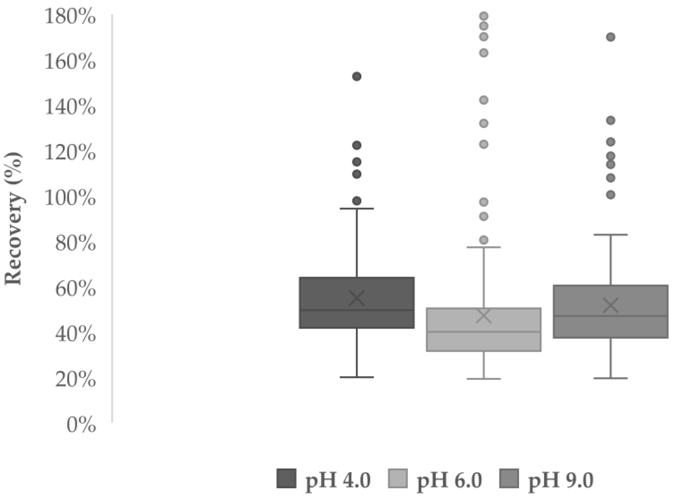
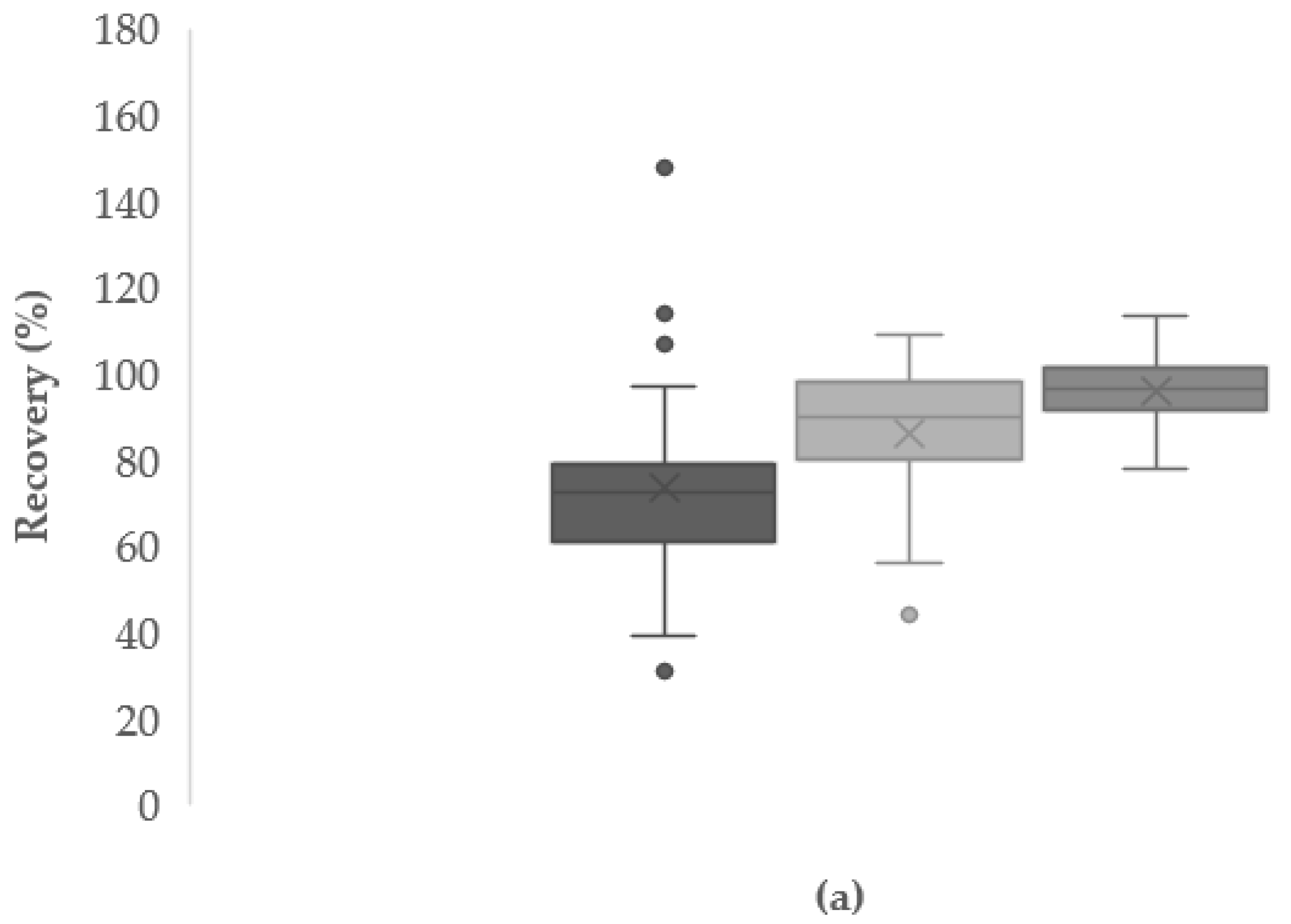
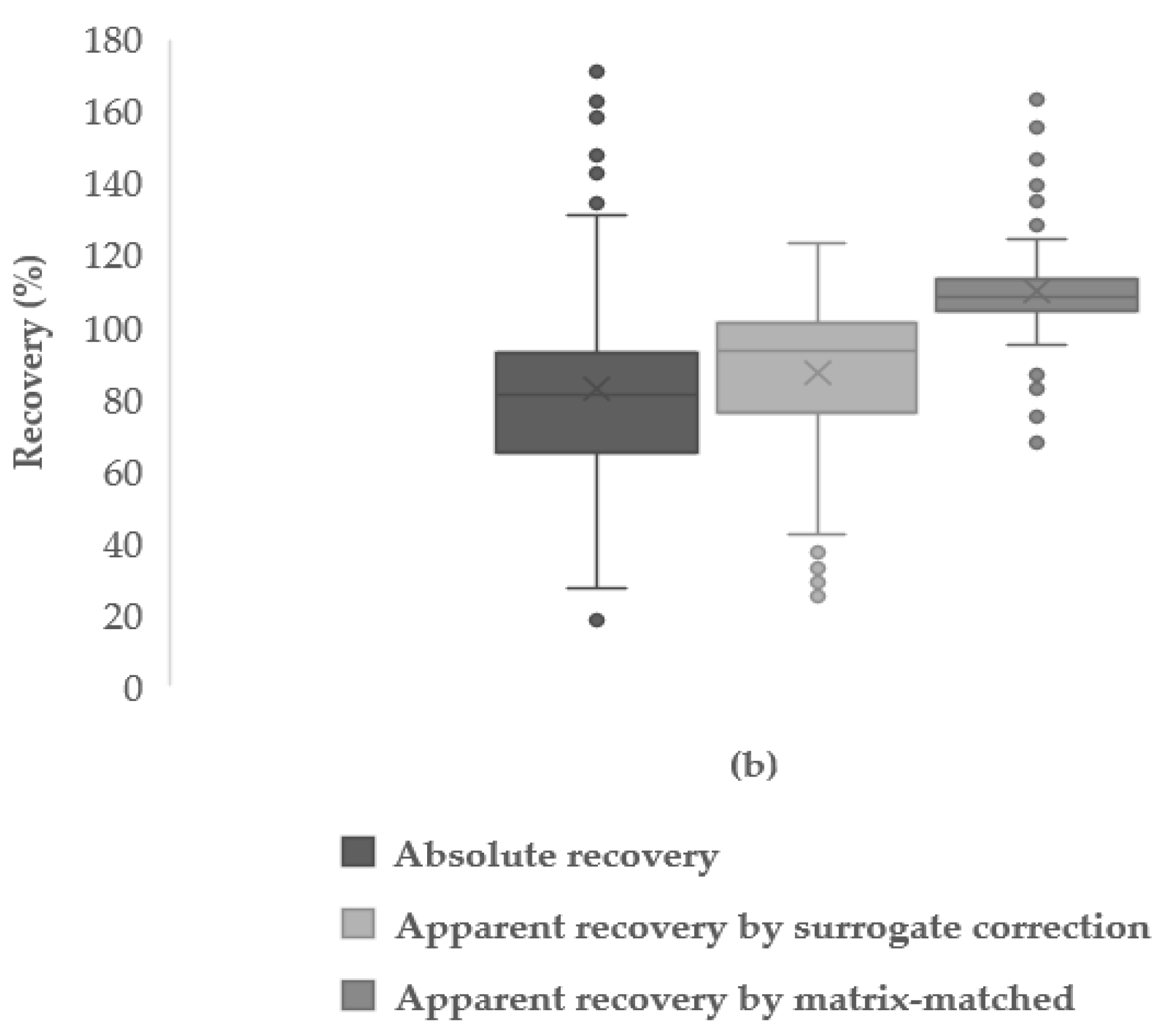
3.4.3. Absolute and Apparent Recoveries

Absolute recoveries (%) were determined as the ratio of the concentrations calculated from the external calibration and the spiked concentration. As for the apparent recoveries, those were calculated using two different approaches: (i) correction using isotopically labelled surrogates and (ii) matrix-matched calibration. The correction using surrogates was applied to each absolute recovery value with every surrogate, and the corrected apparent recovery closest to 100% with the lowest RSD value was chosen for each target analytes when possible. Which surrogate was chosen for each analyte whenever possible is also collected in Supplementary Table S2. In the case of the matrix-matched calibration, the apparent recoveries were determined by the ratio of the concentrations calculated from the curve and the spiked concentration.
Absolute and apparent recoveries obtained at low (10 ng/g) and high (40 ng/g) concentrations are shown as box-plots in Figure 2a,b, respectively, while the results for each analyte are gathered in Supplementary Table S2. Recoveries between 70% and 130% were considered as satisfactory while semi-quantitative analysis was considered for compounds out of this recovery range.
At low concentration (Figure 2a), absolute recoveries were properly determined for 88 analytes (36%), since most of them showed values below 70% (mean value 73%). As for the apparent recoveries, considerably better results were obtained. With surrogate correction, absolute recoveries for 127 analytes (52%) were corrected with the mean value of the apparent recoveries being 86% while with matrix-matched calibration, apparent recoveries for 210 (86%) were properly determined (mean value 96%).
At high concentration (Figure 2b), higher absolute recoveries were obtained since 119 analytes were within the satisfactory range (mean 83%). With surrogate correction, the apparent recoveries for 140 analytes were acceptable (mean value 87%) and using matrix-matched good apparent recoveries between 70% and 130% (mean 110%), were obtained for 220 analytes.
Comparing the apparent recoveries obtained from both strategies, matrix-matched calibration rendered better results than surrogate correction. In order to improve the results obtained using surrogate correction, a larger number of labelled standards is necessary. In this sense, even though matrix-matched calibration requires more lab work, it is more profitable since expensive surrogates can be avoided. On the downside, the sample in which matrix-matched calibration curves are built can differ from the samples analysed and this drawback can be avoided when proper surrogates are available for each target compound.

3.4.4. Instrumental and Procedural Repeatability

Instrumental and procedural repeatability were calculated in terms of RSD of the three injections of the 50 ng/g point from the external calibration and the three replicates analysed at each spiked concentration-level, respectively. For the procedural repeatability, RSD values at low and high concentrations were determined in the case of both absolute and apparent recoveries (four repeatability values for each compound). Since apparent recoveries were calculated by surrogate correction or matrix-matched, the highest value (worst scenario) was set for each analyte in each case. The results are gathered at Table 3 (see the specific values for each analyte at Supplementary Table S2).
As can be seen in Table 3 above, most analytes provided instrumental RSD values lower than 10%, showing the repetitiveness of the UHPLC-qOrbitrap measurements. Considering procedural values, higher RSDs were obtained as expected. Nevertheless, the repeatability was still acceptable with most analytes rendering values below 20%. Moreover, slightly better results were obtained at high concentrations as foreseen.
In a similar multi-target work where 200 xenobiotics were analysed with UHPLC-qOrbitrap, also RSD values lower than 20% were obtained for instrumental repeatability [45]. For procedural repeatability, also RSD values below 20% have been obtained while determining xenobiotics in milk samples, even though no other work has analysed more than 200 compounds [17,23,25,36].

3.4.5. Instrumental and Procedural Limits of Identification (LOIs)

For the calculation of LOIs, the external and matrix-matched calibration points (instrumental, LOIinst, and procedural, LOIproc, values, respectively) were considered under a suspect screening approach using Compound Discoverer 3.1 (see Section 3.5). The lowest concentration values where the analytes could be identified were set as the LOIs (see Supplementary Table S2).
Overall, the LOIs for most compounds were below 10 ng/g. Regarding instrumental LOIs, analytes with higher values than 50 ng/g were naproxen, indomethacin, meclocycline, ethion, 2,6-di-tert-butyl-4-(dimethylaminomethyl)phenol, benzothiazole, fenthion, ciprofloxacin, enoxacin, pendimethalin, oryzalin, danofloxacin, enrofloxacin, glycitin, genistin, celecoxib, and hydrochlorothiazide. As for the procedural LOIs, the following analytes provided values higher than 20 ng/g: bis(methylglycol)phthalate, acyclovir, sotalol, captopril, naproxen, pyrantel, DOP, ethion, indomethacin, ranitidine, meclocycline, montelukast, enoxacin, erythromycin, oryzalin, 2,6-di-tert-butyl-4-(dimethylaminomethyl)phenol, hydroxychloroquine, and glycitin.
Since very few works performing suspect screening or non-target analysis of water-soluble organic xenobiotics in milk are available, identification limits are poorly investigated. Some works use limits of decision to set minimum concentration values at which xenobiotics need to be present in to identify them [31]. However, this parameter is calculated following the EU 2002/657/EC regulation [50] that does not require suspect screening approaches to set the values. Some works have determined LOIs for these kind of xenobiotics but in other complex matrices such as swine manure and fish muscle, with the instrumental values being lower than 30 ng/g [45,51].

3.5. Target Analysis of Commercial and Breast Milk Samples

Target analysis was performed in three commercial (whole bovine milk, raw bovine milk and freeze-dried milk powder) and four breast milk (labelled A, B, C and D) samples, following the details mentioned in Section 2.6. The concentration values were corrected by surrogates or matrix-matched whenever possible, taking into account the results from the validation, and expressed with 95% confidence level (2s, s being the standard deviation of the 3 procedural replicates). Only results that provided lower RSD values than 35% are shown in the Table 4 (commercial milk) and Table 5 (breast milk).
Although several compounds were detected in treated whole and untreated bovine milk samples, most of them were below LOQs, except for acetaminophen (anti-inflammatory drug) and DOP (plasticizer) in the untreated milk, which have been previously determined in bovine milk [52,53]. In the freeze-dried milk powder samples, instead, several diverse compounds were quantified such as, benzothiazole (food additive), caffeine (stimulant), genistein, genistin and glycitin (phytoestrogens) and 2-ethylhexyl-4-dimethylaminobenzoate ((EHDAB, UV filter).
As far as breast milk samples are concerned, caffeine was quantified at high concentration (33–1011 ng/g) in all of the samples, which was not surprising since it is common in several foods and drinks that are regularly taken by humans through the diet, and it has been detected in breast milk in other works as well [25,36]. Moreover, compounds such as DEHP and DOP (plasticizers) were quantified in half of the samples analysed, while benzophenone and EHDAB (UV filters), orlistat (pharmaceutical), caprolactam and triethyl phosphate (industrial chemicals) and benzothiazole (food additive) were punctually detected. Several other xenobiotics were detected below LOQs.

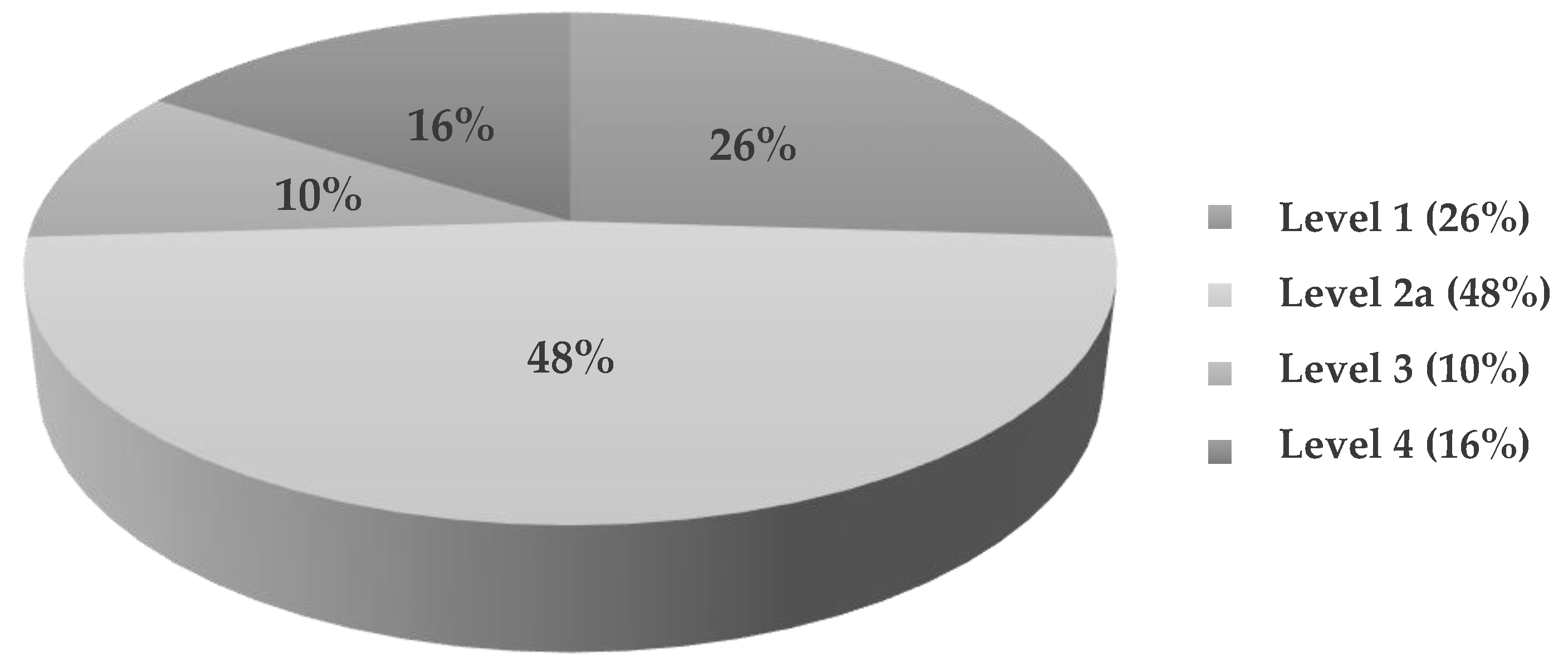
3.6. Suspect Analysis of Commercial and Breast Milk Samples

Suspect screening was performed under the conditions described in Section 2.6, which allowed the identification of 50 xenobiotics of diverse origin (natural or artificial) at different identification levels [54] in the milk samples. The distribution of the confidence levels for the xenobiotics’ identification is represented in Figure 3.
Among the identified compounds, 13 (26%) were identified at level 1 by confirmation via pure standard, precisely, the compounds quantified via target analysis. A wide variety of xenobiotics (24%) were confirmed at level 2a since library data from mzCloud was used for MS2 confirmation. Some ubiquitous xenobiotics in commercial milk samples were 4-indolecarbaldehyde (plant metabolite), bis(2-ethylhexyl) amine (surfactant), isoquinoline (alkaloid) or saccharin (artificial sweetener), while in breast milk samples was frequent to find xenobiotics such as 2-(8-hydroxy-4a,8-dimethyldecahydro-2-naphthalenyl)acrylic acid (sesquiterpene), 2,5-di-tert-butylhydroquinone (industrial chemical), 4-indolecarbaldehyde (algal metabolite), avobenzone (UV filter), piperine (alkaloid), saccharin (artificial sweetener), or shogaol (plant component).
Several other xenobiotics like indoles or quinolones were identified in commercial and breast milks but it was not possible to differentiate between isomers (level 3, 10%). Indoles are industrially used as synthetic favouring compounds for perfume fixative roles, or they can be directly present in tobacco smoke [55]. As for the quinolones, they are broad-spectrum antibiotics which have been previously detected in breast milk samples [56]. For other detected xenobiotics, it was not possible to annotate their MS2 (level 4, 16%).
As it has been observed, some xenobiotics were derived from plants, and are likely less harmful than the artificial ones. All the xenobiotics identified at each level are collected in Table 6 indicating the ones with natural origin, while the extracted ion chromatograms (EICs) of xenobiotics identified at level 1 and 2a in each commercial and breast milk sample are gathered in Supplementary Figures S1–S9.

4. Conclusions

In this work, a multi-target method able to determine more than 200 xenobiotics in commercial and breast milk samples was developed, which was later extended to perform suspect screening using a database that contains almost 18,000 xenobiotic compounds. Although in most works dealing with contaminants in milk samples, mainly non-polar compounds are assessed, in this work, the monitoring of compounds with a wide range of polarities was carried out. Bearing in mind that aim, the optimal conditions for the analytical procedure were set to detect as many compounds as possible in the milk samples with an efficient lipid and protein removal.
In the optimized procedure, the importance of salts, such as anhydrous Na2SO4 and NaCl, was highlighted, which not only made the procedure straightforward due to moisture removal, but also enhanced the recoveries because of the salting-out effect. Moreover, Captiva ND-Lipids successfully removed co-extracted lipids and proteins ensuring high recoveries for the rest of analytes, without the need of a further clean-up using SPE HLB cartridges.
The optimal procedure to determine xenobiotics in milk was validated at two concentration levels by spiking the milk samples with the target 245 analytes at 10 ng/g and 40 ng/g. Most analytes (220) were validated at high concentration showing apparent recoveries between 70% and 130% that were determined by surrogate correction and/or matrix-matched calibration. As for the rest of figures of merit, satisfactory results were obtained considering the complexity of the matrix, the wide variety of analytes and the small sample amount (1 mL) used. However, the use of isotopically labelled compounds should be further studied in the future since only five surrogates were used in this work.
The optimized multi-target procedure was successfully applied to quantify xenobiotics in 3 different commercial milk samples: treated and untreated raw bovine milk and freeze-dried milk powder. The liquid milk samples turned out to be almost free of the targeted xenobiotics since only acetaminophen (anti-inflammatory) and DOP (plasticizer) were quantified in the raw milk samples. However, several compounds were quantified in the milk powder, such as benzothiazole (food additive), caffeine (stimulant), genistein, genistin and glycitin (phytoestrogens), and EHDAB (UV filter).
Regarding breast milk samples, even though caffeine (stimulant) was quantified at the highest concentration in all of the samples, other xenobiotics such as DEHP and DOP (plasticizers), benzophenone and EHDAB (UV filters) were also found. Apart from those xenobiotics, a wide variety of other xenobiotics that were not included in the target list were identified by suspect screening at different confidence-levels including personal and pharmaceutical products, food additives, and disinfectants.
Although the presence of non-polar compounds is often expected in milk samples, the screening of compounds with a broad range of polarities is advisable in biomonitoring programs in order to have more information about the exposome. The suitability of the multi-target and suspect screening method for the analysis of milk samples opens the opportunity to extend the optimized method to other biological fluids such as blood or urine, to get a more holistic understanding of the exposome and find out the relationship between exposure to xenobiotics and adverse health effects.

Supplementary Materials

The following are available online at https://0-www-mdpi-com.brum.beds.ac.uk/2297-8739/8/2/14/s1, Figure S1: Extracted ion chromatograms (EICs) of the identified compounds at confidence level 1 and 2a in commercial treated milk by suspect screening in positive ionization modes, Figure S2: Extracted ion chromatograms (EICs) of the identified compounds at confidence level 1 and 2a in commercial raw milk by suspect screening in positive ionization modes, Figure S3: Extracted ion chromatograms (EICs) of the identified compounds at confidence level 1 and 2a in commercial powder milk by suspect screening in positive ionization modes, Figure S4: Extracted ion chromatograms (EICs) of the identified compounds at confidence level 1 in breast milk A by suspect screening in positive ionization modes, Figure S5: Extracted ion chromatograms (EICs) of the identified compounds at confidence level 2a in breast milk A by suspect screening in positive ionization modes, Figure S6: Extracted ion chromatograms (EICs) of the identified compounds at confidence level 1 and 2a in breast milk B by suspect screening in positive ionization modes, Figure S7: Extracted ion chromatograms (EICs) of the identified compounds at confidence level 1 in breast milk C by suspect screening in positive ionization modes, Figure S8: Extracted ion chromatograms (EICs) of the identified compounds at confidence level 2a in breast milk C by suspect screening in positive ionization modes, Figure S9: Extracted ion chromatograms (EICs) of the identified compounds at confidence level 1 and 2a in breast milk D by suspect screening in positive ionization modes, Table S1: Information and physicochemical properties of the 245 target analytes, Table S2: Validation results for the target 245 analytes: instrumental (UHPLC-qOrbitrap) and procedural (Vortex—Captiva ND-Lipids—UHPLC-qOrbitrap) figures of merit.

Author Contributions

Conceptualization, M.O. and O.Z.; methodology, M.M., M.O., and A.P.; software, M.I. and O.Z.; validation, M.M., and M.O.; formal analysis, M.M. and B.G.-G.; investigation, M.I., M.O., and O.Z.; resources, N.E.; data curation, M.O. and O.Z.; writing—original draft preparation, M.M.; writing—review and editing, M.M., M.O., and O.Z.; visualization, B.G.-G.; supervision, M.O. and O.Z.; project administration, N.E.; funding acquisition, N.E. All authors have read and agreed to the published version of the manuscript.

Funding

This research was funded by the Agencia Estatal de Investigación (AEI) of Spain, the European Regional Development Fund through projects CTM2017-84763-C3-1-R and CTM2017-90890-REDT (AEI/FEDER, EU) and the Basque Government through the financial support as consolidated group of the Basque Research System (IT1213-19). B.G. acknowledges a Juan de la Cierva-Formación fellowship by the Spanish Ministry for the Economy, Industry and Competitiveness (MINECO).

Institutional Review Board Statement

The study was conducted according to the guidelines of the Declaration of Helsinki, and approved by the Ethics Committee of the University of the Basque Country (CEISH-UPV/EHU, BOPV 32, 17/2/2014, M10_2020_230).

Informed Consent Statement

Informed consent was obtained from all subjects involved in the study.

Conflicts of Interest

The authors declare no conflict of interest. The funders had no role in the design of the study; in the collection, analyses, or interpretation of data; in the writing of the manuscript, or in the decision to publish the results.

References

  1. Aceña, J.; Heuett, N.; Gardinali, P.; Pérez, S. Chapter 12: Suspect screening of pharmaceuticals and related bioactive compounds, their metabolites and their transformation products in the aquatic environment, biota and humans using LC-HR-MS techniques. In Applications of Time-of-Flight and Orbitrap Mass Spectrometry in Environmental, Food, Doping, and Forensic Analysis; Pérez, S., Eichhorn, P., Barceló, D., Eds.; Elsevier: Amsterdam, The Netherlands, 2016; pp. 357–378. [Google Scholar]
  2. Chiaia-Hernandez, A.C.; Krauss, M.; Hollender, J. Screening of lake sediments for emerging contaminants by liquid chromatography atmospheric pressure photoionization and electrospray ionization coupled to high resolution mass spectrometry. Environ. Sci. Technol. 2013, 47, 976–986. [Google Scholar] [CrossRef] [PubMed]
  3. Rappaport, S.M. Implications of the exposome for exposure science. J. Expo. Sci. Environ. Epidemiol. 2011, 21, 5–9. [Google Scholar] [CrossRef] [PubMed]
  4. Wild, C.P. Complementing the genome with an “exposome”: The outstanding challenge of environmental exposure measurement in molecular epidemiology. Cancer Epidemiol. Biomark. Prev. 2005, 14, 1847–1850. [Google Scholar] [CrossRef] [PubMed] [Green Version]
  5. Dennis, K.K.; Jones, D.P. The exposome: A new frontier for education. Am. Biol. Teach. 2016, 78, 542–548. [Google Scholar] [CrossRef] [PubMed] [Green Version]
  6. Cui, Y.; Balshaw, D.M.; Kwok, R.K.; Thompson, C.L.; Collman, G.W.; Birnbaum, L.S. The exposome: Embracing the complexity for discovery in environmental health. Environ. Health Perspect. 2016, 124, A137–A140. [Google Scholar] [CrossRef] [Green Version]
  7. Escher, B.I.; Hackermüller, J.; Polte, T.; Scholz, S.; Aigner, A.; Altenburger, R.; Böhme, A.; Bopp, S.K.; Brack, W.; Busch, W.; et al. From the exposome to mechanistic understanding of chemical-induced adverse effects. Environ. Int. 2017, 99, 97–106. [Google Scholar] [CrossRef]
  8. Patel, C.J.; Bhattacharya, J.; Butte, A.J. An Environment-Wide Association Study (EWAS) on type 2 diabetes mellitus. PLoS ONE 2010, 5, e10746. [Google Scholar] [CrossRef]
  9. Vineis, P.; Chadeau-Hyam, M.; Gmuender, H.; Gulliver, J.; Herceg, Z.; Kleinjans, J.; Kogevinas, M.; Kyrtopoulos, S.; Nieuwenhuijsen, M.; Phillips, D.H.; et al. The exposome in practice: Design of the EXPOsOMICS project. Int. J. Hyg. Environ. Health 2017, 220, 142–151. [Google Scholar] [CrossRef]
  10. Andra, S.S.; Austin, C.; Patel, D.; Dolios, G.; Awawda, M.; Arora, M. Trends in the application of high-resolution mass spectrometry for human biomonitoring: An analytical primer to studying the environmental chemical space of the human exposome. Environ. Int. 2017, 100, 32–61. [Google Scholar] [CrossRef] [Green Version]
  11. Exposure and Health. HBM4EU—Science and Policy for a Healthy Future. Available online: https://www.hbm4eu.eu/the-project/exposure-and-health/ (accessed on 2 July 2020).
  12. Yusa, V.; Millet, M.; Coscolla, C.; Roca, M. Analytical methods for human biomonitoring of pesticides. A review. Anal. Chim. Acta 2015, 891, 15–31. [Google Scholar] [CrossRef]
  13. Siddique, S.; Kubwabo, C.; Harris, S.A. A review of the role of emerging environmental contaminants in the development of breast cancer in women. Emerg. Contam. 2016, 2, 204–219. [Google Scholar] [CrossRef]
  14. García Lara, N.R.; Peña Caballero, M. Riesgos asociados al uso no controlado de la leche materna donada. An. Pediatría 2017, 86, 237–239. [Google Scholar] [CrossRef] [PubMed]
  15. Irazusta, A. High-pressure homogenization and high hydrostatic pressure processing of human milk: Preservation of immunological components for human milk banks. J. Dairy Sci. 2020, 103, 14. [Google Scholar] [CrossRef] [PubMed]
  16. Stefanidou, M.; Maravelias, C.; Spiliopoulou, C. Human exposure to endocrine disruptors and breast milk. Endocr. Metab. Immune Disord. Drug Targets 2009, 9, 269–276. [Google Scholar] [CrossRef]
  17. Jadhav, M.R.; Pudale, A.; Raut, P.; Utture, S.; Shabeer, T.A.; Banerjee, K. A unified approach for high-throughput quantitative analysis of the residues of multi-class veterinary drugs and pesticides in bovine milk using LC-MS/MS and GC–MS/MS. Food Chem. 2019, 272, 292–305. [Google Scholar] [CrossRef]
  18. Beyene, T. Veterinary drug residues in food-animal products: Its risk factors and potential effects on public health. J. Vet. Sci. Technol. 2015, 7. [Google Scholar] [CrossRef]
  19. Díaz, R.; Ibáñez, M.; Sancho, J.V.; Hernández, F. Target and non-target screening strategies for organic contaminants, residues and illicit substances in food, environmental and human biological samples by UHPLC-QTOF-MS. Anal. Methods 2012, 4, 196–209. [Google Scholar] [CrossRef] [Green Version]
  20. Plassmann, M.M.; Brack, W.; Krauss, M. Extending analysis of environmental pollutants in human urine towards screening for suspected compounds. J. Chromatogr. A 2015, 1394, 18–25. [Google Scholar] [CrossRef]
  21. Nickerson, K. Environmental contaminants in breast milk. J. Midwifery Women Health 2006, 51, 26–34. [Google Scholar] [CrossRef]
  22. Cano-Sancho, G.; Alexandre-Gouabau, M.-C.; Moyon, T.; Royer, A.-L.; Guitton, Y.; Billard, H.; Darmaun, D.; Rozé, J.-C.; Boquien, C.-Y.; Le Bizec, B.; et al. Simultaneous exploration of nutrients and pollutants in human milk and their impact on preterm infant growth: An integrative cross-platform approach. Environ. Res. 2020, 182, 109018. [Google Scholar] [CrossRef]
  23. Tran, C.D.; Dodder, N.G.; Quintana, P.J.E.; Watanabe, K.; Kim, J.H.; Hovell, M.F.; Chambers, C.D.; Hoh, E. Organic contaminants in human breast milk identified by non-targeted analysis. Chemosphere 2020, 238, 124677. [Google Scholar] [CrossRef] [PubMed]
  24. Rodríguez-Gómez, R.; Jiménez-Díaz, I.; Zafra-Gómez, A.; Ballesteros, O.; Navalón, A. A multiresidue method for the determination of selected endocrine disrupting chemicals in human breast milk based on a simple extraction procedure. Talanta 2014, 130, 561–570. [Google Scholar] [CrossRef] [PubMed]
  25. Baduel, C.; Mueller, J.F.; Tsai, H.; Gomez Ramos, M.J. Development of sample extraction and clean-up strategies for target and non-target analysis of environmental contaminants in biological matrices. J. Chromatogr. A 2015, 1426, 33–47. [Google Scholar] [CrossRef]
  26. Adenuga, A.A.; Ayinuola, O.; Adejuyigbe, E.A.; Ogunfowokan, A.O. Biomonitoring of phthalate esters in breast-milk and urine samples as biomarkers for neonates’ exposure, using modified quechers method with agricultural biochar as dispersive solid-phase extraction absorbent. Microchem. J. 2020, 152, 104277. [Google Scholar] [CrossRef]
  27. Pourchet, M.; Debrauwer, L.; Klanova, J.; Price, E.J.; Covaci, A.; Caballero-Casero, N.; Oberacher, H.; Lamoree, M.; Damont, A.; Fenaille, F.; et al. Suspect and non-targeted screening of chemicals of emerging concern for human biomonitoring, environmental health studies and support to risk assessment: From promises to challenges and harmonisation issues. Environ. Int. 2020, 139, 105545. [Google Scholar] [CrossRef]
  28. Shishov, A.; Nechaeva, D.; Bulatov, A. HPLC-MS/MS determination of non-steroidal anti-inflammatory drugs in bovine milk based on simultaneous deep eutectic solvents formation and its solidification. Microchem. J. 2019, 150, 104080. [Google Scholar] [CrossRef]
  29. Herrera-Herrera, A.V.; Hernández-Borges, J.; Rodríguez-Delgado, M.Á. Fluoroquinolone antibiotic determination in bovine, ovine and caprine milk using solid-phase extraction and high-performance liquid chromatography-fluorescence detection with ionic liquids as mobile phase additives. J. Chromatogr. A 2009, 1216, 7281–7287. [Google Scholar] [CrossRef]
  30. Meng, Z.; Shi, Z.; Liang, S.; Dong, X.; Li, H.; Sun, H. Residues investigation of fluoroquinolones and sulphonamides and their metabolites in bovine milk by quantification and confirmation using ultra-performance liquid chromatography–tandem mass spectrometry. Food Chem. 2015, 174, 597–605. [Google Scholar] [CrossRef]
  31. Kantiani, L.; Farré, M.; Barceló, D. Rapid residue analysis of fluoroquinolones in raw bovine milk by online solid phase extraction followed by liquid chromatography coupled to tandem mass spectrometry. J. Chromatogr. A 2011, 1218, 9019–9027. [Google Scholar] [CrossRef]
  32. Lopes, B.R.; Barreiro, J.C.; Cass, Q.B. Bioanalytical challenge: A review of environmental and pharmaceuticals contaminants in human milk. J. Pharm. Biomed. Anal. 2016, 130, 318–325. [Google Scholar] [CrossRef]
  33. Jiménez-Díaz, I.; Vela-Soria, F.; Rodríguez-Gómez, R.; Zafra-Gómez, A.; Ballesteros, O.; Navalón, A. Analytical methods for the assessment of endocrine disrupting chemical exposure during human fetal and lactation stages: A review. Anal. Chim. Acta 2015, 892, 27–48. [Google Scholar] [CrossRef] [PubMed]
  34. Devanathan, G.; Subramanian, A.; Sudaryanto, A.; Takahashi, S.; Isobe, T.; Tanabe, S. Brominated flame retardants and polychlorinated biphenyls in human breast milk from several locations in India: Potential contaminant sources in a municipal dumping site. Environ. Int. 2012, 39, 87–95. [Google Scholar] [CrossRef] [PubMed]
  35. Asante, K.A.; Adu-Kumi, S.; Nakahiro, K.; Takahashi, S.; Isobe, T.; Sudaryanto, A.; Devanathan, G.; Clarke, E.; Ansa-Asare, O.D.; Dapaah-Siakwan, S.; et al. Human exposure to PCBs, PBDEs and HBCDs in Ghana: Temporal variation, sources of exposure and estimation of daily intakes by infants. Environ. Int. 2011, 37, 921–928. [Google Scholar] [CrossRef] [PubMed]
  36. López-García, E.; Mastroianni, N.; Postigo, C.; Valcárcel, Y.; González-Alonso, S.; Barceló, D.; López de Alda, M. Simultaneous LC–MS/MS determination of 40 legal and illegal psychoactive drugs in breast and bovine milk. Food Chem. 2018, 245, 159–167. [Google Scholar] [CrossRef] [PubMed]
  37. Calafat, A.M.; Slakman, A.R.; Silva, M.J.; Herbert, A.R.; Needham, L.L. Automated solid phase extraction and quantitative analysis of human milk for 13 phthalate metabolites. J. Chromatogr. B 2004, 805, 49–56. [Google Scholar] [CrossRef] [PubMed]
  38. Malarvannan, G.; Kunisue, T.; Isobe, T.; Sudaryanto, A.; Takahashi, S.; Prudente, M.; Subramanian, A.; Tanabe, S. Organohalogen compounds in human breast milk from mothers living in Payatas and Malate, the Philippines: Levels, accumulation kinetics and infant health risk. Environ. Pollut. 2009, 157, 1924–1932. [Google Scholar] [CrossRef]
  39. Jamin, E.L.; Bonvallot, N.; Tremblay-Franco, M.; Cravedi, J.-P.; Chevrier, C.; Cordier, S.; Debrauwer, L. Untargeted profiling of pesticide metabolites by LC-HRMS: An exposomics tool for human exposure evaluation. Anal. Bioanal. Chem. 2014, 406, 1149–1161. [Google Scholar] [CrossRef]
  40. Gago-Ferrero, P.; Schymanski, E.L.; Hollender, J.; Thomaidis, N.S. Chapter 13—Nontarget analysis of environmental samples based on Liquid Chromatography Coupled to High Resolution Mass Spectrometry (LC-HRMS). In Comprehensive Analytical Chemistry; Pérez, S., Eichhorn, P., Barceló, D., Eds.; Elsevier: Amsterdam, The Netherlands, 2016; pp. 381–403. [Google Scholar] [CrossRef]
  41. Hermo, M.P.; Nemutlu, E.; Kır, S.; Barrón, D.; Barbosa, J. Improved determination of quinolones in milk at their MRL levels using LC–UV, LC–FD, LC–MS and LC–MS/MS and validation in line with regulation 2002/657/EC. Anal. Chim. Acta 2008, 613, 98–107. [Google Scholar] [CrossRef]
  42. Gross, J.H. Mass spectrometry. A textbook. Anal. Bioanal. Chem. 2005, 381, 1319–1320. [Google Scholar]
  43. Cortéjade, A.; Kiss, A.; Cren, C.; Vulliet, E.; Buleté, A. Development of an analytical method for the targeted screening and multi-residue quantification of environmental contaminants in urine by liquid chromatography coupled to high resolution mass spectrometry for evaluation of human exposures. Talanta 2016, 146, 694–706. [Google Scholar] [CrossRef]
  44. Menger, F.; Ahrens, L.; Wiberg, K.; Gago-Ferrero, P. Suspect screening based on market data of polar halogenated micropollutants in river water affected by wastewater. J. Hazard. Mater. 2021, 401, 123377. [Google Scholar] [CrossRef]
  45. Musatadi, M.; González-Gaya, B.; Irazola, M.; Prieto, A.; Etxebarria, N.; Olivares, M.; Zuloaga, O. Focused ultrasound-based extraction for target analysis and suspect screening of organic xenobiotics in fish muscle. Sci. Total Environ. 2020, 740, 139894. [Google Scholar] [CrossRef] [PubMed]
  46. Blanco-Zubiaguirre, L.; Zabaleta, I.; Usobiaga, A.; Prieto, A.; Olivares, M.; Zuloaga, O.; Elizalde, M.P. Target and suspect screening of substances liable to migrate from food contact paper and cardboard materials using liquid chromatography-high resolution tandem mass spectrometry. Talanta 2020, 208, 120394. [Google Scholar] [CrossRef] [PubMed]
  47. Weiss, J.M.; Simon, E.; Stroomberg, G.J.; de Boer, J.; de Boer, R.; van der Linden, S.C.; Leonards, P.E.G.; Lamoree, M.H. Identification strategy for unknown pollutants using high-resolution mass spectrometry: Androgen-disrupting compounds identified through effect-directed analysis. Anal. Bioanal. Chem. 2011, 400, 3141–3149. [Google Scholar] [CrossRef] [PubMed] [Green Version]
  48. Agilent Captiva ND and Captiva ND Lipids Method Guide. Available online: https://www.crawfordscientific.com/media/wysiwyg//Literature/Sample_Prep/Agilent/Captiva-ND-and-ND-Lipids-Method-Guide.pdf (accessed on 25 April 2019).
  49. Dualde, P.; Pardo, O.; Fernández, S.F.; Pastor, A.; Yusà, V. Determination of four parabens and bisphenols A, F and S in human breast milk using QuEChERS and liquid chromatography coupled to mass spectrometry. J. Chromatogr. B 2019, 1114–1115, 154–166. [Google Scholar] [CrossRef]
  50. Available online: Http://europa.eu.int/eur-lex/pri/en/oj/dat/2002/l_221/l_22120020817en00080036.pdf (accessed on 19 January 2021).
  51. Solliec, M.; Roy-Lachapelle, A.; Sauvé, S. Development of a suspect and non-target screening approach to detect veterinary antibiotic residues in a complex biological matrix using liquid chromatography/high-resolution mass spectrometry. Rapid Commun. Mass Spectrom. 2015, 29, 2361–2373. [Google Scholar] [CrossRef]
  52. Peng, T.; Zhu, A.-L.; Zhou, Y.-N.; Hu, T.; Yue, Z.-F.; Chen, D.-D.; Wang, G.-M.; Kang, J.; Fan, C.; Chen, Y.; et al. Development of a simple method for simultaneous determination of nine subclasses of non-steroidal anti-inflammatory drugs in milk and dairy products by ultra-performance liquid chromatography with tandem mass spectrometry. J. Chromatogr. B 2013, 933, 15–23. [Google Scholar] [CrossRef]
  53. Lin, J.; Chen, W.; Zhu, H.; Wang, C. Determination of free and total phthalates in commercial whole milk products in different packaging materials by gas chromatography-mass spectrometry. J. Dairy Sci. 2015, 98, 8278–8284. [Google Scholar] [CrossRef] [Green Version]
  54. Schymanski, E.L.; Jeon, J.; Gulde, R.; Fenner, K.; Ruff, M.; Singer, H.P.; Hollender, J. Identifying small molecules via high resolution mass spectrometry: Communicating confidence. Environ. Sci. Technol. 2014, 48, 2097–2098. [Google Scholar] [CrossRef]
  55. PubChem. Hazardous Substances Data Bank (HSDB): 599. Available online: https://pubchem.ncbi.nlm.nih.gov/source/hsdb/599#section=Interactions-(Complete) (accessed on 6 December 2020).
  56. Yalçin, S.S.; Güneş, B.; Yalçin, S. Incredible pharmaceutical residues in human milk in a cohort study from Şanlıurfa in Turkey. Environ. Toxicol. Pharmacol. 2020, 80, 103502. [Google Scholar] [CrossRef]
Figure 1. Boxplots of the recoveries (%) for Oasis HLB clean-up at pH 4.0, 6.0 and 9.0, showing minimum and maximum values (whiskers), percentiles (Q1 and Q3), average (x), median (line) and outliers (points).
Separations 08 00014 g001
Figure 2. Absolute and apparent (surrogate correction and matrix-matched) recoveries (%) spiking milk samples at: (a) 10 ng/g and (b) 40 ng/g.
Separations 08 00014 g002aSeparations 08 00014 g002b
Figure 3. Distribution of the identification confidence levels of the xenobiotics by suspect screening.
Separations 08 00014 g003
Table 1. Results of the experiments to optimize the addition of salts.
ExperimentWithout Oasis HLBWith Oasis HLB
Detected (%)Recoveries (%) *Detected (%)Recoveries (%) *
(i)AcN + NaCl8863–92 (80)889–93 (46)
(ii)AcN + Na2SO4 + NaCl9268–110 (90)829–122 (44)
(iii)AcN + MgSO4 + NaCl8450–110 (78)7830–111 (69)
(iv)1. AcN,
2. AcN + MgSO4 + NaCl
8958–101 (81)7926–118 (50)
(v)AcN:Milli-Q water 0.1% EDTA (95:5, v/v)817–85 (47)Not performedNot performed
* Recoveries are represented as the range and the mean recovery (in brackets) of all detected compounds.
Table 2. Matrix-effect (%) at detection.
ExperimentWithout Oasis HLBWith Oasis HLB
Recoveries (%) *Recoveries (%) *
(i)AcN + NaCl85–115 (109)72–108 (97)
(ii)AcN + Na2SO4 + NaCl92–119 (109)81–112 (101)
(iv)1. AcN,
2. AcN + MgSO4 + NaCl
95–128 (114)83–113 (102)
* Recoveries are represented as the range and the mean recovery (in brackets) of all detected compounds.
Table 3. Instrumental and procedural repeatability values in terms of RSD (%) for absolute (abs.) and apparent (app.) recoveries at low and high concentrations.
RepeatabilityConcentrationNumber of Analytes
<10%10–20%20–50%>50%
Instrumental50 ng/g (final extract)234821
Procedural10 ng/g (milk)Abs. rec.18337169
App. rec.19134137
40 ng/g (milk)Abs. rec.19726157
App. rec.19528157
Table 4. Target analysis results: concentration of xenobiotics (95%, 2 s) in commercial milk samples.
CompoundWhole Bovine Milk (ng/g)Raw Bovine Milk (ng/g)Milk Powder (ng/g)
Acetaminophen mn.d3.2 ± 0.4n.d
Benzothiazole an.dn.d11 ± 6
Caffeine cn.dn.d2.0 ± 0.1
EHDAB mn.dn.d0.70 ± 0.04
Genisteinn.dn.d20.1 ± 0.3
Genistinn.dn.d29 ± 1
Glycitinn.dn.d4.7 ± 0.6
2-Hydroxybenzothiazole<LOQn.d<LOQ
Azoxystrobinn.d<LOQn.d
Benzethoniumn.d<LOQn.d
Bicalutamide<LOQ<LOQ<LOQ
BEHAn.d<LOQn.d
BEHPn.d<LOQn.d
Butylparaben<LOQ<LOQ<LOQ
Carbendazim<LOQn.dn.d
Cortisone<LOQ<LOQ<LOQ
Cotinine<LOQn.d<LOQ
Crotamitonn.d<LOQn.d
Diethyl Toluamide<LOQ<LOQ<LOQ
DOP mn.d<LOQn.d
Enrofloxacin<LOQn.dn.d
Fenpropimorph<LOQ<LOQ<LOQ
Finasteriden.dn.d<LOQ
Ifosfamiden.d<LOQ<LOQ
Methylparaben<LOQn.dn.d
Pirimicarb<LOQ<LOQ<LOQ
Pirimiphos-methyln.dn.d<LOQ
Primidone<LOQ<LOQ<LOQ
Progesterone<LOQn.dn.d
Propiconazolen.d<LOQn.d
Pyrazophos<LOQn.dn.d
Sulfamethazine<LOQn.dn.d
Sulfamethoxazole<LOQn.dn.d
Terbutryn<LOQ<LOQ<LOQ
Superscript: (a) [2H5]-atrazine, (c) [13C3]-caffeine, (m) matrix-matched. Data abbreviations: below limit of quantification (<LOQ), non-detected (n.d). Compounds abbreviations: 2-ethylhexyl-4-dimethylaminobenzoate (EHDAB), bis(2-ethylhexyl)adipate (BEHA), bis(2-ethylhexyl)phthalate (BEHP), di-n-octyl phthalate (DOP).
Table 5. Target analysis results: average concentration of xenobiotics (95%, 2 s) in breast milk samples.
CompoundA (ng/g)B (ng/g)C (ng/g)D (ng/g)
Benzophenonen.dn.d4 ± 1n.d
Benzothiazole45 ± 4n.dn.dn.d
BEHP m45 ± 30n.d48 ± 29n.d
Caffeine c988 ± 12735 ± 233 ± 31011 ± 210
Caprolactam27 ± 2n.dn.dn.d
DOP m39 ± 27n.d41 ± 26n.d
EHDAB mn.dn.dn.d0.58 ± 0.01
MBHB c<LOQn.d4 ± 2n.d
Orlistatn.dn.d2.3 ± 0.4n.d
Triethyl phosphaten.dn.d20 ± 8n.d
2,4-Dinitrophenol<LOQn.dn.dn.d
Azoxystrobinn.d<LOQn.dn.d
Benzethoniumn.d<LOQn.dn.d
Bicalutamiden.d<LOQ<LOQ<LOQ
BEHAn.dn.d<LOQn.d
BPA<LOQn.dn.dn.d
Butylparaben<LOQ<LOQ<LOQ<LOQ
Cotinine<LOQ<LOQ<LOQn.d
Crotamiton<LOQn.d<LOQ<LOQ
DBPn.dn.d<LOQn.d
Diethyl Toluamide<LOQn.d<LOQ<LOQ
Exemestanen.dn.d<LOQn.d
Fluvoxaminen.dn.d<LOQn.d
MBP<LOQn.dn.dn.d
Medroxyprogesterone<LOQ<LOQ<LOQn.d
Methylparaben<LOQn.dn.dn.d
Norfloxacinn.dn.d<LOQn.d
Pindololn.d<LOQn.d<LOQ
Pirimicarb<LOQ<LOQ<LOQ<LOQ
Propiconazole<LOQ<LOQ<LOQ<LOQ
Sotalol<LOQn.dn.d<LOQ
Sulfamethoxazole<LOQn.dn.dn.d
Tamoxifen<LOQn.d<LOQn.d
Trimethoprimn.dn.d<LOQn.d
Triphenylphosphate<LOQn.dn.dn.d
Superscript: (c) [13C3]-caffeine, (m) matrix-matched. Data abbreviations: below limit of quantification (<LOQ), non-detected (n.d). Compounds abbreviations: bis(2-ethylhexyl)phthalate (BEHP), di-n-octyl phthalate (DOP), 2-ethylhexyl-4-dimethylaminobenzoate (EHDAB), methyl 3,5-di-tert-butyl-4-hydroxybenzoate (MBHB), bis(2-ethylhexyl)adipate (BEHA), bisphenol A (BPA), dibutyl phthalate (DBP), methylbenzophenone (MBP).
Table 6. Suspect screening of commercial and breast milk: xenobiotics identified at level 1, 2a, 3 and 4.
Commercial MilkBreast Milk
Annotated CompoundMolecular FormulaExact MasstR (min)123ABCD
[M+H]+
Level 1
AcetaminophenC8H9NO2151.063331.77XXXXX
BEHPC24H38O4390.2770115.90XXXXX
BenzophenoneC13H10O182.0731611.75XXXXXX
BenzothiazoleC7H5NS135.014276.50XXXXX
CaffeineC8H10N4O2194.080382.50XX
CaprolactamC6H11NO113.084062.35XXXXXX
DOPC24H38O4390.2770115.90X<LOIX<LOIX<LOIX
EHDABC17H27NO2277.2041814.50XX<LOIXXX<LOI
Genistein *C15H10O5270.052827.55XXXXXX
Genistin *C21H20O10432.105654.30XXXXXX
Glycitin *C22H22O10446.121303.43XX<LOIXXXX
MBHBC16H24O3264.1725613.60XXX<LOIX<LOIX
Triethyl phosphateC6H15O4P182.07085.70XXXXXX
Level 2a
1,2,3,9-tetrahydro-4H-carbazol-4-one oximeC12H12N2O200.094964.99XXXXXX
3-Acetyl-2,5-dimethylfuranC8H10O2138.068083.12XXXXXX
3-amino-2-phenyl-2H-pyrazolo [4,3-c]pyridine-4,6-diolC12H10N4O2242.080385.07XXX
3-Methyl-5-(5,5,8a-trimethyl-2-methylene-7-oxodecahydro-1-naphthalenyl)pentyl acetateC22H36O3348.2664513.70XXX
4-Indolecarbaldehyde *C9H7NO145.052763.05XX
4-Methoxycinnamic acid *C10H10O3178.062997.04XXXXXX
5-Hydroxymethyl-2-furaldehydeC6H6O3126.031691.63XXXXXX
7-Methyl-2-phenylquinoline-4-carboxylic acidC17H13NO2263.0946312.09XXXXX
AmfepramoneC13H19NO205.146665.66XXXXXX
AvobenzoneC20H22O3310.1568914.40XXXXX
Bis(2-ethylhexyl) amineC16H35N241.2769511.34XXXXX
CarvoneC10H14O150.1044711.72XXXXXX
Citroflex A-4C20H34O8402.2253714.00XXXXXX
DidecyldimethylammoniumC22H47N325.3708512.60XXXXXX
Isoquinoline *C9H7N129.057853.80XXXX
NootkatoneC15H22O218.1670712.95XXXXXX
OPEOC16H26O2250.1932813.07XXXX
Piperine *C17H19NO3285.1364911.81XXXX
Shogaol *C17H24O3276.1725410.70XXX
Tetramethylene sulfoxideC4H8OS104.029591.46XXXXXX
Level 3
2-Oxindole/4-Hydroxyindole/
5-Hydroxyindole
C8H7NO133.052761.54XXXX
2-Hydroxyquinoline/
8-Hydroxyquinoline *
C9H7NO145.052763.95XXXXX
1,5-Isoquinolinediol/
2,4-Quinolinediol
C9H7NO2161.047684.17XXXX
1,5-Isoquinolinediol/
2,4-Quinolinediol
C9H7NO2161.047684.48XXXXXX
Paraxanthine/Theophylline/Theobromine *C7H8N4O2180.064731.69XXX
Level 4
2-Oxindole/4-Hydroxyindole/
5-Hydroxyindole/
6-Methylbenzoxazole
C8H7NO133.052764.68X
Pulegone/D,L-Camphor/Citral *C10H16O152.120127.81XXXX
DL-2-(acetylamino)-3-phenylpropanoic acid/
4-morpholinobenzoic acid
C11H13NO3207.089543.86XXXXXX
2-(8-Hydroxy-4a,8-dimethyldecahydro-2-naphthalenyl) acrylic acid/
2-[(2R,4aR,8R,8aR)-8-hydroxy-4a,8-dimethyl-decahydronaphthalen-2-yl] prop-2-enoic acid/Polygodial
C15H24O3252.1725411.83XXXXXX
5-O-Methylgenistein/5,7-dihydroxy-3-(4-methoxyphenyl)-4H-chromen-4-one/Biochanin A/Glycitein/WogoninC16H12O5284.068476.46XXXXXX
1,4a-dimethyl-9-oxo-7-(propan-2-yl)-1,2,3,4,4a,9,10,10a-octahydrophenanthrene-1-carboxylic acid/KahweolC20H26O3314.1881912.45XXXXX
N-Benzylformamide/PhenacylamineC8H9NO135.068413.13XX
[M-H]−
Level 2a
2-(8-Hydroxy-4a,8-dimethyldecahydro-2-naphthalenyl)acrylic acidC15H24O3252.1725410.82XXXXX
2,5-di-tert-ButylhydroquinoneC14H22O2222.1619812.49XXX
Myristyl sulfateC14H30O4S294.1864812.22XXXXX
SaccharinC7H5NO3S182.999011.38X
Level 4
Chrysin/Daidzein/Abietic acidC15H10O4254.057916.09XXXXXX
* Natural origin.
Publisher’s Note: MDPI stays neutral with regard to jurisdictional claims in published maps and institutional affiliations.

