Article

Gadolinium(III)-DOTA Complex Functionalized with BODIPY as a Potential Bimodal Contrast Agent for MRI and Optical Imaging

by
Matthias Ceulemans
,
Koen Nuyts
,
Wim M. De Borggraeve
and
Tatjana N. Parac-Vogt
*
Department of Chemistry, KU Leuven, Celestijnenlaan 200F, Leuven 3001, Belgium
*
Author to whom correspondence should be addressed.
Submission received: 29 September 2015 / Revised: 10 November 2015 / Accepted: 17 November 2015 / Published: 25 November 2015
(This article belongs to the Special Issue Rare Earth and Actinide Complexes)

Abstract

:
The synthesis and characterization of a novel gadolinium(III) DOTA complex functionalized with a boron-dipyrromethene derivative (BODIPY) is described. The assembly of the complex relies on azide diazotransfer chemistry in a copper tube flow reactor. The azide thus formed is coupled directly with an alkyne via click chemistry, resulting into a paramagnetic and luminescent gadolinium(III) complex. Luminescent data and relaxometric properties of the complex have been evaluated, suggesting the potential applicability of the complexes as a bimodal contrast agent for magnetic resonance and optical imaging. The complex displays a bright emission at 523 nm with an absorption maximum of 507 nm and high quantum yields of up to 83% in water. The proton relaxivity of the complex measured at 310 K and at frequencies of 20 and 60 MHz had the values of 3.9 and 3.6 s−1·mM−1, respectively.

Graphical Abstract

1. Introduction

Magnetic resonance imaging (MRI) is a diagnostic tool that has experienced large growth over the past years. Consequently, the search for highly-efficient, responsive, and tissue-specific markers has followed the same trend. Most of the clinically-used contrast agents are based on gadolinium(III) chelates [1,2,3,4,5,6]. Gadolinium(III), with its symmetric 8S7/2 ground state and large magnetic moment (7.94 µB), is a superior ion to efficiently increase relaxation rates of water molecules [1]. Due to the toxicity of free gadolinium(III) (LD50 = 0.2 mmol·kg−1 in mice), and the fact that relative high concentrations of contrast agents are needed for MRI scan (up to 0.3 mmol per kg body weight) [7,8], strong chelating agents are used to ensure kinetic and thermostatic stability of the gadolinium(III) complex. The most widespread contrast agents used in modern molecular imaging techniques are the acyclic diethylenetriaminepentaacetic acid (Gd(III)-DTPA, Magnevist®, Bayer Healthcare, Berlin, Germany) and the cyclic 1,4,7,10-tetraazacyclododecane-1,4,7,10-tetraacetic acid (Gd(III)-DOTA, Dotarem®, Guerbet, Hongkong, China) [2]. The eight-fold coordination ensures high stability (logK = 25.3 and 20.1 for DOTA and DOTA-propylamide respectively), and allows the binding of one water molecule directly to the metal center [9,10,11,12].
MRI excels in its special resolution and depth penetration, but it suffers from a low sensitivity. Combining it with a complimentary imaging technique would allow for obtaining a better diagnostic tool, as it was recently demonstrated by a successful combination of MRI and PET probes [13,14,15]. The combined contrast agent gives both insight into the morphology (MRI) and information about biomedical processes (PET) of the human body. Another promising imaging modality complementary to MRI is optical imaging, which has a very good sensitivity, but lacks the spatial resolution and depth penetration. The latter drawback can be circumvented by choosing the absorption and emission wavelength close to the biological window [16]. Different optical probes have already been coupled to DTPA or DOTA, such as luminescent polymers in lipophilic aggregates [17], luminescent metal complexes [18,19,20,21,22,23,24,25,26,27], dendrimers [28], and organic dyes such as quinolone [29], rhodamine [30,31], fluoresceine [32], and naphalimide [33,34].
A promising class of organic dyes are 4,4-Difluoro-4-bora-3a,4a-diaza-s-indacene (abbreviated as BODIPY) and its derivatives. This class of small organic molecules generally has very high extinction coefficients, fairly sharp fluorescence peaks, and high quantum yields. The BODIPY core is pretty stable in physiological conditions and relatively insensitive to the environment. Their emission wavelength is tunable by increasing the resonance within the BODIPY core [35]. The BODIPY dyes have been used in a variety of applications such as biological labels and probes [36,37,38], fluorescent probes [39], laser dyes [40,41], light emitting diodes [42], solar cells [43,44], and potential sensitizers in photodynamic therapy [45,46]. Although BODIPYs are versatile compounds with good optical properties, their use in developing bimodal contrast agents remain scarce [47,48,49,50,51,52]. 18F labeled BODIPY derivatives have been recently suggested as bifunctional reporters for hybrid optical/positron emission tomography imaging [53,54]. An example of DOTA ligand functionalized with BODIPY compound was reported in 2010 by Bernhard et al. [47] with the idea to generate a bimodal imaging agent for optical and nuclear imaging (PET/CT) by introducing In(III), Ga(III) and Cu(II) into the DOTA moiety. In 2012 the same group expanded their compound to a DOTAGA derivative (GA = glutaric acid) which gave more stable complexes with the transition metals [48,51,52]. Considering the versatility and favorable optical properties of BODIPY organic dyes, there are surprisingly very few examples of its use as an optical probe in the development of bimodal contrast agents. The main disadvantage of BODIPY dyes is their hydrophobicity, which limits their solubility and therefore hinders their straightforward application as a probe for medical imaging. Although overcoming the solubility issue poses a challenge, it is also beneficial as it was demonstrated that hydrophilic dyes can improve cell permeability [50].
We have recently shown that BODIPY dyes can be functionalized in a copper tube flow reactor via chemistry that converts primary amines into azides using the catalyst generated in situ from the metallic copper [55]. In this paper we further expand this strategy for performing azide-alkyne cycloaddition in order to create a Gd-DOTA-BODIPY derivative. We report on the synthesis and characterization of this novel compound and evaluate its potential as a bimodal contrast agent for MRI and optical imaging.

2. Results and Discussion

2.1. Synthesis of Gd-DOTA-BODIPY Derivative

The main concept used in order to create a Gd-DOTA-BODIPY derivative is based on using click chemistry to couple a BODIPY dye to a DOTA moiety, resulting in a bimodal agent with both optical and paramagnetic entities (Figure 1). The novel BODIPY derivative developed in this work is 5,5-difluoro-1,3,7,9-tetramethyl-10-(2-(4-((2-gadolinium(III)-(4,7,10-tris(carboxylatomethyl)-1,4,7,10-tetraazacyclododecan-1-yl)acetamido)methyl)-1H-1,2,3-triazol-1-yl)ethyl)-5H-dipyrrolo[1,2-c:2′,1′-f][1,3,2]diazaborinin-4-ium-5-uide, presented in Figure 1. The synthesis of the BODIPY core starts with the protection of β-analine with phtalic acid in solvent free conditions. Subsequently reaction with thionyl chloride converts the acid group into an acid chloride, which can react further with two equivalent of 2,4-dimethylpyrrole to form a dipyrromethene derivative. Complexation of this product with borontrifluoride dietherate and a base forms BODIPY-(CH2)2-phtalimide. This product can easily be transformed into BODIPY-(CH2)2-amine by using hydrazine in ethanol, which was clearly demonstrated by the disappearance of the aromatic peaks at 7.77 and 7.88 ppm, and appearance of a new peak at 1.61 ppm in the 1H NMR spectrum (Scheme 1). The final BODIPY product contains an amine function which can be transformed in a copper tube into an azide via a diazotranfer reagent (ISA·H2SO4) giving BODIPY-(CH2)2-azide shown in Figure 2a [56,57].
Figure 1. The Gd-DOTA-BODIPY derivative.
Inorganics 03 00516 g001
Scheme 1. Synthetic procedure for BODIPY-(CH2)2-amine: (a) 160 °C, 6 h; (b) SOCl2, reflux, 3 h; (c) 2 equivalents 2,4-dimethylpyrrole, DCM, 0 °C reflux, 4 h; (d) 10 eq. DIPEA, 11 eq. BF3·Et2O, DCM, 0 °C R.T., overnight; and (e) N2H4·H2O, EtOH, reflux, 3 h.
Inorganics 03 00516 g013
The propargylated Ln(III)-DOTA complex (Ln = La, Gd) that was used for coupling to BODIPY dye is shown in Figure 2b [58,59]. The coupling was performed by using a flow chemistry approach, which can mitigate some safety related issues regarding working with azides [60]. Organic azides can be synthesized from primary amines via a diazotransfer reaction. The potential short shelf life and highly explosive nature of some diazotransfer reagents requires their careful consideration of the safety issues for their handling [61]. Some safety issues can be circumvented by introduction of imidazole-1-sulfonyl azide (ISA) or its hydrogen sulfate salt (ISA·H2SO4) [56,62]. The flow reaction was performed by mixing solutions of BODIPY-(CH2)2-amine and ISA·H2SO4, which is graphically represented in Scheme 2. Upon leaving the reactor, the reaction mixture was directly added to a solution of propargylated Ln(III)-DOTA (Ln = La or Gd), resulting in the final product Ln(III)-DOTA-BODIPY.
Figure 2. (a) Structure of BODIPY-(CH2)2-azide made in a copper tube reactor; (b) Structure of propargylated Gd(III)-DOTA complex used to couple to the azide.
Inorganics 03 00516 g002
Scheme 2. Schematic representation of the flow reactor.
Inorganics 03 00516 g014
The Ln-DOTA-BODIPY complexes have been isolated and purified (see Figure S13) and the diamagnetic lanthanum(III) complex La-DOTA-BODIPY has been characterized by 1H NMR spectroscopy in solution (Figure 3). The 1H NMR spectrum shows a distinct peak of the triazole proton at 7.55 ppm and two protons on the BODIPY core at 6.00 ppm, indicating a successful linkage of the DOTA moiety with the BODIPY derivative. The 1H NMR spectrum also shows broad peaks in the region from 2.20–3.82 ppm which are typical for the protons in the DOTA ring [63].
Figure 3. 1H NMR spectrum of the final clicked La-DOTA-BODIPY derivative measured in deuterated water.
Inorganics 03 00516 g003

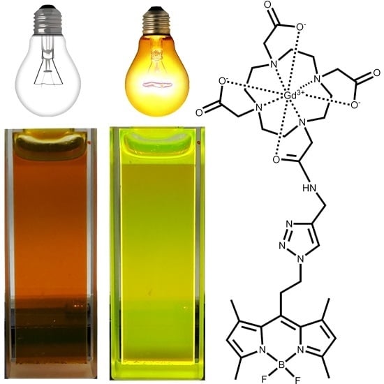
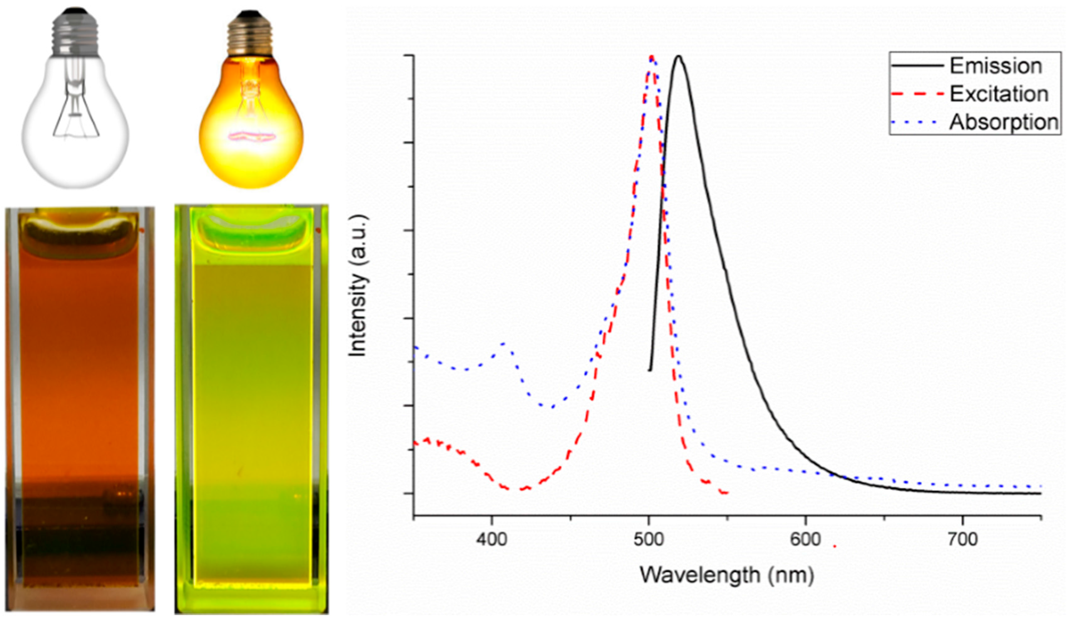
2.2. Photophysical Properties of the Gd-DOTA-BODIPY

The BODIPY derivatives are typically strongly red colored. Most BODIPY derivatives are apolar in nature and dissolve very well in apolar organic solvents, like chloroform or dichloromethane, and will emit a bright green fluorescence. Due to the coupling to hydrophilic Ln-DOTA complex the final Ln-DOTA-BODIPY adducts are water soluble and give bright green fluorescence in aqueous solution. Figure 4 shows a picture of an aqueous solution of the synthesized Gd-DOTA-BODIPY in the absence and in the presence of excitation light at 366 nm. The green fluorescence is clearly visible during excitation. Furthermore, the electronic spectra of Gd-DOTA-BODIPY are given in Figure 4 and depict the characteristic and rather narrow absorption and emission bands which are typical for BODIPY dyes. A main absorption band (blue dotted line) with a maximum λabs(max) at 503 nm is observed for Gd-DOTA-BODIPY in aqueous solution. These visible absorption bands can be assigned to the S0→S1 transition [64]. An additional, considerably weaker broad absorption band is observed in the UV-VIS region around 360 nm, and is attributed to the S0→S2 transition. It should be noted that the optical properties of the BODIPY dyes can be tuned by increasing conjugation through placing different substituents on the BODIPY core. Such substitutions increase the resonance of the whole structure and provide a red shift of the main emission band, which is favorable for biological applications. A tradeoff between tissue penetration and image resolution for in vivo imaging can be made in the optical imaging window (from 665 to 900 nm) [65]. In this region the extinction coefficients of the main sources of absorption such as hemoglobin, deoxyhemoglobin, and water, are at their minimum. However, it must be taken into account that placing additional substituents on the BODIPY usually renders the system more hydrophobic and decreases the solubility of the entire complex in water.
The emission maximum in water of Gd-DOTA-BODIPY (Figure 4) is observed at 523 nm and is in accordance with the previously reported BODIPY derivatives [66]. The excitation maximum (red dashed line) recorded at 560 nm shows a peak at 503 nm.
Figure 4. The emission (black line), excitation (red dashed line) and absorption (blue dotted line) spectrum of Gd-DOTA-BODIPY in water.
Inorganics 03 00516 g004
The fluorescent quantum yields QL of La-DOTA-BODIPY and Gd-DOTA-BODIPY were determined upon ligand excitation by a comparative method, using a solution of Rhodamine 6G (Q = 78%) [67] in water as the standard. The advantage of BODIPY dyes is that the quantum yields of their derivatives are generally high, even in water [39]. The quantum yield was determined according to the following equation:
Q L = Q S × I X I S × A S ( λ e x c ) A X ( λ e x c ) × η X 2 η S 2
In this equation the s and x refer to the standard and the unknown sample respectively, I represents the corrected total integrated emission intensity, A: the absorbance at the excitation wavelength, η: the refractive index of the solution (ηwater = 1.33), and Qs: the quantum yield of the standard (Q = 78%) [67]. The samples are diluted until the absorbance at the excitation wavelength is between 0.02–0.05. Quantum yield is recorded with ligand excitation wavelength of 490 nm and gave values of 80% and 83% for La-DOTA-BODIPY and Gd-DOTA-BODIPY, respectively.

2.3. Relaxivity of Gd-DOTA-BODIPY

The relaxivity r1 is the efficiency of a 1 mM solution of gadolinium(III) agent to shorten the longitudinal relaxation time (T1). According to the Solomon Bloembergen Morgan theory [68,69] several parameters can alter the relaxivity of a contrast agent. High relaxivity can be obtained with a higher amount of water molecules directly bound to the paramagnetic centre (q). Although an easy parameter to adjust, increasing the q value leads to less stable complexes. Other parameters are the distance between gadolinium(III) and water (r), the water residence time (τM), the rotational correlation time of the paramagnetic center (τR), the electronic relaxation time of gadolinium(III) at zero field (τS0) and the correlation time modulating the electronic relaxation (τv). The relaxivity of Gd-DOTA-BODIPY derivative measured at frequencies of 20 and 60 MHz at 310 K and was found to be 3.9 and 3.6 s−1·mM−1 respectively. These values are in close comparison with previously reported values for Gd(III)-DOTA complex, which gave values of 3.5 and 3.1 s−1·mM−1 at 20 and 60 MHz respectively [70]. A slight increase compared to the parent Gd-DOTA complex can be attributed to the increase in molecular weight of the Gd-DOTA-BODIPY complex, resulting in the increase of the rotational correlation time (τR) and in overall increase of the relaxivity. The limiting factors for the increase of Gd-DOTA-BODIPY relaxivity are probably the presence of an amide bond in the DOTA scaffold which may slow down the water exchange rate, or flexible linker used for attaching the BODIPY core to DOTA, which may result in high internal rotations within the Gd-DOTA-BODIPY.

3. Experimental Section

3.1. Materials, Reagents, and Solvents

β-alanine, boron trifluoride diethyl etherate, phosphoryl chloride, thionyl chloride was obtained from Sigma-Aldrich (Bornem, Belgium); Copper(II)sulphate, 2,4-dimethylpyrrole, dry dichloromethane, lanthanum(III) chloride heptahydrate, methanol, sodium ascorbate, triethylamine were purchased from Acros Organics (Geel, Belgium); acetonitrile, diethyl ether, ethyl acetate, petroleum ether, magnesium sulphate from VWR chemicals (Leuven, Belgium); dichloromethane, from hydrogen chloride Fisher Scientific (Loughborough, UK); Gadolinium(III) was obtained from Alfa Aesar (Ward Hill, Shrewsbury, MA, USA) and were used without further purification.

3.2. Characterization

NMR spectra were measured on a Bruker Avance 300 (Bruker, Karlsruhe, Germany) (operating at 300 MHz for 1H NMR spectra, operating at 75 MHz for 13C NMR), Bruker Avance 400 (operating at 400 MHz for 1H NMR spectra, operating at 100 MHz for 13C NMR spectra). Chemical shifts are reported in parts per million (ppm) and are referenced to the internal standard tetramethylsilane. For 13C spectra, residual solvent signals are used as the internal standard. Spectra are taken at room temperature unless otherwise stated. Relaxation rates at 20 and 60 MHz were obtained on a Minispec mq-20, and a Minispec mq-60 (Bruker, Karlsruhe, Germany) respectively.
FT-IR spectra were measured by using a Bruker Vertex 70 FT-IR spectrometer (Bruker, Ettlingen, Germany). ESI-MS measurements were taken on a Thermo Electron LCQ Advantage apparatus (Thermo Scientific, Waltham, MA, USA) with Agilent 1100 pump (Agilent, Waldbronn, Germany) and injection system coupled to Xcalibur data analyzing software. Methanol is used as eluent. TXRF measurements were done on a Bruker S2 Picofox (Bruker, Berlin, Germany) by analyzing approximately 100 ppm gadolinium or europium solutions with respect to a Chem-Lab gallium standard solution (500 µg/mL, 2%–5% HNO3).
The flow setup was constructed with copper GC tubing (0.065′′ inner diameter, Restek, Middelburg, Netherlands) and the reagents are pumped using a Chemyx Fusion 200 syringe pump (Chemyx, Stafford, TX, USA). LC–MS was performed on an Alltech Prevail RP-C18 5 µm 150 mm × 2.1 mm column coupled to an Agilent 1100 degasser, quaternary pump, auto sampler, UV-DAD detector, and thermostated column module coupled to Agilent 6110 single-quadrupole MS. Use of Agilent LC/MSD Chemstation software. HPLC was performed on Waters Delta 600 analytical/preparative system equipped with a Waters 996 Photo Diode Array detector (Waters, Milford, MA, USA). Preparative column: Alltech C18 Prevail 5 µm 150 mm × 22 mm.
UV-VIS absorption spectra were measured on a Varian Cary 5000 spectrophotometer (Agilent, Santa Clara, CA, USA) on freshly prepared aqua solutions in quartz Suprasil® cells (115F-QS) with an optical path-length of 1.0 cm. Excitation and emission data were recorded on an Edinburgh Instruments FS900 steady state spectrofluorimeter (Edinburgh Instruments, Livingston, UK). This instrument is equipped with a 450 W xenon arc lamp and an extended red-sensitive photomultiplier (Hamamatsu R 2658P). Quantum yields were determined by a comparative method with an estimated experimental error of ± 10% using a solution of Rhodamine 6G in water (Q = 78%) as standard. The solutions are diluted to get an optical density lower than 0.05 at the excitation wavelength.

3.3. Synthetic Procedures

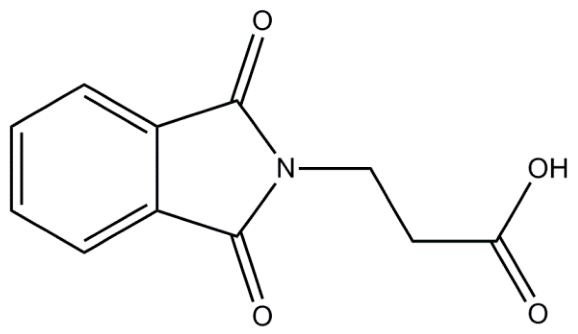
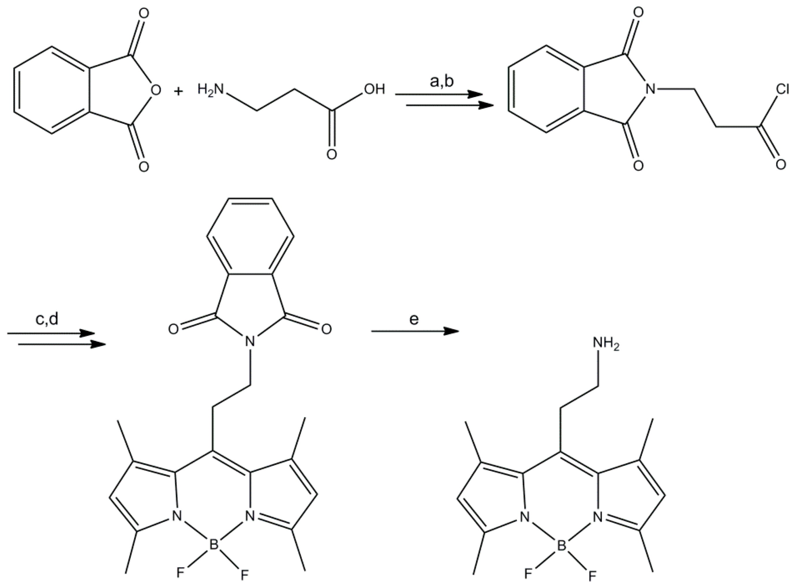
3.3.1. Synthesis of Product (1) (Figure 5)

Figure 5. Chemical structure of Product 1.
Inorganics 03 00516 g005
Phthalic anhydride (1 eq, 40 mmol, 5.92 g) and β-alanine (1 eq, 40 mmol, 3.56 g) were added in a round bottom flask and heated up to 160 °C for 6 h where it became a smelt. The mixture was cooled and the resulting solid was dissolved in DCM. The solution was washed with 0.1 M HCl (3 × 30 mL). The organic layer was dried with MgSO4 and evaporated till dryness, yield: 5.68 g, 25.9 mmol, 65%. CI–MS (MeOH, m/z): calcd: 219.19 g/mol, found: 220.0 g/mol [M + H]+. 1H NMR (300 MHz, CDCl3, δ ppm): 2.80 (t, J = 7.4 Hz, 2H), 4.00 (t, J = 7.4 Hz, 2H), 7.74 (m, 2H), 7.84 (m, 2H); 13C NMR (75 MHz, CDCl3, δ ppm): 32.8, 33.4, 123.4, 132.0, 134.1, 168.0, 176.5.

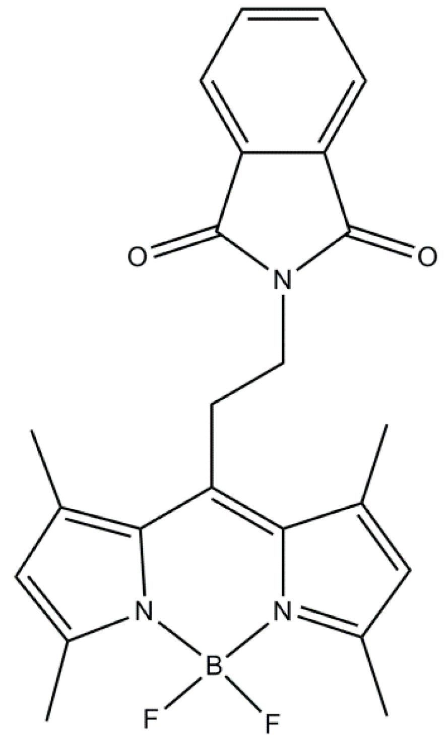
3.3.2. Synthesis of BODIPY-(CH2)2-Phtalimide (2) (Figure 6)

Figure 6. Chemical structure of Product 2.
Inorganics 03 00516 g006
To a round bottom flask product 1 (1 eq, 5 mmol, 1.10 g) and thionyl chloride (5 eq, 25 mmol, 1.82 mL) were added and refluxed for 3 h. The thionyl chloride was removed in vacuo until dryness. The acid chloride was used further without any purification. The yellow residue was dissolved in 30 mL dry DCM and 2,4-dimethylpyrrole (2 eq, 10 mmol, 1.04 mL) was slowly added. The solution was stirred for 30 min at room temperature. The color changed from yellow to red-brown. The solution was subsequently refluxed for 4 h. The solution was then cooled to 0 °C and triethylamine (10 eq, 50 mmol, 6.97 mL) was added. After 5 min of stirring at 0 °C boron trifluoride diethyl etherate (11 eq, 55 mmol, 6.79 mL) was slowly added and the solution was stirred overnight at room temperature. Diethyl ether (100 mL) was added to the solution and the organic layer was washed with water (3 × 50 mL). The organic layer was dried with MgSO4 and concentrated in vacuo. The resulting residue was purified using silica column (eluent: DCM/PET 50:50) yielding a dark orange solid, yield: 0.392 g, 0.9 mmol, 19%. CI–MS (MeOH, m/z): calcd: 421.25 g/mol, found: 421 g/mol [M]+. 1H NMR (300 MHz, CDCl3, δ ppm): 2.54 (s, 6H), 2.67 (s, 6H), 3.42 (t, J = 8.60 Hz, 2H), 3.89 (t, J = 8.60 Hz, 2H), 6.11 (s, 2H), 7.77 (m, 2H), 7.88 (m, 2H); 13C NMR (100 MHz, CDCl3, δ ppm): 14.6, 16.4, 27.7, 39.0, 122.2, 123.5, 131.7, 131.9, 134.3, 139.8, 141.5, 154.9, 168.2.

3.3.3. Synthesis of BODIPY-(CH2)2-Amine (3) (Figure 7)

Figure 7. Chemical structure of Product 3.
Inorganics 03 00516 g007
BODIPY-(CH2)2-phtalimide (2) (1 eq, 0.37 mmol, 158 mg) and hydrazine hydrate (1 eq, 0.37 mmol, 18.8 mg) in 20 mL ethanol was refluxed for 3 h. After which the solution was cooled to room temperature and filtrated with a Millipore 0.45 µM filter and the solvent was concentrated in vacuo until an orange solid, yield: 0.106, 0.36 mmol, 97%. CI–MS (MeOH, m/z): calcd: 291.17 g/mol, found: 282 g/mol [M − F]+, and 291 [M]+. 1H NMR (300 MHz, CDCl3, δ ppm): 1.61, (broad, 2H), 2.45 (s, 6H), 2.51 (s, 6H), 2.99 (m, 2H), 3.14 (m, 2H), 6.05 (s, 2H); 13C NMR (100 MHz, CDCl3, δ ppm): 10.5, 12.6, 28.2, 40.1, 117.9, 127.8, 136.6, 138.9, 150.2.

3.3.4. Synthesis of DO3A-tBu (4) (Figure 8)

Figure 8. Chemical structure of Product 4.
Inorganics 03 00516 g008
The synthesis uses an altered synthesis by Viguier et al. [58]. To a solution of tetraazacyclodedecaan (1 eq, 5.80 mmol, 1 g), sodium bicarbonate (3.5 eq, 20.3 mmol, 1.71 g) in 150 mL ACN under an argon atmosphere a solution of tert-butyl bromoacetate (3.5 eq, 20.3 mmol, 3.0 mL) in 50 mL ACN was added drop wise. The mixture was refluxed for 17 h. After removing the salts via filtration over Celite® the solvent was evaporated and the solid was recrystallized from toluene as a white powder, yield: 2.47 g, 4.8 mmol, 82%. ESI–MS: m/z calcd 516 [M + H]+, 538 [M + Na]+, found 516.0 [M + H]+, 537.5 [M + Na]+. 1H NMR (CDCl3, 300 MHz, δ ppm): 1.46 (s, 27H), 2.88 (t, 12H), 3.08 (t, 4H), 3.30 (s, 2H), 3.38 (s, 4H). 13C NMR (CDCl3, 75 MHz, δ ppm): 28.2, 28.3, 47.5, 49.5, 51.3, 58.1, 81.6, 81.7, 169.8, 170.6.

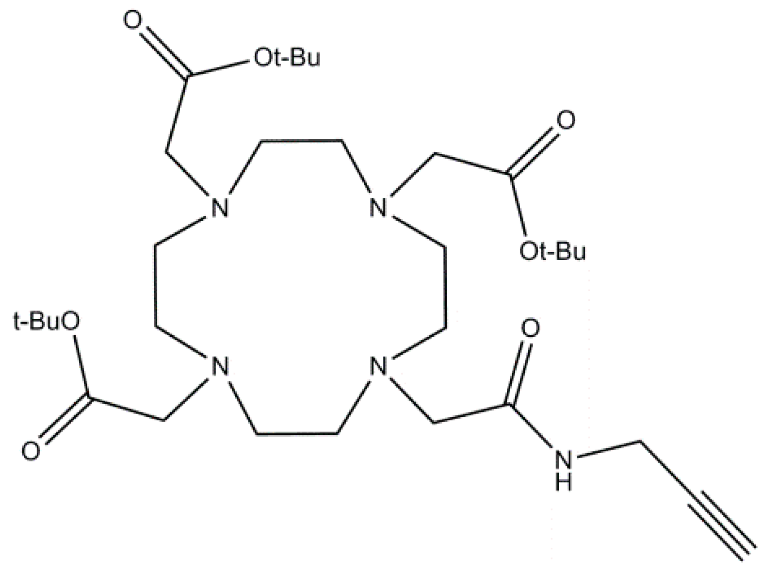
3.3.5. Synthesis of {4,10-Bis-Tert-Butoxycarbonylmethyl-7-[(2-Propynylcarbamoyl)-Methyl]-1,4,7,10-Tetraaza-Cyclododec-1-yl}-Acetic Acid Tert-Butyl Ester (5) (Figure 9)

Figure 9. Chemical structure of Product 5.
Inorganics 03 00516 g009
A solution of DO3A-tBu (4) (1 eq, 772 mg, 1.5 mmol), N-(2-propynyl)chloroacetamide (1.2 eq, 291 mg, 1.8 mmol) and K2CO3 (2 eq, 415 mg, 3.0 mmol) in 70 mL ACN was stirred under nitrogen at reflux temperature for 17 h. After filtration of over Celite®, the solvent was removed in vacuo, the residual mixture was purified by basic alumina (eluens CHCl3:MeOH (98:2)) to give a colorless solid, yield: 1.45 g, 2.4 mmol, 94%. ESI–MS: m/z calcd 610.80 [M + H]+, found 632 [M + Na]+. 1H NMR (CDCl3, 300 MHz, δ ppm): 1.45 (s, 18H), 1.46 (s, 9H), 2.15 (t, J = 2.3 Hz, 1H), 2.52 (broad, 4H), 2.70 (broad, 4H), 2.82 (broad, 4H), 2.91 (broad, 4H), 3.10 (s, 2H), 3.27 (s, 4H), 3.38 (s, 2H), 4.05 (dd, J = 5.5 Hz, J = 2.3 Hz, 2H), 9.27 (t, J = 5.5 Hz, 1H). 13C NMR (CDCl3, 75 MHz, δ ppm): 28.2, 28.3, 28.6, 29.3, 52.1, 52.5, 53.8, 55.2, 56.2, 57.6, 70.2, 72.9, 80.9, 170.8.

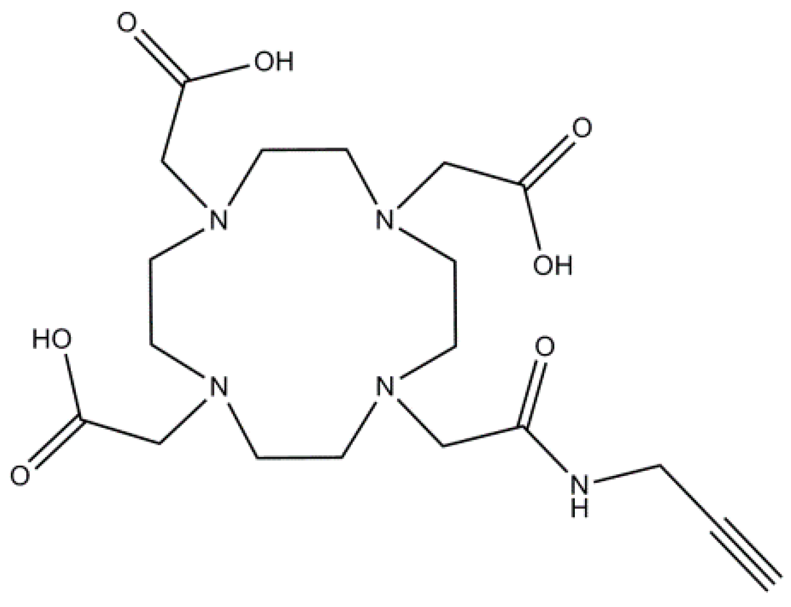
3.3.6. Synthesis of {4,10-Bis-Carboxymethyl-7-[(2-Propynylcarbamoyl)-Methyl]-1,4,7,10-Tetraaza-Cyclododec-1-yl}-Acetic Acid (6) (Figure 10)

Figure 10. Chemical structure of Product 6.
Inorganics 03 00516 g010
Product (5) (1 eq, 1.45 g, 2.4 mmol) was dissolved in 10 mL of a DCM/TFA (50:50) mixture and stirred under inert atmosphere overnight. After the reaction, 20 mL of DCM was added and evaporated (repeated two times), yield: 1.07 g, 2.4 mmol, quantitative. ESI-MS: m/z calcd 442.22 [M + H]+, found 442 [M + H]+, 464 [M + Na]+. 1H NMR (D2O, 300 MHz, δ ppm): 2.42 (t, 1H), 2.48–4.01 (broad, 22H), 3.81 (broad, 2H). 13C NMR (D2O, 75 MHz, δ ppm): 29.1, 29.6, 49.7, 53.7, 58.6, 60.6, 60.8, 69.3, 72.7, 73.5, 78.7, 175.4, 179.4, 179.6.

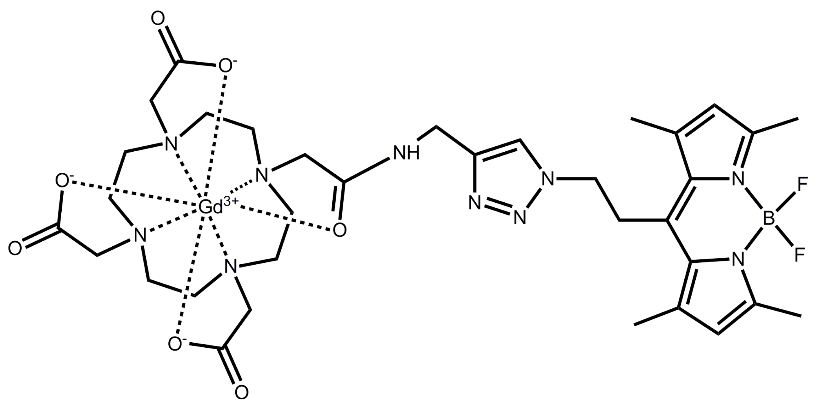
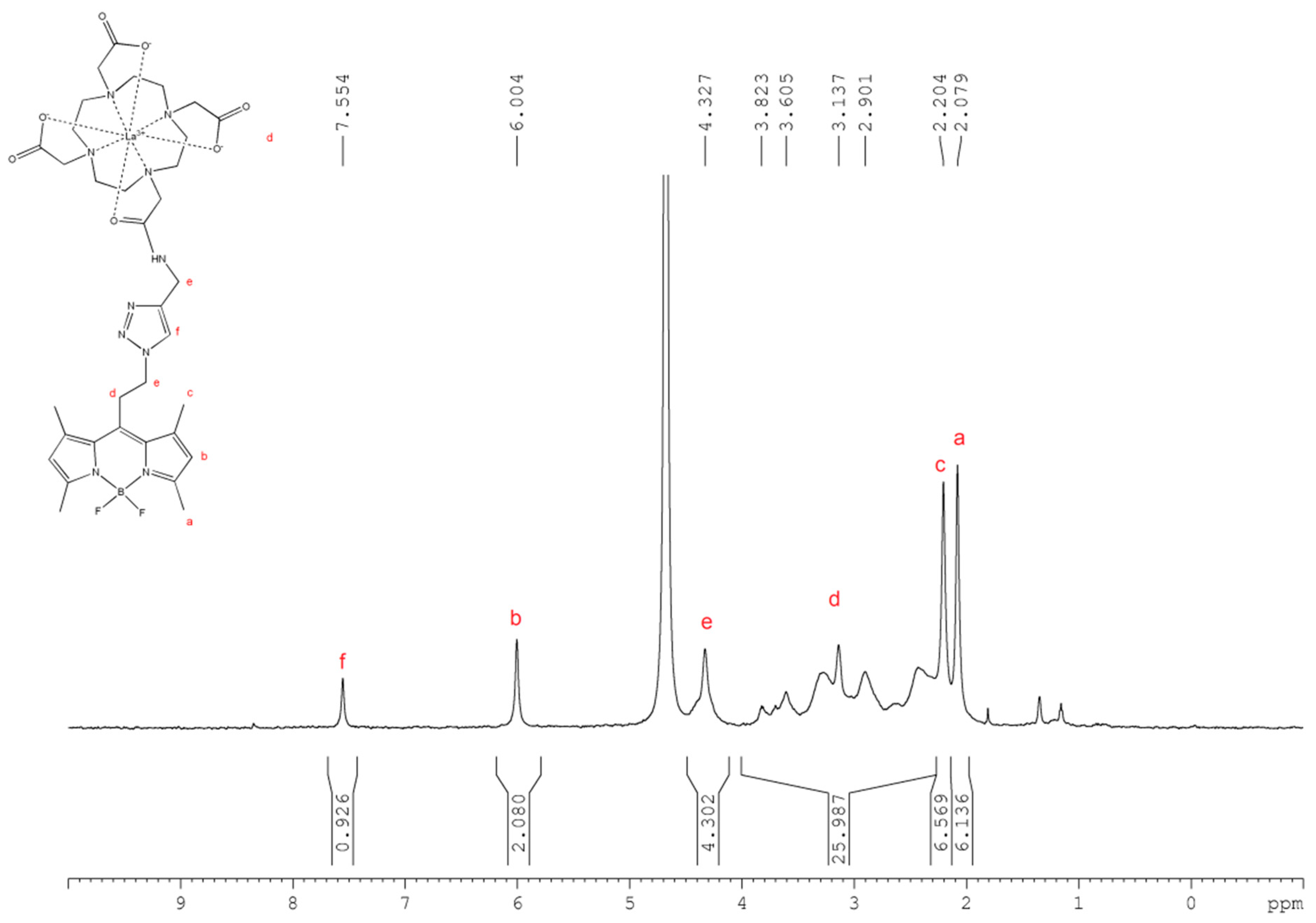
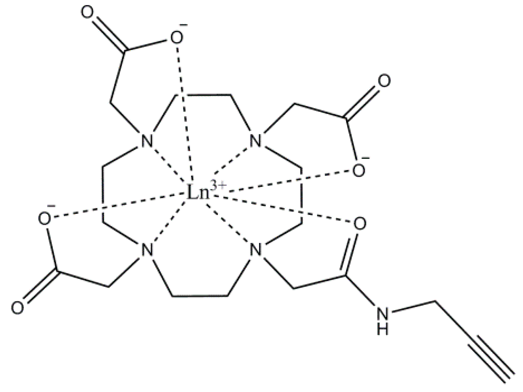
3.3.7. Synthesis of Lanthanide(III) {4,10-Bis-Carboxymethyl-7-[(2-Propynylcarbamoyl)-Methyl]-1,4,7,10-Tetraaza-Cyclododec-1-yl}-Acetic Acid (Figure 11)

Figure 11. Chemical structure of Products 7 and 8.
Inorganics 03 00516 g011
General procedure for complexation of propargylated DOTA: Product (6) (1 eq) was dissolved in 5 mL water and the appropriate lanthanide(III) chloride hydrate (1,1 eq) was added. The pH was adjusted to 5.5 with a 0.1 M KOH solution and stirred overnight at 60 °C. The solvent was evaporated. The residue was redissolved in 5 mL water and Chelex-100 was added and stirred for 2 h. This last step was repeated until no free lanthanide was found with an Arsenazo III indicator [71].
Propargylated Gd-DOTA (7): Yield: 0.446 g, 0.75 mmol, 67%. ESI-MS: m/z calcd 596.70 [M + H]+, found 619.2 [M + Na]+. IR (neat): 3416, 3245, 3098, 2873, 1675, 1609 cm−1.
Propargylated La-DOTA (8): Yield: 0.445 g, 0.77 mmol, 65%. ESI-MS: m/z calcd 478.36 [M + H]+, found 600.5 [M + Na]+. 1H NMR (CDCl3, 300 MHz, δ ppm): 2.42 (t, 1H), 2.48–4.01 (broad, 22H), 3.81 (broad, 2H). 13C NMR (CDCl3, 75 MHz, δ ppm): 29.1, 29.6, 49.7, 53.7, 58.5, 60.6, 60.8, 69.2, 72.7, 73.5, 78.7, 175.4, 179.4, 179.6; IR (neat): 3429, 3258, 3103, 2857, 1673, 1606 cm−1.

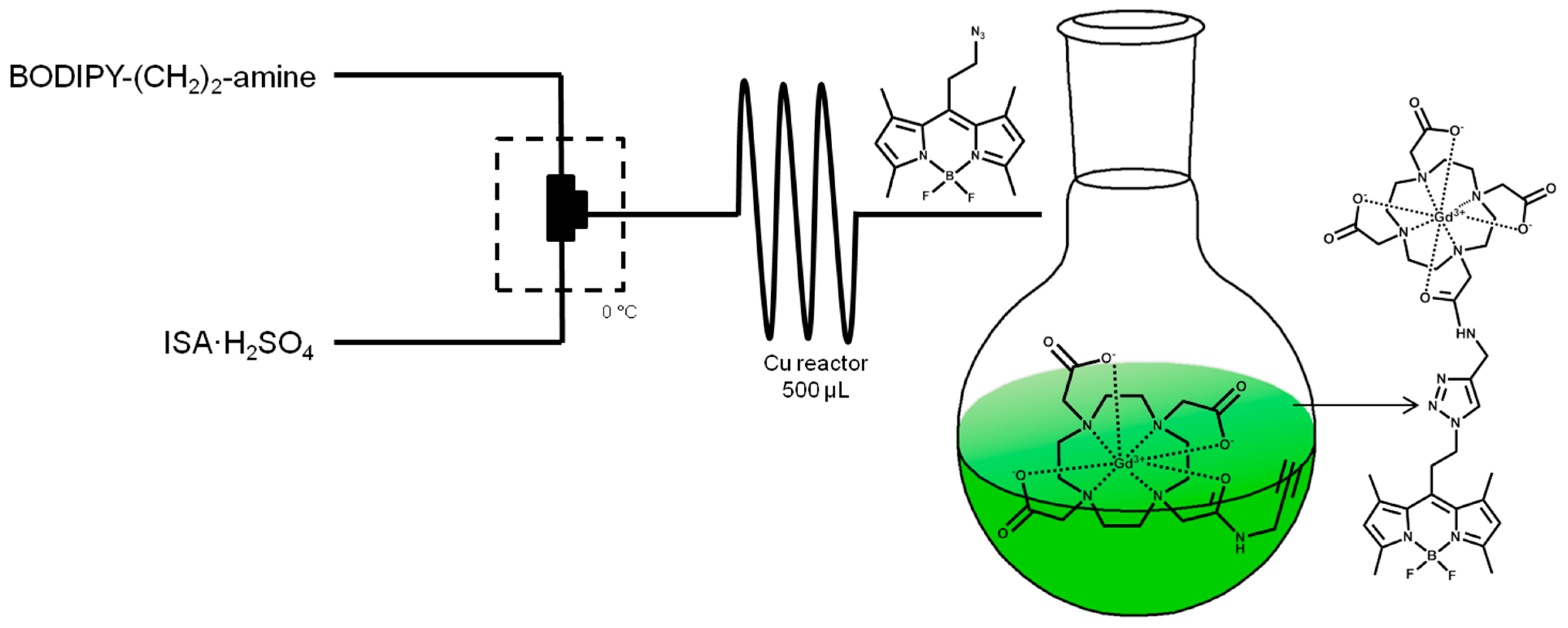
3.3.8. General Procedure Flow

Stock solution A: a 2 mL solution in a degassed mixture of 10:3:3 MeOH:DCM:H2O of 1 eq BODIPY-amine and 11 equivalents of DIPEA.
Stock solution B: a 2 mL solution of three equivalents of ISA∙H2SO4 in a degassed mixture of 10:3:3 MeOH:DCM:H2O.
Both solutions in a separate syringe are combined via a T-mixer in an ice bath before introducing the mixture to the copper tube flow reactor (150 µL) at room temperature. Flow speed is adjusted so the retention time in the reactor is 300 s. The column is stabilized during three reactor volumes. Upon leaving the reactor, the reaction mixture was directly added to a solution of 1.5 equivalent of Ln-DOTA-propargyl and 1.5 eq sodium ascorbate. The mixture is left stirring overnight. The aqueous layer is lyophilized, redissolved in distilled water, purified via HPLC and lyophilized. The products are isolated as bright orange powders.
Product 9 (Gd) (Figure 12): Yield: 13 mg, 0.01 mmol, 6%. ESI–MS: m/z calcd 913.85 [M + H]+, found 913.2 [M + H]+. The concentration used for relaxivity was 1.45 mmol/L and was measured by TXRF against an internal gallium standard.
Product 10 (La) (Figure 12): Yield: 23 mg, 0.03 mmol, 9%. ESI-MS: m/z calcd 895.51 [M + H]+, found 917.8 [M + Na]+ (positive mode), 894.2 [M + e] (negative mode). 1H NMR (D2O, 300 MHz, δ ppm): 2.08 (s, 6H), 2.20 (s, 6H), 2.20–3.82 (broad, 26H), 4.33 (broad, H), 6.00 (s, 2H), 7.55 (s, 1H). 11B NMR (D2O, 80 MHz, δ ppm): 2.16 ppm with respect to B(OMe)3 as a reference.
Figure 12. Chemical structure of Products 9 and 10.
Inorganics 03 00516 g012
After each reaction the copper tube flow reactor is cleansed in a sonication bath for 10 min while introducing a 1:1 mixture of MeOH:triethylamine at 4 mL/min, followed by 20 mL of pentane at 4 mL/min. The reactor is dried with argon and stored under argon.

4. Conclusions

In this paper a novel Gd-DOTA-BODIPY derivative is synthesized via a diazotransfer reaction under flow conditions in a copper tube. The complex is water-soluble and exhibits favorable fluorescent properties, thus offering the possibility for the use of BODIPY adducts for in vivo optical imaging. High quantum yield of 83% is obtained in water, and bright emission was observed at 523 nm. Shifting of the emission to the more optimal red-IR region of the spectrum will be attempted in future by increasing the electronic resonance of the BODIPY by adding appropriate substituents to the BODIPY core. The complex exhibits relaxivities which are comparable to the parent Gd-DOTA complex and, therefore, holds potential as a bimodal contrast agent for MRI and optical imaging.

Supplementary Materials

Supplementary materials can be found at https://0-www-mdpi-com.brum.beds.ac.uk/2304-6740/3/4/0516/s1.

Acknowledgments

Karel Duerinckx is acknowledged for his help with the NMR measurements and Michael Harris is acknowledged for his help with the relaxometric measurements.

Author Contributions

Matthias Ceulemans and Koen Nuyts have performed experimental work and spectroscopic measurements. Wim M. De Borggraeve and Tatjana N. Parac-Vogt have supervised the work and helped with interpretation of the results sand the writing of the final manuscript.

Conflicts of Interest

The authors declare no conflict of interest.

References

  1. Hermann, P.; Kotek, J.; Kubícek, V.; Lukes, I. Gadolinium(III) complexes as MRI contrast agents: Ligand design and properties of the complexes. Dalton Trans. 2008, 9226, 3027–3047. [Google Scholar] [CrossRef] [PubMed]
  2. Pierre, V.C.; Allen, M.J.; Caravan, P. Contrast agents for MRI: 30+ years and where are we going? J. Biol. Inorg. Chem. 2014, 19, 127–131. [Google Scholar] [CrossRef] [PubMed]
  3. Waters, E.A.; Wickline, S.A. Contrast agents for MRI. Basic Res. Cardiol. 2008, 103, 114–121. [Google Scholar] [CrossRef] [PubMed]
  4. Tóth, É.; Helm, L.; Merbach, A. Contrast Agents I. In Topics in Current Chemistry; Krause, W., Ed.; Springer Berlin Heidelberg: Berlin, Germany, 2002; Volume 221, pp. 61–101. [Google Scholar]
  5. Debroye, E.; Parac-Vogt, T.N. Towards polymetallic lanthanide complexes as dual contrast agents for magnetic resonance and optical imaging. Chem. Soc. Rev. 2014, 43, 8178–8192. [Google Scholar] [CrossRef] [PubMed]
  6. Cacheris, W.P.; Quay, S.C.; Rocklage, S.M. The relationship between thermodynamics and the toxicity of gadolinium complexes. Magn. Reson. Imaging 1990, 8, 467–481. [Google Scholar] [CrossRef]
  7. Shellock, F.G.; Kanal, E. Safety of magnetic resonance imaging contrast agents. J. Magn. Reson. Imaging 1999, 10, 477–484. [Google Scholar] [CrossRef]
  8. Bartolini, M.E.; Pekar, J.; Chettle, D.R.; McNeill, F.; Scott, A.; Sykes, J.; Prato, F.S.; Moran, G.R. An investigation of the toxicity of gadolinium based MRI contrast agents using neutron activation analysis. Magn. Reson. Imaging 2003, 21, 541–544. [Google Scholar] [CrossRef]
  9. Accardo, A.; Tesauro, D.; Aloj, L.; Pedone, C.; Morelli, G. Supramolecular aggregates containing lipophilic Gd(III) complexes as contrast agents in MRI. Coord. Chem. Rev. 2009, 253, 2193–2213. [Google Scholar] [CrossRef]
  10. Caravan, P.; Ellison, J.J.; McMurry, T.J.; Lauffer, R.B. Gadolinium(III) chelates as MRI contrast agents: Structure, dynamics, and applications. Chem. Rev. 1999, 99, 2293–2352. [Google Scholar] [CrossRef] [PubMed]
  11. Villaraza, A.J.L.; Bumb, A.; Brechbiel, M.W. Macromolecules, dendrimers, and nanomaterials in magnetic resonance imaging: The interplay between size, function, and pharmacokinetics. Chem. Rev. 2010, 110, 2921–2959. [Google Scholar] [CrossRef] [PubMed]
  12. Sherry, A.D.; Brown, R.D.; Geraldes, C.F.G.C.; Koenig, S.H.; Kuan, K.-T.; Spiller, M. Synthesis and characterization of the gadolinium(3+) complex of DOTA-propylamide: A model DOTA-protein conjugate. Inorg. Chem. 1989, 28, 620–622. [Google Scholar] [CrossRef]
  13. Huang, W.-Y.; Davis, J.J. Multimodality and nanoparticles in medical imaging. Dalton Trans. 2011, 40, 6087–6103. [Google Scholar] [CrossRef] [PubMed]
  14. Xie, J.; Liu, G.; Eden, H.S.; Ai, H.; Chen, X. Surface-engineered magnetic nanoparticle platforms for cancer imaging and therapy. Acc. Chem. Res. 2011, 44, 883–892. [Google Scholar] [CrossRef] [PubMed]
  15. Louie, A. Multimodality imaging probes: Design and challenges. Chem. Rev. 2010, 110, 3146–3195. [Google Scholar] [CrossRef] [PubMed]
  16. Joshi, B.P.; Wang, T.D. Exogenous molecular probes for targeted imaging in cancer: Focus on multi-modal imaging. Cancers (Basel) 2010, 2, 1251–1287. [Google Scholar] [CrossRef] [PubMed]
  17. Soenen, S.J.; Vande Velde, G.; Ketkar-Atre, A.; Himmelreich, U.; de Cuyper, M. Magnetoliposomes as magnetic resonance imaging contrast agents. Wiley Interdiscip. Rev. Nanomed. Nanobiotechnol. 2002, 3, 197–211. [Google Scholar] [CrossRef] [PubMed]
  18. Costa, J.; Ruloff, R.; Burai, L.; Helm, L.; Merbach, A.E. Rigid M(II)L2Gd2(III) (M = Fe, Ru) complexes of a terpyridine-based heteroditopic chelate: A class of candidates for MRI contrast agents. J. Am. Chem. Soc. 2005, 127, 5147–5157. [Google Scholar] [CrossRef] [PubMed]
  19. Livramento, J.B.; Sour, A.; Borel, A.; Merbach, A.E.; Tóth, É. A starburst-shaped heterometallic compound incorporating six densely packed Gd3+ Ions. Chem. Eur. J. 2006, 12, 989–1003. [Google Scholar] [CrossRef] [PubMed]
  20. Livramento, J.B.; Weidensteiner, C.; Prata, M.I.M.; Allegrini, P.R.; Geraldes, C.F.G.C.; Helm, L.; Kneuer, R.; Merbach, A.E.; Santos, A.C.; Schmidt, P.; et al. First in vivo MRI assessment of a self-assembled metallostar compound endowed with a remarkable high field relaxivity. Contrast Media Mol. Imaging 2006, 1, 30–39. [Google Scholar] [CrossRef] [PubMed]
  21. Parac-Vogt, T.N.; Vander Elst, L.; Kimpe, K.; Laurent, S.; Burtéa, C.; Chen, F.; van Deun, R.; Ni, Y.; Muller, R.N.; Binnemans, K. Pharmacokinetic and in vivo evaluation of a self-assembled gadolinium(III)-iron(II) contrast agent with high relaxivity. Contrast Media Mol. Imaging 2006, 1, 267–278. [Google Scholar] [CrossRef] [PubMed] [Green Version]
  22. Paris, J.; Gameiro, C.; Humblet, V.; Mohapatra, P.K.; Jacques, V.; Desreux, J.F. Auto-assembling of ditopic macrocyclic lanthanide chelates with transition-metal ions. Rigid multimetallic high relaxivity contrast agents for magnetic resonance imaging. Inorg. Chem. 2006, 45, 5092–5102. [Google Scholar] [CrossRef] [PubMed]
  23. Dehaen, G.; Verwilst, P.; Eliseeva, S.V.; Laurent, S.; Vander Elst, L.; Muller, R.N.; de Borggraeve, W.M.; Binnemans, K.; Parac-Vogt, T.N. A heterobimetallic ruthenium-gadolinium complex as a potential agent for bimodal imaging. Inorg. Chem. 2011, 50, 10005–10014. [Google Scholar] [CrossRef] [PubMed]
  24. Dehaen, G.; Eliseeva, S.V.; Kimpe, K.; Laurent, S.; Vander Elst, L.; Muller, R.N.; Dehaen, W.; Binnemans, K.; Parac-Vogt, T.N.; Vanderelst, L.; et al. A self-assembled complex with a titanium(IV) catecholate core as a potential bimodal contrast agent. Chem. Eur. J. 2012, 18, 293–302. [Google Scholar] [CrossRef] [PubMed]
  25. Verwilst, P.; Eliseeva, S.V.; Vander Elst, L.; Burtea, C.; Laurent, S.; Petoud, S.; Muller, R.N.; Parac-Vogt, T.N.; de Borggraeve, W.M. A tripodal ruthenium-gadolinium metallostar as a potential αvβ3 integrin specific bimodal imaging contrast agent. Inorg. Chem. 2012, 51, 6405–6411. [Google Scholar] [CrossRef] [PubMed]
  26. Dehaen, G.; Eliseeva, S.V.; Verwilst, P.; Laurent, S.; Vander Elst, L.; Muller, R.N.; de Borggraeve, W.M.; Binnemans, K.; Parac-Vogt, T.N. Tetranuclear d-f metallostars: Synthesis, relaxometric, and luminescent properties. Inorg. Chem. 2012, 51, 8775–8783. [Google Scholar] [CrossRef] [PubMed]
  27. Debroye, E.; Ceulemans, M.; Vander Elst, L.; Laurent, S.; Muller, R.N.; Parac-Vogt, T.N. Controlled synthesis of a novel heteropolymetallic complex with selectively incorporated lanthanide(III) ions. Inorg. Chem. 2014, 53, 1257–1259. [Google Scholar] [CrossRef] [PubMed]
  28. Kuil, J.; Buckle, T.; Oldenburg, J.; Yuan, H.; Borowsky, A.D.; Josephson, L.; van Leeuwen, F.W.B. Hybrid peptide dendrimers for imaging of chemokine receptor 4 (CXCR4) expression. Mol. Pharm. 2011, 8, 2444–2453. [Google Scholar] [CrossRef] [PubMed]
  29. Musonda, C.C.; Taylor, D.; Lehman, J.; Gut, J.; Rosenthal, P.J.; Chibale, K. Application of multi-component reactions to antimalarial drug discovery. Part 1: Parallel synthesis and antiplasmodial activity of new 4-aminoquinoline Ugi adducts. Bioorganic Med. Chem. Lett. 2004, 14, 3901–3905. [Google Scholar] [CrossRef] [PubMed]
  30. Rivas, C.; Stasiuk, G.J.; Sae-Heng, M.; Long, N.J. Towards understanding the design of dual-modal MR/fluorescent probes to sense zinc ions. Dalton Trans. 2015, 44, 4976–4985. [Google Scholar] [CrossRef] [PubMed]
  31. Hüber, M.M.; Staubli, A.B.; Kustedjo, K.; Gray, M.H.B.; Shih, J.; Fraser, S.E.; Jacobs, R.E.; Meade, T.J. Fluorescently detectable magnetic resonance imaging agents. Bioconjug. Chem. 1998, 9, 242–249. [Google Scholar] [CrossRef] [PubMed]
  32. Mishra, A.A.K.; Pfeuffer, J.; Mishra, R.; Engelmann, J.; Mishra, A.A.K.; Ugurbil, K.; Logothetis, N.K. A new class of Gd-based DO3A-ethylamine-derived targeted contrast agents for MR and optical imaging. Bioconjug. Chem. 2006, 17, 773–780. [Google Scholar] [CrossRef] [PubMed]
  33. Zhang, M.; Imm, S.; Bähn, S.; Neubert, L.; Neumann, H.; Beller, M. Efficient copper(II)-catalyzed transamidation of non-activated primary carboxamides and ureas with amines. Angew. Chem. Int. Ed. 2012, 51, 3905–3909. [Google Scholar] [CrossRef] [PubMed]
  34. Jang, J.H.; Bhuniya, S.; Kang, J.; Yeom, A.; Hong, K.S.; Kim, J.S. Cu2+-responsive bimodal (optical/MRI) contrast agent for cellular imaging. Org. Lett. 2013, 15, 4702–4705. [Google Scholar] [CrossRef] [PubMed]
  35. Ulrich, G.; Ziessel, R.; Harriman, A. The chemistry of fluorescent bodipy dyes: Versatility unsurpassed. Angew. Chem. Int. Ed. 2008, 47, 1184–1201. [Google Scholar] [CrossRef] [PubMed]
  36. Yee, M.-C.; Fas, S.C.; Stohlmeyer, M.M.; Wandless, T.J.; Cimprich, K.A. A cell-permeable, activity-based probe for protein and lipid kinases. J. Biol. Chem. 2005, 280, 29053–29059. [Google Scholar] [CrossRef] [PubMed]
  37. West, R.; Panagabko, C.; Atkinson, J. Synthesis and characterization of BODIPY-α-tocopherol: A fluorescent form of vitamin E. J. Org. Chem. 2010, 75, 2883–2892. [Google Scholar] [CrossRef] [PubMed]
  38. Kowada, T.; Maeda, H.; Kikuchi, K. BODIPY-based probes for the fluorescence imaging of biomolecules in living cells. Chem. Soc. Rev. 2015, 44, 4953–4972. [Google Scholar] [CrossRef] [PubMed]
  39. Boens, N.; Leen, V.; Dehaen, W. Fluorescent indicators based on BODIPY. Chem. Soc. Rev. 2012, 41, 1130–1172. [Google Scholar] [CrossRef] [PubMed]
  40. García-Moreno, I.; Amat-Guerri, F.; Liras, M.; Costela, A.; Infantes, L.; Sastre, R.; López Arbeloa, F.; Bañuelos Prieto, J.; López Arbeloa, Í. Structural changes in the BODIPY dye PM567 enhancing the laser action in liquid and solid media. Adv. Funct. Mater. 2007, 17, 3088–3098. [Google Scholar] [CrossRef]
  41. Duran-Sampedro, G.; Agarrabeitia, A.R.; Garcia-Moreno, I.; Costela, A.; Bañuelos, J.; Arbeloa, T.; López Arbeloa, I.; Chiara, J.L.; Ortiz, M.J. Chlorinated BODIPYs: Surprisingly efficient and highly photostable laser dyes. Eur. J. Org. Chem. 2012, 2012, 6335–6350. [Google Scholar] [CrossRef]
  42. Lakshmi, V.; Rajeswara Rao, M.; Ravikanth, M. Halogenated boron-dipyrromethenes: Synthesis, properties and applications. Org. Biomol. Chem. 2015, 13, 2501–2517. [Google Scholar] [CrossRef] [PubMed]
  43. Bessette, A.; Hanan, G.S. Design, synthesis and photophysical studies of dipyrromethene-based materials: Insights into their applications in organic photovoltaic devices. Chem. Soc. Rev. 2014, 43, 3342. [Google Scholar] [CrossRef] [PubMed]
  44. Singh, S.P.; Gayathri, T. Evolution of BODIPY dyes as potential sensitizers for dye-sensitized solar cells. Eur. J. Org. Chem. 2014, 2014, 4689–4707. [Google Scholar] [CrossRef]
  45. González-Béjar, M.; Liras, M.; Francés-Soriano, L.; Voliani, V.; Herranz-Pérez, V.; Duran-Moreno, M.; Garcia-Verdugo, J.M.; Alarcon, E.I.; Scaiano, J.C.; Pérez-Prieto, J. NIR excitation of upconversion nanohybrids containing a surface grafted bodipy induces oxygen-mediated cancer cell death. J. Mater. Chem. B 2014, 2, 4554. [Google Scholar] [CrossRef]
  46. Lim, S.H.; Thivierge, C.; Nowak-Sliwinska, P.; Han, J.; van den Bergh, H.; Wagnières, G.; Burgess, K.; Lee, H.B. In vitro and in vivo photocytotoxicity of boron dipyrromethene derivatives for photodynamic therapy. J. Med. Chem. 2010, 53, 2865–2874. [Google Scholar] [CrossRef] [PubMed]
  47. Bernhard, C.; Goze, C.; Rousselin, Y.; Denat, F. First bodipy–DOTA derivatives as probes for bimodal imaging. Chem. Commun. 2010, 46, 8267. [Google Scholar] [CrossRef] [PubMed]
  48. Bernhard, C.; Moreau, M.; Lhenry, D.; Goze, C.; Boschetti, F.; Rousselin, Y.; Brunotte, F.; Denat, F. DOTAGA-anhydride: A valuable building block for the preparation of DOTA-like chelating agents. Chem. Eur. J. 2012, 18, 7834–7841. [Google Scholar] [CrossRef] [PubMed]
  49. Iwaki, S.; Hokamura, K.; Ogawa, M.; Takehara, Y.; Muramatsu, Y.; Yamane, T.; Hirabayashi, K.; Morimoto, Y.; Hagisawa, K.; Nakahara, K.; et al. A design strategy for small molecule-based targeted MRI contrast agents: Their application for detection of atherosclerotic plaques. Org. Biomol. Chem. 2014, 12, 8611–8618. [Google Scholar] [CrossRef] [PubMed]
  50. Yamane, T.; Hanaoka, K.; Muramatsu, Y.; Tamura, K.; Adachi, Y.; Miyashita, Y.; Hirata, Y.; Nagano, T. Method for enhancing cell penetration of Gd3+-based MRI contrast agents by conjugation with hydrophobic fluorescent dyes. Bioconjug. Chem. 2011, 22, 2227–2236. [Google Scholar] [CrossRef] [PubMed]
  51. Duheron, V.; Moreau, M.; Collin, B.; Sali, W.; Bernhard, C.; Goze, C.; Gautier, T.; Pais de Barros, J.-P.; Deckert, V.; Brunotte, F.; et al. Dual labeling of lipopolysaccharides for SPECT-CT imaging and fluorescence microscopy. ACS Chem. Biol. 2014, 9, 656–662. [Google Scholar] [CrossRef] [PubMed]
  52. Lhenry, D.; Larrouy, M.; Bernhard, C.; Goncalves, V.; Raguin, O.; Provent, P.; Moreau, M.; Collin, B.; Oudot, A.; Vrigneaud, J.-M.; et al. BODIPY: A highly versatile platform for the design of bimodal imaging probes. Chemistry 2015, 21, 13091–13099. [Google Scholar] [CrossRef] [PubMed]
  53. Hendricks, J.A.; Keliher, E.J.; Wan, D.; Hilderbrand, S.A.; Weissleder, R.; Mazitschek, R. Synthesis of [18F]BODIPY: Bifunctional reporter for hybrid optical/positron emission tomography imaging. Angew. Chem. Int. Ed. Engl. 2012, 51, 4603–4606. [Google Scholar] [CrossRef] [PubMed]
  54. Liu, S.; Li, D.; Zhang, Z.; Surya Prakash, G.K.; Conti, P.S.; Li, Z. Efficient synthesis of fluorescent-PET probes based on [18F]BODIPY dye. Chem. Commun. 2014, 50, 7371. [Google Scholar] [CrossRef] [PubMed]
  55. Nuyts, K.; Ceulemans, M.; Parac-Vogt, T.N.; Bultynck, G.; de Borggraeve, W.M. Facile azide formation via diazotransfer reaction in a copper tube flow reactor. Tetrahedron Lett. 2015, 56, 1687–1690. [Google Scholar] [CrossRef]
  56. Fischer, N.; Goddard-Borger, E.D.; Greiner, R.; Klapötke, T.M.; Skelton, B.W.; Stierstorfer, J. Sensitivities of some imidazole-1-sulfonyl azide salts. J. Org. Chem. 2012, 77, 1760–1764. [Google Scholar] [CrossRef] [PubMed]
  57. Hansen, A.M.; Sewell, A.L.; Pedersen, R.H.; Long, D.-L.; Gadegaard, N.; Marquez, R. Tunable BODIPY derivatives amenable to “click” and peptide chemistry. Tetrahedron 2013, 69, 8527–8533. [Google Scholar] [CrossRef]
  58. Viguier, R.F.H.; Hulme, A.N. A sensitized europium complex generated by micromolar concentrations of copper(I): Toward the detection of copper(I) in biology. J. Am. Chem. Soc. 2006, 128, 11370–11371. [Google Scholar] [CrossRef] [PubMed]
  59. Verwilst, P.; Eliseeva, S.V.; Carron, S.; Vander Elst, L.; Burtea, C.; Dehaen, G.; Laurent, S.; Binnemans, K.; Muller, R.N.; Parac-Vogt, T.N.; et al. A modular approach towards the synthesis of target-specific MRI contrast agents. Eur. J. Inorg. Chem. 2011, 2011, 3577–3585. [Google Scholar] [CrossRef]
  60. Hessel, V.; Kralisch, D.; Kockmann, N.; Noël, T.; Wang, Q. Novel process windows for enabling, accelerating, and uplifting flow chemistry. ChemSusChem 2013, 6, 746–789. [Google Scholar] [CrossRef] [PubMed]
  61. Johansson, H.; Pedersen, D.S. Azide- and alkyne-derivatised α-amino acids. Eur. J. Org. Chem. 2012, 2012, 4267–4281. [Google Scholar] [CrossRef]
  62. Goddard-Borger, E.D.; Stick, R.V. An efficient, inexpensive, and shelf-stable diazotransfer reagent: Imidazole-1-sulfonyl azide hydrochloride. Org. Lett. 2007, 9, 3797–3800. [Google Scholar] [CrossRef] [PubMed]
  63. Duimstra, J.A.; Femia, F.J.; Meade, T.J. A gadolinium chelate for detection of β-glucuronidase: A self-immolative approach. J. Am. Chem. Soc. 2005, 127, 12847–12855. [Google Scholar] [CrossRef] [PubMed]
  64. Wang, L.; Verbelen, B.; Tonnelé, C.; Beljonne, D.; Lazzaroni, R.; Leen, V.; Dehaen, W.; Boens, N. UV-VIS spectroscopy of the coupling products of the palladium-catalyzed C–H arylation of the BODIPY core. Photochem. Photobiol. Sci. 2013, 12, 835–847. [Google Scholar] [CrossRef] [PubMed] [Green Version]
  65. Kobayashi, H.; Ogawa, M.; Alford, R.; Choyke, P.L.; Urano, Y. New strategies for fluorescent probe design in medical diagnostic imaging. Chem. Rev. 2010, 110, 2620–2640. [Google Scholar] [CrossRef] [PubMed]
  66. Niu, S. Advanced Water Soluble BODIPY Dyes: Synthesis and Application. Ph.D. Thesis, Université de Strasbourg, Strasbourg, France, July 2011. [Google Scholar]
  67. Olmsted, J. Calorimetric determinations of absolute fluorescence quantum yields. J. Phys. Chem. 1979, 83, 2581–2584. [Google Scholar] [CrossRef]
  68. Solomon, I. Relaxation processes in a system of two spins. Phys. Rev. 1955, 99, 559–565. [Google Scholar] [CrossRef]
  69. Bloembergen, N. Proton relaxation times in paramagnetic solutions. J. Chem. Phys. 1957, 27, 572. [Google Scholar] [CrossRef]
  70. Laurent, S.; Elst, L.V.; Muller, R.N. Comparative study of the physicochemical properties of six clinical low molecular weight gadolinium contrast agents. Contrast Media Mol. Imaging 2006, 1, 128–137. [Google Scholar] [CrossRef] [PubMed]
  71. Onishi, H. Spectrophotometric determination of zirconium, uranium, thorium and rare earths with arsenazo III after extractions with thenoyltrifluoroacetone and tri-n-octylamine. Talanta 1972, 19, 473–478. [Google Scholar] [CrossRef]

